biomaterials: important areas for …nsfbiomatworkshop2012.caltech.edu/report/nsf biomaterials...
TRANSCRIPT

BIOMATERIALS: IMPORTANT AREAS FOR FUTURE INVESTMENT
2012 NSF BIOMATERIALS WORKSHOP

Page 2 Biomaterials: Important Areas for Future Investment
Biomaterials: Important Areas for Future Investment
A National Science Foundation Sponsored Workshop
Held at NSF Headquarters Arlington, VA
June 19-20, 2012
WORKSHOP CHAIR
David A. Tirrell, Ph.D., Ross McCollum - William H. Corcoran Professor Professor of Chemistry and Chemical Engineering, California Institute of Technology CO-ORGANIZERS:
Kristi Anseth, Ph.D., University of Colorado Dennis Discher, Ph. D., University of Pennsylvania Lara Estroff, Ph.D., Cornell University Paula Hammond, Ph.D., Massachusetts Institute of Technology Ashley White, Ph.D., AAAS Science & Technology Policy Fellow, National Science Foundation
LIST OF INVITED PARTICIPANTS Debra Auguste, Harvard University Elia Beniash, University of Pittsburgh Julie Champion, Georgia Institute of Technology Joseph DeSimone, University of North Carolina at Chapel Hill Trevor Douglas, Montana State University Omolola Eniola-Adefeso, University of Michigan Seth Fraden, Brandeis University Delphine Gourdon, Cornell University Daniel Hammer, University of Pennsylvania Ryan Hayward, University of Massachusetts, Amherst Sarah Heilshorn, Stanford University Darrell Irvine, Massachusetts Institute of Technology Derk Joester, Northwestern University Ali Khademhosseini, Harvard University/Massachusetts Institute of Technology Laura Kiessling, University of Wisconsin Efrosini Kokkoli, University of Minnesota LaShanda Korley, Case Western Reserve University Nils Kroeger, B CUBE/Technische Universität Dresden Cato Laurencin, University of Connecticut Jeffrey Linhardt, Google, Inc. Helen Lu, Columbia University SuPing Lyu, Medtronic Inc. Joanna McKittrick, University of California, San DiegoPhillip Messersmith, Northwestern University

Page 3Biomaterials: Important Areas for Future Investment
Liviu Movileanu, Syracuse University Bill Murphy, University of Wisconsin Paul Nealey, University of Wisconsin Celeste Nelson, Princeton University Fiorenzo Omenetto, Tufts University Vanessa Ortiz, Columbia University Alyssa Panitch, Purdue University Darrin Pochan, University of Delaware David Putnam, Cornell University Buddy Ratner, University of Washington Samuel Safran, Weizmann Institute of Science Shelly Sakiyama-Elbert, Washington University Christine Schmidt, University of Texas at Austin Ravi Shanker, Pfizer Inc. William Shih, Harvard University Samuel Stupp, Northwestern University Igal Szleifer, Northwestern University Francis Szoka, University of California, San Francisco Johnna Temenoff, Georgia Institute of Technology/Emory University Amy Wagoner Johnson, University of Illinois at Urbana-Champaign Thomas Webster, Brown University Ulrike Wegst, Dartmouth University David Weitz, Harvard University Joyce Wong, Boston University Ting Xu, University of California, Berkeley
GOVERNMENT AGENCIES
Joseph Akkara, National Science Foundation Krastan Blagoev, National Science Foundation David Brant, National Science Foundation Hugh De Long, Air Force Office of Scientific Research James Drummond, National Institute of Dental and Craniofacial Institute/National Institutes of Health Joycelyn Harrison, Air Force Office of Scientific Research Janice Hicks, National Science Foundation Rosemarie Hunziker, National Institutes of Health Douglas Kiserow, Army Research Office Sheng Lin-Gibson, National Institute of Standards and Technology Andrew Lovinger, National Science Foundation Nadya Lumelsky, National Institutes of Health Michael Markowitz, Department of Energy Nicole Moore, National Cancer Institute/National Institutes of Health Rajesh Naik, Air Force Research Laboratory Dawanne Poree, Army Research Office David Stepp, Army Research Office Ashley White, National Science Foundation/American Association for the Advancement of Science William Zamer, National Science Foundation

Page 4 Biomaterials: Important Areas for Future Investment
Preface
On June 19 and 20, 2012, the National Science Foundation (NSF) convened a workshop in Ar-lington, VA, to assess the status of the field of biomaterials science and engineering and identify especially promising directions for the future of biomaterials research. The workshop partic-ipants included representatives from 47 universities, companies, and federal agencies. This report summarizes the deliberations of the participants and the conclusions of the workshop.
As chairman, I would like to express my appreciation to NSF for supporting the workshop and seeking the advice of the biomaterials research community. David Brant and Joseph Akkara, di-rectors NSF’s Biomaterials Program, provided guidance and support at every stage of the pro-cess. Ashley White, a materials scientist and policy fellow of the American Association for the Advancement of Science (AAAS), worked with us from the beginning of the planning process and offered thoughtful suggestions and critically important logistical support. Anne Hormann, my extraordinary assistant at Caltech, made sure that everything that needed to be done, got done.
I want to express special thanks to the workshop steering committee: Kristi Anseth of the Uni-versity of Colorado; Dennis Discher of the University of Pennsylvania; Lara Estroff of Cornell University; and Paula Hammond of the Massachusetts Institute of Technology. The committee members played essential roles in planning the workshop, leading the discussion groups, and preparing this report. It was a great team to work with, and I’m grateful to each member for their excellent ideas and diligent work.
The steering committee worked closely with a wonderful group of graduate students: Dave Dingal of the University of Pennsylvania; Lawrence Dooling of the California Institute of Tech-nology; Deng Wen (Debra) Lin of Cornell University; Anasuya Mandal of the Massachusetts Institute of Technology; and Mark Tibbit of the University of Colorado. Dave, Larry, Debra, Anasuya, and Mark kept track of the group discussions, prepared materials for workshop feed-back sessions, and were deeply involved in the early stages of preparation of the workshop report. They did a wonderful job.
The workshop report owes a great deal to the efforts of science editor Jim Swyers, who sup-ported the efforts of the steering committee at several stages of the report’s development, and to the talents of Greg Mueller, who designed the report and its cover. I greatly appreciate their contributions.
Finally, I want to acknowledge all of the workshop participants who gave plenary lectures, con-tributed to the education panel, enlivened the discussion groups, offered constructive feedback, and drafted various sections of the report. I hope that this final version of the report conveys the thoughts, imagination, and hard work of the entire group. I also hope it will stimulate further discussion and investments in the field of biomaterials research.
David Tirrell
Pasadena, CA
January 3, 2013

Page 5Biomaterials: Important Areas for Future Investment
Table of Contents List of Invited Participants. . . . . . . . . . . . . . . . . . . . . . . . . . . . . . . . . . . . 2
Preface . . . . . . . . . . . . . . . . . . . . . . . . . . . . . . . . . . . . . . . . . . . . . . 4
Table of contents . . . . . . . . . . . . . . . . . . . . . . . . . . . . . . . . . . . . . . . . . 5
Executive Summary . . . . . . . . . . . . . . . . . . . . . . . . . . . . . . . . . . . . . . 10
Scientific themes . . . . . . . . . . . . . . . . . . . . . . . . . . . . . . . . . . . . 10
Practical impact . . . . . . . . . . . . . . . . . . . . . . . . . . . . . . . . . . . . 11
Needs and recommendations . . . . . . . . . . . . . . . . . . . . . . . . . . . . . 12
Biomaterials education. . . . . . . . . . . . . . . . . . . . . . . . . . . . . . . . . 13
Discussion group summaries . . . . . . . . . . . . . . . . . . . . . . . . . . . . . 15
Hard materials and Composites . . . . . . . . . . . . . . . . . . . . . . . . . . . . 15
Soft Materials . . . . . . . . . . . . . . . . . . . . . . . . . . . . . . . . . . . . . 16
Cell-Material Interactions . . . . . . . . . . . . . . . . . . . . . . . . . . . . . . . 17
Dispersed Systems . . . . . . . . . . . . . . . . . . . . . . . . . . . . . . . . . . . 18
Thin films and Interfaces . . . . . . . . . . . . . . . . . . . . . . . . . . . . . . . 19
Workshop Program . . . . . . . . . . . . . . . . . . . . . . . . . . . . . . . . . . . . . . 21
Section 1. Hard Materials and Composites . . . . . . . . . . . . . . . . . . . . . . . . . 23
1.1 Introduction . . . . . . . . . . . . . . . . . . . . . . . . . . . . . . . . . . . . . . 23
1.2 Opportunities and Challenges . . . . . . . . . . . . . . . . . . . . . . . . . . . . 26
1.2.1 Advanced Structural and Mechanical Characterization
of Biological and Bio-inspired Composites and Hard Materials . . . . . . . . . . 26
1.2.2 Prospecting Genomes and Beyond. . . . . . . . . . . . . . . . . . . . . . . . . . 28
1.2.3 Grand Challenges in HMC Research . . . . . . . . . . . . . . . . . . . . . . . . 28
1.3 Scientific Questions . . . . . . . . . . . . . . . . . . . . . . . . . . . . . . . . . 30
1.3.1 Bioprospecting . . . . . . . . . . . . . . . . . . . . . . . . . . . . . . . . . . . . 31

Page 6 Biomaterials: Important Areas for Future Investment
1.3.2 “Omics,” Bioinformatics, and Phylogeny
as Routes to Materials Discovery . . . . . . . . . . . . . . . . . . . . . . . . . . 31
1.3.3 Characterizing and Exploiting
Amorphous/Poorly Crystalline Phases . . . . . . . . . . . . . . . . . . . . . . . . 32
1.3.4 Understanding and replicating interfaces/interphases
between organic-inorganic,
organic-organic, and inorganic-inorganic materials . . . . . . . . . . . . . . . . . 33
1.3.5 Design of Functionally Graded Systems . . . . . . . . . . . . . . . . . . . . . . . 34
1.3.6 Hierarchical composite materials by design . . . . . . . . . . . . . . . . . . . . . 38
1.3.7 Predictive ability to design de novo composites
with defined property profiles . . . . . . . . . . . . . . . . . . . . . . . . . . . . 40
1.3.8 Genetically Designed Materials . . . . . . . . . . . . . . . . . . . . . . . . . . . 41
1.4 Needs and Recommendations . . . . . . . . . . . . . . . . . . . . . . . . . . . . 42
Section 2. Soft Materials . . . . . . . . . . . . . . . . . . . . . . . . . . . . . . . . . . . 44
2.1 Introduction . . . . . . . . . . . . . . . . . . . . . . . . . . . . . . . . . . . . . . 44
2.2 Opportunities and Challenges . . . . . . . . . . . . . . . . . . . . . . . . . . . . . 46
2.2.1 Mining and emulating the adaptive capacity of diverse natural materials . . . . . . 47
2.2.2 Making matter active and capable of morphogenesis . . . . . . . . . . . . . . . . 48
2.2.3 Probing genome-scale hyper-complexity
for evolved biomaterials . . . . . . . . . . . . . . . . . . . . . . . . . . . . . . . 49
2.3 Scientific Questions. . . . . . . . . . . . . . . . . . . . . . . . . . . . . . . . . . . 51
2.3.1 Which soft biomaterial systems are best suited
for understanding adaptation . . . . . . . . . . . . . . . . . . . . . . . . . . . . . 51
2.3.2 What is living, and what is synthetic . . . . . . . . . . . . . . . . . . . . . . . . . 53
2.3.3 Can we understand biomaterial complexity and make
active matter evolve . . . . . . . . . . . . . . . . . . . . . . . . . . . . . . . . . 55

Page 7Biomaterials: Important Areas for Future Investment
2.4 Needs and Recommendations . . . . . . . . . . . . . . . . . . . . . . . . . . . . 56
2.4.1 Adaptability of hierarchical matrices with bio-complexity. . . . . . . . . . . . . . 56
2.4.2 Hybrid molecules for assembly of nanostructures
and hierarchical materials . . . . . . . . . . . . . . . . . . . . . . . . . . . . . . 58
2.4.3 Cyber-discovery and molecular dynamics of adaptation . . . . . . . . . . . . . . . 59
2.4.4 Living waves of biomaterials . . . . . . . . . . . . . . . . . . . . . . . . . . . . . 60
2.4.5 New tools for hypercomplexity and biomaterials evolution . . . . . . . . . . . . . 61
Section 3. Cell-Material Interactions . . . . . . . . . . . . . . . . . . . . . . . . . . . . 62
3.1 Introduction . . . . . . . . . . . . . . . . . . . . . . . . . . . . . . . . . . . . . . 62
3.2 Opportunities and Challenges . . . . . . . . . . . . . . . . . . . . . . . . . . . . 63
3.2.1 Improving the biocompatibility and longevity of implanted
biomaterials and reducing short- and long-term complications . . . . . . . . . . . 64
3.2.2 Engineer responsive and multifunctional materials
for cellular control . . . . . . . . . . . . . . . . . . . . . . . . . . . . . . . . . . 64
3.2.3 Harness developmental and regenerative biology knowledge
to engineer biomaterials that induce functional regeneration . . . . . . . . . . . . 66
3.2.4 Utilize biomaterials to combat disease and
enable the body’s defense mechanisms. . . . . . . . . . . . . . . . . . . . . . . . 67
3.3 Scientific Questions. . . . . . . . . . . . . . . . . . . . . . . . . . . . . . . . . . 69
3.3.1 How do cells interact with and sense materials . . . . . . . . . . . . . . . . . . . 69
3.3.2 Which signals are necessary to direct desired cell functions . . . . . . . . . . . . . 70
3.3.3 How can we elucidate the key differences between cell-material
interactions in twoand three dimensions . . . . . . . . . . . . . . . . . . . . . . . 71
3.3.4 How can we assess and exploit the value of
in vitro versus in vivo analyses. . . . . . . . . . . . . . . . . . . . . . . . . . . . 73
3.4 Needs and Recommendations . . . . . . . . . . . . . . . . . . . . . . . . . . . . 73

Page 8 Biomaterials: Important Areas for Future Investment
3.4.1 Advanced chemistries that enable probing and
directing cell-material interactions . . . . . . . . . . . . . . . . . . . . . . . . . . 73
3.4.2 Analysis of cellular-level response to biomaterials. . . . . . . . . . . . . . . . . . 75
3.4.3 Tools to assess dynamic cell-induced remodeling of biomaterials . . . . . . . . . . 76
3.4.4 Real-time, in situ 3-D cell monitoring . . . . . . . . . . . . . . . . . . . . . . . . 77
3.4.5 High-throughput/combinatorial methods for biomaterials engineering . . . . . . . 78
3.4.6 Advanced techniques to control the cell-biomaterial environment. . . . . . . . . . 80
Section 4: Dispersed Systems . . . . . . . . . . . . . . . . . . . . . . . . . . . . . . . . 81
4.1 Introduction . . . . . . . . . . . . . . . . . . . . . . . . . . . . . . . . . . . . . . 81
4.2 Opportunities for Dispersed Biomaterials Application . . . . . . . . . . . . . . . . 82
4.2.1 Life sciences - biology and medicine. . . . . . . . . . . . . . . . . . . . . . . . . 82
4.2.2 Energy . . . . . . . . . . . . . . . . . . . . . . . . . . . . . . . . . . . . . . . . 84
4.2.3 Catalysis . . . . . . . . . . . . . . . . . . . . . . . . . . . . . . . . . . . . . . . 84
4.2.4 Environment . . . . . . . . . . . . . . . . . . . . . . . . . . . . . . . . . . . . . 85
4.2.5 Sensing . . . . . . . . . . . . . . . . . . . . . . . . . . . . . . . . . . . . . . . . 85
4.2.6 Summary . . . . . . . . . . . . . . . . . . . . . . . . . . . . . . . . . . . . . . . 85
4.3 Particle Motility . . . . . . . . . . . . . . . . . . . . . . . . . . . . . . . . . . . 86
4.4 Cooperativity & Architecture . . . . . . . . . . . . . . . . . . . . . . . . . . . . 88
4.5 3-D Patterning of Particles . . . . . . . . . . . . . . . . . . . . . . . . . . . . . . 90
4.6 Synthesis – New Materials and New Methods . . . . . . . . . . . . . . . . . . . 91
4.7 Manufacturing Process Scalability and Control . . . . . . . . . . . . . . . . . . . 92
4.8 Computational Models of Dispersed Systems in
Realistic Environments . . . . . . . . . . . . . . . . . . . . . . . . . . . . . . . 94
4.8.1 Development of computational and predictive tools for
addressing the physics of dispersed systems . . . . . . . . . . . . . . . . . . . . 95
4.9 Technological Needs . . . . . . . . . . . . . . . . . . . . . . . . . . . . . . . . . 95

Page 9Biomaterials: Important Areas for Future Investment
4.10 Recommendation: A Particle Foundry . . . . . . . . . . . . . . . . . . . . . . . . 97
4.10.1 Particle Foundry description . . . . . . . . . . . . . . . . . . . . . . . . . . . . 97
Section 5. Thin Films and Interfaces . . . . . . . . . . . . . . . . . . . . . . . . . . . . 99
5.1 Introduction . . . . . . . . . . . . . . . . . . . . . . . . . . . . . . . . . . . . . . 99
5.2 Opportunities and Challenges . . . . . . . . . . . . . . . . . . . . . . . . . . . . 100
5.2.1 Biomedical interfaces. . . . . . . . . . . . . . . . . . . . . . . . . . . . . . . . . 100
5.2.2 Biomolecular factories . . . . . . . . . . . . . . . . . . . . . . . . . . . . . . . . 101
5.2.3 Self-healing and self-reporting materials . . . . . . . . . . . . . . . . . . . . . . . 101
5.2.4 Adaptive interfaces . . . . . . . . . . . . . . . . . . . . . . . . . . . . . . . . . . 103
5.3 Scientific Questions. . . . . . . . . . . . . . . . . . . . . . . . . . . . . . . . . . 103
5.3.1 Understanding the cell-material interface: dynamic interactions
from the molecular to the macroscopic scale. . . . . . . . . . . . . . . . . . . . . 103
5.3.2 How do biological materials sense and repair damage . . . . . . . . . . . . . . . . 104
5.3.3 Understanding the transport of small molecules and biopolymers through
nanopores and nanochannels: inspiration for biomaterial engineering. . . . . . . . 106
5.4 Needs and Recommendations . . . . . . . . . . . . . . . . . . . . . . . . . . . . 107
5.4.1 Well-defined and well-characterized presentation of
biomolecules at interfaces . . . . . . . . . . . . . . . . . . . . . . . . . . . . . . 107
5.4.2 Patterned biological interfaces and interphases . . . . . . . . . . . . . . . . . . . 109
5.4.3 Design and characterization of multifunctional interfaces . . . . . . . . . . . . . . 110
5.4.4 Design and synthesis of stable proteins . . . . . . . . . . . . . . . . . . . . . . . 111
Section 6. Biomaterials Education. . . . . . . . . . . . . . . . . . . . . . . . . . . . . . 112
References . . . . . . . . . . . . . . . . . . . . . . . . . . . . . . . . . . . . . . . . . . . 114
Permissions . . . . . . . . . . . . . . . . . . . . . . . . . . . . . . . . . . . . . . . . . . 121

Page 10 Biomaterials: Important Areas for Future Investment
Executive Summary
What are biomaterials? There is no simple answer, because the intersection of materials science and the biological sciences presents so many important opportunities and challenges. Why do biological materials behave as they do? How can they be so tough, so strong, or so beautifully colored? How are they made? Can we use what we learn about them to design new materials with superior properties and performance or devise better strategies for making materials more cheaply or with better control of structure and function? Can we integrate synthetic materials more effec-tively with biological systems, including the human body? Research on biomaterials addresses all of these questions and many more that are just as important and interesting. So, where should we direct future investments in this field?
On June 19 and 20, 2012, the NSF convened a workshop in Arlington, VA, to assess the status of the field and to identify especially promising directions for future research. The workshop par-ticipants included representatives from 47 universities, companies, and federal agencies (see List of Invited Participants). The program was built around nine plenary lectures, a panel discussion on the subject of biomaterials education, and discussion groups focused on five general areas: (1) Hard Materials and Composites, (2) Soft Materials, 3) Cell-Material Interactions, (4) Dispersed Systems, and (5) Thin Films and Interfaces.
The discussion groups reported back to the full workshop several times to get broader feedback and to look for common themes that tie the field together. An overview of the near-final report was presented, together with a question-and-answer session, at NSF on December 3, 2012, to program directors from NSF and other federal agencies, with web connections to more than 100 scientists and engineers at remote sites.
Scientific themes. Although the discussion groups focused on different classes of materials and different aspects of materials behavior, several broad scientific themes arose again and again. These concepts lie at the core of biomaterials research and represent especially important scientific challenges for the field. Taking full advantage of the promise of biomaterials science and engi-neering will require a deeper understanding of these ideas and better experimental and theoretical methods to address them.
• Complexity – Biomaterials systems are much more complex than their synthetic counter-parts in terms of composition, structure, and function. Their behavior emerges from inter-actions among diverse collections of macromolecules, small molecules, ions, and water. We must better understand the essential elements of complexity in biomaterials, reduce complexity when we can, and develop better theoretical and experimental tools to engineer and interrogate complex materials systems.
• Hierarchy – Structure on multiple length scales ─ from molecular to macroscopic ─ is an essential characteristic of many biomaterials systems. The behavior of such “hierarchical” systems depends not only on each of the structural elements of the hierarchy but also on the critically important interfaces between elements. Thus, we need better tools so that we can probe materials on multiple length scales, obtain a deeper understanding of the roles of hierarchical structure in determining the properties and performance of materials systems,
Page 11Biomaterials: Important Areas for Future Investment
and develop better ways to control hierarchy in engineered materials.
• Dynamics and adaptation – Perhaps no aspect of the behavior of biomaterials is more striking than their ability to adapt to signals and stresses. Indeed, biological materials are dynamic systems, in which properties change in response to changing conditions. Learn-ing the fundamentals of biological adaptation will provide a basis for the design of new materials that autonomously optimize their performance in changing environments, sense and signal damage, and self-repair.
• Healing – The concept of healing is central to biomaterials research in two ways: First, as just described, the healing behavior of natural materials systems provides inspira-tion and guidance in the design of synthetic “self-healing” materials. Second, and perhaps an even more challenging, is the ability to engineer biomaterials that can be used to pro-mote authentic healing processes in human patients after surgery or injury. Although we are far from understanding the fundamentals needed to meet this challenge, success in this endeavor would bring incalculable benefit in terms of improved quality of life.
• Morphogenesis – Controlling size and shape in biology does not require us to use molds. Shells, tissues, organs, and organisms emerge from developmental processes that are en-coded in genetic information. Learning to control such processes would provide founda-tions for fundamentally new approaches to materials manufacturing, implantable medical devices, and regenerative medicine.
Practical impact. Research in biomaterials promises to impact the nation’s health care, energy technology, and manufacturing sectors as well at its environmental quality, safety, and security.
• Health care – Biomaterials already play central roles in the $200-billion-per-year medi-cal device industry, and they currently improve the quality of life for millions of people throughout the world. Further advances in this industry will be critically dependent upon fundamental studies of interfacial phenomena, cell-material interactions, the synthesis and characterization of particulate systems, and the development of materials for use in sensors and diagnostics. An ambitious vision for the future of biomaterials research might include the creation of cell-powered medical implants, virtual patients, materials that anticipate and prevent disease, and implanted devices that restore lost function and adapt and grow with the patient. Realizing this vision will require investment in the kind of fundamental research that lies at the core of the NSF mission.
• Energy technology – Biological systems harvest light, transfer electrons, and convert abun-dant renewable fuels into work and heat. A better understanding of these processes will

Page 12 Biomaterials: Important Areas for Future Investment
enable us to design new sustainable energy systems and directly adapt biological systems for use in energy technologies. Batteries, fuel cells, and energy-efficient catalysis and separations processes all are likely areas to be impacted.
• Manufacturing – Biological systems are remarkable chemical factories, capable of con-verting the simplest, most abundant starting materials (e.g., CO2) into complex, highly functional products without intermediate separation and purification steps. Learning how to harness the catalytic, binding, and transport properties of such systems will allow us to engineer more efficient chemical processes. However, we do not, in general, know how to assemble biological macromolecules into useful material forms without loss of function. If we could do this successfully, complex chemical processes that now require high temperatures and multiple chemical conversion steps and purification could be car-ried out in integrated fashion with substantial savings in cost and energy.
• Environmental quality – Microbial systems already are widely used for environmental protection, most notably in wastewater treatment plants. The successful integration of biocatalytic and materials systems will enable their much broader impact on environ-mental technologies including in situ remediation of contaminated sites. Looking further ahead, it is possible to imagine the development of materials systems that can sense changes in the environment and respond promptly and autonomously to environmental threats.
• Safety and security – The capacity of cells and organisms to sense their surroundings pro-vides a rich source of design principles for engineered systems that protect our homes, communities, and work places from chemical and biological threats to human health. The development of new materials that reliably detect pathogens or pollutants in air, food, and water and can be widely deployed at a manageable cost represents an important opportunity for biomaterials research. The unusual mechanical properties of biological materials also suggest new approaches to the design of systems for protection against physical threats.
Needs and recommendations. Each of the discussion groups was asked to consider the in-vestments that will be needed to realize the important scientific and practical advances that we expect from biomaterials research. Four common themes emerged from their discussions: (1) synthetic tools, (2) methods for in situ characterization, (3) experimental and computational ap-proaches to rapid discovery, and (4) the need for scalable manufacturing processes.
• Synthetic tools – Better methods are needed for controlling architecture at the molecular level and at longer length scales, for patterning and presenting functional macromole-cules, and for engineering biomaterials and biomacromolecules with enhanced stability.

Page 13Biomaterials: Important Areas for Future Investment
An important part of this challenge is the development of high-yield methods that can be implemented successfully on large scales.
• Characterization in situ – The complexity of biomaterials and of the environments in which they are used demand the development of new tools for characterization of struc-ture, properties, and function. The fact that most biomaterials contain water, for exam-ple, limits the utility of conventional methods that require high vacuum. New approaches to the in situ analysis of buried interfaces, cell-material interactions, amorphous systems, and gradient systems are especially needed.
• Rapid discovery – Variations in composition, macromolecular sequence, and supramo-lecular architecture allow the properties of biomaterials systems to be varied within wide limits. However, with so many possible variations, we need to better understand how we can optimize the design of new biomaterials or the evolutionary optimization that determines the behavior of natural ones. To succeed in these efforts, we need better meth-ods for: (1) mining large datasets, (2) combinatorial and high-throughput experiments, and (3) integrating experimental, theoretical, computational, and modeling approaches to rapid discovery.
• A particle foundry – Recently, there has been an explosion of interest in the design and application of particulate biomaterials systems, many of which are responsive to the environment, prepared from multiple components, and contain reporters, ligands, and cargos. The complexity of these systems has generated a compelling demand for new synthetic methods and sensitive analytical techniques for particle characterization and standardization. Progress in this field would be greatly accelerated by the creation of a “particle foundry,” which would provide expertise and shared facilities for particle syn-thesis, scale-up, characterization, standardization, and distribution.
Biomaterials education. Students in materials science and engineering face daunting challeng-es. They must learn substantial elements of chemistry, physics, mathematics, and engineering in order to understand how materials are made, characterized, and used, and, perhaps more im-portantly, how they will be made, characterized, and used in the future. Biomaterials science stretches students still further by also requiring them to master important aspects of the biolog-ical sciences, including molecular, cell, and developmental biology as well as immunology. In addition, because students come to biomaterials research with a variety of backgrounds, such as chemistry, chemical engineering, mechanical engineering, pharmacy, biology, or computer science, they lack a shared set of core ideas and information.
Although the task of educating young biomaterials scientists and engineers seems nearly impos-sible, it must be done. The question is: how should we do it? Five key points emerged from the workshop discussions of this question.

Page 14 Biomaterials: Important Areas for Future Investment
• We must reach diverse audiences – Educational efforts in biomaterials should embrace the development of new courses in biomaterials science and engineering, the enhancement of courses in related fields (e.g., chemical engineering), and the creation of exercises and course offerings for students – including first-year undergraduate students – who have not yet chosen their primary fields of study. We must meet the last challenge, especially, if we are to attract the most talented students to the fields of biomaterials science and engineer-ing.
• We must ensure scientific rigor – Faced with the challenge of introducing students to the field of biomaterials science and engineering, teachers struggle with the tension between depth and breadth. It is essential that the instructors who develop courses in biomaterials avoid the temptation to try to address the full breadth of the field in survey courses that lack the rigor that we expect in other areas of science and engineering.
• We should seek opportunities to engage industrial scientists – Many students are drawn to the study of biomaterials because they want to solve medical problems. The biomaterials, medical devices, and pharmaceutical industries can be rich sources of case studies that illustrate both the contributions that biomaterials can make to the quality of life and the important health care problems that have not yet been solved but that might be amenable to biomaterials solutions. Our universities should draw more heavily on these important resources, both for scientific reasons and for the insights that can be gained into the career opportunities available to biomaterials scientists and engineers. Partnering with industrial colleagues also will enable broader and deeper discussions of important issues, such as professional ethics, interdisciplinary teamwork, communications skills, and cost-benefit analyses.
• We should share things that are working well – Contributors to the panel discussion high-lighted some successful experiments in biomaterials education, including design-based ex-ams, the development of quantitative homework problems based on the research literature, and team-based exercises that draw together students from different academic backgrounds and can bring different perspectives to biomaterials problems. We should find better ways to share these types of educational resources.
• We need to think hard about the biology – The fields of biomaterials science and engineer-ing have been developed primarily by researchers and teachers who come from the physi-cal sciences and engineering. Input from colleagues trained in the biological sciences has been modest. Thus, it is no surprise that courses in biomaterials look very similar to other courses in materials science and that the depth of discussion of biological ideas in these courses is limited. This situation must change, if we are to take full advantage of the power of modern biology to design the biomaterials of the future. The workshop developed no

Page 15Biomaterials: Important Areas for Future Investment
prescription for how to proceed in this arena but encouaged experimentation and commu-nication directed toward the discovery and discussion of effective ways to put the “bio” in biomaterials education.
Discussion group summaries. Each of the five discussion groups was charged with the task of identifying the most important challenges, opportunities, scientific questions, needs, and recom-mendations in its sub-field of biomaterials research. The results of the group discussions are summarized very briefly here. The reader is encouraged to consult the full report for fuller expla-nations and details.
1. Hard Materials and Composites
o Opportunities and challenges
Interfaces in composite materials – control and characterization at the atom-ic level; creation of organic and inorganic interfaces and interphases; char-acterization of structure and properties; modeling of structure-property re-lations, development of predictive models.
Exploiting genomic information; elucidation of the molecular basis of mate-rials biogenesis; genetic engineering of organisms for materials production.
Penetrating biological complexity; identification of critical length scales and levels of hierarchy; strategic biomimicry: can we reduce complexity and capture function?
Engineering morphogenesis; understanding biological morphogenesis; cre-ation and analysis of morphogen gradients; harnessing control of molecular assembly and disassembly to achieve morphogenetic control.
o Scientific questions
Bioprospecting; identification of biological materials with exceptional properties (e.g., from organisms in extreme environments).
Omics, bioinformatics and phylogeny as routes to materials discovery; identification of genetic information encoding biosynthetic pathways; phy-logenetic comparisons; analysis of large data sets.
Characterizing and exploiting amorphous phases; use in synthesis of con-formal coatings and composites.
Buried interfaces; synthesis, simulation, and in situ characterization.
Design of functionally graded systems; preparation and characterization of gradients in composition, structure and function.

Page 16 Biomaterials: Important Areas for Future Investment
Hierarchical composites by design; control across multiple length scales; integration of theory and experiment.
o Needs and recommendations
New characterization tools; non-destructive, highly sensitive, spatially and temporally resolved.
Tools for data mining; databases and material information systems.
Bioreactors with spatial and temporal control.
Scalable methods of synthesis and fabrication.
Theoretical tools for complex “dirty” systems.
2. Soft Materials
o Opportunities and challenges
Mining and emulating the adaptive capacity of natural materials; response to signals and stresses.
Making matter active and capable of morphogenesis; motion, change of shape, production of work, growth.
Probing genome-scale complexity for evolved biomaterials; systems of many components, emergence of form from genetic information, evolution-ary insights into materials optimization.
o Scientific questions
Which soft biomaterials systems are best suited for understanding adapta-tion? Physical and chemical determinants of adaptation in nature; extracel-lular matrices, membranes and membrane fusion; assembly of filaments and viruses.
Blurring the boundaries between natural and synthetic materials; cell-ma-terial composites; materials that sense the environment, exhibit dynamic behavior and do work; cooperativity, crowding, coupled interactions.
Can we understand biomaterial complexity and make matter evolve? Ultrahigh-throughput experiments, combinatorial synthesis, determination of properties at high rates on small samples.
o Needs and recommendations
Adaptability of hierarchical matrices; fundamental understanding of bio-logical matrices; exploitation of covalent and non-covalent interactions in

Page 17Biomaterials: Important Areas for Future Investment
synthetic matrices; methods for sequencing and synthesis of polysaccha-rides.
Hybrid molecules for assembly of nanostructures and hierarchical materials; integration of biological function into synthetic supramolecular systems.
Cyber-discovery; integration of experiment, theory, and simulation.
New tools for understanding complexity; ultrahigh-throughput experimen-tal methods.
3. Cell – Material Interactions
o Opportunities and challenges
Improve biocompatibility of implanted biomaterials; $200 billion annual market in biomedical devices; foreign body reaction com-promises performance; sensors, electrodes, drug delivery devices, vascular grafts.
Engineer responsive and multifunctional materials for cellular control, bidi-rectional signaling, and dynamic adaptation.
Harness developmental and regenerative biology; stem cell renewal and dif-ferentiation, patterning, generation of tissues and organs, cellular de-differ-entiation; temporal regulation of signaling.
Combat disease and stimulate the immune system, suppress pathogens, and program immune cells.
o Scientific questions
How do cells interact with and sense materials? Implanted materials remod-el (e.g., through protein adsorption); what does the cell see?
What signals are needed to direct cell function? Cells integrate multiple signals across length and time scales; context-dependent signaling requires combinatorial methods of study.
What are the key differences between 2-D and 3-D environments? Synthet-ic methods, oxygen transport, in situ analysis.
What can we learn in vitro? Defining and capturing the essential features of the in vivo environment.
o Needs and recommendations
Chemistries to probe and direct cell behavior; dynamic materials; bio-or-

Page 18 Biomaterials: Important Areas for Future Investment
thogonal (“cell friendly”) chemistries; and the capture and release of li-gands.
Analysis of cellular- and molecular-level response to biomaterials; ligand density, receptor clustering, mechanical properties, dynamics, coupling of cues.
Assessment of cell-induced remodeling of materials; protein adsorption, se-cretion, degradation; changes in mechanical properties; in situ strain gaug-es.
Real-time, in situ, 3-D cell monitoring; signaling, receptor presentation, transcriptional and epigenetic events, secretion of cytokines.
High throughput and combinatorial methods.
Generation and analysis of patterns and gradients.
4. Dispersed Systems
o Opportunities and challenges
Nanoparticles for drug delivery; targeting by shape, size, mechanical prop-erties, and molecular recognition.
Bioengineered templates for wires, electrodes, and devices.
Catalysis and reaction engineering; particulate enzymes; compartmental-ized microreactors.
Environmental protection; particles that seek and destroy pollutants.
Sensing; DNA detection, quorum sensing.
o Scientific questions
Particle motility; actively targeted drug delivery; self-orienting photodevic-es; triggered release coupled to motility for fabrication and morphogenesis; reversible adhesion; chemically driven systems.
Cooperative behavior; quorum sensing (“call to action”); communication; emergent behavior; particle consortia.
Patterning of particles; elective and multiplexed detection; targeted deliv-ery.
Scalability and control in manufacturing; continuous processes; templates.
Theoretical approaches to the physics of dispersed systems; water and com-

Page 19Biomaterials: Important Areas for Future Investment
plex aqueous media.
o Needs and recommendations
Analytical tools; non-destructive determination of particle concentration; in situ structure determination; analysis of mechanical properties.
Synthetic tools; direct synthesis of stable dispersed systems; biomolecular synthesis for functional dispersed systems.
Scaling of nano- and micro-technologies to enable standardized investiga-tion.
A particle foundry; synthetic and analytical tools; capacity for scale-up, dis-tribution, standardization, and training.
5. Thin Films and Interfaces
o Opportunities and challenges
Biomedical interfaces; controlling attachment of proteins and cells; pre-serving protein function; measuring interfacial forces; selective adhesion.
Biomolecular factories; harnessing the catalytic, binding and transport properties of proteins.
Self-healing and self-reporting materials; detection and repair of damage.
Adaptive interfaces; rapid attachment and release for locomotion; sensing technologies.
o Scientific questions
Understanding the cell-material interface; molecular to macroscopic scales; mechanisms of force exchange between cells and materials; effects on cell signaling and phenotype.
How do biological materials sense and repair damage? Rupture of sacrifi-cial bonds; single-molecule force spectroscopy; organic-inorganic interfac-es; healing across interfaces.
Understanding transport through nanopores; selective membranes for sepa-ration technologies; molecular sensing; insight into membrane transport in biology.
o Needs and recommendations
Well-defined and well-characterized presentation of biomolecules at inter-

Page 20 Biomaterials: Important Areas for Future Investment
faces; selective adhesion and passivation; sensing and separation; funda-mental studies; bio-orthogonal chemistries; synthesis on templated surfac-es; in situ characterization tools (no high vacuum!).
Patterned interfaces and interphases; composition; topography; physical properties; extension of top-down methods to soft and cellular biomaterials; exploiting the biomolecular assembly.
Design and characterization of multifunctional interfaces; multiple ligands; orthogonal chemistries; spatial and temporal control.
Design and synthesis of stable proteins. Proteins could be better! Design strategies; evolutionary approaches; non-canonical, amino acid building blocks.

Page 21Biomaterials: Important Areas for Future Investment
Workshop Program
Biomaterials: Important Areas for Future Investment June 19-20, 2012
Arlington, VADay 1– Tuesday, June 19th
7:45 – 8:15 am Check-in and refreshments8:15 – 8:20 am Introduction
David Brant and Joseph Akkara, Biomaterials Program Directors, NSF8:20 – 8:25 am Welcome
Cora Marrett, Deputy Director, NSF8:25 – 8:35 am Opening Remarks
Ian Robertson, Director, Division of Materials Research, NSF8:35 – 8:45 am Workshop Program and Goals
David Tirrell, Caltech and Ashley White, AAAS Policy Fellow, NSFPlenary Talks Plenary Session One: Lara Estroff, Moderator8:45 – 9:15 am Learning from Diatoms: Controlling the (Bio)synthesis of Functional
Materials at the Nanoscale Nils Kröger, Technische Universität Dresden
9:15 – 9:45 am Regenerative Engineering: Biomaterials Influences Cato Laurencin, University of Connecticut
9:45 – 10:15 am What’s the pH of a Biomaterial? Igal Szleifer, Northwestern University
10:15 – 10:45 am Break Plenary Session Two: Dennis Discher, Moderator10:45 – 11:15am Clocks in Drops
Seth Fraden, Brandeis University11:15 – 11:45 am Cytoskeletal Forces and Ordering at the Cell-Material Interface
Sam Safran, Weizmann Institute of Science11:45 am – 12:15 pm
Synthetic Surfaces for Controlling Cell Fate Decisions Laura Kiessling, University of Wisconsin-Madison
Lunch and Panel Discussion 12:15 – 1:30 pm Panel Discussion: Biomaterials Education
Buddy Ratner, Moderator Panelists: Sarah Heilshorn, Phil Messersmith, Celeste Nelson, Ravi Shan-ker, Johnna Temenoff
Afternoon Plenary Session Plenary Session Three: Paula Hammond, Moderator1:30 – 2:00 pm Leveraging the Precision Manufacturing Techniques of the Microelectron-
ics Industry for the Design of New Therapeutics and Vaccines Joseph DeSimone, University of North Carolina at Chapel Hill
2:00 – 2:30 pm Self-assembled DNA-nanostructure Tools for Molecular Biophysics William Shih, Harvard University

Page 22 Biomaterials: Important Areas for Future Investment
2:30 – 3:00 pm The Motility and Traction Dynamics of Amoeboid Cells of the Immune System Daniel Hammer, University of Pennsylvania
3:00 – 3:30 pm BreakBreakout SessionsHard Materials and Composites Soft Materials Cell-Material In-teractions Dispersed Systems Thin Films and Interfaces
(Discussion Leader: Lara Estroff) (Discussion Leader: Dennis Discher) (Discussion Leader: Kristi Anseth) (Discussion Leader: Paula Hammond) (Discussion Leader: David Tirrell
Stafford I – 1020 Stafford I – 1060 Stafford I – 365 Stafford I – 375 Stafford II – 575 (before dinner) Stafford I – 370 (after dinner)
3:30 – 6:00 pm Discussions6:00 – 8:00 pm Dinner (on your own or in discussion groups)8:00 – 9:30 pm Prepare feedback to entire group9:30 pm Adjourn
Day 2 –Wednesday, June 20th
Feedback Session 8:00 – 8:30 am Assembly and refreshments8:30 – 10:00 am Breakout sessions report and solicit feedbackMorning Breakout Sessions
Hard Materials and Composites Soft Materials Cell-Material Interactions Dispersed Systems Thin Films and Interfaces
(Discussion Leader: Lara Estroff) (Discussion Leader: Dennis Discher) (Discussion Leader: Kristi Anseth) (Discussion Leader: Paula Hammond) (Discussion Leader: David Tirrell)
Stafford I – 1020 Stafford I – 1060 Stafford I – 365 Stafford I – 375 Stafford I – 370
10:00 am – 12:30 pm
Refine workshop input
Lunch12:30 – 1:30 pm On your ownAfternoon Breakout Sessions1:30 – 4:00 pm Discussion groups write reportsAfternoon Feedback Session 4:00 – 5:00 pm Feedback to NSF5:00 – 5:15 pm Concluding remarks and departure

Page 23Biomaterials: Important Areas for Future Investment
Section 1. Hard Materials and CompositesDISCUSSION LEADER
LARA EStROff, Cornell University
GROUP MEMBERS
ELIA BENIASh, University of PittsbUrgh
tREvOR DOUGLAS, Montana state University
DERk JOEStER, northwestern University
NILS kRöGER, b CUbe/teChnisChe Universität DresDen
CAtO LAURENCIN, University of ConneCtiCUt
hELEN LU, ColUMbia University
JOANNA MCkIttRICk, University of California, san Diego
fIORENzO OMENEttO, tUfts University
JOhNNA tEMNOff, georgia institUte of teChnology/eMory University
AMy WAGONER-JOhNSON, University of illinois at Urbana-ChaMPaign
ULRIkE WEGSt, DartMoUth University
SPECIAL ACkNOWLEDGMENt
DENG WEN (DEBRA) LIN, Cornell University, stUDent PartiCiPant anD ContribUtor to this rePort
1.1 Introduction
The field of biological materials science (often referred to as biomaterials) encompasses the study of materials produced by living organisms as well as “bio-inspired” and “bio-enabled” materials and materials used in medical applications. Unifying the study of all of these materials is the paradigm of Materials Science, which seeks to elucidate structure-proper-ty-function-processing relationships of the materials. Within the larger field of biomaterials research, is the Hard Materials and Composites (HMC) community. The study of HMCs has three specific areas of focus:
Page 24 Biomaterials: Important Areas for Future Investment
1) The structural and mechanical characterization of naturally occurring biominer-als, biopolymers, and composites of the two.
2) The application of strategies learned from biology to create new materials with controlled materials properties.
3) The design of systems to interact with biological systems and to answer ques-tions from biology.
We believe that success and progress in all three of these research areas will lead to fundamen-tal insights into the formation, structure, and function of HMCs. Such insights also will enable us to develop materials for multiple applications, including biomedical devices and tissue en-gineering, energy conversion and storage, water purification, and CO2-capture. Furthermore, the use of organisms and bio-inspired strategies can lead to sustainable (e.g., low temperature and aqueous) production schemes.
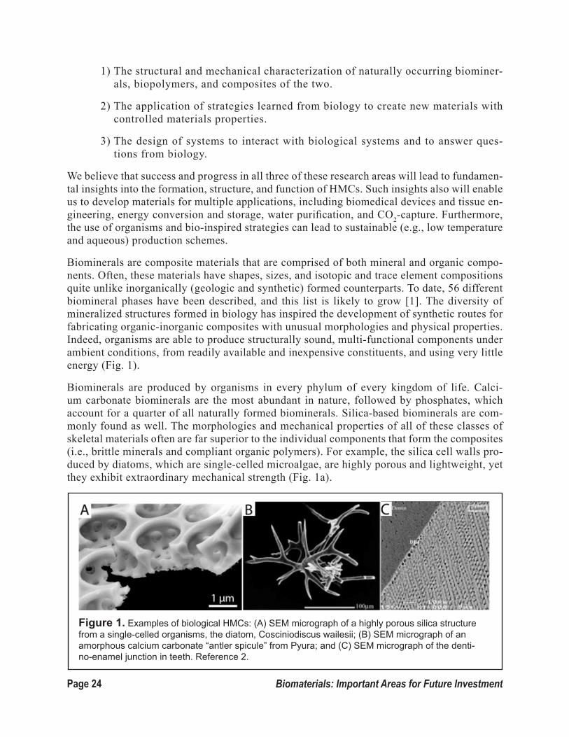
Biominerals are composite materials that are comprised of both mineral and organic compo-nents. Often, these materials have shapes, sizes, and isotopic and trace element compositions quite unlike inorganically (geologic and synthetic) formed counterparts. To date, 56 different biomineral phases have been described, and this list is likely to grow [1]. The diversity of mineralized structures formed in biology has inspired the development of synthetic routes for fabricating organic-inorganic composites with unusual morphologies and physical properties. Indeed, organisms are able to produce structurally sound, multi-functional components under ambient conditions, from readily available and inexpensive constituents, and using very little energy (Fig. 1).
Biominerals are produced by organisms in every phylum of every kingdom of life. Calci-um carbonate biominerals are the most abundant in nature, followed by phosphates, which account for a quarter of all naturally formed biominerals. Silica-based biominerals are com-monly found as well. The morphologies and mechanical properties of all of these classes of skeletal materials often are far superior to the individual components that form the composites (i.e., brittle minerals and compliant organic polymers). For example, the silica cell walls pro-duced by diatoms, which are single-celled microalgae, are highly porous and lightweight, yet they exhibit extraordinary mechanical strength (Fig. 1a).
Figure 1. Examples of biological HMCs: (A) SEM micrograph of a highly porous silica structure from a single-celled organisms, the diatom, Cosciniodiscus wailesii; (B) SEM micrograph of an amorphous calcium carbonate “antler spicule” from Pyura; and (C) SEM micrograph of the denti-no-enamel junction in teeth. Reference 2.

Page 25Biomaterials: Important Areas for Future Investment
Organisms are remarkable biomineral factories because they are able to tightly control the crystal sizes and polymorphs in their skeletons and often stabilize less thermodynamically favorable phases, even amorphous phases (Fig. 1b). It is even more remarkable that some organisms can produce polycrystalline, hierarchical composites, such as mammalian tooth enamel (Fig. 1c, lower right), which is a highly mineralized, hard, damage-tolerant, and abra-sion-resistant tissue. Other mineralized tissues, such as the bone-cartilage interface and the dentino-enamel junction (Fig. 1c, diagonal line), display controlled gradients in structure and mechanical properties that are defined by compositional gradients of the ratio of mineral to organic matrix.
However, understanding and harnessing the design strategies employed by organisms is one of the HMC field’s “grand” challenges. Indeed, based upon the desirable structure-proper-ty-function profiles of biominerals, much effort recently has been devoted to developing a new generation of functional and structural HMCs. These efforts can be divided into two strat-egies: (1) bio-inspired approaches that apply mechanisms learned from biology in synthetic production, and (2) bio-enabled approaches that harness biological organisms directly for
Figure 2. Examples of recent successes in “bio-inspired” and “bio-enabled” approaches. (I) Use of a micropatterned surface, functionalized with a SAM of alkanethiols on gold to template the growth of a large, microporous, single crystal of calcite; (II) Use of a polymer-induced liquid precursor (PILP) phase to infiltrate collagen fibrils; (II) Use of genetically engineered bacterial phage to assemble a cathode material for a lithium ion battery; (IV) Bioengineering single crystal growth by patterning of embryonic primary mesenchyme cells from sea urchins. Reference 3.

Page 26 Biomaterials: Important Areas for Future Investment
sustainable and energy-efficient biotechnological production of advanced materials with desired structures and properties.
The main successes of these types of approaches, to date, include the design of organic small molecules, polymers, and surfaces that can direct crystal growth (Fig. 2, I and II); the use of phage display and related techniques to identify mineral-binding and nucleating peptides (Fig. 3, III); the (genetic) manipulation of organisms, such as magnetotactic bacteria, diatoms, and sea urchins, to grow minerals with altered structures and/or functionalities (Fig. 3, IV); and the use of naturally occurring proteins isolated from organisms, such as sponges, to catalyze the growth of a wide-range of inorganic materials [5]. Recently there also has been some success in fabricating synthetic, “nacre-like” structures with a high ceramic content [6].
Future challenges include moving far beyond “pretty pictures” of proof-of-principle experiments to design de novo materials with desired property-profiles. There also is an ongoing need to dis-cover and evaluate structures from biology that are worth emulating. Such initiatives should be pursued in parallel.
1.2 Opportunities and Challenges
Biological systems have been a source of inspiration for materials synthesis for more than 30 years, yet much of the potential for bio-inspired approaches remains untapped. In particular, our ability to emulate biology and reprogram biological systems for the purposes of materials synthesis remains limited. However, we believe that the field is poised to capitalize upon the promise of bio-inspiration in the design and synthesis of HMCs – now more than ever – given the dramatic advances over the last 10 years in the development of tools for characterizing and manipulating both materials and organisms.
The field also is poised to capitalize on our growing cadre of interdisciplinary researchers. In recent years, it has become increasingly common to train our students to perform multi- and interdisciplinary research and, to create research teams that span the classical disciplines (e.g., materials science, biology, chemistry, physics, and medicine) when they go on to become inde-pendent researchers. This integrated philosophy is proving highly advantageous for biological materials research, because, as a field, it is inherently cross-disciplinary and highly innovative.
Two major opportunities for biomaterials research at this juncture include:
(1) The advanced characterization of biological and bio-inspired HMCs; and
(2) The exploitation of genomic information in the development of new HMCs.
The following sections discuss how these opportunities can be approached.
1.2.1 Advanced Structural and Mechanical Characterization of Biological and Bio-inspired Composites and Hard Materials
Because they are hydrated and structured on multiple length scales, biological composites pres-

Page 27Biomaterials: Important Areas for Future Investment
ent difficult challenges in structural characterization. In some cases, for example, they con-tain organic material embedded non-periodically and at low concentrations in a host matrix with a high electron-scattering cross-section. Given these challenges, in recent years, multiple high-resolution techniques have been used to obtain 3-D nanometer-scale images of biological composites such as bone, teeth, mollusk shells, and other marine animal skeletal units (Fig. 3). Some of these techniques can be used to image samples in their native, hydrated state and simul-taneously provide structure and composition information, with sub-micron resolution.
These high-resolution techniques include:
• Electron microscopy and tomography (including cryoEM) (Fig. 2, II; Fig. 3, I).
• Atom-probe tomography (Fig. 3, II).
• X-ray scattering technologies (e.g., simultaneous SAXS/WAXD, micro/nano-CT, XANES, EXAFS, X-PEEM) (Fig. 3, III).
• Solid-state NMR.
• Vibrational spectromicroscopies (IR and Raman).
In addition to these high-resolution, static techniques, in situ characterization methods that al-low for real-time monitoring of growth processes (both biologic and inorganic) are now avail-able (e.g., liquid-cell TEM, liquid-cell AFM, and confocal microscopy with ultra-fast cameras) (Fig. 3, IV).
Every time one of these characterization techniques is employed to examine a biological ma-terial, we gain important new insights. Indeed, the last 10 years have seen a paradigm shift in our understanding of the mechanisms of biomineral formation and the structure of biological materials at the molecular-to-nanoscale level. In particular, it is now commonly accepted that rather than growing crystals from supersaturated solutions, in both invertebrate and vertebrate organisms, the first phase to form is an amorphous precursor, which is then transformed to form the thermodynamically more stable mineral phases (Fig. 2, II; Fig. 3, III). This insight has rev-olutionized our understanding of how organisms can generate the stunningly complex morphol-ogies of biological HMCs.
In addition, many biominerals previously described as “single crystals” recently have been sug-gested to have meso- to nanoscale sub-grain structures [7]. As a result, our understanding of the mechanistic origins of the desirable properties of biological HMCs has fundamentally changed. Therefore, not only do we have new paradigms regarding materials synthesis (e.g., amorphous/disordered phases transforming via kinetically controllable mechanisms) but also new insights into novel structure-property-function relationships.
To complement the structural characterization of biological HMCs, efforts also are being devoted to characterizing and modeling the materials properties of the natural and synthetic composites. To date, most of this work has focused on mechanical properties (e.g., nano- and microinden-tation as well as micromechanical testing to complement well-established macroscopic tech-niques); however, considerable effort also has been devoted to looking at the optical and mag-netic properties of biominerals. The current challenge is to “close the loop” and identify the key

Page 28 Biomaterials: Important Areas for Future Investment
structural features and length scales that determine any given properties profile. Applying this fundamental knowledge to design strategies for synthetic systems represents an important and promising direction for future investment.
1.2.2 Prospecting Genomes and Beyond
The second major advance over the past 10 years that has directly impact-ed HMC development was achieved through genomic and proteomic re-search. These methodologies have greatly facilitated the identification of novel proteins that play key roles in the formation of biological materials. Furthermore, the rapidly increasing number of completed genome sequenc-es will enable in-depth, evolutionary comparison of mineral-forming organ-isms and provide an entirely new angle for elucidating the molecular basics of HMC biogenesis [8, 9].
The expanding tool set of techniques for manipulating the genomes of or-ganisms has made it possible to envi-sion genetically designing organisms for the biotechnological production of non-natural biological HMCs (i.e., syn-thetic biology) (Fig. 4). This goal is still highly ambitious, but the science of using organisms as factories for gen-erating new materials, adaptive materi-als, and materials for medical and en-ergy applications finally appears to be within reach. The enormous potential payoff justifies the substantial invest-ment that will be required to develop such technologies.
1.2.3 Grand Challenges in HMC Research
Within this context of rapidly developing materials characterization (Section 3.2.1) and ge-
Figure 3. Examples of high-resolution character-ization techniques for biominerals: (I) ADF-STEM tomographic image revealing a network of organic fibers (agarose gel) incorporated into a single crystal of calcite; (II) Atom probe tomography of buried interfaces within a chiton tooth. Chemical informa-tion regarding the inorganic (iron oxide) and organic (chitin fibers) is revealed; (III) (A) XANES-PEEM image of a tri-branched spicule from a sea urchin embryo and (B) RGB map of the three different phases of calcium carbonate identified in the spic-ule; (IV) Time-lapse images taken from a liquid cell TEM experiment, in which oriented-attachment of nanoparticles is imaged with atomic detail for an iron oxide system. Reference 4.

Page 29Biomaterials: Important Areas for Future Investment
netic manipulation techniques (Section 3.2.2), we have identified the following three grand challenges related to biological and bio-inspired HMCs:
(1) Characterizing and controlling the atomic-scale structure and chemistry of HMCs.
(2) Penetrating biological complexity in length scales and molecular diversity.
(3) Engineering the morphogenesis of biological materials.
These challenges are discussed in greater detail below.
1. Characterizing and Controlling Atomic-Scale Structure and Chemistry of Hetero-In-terfaces in Composite Materials
This challenge involves:
a. Creating complex organic-inorganic, inorganic-inorganic, and organic-organ-ic interfaces and interphases in bio-inspired composite materials.
b. Interrogating the structure and properties of these interfaces and interphases in both biological and synthetic materials.
c. Modeling the structure-property relationships in these materials and develop-ing predictive models for the design of new materials.
2. Penetrating Biological Complexity in Length Scales and Molecular Diversity
This challenge involves:
a. I d e n t i f y i n g which length scales and fea-tures of bio-logical materi-als contribute significantly to emergent func-tionality, prop-erties, and per-formance.
b. D e v e l o p i n g structure-based predictive mod-els of hard/soft composite ma-terials.
c. Applying this
Figure 4. (A) Genetic engineering of diatoms (C. fusiformis) to express green fluorescent protein (GFP), encased within the silica cell walls. GFP expression levels are under control of a nitrogen source (NO3, NH4, or N). On the right are fluorescence color images of the genetically modified diatoms, with green being indicative of GFP expression. The red color results from the autofluorescence of the chloroplasts; (B) Incorporation of an active enzyme (hydroxylaminobenzene mutase [habB mutase], which breaks down hydroxyaminobenzene to produce 2-ami-nopehnol [far right image]) within the silica cell wall of T. pseud-onana for catalytic applications. Reference 10.

Page 30 Biomaterials: Important Areas for Future Investment
knowledge to the design and synthesis of new composite materials and under-standing the contributions of each length scale (atomic-nano-micro-macro) to the overall property profile and function of the final material.
d. Ultimately, developing “strategic biomimicry,” which requires knowing enough about how a biological system functions to selectively/strategically identify the key components and features that need to be represented in a syn-thetic material. For example, can a material with two levels of hierarchy be designed to have the same property profile as bone, which has seven levels? If so, which two of the seven levels do we need to mimic?
3. Engineering Morphogenesis of Biological Materials
This challenge involves:
a. Understanding how organisms process materials at the single-cell and tissue level in a sequence of genetically controlled events.
b. Imaging and creating morphogen gradients at multiple length scales.
c. Understanding and harnessing the kinetic processes of formation/dissolution of complex structures to engineer precise control of fabrication, assembly, remodeling, (bio)activity, and degradation (or lack of) for HMCs.
d. Reprogramming the biosynthetic machinery towards scalable, sustainable, and massively parallel synthesis of hierarchically nano- to microstructured composite materials.
e. Combining biological strategies with conventional and advanced materials methods.
1.3 Scientific Questions
As described in the previous section, biological and bio-inspired HMCs present many op-portunities for future investment. Here, we describe several key scientific questions, both fundamental and applied, that must be addressed in order to develop the capabilities needed to achieve the next generation of bio-inspired HMCs.
These questions represent critical areas in which a better understanding must be achieved in order to generate materials with the desired structural and functional profiles. In each case, there are parallel needs for the better characterization of biological materials as well as the development of synthetic methods and computational models based upon what we learn from biology.
We must emphasize that our success and progress in all of these areas will have broader im-plications. Indeed, they will lead to new insights into the formation, structure, and function of HMCs whose development can impact technological innovation related to health care, energy production, and environmental protection, to name a few examples.

Page 31Biomaterials: Important Areas for Future Investment
1.3.1 Bioprospecting
Nature has evolved numerous materials that enable organisms – from single-cell algae and viruses to vertebrates – to thrive in their ecological niches. To date, however, the biological materials com-munity has focused on studying a limited set of either relatively abundant or medically relevant biological materials (e.g., collagen, cellulose, chitin, bone, teeth, mollusk shells, etc.). Thus, the HMC community may have “pigeon-holed” itself by its choice of model organisms so far.
Because only a small number of the existing materials and organisms have been studied, a number of technologically interesting biological materials and many more organisms have not yet been examined for their materials properties. Indeed, many more still await discovery. For example, anyone who has walked on the beaches of New England is familiar with the whelk egg case, which is a hard, brown, twisted structure that resembles a miniature Hawaiian lei, only without the neon colors (Fig. 5). Yet, it was not until 2009 that the unique protein from which it is formed was char-acterized and its deformation mechanism studied [11].
Thus, there is good reason to expect that thoughtful investments in “bioprospecting” will allow the biomaterials community to identify new biological materials with exceptional properties (e.g., from organisms living in extreme environments) and make them available to other HMC research-ers for studies on structure-property-function correlations.
Successful bioprospecting requires multidisciplinary teams consisting of scientists trained in ma-terials science along with life scientists (e.g., zoologists, marine biologists, plant biologists, etc.), so that, once identified, the structures and properties of the novel materials can be readily charac-terized. Additionally, new methods are needed to enable laboratories to cultivate such novel organ-isms and to make them accessible to the wider community for genomic and genetic engineering studies. Through these efforts, new model organisms can be established that are representative of nature’s technologically interesting biological materials portfolio.
1.3.2 “Omics,” Bioinformatics, and Phylogeny as Routes to Materials Discovery
With the advent and rapid evolution of “omics” (e.g., genomics, transcriptomics, proteomics, mi-crobiomics, etc.) techniques, it is now possible to identify the genetically-encoded machineries for the biosynthesis of materials in both existing model organisms and newly discovered organisms. For example, the results of sequencing the genomes of diatoms and sea urchins have enabled the identification of key components of their biomineral-forming machineries [8, 9].
In addition, the growing bioinformatics data set on the molecular phylogeny of organisms (i.e., their evolutionary relationship) and their ecological niches is providing us with important informa-tion on structure-function correlations in the biological materials that they produce. Understanding the structure-function correlations of similar biomaterials from different organisms may reveal evolutionary relationships that are not immediately obvious from molecular phylogenetic analysis. Such a combination of evolutionary and functional analysis of biomaterials will greatly assist us in identifying the sets of genes intimately involved in biomineral morphogenesis.
These types of analyses will continue to provide enormous amounts of data that will need to be analyzed. Indeed, understanding the genetic and molecular basis of morphogenesis of biominerals

Page 32 Biomaterials: Important Areas for Future Investment
requires the efficient analysis of massively large data sets, which are likely to get larger in an ac-celerating fashion. Thus, we will need to develop and implement a set of advanced bioinformatics tools to handle the analysis of these types of data.
Such expertise, however, is currently lacking in the biological materials science community and needs to be acquired in the short term through collaborations with other research fields. This ana-lytic capacity also needs to be made sustainable by incorporating bioinformatics in the education of the next generation of biological materials scientists and engineers (see Education section).
1.3.3 Characterizing and Exploiting Amorphous/Poorly Crystalline Phases
The use of amorphous building materials and precursors is a widespread phenomenon in biomin-eralization. Amorphous precursors are far from equilibrium, but they are kinetically trapped and typically have lifetimes between hours and days. The precursor strategy is thought to play a major role in the unparalleled ability of biological organisms to: control polymorph and crystal shape; introduce smooth, curving surfaces; and impart outstanding mechanical properties to organic-in-organic composite materials.
The mechanism of the biologically controlled disorder-to-order transformation in biomineralizing organisms remains unclear. Our lack of understanding of this process is due to the challenges of characterizing the structure of the mineral precursors, which lack long-range order and are un-stable. Significant improvements in the sensitivity of in situ X-ray or electron scattering-based approaches, coupled with a better understanding of synthetic model systems and the complexity of computational simulations, will likely be required to address the particular challenges of investi-gating these processes in biological systems.
Despite our limited understanding of the structures of amorphous precursors and their phase trans-formations, the interest in developing synthetic capabilities based upon this strategy has grown significantly in recent years. Indeed, investigations of polymer-stabilized amorphous precursors and polymer-induced liquid precursors have shown wetting and infiltration behaviours that prom-
Figure 5. (A) Photograph of a whelk egg case (total length: 1 m); (B) Schematic representation of the proposed protein structure, which converts, reversibly, from an alpha-helical native state to an extended beta-sheet state. This reversible change in protein secondary structure is proposed to be responsible for the observed high reversible extensibility and shock absorbing properties of the poly-mer egg case. Reference 11.

Page 33Biomaterials: Important Areas for Future Investment
ise to open up new avenues for creating novel conformal coatings or composite architectures with oriented fibrous soft matter Fig. 2, I and II; Fig. 6B). The importance of confinement for the sta-bilization of amorphous precursors also is beginning to emerge as a synthetic strategy (Fig. 6A).
1.3.4 Understanding and replicating interfaces/interphases between organic-inorganic, organic-organic, and inorganic-inorganic materials
Buried interfaces (i.e., interfaces that are occluded within the final composite material or function-al structure) are crucial to a variety of biological processes and applications, including:
• Biological wet/dry adhesion of organisms ranging from barnacles to geckos.
• Biological control over mineral growth.
• Mechanical properties in high toughness, wear-resistant tissues, such as tooth or bone.
• Tissue engineering.
• Functionalized core@shell nanomaterials for biomedical imaging and drug-delivery applications.
• Bio-lubrication, for example, at the articular cartilage surface in joints.
The pivotal role of organic/inorganic interfaces in other areas (e.g., lithium ion battery perfor-mance and safety, dye-sensitized solar cells, nano-dielectrics for organic field-effect transistors, flexible displays, etc.) is emerging. The rational design of bio-inspired materials for applications ranging from biomedicine to sensors and catalysts to high-toughness composites, depends on a detailed understanding of the structure, chemistry, routes of assembly, mechanical properties, and wear processes at these interfaces. However, with typical length scales ranging from sub-nano-meter to macroscopic dimensions, combined with the complexities arising from the hybrid (e.g., hard/soft, organic/inorganic, etc.) character of these materials, the synthesis, simulation, and in situ functional characterization of such buried interfaces is a significant challenge.
Nevertheless, imaging atomic-scale, buried interfaces, interphases, and dynamic processes occurring at these locations is integral to understanding biological design strategies. It also is integral to building quantitative models in simulations and to controlling processing parameters, structure, and performance of bio-inspired functional materials. Although tre-mendous progress has been made in terms of electron tomography, for example, in imaging synthetic and biogenic single crystals (see Fig. 3, I), quantitative imaging techniques based on synchrotron X-ray micro/nanoprobes (see Fig. 3, III) and nanoSIMS will play increas-ingly important roles in characterizing both chemically and structurally complex materials. Atom probe tomography, the only current technique available with sub-nanometer spatial resolution and unbiased chemical sensitivity across the periodic table, also offers tremen-dous opportunities (see Fig. 3, II).

Page 34 Biomaterials: Important Areas for Future Investment
However, significant challenges remain to be solved in the application of these techniques to new materials, especially in the implementation of cryogenic sample preparation and imaging for optimum sample preservation and in statistical data analysis. Considerable syn-ergy is anticipated from correlative imaging approaches (e.g., fluorescence/electron mi-croscopy or electron tomography/atom probe), and the development of liquid-cell scanning and transmission electron microscopy heralds our ability in the near future to investigate dynamic processes at interfaces with unprecedented resolution (see Fig. 3, IV).
We anticipate that within the next decade many technical hurdles to investigating buried interfaces in biological model systems will be cleared. Indeed, as new imaging modalities become available, it will become even more important to draw together teams with the ex-pertise to: (1) manipulate biological model systems genetically; (2) structurally characterize the resulting structures; (3) computationally model the resulting data; and (4) build in vitro model systems.
Based on recent progress in our understanding of the incorporation of organics into growing crystals (13,14), the tremendous development of soft-matter self-assembled systems (see Section 2 of this report), and the emerging role of confinement for nucleation and growth [15, 16], there is significant potential in the integration of these areas. The rational design of 3-D matrices with advanced features mirroring those of biology (e.g., stimulus-response, compartmentalization, and self-degradation for control of both classical and non-classical nucleation and growth) offers a number of very exciting prospects.
1.3.5 Design of Functionally Graded Systems
Gradients of composition and functional properties are pervasive in industrial and biological systems and are hallmarks of complex systems with multi-functionality. In particular, they exist in native mineralized tissues (e.g., the osteochondral interface, bone-tendon and bone-ligament interfaces, and the dentino-enamel and dentin-cementum junctions in teeth) [17]. For example, in a healthy joint, the transition from the compliant, gel-like, cartilage tissue to the stiff bone
Figure 6: (A) Amorphous calcium carbonate precursor nanoparticles stabilized by confinement in liposomes, imaged by cryoTEM; (B) A solution of amorphous CaCO3 precursor particles can infil-trate a colloidal crystal (schematic on left) and crystallize into a microporous single crystal of calcite (SEM image on right). Reference 12.

Page 35Biomaterials: Important Areas for Future Investment
tissue is made through a zone of calcified cartilage, which has a modulus between those of bone and cartilage (Fig. 7.I). The human tooth has multiple graded interfaces as schematically illus-trated in Fig. 7.II. In addition to static gradients, there also are examples in biology of function-ally graded materials that evolve with time and during development, such as the growth plate in developing bone. In all of these systems, the gradient in functionality is achieved via spatial and/or temporal compositional and/or structural gradients.
Despite the prevalence of these graded interphases in biology, current generation biomaterials are usually homogenous materials. Thus, one of the field’s biggest challenges remains the in-tegration of these synthetic, homogenous materials into the living tissue. An ideal biomedical material should fully integrate with the target tissue at all levels of hierarchy and respond to physiological changes over time.
Two pressing questions in this research area include: (1) how do we image physiologically rele-vant gradients with molecular-level resolution and biochemical sensitivity in a 3-D, time-depen-dent manner; and (2) how do we create functional gradients in synthetic systems.
The ability to characterize and create nanostructured, functionally graded systems is relevant to the design of highly integrated composite materials for a variety of applications – ranging from regenerative medicine to structural materials. Within the regenerative medicine community there is an emerging understanding that interfaces between tissues are the key to the restoration of function for many pathologies. This insight, coupled with advances in biology and fabrication methods, makes functionally graded materials a timely and important topic of investigation.
The first set of fundamental science questions in this area involves a better understanding of the composition, structure, and function of gradients in biological materials. As mentioned previ-ously, characterizing such materials requires techniques that can probe multiple hierarchical lev-els, provide both spatial and temporal resolution regarding changes in molecular composition, nano- to mesoscale structure, and mechanical and biological function. Developing such tech-niques will enhance our ability to characterize biological materials and to fabricate materials that emulate the properties of a given tissue. Particular emphasis should be placed on techniques with sufficient temporal resolution to elucidate how the functional gradients are formed in time, as well as the dynamics of their mechanical performance.
For mechanical testing, the techniques must be sensitive over several orders of magnitude in force and displacement, and multiple methods must be coupled to be able to integrate information over the entire gradient structure. For example, recent advances in ultrafast, confocal microscopy, cou-pled with a shear-stage, can be used to perform dynamic testing of tissues, such as cartilage [18]. Additionally, finite element modeling of force distribution through anisotropic domains reflective of biological tissues would help us understand responses to mechanical stimuli, such as pressure. A key component of any characterization study is the identification of which hierarchical, tempo-ral, and spatial parameters from the model biological system are critical to include in designs for synthetic functionally graded systems.
In parallel with an improved, higher-resolution characterization of biological, functionally graded composite systems, the second set of research directions in this area involves the design and syn-thesis of new materials. There are two general types of strategies: (1) gradient imprinting, and (2) gradient generators.

Page 36 Biomaterials: Important Areas for Future Investment
Gradient imprinting in-volves creating struc-tural or compositional gradients using estab-lished nano- to micro-fabrication techniques. Examples of gradient imprinting include the formation of mineral or protein gradients by simple layering tech-niques in a gel, fiber alignment gradients using extrusion or electrospinning tech-niques, and stiffness gradients made by varying crosslinker densities.
Gradient generators are systems in which molecular, biomolec-ular, or genetic cues are introduced aniso-tropically into a ho-mogenous material ─ with or without cells ─ to generate de novo structural and/or com-positional gradients. Although gradient generators offer more potential than gradient imprinting techniques, they also present more challenges. A key to the development of new gradient gener-ators is the identifi-cation of potential methods for developing anisotropic temporal and spatial “signals” within initially homogeneous materials. Examples of acellular approaches include using reaction-diffusion processes within hydrogels to create compositional gradients and using microfluidic gradient generators to pattern 2-D surfaces. Future potential directions in acellular gradient generation include taking advantage of additional external (or internal) forces, such as gravity, electrical, thermal, magnetic, and optical forces to form gradients in liquid or viscous systems to avoid problems associated with microfluidity.
Figure 7. (I) A plot of mineral density, as determined by FTIR micros-copy, across the bone-cartilage interface. The different zones of the tissue are labeled as bone, calcified cartilage (CC), deep- and mid-zone cartilage, and superficial zone (SZC); (II) Schematic cross-sec-tion of a human tooth, highlighting the hierarchical structure and multiple graded-interfaces (e.g., dentino-enamel junction (DEJ) and the dentine-cementum junction (CDJ). The changes in stiffness as a function of location are given on the right. Reference 17.

Page 37Biomaterials: Important Areas for Future Investment
Perhaps the greatest potential exists in harnessing cells (e.g., mammalian, bacterial, etc.) or viruses to create gradients for us. For example, gradients could be generated by:
• Co-culturing different cell-types/organisms in a pattern to generate matrix gradients.
• Patterning growth factors or growth factor binding molecules in a gradient to induce selective responses of the cells/organisms.
• Creating transfection vector gradients to induce spatial gradients in cells/organism function.
• Using mechanical and/or electrical stimulation to induce gradient formation by bioor-ganisms.
• Utilizing gradient imprinting to create a system containing an “inductive structural gradient,” where cells introduced to a gradient imprinted material create additional gradients in the system or alter the original platform.
For all of these approaches to making gradients, the following four issues need to be ad-dressed:
1. Achieving appropriate spatial resolution in fabrication/synthesis;
2. Achieving appropriate temporal resolution/kinetic control in both the fabrication, as well as the lifetime of the functionally graded material (i.e., controlling how the mate-rial and its properties evolve with time);
3. Characterizing the interaction of temporal and spatial cues in the generation of the gradients;
4. Determining the scalability of the techniques. Although important and useful informa-tion can be gleaned from small sample sizes, the next challenge is to fabricate materi-als at a scale that is useful for applications.
Developing organism-based gradient generators has its own set of unique challenges, includ-ing: selecting the appropriate organism(s); selecting culture environments that are compatible with achieving spatial and temporal control; and the reproducibility of generated gradients due to inherent “biological noise.”
Additional advances in the area of functionally graded materials will come from developing adaptable and tunable gradient systems. By understanding how gradients grow and evolve with time, we can design adaptable systems that have one gradient profile (i.e., one “equilib-rium”) and then change at a later time, on demand, to another “equilibrium” gradient. Such a system would mimic growth and development in biology, as well as harness the idea of metastable states from materials science. Related to adaptable systems are tunable gradient systems that can be “frozen” at arbitrary points prior to reaching equilibrium.
Finally, the integration of the ideas discussed in this section with those in the next section will be critical to any successful design of hierarchical materials. The extension of gradient formation to multiple length scales is a crucial next step.

Page 38 Biomaterials: Important Areas for Future Investment
1.3.6 Hierarchical composite materials by design
Biology uses hierarchical composites to tailor local properties to a required set of design require-ments and to introduce multifunctionality into a single tissue [19]. In addition, biological hierar-chical composites can be self-healing, adaptive, and damage tolerant due to multiple toughening mechanisms over a range of length scales (Fig. 8). Despite the promise of hierarchical materials, to-date, the maximum number of hierarchical levels currently realized in synthetic materials is limited to two or three.
Biological composite materials, such as bone or mollusk shells, are hierarchical nanocomposites with properties that are uniquely optimized for their functions. Specifically, these materials:
o Are built of nano-sized building blocks such as mineralized collagen fibrils (bone) or mineral tablets (molluscan nacre).
o Feature molecularly well-defined interfaces and interphases that connect the levels to-gether (see Sections 1.3.4 and 1.3.5).
o Have functional properties that are heterogeneous and change from one level of hierarchy to the next and from one location to another to optimize their functional performance.
o Possess self-healing and adaptive properties.
o Have dynamic interactions between macromolecular assemblies and mineral phases both during their formation as well as in the fully mature form.
Despite major advances in our understanding of these materials, a number of critical issues, both basic and applied, remain unresolved. For example, as with functionally graded systems, we need to better characterize and understand the formation processes and structure-function-property pro-files of hierarchical biological materials. One of the issues we face is that the properties of these materials are emergent (i.e., they are not predictable from our understanding of each individual building block).
In addition, we currently lack experimental data that is sufficiently detailed to allow us to de-velop theoretical and computational tools that will enable us to design such materials with the desired property profile (see Section 3.3.7). For example, even for bone, which is perhaps the quintessential example of a biological hierarchical composite material, with seven well-defined levels of hierarchy [21], we neither fully understand how each level is formed nor what role each level plays in determining the overall functions of the material. The combination of overlapping interphases/interfaces at different length scales from molecular to microscopic, all of which contain water, as well as organic and inorganic components, requires a unique set of character-ization tools and techniques. Additionally, these tools and techniques must couple high-resolu-tion methods traditionally used in materials science/chemistry/physics with methods from the biological sciences (e.g., hydrated samples and cryo techniques).
A key to better understanding the formation mechanisms of inorganic-organic assemblies are stud-ies on the:
• Cooperative formation of mineral/organic assemblies: e.g., active transport versus diffu-sion, self-assembly versus cellular-assembly.

Page 39Biomaterials: Important Areas for Future Investment
• Kinetic processes amenable to regulation: e.g., molecular feedback loops versus cellular instruction.
• Multifunctional proteins, multifunctional minerals: elemental/mineral sinks and sources, “dirty” crystals for improved solubility, peptides to control mineralization processes, me-chanical properties, pH variations, etc.
The second set of new directions in this area involves the development of new synthetic methods. Although we now can create biomimetic structures recapitulating the first level of hierarchy, via macromolecular self-assembly and sophisticated regulation of mineralization, we do not have the technological means to design and produce nanomaterials with multiple levels of hierarchy.
The major difficulty we face in addressing hierarchical complexity is our current inability to bridge the sub-micron and micron levels. Thus, the translation of even the current understanding of biolog-ical systems into synthetic (bench-top) materials and systems is slow and difficult. For example, in most synthetic hierarchical materials, sharp interfaces rather than gradient interphases typically are formed. Gradient interphases are typical of natural materials (see Section 1.3.5). The integration of molecularly well-defined and functionally graded interphases over a range of length scales, into syn-thetic hierarchical structures, remains a key challenge for this field.
When designing the next generation of synthetic systems, it is essential to ask the following questions:
1. How many levels of hierarchy do we need to achieve our target function?
2. What is the role of each level and is it required also in a syn-thetic system?
3. How will the levels be assembled?
4. How many processes and processing steps are required?
5. How scalable are these fabrication methods?
Depending on the critical property for the end use of the bioinspired material (e.g., fracture resistance, energy absorption, electrical/optical/magnetic sensing, etc.), dif-ferent fabrication methods may be required. To achieve these goals, we need new
Figure 8. Schematic illustration of bone deformation in re-sponse to tensile load. The distribution of stress is illustrated at three levels in the structural hierarchy: the tissue level (left), min-eralized collagen fibril array (center), and an individual collagen fibril with platelets of hydroxyapatite mineral (right). Reference 20.

Page 40 Biomaterials: Important Areas for Future Investment
techniques for replicating desired features at the nanoscale both in 2-D and in 3-D (e.g., self-assembly, multichannel printing) as well as for co-processing multiple phases (e.g., multiple proteins, polymers and ceramics, etc.) to form composites and gradients. In addition, efforts should be made to translate low-yielding, synthetic routes into higher-yield processes. It is possible that organisms could be har-nessed to perform some of these tasks (see Section 1.3.8).
One of the major challenges in making multi-scale, hierarchical materials is linking manufacturing at different scales. We routinely manipulate matter at nanoscale via self-assembly and mineral particle manipulation by supramolecular complexes. Where we have a harder time, however, is operating in the meso and micro scales (i.e., microns to the hundredths of microns scale). Although, in some cases, we are able to create sophisticated mesoscale objects, we currently do not have the means to incorporate them into a larger hierarchical design.
One possible approach to meeting this challenge is to create an artificial microscale materials synthesis device. Such a device would have:
1. Synthetic machinery (with chemical [ATP/ADP], light, or thermal sources of energy) to produce organic and mineral components.
2. Motility, for example, by having internal magnetosomes to guide movement within a mag-netic field.
3. A secretory apparatus that will deliver the material (e.g., a protein-mineral complex) to the appropriate location.
4. Devices that work in parallel and, ideally, with the ability to deposit different materials in an orchestrated fashion.
5. Methods to accelerate growth at the nanoscale into the mesoscale and, ultimately, to the macroscale
1.3.7 Predictive ability to design de novo composites with defined property profiles
Although the theory and modeling of traditional composite materials is fairly well-established in the materials science community, understanding the structure-property relationships in bi-ological and bio-inspired, hierarchical composites and functionally graded materials remains poorly addressed by current theory, modeling, and computational approaches. For example, there are very few studies at the molecular scale that have bridged models of inorganic crystal nucleation and growth with coarse-grained approaches to study the self-assembly of biopoly-mers. Similarly, only a few studies have been published that address the mechanical interplay of the different hierarchical levels from the molecular to the macroscale.
Thus, there is a critical need for a theory-based, “strategic biomimicry,” which will enable us to identify which and how many levels of the hierarchy need to be emulated in a model system to sufficiently de-scribe it. Addressing this need requires us to bring together theorists from both hard and soft materials communities and pair them with experimentalists who are focused on developing simple model sys-tems amenable to theoretical exploration and modeling. A small number of model systems that focus on
Page 41Biomaterials: Important Areas for Future Investment
individual levels within the hierarchical structure need to be developed, such as a model for organic-in-organic interfaces at the molecular scale. Then these individual model systems – with limited levels of hierarchy and complexity – can be combined over a range of length scales.
In addition to bringing together the hard and soft materials communities, we also need to engage theo-rists and modelers with expertise in multi-scale and/or a range of length scales. A key to any successful program in this area will include the ability to predict appropriate structure-property relationships (e.g., mechanical, optical, electrical, etc.) and to explore how different levels of the hierarchy are linked to achieve performance. Finally, and perhaps most computationally challenging, will be the ability to de-velop models of the kinetics of formation, coupled with a prediction of static, final structure-property relationships.
1.3.8 Genetically Designed Materials
The rapid and continuing advancements in DNA synthesis and sequencing capabilities have opened up the possibility for us to understand the genetic basis of biological materials properties and to design non-natural materials with desired functionalities through biotechnological routes.
In the last 15 years, we have made tremendous progress in understanding biological materials synthesis at the structural, compositional, and mechanistic levels. The next step is to rationally design new mate-rials based on this understanding and to harness organisms to manufacture materials for non-biological applications with programmed structures and properties (Fig. 9).
We suggest three complementary approaches to do so. This list is by no means exhaustive and should be taken as a starting point:
1. Combining minimal genetic cassettes that control materials synthesis in a host or synthetic organism. A recent example of a magnetotactic bacterium with two sets of magnetosomes – one set to produce iron oxide and the other set to produce iron sulfide – illustrates the first step in such a process [23]. Looking further ahead, one could create metamaterials with photonic and magnetic properties by, for example, inserting a bacterial magnetosome cas-sette into Morpho butterflies.
2. Forward engineering of organismal materials synthesis to incorporate novel, orthogonal functionality. For example, silkworms could be engineered to incorporate a stretch-activat-ed protein sensor to create force-aware textiles.
3. Expropriating systems-level biological self-assembly and morphogenetic mechanisms to in vitro and cell-free systems. For example, single crystals could be engineered by co-opting crystal-depositing cells [24].
All of these approaches rely on the chemical synthesis of DNA and the molecular genetic manipulation of genomes, and they can be expanded to introduce exogenous genes or genes that encode for proteins with unnatural amino acids. In principle, these tools already are in place. Yet, we envision applying them at a scale (e.g., genetic islands, chromosomes, entire genomes, etc.) that is extremely challenging and only recently has become – in principle – achievable. A key challenge is the use of model organisms for materials synthesis (e.g., cockroaches, beetles, butterflies, silkworms, sea urchins, etc.) that have not yet been fully analyzed on the “omics” level and for which methods for genetic manipulation have not yet been established.

Page 42 Biomaterials: Important Areas for Future Investment
1.4 Needs and Recommendations
To tackle the scientific questions discussed in Section 1.3, the following set of technological advances are recommended:
• New characterization tools and methodologies that are:
o Non-destructive.
o Highly sensitive (small amounts of material, high temporal resolution).
o High-content (multiplex assays to combine spatial and temporal information from a single sample).
• Biomolecular analysis platforms (especially mass spectrometry and [solid-state] NMR spec-troscopy) that are specifically equipped for the analysis of challenging biological materials sam-ples (e.g., insoluble organic and composite matrices; biominerals with elemental compositions that are not straightforward to analyze by NMR).
• Shared user facilities with state-of-the-art characteriza-tion tools and inter/cross-dis-ciplinary expertise.
• Development of databas-es and material information systems on structures, prop-erties, and functions and tools for systematic data mining (e.g., a BioMaterials Genome).
• New bioreactors with im-proved spatial and temporal control.
• New material synthesis and fabrication methods, includ-ing processing approaches that can be scaled up from the bench-top to large-scale applications.
• High throughput platforms in combination with high-reso-lution printing/fabrication.
• Multiplex assays to combine spatial and temporal infor-
Figure 9. Schematic representation of how genetic engineering of organisms can lead to new materials de-velopment, beginning with genetic engineering based on biological information. One the organism is determined to produce a biomaterial with the desired properties, it can be bread and cultivated on a large scale to produce a biofunc-tional material. Reference 22.

Page 43Biomaterials: Important Areas for Future Investment
mation from a single sample.
• More basic theory in structure-property relationships for “dirty” biological systems.
• Expansion of graduate studies curriculum in biological materials to include:
o Bioinformatics
o Characterization techniques that cover a range of size scales
o Modeling/multivariate analyses that will allow integration of data at multiple size scales and/or time points

Page 44 Biomaterials: Important Areas for Future Investment
Section 2. Soft Materials DISCUSSION LEADER
DENNIS E. DISChER, University of Pennsylvania
GROUP MEMBERS
SEth fRADEN, branDeis University
vANESSA ORtIz, ColUMbia University
ALySSA PANItCh, PUrDUe University
DARRIN POChAN, University of Delaware
SAMUEL SAfRAN, the weizMann institUte
ShELLy SAkIyAMA-ELBERt, washington University
IGAL SzLEIfER, northwestern University
DAvID WEItz, harvarD University
tING XU, University of California, berkeley
SPECIAL ACkNOWLEDGMENt
DAvE DINGAL, University of Pennsylvania, stUDent PartiCiPant anD ContribUtor to this rePort
2.1 Introduction
Soft, wet matter pervades the biological world, and skin provides a perfect example. A light poke readily deforms the skin, which quickly springs back. However, the response of the mat-ter that makes up ‘skin’ involves both passive responses and poorly understood, energy-con-suming active responses. Indeed, poking the skin with too hard or too sharp of an object will bruise or even cut it, initiating a cascade of far more active processes that promote wound healing. Even before that, however, the damage will be anticipated, stress will be sensed, and the tissue will be primed to quickly react.
Due to skin’s resiliency and ability to self-repair, it certainly seems useful to emulate it in var-ious biomaterials and devices. However, this is not as easy as it might seem. At the microscop-ic scale, skin has extremely complex biomaterials residing both outside cells as extracellular

Page 45Biomaterials: Important Areas for Future Investment
matrix and inside cells delimited by membranes (Fig. 1).
The specific biomaterials of skin ─ or any other tissue ─ are invariably biopolymers of many types. All of them are permeated by water, which impacts everything from how the molecules are made to how they perform. Over the past several centuries, many components of cells and the extracellular matrix have been isolated, processed, applied, and then further used to motivate the synthesis of other materials, including polymers and block copolymers, colloids, gels, liquid crystals, membranes, and micelles.
In fact, polymer science is often said to have started with the controlled modification of plant cellulose in the mid-19th century to produce the first synthetic thermoplastic polymer. Liquid crystals also have natural origins in the lipids that self-organize into micelles and mem-branes, but liquid crystals are now ubiquitous in displays of modern electronic devices due to their ability to respond to boundary conditions and applied electric fields.
The modern field of soft matter also has benefited enormously from advances in physical characterization, which in recent times has motivated a broad array of new microscopies, new and large facilities for atomic resolution studies with various types of particles (neu-trons, electrons, high energy x-ray photons, etc.), and new methods for high-precision force measurement and rheology (the study of flowing matter). Molecular-scale simulation is now being partnered with theory, which is driving the field forward and explaining accepted experimental results or addressing controversial findings as well as making predictions that
Figure 1: Skin is typical of biological soft matter. The dermis, which is layer of skin beneath the epidermis and cushions the body from stress and strain, provides tensile strength and elastic-ity to the skin through an extracellular matrix composed of collagen fibers microfibrils, and elastic fibers, embedded in proteoglycans. Reference 25.

Page 46 Biomaterials: Important Areas for Future Investment
can challenge curious experimentalists.
Theoretical work has been essential in providing a predictive, mathematical understanding of many current soft matter systems, and Nobel Prizes for these discoveries abound. Pierre-Gilles de Gennes, who often is referred to as the “founding father of soft matter,” for exam-ple, received the Nobel Prize in Physics in 1991 for predicting that the order parameter from simple thermodynamic systems can be applied to the more complex cases found in soft matter and, in particular, to the behaviors of liquid crystals and polymers .
The field of soft materials, thus, has a long tradition of partnering rigorous physics and chem-ical synthesis with biochemistry and biophysics to create new types of matter in exciting areas for cross-disciplinary research. Entire industries, from cosmetics and pharmaceuticals to foods and biomedical devices, deal rigorously every day with many aspects of soft bioma-terials, including for example rheological properties and colloidal stability. The many insights and new materials gained from these endeavors as well as the training of the next generation of soft material researchers have helped to position the field well in terms of broader societal impacts.
In this new millennium, soft material R&D is being further bolstered by biology’s “Infor-mation Age” which is producing catalogs of full genomes for many species (and even indi-viduals) along with their accompanying proteomes and additional high content, annotated descriptions. These new catalogs will be used by modern soft matter scientists and engineers as inspiration, motivation, and even blueprints for the development of new soft biomaterial systems that cannot yet be imagined.
2.2 Opportunities and Challenges
Opportunities for soft matter science and engineering include, for example, implantation in humans and animals to support or replace failed tissues and organs. However, applications of novel biomaterial systems extend far beyond biomedical uses. Three broad challenges are discussed below as an illustrative vision for the field, starting with near-term opportunities and progressing to long-term challenges.
First, as the previously described example of skin portrays, when compared to modern materi-als, there remain major opportunities to mine and emulate the adaptive capabilities of diverse natural systems. A second major opportunity is the development of soft biomaterials that an-ticipate damage and, ultimately, repair it, and are even capable of morphogenesis. Thirdly, the time appears right to begin probing and understanding the underlying rules that biology draws from genomes, with their tens of thousands of components expressed dynamically in varying proportion, in order to develop adaptive and active biomaterials.
The challenges in experimentation, theory, and computation are enormous but have the po-tential to transform everyday devices just as polymers and liquid crystals have done. Each challenge will require cross-disciplinary experimental and theoretical approaches with chem-istry, physics, materials engineering, and increasingly sophisticated uses of modern biology. Cross-program training support and resource investments are, of course, essential.

Page 47Biomaterials: Important Areas for Future Investment
2.2.1 Mining and emulating the adaptive capacity of diverse natural materials
Extracting a tissue with a micelle-forming detergent removes cells together with other soluble materials and, thereby, generates an insoluble matrix scaffold (Fig. 2). These scaffolds are now being recellularized with patient stem cells and implanted in humans to successfully regenerate organs. Tracheas, for example, possess complex geometry at multiple scales and have been used as adaptive grafting scaffolds in recent, high profile clinical trials [26]. As of yet, synthetics cannot replicate such properties, which illustrates how valuable mining nature for biomaterials remains.
Biological matrices are particularly inspiring as they are monolithic and flexible as well as wet and multifunctional. Although collagens are the most abundant proteins in the body, matrices are invariably built from numerous, spatially-constrained components and interactions, and they har-bor various types of signals and cues that promote adaptive responses from cells. Matrices also incorporate key functionalities, such as growth factor storage and triggered release.
Thus, there is a basic need for methods to better characterize critical material interactions and for methods to accurately identify the minimal requirements to recapitulate adaptive behaviors in such natural soft materials. Matrices are prototypical in posing a number of major theoretical and computational problems that should aim to enhance predictability. However, matrices also raise a broader set of illustrative and challenging questions.
For example, how do we understand and mod-el natural matrices? Do we need to develop better detergents for tissue deconstruction or for dissolution of biofilms? Can hybrid materials or even pure synthetics minimize complexity but still function either upon im-plantation or in completely non-biological ap-plications? Might ‘omics’ methods and bioin-formatics that have emerged with genomics in the last few years help to elucidate and even inform theory?
Here, the opportunity – and challenge – is to mine, minimize, make, and model natural ma-terial systems of which extracellular matrices constitute just one major example. New mi-cellizing detergents, tissue implants, and hy-brid materials are likely to emerge from these endeavors. Even when reductionism is em-ployed in bottom-up design approaches, such as with peptides modified by polymers, the material systems are often ‘super-complex’ with confounding issues, such as protein fold-ing. Therefore, it is timely to invest in this area ─ not only because the ongoing revolution in genomics and proteomics is allowing better
Figure 2: A detergent-decellularized chick-en heart. The remaining structure is extra-cellular matrix and allows cells to attach and grow into functional tissue such as a beat-ing heart. It also illustrates the use of deter-gent molecules commonly used in micellar, soft matter assemblies. Reference 27.

Page 48 Biomaterials: Important Areas for Future Investment
compositional characterization than ever before but also because of recent key advances in the synthesis, characterization, and molecular biology of protein expression. Beyond such matrices for medical application, other natural biological systems, including microbes and plants, remain largely untapped for modern soft materials and for bio-inspired synthetics.
2.2.2 Making matter active and capable of morphogenesis
The self-healing of skin suggests at least one very active and common mechanism for tissues: cells migrate into the wound, come in contract with each other to close the wound, and synthesize new matrix to replace lost tissue. Imagine, then, a torn shirt that, like skin, could repair itself by using ambient light as a fuel source. Or, perhaps better, a shirt that first senses the possibility of a tear and quickly fortifies itself to limit or prevent the damage.
How might we design soft matter that is active and capable of self-repair or morphogenesis? The beating heart is a model of stimulated activity that might be co-opted for such purposes. Although some cell types and pieces of tissue move under their own volition, many cells and tissues respond to triggers by changing shape and moving. Even heart cells that don’t normally move from point-A to point-B can be integrated into a suitably soft material design that swims like a jellyfish (Fig. 3).
Current theories have predicted how applied stresses and other changes in the physical en-vironment trigger structural reorganization, extending to cell and collective motion. For ex-ample, symmetry breaking seems essential and exploits anisotropies in shape or properties. Furthermore, theoretical approaches have been developed in the last half-dozen years that ac-count for the internal consumption of ener-gy by an object such as a cell, which is sub-ject to randomizing Brownian forces, and decades of theory and experiment in liquid crystals is informing these new non-equilib-rium theories and computations. However, many of these predictions are untested, and the many component degrees of freedom in living systems are not so easily or credibly simplified into more simple, coarse-grained objects, such as spheres or rods.
Regardless of the systems involved, active materials raise a broad set of challenging questions. For example, how do we make microscopic materials that can exhibit fu-eled, dynamical behavior and can do work that is genuinely useful after sensing and responding to their microenvironment, in-cluding transient stimuli? Can we design materials that cooperatively build them-selves, such as tissues, and might even secrete their own matrix for permanence? Can we produce materials composed of
Figure 3: An artificial jellyfish made of sili-cone and rat heart cells has the ability to swim in water when subjected to an electric field. Reference 28.

Page 49Biomaterials: Important Areas for Future Investment
identical ‘units’ that, in response to an external cue, can differentiate their structures, rhe-ological properties, material patterns, and properties depending on ‘cell’ state or patterned coupling to environment?
In addition to motions of active materials, growth processes are of equal or greater interest. A new organism begins with one cell that divides to produce two identical daughter cells and, then, it might make 10 more generations (i.e., 1024 cells from a single cell) before differentiation occurs. Proliferation then expands each type of specialized cell, which is dedicated to a set of tasks, such as filtration (e.g., the kidneys), pumping (e.g. the heart), or computing (e.g., the brain). Reaction-diffusion processes that are confined to droplets but also interacting in defined spatial arrangements might help establish experimental princi-ples akin to proliferation.
The grand challenge at this point in time is to learn from theory as well as nature and make active material systems that move, re-shape, and, perhaps, even grow. Studies of cell systems can be insightful in this same context, provided the focus is on energy, motion, and collective function as a living material system.
Thus, it seems very timely to invest in this field of research not only because of the existing theories but also because cell culture systems are now sufficiently well developed to exploit. For example, heart cells that contract rhythmically at 1 Hz (in humans) or 5 Hz (in mice) are now routinely generated with increasing purity in many laboratories from induced pluripotent stem cells. Furthermore, microfluidic methods are now routinely able to generate cell-size droplets or capsules with controlled chemistry inside, thus, creating a diversity of opportu-nities for micro-responsive materials that couple and communicate by light, contact, or se-cretion. Coupling to asymmetric shapes and other complicated geometries will usher in new fields of active biomaterials that move and swarm in liquid and perhaps other media.
2.2.3 Probing genome-scale hyper-complexity for evolved biomaterials
Current man-made materials are far from achieving the type of performance observed in na-ture. Indeed, a typical commercial blend or soft materials composite typically contains only a handful of different polymer types; whereas, the typical mammalian cell will invariably draw from thousands or more structural and structure-signaling genes during its development. Al-though mammalian cells have evolved over eons from far fewer genes in simpler organisms, the most advanced tissue systems are ‘hyper-complex’ in their composition and top-down properties.
This disparity likely has its basis in the fact that no biological system functions exclusively on a single length scale. Cellular assembly is self-directed both with and within a diversity of proteins, lipid membranes, carbohydrate gels, and nucleic acid chains. Biological systems arrange hierarchically into intricate, often fractal structures and invariably under non-ideal conditions. Although key cellular and intermolecular interactions are diverse, they seemingly are well regulated and typically malleable at only a few kcal/mol. Thus, modern genomics and genetics are laying the foundation for us to consider exploiting the useful materials and material concepts hidden within cells and/or tissues.

Page 50 Biomaterials: Important Areas for Future Investment
Chemical biology as well as classic pharmacological reagents are providing new routes to modify molecules and organelles within cells or just on the surface of individual cells and, thereby, creating diverse libraries of cells and secretions to probe. Microfluidics also is pro-viding a means to manipulate and isolate singles cells in addition to large arrays of soft, wet materials (Figure 4).
Although biology provides the motivation and modern routes to manipulate cells and tissues, the perspective of biomaterials science is needed to address the broad scope of questions re-lated to soft-material discovery and development, such as:
• What are the elements of a theory that can focus on such extremely complex compositional mixtures?
• Can we learn something from theories of evolution that seek optimization of such materials?
At present, this final set of grand challenges constitutes a long-term set of goals with few cur-rent illustrative examples. However, hyper-complexity does seem approachable ─ and timely ─ via high-throughput screening on a genomic scale space, on the mesoscopic length scale, and in an evolutionary trial-and-error mode. Bottom-up and top-down materials synthesis also is timely, but the characterization of materials properties requires transformative ideas to create a ‘Bio-Material-omics,’ including the development of informatics that lends itself to property correlations and models.
Adaptive soft materials are multifunctional and, often, sufficiently compliant for cell forces to deform them. Most cells residing in tissues must adhere to matrices to survive, and once they adhere, they pull and push on the substance for guidance in decision-making. Biomolecules
Figure 4: One-step formation of multiple emulsions in microfluidics. Reference 29.
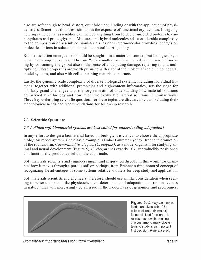
Page 51Biomaterials: Important Areas for Future Investment
also are soft enough to bend, distort, or unfold upon binding or with the application of physi-cal stress. Sometimes this stress stimulates the exposure of functional cryptic sites. Intriguing new supramolecular assemblies can include anything from folded or unfolded proteins to car-bohydrates and proteoglycans. Mixtures and hybrid molecules add considerable complexity to the composition of assembled biomaterials, as does intermolecular crowding, charges on molecules or ions in solution, and spatiotemporal heterogeneity.
Robustness often emerges – or should be sought – in a materials context, but biological sys-tems have a major advantage. They are “active matter” systems not only in the sense of mov-ing by consuming energy but also in the sense of anticipating damage, repairing it, and mul-tiplying. These properties are worth pursuing with rigor at the molecular scale, in conceptual model systems, and also with cell-containing material constructs.
Lastly, the genomic scale complexity of diverse biological systems, including individual hu-mans, together with additional proteomics and high-content informatics, sets the stage for similarly grand challenges with the long-term aim of understanding how material solutions are arrived at in biology and how might we evolve biomaterial solutions in similar ways. Three key underlying scientific questions for these topics are discussed below, including their technological needs and recommendations for follow-up research.
2.3 Scientific Questions
2.3.1 Which soft biomaterial systems are best suited for understanding adaptation?
In any effort to design a biomaterial based on biology, it is critical to choose the appropriate biological model system. One classic example is Nobel Laureate Sydney Brenner’s promotion of the roundworm, Caenorhabditis elegans (C. elegans), as a model organism for studying an-imal and neural development (Figure 5). C. elegans has exactly 1031 reproducibly positioned and functionally productive cells in the adult male.
Soft materials scientists and engineers might find inspiration directly in this worm, for exam-ple, how it moves through a porous soil or, perhaps, from Brenner’s time-honored concept of recognizing the advantages of some systems relative to others for deep study and application.
Soft materials scientists and engineers, therefore, should use similar consideration when seek-ing to better understand the physicochemical determinants of adaptation and responsiveness in nature. This will increasingly be an issue in the modern era of genomics and proteomics,
Figure 5: C. elegans moves, feeds, and lives with 1031 cells positioned (in matrix) for specialized functions. It represents how the making choices among many biosys-tems to study is an important first decision. Reference 30.

Page 52 Biomaterials: Important Areas for Future Investment
and one dare not propose a singular answer when fundamentals are the goal. Although other systems and sources can provide major new insights and advantages as well, a strong case can be made for studying the extracellular matrix derived from mammals,
Indeed, the extracellular matrix is the major building block of solid tissue shapes and confers me-chanical properties that are characteristic of a species and even specific organs. Work on matrix is not new. Leather is an ancient, matrix-based material that has been used around the world for thousands of years for all manner of straps, saddles, or clothing, plus many other types of soft or stiff structures and mechanical devices. The ancient arts of making and modifying the many forms of leather are infused with chemistry and physics. However, until more recently, the adaptability and responsiveness of extracellular matrix had not been fully exploited.
This began to change several decades ago when it was discovered that the extracellular matrix is not the passive residence of cells but is highly interactive and plays a major role in health and disease. Today, many in the biomedical community are extracting donor tissue of cells and loose material and then growing host cells within the natural scaffold of extracellular matrix, either before or after implantation. For example, replacement heart valves are being isolated from an-imals and modified by crosslinking with glutaraldehyde for implantation into people with heart valve disease.
On the other hand, the composition-structure-property relationships of such industry-generated materials still are not well delineated in relation to adaptation in performance and failure, especial-ly when considering that implanted valves must open and close with large deformation in vivo up to 100 million cycles per year for 10 years or more. Thus, to more fully mine and engineer matrix materials, complications of interactions with different cell types and biofluids must motivate both bottom-up approaches and to top-down methods.
Over the last several decades, the in vitro development of relatively simple biological structures, such as vesicles, DNA/RNA condensates, and actin gels, has been successfully imitated, pro-viding us with a wealth of information on the basic structures and properties of these soft, wet materials. Some of these insights have been exploited in drug and gene delivery with significant clinical impact, although major challenges exist as exemplified by the major unsolved problem of delivering highly charged nucleic acid to a desired site of disease or dysfunction in the body; we often know exactly what sequence(s) we want to deliver but simply don’t know how to de-liver it to a specific site.
We also do not yet understand the molecular adaptation mechanisms by which a lipid vesicle (lipo-some), for example, contacts and fuses with a cell membrane as well as how a completely synthetic polymer vesicle (polymersome) degrades and disrupts the low-pH endolysosome within a cell. Molecular dynamics and coarse-grained simulations have provided detailed insights into some of these and other processes. On a different scale, gels of actin filaments and other cytoskeletal com-ponents, along with assemblies of rod-like viruses, have served as mesoscopic models of liquid crystals. The adaptiveness of such collective systems is evident in large responses to mechanical stresses, boundaries, and solution conditions, as measured by rheometry and a diversity of scatter-ing methods.
Non-affine and heterogeneous responses with glassiness, such as in crosslinking, are common issues and they are even captured, to some extent, in considerable theory and simulation efforts of networks. The temperature is not usually varied in such systems because the constituent proteins

Page 53Biomaterials: Important Areas for Future Investment
that assemble into the filaments are prone to unfold and agglomerate, which also could occur with mechanical work on any protein system, including extracellular matrix. However, some of the same physicochemical questions of interactions, charge, order, glassiness, and unfolding, thus, arise in these reductionist systems as well as in the deconstructed matrices.
The richness of systems choices extends to even more complex and dynamic natural biological systems, including plants, worms (such as the aforementioned C. elegans), or the mucus that sim-ilar creatures secrete. From a rigorous materials perspective, biology largely has been untapped for adaptive biomimetics in both biological and non-biological applications. However, at the same time, we have an increasing number of genomes that provide us a key first-level of information on sequence and even abundance.
Once expressed and assembled, the complexity of biological materials allows them to utilize en-ergy sources to change molecular conformations that contribute to motion, adaptation, and fur-ther display of signals. The bottom line is that we need to consider the diversity of bio-systems, employing better-designed experiments that help to reveal key interactions, essential mesoscale physics, and critical signals to produce desired material behavior, especially when cells are added.
Clearly, a better knowledge of minimal physicochemical requirements of adaptive soft natural sys-tems will aid us in designing and synthesizing materials with the desired functions. Physics- and chemistry-based theory and modeling must be increasingly brought to bear on these phenomena and aided by molecular simulations to clarify structure-property relationships. How else can one ensure that natural materials maintain or that synthetic materials reproducibly achieve the physico-chemical properties and assembled states suitable for adaptation after either implantation or even in non-biological applications, such as smarter iPad covers?
2.3.2 What is living, and what is synthetic?
Peering through a microscope nearly two hundred years ago, the botanist Robert Brown observed a continuous jittery motion among small lipid and starch organelles ejected from pollen grains. He concluded this motion was not indicative of life, because similar ‘Brownian’ motions were observed fifty years earlier for particles of inorganic matter in studies of the physiologist and chemist, Jan Ingenhousz (who discovered photosynthesis). Brown’s conclusion proved correct of course. Current developments of active materials and those that are being theorized about or yet to be contemplated will increasingly obscure differences with living matter. However, there are a myriad of underlying topics and questions that must be addressed if they are to become a reality.
A material system that first senses danger signals and, then, prepares for damage by changing its structure and properties is one variation of an active material that we might for now call a “preemp-tively adaptive” material. However, all such materials, including cell-material constructs, are far from equilibrium, with an energy flow from source to sink (e.g., covalent bonds in ATP to protein motion) that is generally apparent, at least at a coarse-grain level.
Continuum hydrodynamic theories have been developed that account for both the internal con-sumption of energy by tissue-like objects and the coupling of the resultant stresses to motion. The experimental implementation of these theories is just beginning but could yield materials that exhibit predicted dynamical behavior and can do work, while also sensing their environment

Page 54 Biomaterials: Important Areas for Future Investment
and changing dynamics in response. Self-healing materials, for exam-ple, might use a cell-biomaterial construct or purified proteins and transport matter along directed tra-jectories that dominate the Brownian modes of both cells and molecules.
Although new approaches, rang-ing from molecular motor stepping to cell-scale wound healing with active contractility, are needed to exploit these processes, emergent properties of the collective are a particular challenge. Indeed, recent experimental realizations of inter-acting cilia highlight emergent ac-tive behavior (Fig. 6). Such features often are not easily predicted from molecular designs, due to cooperativity, crowding, and more subtle coupled interactions.
Time scales in cell/material systems can be limited by elastic or viscoelastic responses in water or in more viscous biofilms. In particular, the reorganization of matrix and cytoskeleton can propa-gate and dissipate with an unknown, but effective, “speed of sound” that prohibits long-range in-teractions. Also unknown is the crossover from adhesive strength and cell or matrix cohesiveness, as such transitions can be subtle. Local equilibrium states, for example, could be separated by force-driven conformations of select sub-structures and in select domains.
In the case of focal adhesions that cells use to feel materials and migrate, conformational changes initiate growth and/or rectify assembly. Nonlinear responses in biological systems are likely to include unfolding responses to force, such as the unfolding that occurs for the major protein of blood clots, fibrin, which aligns and, then, unfolds and bundles under mechanical stress. Synthetic materials might use similar responsiveness for sharp hysteretic transitions, and some recent protein designs have illustrated such possibilities.
Nonlinear responses to stress of biomolecules imply limits to linear biomechanics at the cellular level. We need to clarify, therefore, how elasticity and viscosity emerge in reconstituted cytoskel-etal-like systems of actin and intermediate filaments as well as microtubules, with an eye toward better-controlled synthetics in composites with similar “frictional” interactions. Some cells use stress fibers to pull on matrix, and the stresses somehow couple to an isotropic cytoskeleton within the cell as well as other organelles, such as the nucleus. Many such processes motivate deeper study.
Although we often take for granted that many cells are motile or, for example, that muscle pumps food through the gut as well as blood through the heart, the impact and contribution of cell prolif-eration in these processes is too often not considered intrinsic to their function. Even in the highly ciliated adult gut, for example, many cells divide every day or two even though the tissue shows no net growth. Of course, repair, regeneration, and embryogenesis require cell division, but the differ-
Figure 6: A synthetic system with cilia (left) vs a pollen grain with cilia (right). Such active assemblies blur the boundaries between living and synthetic systems. Refer-ence 31.

Page 55Biomaterials: Important Areas for Future Investment
entiation and specialization of structure and function also occurs at division and/or in response to microenvironmental cues. The study of living materials that suitably grow and regionally special-ize in response to heat, stress, light, or related threats would be a logical second major phase of the broader developments in active matter. Theories and simulations of tumor growth are instructive and also suggest the need for tight control of such systems.
2.3.3 Can we understand biomaterial complexity and make active matter evolve?
A long-term goal of soft matter science and engineering is to delve deeply into nature’s mechanisms so that we can identify new functionality in biological systems and biomaterials and design sys-tems that intelligently evolve. As Darwin wrote in the Origin of Spe-cies one-and-a-half centuries ago, “natural selection would probably favor different varieties.” Indeed, among Darwin’s finches there are beaks of many different shapes, sizes, and effective strengths to meet the physical demands of different diets in different envi-ronments (Fig. 7). At a molecular scale, all we know at present is that one calcium handling protein differs in level between the finch-es [34]. We need to better under-stand such differences at a more advanced level of molecular detail. If successful, this will allow us to harness some of the vast solutions available to biology in order to produce useful materials or biolog-ical function. Given the large sizes of genomes (e.g., ~20,000 human genes), the scope of the problem is hyper-complex.
To develop a better molecular un-derstanding of biodiversity, we might use theory to build on avail-able bioinformatics to provide us with an understanding of the large number of possibilities in relation to a solution that ‘works’ rather than a global optimum. All possi-ble combinations of all the bases in the genome, for example, yield
Figure 7: The diversity of Darwin finch beaks. The war-bler finch (top) has a thin, sharp beak best suited for spear-ing insects; whereas ground finches (center) have shorter, more robust beaks that are adapted for eating seeds found on the ground. Those of cactus finches (bottom) are shaped for getting seeds from cacti. Reference 32.

Page 56 Biomaterials: Important Areas for Future Investment
too many combinations to explore, even with the power of evolution. Thus, we must learn to test a vast number of potential solutions, while still recognizing that we can still only test a very small fraction of available solutions. To overcome this inevitable limitation, we must combine new techniques that enable ultra-high-throughput experiments, with a new theoretical understanding of problems and novel computational approaches to enhance our understanding of the processes.
The experimental methods will undoubtedly require rapid preparation and testing of very large numbers of samples in very small volumes. In addition, they will require new techniques to mea-sure the desired physical properties of these small samples at high rates. If this work is successful, it can be harness biological systems to produce chemicals and new and improved proteins, en-zymes, catalysts, and antibodies for materials purposes or as diagnostic or therapeutic chemicals. It also will use biological systems to directly synthesize new biomaterials.
2.4 Needs and Recommendations
2.4.1 Adaptability of hierarchical matrices with bio-complexity
The development of new hydrogel materials will allow us to build upon recent successes in the application of simple hydrogels and pursue matrix-like multifunctionality for tissue-like adaptability. In multi-component strategies, mixtures of covalent and physical interactions will increasingly be made to make hydrogels change with time and respond to stimuli.
Single types of intermolecular interactions will be inadequate, however, and complexity will be inherent to increasing the functionality of hydrogels. Although past hydrogel work, with its focus on hydrophilic, primarily covalent molecular networks, has been foundational, supra-molecular physical networks are increasingly being made and provide an opportunity for us to develop tissue-matrix-like organized composites of fiber plus filler (Figure 8). Indeed, different gels have been developed for different singular functions, such as a biocompatible network that displays or delivers a peptide for interaction with a desired cell type for drug delivery or for tissue engineering. However, networks are now being designed with more than one function (e.g., networks that contain a particular peptide for delivery to specific cells) and that also de-
Figure 8: A scanning electron microgram of collagen in bovine skin (left) and collagen polymerized in vitro (right), illustrating the natural system of fibers and crosslinking fillers versus the naturally derived synthetic. Reference 33.

Page 57Biomaterials: Important Areas for Future Investment
grade enzymatically over time. Characterizing these materials by novel scattering, imaging, and rheology methods, especially when cells are present, offers major opportunities for innovation.
A fundamental understanding of natural biological matrices should complement the develop-ment of synthetic hydrogels. Among biopolymers, methods to sequence and synthesize nucleic acid polymers and proteins are routine, but there are few good methods for fully sequencing and synthesizing many complex polysaccharides and glycosaminoglycans (GAGs), for example. These macromolecules are abundant in the extracellular matrix and contribute viscoelastic and biochemical properties. Proteoglycans interact with other GAGs and proteins to promote hydra-tion and the assembly of matrix as well as growth factor storage. Hyaluronic acid and chondroi-tin sulfate in cartilage, for example, bind water strongly and contribute its compressive strength. They also have large excluded volumes that can protect the macromolecules to which they are bound by limiting the access of proteases. Synthetic polyethylene glycol (PEG) can have a similar function when attached to proteins. GAGs also contribute to lubricity, as exemplified by the near frictionless articular cartilage surface, and they motivate consideration of mucins and other mucus components that have related structures and partially overlapping functions in many other tissues.
Particularly attractive to soft biomaterials work is the prospect of combining the disparate chemistries of constituent network molecules, such as GAGs, with fiber-forming polypep-tides. Transient molecular interactions via hydrogen bonding and electrostatics can provide cells with the freedom to move and remodel, while the fibers add form to scaffold structures. Changes to one or both components in response to environmental stimuli should allow the systems to adapt with time.
Although the examples presented here are largely based on polysaccharides derived from eu-karyotes, other polysaccharides also are in need of study. For example, the biofilms produced by bacteria protect them from antibiotics and provide an extracellular matrix to enhance their adhe-sion to surfaces. Thus, an increased understanding of key biofilm parameters should allow us to design antibiotics with improved biofilm penetration and also could facilitate our development of novel methods of preventing biofilm formation.
In both the understanding of biological networks and discovery of new materials, the grand chal-lenges of complexity and materials with multifunctional, morphogenetic, and emergent prop-erties must be embraced. The biomaterials community already has made great progress in the development and understanding of hydrogel materials and matrices, and the continued promise of the opportunities presented by these materials warrants enhanced support.
With newer characterization methods, including liquid chromatography coupled with mass spectrometry and high-resolution elemental mass analysis, we soon should be able to fully char-acterize GAG sequences. However, newer chemical methods are needed to synthesize GAGs of substantial length and provide sequence control. In addition, better analysis and synthesis of GAGs will enable the development of an in-depth understanding of how GAGs interact with other molecules and allow the identification of the key intermolecular associations that lead to the unique properties that GAGs impart to materials. Emulating these adaptive interactions in hybrid or synthetic materials also will impart multi-functionality, ranging from physical assem-bly and enhanced mechanical properties to growth factor sequestration and the rationale design of lubricating coatings.

Page 58 Biomaterials: Important Areas for Future Investment
2.4.2 Hybrid molecules for assembly of nanostructures and hierarchical materials
Silk, sea shells, and the photosynthetic machinery are examples of materials that have been extensively studied in recent decades for their desirable properties and functions, including their adaptability. It is now clear that to duplicate what nature has achieved through eons of evolution is a daunting task. Indeed, natural proteins clearly possess structures and proper-ties that synthetic polymers currently cannot remotely match. Nevertheless, new approaches should incorporate what nature offers into synthetic materials in order to achieve targeted properties, ranging from structural to optical, mechanical, electronic, or biological.
Indeed, now is the right time to pursue new families of soft hybrid matter by exploiting recent developments in molecular and cell biology, chemical biology, biochemistry, and biophysics. Current methods of protein synthesis, site-specific protein modification, and control over dif-ferent cellular pathways are extremely powerful, and protein crystal structures are available or modeled by homology for many water-soluble proteins to help define interactions. In ad-dition, the intricacies of protein folding is increasingly being understood from first principle atomistics, and de novo design capabilities are enabling the materials community to explore sequences that are both natural and non-natural. In parallel, our fundamental understand-ing of polymeric nanostructures and polymer phase behavior in thin films, at interfaces and under geometric confinement has reached unprecedented levels. Highly versatile synthesis approaches, such as living free radical polymerization, are now routine in most polymer labs. Thus, for the first time, functional hybrid biomaterials of proteins and polymers can be real-ized.
New supramolecular material systems (e.g., Figure 9) will allow us to produce soft materials that exhibit active, healing, and morphogenetic-like behaviors. These materials can help elu-cidate biological systems and also mimic biological behavior in novel, non-biological appli-cations. Even in choosing polypeptides, there currently are limitless combinations of natural and non-natural amino acids with which to construct new molecules. Complexity is provided by non-natural amino acids (e.g., synthetic side chains, beta amino acids, peptoid backbones, etc.) as well as by hybrid backbones that are part peptidic and part non-peptidic polymer. The beautifully complicated nanostructures that have been assembled from the elegant simplicity of DNA base pairing are examples of inspiring structures that generally are possible with biomolecules.
Figure 9: Hybrid materials of completely synthetic polymers, folded peptides, and functional ligands build on a diversity of recent advances that enable hierarchical assembly into meso-struc-tured materials such as oriented films. Reference 34.

Page 59Biomaterials: Important Areas for Future Investment
As with proteins, interactions between polypeptide hybrid molecules are much more compli-cated and more dependent on solution conditions, which is why nature is built from proteins and not from DNA. Proteins have the electrostatic possibilities of DNA but, unlike DNA, also have many hydrophobic choices to drive molecular assembly in water. Indeed, polypep-tide-based vesicles are now well-established, alongside liposomes and polymersomes, and illustrate the potential opportunities for a wider range of surface-active and detergent-like polypeptides.
There are at least two avenues to explore in the creation of new soft materials with hybrid mo-lecular assemblies. One could look to natural proteins and assemblies for examples of struc-tures useful in a non-natural assembly. For example, taking a natural protein and its inherent tertiary structure as a model system, one can think of using the protein as a building block in a multi-molecular assembly before or after altering it to have some desired intermolecular interaction or function. Protein structures from animals, plants, insects, and more can be used in the construction of such materials.
Computationally predicted molecules also will increasingly be incorporated into new soft ma-terial constructions. Many natural and non-natural amino acids can be incorporated into new structures, and computational screening of sequences for desired intra- and intermolecular structure formation would be extremely useful. Such virtual screening methods will be par-ticularly useful in assessing various environments that might range from mixtures of different assemblers and chaperones for folding or assembly to steric crowders and fillers. Partnering theory/simulation and experimental work in new molecular systems for bottom-up assembly into new soft-matter systems is critical to the advancement of this field of science.
2.4.3 Cyber-discovery and molecular dynamics of adaptation
The rigorous traditions of soft-matter science demand that we elaborate the molecular and physical bases of biomaterials in relation to their mesoscopic and macroscopic behaviors. We need to be able to predict the activity of a given system under different conditions and environ-ments. Computational and theoret-ical advances also should facilitate the design of novel materials with properties that enable new appli-cations. In particular, theoretical approaches that focus on generic properties at the mesoscale and modeling of the interactions and assemblies at the nanoscale should support and complement compu-tational methods that use either molecular- or coarse-grained ap-proaches for more specific and dynamical insight. Integration will help to quantify and clarify the na-noscale properties of biomaterials
Figure 10: Molecular dynamics simulation of a single molecule (horizontal, top) interacting with a molecular film (vertically aligned molecular chains at bottom). Ref-erence 35.
Page 60 Biomaterials: Important Areas for Future Investment
in relation both to key molecular interactions and to how properties scale up to macroscopic and mesoscopic behaviors.
Computational studies will become increasingly important in the discovery and design pro-cesses of new biomaterials due to developments in both computing technology and modeling approaches. In the pharmaceutical industry, for example, in silico screening of large libraries of compounds is now regularly performed to identify lead compounds as new drug candidates. In a similar spirit, polymer and biomacromolecule libraries could be screened to identify opti-mum polymeric biomaterials tailored to specific material properties for a designed application (see Figure 10). This approach holds the potential to reduce the burden of synthesizing large numbers of polymers for material property characterization and bioassays.
Although some of the computational challenges include lack of models that properly describe the physics or chemistry and hardware limitations, an equally important obstacle is poor com-munication with experimentalists. In fact, relatively few experimentalists are trained well enough in the computational analysis of biomaterials to provide the needed guidance, and there also is a dearth of easy-to-use software that can be used to provide the desired infor-mation. Many theoreticians lack sufficient training in biology to help them identify the most important bottlenecks. By the same token, experimentalists often are unaware of both the ca-pabilities and the limitations of their theoretical and computational methods. Thus, theory and computation need to be better integrated in the biomaterials field, and concerted efforts are needed to break down traditional barriers so that experimentation and computation become core components of any biomaterials curriculum (see the Education section).
In addition, cross-disciplinary research teams of theory/computation/experimentation should be assembled to tackle key parts or entire problems in processing, synthesis, and property characterization. Computational packages and analysis strategies might then emerge to make it easier for scientists who are not trained in computational modeling to use software that provides the needed insights. The last decade of research in computational modeling methods used for prediction of polymeric biomaterial properties and biological responses to polymer surfaces is partially represented in the online Biomolecular Absorption Database (BAD) [36], which also includes computational resources specifically designed to perform in silico pre-dictions for materials of interest. A plethora of online bioinformatics tools widely used in genomics, thus, is extending into biomaterials.
2.4.4 Living waves of biomaterials
In a bottom-up approach to developing active materials, a minimal subset of biological agents are purified and reconstituted. For example, ciliary motion has been generated in a system of purified microtubules (MT), MT motors, ATP, and generic linkers that bundle the microtu-bules. Many such bundles attached to a substrate exhibit metachronal waves that are difficult to distinguish from those observed in living animals (Figure 6). Gels that can crawl or swim like bacteria or protozoa also can be envisioned, although different proteins and processes of assembly, no doubt, will be needed for their development. Directing the motion of such sys-tems externally using chemophoresis and photophoresis is a promising approach.
Ideally, the field would use robust synthetic versions of the biological components, including

Page 61Biomaterials: Important Areas for Future Investment
artificial motors and tracks for the motors. An intermediate goal might be to use DNA origami systems, since these have some potential for activity given the programmability of self-assem-bly and catalysis. Design limits on the number of competing complementary oligonucleotides and on mechanical strength need to be better understood, however. Also critical is a better understanding of kinetic limits on assembly.
On a very different scale are active materials that make use of living cells in niches or colo-nies, some of which might be specialized to sense and communicate a type of threat or danger signal, while others act to move, contract, secrete, and repair. Mammalian cells could be used in novel adaptive hydrogels or, perhaps, microbial cells could be used in controlled biofilms. The needs in this area range from materials that control cells to real time characterization, with tools ranging from fluorescence reporters and force sensors to analytical mass spectrometry.
2.4.5 New tools for hypercomplexity and biomaterials evolution
Microarrays used around the world today address all types of genomic questions and are truly remarkable devices. In one type of microarray, for example, one million cell-size spots of distinct nucleic acid chemistry are positioned next to each other in roughly a 1-square-centi-meter area, with numbered rows and columns that can be probed optically with dyes. The end result is a microarray assay, for example, of mRNA expression from every human gene with a redundancy of several dozen spots or more per gene. This suggests there is at least some feasibility to the idea of probing the biomaterial consequences of hypercomplexity within and between genomes.
However, the field will clearly need many new tools and techniques in combinations of bot-tom-up (material mixtures) and top-down (genetic screens) ultra-high-throughput experimen-tation. Insightful characterization with such devices, which include electromechanical probes, for example, must be matched to computational resources for information gathering, analysis, exploration, and modeling.
Page 62 Biomaterials: Important Areas for Future Investment
Section 3. Cell-Material Interactions DISCUSSION LEADER
kRIStI ANSEth, University of ColoraDo
GROUP MEMBERS
ALI khADEMhOSSEINI, harvarD MeDiCal sChool
SARAh hEILShORN, stanforD University
LAURA kIESSLING, University of wisConsin
JEffREy LINhARDt, google, inC.
WILLIAM MURPhy, University of wisConsin
CELEStE NELSON, PrinCeton University
BUDDy RAtNER, University of washington
ChRIStINE SChMIDt, University of texas
JOyCE WONG, boston University
SPECIAL ACkNOWLEDGMENt
MARk tIBBItt, University of ColoraDo, stUDent PartiCiPant anD ContribUtor to this rePort
3.1 Introduction
Cells interact with their external environments through a variety of cell membrane surface receptors, which bind to immobilized or free ligands with high affinity and specificity. These interactions, which regulate a variety of cell functions, including adhesion, apopto-sis, migration, proliferation, secretory properties, and phenotype, provide the cell with vital information about the chemical and physical properties of the surrounding environment. A more complete understanding of these cell-material interactions and the development of techniques to direct and control them is essential to the advancement of the biomaterials field.
To date, a variety of biomaterials have been derived from naturally occurring substances, including extracellular matrix components (see Soft Materials section), synthetic chemis-

Page 63Biomaterials: Important Areas for Future Investment
tries that rely on adsorption of endogenous biomacromolecules, or synthetic analogs of the extracellular matrix that present ligands to the cell in a highly defined manner. These bio-materials, which have been developed to promote cellular interactions of interest, generally are designed to:
• Minimize unintended or undesired cell interactions (e.g., foreign body response, infec-tions, etc.).
• Interact with and signal to endogenous cells (e.g., wound healing, vaccines, etc.).
• Deliver and direct cell behavior, with their applications ranging from directing stem cell fate to tissue regeneration.
However, studies of these biomaterials often have led to only to a basic understanding of how cells interact with their surrounding environments. Thus, our ability to predict the fate of cell-material interactions is still quite limited. Due to our limited knowledge of how biomaterials interact with their surroundings, medical devices using them as components often fail. Indeed, in vitro exper-iments often do not predict in vivo performance: drugs often are delivered with numerous side effects; stem cells sometimes do not differentiate efficiently and reliably when they are implanted; and engineered tissues often have inferior properties compared to the endogenous tissues they’re designed to replace. A key to overcoming these challenges is an improved understanding of what causes a biomaterial to fail, often beginning at the very basic concepts of how a cell senses its extracellular environment.
This chapter begins with a discussion of the major opportunities and significant challenges that must be addressed to propel forward advances in biomaterial applications. This is followed by a an examination of the critical scientific questions that need to be answered in order for us to bridge gaps in knowledge and fuel application-oriented advancements. The chapter concludes with our recommendations for new or improved technologies and technological approaches that may play key roles in finding answers to many of these questions.
3.2 Opportunities and Challenges
Although biomaterials have had a profound impact on our modern quality of life, it is interesting to note that the term “biomaterial” did not exist until 1950, when Harold Ridley and John Char-ley pioneered biomaterial systems for intraocular lenses and hip joint replacements, respectively. Fast-forward, and, today, the U.S. market for medical devices is estimated at $200 billion annually and it is predicted to grow to $415 billion annually by 2016.
Despite the huge growth potential of biomaterials, important questions remain to be answered before the field can realize its full potential. For example, how will the field evolve to meet the sci-entific and technological challenges? How will universities prepare the future workforce to power this industry? Certainly, understanding and manipulating cell-material interactions will be critical to fueling these advancements, and this understanding will likely impact fields beyond biomedical devices, including biofuels, environmental sensing, antimicrobials, smart materials, and more.
Imagine what we could create, if we could engineer biomaterials that could talk and even lis-ten to cells to unlock the potential of their basic biology to make smarter and more biologically

Page 64 Biomaterials: Important Areas for Future Investment
compatible devices and technologies. For example, could we engineer an internal “band aid” that would heal damaged or diseased tissues? Or, could we develop implantable microdevices, such as pacemakers, that would be powered by local cell processes rather than a battery. Imagine also a “virtual patient” in a petri dish; that is, growing miniaturized organs or tissue units to test drugs, personalize treatments for cancer, or predict disease. Finally, what kinds of devices might we be able to create, if we had responsive and adaptive biomaterials that would sense and treat diseases before they were otherwise apparent? Clearly, although the challenges are grand, the opportunities are many.
The rest of this chapter discusses four broad challenges, each of which will require an investment in time and resources, advances in training, development of new technologies, and integration of research communities to overcome.
3.2.1 Improving the biocompatibility and longevity of implanted biomaterials and reducing short- and long-term complications
Millions of medical devices fabricated from biomaterials are implanted into human patients each year, saving countless lives and greatly improving our quality of life. However, the complication rate of implanted biomaterials is unacceptably high; the devices frequently fail after varying peri-ods in the body, and they never perform as well as the biological part they were intended to replace or augment. Examples where advanced materials could significantly improve the durability and dependability of such devices include implanted sensors, electrodes, drug delivery devices, small diameter vascular grafts, breast implants, stents, and intraocular lenses.
A central component to the development of improved biomaterials is the ability to define and un-derstand “biocompatibility.” In 1987, an international consensus conference in Europe formalized a definition of biocompatibility, which has been widely used ever since, as:
“the ability of a material to perform with an appropriate host response in a specific application [37].”
This excellent definition highlights two key issues: (1) how the living organism responds to the material and (2) that materials are used in numerous specific applications in medicine. On the other hand, there is some ambiguity in this definition of biocompatibility.
For example, consider this. The following ten different, highly purified materials are implant-ed separately as small disks subcutaneously in mice: polyethylene, titanium, Teflon, platinum, silicone rubber, polyurethane, zirconia, poly(2-hydroxyethyl methacrylate) hydrogel, tissue cul-ture polystyrene, and polyisobutylene. To subcutaneous implantation of such materials in humans requires that each has passed a routine set of toxicology assays (i.e., to ensure that they do not leach undesirable substances into surrounding tissue and show almost no endotoxin). Also, each specimen must be implanted in such a way that localized movement of the sample and mechanical distortions of the implant site are minimized.
However, when we examine the implants after one month, what we typically will find is that all of these materials have healed indistinguishably, and the observed healing is characterized by avas-cular, collagenous capsules surrounding each implant. This is the classic “foreign body reaction,”

Page 65Biomaterials: Important Areas for Future Investment
or FBR, and it is an acceptable outcome to regulatory agencies for a biocompatible medical device.
However, this does not represent optimal healing for medical device performance, in many cases. Indeed, we’re still not sure what drives the FBR reaction. When we look at the adsorption of bio-logical macromolecules (e.g., proteins) to these materials in the laboratory, each shows a unique interface. Each also shows unique interactions for influencing cell attachment and growth in vitro. Yet, they often all heal similarly in vivo, developing the FBR reaction, which is clearly related to the inflammation process. In fact, it might best be referred to as “low-level, chronic inflammation.”
Thus, the grand challenge in this area is to master cell-biomaterial control such that all biomateri-als – and medical devices fabricated from them – can be made to heal in a non-complicated, recon-structive, and integrated fashion. Such an understanding will improve the device’s performance, extend its longevity, and reduce the costs associated with its eventual failure and replacement.
3.2.2 Engineer responsive and multifunctional materials for cellular control
Over the last few decades researchers have begun to reveal the material and chemical properties that are needed to develop functional and dynamic biomaterials. For materials to exhibit the exquisite cellular control present in all living systems, they must be able to engraft cells, direct their differentia-tion and proliferation, and control their migration. These outcomes depend on both bidirectional sig-naling and dynamic adaptation of the cellular environment (Fig. 1).
Biological phenomena are in-creasingly recognized as dynam-ic, multicomponent processes, in which cells listen to signals in their environment but also talk back to it in meaningful ways. Similarly, new generations of biomaterials are being developed to adapt and respond to changes in cell pheno-type and the organization of mul-ticellular assemblies. Responsive biomaterials can be designed to be adaptive and, thus, amenable to cell-mediated remodeling (e.g., protease-labile hydrogels). They also can be tailored to respond in controllable ways to physi-cochemical inputs (e.g., pH-re-sponsive, temperature-responsive materials) or specific biological inputs (e.g., antigen-responsive, ligand-responsive materials). As a result, we might envision using
Figure 1: The “branching” morphogenesis that occurs as a result of cell-material interactions in many tissue types, such as mammary, pulmonary, and salivary. Proper branching depends on regulated interactions between the epithelium and extracellular matrix (ECM) proteins, mech-anotrandsuction of signals at the branches leading edges, dynamic remodeling of the ECM through protein deposition and secretion of enzymes, as well as cellular outgrowth caused by high mechanical fields at the branches leading edge. Although this is specific to branching morphogenesis, similar interactions are critical in all morphogenetic phe-nomena. Reference 38.

Page 66 Biomaterials: Important Areas for Future Investment
biomaterials to dynamically manipulate and monitor biological systems.
It is especially timely to pursue the development of biomaterials with emergent properties. The groundwork already has been laid by four key findings that were made over the past decade: (1) biomarkers produced by cells and associated with physiological or pathological phenotypes have been identified and new ones continue to emerge; (2) specific intermolecular interactions have been systematically catalogued that could be used to enable bioresponsiveness; (3) the development of new materials chemistries that intrinsically adapt and remodel as a result of cell behavior; and (4) the development of new materials chemistries that adapt and remodel in a “user-defined” manner as dictated by the experimenter and can be used to modulate cell-material interactions.
Thus, the field is now poised for development and application of biomaterials that both talk and listen to cells and multicellular assemblies so that they can understand and manipulate cell behav-ior. A recent example of this type of a multifunctional, responsive biomaterial is the development of immunomodulatory vaccines that generate coordinated cellular immune responses, including the activation of CD8+ cytotoxic T lymphocytes by dendritic cells (DC) to enhance host immu-nity. Regulating the DC network involves both the recruitment of DCs and the targeted and pro-longed delivery of antigen to them, which, in turn, facilitates the activation of mature DCs. Mature cells then present co-stimulatory molecules and cytokines capable of propagating the appropriate T-cell response. Although it is rather complex, this system is still relatively primitive when com-pared to many naturally occurring hierarchical biological systems.
3.2.3 Harness developmental and regenerative biology knowledge to engineer biomaterials that induce functional regeneration
Advances in genomics have made it clear that the development of all organisms – from worm to human – depends on fundamental conserved signaling pathways. It is exciting to consider the pros-pect of incorporating this new knowledge into the design of biomaterials that promote regeneration (Fig. 2), including using them to generate unlimited supplies of adult and pluripotent stem cells and devising materials that facilitate the repair of complex tissues and organs. We also may be able to develop a broad new range of biomaterials that can serve as scaffolds for tissue or organ gen-eration. These developments would revolutionize the transplant and regenerative medicine fields.
The process of developing biomaterials for tissue regeneration must embrace at least four funda-mental steps: (1) stem cell self-renewal (e.g., propagation of hematopoietic stem cells); (2) stem cell differentiation (e.g., differentiation to specific lineages such as pancreatic cells); (3) asymmet-ric differentiation to initiate patterning (i.e., the first step in tissue generation); and (4) organizing cells into tissues and, ultimately, organs.
All four of these outcomes involve temporal regulation of signaling at multiple levels––between cells and their substratum, between neighboring cells, and among the entire cellular population. Inherently, interactions between cells and materials (e.g., extracellular matrix proteins, other cells, organic crystals, or biofilm proteoglycans) mediate these cellular processes in living systems.
For us to better engineer materials that encourage desired cell functions, we need to understand how cell-material interactions regulate cell behavior. For example, we need to better understand

Page 67Biomaterials: Important Areas for Future Investment
the connection between cancer cells and pluripotent stem cells. We can use the knowledge that we would gain from being able to selectively promote or inhibit cell fate decisions not only to advance materials science in regenerative medicine but also to directly eliminate and/or manage certain diseases. In addition, materials that induce the selective dedifferentiation of cells could be used to activate regenerative properties for organ or tissue repair. We also increasingly recognize that the cells (e.g., adult stem cells) and signaling molecules (e.g., cytokines) already present in vivo can promote wound healing. Biomaterials may be used to “harness” these endogenous signals. However, our current ability to design materials with any of these attributes is either nascent or non-existent.
3.2.4 Utilize biomaterials to combat disease and enable the body’s defense mechanisms
Cancer, diabetes, obesity, and infection are among the most important public health challeng-es in the U.S. and worldwide, and there are numerous potential applications where cell-bioma-terial interactions can be exploited to combat these deadly, costly diseases. Specifically, biomaterials can be designed to enable early disease detection, to prevent diseases prior to their occurrence, or to treat chronic illnesses and acute injury.
For example, if we can modify biomaterials as targets and contrast agents to be used with multiple different imag-ing modalities (e.g., light spectroscopy, ultrasound, MRI, etc.), we can use them to detect and, ulti-mately, treat early cancer or infection. We also can use them as diagnostic markers for identifying disease states at early stages. Similarly, if we can develop materials that inhibit the formation of specific biofilms in the gastrointestinal tract, we could prevent patho-gen-borne infection. We
Figure 2: Learning from natural regeneration. A major challenge in the field of biomaterials is to understand and harness the regen-erative capacity in biology in order to engineer materials that can self-repair in the case of acute injury. For example, the zebrafish is able to repair its heart after acute injuries. Histological sections demonstrate an intact zebrafish heart (A and C) and a heart after 20% resection (B), which induced nucleated erythrocytes (indicated by arrows) to fill the clot after injury (D). Over the course of the 60-day (E-H) healing process, post-resection staining revealed that the zebrafish is able to regenerate damaged cardiac muscle (brown), and, by day 60, there is little fibrin remaining (blue). Further, the ventricular section surface area (I) is restored to normal values by 60 d. Reference 39.

Page 68 Biomaterials: Important Areas for Future Investment
also could use biomaterials that facilitate islet viability and release insulin reliably over the course of decades to better manage and mitigate the consequences of diabetes.
Biomaterials can be further exploited to augment the human body’s native defense system by priming the immune system to better target and eradicate cancer cells, pathogens, and auto-immune diseases, or to expedite wound healing following acute injury. One also might envi-sion implementing materials-based strategies to improve organism function and healing by understanding how cells interact with each other – and with the other tissues of the body – to maintain homeostasis, heal wounds, and fight disease.
Biomaterials also can be used to induce desired responses from the microorganisms inhabiting the human body or that humans come in contact with to improve the body’s health and/or de-fense mechanisms. For example, biomaterials might be engineered to aid in the wound healing process, while simultaneously preventing pathogen and microbial infections. Furthermore, biomaterial patches or soluble strips might be developed for use in the oral cavity, reproductive/digestive tracts, or nasal passages to trap and disable pathogens be-fore they enter the body.
In addition to inhibiting dis-ease-causing pathogens, biomate-rials also could be designed that would enhance the microorgan-ism flora living in the digestive system, for example, to facilitate overall digestive health as well as aid in a person’s natural ability to combat disease through better nutrition. In addition, new ma-terials-based systems might be developed that harness the pow-er of cells as hybrid biomaterial/cell devices to seek out a partic-ular disease cell or pathogen and to deliver treatments to injured tissues or organs. For example, biomaterials might be integrat-ed with immune cells, where the cells would direct the payload within the biomaterial to the site of disease or injury. Utilizing the unique developments from the biomaterials community, the field is now poised to realize such goals.
Figure 3: Active materials that exploit the body’s de-fense mechanisms. Organisms have evolved complex defense mechanisms to suppress foreign pathogens while supporting normal function. Although we have a fundamental understanding of the workings of these systems, we have a limited ability to activate them with cell-material interactions to accelerate the body’s disease suppression mechanisms. For instance, one can imagine isolating appropriate cells (e.g., dendritic cells or killer T cells) from the body, activating them with specific cell-ma-terial interactions, and placing them back into the body to confer protective immunity or to actively fight a disease. Similar paradigms might employ implantable materials to recruit inactivated cells to the material interface, activate the cells, and then encourage them to leave the bioma-terial and patrol throughout the body prevent or combat disease. Reference 40.

Page 69Biomaterials: Important Areas for Future Investment
3.3 Scientific Questions
3.3.1 How do cells interact with and sense materials?
Although hundreds of different materials have been used and studied, to date, with regard to their interactions with biological systems, little is known about the materials properties that drive cell responses. Most biochemical interactive models typically used for material design, such as wheth-er interacting molecules are hydrophilic/hydrophobic or electrically-charged/neutral, have failed to predict actual cell behavior. In order to rationally design biomaterials most effectively for a broad range of biological applications, it is critical to understand how cells interact with and sense a biomaterial that is immersed in complex aqueous media, such as blood, serum, mucus, or gas-trointestinal fluids.
Indeed, cells rarely, if ever, see the “as-designed” biomaterial in any real application. All materi-als, once exposed to biological aqueous media, are rapidly covered with a layer of biomolecules, such as proteins, glycans, and lipids. However, this “biomolecular” layer often is dominated by protein adsorption and is the true material with which the cells are interacting. This is known as biomolecular adsorption, and considerable adsorbed protein can be measured only one second after a biomaterial is placed in a protein solution. Therefore, to understand and predict how cells will respond to different materials, it’s first necessary to understand how biomolecules interact at these interfaces.
The first studies of biomolecular adsorption to materials took place in the 1920s and 1930s, but progress in understanding this phenomenon was slow until the advent of modern measuring tools and techniques. However, starting in the 1960s and 1970s, medical device researchers began to intensively analyze these adsorbed protein films with a variety of tools. Some of these tools used to quantify and characterize biomolecules at interfaces included radiolabeling, antibody probes, sur-face plasmon resonance, quartz crystal microbalance, electron spectroscopy for chemical analysis, secondary ion mass spectrometry, circular dichroism, and infrared spectroscopy.
From studies using these and other methods, two observations are clear. First, a protein or other biomolecule in contact with a synthetic surface may ─ and most likely will ─ alter its conforma-tion over time on the surface. This conformational alteration can be slow or fast, and the denatured protein may elicit a different reaction from a cell than the same protein in its native configuration. Second, from the complex biomolecular mixtures that comprise cell culture media, serum, or plas-ma, each type of surface will fractionate the protein pool and concentrate certain proteins more than others at the surface. Since cells respond to specific signals, the molecules that dominate the surface may drive the cellular response. Figure 4 expand on the types of reactions that proteins can have with biomaterial surfaces.
We possess a limited understanding of how a given surface impacts the adsorption and function of the biomolecules that dictate cell interactions, which is a major gap in our scientific knowledge. Since this adsorbed film mediates all surface-cell interactions, understanding it is essential to the design of biomaterials that facilitate desired interactions while inhibiting undesired ones. Specifically, we need the ability to predict which biomolecules will be adsorbed from a complex mixture based on surface chemistry and structure and to understand how the surface will influence the presentation of these molecules. Advances in our knowledge on these topics likely will come from studying protein adsorption to highly-defined, well-characterized surfaces (e.g., self-assembled monolayers) and from theoretical studies (e.g., molecular dynamics, Monte Carlo simulations, etc.).

Page 70 Biomaterials: Important Areas for Future Investment
3.3.2 Which signals are necessary to direct desired cell functions?
We know that cells integrate multiple sig-nals at the biomaterial interface to deter-mine cell function (e.g., homeostasis, pro-liferation, migration, and differentiation). In addition, biomaterial surfaces often present both biophysical (e.g., mechanical, electrical, or topographical) and biochem-ical signals that vary on multiple length and time scales. Signals also can arise from neighboring cells that can consist either of the same type of cells or multiple cell types.
Owing to the complexity of signals that can arise at multiple length- and time-scales, we still have a poor understanding of the minimal requirements of the fundamental signals that are necessary to direct desired cell functions. Furthermore, signals are typically presented to cells in a complex fluid environment, in which their ability to access a particular signal is often unclear. This includes single-cell behavior as well as emergent properties from collections of cells that may consist of multiple cell types.
A particularly pressing question addresses the relative roles of biophysical versus bio-chemical signals in controlling cellular out-comes; the answer to this question is likely to be highly context dependent. For exam-ple, how does a cell’s response to substrate physical properties depend on the local cell density? Under what specific environmen-tal conditions do specific signals dominate? How is signal response modulated on materials with gradients or patterns in physical or chemical properties? Can we recapitulate such heterogeneities during normal development or the repair/wound healing process into engineered biomaterials? Can we develop tools to screen the inputs and outputs in a high-throughput fashion?
To answer these and other questions, we need tools and model systems that will help us understand how cells process signals from their extracellular environments and transfer that information to the nucleus or to other parts of the cell. We also must understand reciprocal, “inside-out” signaling. However, it is critical to have quantitative tools in order to make meaningful comparisons between the different experimental systems that will be needed to derive this knowledge. This issue is fur-ther complicated by the inherent differences in cell sources. One means of moving the field forward
Figure 4: It is increasingly clear that cells implanted in the body interact with biomolecules that are presented at material surfaces and not with the materials themselves. The manner in which these biomolecules (e.g., proteins, glycans, and small molecule ligands) interact with the surface ultimately regulates how a cell will interact with that material and what cellular functions the material will elicit. Our nascent knowledge of protein-biomaterials interactions indicates that proteins can undergo a myriad of physical and chemical alterations at the material interface. For instance, proteins can preferentially adsorb to or desorb from a surface (top), which can lead to fractionation of protein solutions or displace-ment of proteins previously at the interface. Similarly, proteins often alter their conformation, or reconform, during adsorption to a surface or orient based on surface density of the protein. Finally, proteins can be activated or inactivated by interacting with specific material surfaces. All of these alterations lead to significantly different surfaces that a cell will encounter at a material interface. These interactions can drastically alter the cellular response to biomaterial surfaces. Reference 41.

Page 71Biomaterials: Important Areas for Future Investment
is to have integrated imaging tools that can inform in real time and in a minimally invasive manner the evolving cell response. Matthias Lutolf’s laboratory in Switzerland recently demonstrated an approach to quantifying the relationship between external signals and cell function [42], in which they used synthetic hydrogel arrays loaded with combinations of stem cell niche signals to iden-tify artificial niches that support extensive neural stem cell self-renewal. Several other groups are using similar array-based biomaterials to explore complex combinations of signals and to address questions of context-dependent signaling.
One important characteristic of biomaterials is their ability to present multiple binding groups to cells and present these groups in specific orientations. These attributes are valuable because they allow biomaterials to mediate the formation of multi-protein complexes, which often are the critical mediators of signal transduction. The aberrant assembly of signaling proteins can be del-eterious to the cell. For example, dysregulation of signaling protein complexes occurs in cancer cells and in the presence of some viral pathogens. In contrast, the directed formation of specific signaling complexes can promote desired biological responses, such as wound healing or stem cell differentiation.
There are examples in which biomaterials already have been designed to promote specific re-sponses; however, our ability to identify exactly what biomaterial attributes give rise to specific responses remains limited. Thus, methods are needed to distinguish how different parameters in-fluence signaling. For example, the identity of the ligands presented to cells, the density at which those groups are displayed, the use of combinations of groups, and the orientation of those combi-nations all can influence cellular responses. These different parameters represent only some of the biomaterial attributes that can alter cell behavior; other critical attributes include the mechanical properties of the surface and its topology.
3.3.3 How can we elucidate the key differences between cell-material interactions in two and three dimensions?
Our fundamental need to understand the interactions between cells and materials began almost 100 years ago with the advent of cell and tissue culture. Early efforts in cell culture were bolstered by the successful use of cells as bioreactors to propagate viruses for the development of vaccines. Since the mid-1950s, isolated human cells have been cultured on 2-D glass and plastic surfaces for a variety of purposes. Today, however, the field is coming to recognize that there are fundamental differences between the behaviors of most cell types depending on the dimensionality of their surrounding environment.
Even in the early days of tissue culture, it was clear that 2-D culture environments were ill-suited for maintaining tissue-specific structure and function. For example, hepatocytes isolated from the liver and plated on the surface of 2-D polystyrene de-differentiate and fail to maintain liver-specif-ic function. Beginning in the 1970s, investigators realized that embedding certain cell types within three-dimensional (3-D) hydrogels, such as those comprised of type I collagen or Matrigel® (a reconstituted mixture of extracellular matrix proteins derived from the Engelbreth-Holm Swarm [EHS] mouse sarcoma cell line) could enable these cells to regain their tissue-specific phenotypes.
Tumor cells also have been shown to exhibit distinctly differen behaviors in 2-D compared to 3-D environments, with many cancer cell lines resistant to chemotherapeutics in 2-D but susceptible

Page 72 Biomaterials: Important Areas for Future Investment
when embedded within 3-D hydrogels. Nevertheless, it is surprisingly unclear what distinguish-es a 3-D biomaterial from its 2-D counterpart from the perspective of the cell. Understanding these distinctions is critical both for basic research and clinical and commercial applications.
Our current understanding has been sorely limited by the conventional biomaterials popular within and available to the research community. For example, there are numerous chemical, physical, and mechanical differences between the 2-D surface of a polystyrene dish and the 3-D fibrous environment of a collagen gel (Figure 5). These differences include: (1) the chemical na-ture of the cell-material interface (e.g., unidentified adsorbed protein versus collagen fiber); (2) the elastic modulus of the material (e.g., GPa versus kPa); (3) the topography (essentially flat versus interwoven nano-scale fibers); (4) the spacing of available ligands (monolayer packing on a sur-face versus mesh-like organization); (5) the dimensional presentation of ligands (only on the ven-tral surface of the cell versus surrounding the entire cell); (6) the effective diffusion coefficients for oxygen and other small molecules (unhindered diffusion through an aqueous environment versus pore-limited diffusion through a hydrogel); (7) the ability of the cell to remodel its environment (limited versus limitless); and (8) the ability to interact with neighboring cells (in a plane versus in all directions, etc.).
It is unclear which of these differences is/are the most important features that cause cells to adopt distinct phenotypes, because it has been difficult to experimentally vary one of these parameters inde-pendently of the others using a conventional material, such as type I collagen.
Fortunately, our research toolbox has expanded significantly over the past decade, and the biomaterials community is now well positioned to address this major unanswered question. Tailor-made materials might be utilized to manipulate each of the parameters described above in both 2-D and 3-D settings, with all other parameters held constant. Thus, continued investment in this toolbox is necessary to cre-ate new technologies that will enable biomaterials researchers to tackle these issues and better define
Figure 5: 2-D and 3-D interactions. Cells placed in 2-D culture (A) become polarized and orient in a specific direction that confines their interactions with adsorbed proteins (yellow fiber) through integrin binding (brown) receptors to specific ligands (green) located on specific regions of the cell. This polarization also limits their interactions with media components and soluble factors (orange receptors and red ligands) on the opposite side of the cell, while confining cell-cell interactions and migration to a plane. In contrast, 3-D culture (B) conditions enable isotropic interactions with extra-cellular biomolecules and chemical factors through receptor-ligand binding on all surfaces of the cell as well as isotropic cell-cell interactions and migration. Reference 43.

Page 73Biomaterials: Important Areas for Future Investment
the critical differences between cellular functions dependent on their 2-D vs 3-D microenvironment.
3.3.4 How can we assess and exploit the value of in vitro versus in vivo analyses?
To better utilize biomaterials that direct and control cell-material interactions, it is critical that we understand the benefits and limitations of both in vitro and in vivo analyses. In vitro studies – either in 2-D or 3-D – cannot completely replicate the complex and dynamic environment in the human body. However, it is important to recognize that the context of biomaterial performance ranges from in vitro predictions of in vivo responses to pharmacological or therapeutic agents to short-term or long-term implantation in the body.
A key challenge for the biomaterials field and for the successful translation of biomaterials-based devices and therapeutics is the ability to better predict how a particular biomaterial will behave under different physiologic conditions and in various timescales. This challenge can be addressed through a better systematic analysis of in vivo conditions under specific injury or disease states and the design of in vitro systems that better replicate the complex chemical, biological, and physical milieu. In particular, we currently have a poor understanding of the minimal features of the in vivo environment that are required to recapitulate its functional properties. In addition, studies to better correlate in vitro results with in vivo studies could provide a data bank with which to better predict in vivo performance.
Specifically, cell-material interactions often diverge between in vitro testing and in vivo perfor-mance, and this often leads to material failure or contraindications. The use of systems biology and computational studies will certainly be invaluable to the development of theoretical models that will be useful in predicting in vivo outcomes before such studies are performed. The development of tissue-engineered equivalents as simulation chambers also will facilitate bench-top testing of a number of therapeutics, as will the development of more accurate bioreactors that advance our ability to replicate in vivo scenarios. To approach these concepts in a research laboratory, the field will need to characterize the essential characteristics of the in vivo milieu and recapitulate them in vitro.
Finally, there is still a need for more in vivo studies, particularly involving more complex tissue environments and long-term functional studies. Although many model in vivo systems exist, these experimental set-ups are costly and not always indicative of a biomaterial’s performance in hu-mans or other organisms. It is critical, therefore, to continue to explore and analyze in vivo models that can be employed in the late-stage testing of biomaterials to provide information on their safe-ty, efficacy, and longevity.
3.4 Needs and Recommendations
3.4.1 Advanced chemistries that enable probing and directing cell-material interactions
Within microbial colonies and complex tissues, cells proliferate, exhibit diverse phenotypes, and select specific fate decisions in response to dynamic cues in their environments. Considering the importance of dynamic signal presentation, it is clear that biomaterials that can respond to or alter the spatial and temporal properties of the cellular microenvironment would be useful in wide range

Page 74 Biomaterials: Important Areas for Future Investment
of applications – from preventing pathogenesis to promoting regeneration.
Given the potential benefits in terms of human health and cost of care, creating dynamic mate-rials should be a top priority in biomaterials research. This goal represents a key technological challenge, however, because most of today’s biomaterials are static and limited to pre-defined properties.
We also need to generate materials that exert a level of control and plasticity over their microen-vironment, a level which currently is attained only in physiological niches. Specifically, there is a critical need for advanced biomaterials that can deliver controlled signals to cells and also respond to specific biomarkers and report changes in cell/tissue properties. Such materials may provide unique advantages, for example, as tissue engineering scaffolds, matrices for cell-based drug/toxin screening, point-of-care biosensors, and drug delivery systems.
Figure 6: Dynamic biomolecular chemistries. To better understand how cells respond to dynam-ic changes in the extracellular environment, an array of biomolecular chemistries are needed that allows specific alterations in material properties in the presence of cells and in real-time. Such chemistries offer the ability to control material properties in four dimensions, 3D space and time. (A) Anseth et al. [44] have demonstrated the use of photolabile chemistries within poly(ethylene glycol) (PEG)-based hydrogels to enable dynamic changes in material elasticity. (B) Burdick et al. [45] have illustrated the use of photopolymerization in hyaluronic acid hydrogels to increase polymer density and elasticity and examined its effects on cell spreading and differentiation. (C) Murphy et al. [46] employed changes in protein conformations within the backbone of hydrogels to elicit vol-ume changes and defined release of chemical factors.

Page 75Biomaterials: Important Areas for Future Investment
The development and implementation of advanced material chemistries that enable biorespon-siveness, biomolecular sequestering, contextual ligand presentation, and spatio-temporal control are vital in pursuing this goal. For example, bio-orthogonal, cell-friendly, chemical reactions are currently providing the means to spatially pattern peptides, proteins, glycans, and small molecule ligands and, thereby, alter the cell-biomaterial interaction.
The controlled immobilization or release of signals (e.g., growth factors) is allowing new bio-logical hypotheses to be tested in high-throughput formats. Nevertheless, new chemistries still are needed to: (1) enable specific molecular recognition and expand bioresponsiveness; (2) con-trol nanometer-scale, ligand-receptor interactions in well-defined signaling contexts; (3) deliver multiple, distinct signals to cells in a spatially and temporally defined manner (from nano- to meso-scale) to explore signaling synergy, antagonism, or non-linearity; and (4) explore the impli-cations of nano- to meso-scale organization of biochemical ligands (e.g., multivalency, induced multicellular assembly).
3.4.2 Analysis of cellular-level response to biomaterials
Materials immersed in complex aqueous media rapidly adsorb biomolecules that potentially alter the chemical and mechanical interface that cells sense. In order to predict the response of a cell to a biomaterial, we need to understand what features of that biomaterial are sensed and interpreted by the cell. In other words, what does the cell see, feel, hear, taste and touch? Understanding this cell-level perspective (Fig. 7) will require new enabling tech-nologies.
All cells interact with their sur-rounding microenvironment via cell-surface receptors that bind to ligands. Once bound to a li-gand, these receptors can be clus-tered together within the plasma membrane and activated through strain-mediated unfolding and/or proximity-induced phosphor-ylation. These phenomena create a number of fundamental ques-tions that require answers if the biomaterials field is to progress. Can we create biomaterials that promote varying degrees of re-ceptor clustering? Are there ma-terials that already have ligands located at fixed positions on the surface? Are there materials
Figure 7: Example of how tools can be used to analyze the cell-material interface. Biomaterials are often applied in complex environments, such as cell culture media, bodily flu-ids, or tissues, and they are remodeled by the cells that they encounter in vitro or in vivo. Thus, the biomaterial interface is far from static, and new tools and methods are needed that not only allow us to quantitatively assess the surface chemis-try, composition, and morphology but also allow for real-time measurements in a non-invasive manner. The challenges often include detection of low concentration of biological molecules, direct measurements of biomolecular interactions (e.g., ligand-receptor interactions), making measurements over many time and size scales, and translating method-ologies to three-dimensional material microenvironments. Reference 47.

Page 76 Biomaterials: Important Areas for Future Investment
with gradients in ligand spacing? Are there “multi-color” materials with more than one type of ligand spaced relative to each other?
In addition to the chemical nature of the ligand available on the surface, it is now clear that cells are responsive to the mechanical properties of their surroundings. Can we create bio-materials in which the ligand spacing and elastic moduli are independently tuned and con-trolled? Does the elastic modulus of the material affect the ability of the cell to cluster its receptors? Are these parameters coupled, synergistic, or orthogonal in the response of the cell, and can we harness this knowledge in the design of functional biomaterials?
To facilitate the development of these enabling technologies and address such fundamen-tal questions, the next generation of analysis tools is needed to assay the chemical and mechanical nature of materials both at surfaces and in the bulk. We currently are able to probe surface-level functionality in well-defined systems (e.g., through the use of XPS, MALDI-TOF, or circular dichroism). However, we are unable to determine what these de-fined surfaces truly present to the cell in culture. Furthermore, as 3-D culturing becomes more prevalent, it is critical to develop tools that assay bulk material properties for both chemical and mechanical functionality. In order to promote the rational design of the next generation of materials, we will need to understand ─ in a more defined manner ─ how materials influence biomolecule adsorption and, ultimately, cell function.
3.4.3 Tools to assess dynamic cell-induced remodeling of biomaterials
As cells interact with biomaterials there is a dynamic remodeling of the material that in-fluences cell response, molecular stability, material identity, and chemical structure, all of which, ultimately, affect a biomaterial’s performance. To date, the tools to assess the func-tional outcomes of this remodeling are limited. Instead, we often rely on predictions from the initial material properties. To better engineer and maintain devices with increased functionality, longevity, and stability, it is necessary to assess and quantitate how bioma-terials are dynamically remodeled by the cell (i.e., how does the stability of a biomaterial or biomolecular interface change with time, what molecules adsorb to or desorb from the material, and how does the cell integrate these dynamic signals?)
Many cell-material interactions occur at interfaces and are facilitated by biomolecular in-teractions between adsorbed molecules and the cell surface. After cells initially experience the surface, there is a bidirectional interplay between the cell and the material that leads to the secretion of new molecules (e.g., proteins, glycans, cytokines, etc.) by the cell, which further alter the chemical composition of the material interface and the function of the interacting cell. This dynamic reciprocity can be confounded by specific or non-specific adsorption of molecules from the local environment encountered by the material.
As tools are developed to characterize material interfacial and bulk properties, it also will be necessary to consider techniques to quantify these properties in real time during cell-material interactions. Specifically, tools are needed to assess chemical composition and molecular orientation at the interface in real time as well as methods to quantify mate-rial integrity during cell-material interactions. Further, it is important to understand how the cell interprets these dynamic changes in material properties with improved in situ,

Page 77Biomaterials: Important Areas for Future Investment
cell-monitoring techniques. Only in this manner can we begin to couple the fundamental understanding of cellular restructuring of biomaterials with biomaterial design and fabri-cation.
Adaptive and responsive biomaterials currently are emerging that enable active remodel-ing of material properties by the cell through the incorporation of protease and hydrolase sensitive moieties. As these materials are applied as cell culture templates, non-fouling substrates, and drug delivery vehicles, it is increasingly important to understand and pre-dict how material properties evolve while interacting with cells. During this evolution, the cell may remove or add mass to the biomaterial as well as deposit or remove biomolecules at the biomaterial interface.
3.4.4 Real-time, in situ 3-D cell monitoring
Cellular interactions with biomaterials are inherently dynamic, yet the toolkit currently available to characterize these interactions typically relies on static, end-point analyses. Therefore, there is a critical need within the biomaterials community to develop technolo-gies that enable real-time in situ monitoring of cell-material interactions. This technolog-ical bottleneck is even more apparent when surveying the suite of techniques available to assess cellular interactions within 3-D materials, both in vitro and in vivo.
By definition, real-time monitoring requires non-destructive evaluation of the cell-materi-al construct. In addition, the desirable features of monitoring techniques include the abil-ity to yield quantitative data and directly link meaningful cell functions with predictive capability and the use of probes and/or protocols that are as non-perturbing as possible. For example, although fluorescence microscopy is an indispensable tool for characteriz-ing cell-biomaterial interactions, the fluorescence labels themselves can often perturb the cellular response, limiting their use in real-time analyses. Furthermore, fluorescence im-aging is typically limited to 100 micrometers or less in depth, which necessitates the use of destructive sample preparation techniques for large cell-material constructs.
Several recent technological advances, however, suggest that the field is poised to devel-op new innovative characterization techniques that will overcome such limitations. These innovations include advances in super-resolution microscopy (e.g., STORM and PALM), non-linear optical microscopy (e.g., CARS and SHG), and progress in the design of bio-molecular probes for in vivo imaging modalities (e.g., bioluminescence, MRI, and ultra-sound). Furthermore, as biomaterials scientists continue to integrate concepts from de-velopmental biology and regenerative medicine into their biomaterial designs, the ability to quantitatively detect dynamic, cellular-level events will become ever more imperative.
New techniques and probes for quantifying cellular changes in biochemical signaling, receptor presentation, cytokine secretion, transcriptional regulation, epigenetic events, differentiation, electrical activity, and cell cycle progression would all enable the testing of new hypotheses about cell-material interactions. Another critical need for the field to progress is the characterization of dynamic biophysical events. This includes the de-velopment of in situ strain gauges and other techniques to quantify the local, dynamic mechanical stresses and strains of cells and the cell-material interface (Fig. 8). Adding

Page 78 Biomaterials: Important Areas for Future Investment
complexity to these analyses, cell-induced remodeling of biomaterials often results in changes in the stress-strain relationship over time, requiring dynamic calibration of the in situ measurement.
3.4.5 High-throughput/combinatorial methods for biomaterials engineering
The cellular microenvironment is highly complex and directs cell behavior through various phys-ical and biological signals, such as cell-cell contact, cell-extracellular matrix (ECM), and cell-sol-uble factor interactions. The sum of these signals is nonlinear, as they interact in synergistic or antagonistic manners to regulate cellular responses, such as proliferation, migration, self-renewal, and differentiation. Thus, assessing the effects of individual signals in these complex networks may not be sufficient to fully understand cell-microenvironment interactions.
Indeed, understanding the complexity of biological outcomes requires the ability to analyze the in-teractions between various signaling pathways in a biologically relevant manner. Even with a lim-ited number of biological inputs, the various potential interactions quickly generate a large number of experimental conditions that cannot easily be recreated using conventional assays. Thus, the
Figure 8: Tools to assess dynamic cell-induced remodeling of materials. Dynamic crosstalk occurs between cells and the material interface when cells are introduced to a biomaterial. For instance, cells deposit proteins that alter the composition of the material interface, express enzymes that dynamically remodel the extracellular environment, and secrete chemical factors that alter the signaling environment of local cells. To fully understand how cell function evolves over time at the biomaterial interface, it is critical to develop tools that allow the experimenter to monitor the dynamic remodeling of the extracellular environment during the time course of cell-material interactions. For instance, microrheology is being applied to understand how the expression of matrix metallopro-teinases influences the local elasticity of peptide-modified hydrogels during 3-D cell encapsulation. Specifically, this technique can reveal the transition of the near cell environment from a gel (a) to a viscoelastic fluid or liquid in response to enzymatic degradation (b). Reference 48.

Page 79Biomaterials: Important Areas for Future Investment
ability to analyze cell-material interactions with a biologically relevant set of input signals requires the development of new technologies. With respect to biomaterials, the variations in signals could include different types of chemistries, selective adsorption of proteins on the surfaces, and vari-ous degrees of mechanical stiffness, topography, and degradability, among others. Furthermore, the interaction of the biomaterial with other soluble factors and cells could also provide different biological outcomes.
To address the complexity associated with the large number of signals present in the cellular en-vironment, high-throughput screening technologies increasingly have been used to elucidate the effects of individual or combinations of micro-environmental factors for directing cellular fate decisions. For example, different biomaterials can be printed on a glass slide to study the interac-tion of cells with a large number of materials. Additionally, peptides, growth factors, antibodies, drugs, or toxins can be incorporated within or immobilized on these biomaterials to investigate the influence of small molecules or their different compositions on the regulation of cell behavior in a high-throughput fashion. Miniaturization reduces the amount of reagents required and allows more conditions to be assayed.
In an early example of high throughput analysis, Langer and colleagues (Figure 9) developed large libraries of biomaterials and assessed the ability of these ma-terial libraries to control the dif-ferentiation of human embryonic stem cell self-renewal and differ-entiation. These synthetic mate-rial libraries also have been used for other types of systems such as liver and bone. In addition, cells can be printed within hydrogels in 3-D to study cell behaviors, such as differentiation, in more biomimetic systems. These stud-ies have led to unexpected and novel cell-material interactions and provide valuable information about synergies and interactions that could drive future discoveries related to developing biomateri-als that instruct cells to particular fates. This approach is a radical change from traditional methods of developing new biomaterials, where polymers are developed and tested individually for their effects on cells.
In addition to medical applica-tions, such technologies also could be applied to a range of
Figure 9: Combinatorial methods for screening cell-ma-terial interactions. To elucidate the synergistic or competi-tive effects of multiple combined extracellular signals, such as chemical factors, cell-protein binding, and mechanical forces, combinatorial methods have been developed that enable the experimenter to rapidly screen broad arrays of extracellular signals. These systems have enabled us to discern which cell-material interactions are critical in the maintenance of pluripotency in human embryonic stem cells. Although these high-throughput platforms are useful in understanding critical aspects of cell-material interactions, further developments are required to translate these ideas to 3-D interactions and to be able to probe higher-dimensional data sets. Reference 49.

Page 80 Biomaterials: Important Areas for Future Investment
non-medical applications. For example, they can be used to optimize material properties that in-duce or inhibit bacterial biofilm formation for coatings or to enhance the properties of microcarri-ers within bioreactors for generating products relevant to the biotechnology industry.
3.4.6 Advanced techniques to control the cell-biomaterial environment
A variety of technologies have been developed to impart cell-instructive cues into biomate-rials, including advanced chemistries for spatial and temporal patterning, microlithography techniques, and nano-patterning methods. Often these techniques can be multiplexed to create biomaterials with a combination of biochemical, structural, and mechanical cues.
Beyond the myriad of cues designed explicitly into a biomaterial to elicit specific cellular respons-es, the surrounding cell-biomaterial environment provides an important context that can mediate cellular events. For example, recent advances in bioreactor design can be used to incorporate dy-namic mechanical, chemical, and/or electrical stimulation to cell-biomaterial constructs. The body itself also can be a potent bioreactor that provides a variety of stimuli and improved transport to cell-biomaterial constructs.
Successfully scaling up the size of cell-laden biomaterials beyond the diffusion limit will require new technologies that promote efficient transport of nutrients and oxygen. These new tools may include innovative fabrication strategies to create interconnected networks within biomaterials via bottom-up (e.g., self-assembly) or top-down (e.g., microfluidic) approaches. Another challenge within this area is the precise application of inhomogeneous environments to the cell-biomaterial construct. For example, gradients of gases, cytokines, and growth factors can be applied within microfluidic devices to induce asymmetric cellular responses. These types of techniques should prove useful in the design of in vitro tissue mimics of disease (e.g., cancer) and in the engineering of tissue interfaces that require the coordinated action of multiple cell types.

Page 81Biomaterials: Important Areas for Future Investment
Section 4: Dispersed SystemsDISCUSSION LEADER
PAULA hAMMOND, MassaChUsetts institUte of teChnology
GROUP MEMBERS
DEBRA AUGUStE, harvarD University
JULIE ChAMPION, georgia institUte of teChnology
JOSEPh DESIMONE, University of north Carolina at ChaPel hill
OMOLOLA ENIOLA-ADEfESO, University of MiChigan
DARRELL IRvINE, MassaChUsetts institUte of teChnology
DAvID PUtNAM, Cornell University
RAvI ShANkER, Pfizer inC.
WILLIAM ShIh, harvarD University
fRANCIS SzOkA, University of California, san franCisCo
SPECIAL ACkNOWLEDGMENt
ANASUyA MANDAL, MassaChUsetts institUte of teChnology, stUDent PartiCiPant anD ContribUtor to this rePort
4.1 Introduction
A number of biological systems exist as stabilized, discrete entities within the wa-ter-based milieu of living systems. Man-made materials also can be designed as nano- to microscale water-dispersed colloids that can engage, interact with, or mim-ic biology in a myriad of fashions (Fig. 1). Dispersed biomaterials include nanomate-rials, such as micelles and nanoparticles, as well as particles ranging from a few mi-crons to submillimeter in size, including liposomes, microparticles and microgels.
Discrete units that are the size of cells, sub-cellular components, or smaller, for which the surface and the interior of the particles can be compositionally controlled, provide a means of probing, manipulating, or complementing biological behavior. On the other hand, systems that

Page 82 Biomaterials: Important Areas for Future Investment
have been designed to imitate or assimilate elements in nature can exhibit high levels of function that can be harnessed for the gen-eration of new and promising structures and devices.
Indeed, as new synthet-ic routes and processes are developed to gener-ate organic, inorganic, and hybrid composite particles with a range of shapes, sizes, and compositions, the pos-sibilities for combined function and cell-, tis-sue-, and environment-responsive behaviors in these materials abound. As described briefly below, dispersed biomaterials systems hold the potential to significantly impact several dif-ferent areas of application, including drug delivery and medical imaging, in situ tools that enhance the understanding of cellular behavior, highly advanced sensing elements, catalyt-ic substrates, and environmental micro-reactors. All of these important biomaterials design goals, however, require foundational materials chemistry and physics.
4.2 Opportunities for Dispersed Biomaterials Application
4.2.1 Life sciences - biology and medicine
Over the past several decades, research and development in the use of dispersed particles in biomedical applications has accelerated rapidly. Submicron-scale materials systems, for example, now can be tuned to optimize the incorporation of hydrophilic or hydrophobic mol-ecules or designed to serve as systemic drug carriers that target different cell types based on molecular targeting and size. Nanoparticles have been of particular interest for developing in vivo medical applications, such as treatments for cancer and neurological diseases, because the biodistribution of nanoscale objects is highly influenced by size. Thus, they are particu-larly attractive for drug delivery due to the enhancement of their concentration in different regions of the body based on tissue vascularity, the presence of leaky or damaged vasculature, macrophage uptake, and the nature of the clearing process in the body.
Efforts to manipulate particle-cell interactions through biomaterials composition ─ to enhance interactions with targeted cells or tissues while minimizing engagement with non-targeted cells ─ have led to a number of advances in this field. In particular, stable block copolymer micelles, liposomes, and degradable polymeric nanoparticles recently have been introduced into clinical practice or are in the near-final stages of clinical trials. For example, the exploita-
Width of DNA3 nm
HIV VirusDiameter 90 nm
Largest virusDiameter 500 nm
Red Blood CellDiameter 7 μm
Pollen grainDiameter 50 μm
DiatomDiameter 100 μm
10-510-9 10-710-8 10-6 10-4
Janus ParticlesDiameter 80 μm
Drug Delivery Nanoparticle
Diameter 100 nm
Carbon NanotubeLength: 50 nmDiameter 2 nm
Spherical Dendrimer
Diameter 1-10 nm
Polymersome/Polymer VesicleDiameter 1 μm
Cell encapsulating hydrogel
Pore size 10 μm
Figure 1. Examples of naturally occurring and synthetic dispersed biomaterials systems. Reference 50.
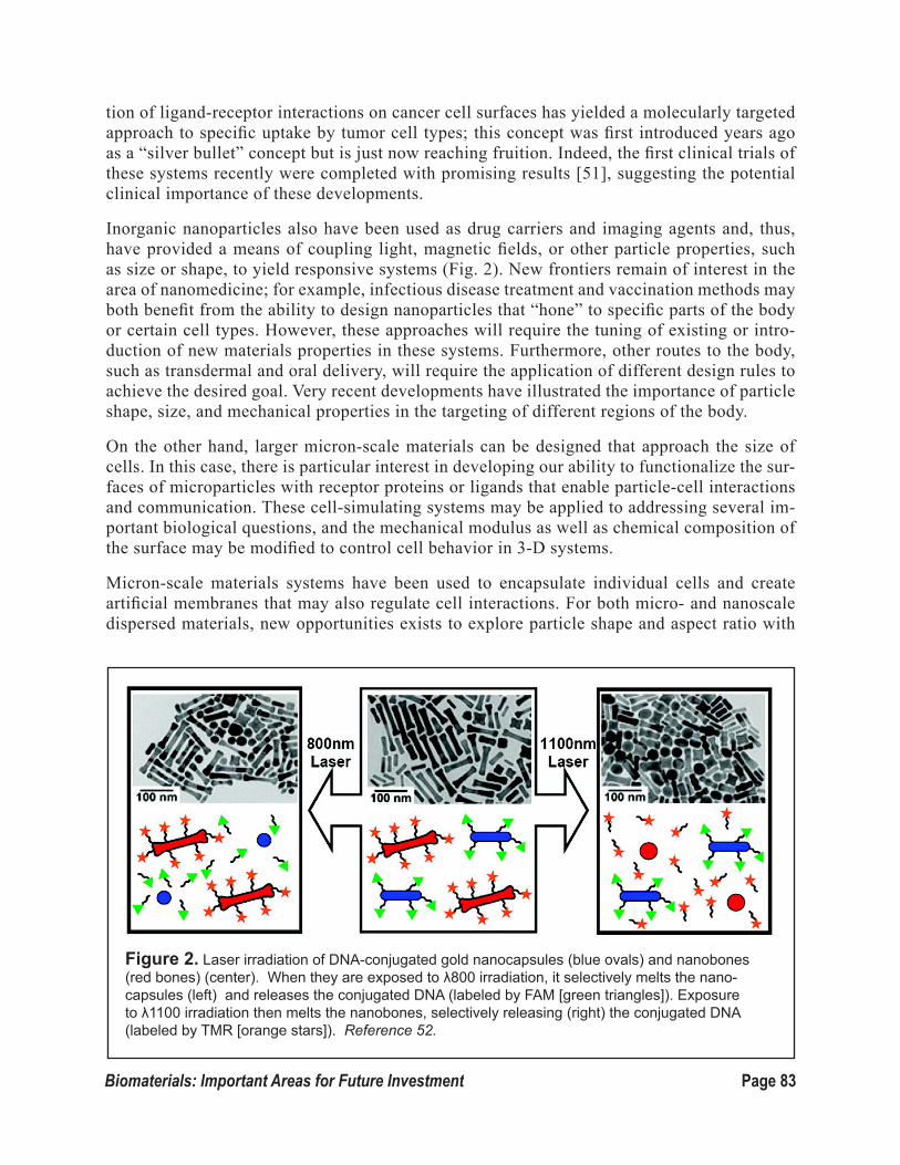
Page 83Biomaterials: Important Areas for Future Investment
tion of ligand-receptor interactions on cancer cell surfaces has yielded a molecularly targeted approach to specific uptake by tumor cell types; this concept was first introduced years ago as a “silver bullet” concept but is just now reaching fruition. Indeed, the first clinical trials of these systems recently were completed with promising results [51], suggesting the potential clinical importance of these developments.
Inorganic nanoparticles also have been used as drug carriers and imaging agents and, thus, have provided a means of coupling light, magnetic fields, or other particle properties, such as size or shape, to yield responsive systems (Fig. 2). New frontiers remain of interest in the area of nanomedicine; for example, infectious disease treatment and vaccination methods may both benefit from the ability to design nanoparticles that “hone” to specific parts of the body or certain cell types. However, these approaches will require the tuning of existing or intro-duction of new materials properties in these systems. Furthermore, other routes to the body, such as transdermal and oral delivery, will require the application of different design rules to achieve the desired goal. Very recent developments have illustrated the importance of particle shape, size, and mechanical properties in the targeting of different regions of the body.
On the other hand, larger micron-scale materials can be designed that approach the size of cells. In this case, there is particular interest in developing our ability to functionalize the sur-faces of microparticles with receptor proteins or ligands that enable particle-cell interactions and communication. These cell-simulating systems may be applied to addressing several im-portant biological questions, and the mechanical modulus as well as chemical composition of the surface may be modified to control cell behavior in 3-D systems.
Micron-scale materials systems have been used to encapsulate individual cells and create artificial membranes that may also regulate cell interactions. For both micro- and nanoscale dispersed materials, new opportunities exists to explore particle shape and aspect ratio with
Figure 2. Laser irradiation of DNA-conjugated gold nanocapsules (blue ovals) and nanobones (red bones) (center). When they are exposed to λ800 irradiation, it selectively melts the nano-capsules (left) and releases the conjugated DNA (labeled by FAM [green triangles]). Exposure to λ1100 irradiation then melts the nanobones, selectively releasing (right) the conjugated DNA (labeled by TMR [orange stars]). Reference 52.
Page 84 Biomaterials: Important Areas for Future Investment
regard to cell interactions. In addition, they may be used to explore mechanical stiffness and released or surface-bound chemical factors as cues to address important questions about cell behavior and cell-cell interactions.
4.2.2. Energy
The coupling of synthetic and biological or bio-inspired elements in dispersed materials sys-tems can lead to unique functions and capabilities relevant to energy applications. Excellent examples of coupled systems exist in biology for harvesting light, managing holes and elec-trons, and converting energy to fuels. For example, by conjugating photosystem proteins to lipid-like molecules, it is possible to create pre-organized structures that bind to carbon nano-tube surfaces and provide a means of harvesting light, coupled with rapid-electron transport for solar cells [53]. Reversible self-assembly of the micellar photocomplexes has been used as a mode of self-repair in these systems and is inspired by the natural self-repair observed in plant leaves. Dispersed nanomaterials also may be generated using biological systems as a template – for example, the use of filamentous phage as a bio-engineered template for met-al or metal oxide nanowires [54] or the grafting of polymeric species to the backbones or sidegroups of proteins or peptides with specific 3-D superstructures. These kinds of hybrid chemical-biological systems can be tuned to generate important organic or inorganic nano- to micro-scale components for batteries, solar cells, and electrochemical capacitors. In some cases, biological function may be preserved or modified in the presence of synthetic or supra-molecular modifications, thus, leading to new types of biomaterials.
4.2.3 Catalysis
Dispersed systems are of interest in the field of catalysis as a means of generating suspended micro- or nanoreactors within a liquid medium. Nanomaterials have been studied extensively in the field of catalysis, including their use in fuel cells, biofuel conversion, water splitting, electrochemical catalysis, and other applications relevant to the United States’ national inter-est. Although a number of inorganic metal and metal oxide particles of various size and shape have been generated using traditional synthetic routes, there is the potential for biomaterials systems to provide more advanced forms of catalytic systems. For example, it may be pos-sible to couple enzymes with catalytic metal or metal oxide cores in stabilized liposomes or polymersomes.
The ability to tune the interior and exterior content of disperse biomaterials systems would allow us to design an exterior membrane with specific barrier properties that can mitigate the molecular diffusion of reactants and products to the reactive core, while achieving an envi-ronment within the core that is optimized for catalytic reaction. Incorporating highly acidic phosphonic acid groups, for example, might be used to attain low-pH environs for reaction in the presence of metal or metal oxide catalytic nanoparticles. These kinds of applications are of particular interest to the biomaterials community, because the reaction is not limited in terms of time scale or volume, and micron-scale particles can be readily retained or separated from the media following catalysis and reused or regenerated.

Page 85Biomaterials: Important Areas for Future Investment
4.2.4 Environment
By bringing the world of biomaterials together with that of traditional fields of catalysis, oth-er new concepts may arise. For example, it may be possible to generate micron-scale hydrogel particles that contain coupled enzymatic components with inorganic catalytic species capable of breaking down common organic pollutants, such as steroids or other chemical by-products in sin-gle- or multi-step processes. The generation of water-dispersible micro- or nanoreactor particles with complex function, thus, would create environmental mediation systems inspired by algae or bacteria designed to break down pollutants to generate clean water. Additional opportunities in the area of environmental management lie in the potential to develop safe biomaterials that manage the presence of water, acidity, and critical nutrients and mediate the presence of toxic chemicals, while existing as dispersed particles in soil.
4.2.5 Sensing
The ability to incorporate responsive features into nano- and microparticles can lead to biomate-rials that serve as active components in many different sensing applications. Materials chemistry approaches that enable highly specific responses to molecular binding, enzymatic cleavage, pH, temperature, and ion content can be coupled with materials that generate a measurable signal by coupling an electrical, optical, or chemical response to molecular transformations, conformation-al change, larger-scale clustering, or the hierarchical assembly of multiple particles. There are multiple advantages to using dispersed biomaterials systems for sensing applications. Individual elements can be used to generate multiple sensing components that can be readily constituted and tailored to address specific sensing applications. In addition, the clustering of individual sensing molecules to the surfaces of microparticles or microgels, for example, can lead to enhanced sur-face area coverage and a significant amplification of signal in molecular beacon systems designed for DNA detection [55]. The use of cooperative behavior between individual disperse particles can lead to quorum sensing [56], which can be made extremely sensitive to environmental change while enabling a greatly enhanced signal.
Incorporating elements responsive to specific proteases, peptides, cytokines, or nucleic acids great-ly broadens the range of applications for sensing and detection. By generating materials that can exist in the biological environment as independent monitors of biological function, it may be pos-sible to engineer new generations of materials that help predict ─ as well as ─ control biological behavior and would serve as critical tools for biology and diagnostic medicine. These same tools also could be readily adapted to monitoring the environment, including detecting water potability, changes in wildlife habitat, and diseases at an early stage.
4.2.6 Summary
To attain the promise of the potential applications for dispersed biomaterials described here, a great deal of fundamental science and engineering is needed to develop new materials systems with precise control of size, shape, and function. The ability to enable particles to take on specific tasks as individually operable elements or as coordinated and responsive assemblies will yield new opportunities. If we can generate these systems in a reproducible and scalable manner, it also will
Page 86 Biomaterials: Important Areas for Future Investment
allow us to translate them to critical areas of need and open up new areas of application.
We have identified seven key scientific challenges that must be addressed in the upcoming decade to allow the kinds of materials functions that yield revolutionary new capabilities and advancements in the fields of medicine, biology, catalysis, energy, and the environment. These challenges include the exploration of new function and materials behavior that represent true frontiers of the biomaterials field as well as fundamental but critical needs that must be met to ensure these advancements take place. The National Science Foundation is particularly suited to address these critical materials science challenges in areas that range from fundamental particle physics to new polymeric and inorganic biomaterials synthesis and the development of novel computational tools.
The seven challenges are:
1. Particle motility.
2. Cooperative assembly.
3. 3-D patterning of particles.
4. New synthetic methods and materials.
5. Scalable and reproducible fabrication processes.
6. Computational models of dispersed systems in realistic environments.
7. More advanced experimental and computational tools for addressing the physics of dispersed systems
4.3 Particle Motility
Motility is a common attribute of living systems, which is shared across evolution – from protozoa to humans. The ability to actively convert potential energy into motion, motility underlies a vari-ety of diverse higher-order processes in biology, ranging from nutrient acquisition by bacteria to recruitment of immune cells to sites of infection to morphogenesis in the embryo. Motility enables living systems to undergo movements that may or may not be aligned with the physical forces in the environment. For example, a cell can migrate perpendicular to the forces of blood flow to exit a blood vessel and extravasate into a tissue.
The ability to create synthetic particles mimicking biological motility would enable bioengineers to develop diverse technologies, ranging from self-healing materials to solar cells with light-chasing photocenters as well as nanomedicines that truly exhibit “active targeting” to disease sites. Thus, a key scientific challenge to the field is the design of synthetic, dispersed systems that can mimic the modes of motility found in nature, using either biomimetic or wholly artificial processes.
Like the other grand challenges identified here, this not a trivial extension of existing science and technology. Achieving this vision will require fundamental advances in our understanding of bio-logical models for microscale motility, new methods of particle synthesis, and the development of

Page 87Biomaterials: Important Areas for Future Investment
new types of active nanosystems. All of these important biomaterials design goals require foun-dational materials chemistry and physics, which the National Science Foundation is highly suited to support.
Current work has indicated the potential power of hybridizing the motility of living cells with synthetic materials in the arena of medicine. For example, bacteria conjugated with DNA-carry-ing nanoparticles have been shown to actively invade host cells to deliver nanoparticle payloads into cells in vivo [57]. In addition, drug-loaded nanoparticles internalized by cells or linked to cell surfaces allow tiny doses of drug to be actively carried by their cellular “chaperone” to disease sites to stimulate potent therapeutic responses without systemic side effects in animal models (Fig. 3). Recently, a few pioneering theoretical and experimental studies have explored approaches for designing active motion into wholly synthetic particle systems, including com-putational models for swimming particles [59], repair-and-go capsules that roll and stop on a surface on cue [60], and the design of structures that can form an actin polymer network [61,62].
The design of synthetic particles with intrinsic motility – especially guided motility that imitates the directed migration processes of cells such as chemotaxis – would create new paradigms for medicine, sensing, and even the synthesis of materials. By employing design principles found in biology, motile drug-delivery particles could be designed to detect and move up spatial chemical gradients in the environment, for example to sense and report on toxins or contaminants in a water supply. In addition, we could design synthetic particles capable of combining motility with triggered release of material payloads to control the deposition and self-assembly of ma-terials for fabrication or repair of organic or inorganic devices. The ability of nanomaterials to move in or toward a specific stimulus might lead to microscale devices that can move and orient in 3-D to maximize function (e.g., self-contained microscale solar-fuel generators that orient in the direction of light’s greatest intensity.
In the field of nanomedicine, the phrase “active targeting” of particulate drug carriers refers to particles decorated with ligands that allow binding to specific target cells [63, 64]. However, such active-targeting still relies on blood flow, convection, and diffusion to bring a particle to the target cell, and most nanoparticle drug-delivery agents are cleared from the blood by the liver and spleen without ever reaching their target site. Motile particles that could actively move through tissues, invade through the epithelium of mucosal surfaces, or extravasate from the blood into tis-sues could fundamentally alter the current notion of active targeting.
At the single-cell level, particles that actively invade cells or nav-igate to specific subcellular com-partments (e.g., cytosol, mitochon-dria, nucleus, etc.) could be used to deliver therapeutics or report on signals within a cell. Particles able to home to specific sites in tissues
Figure 3. Nanoparticles (NPs) conjugated to immune cells known as T cells (left) are carried by the cells through tissue (center) to target disease sites, in this case a tumor cell (labeled EL4) (right). Nanoparticles appear yellow in first frame, and pink in frames 2 and 3; the green stained spot in frame 3 is the synapse formed by the T cell to trigger an immune response to the tumor. Reference 58.

Page 88 Biomaterials: Important Areas for Future Investment
or within cells would represent powerful tools for studying the biology of living systems and provide a basis for new kinds of in vivo diagnostics.
A key to motility is symmetry breaking, which might be intrinsic in anisotropic particles or might be induced in response to a stimulus in deformable symmetric particles. Living systems achieve motility through a complex system of regulated polymerization/depolymerization reactions of pro-teins to form intracellular polymeric fibers that can adhere to and disjoin from their surroundings and generate mechanical force. Thus, strategies for continuous-force generation in synthetic par-ticles must be developed. One approach may be the use of biomimetic routes, building on early work which studied reconstituted actin polymerization in elastic vesicles and using myosin motors or other native proteins that might be harnessed to synthetic biological components.
On the other hand, wholly synthetic concepts of generating propulsion also are promising. For example, the generation of propulsion from carbon nanotubes may produce a rapid means of trans-port [66]. In addition, the use of self-accelerating or self-regulating chemical couples, such as the Belousov-Zhabotinsky (BZ) reaction [66-68] or similar cyclical chemical redox reactions that can yield self-oscillating microgels and actuating mechanisms that are triggered by pH or ionic gra-dients, also may produce motility. Gradients in the presence of specific proteases, chemical con-taminants, or physical triggers also might produce oriented or guided transport along chemically, electrically, or mechanically defined pathways.
4.4 Cooperativity & Architecture
Since dispersed systems are comprised of many discrete entities, a grand challenge is to un-derstand, control, and exploit the ability of the individual units to cooperate with each other or assemble into complex architectures. Nature provides the inspiration for addressing this challenge, with one example being quorum sensing bacteria [56]. These are individual “parti-cles” that communicate with each other and detect chemical and environmental changes that lead to their organization into biofilms with new coordinated functionality that enhances their persistence well beyond that of a single individual. Inspired by this biological behavior, we could engineer dispersed particulate systems to exhibit similar behavior in response to chem-ical (or physical) stimuli. Indeed, we would leverage the potential for triggered coalescence of dispersed particles resulting in dramatic changes in the physical properties of the system for a wide range of applications, including those in the sensing, energy, environmental, and biomedical arenas.
Addressing this challenge would enable us to fabricate complex biomaterials with function-ality far beyond what is feasible today. An example of the potential for such a system, which also is inspired by nature, is a “microleaf.” One could imagine particles with different func-tionalities, such as light collection, catalysis and separation, which assemble into a complex structure capable of adapting to changing light conditions, converting light and CO2 into use-ful products, and transporting those products for collection. These are all activities carried out by natural leaf structures (Fig. 4). In order to achieve such functional complexity, however, significant technological advances must be made in a variety of areas.
In terms of the design of individual units, adaptive and responsive behaviors to physical, chemical, or biological stimuli are required. Stimuli could range from mechanical forces, tex-

Page 89Biomaterials: Important Areas for Future Investment
ture, or topographical changes, electrical or magnetic fields, chemical gradients, or specific bio-molecular interactions.
Biomaterials, as opposed to non-biolog-ical materials, are uniquely positioned to be able to sense these stimuli. The literature is full of biological systems that respond to such stimuli, and there is an opportunity to mine specific sensing molecules from living cells or design synthetic materials that mimic or have novel engineered detection capabilities.
The critical aspect in moving from in-dividual units to an assembled or coop-erative ensemble is the ability to com-municate. This will require a fundamental understanding of how current dispersed systems – from complex individual living systems (e.g., eukaryotic cells, bacteria, and viruses) to simpler biomolecules (e.g., proteins or nucleic acids) – communicate. Signal transduction mechanisms, therefore, must be studied to identify common themes that can be translated into synthetic biomaterials, including self-recognition, stimuli-sensitive aggregation/de-aggrega-tion, controlled molecular folding/unfolding, changes in catalytic or degradation activity, or different forms of energy transfer. Harvested or synthesized proteins or peptides may be key components that allow dispersed entities to communicate with one another, and specific par-ticle-particle interactions can be manipulated through the attachment of ligand and receptor proteins from cells onto synthetic particle surfaces. The engineering of natural proteins or synthetic coupling with responsive polymers can lead to modified systems with a self-attrac-tive binding function that is regulated by secondary molecules acting as switches or by other stimuli, such as light or pH, for example.
The defining feature of this challenge is the ability to channel individual sensing and communication into new properties or behaviors displayed by the group that were not feasible from single individuals. Such cooperativity would indicate novel or enhanced function of the ensemble compared to the individual components. Complex assembled architectures exhibit organization over multiple length scales, ranging from the individual particle scale to many scales larger. These two phenomena – cooperativity and assembled architecture – can be interdependent, cause-and-effect, or spontaneous above a given con-centration. In any case, we must develop a fundamental understanding of how the prop-erties of the ensemble relate to the properties of the individuals and the environment. To accomplish this, we will need to uncover the kinetic or thermodynamic dependence and, ideally, manipulate it.
Also required is the ability to control whether individuals assemble or respond simultane-ously or whether small ensembles form and are gradually extended or decreased in size by the inclusion or loss of other individuals. An incredible technological advance would be to build a biomaterial capable of undergoing these processes in a truly reversible fashion (i.e.,
Figure 4. Schematic of a natural leaf structure, which is a complex architecture of discrete com-ponents with different functions that interact with each other. Reference 69.

Page 90 Biomaterials: Important Areas for Future Investment
the ability to respond to changes in the environment or time). This challenge is significant and will require contributions in the areas of mechanical/chemical/biological sensing, signal transduction, and interfacial interactions between entities ranging from small molecules to macromolecules to colloids as well as with larger-scale assemblies.
4.5 3-D Patterning of Particles
Spatial control of particle chemistry is a unique challenge due to issues of dimension and curvature on particle surfaces. As particle technology moves from homogeneous to heterogeneous, the field of biomaterials will require new chemistry and analytical tools that enable us to control the organi-zation of single and/or multiple molecules and characterize their reproducibility. Such patterning will create a new class of biomaterials, with altered reaction kinetics, adhesion, and cell/material interactions. Illustratively, 3-D patterning comprises the idea of organizing structural complexity within or on the surface of a nanoparticle or “painting” a particle with specific chemical and phys-ical features with molecular scale precision.
Surface chemistry and presentation, as is evident in nature, govern physical and chemical phe-nomena. For example, the alternating striations of covalently bound hydrophobic and hydrophilic moieties on the nanometer length scale of small gold nanoparticles can yield surfaces that directly penetrate cell membranes without rupture, thus, enabling cell internalization (Fig. 5). In addition, patterns may emerge - given changes in the local environment – that may be utilized to control surface characteristics. Rearranging molecules within lipid rafts is observed in the binding of cy-tokine-activated endothelial cells to leukocytes. Such ordering, either prescribed or adapted, may guide interactions between two dispersed entities or between a particle and a biological element, such as a cell or a tissue membrane.
Control over ligand organization may be di-rected towards the detection of agents that exhibit similar chemistry but distinct orga-nization. Such complementarity and altered dissociation kinetics may allow for targeted selection and separation. Enhanced targeting of cancer and activated endothelial cells, for example, has been described in vivo and in vitro by the ordering of single and multiple ligands, respectively [71,72].
These initial efforts will lay the foundation for how molecular-level control over ligand display and may overcome challenges in cell targeting. The presentation of specific chem-ical or biomolecular cues on surfaces would provide a new means of probing cell interac-tions with nanomaterials, and it may provide a means of regulating or avoiding immune re-sponse or toxic effects of various nanomateri-als in the body. Biological studies of cell-cell
Figure 5. A schematic of striped gold nanoparticles formed through the introduction of hydrophobic and hydrophilic thiol ligands to form regions of alternating wetting characteris-tics that enable direct cell membrane penetra-tion. Reference 70.

Page 91Biomaterials: Important Areas for Future Investment
behaviors also may inform the design of microparticles and gels that present patterned surfaces to regulate or induce cellular behavior, thus, influencing in vitro and in vivo cell function. Fi-nally, such studies will provide a means of supporting the culture of cells that maintain their specific function based on such interactions.
On the other hand, the patterning of dispersed biomaterials can involve generating isolated compartments within particles that might serve as reservoirs with different components for par-ticle-based microreactors. Patterning might also involve the design of specific self-assembling systems that organize in very specific arrangements due to the presentation of specific coopera-tive surfaces to their complementary surfaces, thus, providing a precursor to cooperative assem-blies (see Soft Materials section).
4.6 Synthesis – New Materials and New Methods
One of the great challenges in achieving new, highly tunable dispersed systems is the ability to control their chemical composition, self-assembling behavior, shape, and structure from the mo-lecular to the mesoscale. Although the chemistry community has mastered a number of synthetic approaches, the ability to precisely control the dispersity, architecture, and chemical composi-tion of these materials is still non-existent. A combination of new and emergent approaches that enable control of these features will provide critical tools for new generations of biologically inspired and bio-derived materials.
Nevertheless, a number of chemical approaches have been developed that do enable us to con-trol the design of biomolecules. These approaches provide, for example, a means of creating polypeptide backbones using synthetic routes; however, the resulting materials still exhibit tre-mendous polydispersity. The introduction of co-monomers leads to polymer species that vary significantly in distribution and sequence, thus yielding molecules that cannot undergo folding into secondary and tertiary structures, and, therefore, they lack specific interactions with recep-tors and biological systems, which are greatly needed in the biomaterials area.
Facile, robust, synthetic methods for precision polymer systems is an area where new approach-es are required for generating self-assembling, dispersed materials that can present structure and function in a controlled and predictable fashion. Furthermore, there are few rapid synthetic tools available to make other biomolecular backbones, such as nucleic acids, polysaccharides, and posttranslationally-modified proteins, on large scales using easily implemented chemical approaches.
Currently, there is a revolution in rapid and quantitative “click” chemistries, which has led to a ready means of directly modifying materials with high fidelity; however, the next frontier requires the ability to control chain composition unit-by-unit, while enabling control of chain length and end groups. Some examples of new directions in this area include using existing bio-macromolecules as a scaffold for synthetic-biologic hybrid systems. Another potential general strategy for generating new self-assembling systems is the pre-programmed genetic design of protein modules that self-assemble into organized particles; this approach is guided by design rules and computation that predicts folded structure and guides materials design [73].
A number of new avenues in the use of genetically modified biological systems are enabling

Page 92 Biomaterials: Important Areas for Future Investment
us to use of cells as factories for the production of precise proteins, biomacromolecules and non-biological polymeric species in large quantities (Fig. 6).
The use of recombinant approaches can lead to highly controlled materials systems and offer great promise for the generation of new materials for the field. There are limitations to the conventional or current recombinant methods, however, in terms of the types of backbones and chemical com-positions that can be incorporated using only cellular means.
New recombinant approaches might include hybrid methods that utilize cellular machinery in combination with other synthetic methods to generate complex molecules, such as polysaccha-rides, with a level of precision that does not yet exist in nature [75]. Hybrid approaches that might combine these techniques could introduce polymer chains that contain biologic and synthetic moi-eties along the backbone in specific regions, as well as the attachment of nanoscale materials with precise shape and architecture to create hierarchical and multiscale dispersed systems that have engineered function, ranging from electrochemical and mechano-optical to biological activities. Early demonstrations of this capability have been shown in the directed polymer-like assembly of nanospheres and nanowires [76]. In addition, new developments in the ability to turn genes on and off in response to different stimuli provide a fresh opportunity to develop novel recombinant technologies that introduce different molecular segments at different time points in macromolecule synthesis to create carefully sequenced materials systems on demand.
4.7 Manufacturing Process Scalability and Control
One of the keys to executing good basic science, especially in the area of dispersed systems, is hav-ing access to materials that are readily reproducible, uniform in quality and characteristics, can be made to have new characteristics, and well characterized and understood from a detailed chemical and morphological point of view. Challenges in reproducibility, scalability, and manufacturability threaten to impede the full exploitation of these promising materials. New manufacturing methods and microfabrication processes, therefore, must be developed in the biomaterials community to allow for the translation of important develop-ments from the bench to application, and the development of different means of generating nano- and microparticle systems will also pro-vide a platform for the design of new structures that would not be accessible through traditional means.
In the design of dispersed biomaterials, it often is essential to understand the whole life cycle of the material. The successful development of a dispersible biomaterial is measured by an inno-vative design, good manufacturability, and high reliability for applications. Manufacturability and reliability need to be considered as intrinsic parts of the material design stage. In industry,
Figure 6. Schematic of use of cellular machinery to generate linear carbohydrate polymers. Reference 74.

Page 93Biomaterials: Important Areas for Future Investment
this concept is often referred to as “Design to Manufacturability and Reliability.”
The ability to generate many of the kinds of dispersed systems described in the previously stated scientific challenges requires the formation of nano- and microparticles with very-low polydisper-sity, well-defined surface chemistries, and, in some cases, control of spatially organized features within the interior or the surface of the nanoparticle. Many particle synthesis approaches, on the other hand, yield relatively high polydispersities and stochastically arranged surface or bulk inte-rior features.
The function of these systems often relies on size or chemical surface characteristics, thus, making reproducibility critical for achieving new levels of functionality. For applications in biomedicine, in particular, there also are different biologic outcomes anticipated for particles that differ in size or chemistry.
Finally, manufacturing large quantities of dispersed systems at reasonable costs and speed would be critical for many applications in energy storage or the environment. There is, therefore, a strong need to synthesize and manufacture particles that achieve these desirable attributes, and new approaches must be deployed to achieve these goals. Examples of two such strategies are: (1) the use of contin-uous manufacturing methods (vs batch) and (2) templated synthesis methods.
Most dispersed systems used in scientific research are made using batch-reactor designs for a num-ber of reasons, including ease of use, cost effectiveness, and the culture of the scientific community which pioneers many new dispersed materials, including most nanomaterials. Batch reaction sys-tems, however, are plagued by excursions during a synthetic run that include drifts in the feed ratio of the reactants, temperature and pressure excursions, and mixing issues. These changes often intrinsically lead to heterogeneities in chemical composition, particle size, surface chemistry, and many other attributes. In contrast, in a continuous reaction system, reactants and products are continuously added and withdrawn. This results in steady state conditions of reactant and product concentrations and better con-trol of temperature and mixing. This level of control lends itself to better product uni-formity.
Continuous processes can exist in two forms as well: (1) stirred tank reactors, and (2) roll-to-roll web-based processing. Roll-to-roll continuous processes using templat-ed methods to make particles that achieve higher uniformity intrinsic to a continuous stirred tank reactor, and also can yield high-ly regular and reproducible particles with a broad range of shapes due to the ease with
Figure 7. (Top) Continuous thin-mold manu-facturing for the PRINT nanoparticle production process. Patterned surface can be seen in green. (Bottom) Polymer nano- and microparticles pat-terned using PRINT micromolding process with molds of various shapes and sizes. Reference 77.
Page 94 Biomaterials: Important Areas for Future Investment
which a pattern can be transferred in the molding process (Fig. 7). Another continuous processing route includes the use of microfluidic reactors to generate microparticles; by combining light-in-duced polymerization with the controlled laminar flow environment within a fluidic microchannel, it is possible to create highly detailed patterned microparticles with great reproducibility [78].
4.8 Computational Models of Dispersed Systems in Realistic Environments
Whether for water purification, biomedicine, or catalysis, the application of inherently nano- and microparticle dispersed systems occurs in a highly complex aqueous environment. However, due to severe limitations in recreating complex natural environments in laboratory experiments, cur-rent experimental designs and characterization processes for many of these systems often are done in an oversimplified environment. Access to computational models that can build in the complexi-ty of the natural aqueous environment can help inform the design of dispersed systems to achieve more precise functionality and inform materials design.
Indeed, computational tools are of critical importance in the design of dispersed systems for func-tionality in biologically relevant environments. For example, nanocarriers of different sizes, ma-terials, and shape are ubiquitously designed for use in diagnostic and delivery of therapeutics in human diseases. The design of these systems often is done, however, with a limited understanding of the complex in vivo environment in which the systems must exert their functionality. In vitro experiments grossly underestimate human physiology, and animal models often exhibit subtle differences in their physiology relative to the human body. Although more realistic experimental models of biological systems have emerged in recent literature, these are still limited in their ca-pacity to fully represent the in vivo environment. For example, recent literature has explored the capacity of micro- and nanoparticles to bind to the vascular wall in human blood flow with in vitro assays; however, these assays are unable to faithfully represent the dimensions and blood flow characteristics of human circulation. Similarly, current computational models primarily focus only on the fluid dynamics of the human vascular system.
Future computational models must be developed that have the capacity to build up to multiple scales and incorporate the different cellular components of human blood so that they can begin to expose experimental researchers to critical design criteria that must be met for dispersed imaging and delivery systems. Such complex flow models also are relevant in non-biological environ-ments, including the use of dispersed systems in water purification, neutralization of aerosolized chem/bio-warfare, and in oil-well drilling. On another front, there also is the need for models that can realistically describe the physics of cell motility, differentiation, and proliferation. Access to such models can precipitate the design of complex, functional dispersed systems, when they are coupled with materials computational models.
Although computational power and capacity has rapidly evolved in the last decade, it still has significant limitations for addressing the complexity of dispersed systems in complex environments. These limitations may be associated with the lack of appropriate modeling tools that take into consideration thoroughly defined biological environments and biological constraints. Additional key constraints include the computing power needed to handle com-plex computations and the need for a more accessible means of generalizing certain biological milieu. As such, cross communications and close interactions between computational and theoretical scientists and experimental biomaterials scientists would significantly accelerate

Page 95Biomaterials: Important Areas for Future Investment
the evolution of this next frontier in biomaterials research as it relates to dispersed systems.
4.8.1 Development of computational and predictive tools for addressing the physics of dispersed systems
Knowledge of the fundamental thermodynamics and kinetics governing the formation of myr-iad architectures of dispersed systems, starting from solvated molecular materials of diverse structural origin (biological, bio-mimetic, bio-inspired or synthetic) is of paramount impor-tance for the design of new materials.
The development of qualitative predictive tools and quantitative models of the thermodynam-ics as well as kinetics of assembly of single or multi-component systems leading to self-assem-bly (thermodynamics), directed assembly (kinetics and thermodynamics), or process driven assembly (primarily kinetics) needs inter-disciplinary collaboration among physicists, chem-ists, biologists, and material scientists. Since the dispersed systems field historically has been driven by experimental observations, it is imperative to bring together much of the phenome-nology into predictive computational tools at multi-scale levels. Furthermore, there is a need for more systematic experimentation that explores the basic interactions between particles and water in dilute and crowded environments and the impact of aqueous conditions on properties such as surface hydration, size, and permeability. Such experiments would be complementary to computational modeling and would further inform the field about the anticipated behavior of dispersed systems as individual units and assemblies.
The driving forces of enthalpy and entropy of molecules dissolved in solvents during the cre-ation of sub-nano size clusters, followed by precursor nano-size assemblies in solvents and, ultimately, to the formation of disperse particles of various length scales (nano, micro, meso, etc.), requires a detailed and fundamental understanding of these processes. Detailed compu-tational analysis of the specific nature of the molecular material (i.e. biological, biomimetics, bio-inspired, or synthetic materials) and their mechanisms for formation of dispersed systems could provide significant insights to the design of novel materials as well as optimization of dispersed particle constructs. Additionally the thermodynamics and kinetics associated with the manufacturing process (e.g. micro-fluidic systems, stirred tank systems, batch, or continu-ous process) may require not only molecular dynamic simulations but also the use of compu-tational fluid-dynamic approaches.
Developing models for predicting the particle size distribution, polydispersity, etc. also will enable us to optimize the fabrication and scale-up of dispersed systems manufacturing. The next level of modeling required by the biomaterial community is to understand and predict the movement and transport of dispersed systems in its intended-use environment (physical or biological).
4.9 Technological Needs
To address the challenges of dispersed biomaterials systems research and development, a number of key technological needs must be met. These needs include:
Page 96 Biomaterials: Important Areas for Future Investment
• Advanced characterization and analytical tools: The tools that often are effective for bulk materials systems are much less useful for examining nanoparticles, microparti-cles, and other dispersed systems due to constraints of the small size and mass of these materials. Furthermore, the very nature of the dispersed system is best understood in its aqueous milieu as a dispersion, making many more traditional techniques, such as electron and X-ray microscopy, less relevant. Some specific examples of advanced characterization and analytical tools include:
o Non-destructive Particle Concentration – There is a strong need to develop instru-ments that can nondestructively report the number and concentration of nanopar-ticles in a small volume of fluid along with additional properties, such as mass concentration, zeta potential, and density. Such methods would be particularly relevant to the examination of nanoparticles in their native environment based on application, such as blood serum or plasma, ocean or river water, etc.
o In-Situ Structural Characterization – It is critical to be able to observe surface characteristics of a dispersed system without modifying or changing its inter-actions with the surrounding environment. For example, for nanoparticles with structured surfaces containing ligands or specific surface functional groups, it is possible that rearrangements take place on the particle surface when they are en-gaging with cells or exposed to a different pH or ionic content. A means of imag-ing particle surfaces in situ, while maintaining nanoscale resolution, would greatly impact the design of new responsive disperse materials. Furthermore, techniques that determine fractional surface composition, degree of hydration, and relative amounts of ordering at the particle surface are key to further understanding dis-persed biomaterials systems.
o Nano-biomechanics of dispersed systems - Mechanical properties play a key role in a number of biomaterials applications, and they can be critical to the nature of cellular interactions, particle flow, and directed assembly behavior; however, it is relatively difficult to directly determine the mechanical properties of nanoparti-cles. New methods therefore need to be developed to understand the mechanics of systems of very small scale.
• New dispersed materials synthetic tools: A number of exciting chemistries recently have been introduced in the materials science field; however, it will require some adap-tation of known chemical methods to provide a means of synthesizing particles directly in aqueous media to make dispersed materials with a minimum of added surfactant or stabilizer. Furthermore, more effective and controlled methods of synthesizing biomol-ecules ─ ranging from peptide ligands to large polysaccharides with control of size and composition ─ would enable us to generate new functional dispersed systems.
• Scaling of nano- and microtechnologies for large-batch investigations: The ability to ex-plore the range of applications described as potential areas for dispersed materials requires the characterization and examination of much larger quantities of particles than might typ-ically be produced in a laboratory setting. Methods for producing systems of interest at

Page 97Biomaterials: Important Areas for Future Investment
larger scale would provide a route to improved and broader characterization of this unique set of biomaterials as well as more effective and rapid translation of experimental results to application.
• Theory and computational hub: The ability to use molecular- and physicochemical theory to predict materials properties, interactions with water and other biomolecules, and self-as-sembly or binding behavior is key to the design of new dispersed systems. Centralized hubs provide an opportunity to bring experimentalists, theorists, and computational scientists together to develop realistic models.
4.10 Recommendation: A Particle Foundry
One means of addressing many aspects of the technological needs described above is the concept of a shared “Particle Foundry.” The three past decades have seen an explosion of bio-inspired or biocompatible materials for the manufacture and production of dispersed particles covering length scales from the nano to the meso. These particles often are responsive to the environment, prepared from multiple components, and contain reporters, ligands, and cargos. This complexity has generated a compelling demand for synthetic techniques to prepare novel components and for applying sensitive analytical and imaging techniques to characterize the starting components and resulting particles.
However, many of these techniques require expensive instruments that necessitate specialized training to operate and frequent calibration to maintain accuracy and subtleties in the inter-pretation of the outputs. These instruments are complex, expensive, and, thus, not well suited for purchase and maintenance by individual researchers. In addition, the complexity of the dispersed particles and their interactions with the environment necessitates a modeling and computation component that, again, is better suited to a specialized/dedicated facility rather than to individual investigators. The proposed Particle Foundry (see outline below) would provide the research community a unique educational/ training resource for the manufacture and production of particles by batch, continuous, or microfluidic methodologies.
4.10.1 Particle Foundry description
Staffing: Sufficient research and support staff and infrastructure are needed to implement and sustain a central facility for the application of state-of-the-art analytical methods for dis-persed system characterization. Staff also will be needed for the development of new methods of analysis, the characterization of submitted dispersed systems, the production of reference dispersed systems, the scale-up of dispersed particles to pilot-scale quantities, and the prepa-ration of other critical components.
Synthesis and Manufacture: Oftentimes individual investigators develop specialized poly-mers, ligands, or reporters that would be useful to the broader research community. Because these compounds typically are not commercially available, an important function of the Foundry would be to collect these compounds from investigators and scale-up their produc-tion from the typical 100-mg-range research scale to the 10 gram – 100 gram range. This

Page 98 Biomaterials: Important Areas for Future Investment
service would provide uniform material for application specific investigations. The Particle Foundry then could provide validated specialized reporters, ligands, or other components as well as canonical dispersed particles as standards for calibration to the research community. It also would provide components to enable particle tracking/detection and equipment and expertise for small batch scale-up.
Analytical Techniques: There is a critical need in the dispersed particle community for high resolution instruments to analyze, visualize, and characterize the molecular structure of the dispersed particle components as well as their composition, surface characteristics (e.g., zeta potential, diameter, polydispersity, morphology [architecture], ligand density and ligand sur-face distribution, and hydrophobicity/wettability). The Foundry personnel would develop and validate methods to enable the detailed characterization of the particles. These techniques will help to standardize the analysis of particles among different investigators.
Characterization: In addition to methods development, the Foundry will provide a service or assist investigators in the analysis of the properties of investigator-produced particles. A centralized analytical service would create cost effective, timely, comprehensive analyses of particle characteristics with greater uniformity and independent assessment of particle char-acteristics that enable a comparison among the materials created by various investigators. This characterization would provide a database of particle characteristics that investigators could interrogate to identify particles/compositions that might be applicable to their particular needs.
Training component: The Foundry will catalyze the transfer of knowledge, materials, and techniques among laboratories, including maintaining a web site, presenting training sessions to insure quality control, and introducing improvements in the manufacture of components and dispersed systems. The Foundry would have internships for students and bench space for visiting scientists to enable them to prepare dispersed systems and to learn cutting-edge tech-niques for the production or analysis of dispersed particles.
Global Impact: The Foundry would provide the infrastructure for the production and analysis of dispersed particles, computational modeling cluster, training to investigators, and opportu-nities for collaboration with international investigators. It also would generate new methods and analytical techniques to enable the high-resolution characterization of multi-component particles.
Page 99Biomaterials: Important Areas for Future Investment
Section 5. Thin Films and InterfacesDISCUSSION LEADER
DAvID tIRRELL, California institUte of teChnology
GROUP MEMBERS
DELPhINE GOURDON, Cornell University
DANIEL hAMMER, University of Pennsylvania
RyAN hAyWARD, University of MassaChUsetts aMherst
EfROSINI kOkkOLI, University of Minnesota
LAShANDA kORLEy, Case western reserve University
SUPING LyU, MeDtroniC, inC.
PhILIP MESSERSMIth, northwestern University
LIvIU MOvILEANU, syraCUse University
PAUL NEALEy, University of wisConsin
SPECIAL ACkNOWLEDGMENt
LAWRENCE DOOLING, California institUte of teChnology, stUDent PartiCiPant anD ContribU-tor to this rePort
5.1 Introduction
Many biomaterials function as thin films or in interfacial environments. Injected or im-planted biomaterials form critically important interfaces with surrounding cells, tissues and fluids; adhesive interactions can dictate the success or failure of surgical procedures; sen-sors and diagnostic devices operate primarily through interfacial interactions; and some of nature’s most intriguing materials systems (e.g. mineralized structures) are assembled through deposition and interfacing of multiple thin layers. If we are to succeed in our efforts to understand and design biomaterials systems, we must understand more thoroughly the behavior of thin films and interfaces.
New methods for imaging of surfaces and thin films, for scattering from surfaces, and for

Page 100 Biomaterials: Important Areas for Future Investment
measuring interfacial forces are providing important new insights into the roles of inter-facial phenomena in determining biomaterials performance. For example, scanning probe microscopy, super-resolution microscopy, grazing incidence diffraction, and the surface forces apparatus are allowing us to interrogate surfaces and interfaces in new ways. Sin-gle-molecule spectroscopy and molecular force measurements also now enable researchers to examine the behavior of individual molecules and cells tethered to surfaces, and trac-tion-force microscopy is beginning to reveal the complex mechanisms and consequences of the exchange of forces that occurs at the interfaces between cells and their surroundings.
We also are learning how to tailor surfaces and interfacial environments more effectively through improved methods of materials synthesis and processing. The topographic features and chemical functionality of surfaces can now be controlled on length scales of nanome-ters, and there is a growing appreciation of the importance of such control, both in funda-mental studies of cell-material interaction and in efforts to engineer biomaterials for useful function. The following paragraphs describe some of the most important opportunities, challenges, scientific questions, and technological needs in the field of interfacial biomate-rials research.
5.2 Opportunities and Challenges
5.2.1 Biomedical interfaces
Hundreds of billions of dollars are spent on medical devices each year in the United States, and millions of people depend on the performance of such devices to maintain a satisfactory quality of life. In many of these devices, biomaterials form critically important interfaces with cells and tissue fluids, and the key modes of failure involve interfacial phenomena that are poorly understood. Surfaces of implanted medical devices are covered with proteins of unknown composition, orientation and conformation; they trigger foreign body reactions (see Cell-Materials Interactions section); and they can serve as sources of devastating in-fections (Fig. 1).
Addressing these issues and creating new generations of devices that can function effective-ly over long periods of time without repeated inter-vention will require new insights into the structure and dynamics of complex interfacial systems. In particular, we must better understand how proteins and cells attach to surfaces and how we can re-cruit specific proteins or inhibit their attachment, as needed. How do we ensure that protein structure is preserved upon attachment? Can we measure the interfacial forces involved in molecular and cellular contacts at biomaterials surfaces? Can we engineer those forces to make strong surgical ad-hesives but also materials that suppress unwanted tissue adhesion or bacterial colonization? And, can we develop the analytical tools needed to address
Figure 1. Bacteria growing on the luminal surface of an in-dwelling cath-eter. Reference 79.
these issues, and especially to study biological interfaces directly, without the need for dehydration and high-vacuum imaging systems? There are many compelling opportunities here to improve peoples’ quality of life, but these opportunities will not be realized without sustained commitment and new ideas from the materials community.
5.2.2 Biomolecular factories
Many of the most familiar and essential elements of modern life result from chemical man-ufacturing processes. For example, computer chips are assembled through an astonishing sequence of chemical processing steps that must be controlled on size scales of nanometers and with compositional tolerances of parts per billion. In addition, we clothe ourselves in synthetic fibers and protect our homes with paints made from long-chain polymers. And, we run our cars on gasoline that is made by breaking down and refining crude oil, which comes from long-decayed biological organisms.
Biological systems are also remarkable chemical factories, capable of converting the sim-plest, most abundant starting materials (e.g., CO2) into complex, highly functional products without intermediate separation and purification steps. They do this largely through the exquisitely selective actions of proteins, which recognize individual molecules with high specificity, catalyze chemical transformations under mild conditions, and deliver their met-abolic products to the cellular and extracellular destinations where they are needed.
Although it is easy to imagine harnessing the catalytic, binding, and transport properties of proteins to engineer efficient chemical processes, we do not, in general, know how to as-semble the proteins into useful forms without loss of function. Chemical modification of proteins often interferes with substrate binding or causes proteins to unfold and lose activi-ty. Attaching proteins to surfaces to make catalytic particles or selective membranes also is an uncertain proposition (Fig. 2). If we could do this successfully, however, complex chem-ical processes that now require high temperatures and multiple steps of chemical conversion and purification could be carried out in integrated fashion with substantial savings in cost and energy. Development of reliable methods for protein assembly on surfaces, thus, is an import-ant challenge for the biomaterials community.
5.2.3 Self-healing and self-reporting materials
The need for continual repair and replacement of damaged or failed structural materials remains one of the grand challenges facing society today, as it has been for hundreds or thousands of years. The limited lifetime of commonly used materials impacts virtually all aspects of life. For exam-ple, although medical implants enjoy widespread success in a variety of clinical settings, current medical implant materials do not have the same longevity and robustness of the tissues they are

Page 101Biomaterials: Important Areas for Future Investment
these issues, and especially to study biological interfaces directly, without the need for dehydration and high-vacuum imaging systems? There are many compelling opportunities here to improve peoples’ quality of life, but these opportunities will not be realized without sustained commitment and new ideas from the materials community.
5.2.2 Biomolecular factories
Many of the most familiar and essential elements of modern life result from chemical man-ufacturing processes. For example, computer chips are assembled through an astonishing sequence of chemical processing steps that must be controlled on size scales of nanometers and with compositional tolerances of parts per billion. In addition, we clothe ourselves in synthetic fibers and protect our homes with paints made from long-chain polymers. And, we run our cars on gasoline that is made by breaking down and refining crude oil, which comes from long-decayed biological organisms.
Biological systems are also remarkable chemical factories, capable of converting the sim-plest, most abundant starting materials (e.g., CO2) into complex, highly functional products without intermediate separation and purification steps. They do this largely through the exquisitely selective actions of proteins, which recognize individual molecules with high specificity, catalyze chemical transformations under mild conditions, and deliver their met-abolic products to the cellular and extracellular destinations where they are needed.
Although it is easy to imagine harnessing the catalytic, binding, and transport properties of proteins to engineer efficient chemical processes, we do not, in general, know how to as-semble the proteins into useful forms without loss of function. Chemical modification of proteins often interferes with substrate binding or causes proteins to unfold and lose activi-ty. Attaching proteins to surfaces to make catalytic particles or selective membranes also is an uncertain proposition (Fig. 2). If we could do this successfully, however, complex chem-ical processes that now require high temperatures and multiple steps of chemical conversion and purification could be carried out in integrated fashion with substantial savings in cost and energy. Development of reliable methods for protein assembly on surfaces, thus, is an import-ant challenge for the biomaterials community.
5.2.3 Self-healing and self-reporting materials
The need for continual repair and replacement of damaged or failed structural materials remains one of the grand challenges facing society today, as it has been for hundreds or thousands of years. The limited lifetime of commonly used materials impacts virtually all aspects of life. For exam-ple, although medical implants enjoy widespread success in a variety of clinical settings, current medical implant materials do not have the same longevity and robustness of the tissues they are
Figure 2. Idealized image of pro-teins presented with well-defined structure and orientation (left) and in more typical disordered fashion (right). Reference 80.

Page 102 Biomaterials: Important Areas for Future Investment
intended to replace. The idea of building self-healing mechanisms into synthetic materials has stimulated a wide range of innovative research.
Self-healing may be intrinsic or extrinsic to a material; in the case of polymers, examples of intrinsic phenomena that can give rise to self-healing include diffusion of polymer chains across a fracture interface upon heating above the glass transition or chemical-bond forma-tion between transient reactive species created as a result of molecular scission during frac-ture. A more commonly employed self-healing mechanism for polymers is to incorporate extrinsic factors within the polymer matrix, such as microencapsulated monomers, which upon local release into a newly created defect (e.g. a slowly propagating crack) give rise to new polymer formation to heal the defect. Research in self-healing polymers is leading to rapid progress in this area, including the development of polymer coatings that can be repaired via exposure to ultraviolet light.
Many biological tissues have high damage tolerance and the capacity to self-heal when damaged. Therefore, they are important sources of information and guidance in this endeav-or. Self-healing mechanisms can be cell-orchestrated. An example of this is long-bone frac-ture repair via the initial formation of a fracture callous followed by cancellous and, ulti-mately, cortical bone formation. However, self-healing is ubiquitous in nature on a much finer level. Molecular-level phe-nomena, such as sacrificial bond rupture under applied load and re-formation in the absence of load, are examples of this. These molecular-level physical concepts remain poorly un-derstood, yet have enormous potential to inform the design of novel self-healing materials for practical applications. A significant opportunity exists to expand this effort, along with research that aims to reveal the physical, chemical and mechanical phenomena underlying self-healing behavior in biological materials. These activities promise to produce signif-icant societal benefits by leading to better materials that can be integrated into high-tech devices, consumer and construction goods, and healthcare devices. A related area worthy of invest-ment is self-reporting materials, including materials that are capa-ble of signaling the presence of damage, ideally, while in service or in situ. Important goals in this area include the design of mac-romolecules with force-sens-ing components integrated into the molecular design. So-called ‘mechanophores’ have recently been developed and shown to act as molecular strain sensors when embedded in polymer ma-trices, allowing the in situ visu-alization of microcracks. Such materials can provide the means
Figure 3. Self-healing materials with vascular networks. (a) The dermis of the skin. (b-d) A vascularized synthetic epoxy system with embedded catalyst. Note the excess healing fluid on the surface in (d). Reference 81.

Page 103Biomaterials: Important Areas for Future Investment
intended to replace. The idea of building self-healing mechanisms into synthetic materials has stimulated a wide range of innovative research.
Self-healing may be intrinsic or extrinsic to a material; in the case of polymers, examples of intrinsic phenomena that can give rise to self-healing include diffusion of polymer chains across a fracture interface upon heating above the glass transition or chemical-bond forma-tion between transient reactive species created as a result of molecular scission during frac-ture. A more commonly employed self-healing mechanism for polymers is to incorporate extrinsic factors within the polymer matrix, such as microencapsulated monomers, which upon local release into a newly created defect (e.g. a slowly propagating crack) give rise to new polymer formation to heal the defect. Research in self-healing polymers is leading to rapid progress in this area, including the development of polymer coatings that can be repaired via exposure to ultraviolet light.
Many biological tissues have high damage tolerance and the capacity to self-heal when damaged. Therefore, they are important sources of information and guidance in this endeav-or. Self-healing mechanisms can be cell-orchestrated. An example of this is long-bone frac-ture repair via the initial formation of a fracture callous followed by cancellous and, ulti-mately, cortical bone formation. However, self-healing is ubiquitous in nature on a much finer level. Molecular-level phe-nomena, such as sacrificial bond rupture under applied load and re-formation in the absence of load, are examples of this. These molecular-level physical concepts remain poorly un-derstood, yet have enormous potential to inform the design of novel self-healing materials for practical applications. A significant opportunity exists to expand this effort, along with research that aims to reveal the physical, chemical and mechanical phenomena underlying self-healing behavior in biological materials. These activities promise to produce signif-icant societal benefits by leading to better materials that can be integrated into high-tech devices, consumer and construction goods, and healthcare devices. A related area worthy of invest-ment is self-reporting materials, including materials that are capa-ble of signaling the presence of damage, ideally, while in service or in situ. Important goals in this area include the design of mac-romolecules with force-sens-ing components integrated into the molecular design. So-called ‘mechanophores’ have recently been developed and shown to act as molecular strain sensors when embedded in polymer ma-trices, allowing the in situ visu-alization of microcracks. Such materials can provide the means
for identifying incipient damage and preventing catastrophic failures. The applications of such materials are far-ranging and include structural components in medical and aerospace assemblies.
5.2.4 Adaptive interfaces
The extracellular matrix (ECM) is an example of a dynamic biological material that pro-vides not only structural support but also the signaling, transport, and regulatory cues nec-essary for cell growth and regulation. Studies of the complexity and dynamics of biological systems, such as the ECM, has led to an increasing appreciation of the opportunities that could be realized through the development of new responsive, or dynamic, materials that emulate the diverse, adaptive functions of biological surfaces. In the context of thin films and interfaces, chemical, physical, and biological triggers can be utilized to modulate archi-tecture, signaling, transport, repair, and adhesive properties. Can we, for example, create surfaces that adhere rapidly and robustly to other surfaces, but they then release quickly in response to chemical or mechanical triggers? Or, can we create surfaces that change their optical properties upon exposure to molecules that we would like to detect at low concen-tration (Fig. 4)?
Important opportunities exist in the design of adaptive surfaces through the construction of biomaterials that can access multiple (and potentially interacting) pathways for dynamic response. Understanding and exploiting the spatial and temporal response to external cues requires both theoretical and computational insight, along with advances in synthetic and manufacturing strategies.
5.3 Scientific Questions
5.3.1 Understanding the cell-ma-terial interface: dynamic inter-actions from the molecular to the macroscopic scale
Virtually every cell in the body is subjected to mechanical stimuli, and many respond to external forc-es by altering their differentiation, structure, and function. Mechano-biology lies at the intersection of biophysics, biomaterials science, and engineering, and aims to under-stand how mechanical signals alter cell behavior, whether they result from material properties, wheth-er from external applied forces or cell-generated contractility.
Figure 4: Changes in texture of liquid crystal droplets upon exposure to endotoxin. The top row of images show texture prior to exposure; the bottom row shows droplets after exposure. Images F and I were obtained by bright-field microscopy; G and J in polarized light. Images H and K show the change in liquid crystal texture. The ordering transition is triggered by endotoxin concentrations below 1 pg/mL. Reference 82.

Page 104 Biomaterials: Important Areas for Future Investment
Among the important goals in this area of research is to achieve optimal materials designs and experimental methods for the study of mechanobiology. More specifically, we must learn: (i) how to address the ways cells sense force at all length and times scales; (ii) how the cytoskeleton and adhesion sites can both transduce and produce force; (iii) how inte-grins and other molecules sense and respond to mechanical force; and (iv) how cells act cooperatively in response to force at the tissue level.
Also still unclear to us are the effects of force on developing cells, terminally differentiated cells, and diseased cells.
A second major challenge is to address the ways in which forces are provided to cells, name-ly in the mechanics of: (i) the extracellular matrix (Fig. 5) and (ii) bio-inspired artificial ma-trices (both 2-D and 3-D) and the mechanisms by which physical forces influence signaling and alter cell behavior. The identification of instructive model systems will be essential to the identification of the mechanobiological mechanisms used by cells, and to the control of these mechanisms in tissue engineering and biomaterials applications.
A second major goal is the integration of theoretical, modeling and simulation methods with experimental tools to address mechanobiological questions about in situ and dynamic me-chanical and biochemical characterization at the molecular (protein, water), macromolecu-lar assembly (fibers, monolayers), microscopic (thin films, tissues) and macroscopic (organ, body) scales. At the molecular and macromolecular assembly levels, atomic force micros-copy, optical and magnetic tweezers, and fluorescence resonance energy transfer have been used for force sensing, while 2-D (and the recently developed 3-D traction force micros-copy) and the surface forces apparatus are providing powerful tools to map force gradients at the microscopic/cellular level. Nevertheless, an ambitious interdisciplinary (theoretical and experimental) effort must be undertaken, if we are to achieve time-resolved and in situ me-chanical and biochemical characterization of such complex systems driven out of equilib-rium.
5.3.2 How do biological materials sense and repair damage?
Interfacial molecular phenomena are essen-tial to the self-repair of biological materials. Examples include sacrificial bond rupture and revealing of ‘hidden length’ in proteins in soft connective tissues. A prototypical example is titin, a load-bearing protein in muscle. Single molecule force spectroscopy has revealed the reversible unfolding of titin subdomains un-der applied loads (Fig. 6), which is a signif-icant source of energy dissipation. Recently, this concept was applied to the design of an
Figure 5. A confocal microscope image of cultured fibroblast cells (membrane dye false-colored blue) interfacing with their fluorescently-labeled 3-D extracellular matrix (false-colored green). Image is 220 by 220 um. Reference 83.

Page 105Biomaterials: Important Areas for Future Investment
engineered biomimetic polymer possessing molecular features similar to those of titin, and it translated into strong, extensible, and resilient macroscopic behaviors in the syn-thetic material.
Biological composites, such as nacre and bone, are replete with organic-inorganic in-terfaces and serve as useful models in this area. In such mineralized biological tissues, macromolecular constituents (especially proteins) are present at the inorganic-organic interface and appear to play important mechanical roles by dissipating energy through breaking and then remaking sacrificial bonds under applied loads. Such organic mole-cules can be considered as interfacial “glues” in highly filled systems, and the molecular features of these systems that give rise to macroscopic self-healing properties can pro-vide clues to making synthetic composite materials with similar characteristics.
The guiding concepts provided by studies of bone, teeth, nacre, and other biological tis-sues include the use of a relatively small amount of organic “glue” in an otherwise pri-marily inorganic hierarchical structure, the use of entropic elasticity in the organic glue molecules, and significant energy dissipation through the breaking of weak bonds rather than non-recoverable covalent bonds. More detailed molecular studies of these phenom-ena are needed as well as the translation of this information into practical materials that capture self-healing behavior through interfacial design.
The concept of sacrificial and reversible bonds at the organic-inorganic interface of syn-thetic composites has not been extensively explored. In fact, self-healing concepts are only beginning to be applied to composite materials. For example, through the incorporation of monomers within hollow fibers embedded in a polymer matrix, fracturing the hollow fiber can give rise to monomer release and polymerization to repair the defect created by crack propagation. Notwithstanding these recent accomplishments, only limited progress has been made in development of high strength, self-healing composite materials.
With respect to self-healing com-posites intended for use in wet en-vironments, the challenges are even greater due to complications related to the adverse effects of water on interfacial adhesion. The develop-ment of interfacial strategies that can lead to strong and self-healing interfaces in both dry and wet envi-ronments is needed. Improvements in other areas of composites re-search may matter little in real per-formance terms, if crack propaga-tion readily occurs through a weak interface.
It is important to point out that not all biological tissues have inherent self-healing or regenerative capac-
Figure 6. Simulated unfolding of the protein titin, showing successive unfolding of immunoglobulin domains. Reference 84.

Page 106 Biomaterials: Important Areas for Future Investment
ity. For example, enamel, a nearly organic-free mineralized tissue composed primarily of calcium phosphate crystals, is generally considered to be incapable of healing from traumatic injury or carious defects. However, even in the apparent absence of significant organic strengtheners, elegant hierarchical structural organization arising from strict con-trol over mineral formation and organization during morphogenesis is employed to make enamel highly damage tolerant. Thus, enamel formation is an example where material processing is crucial.
Although there are several outstanding examples of materials fabricated in the laborato-ry by applying physicochemical principles taken from the growth of natural composites, these are often limited to thin films or microscopic samples. A key challenge here is to develop fabrication/processing approaches that are capable of integrating molecular, self-healing phenomena with complex, hierarchical control over multiple length scales in materials with practical dimensions.
5.3.3 Understanding the transport of small molecules and biopolymers through nanopores and nanochannels: inspiration for biomaterial engineering
The transport of small molecules and biopolymers through nanopores in membranes is a ubiquitous process in biology. Despite recent advances in understanding how proteins traverse natural nanopores or their synthetic counterparts, the underlying mechanisms of translocation are not yet clear. The careful study of polymer translocation through nanopores will yield an important understanding of the mechanisms that allow commu-nication between compartments separated by membranes. For example, we will better understand how RNAs and proteins traverse the nuclear pore complex and how proteins cross the mitochondrial membrane and are degraded by the proteosome. Molecular in-sights resulting from these studies can be implemented to enhance protein and nucleic acid detection and sequencing using nanochannels and engineered nanopore devices.
Understanding how small molecules penetrate cells through protein channels will have potential benefits for drug and antibiotic design. For example, the opportunistic human pathogen Pseudomonas aeruginosa is a persistent threat for infectious disease in hospitals. This organism uses substrate-specific outer membrane channels for the uptake of small water-soluble nutrients required for cell growth and function. Deciphering the transport of small molecules across these channels will greatly advance our knowledge about how this dangerous pathogen operates. Such explorations must include an integrated approach em-ploying synthetic chemistry, protein crystallography, and molecular engineering as well as biophysical and computational studies. We still do not know what structural modifications within the channel are required for transport, nor do we understand the role of water in the translocation process. Moreover, because substrate-specific protein channels are present in all gram-negative bacteria (Fig. 7), the outcomes of these investigations will be pertinent to many pathogens. Studies of this kind will provide new starting points for combating the growing threat from antibiotic resistant pathogens.
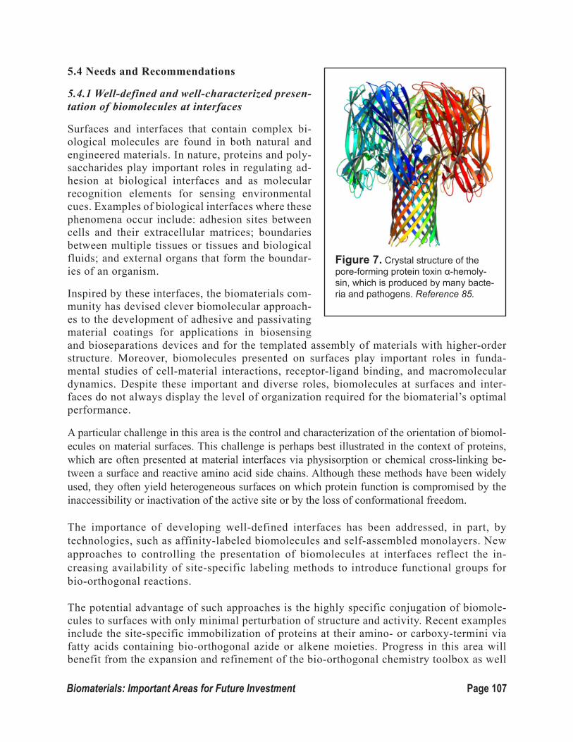
Page 107Biomaterials: Important Areas for Future Investment
5.4 Needs and Recommendations
5.4.1 Well-defined and well-characterized presen-tation of biomolecules at interfaces
Surfaces and interfaces that contain complex bi-ological molecules are found in both natural and engineered materials. In nature, proteins and poly-saccharides play important roles in regulating ad-hesion at biological interfaces and as molecular recognition elements for sensing environmental cues. Examples of biological interfaces where these phenomena occur include: adhesion sites between cells and their extracellular matrices; boundaries between multiple tissues or tissues and biological fluids; and external organs that form the boundar-ies of an organism.
Inspired by these interfaces, the biomaterials com-munity has devised clever biomolecular approach-es to the development of adhesive and passivating material coatings for applications in biosensing and bioseparations devices and for the templated assembly of materials with higher-order structure. Moreover, biomolecules presented on surfaces play important roles in funda-mental studies of cell-material interactions, receptor-ligand binding, and macromolecular dynamics. Despite these important and diverse roles, biomolecules at surfaces and inter-faces do not always display the level of organization required for the biomaterial’s optimal performance.
A particular challenge in this area is the control and characterization of the orientation of biomol-ecules on material surfaces. This challenge is perhaps best illustrated in the context of proteins, which are often presented at material interfaces via physisorption or chemical cross-linking be-tween a surface and reactive amino acid side chains. Although these methods have been widely used, they often yield heterogeneous surfaces on which protein function is compromised by the inaccessibility or inactivation of the active site or by the loss of conformational freedom. The importance of developing well-defined interfaces has been addressed, in part, by technologies, such as affinity-labeled biomolecules and self-assembled monolayers. New approaches to controlling the presentation of biomolecules at interfaces reflect the in-creasing availability of site-specific labeling methods to introduce functional groups for bio-orthogonal reactions.
The potential advantage of such approaches is the highly specific conjugation of biomole-cules to surfaces with only minimal perturbation of structure and activity. Recent examples include the site-specific immobilization of proteins at their amino- or carboxy-termini via fatty acids containing bio-orthogonal azide or alkene moieties. Progress in this area will benefit from the expansion and refinement of the bio-orthogonal chemistry toolbox as well
Figure 7. Crystal structure of the pore-forming protein toxin α-hemoly-sin, which is produced by many bacte-ria and pathogens. Reference 85.

Page 108 Biomaterials: Important Areas for Future Investment
as new methods for incorporating the appropriate functional groups into different classes of biomolecules. In the longer term, the need for such conjugation methods might be elimi-nated altogether by the development of new approaches for biomolecular synthesis directly on templated surfaces. This is envisioned most easily for molecules, such as oligopeptides, oligonucleotides and lipids, where current solid-phase synthesis methods on cleavable resin supports might be adapted to more relevant surfaces and interfaces. Longer polypeptides, polynucleotides, and, particularly, carbohydrates will pose more significant challenges and will likely require more advanced synthetic methods.
In parallel with methods for engineering well-defined interfaces, there also will be a need to develop tools to characterize such materials. Many thin films are well suited to charac-terization by optical microscopy since the entire thickness of the film can be captured in a single focal plane. Although most structural details are inaccessible by these methods, due to the diffraction limit, some information pertaining to biomolecular orientation and confor-mation, interactions among biomole-cules, and macromolecular dynamics may be inferred by fluorescence res-onance energy transfer or super-reso-lution microscopy. Atomic force mi-croscopy, electron microscopy, and surface plasmon resonance also pro-vide important tools for characteriz-ing biomolecules on surfaces.
Protein orientation on surfaces has been investigated by attenuated total reflection Fourier transform infra-red spectroscopy (Fig. 8), near-edge X-ray absorption fine structure spec-troscopy, sum frequency generation vibrational spectroscopy, and time-of-flight secondary ion mass spec-trometry. Improved access to instru-mentation and new analysis tools for the complex data sets they generate will be required for full exploitation of these characterization methods.
An important challenge encountered in the use of some of these charac-terization methods is the requirement for high vacuum environments and dehydrated biomaterials. Under these conditions, the true structure and ori-entation of biomolecules as well as their interactions with water may be lost. Understanding the role of water and more complex media is import-
Figure 8. High-resolution imaging of bacterial protein S-layers. (a) High-resolution topograph of the inner S-layer surface (outline: hole position with 90-nm diameter). (b) Fourier-filtered image of a. (c) Average (left) and symmetrized average (right) topo-graphs. (d) High-resolution topograph of the outer S-layer surface (outline: hole position with 200-nm diameter). (e) Fourier filtered image of d. (f) Average (left) and symmetrized average (right) topographs. Outlines in c and f delineate the structural pore in the center of the flower (F) and triangular (T) shaped surfaces of 15 Å in diameter. Dashed outlines in c and f delineate the structural pore in the center of the flower (F) and triangular (T) shaped surfaces of 15 Å in diameter. Scale bars, 90 nm (a), 20 nm (b), 10 nm (c), 50 nm (d), 20 nm (e) and 10 nm (f). Reference 86.

Page 109Biomaterials: Important Areas for Future Investment
ant not only for the characterization of biomolecules at interfaces but also for an accu-rate assessment of a biomaterial’s performance. Biomolecules have evolved to function in aqueous solutions with pH, ionic strength, osmolarity, and other properties determined by environmental conditions. Although these phenomena are, at least partially, understood in bulk solutions, interfaces add extra complexity due to surface tension, wetting, and mass transfer effects. In addition to improved characterization tools that operate under ambient conditions, new theoretical and modeling frameworks would provide greater insight into the interaction between biomolecules and complex solutions at interfaces.
Achieving well-defined, well-characterized presentations of biomolecules on surfaces and at interfaces would have a significant impact on the field. In the biological and chemical sci-ences, for example, it would permit more accurate measurements of receptor-ligand interac-tions, biomolecular activity, and the dynamics of macromolecules. In the field of biomedi-cal and bio-inspired devices, it would allow for reduced biomolecule loading of biosensors and biocatalytic materials as well as the improved sensitivity of these devices. Similarly, the improved characterization of interfacial phenomena in biomaterials will guide the rational design of biomolecules that are ideally suited to function in thin films and interfaces.
Many of these concepts are likely to be addressed on 2-D thin film surfaces. However, the outcomes will be applicable to a larger class of interfacial materials including dispersed colloidal systems and 3-D bulk materials with buried interfaces.
5.4.2 Patterned biological interfaces and interphases
There is abundant evidence that the rigid, planar, and chemically homogeneous surfaces classically used for interfacial characterization are fundamentally limited in the insight they can provide us about biological interfaces. Indeed, interfaces in biology are not simple and sharply defined but, instead, are instead complex “interphases,” in which nanoscale structural and chemical features are important. Furthermore, biological interphases are, in many cases, both rela-tively soft and dynamic in character.
Considerable progress has been made over the past decade on techniques for nanoscale patterning of to-pography and biomolecu-lar functionalization on surfaces through top-down methods based on dip-pen, electron-beam or X-ray nanolithography or bot-tom-up methods, such as
Figure 9. Confocal micrographs of F-actin stained smooth muscle cells on (A) nano-im-printed poly(methyl methacrylate (PMMA) at low cell density, (B) nano-imprinted PMMA at high cell density, (C) nanopatterned polydimethylsiloxane (PDMS) at low cell den-sity, (D) nano-patterned PDMS at high cell den-sity, (E) non-patterned PMMA, and (F) glass cover slip. Reference 87.

Page 110 Biomaterials: Important Areas for Future Investment
block copolymer self-assembly. Such nanopatterns (Fig. 9) have been shown to have wide-ranging effects on cell behavior. However, a major challenge is to extend these tech-niques for precise nanoscale surface patterning to materials that recapitu-late biologically relevant physical properties ─ namely, that are soft and that show the nonlinear elasticity characteristic of filamentous biopoly-mer gels. Furthermore, the integra-tion of these patterning strategies with methods to sense and apply forc-es to cultured cells, and their applica-tion to materials whose properties can be engineered to change over time in well-defined ways, would represent important tools for fundamental stud-ies of biointerfaces. Although top-down lithographic methods have advanced to a point where their use in pat-terning soft and dynamic materials at the nanoscale can be envisioned, recent advances in the assembly of biomolecules, including peptide amphiphiles, engineered proteins, and DNA, into both periodic and aperiodic structures have suggested that such complementary strategies also hold great potential.
Numerous studies in recent years have demonstrated that physical considerations can be as important as chemical interactions at biointerfaces. For example, changes in material stiffness alone are often sufficient to determine how stem cells differentiate. However, the field is in the early stages of understanding how chemical and physical characteristics interact. In addition to the coupling of these factors at the cellular level, it is important to consider their interplay in self-assembly of ─ and interactions between ─ biomolecules. For example, the tethering of a biomolecule to a surface or its incorporation into a supramolecular structure can introduce physical effects that modify biomolecular recognition events in ways that remain poorly un-derstood.
5.4.3 Design and characterization of multifunctional interfaces
In many cases, the use of a single bioactive ligand to functionalize a biomaterial is insufficient to support or promote desired biological phenomena, such as long-term cell adhesion, surviv-al, and maintenance of cellular phenotype. The ability to design multifunctional interfaces, therefore, is becoming increasingly important, as many biomaterials require the presence of multiple ligands and chemical cues.
Figure 10. Peptide flowers in a polymer matrix. Reference 88.

Page 111Biomaterials: Important Areas for Future Investment
The design of interfaces capable of presenting more complex signals, ultimately, will allow us to explore combinatorial approaches, which could, in turn, contribute to advances in the field of functional biomaterials with applications in biosensors, tissue engineering, and regenera-tive medicine. Especially in the area of tissue engineering, 3-D cell culture is critically im-portant and, in this case, the design of multifunctional interfaces is more challenging. Indeed, in addition to the complexity mentioned above, there is the extra element of interfaces that are buried within the 3-D structure and that may provide extra confinement to the cells. The effect of this confinement and the characterization of the buried interfaces are critically important for the success of biomaterials as 3-D scaffolds. Another key challenge is the development of tools and techniques that will allow us to accurately characterize the composition of the different interfaces within 3-D structures as well as their effects on cell fate.
5.4.4 Design and synthesis of stable proteins
Sensors containing proteins and protein-based complexes represent powerful alternatives to current analytical technologies in environmental monitoring and biomedical diagnosis, be-cause of the extraordinary specificity and selectivity of protein-ligand recognition. Substantial advances in protein engineering have enabled the design, synthesis, and purification of pro-teins intended for complex tasks in biotechnological applications. Despite these benefits, one persistent limitation is the lack of a methodology for preparing protein-containing interfaces that can be integrated into high-throughput and microfluidic devices and used in a broad range of biosensing applications.
A major challenge that needs to be addressed is the preparation of protein-based complexes that are robust, versatile, and tractable under a wide spectrum of detection conditions, includ-ing acidic and basic pH, low and high ionic strength, high osmotic stress, and wide variations in temperature. One possibility will be the redesign of proteins based upon native scaffolds and the implementation of additional features, such as insertion of non-canonical amino acids and arrays of stabilizing ion-pair and disulfide-bond interactions.
For example, highly stable native scaffolds are found in beta-barrel outer-membrane proteins and pore-forming toxins. The unusual robustness of such structures, encompassing arrays of anti-parallel beta strands, makes them convenient models for biosensing technologies using, for example, single-molecule stochastic detection. The fundamental advantageous traits of employing protein-based devices include accurate information on their structure and, in many instances, at atomic resolution, as well as the ability to engineer well-designed groups at var-ious strategic locations. Moreover, stabilized proteins also will be employed as drug-delivery carriers or integrated as controllable gates and nanovalves for drug-loaded vesicles.

Page 112 Biomaterials: Important Areas for Future Investment
Section 6. Biomaterials EducationStudents in materials science and engineering face daunting challenges. They must learn sub-stantial elements of chemistry, physics, mathematics and engineering in order to understand how materials are made, characterized and used, and perhaps more importantly, how they will be made, characterized and used in the future. Biomaterials science stretches students still further, by re-quiring them also to master important aspects of the biological sciences, including molecular bi-ology, cell biology, developmental biology and immunology. And because students come to bio-materials research with backgrounds in chemistry, chemical engineering, mechanical engineering, pharmacy, biology and computer science, they lack a shared set of core ideas and information. The task of educating young biomaterials scientists and engineers seems nearly impossible, but it must be done. How should we do it?
The Workshop addressed this question in a panel discussion led by Buddy Ratner of the University of Washington, with contributions from Sarah Heilshorn of Stanford University, Phillip Messer-smith of Northwestern University, Celeste Nelson of Princeton University, Ravi Shanker of Pfizer, and Johnna Temenoff of the Georgia Institute of Technology. Several key points emerged.
We must reach diverse audiences. Educational efforts in biomaterials should embrace the de-velopment of new courses in biomaterials science and engineering, the enhancement of courses in related fields (e.g., chemical engineering), and perhaps most challenging, the creation of exercises and course offerings for students – including first-year undergraduate students – who have not yet chosen their primary fields of study. We must meet the last challenge if we are to attract the most talented students to the fields of biomaterials science and engineering.
We must ensure scientific rigor. Faced with the challenge of introducing students to the field of biomaterials science and engineering, teachers struggle with the tension between depth and breadth. This is true in any field, of course, but it is an especially difficult struggle in light of the many disciplines that bear on biomaterials research and development. It is essential, therefore, that the instructors who develop courses in biomaterials make difficult choices, and that they avoid the temptation to try to address the full breadth of the field in survey courses that lack the rigor that we expect in other areas of science and engineering. Scientific rigor should be the first priority.
We should seek opportunities to engage industrial scientists. Many students are drawn to the study of biomaterials because they want to solve medical problems. The biomaterials, medical devices, and pharmaceutical industries can be rich sources of case studies that illustrate both the contributions that biomaterials can make to the quality of life and the important health care prob-lems that have not yet been solved but that might be amenable to biomaterials solutions. Our uni-versities should draw more heavily on these important resources, both for scientific reasons and for the insights that can be gained into the career opportunities available to biomaterials scientists and engineers. Partnering with industrial colleagues will also enable broader and deeper discussion of important issues such as professional ethics, interdisciplinary teamwork, communications skills and cost-benefit analyses.
We should share things that are working well. Contributors to the panel discussion highlighted some successful experiments in biomaterials education, including design-based exams, the devel-

Page 113Biomaterials: Important Areas for Future Investment
opment of quantitative homework problems based on the research literature, and team-based ex-ercises that draw together students from different academic backgrounds who can bring different perspectives to biomaterials problems. We should find ways to share things that are working well. We anticipate growing opportunities to shape the progression of biomaterials education through sharing of resources between departments and disciplines. The rapid development of substantive online educational content by many universities should facilitate the incorporation of new bio-materials concepts into classroom materials and allow students to participate in the evolution of curricular material.
We need to think hard about the biology. The field of biomaterials science and engineering has been developed primarily by researchers and teachers who come from the physical sciences and engineering. Input from colleagues trained in the biological sciences has been modest. Thus, it is no surprise that courses in biomaterials resemble other courses in materials science and an in-depth of discussion of biological ideas is limited. This situation must change, if we are to take full advantage of the power of modern biology to design the biomaterials of the future. The Workshop participants offered no prescription for how to proceed here, but they encouraged experimentation and communication directed toward the discovery and discussion of effective ways to put the “bio” into biomaterials education.

Page 114 Biomaterials: Important Areas for Future Investment
References:
1. Dove PM, DeYoreo JJ, and Weiner S (2003). Biomineralization. The Mineralogical Soci-ety of America: Washington, DC. 54:381.
2. (A) Courtesy of Kröger Lab, TU Dresden, Germany
(B) Addadi L., Raz S., and Weiner, S. (2003). Taking advantage of disorder: Amorphous calcium carbonate and its roles in biomineralization. Adv Mater. 15: 959-970.
(C) Courtesy of Beniash Lab, University of Pittsburgh.
3. (I) Aizenberg J., Muller DA, Grazul JL, and Hamann DR (2003). Direct fabrication of large micropatterned single crystals. Science 299 (5610): 1205-1208.
(II) Nudelman F, Pieterse K, George A, Bomans PHH, Friedrich H, Brylka LJ, Hilbers PAJ, de With, G, and Sommerdijk N (2010). The role of collagen in bone apatite forma-tion in the presence of hydroxyapatite nucleation inhibitors. Nature Mater. 9 (12): 1004-1009.
(III) Lee YJ, Yi H, Kim WJ, Kang K, Yun DS, Strano MS, Ceder G, and Belcher AM (2009). Fabricating genetically engineered high-power lithium-ion batteries using multi-ple virus genes. Science. 324(5930):1051-1055.
(IV) Wu CH, Park A, and Joester D (2011). Bioengineering single crystal growth. J Am Chem Soc. 133(6):1658-61.
4. (I) Li H, Xin HL, Muller DA, and Estroff LA (2009). Visualizing the 3D internal struc-ture of calcite single crystals grown in agarose hydrogels. Science. 326(5957):1244-1247.
(II) Gordon LM and Joester D (2011). Nanoscale chemical tomography of buried organ-ic-inorganic interfaces in the chiton tooth. Nature. 13;469(7329):194-197.
(III) Gong YU, Killian CE, Olson IC, Appathurai NP, Amasino AL, Martin MC, Holt LJ, Wilt FH, and Gilbert PU (2012). Phase transitions in biogenic amorphous calcium car-bonate. Proc Natl Acad Sci U S A. 2012 Apr 17;109(16):6088-93.
(IV) Li D, Nielsen MH, Lee JRI, Frandsen C, Banfield JF, and De Yoreo JJ (2012). Di-rection-specific interactions control crystal growth by oriented attachment. Science. 336 (6084):1014-1018.
5. Brutchey RL and Morse DE (2008). Silicatein and the translation of its molecular mech-anism of biosilicification into low temperature nanomaterial synthesis. Chem Rev. 108 (11): 4915-4934.
6. Munch E, Launey ME, Alsem DH, Saiz E, Tomsia AP, and Ritchie RO (2008). Tough, bio-inspired hybrid materials. Science. 322 (5907): 1516-1520.
7. Niederberger M and Colfen H (2006). Oriented attachment and mesocrystals: Non-classi-

Page 115Biomaterials: Important Areas for Future Investment
cal crystallization mechanisms based on nanoparticle assembly. Phys Chem Chem Phys. 8 (28): 3271-3287.
8. Mann K, Wilt FH, and Poustka AJ (2010). Proteomic analysis of sea urchin (Strongylo-centrotus purpuratus) spicule matrix. Proteome Science. 8:33.
9. Armbrust EV, Berges JA, Bowler C, Green BR, Martinez D, Putnam NH, Zhou SG, Allen AE, Apt KE, Bechner M, Brzezinski MA, Chaal BK, Chiovitti A, Davis AK, Demarest MS, Detter JC, Glavina T, Goodstein D, Hadi MZ, Hellsten U, Hildebrand M, Jenkins BD, Jurka J, Kapitonov VV, Kroger N, Lau WWY, Lane TW, Larimer FW, Lippmeier JC, Lucas S, Medina M, Montsant A, Obornik M, Parker MS, Palenik B, Pazour GJ, Richard-son PM, Rynearson TA, Saito MA, Schwartz DC, Thamatrakoln K, Valentin K, Vardi A, Wilkerson FP, and Rokhsar DS (2004). The genome of the diatom Thalassiosira pseud-onana: Ecology, evolution, and metabolism. Science. 306 (5693):79-86.
10. Kroger N and Poulsen N (2008). Diatoms - from cell wall biogenesis to nanotechnology. Anny Rev Genet. 42:83-107.
11. Miserez A, Wasko S, Carpenter CF, and Waite JH (2009). Non-entropic and reversible long-range deformation of an encapsulating bioelastomer. Nature Mater. 8 (11): 910-916.
12. (A) Courtesy of D. Joester Lab, Northwestern University.
(B) Li C and Qi LM (2008). Bioinspired fabrication of 3-D ordered macroporous single crystals of calcite from a transient amorphous phase. Angew Chem Int Ed. 47 (13): 2388-2393.
13. Lee YJ, Yi H, Kim WJ, Kang K, Yun DS, Strano MS, Ceder G, and Belcher AM (2009). Fabricating genetically engineered high-power lithium-ion batteries using multiple virus genes. Science 324 (5930): 1051-1055.
14. Kim YY, Ganesan K, Yang P, Kulak AN, Borukhin S, Pechook S, Ribeiro L, Kröger R, Eichhorn SJ, Armes SP, Pokroy B, and Meldrum FC (2011). Artificial biominerals - in-corporation of copolymer micelles in calcite single crystals. Nat Mater. 10: 890-896.
15. Tester CC, Brock RE, Wu CH, Krejci MR, Weigand S, and Joester D (2011). In vitro synthesis and stabilization of amorphous calcium carbonate (ACC) nanoparticles within liposomes. Cryst Eng Comm. 13 (12): 3975-3978.
16. Stephens CJ, Ladden SF, Meldrum FC, Christenson HK (2010). Amorphous Calcium Carbonate is Stabilized in Confinement. Adv Funct Mater 20 (13): 2108-2115.
17. Dunlop JWC, Weinkamer R, and Fratzl P (2011). Artful interfaces within biological ma-terials. Materials Today 14 (3): 70-78. This reference needs to be cited twice in the text - once before the figure and in the figure, which is now labeled Reference 18. Reorder the rest of the references.
18. Buckley MR, Bergou, AJ, Fouchard J, Bonassar LJ, and Cohen I (2010). High-resolution spatial mapping of shear properties in cartilage. J Biomech. 43: 796-800.

Page 116 Biomaterials: Important Areas for Future Investment
19. Dunlop JWC and Fratzl P (2013). Multilevel architectures in natural materials. Scripta Materialia 68 (1): 8-12.
20. Gupta HS, Seto J, Wagermaier W, Zaslansky P, Boesecke P, and Fratzl P (2006). Coop-erative deformation of mineral and collagen in bone at the nanoscale. Proc Natl Acad Sci USA. 103 (47): 17741-17746.
21. Weiner S and Wagner HD (1998). The material bone: Structure mechanical function rela-tions. Ann Rev Mater Sci. 28: 271-298.
22. Kim SW, Park KY, Ryu J, Ko JW, Cho W, Kim SM, Park CB, and Kang K (2012. Energy storage in in vivo synthesizable biominerals. RSC Advances 2 (13): 5499-5501.
23. Lefevre, CT, Menguy N, Abreu F, Lins U, Posfai M, Prozorov T, Pignol D, Frankel RB, and Bazylinski DA (2011). A cultured greigite-producing magnetotactic bacterium in a novel group of sulfate-feducing gacteria. Science 334 (6063):1720-1723.
24. Wu CH, Park A, Joester D (2011). Bioengineering single crystal growth. J Am Chem Soc. 133 (6):1658-1661.
25. Courtesy of D.E. Discher Lab, University of Pennsylvania.
26. Badylak SF, Weiss DJ, Caplan A, and Macchiarini P (2012). Engineered whole organs and complex tissues. Lancet. 379(9819):943-952.
27. Courtesey of D.E. Discher Lab, University of Pennsylvania.
28. Nawroth JC, Lee H, Feinberg AW, Ripplinger CM, McCain ML, Grosberg A, Dabiri JO, Parker KK. A tissue-engineered jellyfish with biomimetic propulsion. Nat Biotech-nol. 2012 Aug;30(8):792-7.
29. Abate AR, Thiele J, Weitz DA (2011). One-step formation of multiple emulsions in mi-crofluidics. Lab Chip. 11(2):253-258.
30. Available at http://en.wikipedia.org/wiki/File:Adult_Caenorhabditis_elegans.jpg
31. Courtesy S. Fraden Lab, Brandeis University.
32. Abzhanov A, Kuo WP, Hartmann C, Grant BR, Grant PR, and Tabin CJ (2006). The calmodulin pathway and evolution of elongated beak morphology in Darwin’s finches. Nature. 442(7102):563-567.
33. Paderi JE, Sistiabudi R, Ivanisevic A, and Panitch A (2009). Collagen-binding peptido-glycans: biomimetic approach to modulate collagen fibrillogenesis for tissueengineer-ing applications. Tissue Eng Part A. 15(10):2991-2999.
34. Courtesy T. Xu lab, Berkeley National Laboratory.
35. Courtesy D.E. Drisher Lab, University of Pennsylvania
36. Vasina EN, Paszek E, Nicolau DV Jr., and Nicolau DV (2009) The BAD project: data min-
Page 117Biomaterials: Important Areas for Future Investment
ing, database and prediction of protein adsorption on surfaces. Lab Chip. 9: 891-900.
37. Williams DF, editor (1987). Definitions in Biomaterials, Progress in Biomedical Engineer-ing, 4. Elsevier Publishers, Netherlands.
38. Gjorevski N and Nelson CM (2010). Branch formation during organ development. WIREs Sys Bio and Med. 2:734-741.
39. Poss KD, Wilson LG, and Keating MT (2002). Heart regeneration in zebrafish. Science 13:2188-2190.
40. Kim J and Mooney DJ (2011). In vivo modulation of dendritic cells by engineered materi-als: Towards new cancer vaccines. Nano Today. 6:466-477.
41. Coutesy B. Ratner lab, University of Washington.
42. Gobaa S, Hoehnel S, Roccio M, Negro A, Kobel S, and Lutolf MP (2012). Artificial niche microarrays for probing single stem cell fate in high throughput. Nat Methods. 8: 949-955.
43. Courtesy K. Anseth Lab, University of Colorado.
44. Kloxin AM, Kasko AM, Salinas CN, and Anseth KS (2009). Photodegradable hydrogels for dynamic tuning of physical and chemical properties. Science. 324:59-63.
45. Khetan S, Katz JS, and Burdick JA (2009). Sequential crosslinking to control cellular spreading in 3-dimensional hydrogels. Soft Matter. 5:1601-1606.
46. King WJ, Pytel NJ, Ng K, and Murphy WL (2010). Triggered drug release from dynamic microspheres via a protein conformational change. Macromol Biosci. 10, 580-584.
47. Courtesy M. Tibbitt, from K. Anseth Lab, University of Colorado
48. Courtesy K.M. Schultz, from K. Anseth Lab, University of Colorado.
49. Anderson DG, Levenberg S, and Langer R (2004). Nanoliter-scale synthesis of arrayed biomaterials and application to human embryonic stem cells. Nat Biotech 22 (2004) 863-866.
50. Courtesy P. Hammond Lab, Massachusetts Institute of Technology51. Hrkach J, Von Hoff D, Ali MM, Andrianova E, Auer J, Campbell T, De Witt D, Figa M,
Figueiredo M, Horhota A, Low S, McDonnell K, Peeke E, Retnarajan B, Sabnis A, Schnip-per E, Song JJ, Song YH, Summa J, Tompsett D, Troiano G, Hoven TV, Wright J, LoRusso P, Kantoff PW, Bander NH, Sweeney C, Farokhzad OC, Langer R, and Zale S (2012). Pre-clinical development and clinical translation of a PSMA-targeted docetaxel nanoparticle with a differentiated pharmacological profile. Sci Transl Med. 4(128):128ra39.
52. Wijaya A, Schaffer SB, Pallares IG, Hamad-Schifferli K (2009). Selective release of mul-tiple DNA oligonucleotides from gold nanorods. ACS Nano. 3:80-86.
53. Ham MH, Choi JH, Boghossian AA, Jeng ES, Graff RA, Heller DA, Chang AC, Mattis A,

Page 118 Biomaterials: Important Areas for Future Investment
Bayburt TH, Grinkova YV, Zeiger AS, Van Vliet KJ, Hobbie EK, Sligar SG, Wraight CA, and Strano MS (2010). Photoelectrochemical complexes for solar energy conversion that chemically and autonomously regenerate. Nat Chem. 2:929-936.
54. Lee YJ, Yi H, Kim WJ, Kang K, Yun DS, Strano MS, Ceder G, and Belcher AM (2009). Fabricating genetically engineered high-power lithium-ion batteries using multiple virus genes. Science. 324: 1051.
55. Dai XG, Yang W, Firlar E, Marras SAE, and Libera M (2012). Surface-patterned micro-gel-tethered molecular beacons. Soft Matter. 8: 3067-3076.
56. Waters CM and Bassler BL (2005). Quorum sensing: cell-to-cell communication in bacte-ria. Annu Rev Cell Dev Biol. 21:319-46.
57. Akin D, Sturgis J, Ragheb K, Sherman D, Burkholder K, Robinson JP, Bhunia AK, Mo-hammed S, and Bashir R (2007). Bacteria-mediated delivery of nanoparticles and cargo into cells. Nat Nanotechnol. 2: 441-449.
58. Stephan MT, Moon JJ, Um SH, Bershteyn A, Irvine DJ (2010). Therapeutic cell engineer-ing with surface-conjugated synthetic nanoparticles, Nat Med. 16:1035-1041; and Stephan MT, Stephan SB, Bak P, Chen J, and Irvine DJ (2012). Synapse-directed delivery of immu-nomodulators using T-cell-conjugated nanoparticles. Biomaterials. 33: 5776-5787.
59. Pooley CM and Balazs AC (2007). Producing swimmers by coupling reaction-diffusion equations to a chemically responsive material. Phys Rev E Stat Nonlin Soft Mat Phys. 76(1 Pt 2): 016308.
60. Kratz K, Narasimhan A, Tangirala R, Moon S, Revanur R, Kundu S, Kim HS, Crosby AJ, Russell TP, Emrick T, Kolmakov G, and Balazs AC (2012). Probing and repairing damaged surfaces with nanoparticle-containing microcapsules. Nat Nanotechnol. 7:87-90.
61. Liu AP and Fletcher DA (2005). Photopatterning of actin filament structures. Nano Lett. 5(4):625-628.
62. Stachowiak JC, Richmond DL, Li TH, Liu AP, Parekh SH, and Fletcher DA (2008). Unil-amellar vesicle formation and encapsulation by microfluidic jetting. Proc Natl Acad Sci U S A. 105: 4697-4702.
63. Farokhzad OC and Langer R (2009). Impact of nanotechnology on drug delivery. ACS Nano. 3(1):16-20.
64. Davis ME, Chen ZG, and Shin DM (2008). Nanoparticle therapeutics: an emerging treat-ment modality for cancer. Nat Rev Drug Discov. 7:771-782.
65. Choi W, Hong S, Abrahamson JT, Han JH, Song C, Nair N, Baik S, and Strano MS (2010). Chemically driven carbon-nanotube-guided thermopower waves. Nat Mater. 9:423-429.
66. Chen IC, Kuksenok O, Yashin VV, Moslin RM, Balazs AC, and Van Vliet KJ (2011). Shape- and size-dependent patterns in self-oscillating polymer gels. Soft Matter 7: 3141-3146.

Page 119Biomaterials: Important Areas for Future Investment
67. Delgado J, Li N, Leda M, Gonzalez-Ochoa HO, Fraden S, and Epstein IR (2011). Coupled oscillations in a 1D emulsion of Belousov-Zhabotinsky droplets. Soft Matter. 7:3155-3167.
68. Zhang Y, Li N, Delgado J, Zhou N, Yoshida R, Fraden S, Epstein IR, and Xu B (2012). Structural modulation of self-oscillating gels: changing the proximity of the catalyst to the polymer backbone to tailor chemomechanical oscillation. Soft Matter. 8: 3056-3061.
69. Available at: http://dbscience3.wikispaces.com/Drew)
70. Verma A, Uzun O, Hu Y, Hu Y, Han HS, Watson N, Chen S, Irvine DJ, and Stellacci F (2008). Surface-structure-regulated cell-membrane penetration by monolayer-protected nanoparticles. Nat Mater. 7: 588-595.
71. Poon Z, Chen S, Engler AC, Lee HI, Atas E, von Maltzahn G, Bhatia SN, and Hammond PT (2010). Ligand-clustered “patchy” nanoparticles for modulated cellular uptake and in vivo tumor targeting. Angew Chem Int Ed Engl. 49: 7266-7270.
72. Gunawan RC, Almeda D, and Auguste DT (2011). Complementary targeting of liposomes to IL-1 alpha and TNF-alpha activated endothelial cells via the transient expression of VCAM1 and E-selectin. Biomaterials. 32: 9848-9853.
73. King NP, Sheffler W, Sawaya MR, Vollmar BS, Sumida JP, Andre I, Gonen T, Yeates TO, and Baker D (2012). Computational design of self-assembling protein nanomaterials with atomic level accuracy. Science. 336(6085): 1171-1174.
74. Laura K. Greenfield, Chris Whitfield (2012). Synthesis of liposaccharide O-antigens by ABC transporter-dependent pathways. Carbohydr Res.356:12-24.
75. Connor RE and Tirrell DA (2007). Non-canonical amino acids in protein polymer design. Polymer Reviews. 47(1): 9-28.
76. Liu K, Nie ZH, Zhao NN, Li W, Rubinstein M, and Kumacheva E (2010). Step-growth polymerization of inorganic nanoparticles. Science. 329(5988):197-200.
77. Canelas DA, Herlihy KP, and DeSimone JM (2009). Top-down particle fabrication: con-trol of size and shape for diagnostic imaging and drug delivery. Wiley Interdiscip Rev Nanomed Nanobiotechnology. 1: 391-404.
78. Bong KW, Pregibon DC, and Doyle P9S (2009). Lock release lithography for 3-D and composite microparticles. Lab Chip. 9(7): 863-866.
79. Courtesy of Centers of Disease Control and Prevention
80. Lee YS and Mrksich M (2002). Protein chips: from concept to practice. Biotechnol. 20: s14-8.
81. Toohey KS, Sottos NR, Lewis JA, Moore JS, and White SR (2007). Self-healing materials with microvascular networks. Nat Mater. 6(8):581-5.
82. Lin IH, Miller DS, Bertics PJ, Murphy CJ, de Pablo JJ, and Abbott NL (2011). Endotox-

Page 120 Biomaterials: Important Areas for Future Investment
in-induced structural transformations in liquid crystalline droplets. Science. 332: 1297-1300.
83. Courtesy D. Tirrell Lab, California Institute of Technology
84. Available at http://www.ks.uiuc.edu/Research/smd_imd/titin/
85. Tanaka Y, Hirano N, Kaneko J, Kamio Y, Yao M, Tanaka I (2011). 2-Methyl-2,4-pen-tanediol induces spontaneous assembly of staphylococcal α-hemolysin into heptameric pore structure. Protein Science. 20: 448-456.
86. Gonçalves RP, Agnus G, Sens P, Houssin C, Bartenlian B, Scheuring S (2006). Two-chamber AFM: probing membrane proteins separating two aqueous compartments. NatMethods. 3: 1007-1012.
87. Courtesy D. Tirrell Lab, California Institute of Technology
88. Mardilovich A and Kokkoli E (2005). Patterned biomimetic membranes: effect of concen-tration and pH. Langmuir. 21 (16):7468-7475.

Page 121Biomaterials: Important Areas for Future Investment
Permissions
Section 1:
Figure 1A: (See Reference 2)
Figure 1B: Reprinted by permission of Composites Worldwide Inc. (Reference 2)
Figure 1C: (See Reference 2)
Figure 2.I: Reprinted by permission of the American Association the Advancement of Science (Reference 3)
Figure 2.II: Reprinted by permission of the Nature Publishing Group (Reference 3)
Figure 2.III: Reprinted by permission of the American Association the Advancement of Science (Reference 3)
Figure 2.IV: Reprinted by permission of the American Chemical Society (Reference 3)
Figure 3.I: Reprinted by permission of the American Association the Advancement of Science (Reference 4)
Figure 3.II: Reprinted by permission of the Nature Publishing Group (Reference 4)
Figure 3.III: Reprinted by permission of the National Academy of Science, USA (Reference 4)
Figure 3.IV: Reprinted by permission of the American Association the Advancement of Science (Reference 4)
Figure 4: Reprinted by permission of Annual Reviews, Inc. (Reference 10)
Figure 5: Reprinted by permission of the Nature Publishing Group (Reference 11)
Figure 6A: (see Reference 12)
Figure 6B: Reprinted by permission of John Wiley & Sons (Reference 12)
Figure 7: Reprinted by permission of Elselvier BV (Reference 17)
Figure 8: Reprinted by permission of the National Academy of Science, USA (Reference 20)
Figure 9: Reprinted by permission of RSC Publishing (Reference 22)
Section 2
Figure 1: (See Reference 25)
Figure 2: (See Reference 27

Page 122 Biomaterials: Important Areas for Future Investment
Figure 3: Reprinted by permission of the Nature Publishing Group (Reference 28)
Figure 4: Reprinted by permission of the Royal Society of Chemistry (Reference 29)
Figure 5: (See Reference 30)
Figure 6: (See Reference 31)
Figure 7: Reprinted by permission of the Nature Publishing Group (Reference 32)
Figure 8: Reprinted by permission of Mary Ann Liebert, Inc. (Reference 33)
Figure 9: (See Reference 34)
Figure 10: (See Reference 35)
Section 3:
Figure 1: Reprinted by permission of John Wiley & Sons (Reference 38)
Figure 2: Reprinted by permission of the American Association the Advancement of Science (Reference 39)
Figure 3: Reprinted by permission of Elsevier, Inc. (Reference 40)
Figure 4: (See Reference 41)
Figure 5: (See Reference 43)
Figure 6A: Reprinted by permission of the American Association the Advancement of Science (Reference 44)
Figure 6B: Reprinted by permission of the Royal Society of Chemistry (Reference 45)
Figure 6C: Reprinted by permission of John Wiley & Sons (Reference 46)
Figure 7: (See Reference 47)
Figure 8: (See Reference 48)
Figure 9: Reprinted by permission of the Nature Publishing Group (Reference 49)
Section 4:
Figure 1: (See Reference 50)
Figure 2: Reprinted by permission of the American Chemical Society (Reference 52)
Figure 3: Reprinted by permission of Elsevier, Inc. (Reference 58)

Page 123Biomaterials: Important Areas for Future Investment
Figure 4: (See Reference 69)
Figure 5: Reprinted by permission of the Nature Publishing Group (Reference 70)
Figure 6: Reprinted by permission of Elsevier, Inc. (Reference 74)
Figure 7: Reprinted by permission of John Wiley & Sons (Reference 77)
Section 5:
Figure 1: (See Reference 79)
Figure 2: Reprinted by permission of John Wiley & Sons (Reference 80)
Figure 3: Reprinted by permission of the Nature Publishing Group (Reference 81)
Figure 4: Reprinted by permission of the American Association the Advancement of Science (Reference 82)
Figure 5: (See Reference 83)
Figure 6: (See Reference 84)
Figure 7: Reprinted by permission of John Wiley & Sons (Reference 85)
Figure 8: Reprinted by permission of the Nature Publishing Group (Reference 86)
Figure 9: (See Reference 87)
Figure 10: Reprinted by permission of the American Chemical Society (Reference 88)
