biomaterials ws2008/09 testing biomaterialsgenome.tugraz.at/biomaterials/biomat-11.pdf ·...
TRANSCRIPT

Biomaterials WS2008/09
1Institute for Genomics and Bioinformatics, TU Graz / Austria Courtesy R. Zenz
Testing Biomaterials
How to characterize the material that will be processed into a medical device/implant?
How biomaterials can be evaluated to determine if they are biocompatible?
How biomaterials can be evaluated to determine whether they function appropriately in the in vivo environment?
How can testing criteria be defined to proper evaluate a given biomaterials application?
– Some biomaterials complete their intended function in seconds– Others are implanted for lifetime (10-70 years?)

Biomaterials WS2008/09
2Institute for Genomics and Bioinformatics, TU Graz / Austria Courtesy R. Zenz
Testing Biomaterials
Standards: Consensus standards are documents developed by commitees to represent consensus opinions on test methods, devices, or procedures. Following these standards when testing new materials and/or devices is an advantage, but not mandatory, for getting marketing approval.Committees exist at national and international levels. (remember that several metals are even named by their ASTM standards). News and updates regarding european standards can be found at the eupean society for biomaterials webpage: http://www.esbiomaterials.eu/main/index.php. The ESB is a member of the International Union of Societies for Biomaterials Sciences and Engineering (IUS-BSE)
Technical Committee 194 of the International Organization for Standardization (ISO) meet every springSet of documents 10993 (FDA’s version #G95-1):
– 10993-1: "Guidance on Selection of Tests." – 10993-2: "Animal Welfare Requirements." – 10993-3: "Tests for Genotoxicity, Carcinogenicity, and Reproductive Toxicity." – 10993-4: "Selection of Tests for Interactions with Blood." – 10993-5: "Tests for Cytotoxicity—In Vitro Methods." – 10993-6: "Tests for Local Effects after Implantation." – 10993-7: "Ethylene Oxide Sterilization Residuals." – 10993-9: "Degradation of Materials Related to Biological Testing." – 10993-10: "Tests for Irritation and Sensitization." – 10993-11: "Tests for Systemic Toxicity." – 10993-14: “Materials Evaluation."
Biomaterials WS2008/09
3Institute for Genomics and Bioinformatics, TU Graz / Austria Courtesy R. Zenz
Testing Biomaterials
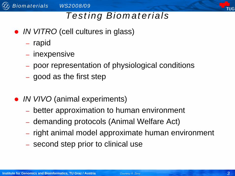
IN VITRO (cell cultures in glass)– rapid– inexpensive– poor representation of physiological conditions– good as the first step
IN VIVO (animal experiments)– better approximation to human environment– demanding protocols (Animal Welfare Act)– right animal model approximate human environment– second step prior to clinical use

Biomaterials WS2008/09
4Institute for Genomics and Bioinformatics, TU Graz / Austria Courtesy R. Zenz
Testing BiomaterialsTesting always leads to experimental variability, particulary tests in living systems.
– The more complex the system (e.g. Humans vs. cultured cells) the larger the variability that might be expected.
Statistics should be used at two steps in testing biomaterials.– Before an experiment is performed, statistical experimental design will
indicate the minimum number of samples that must be evaluated to yield meaningful results.
– After the experiment statistics will help to extract maximum useful information.
Detailed protocols are provided by:– ASTM (American Society for Testing and Materials) and the – ISO (International Standards Organization) – FDA (Food and Drug Administration) – NIH (National Institute of Health)– The EU has its own directives in addition to the ISO standards.
Sometimes individual EU-member states has additional, national demands.

Biomaterials WS2008/09
5Institute for Genomics and Bioinformatics, TU Graz / Austria Courtesy R. Zenz
Testing Biomaterials
Cytotoxicity means to cause toxic effects at the cellular level:– death, – alterations in cellular membrane permeability, – enzymatic inhibtion,... at the cellular level.
Evaluation by methods that use isolated, adherent cells in culture to measure cytotoxicity and biological compatibility.
– Cells used for culture are most often established cell lines from cell banks (e.g. American Type Tissue Culture Collection)Cultured cell lines can be reproducibly used in many different laboratories, providing comparable results usefull for generating databases
– Primary cells (with the exception of erythrocytes for hemolysis assays) are seldom used.Primary cells come directly from living tissue (can only be propagated a few generations in culture),have different genetic backgrounds, giving very high statistical varation in test outcome

Biomaterials WS2008/09
6Institute for Genomics and Bioinformatics, TU Graz / Austria Courtesy R. Zenz
Testing Biomaterials
Toxicity:– A toxic material is defined as a material that releases a chemical
in sufficient quantities to kill cells either directly of indirectly through inhibition of key metabolic pathways.
– The number of cells that are affected is an indication of the dose and potency of the chemical.
– If an animal is exposed to an atmosphere containing a noxious substance (exposure dose), only a small portion of the inhaled substance will be absorbed and delivered to the internal organs and cells (delivered dose).
– Cell culture methods evaluate target cell toxicity by using delivered doses of the test substance used. –Whereas tests in whole animals relate to the exposure dose... Often resulting in different measurements of sensitivity in the two systems. To compensate for this difference in vivo local toxicity models are applied (direct delivery to specific organs)

Biomaterials WS2008/09
7Institute for Genomics and Bioinformatics, TU Graz / Austria Courtesy R. Zenz
Testing Biomaterials
A highly sensitive test system is desirable for evaluating the potential hazards of biomaterials.
– In cell culture the variables of metabolism, distribution, inflammation, and absorption are minimized and the dosage per cell is maximized to produce a highly sensitive test system. Testing at this high margin is considered a safety factor for interpolating results to whole humans
– Typical sources of toxic materials: extractables• additives for manufacturability: plasticizers, antioxidants,
monomers• Leackage from the basic material itself: cobalt, nickel from
metal alloys; fluorinated polyesters from Dacron fibers; etc.

Biomaterials WS2008/09
8Institute for Genomics and Bioinformatics, TU Graz / Austria Courtesy R. Zenz
Testing Biomaterials
Migration of chemicals from a solid phase material into liquid solvent is controlled by:
– Diffusional resitstance within the solid– Chemical concentration– Time– Temperature– Fluid turbulence at the solid-solvent interface– ....
Preparation of extractions of biomaterials have been carefully standardized to improve the reproducibility of the data.
Complete dissolution of biomaterial is an alternative approach for in vitro testing. But:
– May create degradation products that do not occur in the clinical application.

Biomaterials WS2008/09
9Institute for Genomics and Bioinformatics, TU Graz / Austria Courtesy R. Zenz
Testing Biomaterials
Three morphological* cell culture assays are primarily used for evaluating biocompatibility:
– Direct contact– Agar diffusion– Elution
For the results to be comparable the following parameters must be standardized:
– Number of cells– Growth phase of cells– Cell type – Duration of exposure– Test sample size (geometry, density, shape, thickness)– Surface area of test sample must be carefully controlled
Readout: Within the given quantification range:– Dose-response curves– exposure-effect relationships (Klaassen, 1986)
*the outcome of Morphological assays is measured by observation of changes in cell morphology (structure, appearance)

Biomaterials WS2008/09
10Institute for Genomics and Bioinformatics, TU Graz / Austria Courtesy R. Zenz
Testing Biomaterials
Direct contact– monolayer, confluent cell culture, L-929 mouse fibroblasts– biomaterial in direct contact– 24 hours, 37±1°C– cells may
• change morphology• die• lose adherence to dish
– Cells are fixed and stained (hematoxylin blue: stains live adherent cells)
– toxicity=dead/live !Why L-929?
– easy to maintain– good correlation with animals tests (Northup 1986)– Resemble fibroblasts present in wound healing (=often the first
cells to attach to implanted biomaterials in vivo)– In specific cases other, similar cell types may be used
Biomaterial

Biomaterials WS2008/09
11Institute for Genomics and Bioinformatics, TU Graz / Austria Courtesy R. Zenz
Testing Biomaterials
A confluent monolayer (100 x magnification) of well-defined L929 mouse fibroblast cells exhibiting cell-to-cell contact. This appearance is indicative of a non-cytotoxic (negative) response
L929 mouse fibroblast cells (100 x magnification) that illustrate a positive cytotoxic reaction; the considerable open areas between cells indicate that extensive cell lysis (disintegration) has occurred.
Biomaterials WS2008/09
12Institute for Genomics and Bioinformatics, TU Graz / Austria Courtesy R. Zenz
Testing Biomaterials
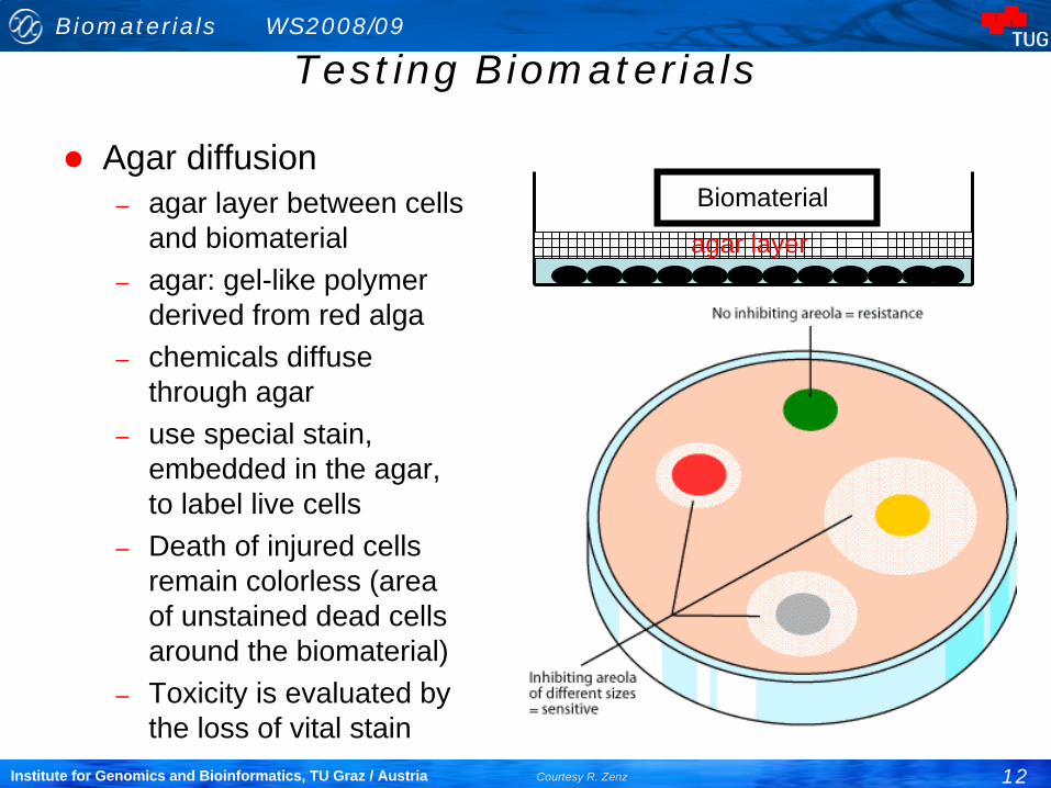
Agar diffusion– agar layer between cells
and biomaterial– agar: gel-like polymer
derived from red alga– chemicals diffuse
through agar– use special stain,
embedded in the agar, to label live cells
– Death of injured cells remain colorless (area of unstained dead cells around the biomaterial)
– Toxicity is evaluated by the loss of vital stain
Biomaterial
agar layer

Biomaterials WS2008/09
13Institute for Genomics and Bioinformatics, TU Graz / Austria Courtesy R. Zenz
Testing Biomaterials
An agar diffusion flask containing a sample of positive control material. The discoloration that extends outward from the material indicates that the presence of the sample has caused the cells to lyse, losing the vital stain incorporated in the agar layer.

Biomaterials WS2008/09
14Institute for Genomics and Bioinformatics, TU Graz / Austria Courtesy R. Zenz
Testing Biomaterials
Elution
– prepare extract of a material– how? Standardization needed!
(0.9% sodium chloride or serum free medium)
– chemicals will leak into solution– apply solution to cell-culture
(48h at 37°C)– perform stain based viability
tests, microscopic evaluation.– experience in recognizing cell
culture morphology is required.
Biom
aterial

Biomaterials WS2008/09
15Institute for Genomics and Bioinformatics, TU Graz / Austria Courtesy R. Zenz
Testing Biomaterials
The methodologies for the three primary cell culture assays are described in the:
– U.S: Pharmacopeia (Pharmakopöe, amtl. Arzneibuch)
And standards published by the:– ASTM (American Society for Testing and Materials)– BSI (British Standards Institute)– ISO (International Standards Organization)
Pharmacopeial* assays are legally required by the ministries of health in the US, Europe, Australia, Japan, and other countries. The ISO standards are expected to gradually replace national standards in Europe
* Pharmacopeia = a compendium containing directions for the identification of samples and the preparation of compound medicines, published by the authority of a government or a medical or pharmaceutical society. In this particular case referring to the methods listed above.

Biomaterials WS2008/09
16Institute for Genomics and Bioinformatics, TU Graz / Austria Courtesy R. Zenz
Testing Biomaterials

Biomaterials WS2008/09
17Institute for Genomics and Bioinformatics, TU Graz / Austria Courtesy R. Zenz
Testing Biomaterials
After the cytotoxicity profile more application-specific tests are performed to assess the biocompatibility of the material:
– Products for in vitro fertilization procedures would be tested for adverse effects on a very low cell population.
– A new material for culturing cells would be assayed by comparing growth rates of cells in contact with the new material with those of currently marketed materials.
– Current experience: a material non-toxic in vitro will be non- toxic in in vivo assays.
– But: the clinical acceptability of a material depends on many different factors; target cell toxicity is but one.

Biomaterials WS2008/09
18Institute for Genomics and Bioinformatics, TU Graz / Austria Courtesy R. Zenz
Testing Biomaterials
In vivo testing: critical for development of clinical devices– In vitro tests cannot replace in vivo tests:
• no inflammation• no immune response• single cell type• no tissue remodeling• No acquired toxicity through processing (eg the liver modifies many foreign
compounds)– In vivo tests provide:
• interactions of different cell types• effects of hormonal factors• interactions with extracellular matrix• interactions with blood-borne cells, proteins and molecules• Overall determination of: wether the device performs as intended and
provides no significant harm to the patient or user.
– The ISO 10993 standard, “Biological evaluation of Medical Devices” presents a systematic approach to the in vivo assessment of tissue compatibility of medical devices. The Standard is extended and upgraded continuosly

Biomaterials WS2008/09
19Institute for Genomics and Bioinformatics, TU Graz / Austria Courtesy R. Zenz
Testing Biomaterials
Implant effects can be simulated in vivo:
– insoluble particulate materials released by implants– interaction of biological factors with the implant– mechanical loading experienced by device– Time is an important variable (implant–related factors act with
different time constants on the biological factors)
– The tissue response to an implant is the cumulative physiological effect of:
• Modulation of the acute wound healing response due to the surgical trauma of implantation and the presence of the implant.
• The subsequent chronic inflammation reaction, and• Remodeling of surrounding tissue as it adopts to the implant.

Biomaterials WS2008/09
20Institute for Genomics and Bioinformatics, TU Graz / Austria Courtesy R. Zenz
Testing Biomaterials
Mechanical loading experienced by biomaterial:
– increased local strain due to movement of device with respect to tissue:
• hyperplasia (increased scar tissue, thicker fibrous encapsulation)
– reduction in tissue strain due to presence of implant
• implant takes all load: tissue undergoes atrophy (stress shielding)

Biomaterials WS2008/09
21Institute for Genomics and Bioinformatics, TU Graz / Austria Courtesy R. Zenz
Testing Biomaterials
Implant sites in animal models:– Similarity to the site to be employed in human use of the medical
device.– The healing and remodeling characteristics of the 4 basic types
of tissue should be considered:• Connective tissue• Muscle• Epithelia• Nerve
– In selecting an implant site consider the following:• Vascularity• Nature of the parenchymal cells (capability for mitosis and
migration: determine the regenerative capability of the tissue)• Presence of regulatory cells (macrophages and histiocytes)• Effects of mechanical strain (hyperplasia, atrophy)

Biomaterials WS2008/09
22Institute for Genomics and Bioinformatics, TU Graz / Austria Courtesy R. Zenz
Testing Biomaterials
Surgical wounds in avascular tissue (e.g. cornea, inner third of meniscus) may not heal:
– limited potential of the proliferation and– migration of surrounding parenchymal cells into the wound site.– Gaps between an implant and surrounding avascular tissue can
remain indefinitely.
Implant sites in vascular tissue in which the parenchymal cell does not have the capability for mitosis (e.g. nerve tissue) heal by the formation of scar.
Macrophages, along with fibroblasts of the scar often form a definable layer of cells that surround an implant: „fibrous encapsulation“.

Biomaterials WS2008/09
23Institute for Genomics and Bioinformatics, TU Graz / Austria Courtesy R. Zenz
Testing Biomaterials
For orthopedic prostheses bone has been used as the site of implantation:
– But the densitiy of bone formation depends on the site of implanation:
• Cortical and cancellous bone differ in vascularization and the size of the pool of preosteoblasts that proliferate in response to surgery.
Cutaneous or subcutaneous sites chosen to assess biocompatibility
– readily accessible– thickness of fibrous capsule measure of biocompatibility– guinea pig

Biomaterials WS2008/09
24Institute for Genomics and Bioinformatics, TU Graz / Austria Courtesy R. Zenz
Testing Biomaterials
Paravertebral muscle of rats, rabbits, and dogs to detect toxic leach:
– Due to the relative motion between the implant and surrounding muscle and the
– limited capability of the skeletal muscle for regeneration, – scar tissue forms around the implant.– Thickness of fibrous encapsulation measure of biocompatibility.
Epithelia:– E.g.: Substances that might be used as temporary covering
materials to facilitate re-epitheliazation of skin wounds.– Epidermal wounds have been produced experimentally by:
• Heat• Chemical agents• Excision of tissue

Biomaterials WS2008/09
25Institute for Genomics and Bioinformatics, TU Graz / Austria Courtesy R. Zenz
Testing Biomaterials
Materials for vascular prostheses have been evaluated for their blood compatibility as replacements segments in selected vesssels in various animal models:
– Carotis-jugular and– Femoral arteriovenous shunts
Nerve:– Nerve cells do not have the capability for division– The elongation of several axons allow a degree of regeneration
across defect sites.– Certain matrices facilitate the elongation of such axons thereby
accelerating the regeneration of the nerve and restoration of some function:
• Peripheral nerves of rats

Biomaterials WS2008/09
26Institute for Genomics and Bioinformatics, TU Graz / Austria Courtesy R. Zenz
Testing Biomaterials
Controls for in vivo investigations of tissue compatibility can include:
– Contralateral intact tissues as anatomic controls:• No implant is inserted; the amount of scar formed can help to
evaluate the fibrous capsule formation around the implant at the test site.
– Sham-operated controls:• E.g.: A shame-operated limb can display the effects of
altered load bearing on the recipient tissue.
– Material and device controls• E.g.: femoral stems of total hip replacement prostheses
should be of identical shape and size.

Biomaterials WS2008/09
27Institute for Genomics and Bioinformatics, TU Graz / Austria Courtesy R. Zenz
Testing Biomaterials
Evaluation of tissue reaction:– Histology and histochemistry:
• Qualitative determination of the relative numbers of various cell types.
– Immunoshistochemistry:• Allows specific cell types and extracellular matrix
components around an implant to be identified.– Transmission electron microscopy (TEM):
• Ultrastructural examination of cells at the interface of implants.
– Scanning electron microscopy (SEM)– Biochemistry:
• Level of inflammatory mediators• But: Manipulation of tissues at or after explantation of a
biomaterial can dramatically alter the production and release of cellular mediators.

Biomaterials WS2008/09
28Institute for Genomics and Bioinformatics, TU Graz / Austria Courtesy R. Zenz
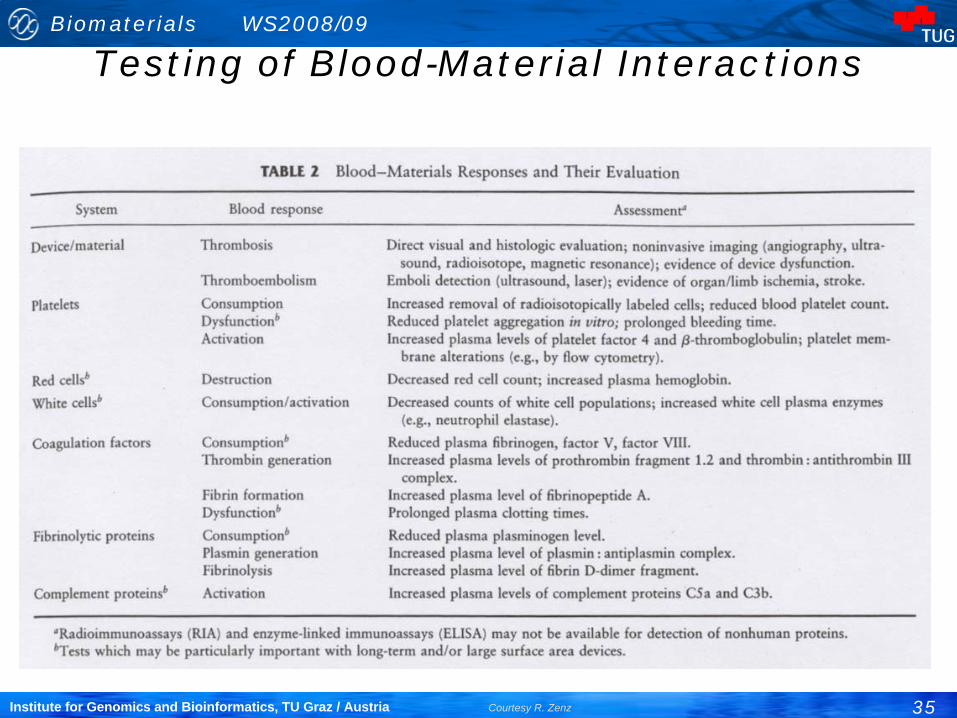
Testing of Blood-Material Interactions
Many devices and materials used have blood contact:– Heart-lung machine– Hollow fiber hemodialyzer for treatment of kidney failure– Catheters for blood access and– Blood vessel manipulation (angioplasty)– Heart assist devices– Stents– Prosthetic heart valves– Vascular grafts
A device made of blood-compatible materials is not automatically blood compatible!No widley recognized, standard list of blood compatibility tests exists.

Biomaterials WS2008/09
29Institute for Genomics and Bioinformatics, TU Graz / Austria Courtesy R. Zenz
Testing of Blood-Material Interactions
Many existing devices are frequently modified to improve durability and mechanical characteristics.
– Changes may also affect blood response (is not entirely predictable): testing is required to document safety
The performance of many existing devices is also less than optimal:
– Prolonged heart lung machine can produce a tendency to severe bleeding.
– Mechanical heart valves occasionally shed emboli to the brain, producing stroke.
– Many devices are only „safe“ when anticoagulating drugs are used (e.g. Oxygenator, heart valves, hemodialyzer).

Biomaterials WS2008/09
30Institute for Genomics and Bioinformatics, TU Graz / Austria Courtesy R. Zenz
Thrombogenicity
Local effects:
– A thrombogenic device may cause the accumulation of various blood elements (thrombus formation).
– Cardiovascular devices may also exhibit regions of disturbed flow or stasis which lead to formation of blood clots.
– These local effects can compromice device function:
• Delivery of blood through artificial blood vessels• Mechanical motion of heart valves• Gas exchange through oxygenators• Removal of metabolic waste (hemodialyzer)

Biomaterials WS2008/09
31Institute for Genomics and Bioinformatics, TU Graz / Austria Courtesy R. Zenz
Thrombogenicity
The local blood reaction may produce systemic effects:
– Thrombi may detach (embolize) and impair blood flow in peripheral vessels.
– Chronic devices may „consume“ circulating blood elements:• Mechanical destruction of red blood cells by heart prostheses
or dialyzers.• Removal of platelets as a result of continuing thrombus
formation.• Mediators of inflammatory responses and vessel tone may
be produced or released from cells (platelets, white cells,..).

Biomaterials WS2008/09
32Institute for Genomics and Bioinformatics, TU Graz / Austria Courtesy R. Zenz
Thrombogenicity
The types of devices used are:– numerous, exhibit complex flow geometries, and are
continuously evolving.
The possible blood responses are:– numerous, complex, dynamic, and not fully understood.
It is difficult and expensive to measure device thrombogenicity in an extensive and systematic way (experiment. animals or humans).Alternative interpretations can be applied to data from „blood compatibility“ tests.

Biomaterials WS2008/09
33Institute for Genomics and Bioinformatics, TU Graz / Austria Courtesy R. Zenz
Testing of Blood-Material Interactions
We cannot generally:– Extrapolate results obtained under one set of test conditions to
another set of conditions.– Use short-term testing to predict long-term results.– Predict in vivo performance of a device based on blood-material
interactions of materials per se in idealized flow geometries.
3 factors contribute to thecoagulation of the blood:
– The blood chemistry– The blood-contacting surface– The flow regime Virchow´s triade (1856)

Biomaterials WS2008/09
34Institute for Genomics and Bioinformatics, TU Graz / Austria Courtesy R. Zenz
Testing of Blood-Material Interactions
The source and methods for handling blood can have important effects on blood material interactions.Initial adhesiviness of blood platelets for artificial surfaces appears to be
– low in man and some primates and– high in the dog, rat and rabbit.
Animal blood donors are relatively homogenous:– Age, health status, blood response
In vitro testing generally requires anticoagulation of the blood (can have profound effects).In vivo testing and the use of extracorporeal circuits are also commonly performed with anticoagulats:
– Sodium citrate (chelates Ca2+)– Heparin (used to block thrombin)
Biomaterials WS2008/09
35Institute for Genomics and Bioinformatics, TU Graz / Austria Courtesy R. Zenz
Testing of Blood-Material Interactions
Biomaterials WS2008/09
36Institute for Genomics and Bioinformatics, TU Graz / Austria Courtesy R. Zenz
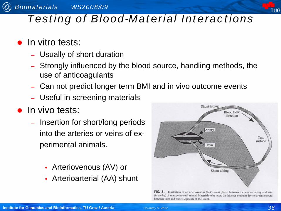
Testing of Blood-Material Interactions
In vitro tests:– Usually of short duration– Strongly influenced by the blood source, handling methods, the
use of anticoagulants– Can not predict longer term BMI and in vivo outcome events– Useful in screening materials
In vivo tests:– Insertion for short/long periods
into the arteries or veins of ex-perimental animals.
• Arteriovenous (AV) or• Arterioarterial (AA) shunt

Biomaterials WS2008/09
37Institute for Genomics and Bioinformatics, TU Graz / Austria Courtesy R. Zenz
Testing Biomaterials
Sensitization:– Prolonged contact with a chemical substance that interacts with
immune system– Skin widely used since most reactions to biomaterials are cell-
mediated type– Dermal sensitization
marked by redness and swelling
Sensitization (rash) to latex gloves

Biomaterials WS2008/09
38Institute for Genomics and Bioinformatics, TU Graz / Austria Courtesy R. Zenz
Testing Biomaterials
Sensitization test methods (guinea pigs):repeated patch (Buehler):
– Induction phase: expose shaved back directly to material under occlusive dressings. 6 hours/day, 3 days/week, 3 weeks
– Recovery phase: 2 weeks rest to allow for development of response
– Final exposure
maximization (Magnuson-Kligman): used for materials that will contact areas other than the skin:
– fluid extracts of test material preparedin saline or vegetable oil
– inject extract with an adjuvant agent that will enhance immune response
– two weeks rest– apply extract topically
Positive maximization test in guinea pig
Biomaterials WS2008/09
39Institute for Genomics and Bioinformatics, TU Graz / Austria Courtesy R. Zenz
Testing Biomaterials
Irritation: local tissue response characterized by the usual signs of inflammation:
– redness– swelling– heat– pain
In vivo tests for irritation:– intracutaneous– primary skin– ocular

Biomaterials WS2008/09
40Institute for Genomics and Bioinformatics, TU Graz / Austria Courtesy R. Zenz
Testing Biomaterials
Intracutaneous test:– albino rabbits– prepare fluid extract under controlled temperature, duration,
material surface/volume ratio (water and oil based solvent)– extract injected into multiple sites the skin (+ control injections)– observe for evidence of
redness and swelling at 24h, 48h, 72h
– aggressive test, extract pre-pared under exaggerated conditions
• maximizes the chance of finding irritant chemical Intracutaneous irritation
test using albino rabbits

Biomaterials WS2008/09
41Institute for Genomics and Bioinformatics, TU Graz / Austria Courtesy R. Zenz
Testing Biomaterials
Primary skin test– less aggressive than intracutaneous– placement of material on shaved back of albino
rabbits– cover with occlusive dressing– apply between 4-24 hrs– observe for 72 hrs– score for redness and swelling– compare with known values for primary skin irritation– categorize the response: negligible, slight, moderate,
severe

Biomaterials WS2008/09
42Institute for Genomics and Bioinformatics, TU Graz / Austria Courtesy R. Zenz
Testing Biomaterials
Ocular test:– used for eye contact products– fluid extracts (occasionally solids or powders)– placed directly into the pocket of the lower eyelid of an albino
rabbit– other eye untreated, control– observe regularly up to 72 hours– score based on:
• swelling and redness of conjunctiva• response of iris to light• corneal opacity• presence of discharge

Biomaterials WS2008/09
43Institute for Genomics and Bioinformatics, TU Graz / Austria Courtesy R. Zenz
Testing Biomaterials
Systemic effects:– Effects of released chemicals on liver, heart, kidneys, and brain– Mice and rats; Various routes of application
• dermal• inhalation• intravenous• intraperitoneal• oral
– Application:• fluid extracts (intraperitoneal or intravenous)• implantation of material (particularly biodegradable ones)
(intramuscular, intraperitoneal, subcutaneous)– Collect
• blood samples (hematology, serum chemistry)• tissue samples (pathology)

Biomaterials WS2008/09
44Institute for Genomics and Bioinformatics, TU Graz / Austria Courtesy R. Zenz
Testing Biomaterials
A hierachy of testing, starting with in vitro systems and progressing through functionality implants in situ is im-plied.