biomechanical characterization and modeling of human tmj …
TRANSCRIPT
Clemson UniversityTigerPrints
All Dissertations Dissertations
5-2015
Biomechanical Characterization and Modeling ofHuman TMJ DiscGregory WrightClemson University, [email protected]
Follow this and additional works at: https://tigerprints.clemson.edu/all_dissertations
Part of the Biomechanics Commons
This Dissertation is brought to you for free and open access by the Dissertations at TigerPrints. It has been accepted for inclusion in All Dissertations byan authorized administrator of TigerPrints. For more information, please contact [email protected].
Recommended CitationWright, Gregory, "Biomechanical Characterization and Modeling of Human TMJ Disc" (2015). All Dissertations. 1533.https://tigerprints.clemson.edu/all_dissertations/1533
BIOMECHANICAL CHARACTERIZATION AND MODELING OF
THE HUMAN TMJ DISC
A Dissertation
Presented to
the Graduate School of
Clemson University
In Partial Fulfillment
of the Requirements for the Degree
Doctor of Philosophy
Bioengineering
by
Gregory John Wright
May 2015
Accepted by:Hai Yao, PhD, Committee Chair
Zhi Gao, PhD
Michael Kern, PhD
Martine LaBerge, PhD

ii
ABSTRACT
Temporomandibular joint (TMJ) disorder affects over 10 million people in the US each
year. The signs and symptoms of temporomandibular joint disorders (TMDs) include
limited mouth opening, clicking and locking of the jaw, and significant pain in the
craniofacial region. A majority of these cases involve the pathology of the TMJ disc, a
large fibrocartilage responsible for joint function. Treatments aimed at restoring joint
function, such as corticosteroid injections and joint replacements, have been met with
limited success due to the gap in understanding about the complex biomechanical
function of the TMJ disc and the events that lead to pathology of the disc. As a result,
significant advances in research are essential to understand the pathophysiology of joint
degeneration for early diagnosis and management. It is generally believed that
pathological mechanical loadings, e.g. sustained jaw clenching or malocclusion, trigger a
cascade of molecular events leading to TMJ disc degeneration. A deeper understanding
of the biomechanics, i.e. mechanical environment and effect on the nutrient environment,
could lead to developments in TMJ disorder diagnosis and management. Therefore, the
objective of this research study is to determine the mechanical and transport properties of
the human TMJ disc and begin to simulate joint biomechanics using patient specific finite
element models. Our central hypothesis is that sustained mechanical loading can alter
solute transport and nutrient levels in the TMJ disc as well as mechanical function

iii
resulting in disc derangement and degeneration. Aim 1: Determine mechanical
properties of human TMJ discs and correlate the mechanical properties to the tissue
composition and structure. Aim 2: Determine strain-dependent transport
properties of human TMJ discs. The outcome of this study will yield a model of the
human mechanical environment of the TMJ disc that will build a pathway between
biomechanics and pathobiology.
iv
ACKNOWLEDGMENTS
My sincerest thank you goes out to all those that stood with me during this work. I
could not have completed this work without the support and encouragement from many
people. In addition, I would like to extend special appreciation to several individuals.
I would like to thank my advisor, Dr. Hai Yao, for his patience, guidance and
commitment to my training and success, and for the amount of time he dedicated to my
development.
I would like to thank my committee members Dr. Martine LaBerge, Dr. Bruce Gao,
and Dr. Michael Kern for their wisdom and support through my study.
I would like to additionally thank Dr. Richard Swaja and Dr. Ed Krug for timely
advice during my study, and a special thank you to Maria Torres and LuAnne Harley.
I want to thank my friends and lab partners, Dr. Yongren Wu, Dr. Wenjun He, Dr.
Andy Shi, Sarah Cisweski and David Butts for your consistent support and help in
moments that made this work possible.
I would like to thank my brother Rob Wright and my family for all their love and
encouragement. I would also like to thank Mike and Lynn Varn, and John A. Zeigler for
their tremendous support.
And finally I would like to give the deepest thank you to my love and my all
Meredith Varn, who believed in me and the work. Thank you for showing me the
moments along the way. This work is dedicated to you.
Funding for this research was provided by the National Institutes of Health for T32
DE017551 and F31 DE023486.

v
TABLE OF CONTENTS
ABSTRACT ........................................................................................................................ ii ACKNOWLEDGMENTS ..................................................................................................iv
LIST OF TABLES ............................................................................................................ vii
LIST OF FIGURES ......................................................................................................... viii
1 GENERAL INTRODUCTION ...................................................................................1
1.1 Specific Aims ...........................................................................................................8
2 A REVIEW OF TMJ DISC BIOMECHANICS ......................................................15
2.1 Anatomy and Biomechanical Function ..................................................................16
2.2 TMJ Disc Cell Characterization and Extracellular Matrix Composition...............19
2.3 Temporomandibular Joint Disorder .......................................................................28
2.4 TMJ Disc Mechanical Properties ...........................................................................42
2.5 Transport Properties of the TMJ Disc ....................................................................57
2.6 Modeling the TMJ Disc .........................................................................................61
2.7 Proposed Research and Scope of Project ...............................................................71
3 TRANSPORT PROPERTIES IN HUMAN TMJ DISCS .......................................72
3.1 Abstract ..................................................................................................................72
3.2 Introduction ............................................................................................................73
3.3 Materials and Methods ...........................................................................................75
3.4 Results ....................................................................................................................81
3.5 Discussion ..............................................................................................................91
4 TRANSPORT PROPERTIES IN PORCINE ARTICULAR CARTILAGE ........97
4.1 Abstract ..................................................................................................................97
4.2 Introduction ............................................................................................................98
4.3 Materials and Methods .........................................................................................100
4.4 Results ..................................................................................................................105
4.5 Discussion ............................................................................................................111
5 FIXED CHARGE DENSITY IN HUMAN TMJ DISCS ......................................119
5.1 Abstract ................................................................................................................119
5.2 Introduction ..........................................................................................................120
5.3 Materials and Methods .........................................................................................128
Page

vi
Table of Contents (Continued)
5.4 Results ..................................................................................................................134
5.5 Discussion ............................................................................................................142
6 TENSILE PROPERTIES OF THE HUMAN TMJ DISC ....................................148
6.1 Abstract ................................................................................................................148
6.2 Introduction ..........................................................................................................149
6.3 Materials and Methods .........................................................................................153
6.4 Results ..................................................................................................................162
6.5 Discussion ............................................................................................................170
7 GENERAL CONCLUSIONS AND FUTURE WORK .........................................178
7.1 Future Work .........................................................................................................182
7.2 Finite Element Model ..........................................................................................186
8 APPENDIX................................................................................................................192
8.1 Abstract ................................................................................................................192
8.2 Introduction ..........................................................................................................193
8.3 Materials and Methods .........................................................................................196
8.4 Results ..................................................................................................................201
8.5 Discussion ............................................................................................................207
9 REFERENCES .........................................................................................................212
Page

vii
LIST OF TABLES
Table
Table 1 Regional collagen content of various animal models. ......................................... 23
Table 2 Comparison of electrical conductivity and tissue porosity in an unstrained state
between the TMJ disc and other cartilaginous tissues (mean±sd). ..................... 95
Table 3 Formulations of intracellular-type solutions used for cartilage storage. ........... 117
Table 4 Conductivity measurement steps ....................................................................... 134
Table 5 Curve fitting parameters for each level of strain ............................................... 166
Table 6 Maximum force and deflection .......................................................................... 206
Page

viii
LIST OF FIGURES
Figure
Figure 1 Cross-sectional view of temporomandibular joint and overhead view of TMJ
disc outlining specific regions [1] ...................................................................... 16
Figure 2 SEM image from porcine TMJ disc ................................................................... 22
Figure 3 A diagram of proteoglycans with fixed negative charges inside a collagen
network .............................................................................................................. 26
Figure 4 Biochemistry comparison of the TMJ disc between species .............................. 27
Figure 5 Possible effects of mechanical dysfunction in the TMJ disc .............................. 32
Figure 6 Example plots of creep test and stress relaxation test ........................................ 45
Figure 7 TMJ disc with anteroposterior and mediolateral sample orientations for
tensile test........................................................................................................... 46
Figure 8 Morphological comparison between species ...................................................... 54
Figure 9 Comparison of the tensile modulus by species................................................... 56
Figure 10 Ultra sonic jaw motion analyzer ....................................................................... 67
Figure 11 Maximum principal stresses on the inferior surface of the human
TMJ disc............................................................................................................. 69
Figure 12 (a) Schematic of specimen preparation. (b) Schematic of apparatus
for measuring electrical conductivity................................................................. 78
Figure 13 Effect of compressive strains on regional distribution of electrical
conductivity by sex ............................................................................................ 83
Figure 14 Effect of compressive strains on regional distribution on porosity by sex....... 86
Figure 15 Effect of compressive strains on regional distribution of ion diffusivity
by sex ................................................................................................................. 88
Figure 16 The effect of sex on the strain-dependent electrical conductivity .................... 90
Figure 17 Association between the electrical conductivity and the porosity
(water volume fraction)...................................................................................... 91
Figure 18 Chondrocyte viability assessed by resazurin reduction reaction .................... 106
Figure 19 Comparison of chondrocyte metabolic function by resazurin reduction
during 4-day recovery period after 1 month of hypothermic storage .............. 107
Figure 20 Electrical conductivity of cartilage plugs after 30 days based on
storage method compared with control fresh samples. .................................... 110
Figure 21 The effect of fixed charge density on the solute permeabilities
in cartilage ........................................................................................................ 124
Page

ix
List of Figures (Continued)
Figure
Figure 22 Sample preparation and conductivity chamber using the 4-wire
resistance method ............................................................................................. 133
Figure 23 Fixed charge density by region (male and female combined, n=12). ............ 135
Figure 24 Male and female comparison of FCD by region (n=6). ................................. 136
Figure 25 FCD vs GAG weight per wet tissue weight plot for linear regression ........... 138
Figure 26 Association between fixed charge density and GAG content per wet
weight of tissue. ............................................................................................... 139
Figure 27 Association between fixed charge density and age ........................................ 140
Figure 28 Association between fixed charge density and % water content .................... 141
Figure 29 Fixed charge density graphed in comparison to other cartilage types. ........... 143
Figure 30 GAG content graphed in comparison to other cartilage types. ...................... 144
Figure 31 Specimen preparation and specimen attached to tensile grips for testing ...... 154
Figure 32 A temperature controlled PBS chamber and a Bose 3200 axial tester ........... 155
Figure 33 An example of the stress-relaxation response to incremental increases
in strain............................................................................................................. 156
Figure 34 The reduced relaxation function G(t) of a solid with a continuous
relaxation spectrum S(τ)=c/τ for τ1<τ<τ2 from Neubert 1963, reprinted
Fung [56]. ......................................................................................................... 159
Figure 35 Example plot of stress relaxation test with each relaxation curve divided
into their respective strain increments before normalizing and curve
fitting with quasilinear viscoelastic theory. ..................................................... 160
Figure 36 Quasilinear Viscoelastic Theory Curve fitting of reduced relaxed function
for a single specimen. ...................................................................................... 161
Figure 37 Example curve fit of the reduced relaxation function for 20% strain ............ 162
Figure 38 Bar plot of the instantaneous and relaxed modulus according to sex ............. 163
Figure 39 Regional instantaneous and relaxed modulus for combined male
and female groups ............................................................................................ 164
Figure 40 Results of human tensile instantaneous and relaxed energy by region .......... 165
Figure 41 Detamore et. al. results from porcine testing for comparison [24] ................. 167
Figure 42 Compilation of results from previous tensile studies emphasizing
the variation in tensile modulus [23, 70, 138, 142] ......................................... 168
Page
x
List of Figures (Continued)
Figure
Figure 43 Results of Detamore et al. porcine tensile instantaneous and relaxed
energy for comparison [24] .............................................................................. 169
Figure 44 The dynamic stereometry system consists of (A) high resolution
MRI, (B) 6 DOF jaw tracking, and (C) Algorithms to reconstruct
dynamic TMJ anatomy into a finite element model. ....................................... 184
Figure 45 Showing the MRI/CT reconstruction of a TM joint MRI/CT to
meshed solid Finite Element model ................................................................. 185
Figure 46 Schematic of integrating dynamic measuring system .................................... 187
Figure 47 The resulting FE model conducted in Abaqus shows the stress translation
profile within the disc based on normal jaw opening and closing. .................. 190
Figure 48 a) Pedicle screw fixation on T3 and T4. b) 4-Rib Construct fixation
on ribs 3-6. ....................................................................................................... 198
Figure 49 showing placement of pedicle screw and hook constructs by
fluoroscopy in the animal OR .......................................................................... 199
Figure 50 Test configuration for full spine bending ....................................................... 200
Figure 51 Pedicle Screw Results: Fixation failure was recorded on all six
spines tested. Failure was pedicle screw pull out. ........................................... 202
Figure 52 Rib Construct Results: No fixation failure was observed on all six
spines tested. Each bending test reached the maximum spine deflection
allowed by the testing system .......................................................................... 203
Figure 53 Example failure curves of pedicle screw (red) and 4 rib construct (black). ... 204
Figure 54 showing maximum force and maximum deflection for the pedicle screw
and 4 rib construct group ................................................................................. 206
Page
1
1 GENERAL INTRODUCTION
The temporomandibular joint (TMJ) is a joint unlike any other in the body. The
articular tissues of the joint along with the TMJ disc are derived from neural crest cells
which give the tissues a unique cell type and matrix compared to articular cartilage of the
knee and the intervertebral disc in the spine [1]. These tissues give rise to the unique
diarthroidial function and translation of the jaw [2-6]. But these tissues may also be
responsible for the unique onset of TMJ disorder (TMD) which involves a much younger
population 25-45 than many other cartilage disorders and could contribute to a significant
gender disparity, with women being affected 2-3 times more than men [7, 8].
In 2005 the NIDCR (National Institutes of Dental and Craniofacial Research)
launched the OPPERA (Orofacial Pain Prospective Evaluation and Risk Assessment)
program. The program was a prospective evaluation to determine which risk factors were
at play in developing temporomandibular joint disorders (TMDs) [9-13]. Although it
confirmed the gender disparity but challenged population age, the study found that a
complex range of risk factors, including gender, genetic, and psychological factors, were
likely correlated, unlike many other cartilage disorders that feature injury or age related
causes. In 2012, the NIDCR director declared TMDs a top priority, establishing research
on TMDs a major research initiative of the NIDCR. This was an effort to bring TMJ
research up to speed with current advancements in biomedical research as few treatments
have advanced in TMJ disorder while dramatic progress has provided much more success
in other joints. One of the key areas of research focuses on the large articular TMJ disc, a
2
cartilage tissue responsible for a significant portion of biomechanical function of the
joint. Most researchers agree that a large majority of late stage progression of TMDs
involves the loss of the biomechanical function of the TMJ disc [5, 14-16]. In the early
1990’s, researchers began to characterize the cell type and extracellular matrix (ECM)
composition of the TMJ disc in an effort to begin to understand the cellular and matrix
environment that may contribute to the molecular events leading to degradation [17-22].
During that time and increasingly in the early 2000’s, a significant number of
biomechanics studies began to correlate matrix composition with mechanical strength of
the TMJ disc in an effort to begin to understand the disc’s biomechanical capacity for
joint loading and ECM mechanisms for load dissipation [23-34].
In the past two decades, these animal models were used to test TMJ disc tissue
[14, 35]. These models have provided a significant understanding in the field of TMJ
disc biomechanics. However, due presumably to the scarcity of human tissue, human
studies were relatively few compared with animal studies. Therefore, the work of this
dissertation seeks to provide a better understanding of the human biomechanical
environment by determining the human transport and biomechanical properties using
human cadaveric tissue. Currently large animal models such as porcine and bovine
models are used to conduct biomechanics studies, such as tissue biomechanical
characterization and tissue engineering implantation. Results from human
characterization will allow researchers working with these large animal models to better
compare the results to the human mechanical environment. In addition, this dissertation
will determine the biomechanical properties of the disc with new electrokinetic

3
methodologies which can determine the solid, fluid and charged matrix effects on the
TMJ disc and measure the diffusion of ions within the disc under strain. This new
understanding could provide valuable insight into the complex biomechanical function of
the TMJ disc. Data from these studies will be used to begin to model the in vivo
mechanical environment of the disc.
This research study will use human tissue properties to develop a model of the
TMJ disc based on the solid matrix, interstitial fluid and charged ion phases of the disc in
reaction to mechanical load. The timeliness of this project comes with the wide variety
of large animal models such as pig and bovine models being used to make biomechanical
assumptions about the behavior of human TMJ disc. Current finite element models use
porcine tissue modulus and diffusivity values to construct human models from MRI and
patient anatomy. A comprehensive characterization of human tissues will better equip
scientists to judge the in vivo multiphasic environment of the disc as well as enhance our
understanding of previous and future animal studies due to the comparison with a human
baseline. As an outcome, the combined stress, strain, electrical conductivity and fixed
charge density of the disc will be modeled to investigate the in vivo physiochemical
environment of the disc. Future animal studies may then reference this study as a basis
for the biomechanical behavior of the disc compared to the human.
The purpose of this study is to experimentally determine the diffusivity, fixed
charge density, and viscoelastic, tensile material properties of the disc so that we can
better understand the TMJ disc biomechanical function. As a result, we may be able to
identify key biomechanical factors that significantly alter the disc’s homeostasis, such as

4
condyle translation against weak collagen fiber alignment within the disc or prolonged
clenching (bruxism) effects on nutrient transport across the disc. Our overall long term
goal is to understand the TMJ disc’s biomechanical function from a macroscopic to
microscopic scale, which includes understanding TMJ disc biomechanical function and
the influence of mechanical loading on cell metabolism and matrix production. Finally,
if we can set a baseline for human biomechanical function we may be able to model more
accurately the events that lead to TMJ pathology. This understanding would allow us to
develop better prevention strategies and treatments in the future.
Anatomy, Composition, and Structure of the TMJ disc
The TMJ disc is a dense, avascular fibrocartilage (19 mm x 14 mm) interposed
between the condyle of the mandible and the glenoid fossa of the temporal bone [2, 25].
The disc articulates with the condyle during function, acting to lubricate the joint and
shield articular surfaces from large contact stresses [36, 37]. Finite element models have
determined the TM joint to be a load bearing joint, while additional studies have
indicated the TMJ disc is central to joint function [38-44]. Many TMJ disc disorders
involve mechanical dysfunction of the disc [45-48]. Therefore, understanding the precise
mechanical function of the disc is central to understanding dysfunction [3, 49-51]. It was
through failed implants and massive FDA recalls along with biomechanical studies that
we learned the disc is a load bearing cartilage structure that performs a complex
biomechanical role in jaw movement and mastication [52]. A disruption in this complex
biomechanical function initiates damage in the ECM architecture and corresponds to
changes in cell synthesis and maintenance of the surrounding ECM [4, 49, 53-56].

5
The TMJ disc has a very unique cell type, given the designation fibrochondrocyes
since they maintain multiple cell type characteristics [1, 17, 26, 35]. The TMJ disc is
mainly composed of an abundant amount of water ~70-85% and type I collagen (75~90%
dry weight) with small amounts of proteoglycan containing fixed charges (~3-10% dry
weight) [14, 25, 27, 57]. SEM studies have found type I collagen aligned in the anterior-
posterior direction within central regions of the disc [24, 58]. By comparison, the outer
bands of the disc assume a random, circumferential orientation of type I collagen
alignment [24, 58]. The combination of different collagen fiber alignment is likely
responsible for the disc’s complex biomechanical function requiring anisotropic
mechanical properties between regions of the disc.
Solute Diffusion and Nutrition: A Potential for a Large Nutrient Concentration
Gradient
Transport of solutes and fluids is a large directive for bioengineers specifically in
the field of cartilage tissue engineering [3, 49-51]. In avascular cartilage, passive
diffusion is responsible for nutrient transport across tissues [53, 59, 60]. In humans,
nutrients are required to diffuse over long distances due to the size of the tissues.
Interestingly, these distances are much smaller in small animal models such as the mouse
and rabbit models. The smaller differences could offer one possible explanation as to
why tissue engineering implants and growth factor delivery therapies have been
successful in small animals but not in humans. This gives greater emphasis for the need
to develop a human model of nutrient transport properties. We do not currently have a
constitutive relation for the tissue to predict solute diffusion accurately in the presence of
6
mechanical loading accounting for additional factors such as the charge density of the
tissue, typical in many cartilage types.
Nutrients required by disc cells for maintaining disc health are supplied by
synovial fluid at the margins of the disc as well as nearby blood vessels [61]. The
transport of small nutrients (i.e., oxygen and glucose) within the TMJ disc depends
primarily on diffusion. The balance between the rate of nutrient diffusion through the
matrix and the rate of consumption by disc cells establishes a concentration gradient
inside the disc [61-63]. Our studies have shown that solute diffusivities in the TMJ disc
are only ~50% of the values in IVD tissues of the spine [58, 61, 62]. Furthermore, our
cell metabolic studies have shown that the oxygen and glucose consumption rates of TMJ
disc cells are at least 5 times and 2 times higher respectively, than cells from articular
cartilage and IVD [64]. These results suggest that a steep nutrient concentration gradient
likely exists in the TMJ disc and that this nutrient environment is uniquely vulnerable to
pathological mechanical loadings (e.g. clenching/bruxism and trauma). Therefore, a
central aim of this research is to determine the transport properties of the human TMJ
disc and to understand the influence of mechanical loading upon these properties.
Mechanical Function of the TMJ Disc: Importance of Strain Dependent Properties
Modeling the physical signals within biologic tissue allows researchers to
simulate the physicochemical environment surrounding cells in response to joint loads.
Rather than focus on tissue composition and static mechanical behavior, the focus of this
dissertation is on the dynamic biomechanical environment of the TMJ disc. The TMJ
disc’s unique ECM environment requires the characterization of the viscoelastic strain-
7
stress relationship. Experiments were conducted at the tissue level to determine the
material properties using appropriate biphasic (solid, fluid) and triphasic (solid, fluid, and
charged ion phase) model assumptions. These material property values were used to
construct a computer generated finite element model of the tissue based on patient
anatomy from reconstructed MRI/CT scans.
TMJ disc degeneration is likely multifactorial in origin, however, pathological
loading such as sustained clenching (bruxism) and injury could trigger a cascade of
molecular events which could lead to disease in certain individuals [65]. Sustained
mechanical loading has been shown to affect tissue response to load, such as the loss of
fluid pressurization from fluid exudation. In addition, increased loading can cause
increased friction through loss of lubrication leading to excessive tissue deformation and
wear. Under normal physiologic conditions, mechanical loading at the tissue level effects
physicochemical signals at the cellular level initiating cell responses to generate ECM
capable of withstanding the loading environment. In the case of disc degeneration,
changes in tissue morphology and biochemistry occur in which the tissue can no longer
support the loading environment resulting in further damage to the tissue. The molecular
cascade of events is still poorly understood, but a significant research objective is to
understand the primary mechanical environment supported by the tissue and the
corresponding ECM structure. Once this biomechanical characterization of the tissue is
elucidated, we can begin to understand the central role pathological mechanical loading
plays in disc degeneration.

8
1.1 Specific Aims
The goal of this research study is to characterize the mechanical properties and transport
properties of human TMJ disc tissues to develop a finite element model of the TMJ disc
to understand the nutrient environment under mechanical strain. Our central hypothesis
is that sustained mechanical loading can alter solute transport and nutrient levels in the
TMJ disc as well as mechanical function resulting in disc derangement and degeneration.
Aim 1: Determine mechanical properties of human and porcine TMJ discs and
correlate the mechanical properties to the tissue composition. Knowledge of
mechanical properties of the TMJ disc and its relationship with tissue composition is
crucial for elucidating the mechanical function of the TMJ disc and studying the effect of
mechanical strain on fluid and nutrient transport. Therefore, we will: 1a) obtain the
tensile instantaneous and relaxed moduli using stress relaxation tensile test.
Aim 2: Determine strain-dependent transport properties of human and porcine
TMJ discs. The rates of fluid and solute transport in tissue are mainly governed by
transport properties, i.e., hydraulic permeability and solute diffusivity, respectively. The
TMJ disc is a charged, hydrated soft tissue. Due to the mechano-electrical coupling
effect, the measured transport properties depend on the fixed charge density of the ECM.
Therefore, we will: 2a) simultaneously determine hydraulic permeability, fixed charge
density, and electrical conductivity of TMJ disc under various mechanical strains using
our novel methods; 2b) obtain ion diffusivities from electrical conductivity data and
develop new constitutive relationships between transport properties (hydraulic

9
permeability and solute diffusivity) and tissue hydration to establish strain-dependent
transport properties.
Framework and Content: A Dissertation Based on a Combined Experimental and
Theoretical Approach
This dissertation addresses the relatively scarce amount of human mechanical
properties available on the TMJ disc. The lack of human properties is likely the result of
the availability of human tissues. It is the unique research environment of a
Bioengineering Program located on the same campus as a Dental School that gives access
to these human tissues. The central focus of this research is the mechanical function of
the human TMJ disc. Is it similar to the IVD and knee cartilage in terms not only of
material strength but how the cartilage dissipates loading? This analysis is also
correlated with the biochemical make-up of these specimens. In addition to mechanical
function, this dissertation addresses another main concern for TMJ research, to
understand the transport properties of the TMJ disc whereby the avascular tissue is
supplied with nutrients. This problem is central to understanding disease mechanisms in
the onset of mechanical loading and is also fundamental to tissue engineering.
In Chapter 2, the background and significance for this research study was presented.
The section defined the TMJ disc’s unique cellular, anatomical, and biochemical
composition as it relates to the disc’s biomechanical function. After the disc’s
composition was established, TMJ disorders and their risk factors were presented along
with current treatments. The limited success of current treatments combined with the

10
limited clinical tools that track the progression of TMJ disorder emphasized the need to
define the TMJ disc’s biomechanical function so that healthy function can be understood.
A detailed review of past biomechanical studies was described, including tensile, shear,
and compression studies of the TMJ disc. These biomechanical tests were described by
methodology and species. Finally, finite element models of the disc were presented
based on assumptions about TMJ disc material properties and mechanical function with
and without TMJ disc displacement.
In Chapter 3, a study was conducted to determine the electrical conductivity of human
TMJ discs. The electrical conductivity was determined based on 0%, 10% and 20%
mechanical strains for five disc regions (anterior, lateral, intermediate, medial and
posterior). These results were compared for male and female tissues, along with porcine
tissue results, which indicated that mechanical strain significantly affected
conductivity/diffusivity of the TMJ disc. This supports our overall hypothesis that
mechanical strain could play a significant role in impeding nutrient transport across the
tissue.
In Chapter 4, cartilage storage solutions were investigated using the electrical
conductivity method. The ECM permeability of articular cartilage samples in different
solutions were evaluated for permeability using conductivity methods similar to Chapter
3. In clinical use, donor osteochondral tissue grafts are stored up to 42 days before
11
transplantation. In this study, a 30 day storage period was used to evaluate storage
solution effects on cell viability and matrix permeability. Ideally, the optimum storage
solution preserves both the cell viability and the matrix. This study evaluated two
intracellular-type preservation solutions compared with two extracellular-type storage
solutions. This study found that extracellular isotonic culture medium (DMEM 10%
FBS) preserved cell viability and matrix permeability better than other storage solutions.
The results for conductivity were similar to previous studies of articular cartilage.
Techniques discovered in this study were applied to the TMJ disc. First, the conductivity
method was improved to investigate slight changes in matrix permeability by using a
hypotonic bathing solution. This reduced the osmotic pressure surrounding the tissue and
exaggerated permeability changes caused by the depletion of glycosaminoglycans
(GAGs) which enhanced the sensitivity of the conductivity method. Further, we
measured not only the change in matrix permeability of the cartilage but also changes in
the charge density of the matrix (fixed charge density) by using the conductivity
measurements in different ionic salt concentrations. The technique derived in this study
was used on the TMJ disc and presented in Chapter 5.
In Chapter 5, the fixed charge density (FCD) of the human TMJ disc was determined
using a two point electrical conductivity method. The FCD values were determined for
male and female TMJ discs. The fixed charge density is a material property of cartilage.
The FCD is determined by the amount of proteoglycan with sulfated GAG chains
carrying fixed negative charges. This property plays a significant mechanical role in

12
articular cartilage as described by the triphasic theory and measured by Maroudas et al.
and Mow et al [66-69]. Fixed negative charges in cartilage play a significant role in load
support by resisting interstitial fluid exudation upon mechanical compression and by
steric interference when GAG chains are compressed closer to one another with like
charges repelling. The fixed charge density also affects the diffusivity of the tissue,
which previously we have considered negligible. The GAG content was also measured in
these tissues and correlated with FCD content. This study brings the TMJ research
community in biomechanics to the level of current studies of articular cartilage and IVD
studies aimed at advanced triphasic modeling of the solid, fluid and charged solid matrix
components of the TMJ disc.
In Chapter 6, the tensile mechanical strength of human TMJ discs was determined by
incremental tensile stress-relaxation methods. To our knowledge, the tensile properties
of human TMJ disc tissue have only been compared in few studies before (i.e. one study
used two discs from a discectomy removing degenerated tissue) [70, 71]. This study used
24 discs from 12 cadavers. The samples in this study were loaded at a uniform load rate.
The tensile moduli, expressed as instantaneous and relaxed modulus, were compared to
previous studies using porcine tissues. The sample’s stress response to increments of
constant strain was also analyzed through YC Fung’s quasilinear viscoelastic theory
which accounts for nonlinear behavior in viscoelastic response [56]. The theory has been
used to analyze tendon and other articular cartilaginous tissues.

13
In Chapter 7, current progress was presented on the formulation of a finite element (FE)
model based on experimentally measured biomechanical properties of the TMJ disc. A
2D axial symmetric FE model was first described followed by a 3D FE model of the
human TMJ disc. The 3D FE model was explored through its creation of the solid model
of patient anatomy using Mimics software and MRI/CT scans. Finally, a dynamic model
was constructed using rigid body jaw tracking data collected from patients. Future
simulations were discussed such as modeling the stress field translation of the condyle
acting on the disc which could be used for diagnosing pathologies based on patient
specific joint function.
In Appendix 1, a spine biomechanics study was presented. The translational study was
completed with the MUSC Orthopaedic Surgery Department. The study investigated two
methods of surgical instrumentation in upper thoracic fixation used to treat pediatric
patients (8-12 years old) with severe early onset scoliosis (COBB angle > 45) in the
presence of severe kyphosis (>45 degrees or hyperkyphosis). The control method was a
commonly used method whereby pedicle screws were used in upper thoracic fixation but
experience pullout failure in the presence of kyphosis. The test method developed by the
pediatric spine surgeon, used lamina hooks for fixation on the ribs to prevent pullout
failure. The implants were applied to an immature porcine model (n=6 for each group)
with the full spine intact, so a pure bending load could be applied to the spine to mimic
kyphosis. The study found the rib construct to be superior in maintaining a larger angle

14
of deflection and carried a similar load without failure, whereas the pedicle screw fixation
experienced dramatic pullout failure in all six samples.

15
2 A REVIEW OF TMJ DISC BIOMECHANICS
TMDs are a major musculoskeletal disorder. In the US, over 10 million people
are affected with signs and symptoms of the disorder (NIDCR 2010). Of those that seek
treatment, over 70% suffer from disc derangement believed to be a consequence of TMJ
disc degeneration [16, 65].
Currently there is a gap in understanding of how mechanical injury can bring
about molecular processes that lead to disc degeneration. Our hypothesis is that sustained
mechanical loading can alter solute transport and nutrient levels in the TMJ disc, thereby
altering the cellular metabolism, tissue composition, and mechanical function leading to
degeneration. The rationale for this literature review comes on the basis of our findings
in the lab and the absence of knowledge dealing with the combined effect of mechanical
strains damaging the nutrient environment within the human TMJ disc.
Current methods aimed at understanding the mechanical function of the TMJ disc
can only investigate in vitro tissue mechanical properties along with the nutrient transport
properties based on animal models. With all tests results combined, however, the level of
knowledge is still not enough to gain an accurate portrait of the tissue under physiologic
mechanical loads in vivo. In order to simulate the TMJ disc in vivo, finite element (FE)
methods must use tissue properties along with the patient’s specific anatomy and
mechanics to gain a better understanding of mechanical effects that could lead to
degeneration.
First, before proposing an experimental plan, a thorough examination of the
literature must provide a firm foundation of discovery and room for advancement. The
16
literature review provides a perspective from previous mechanical and transport studies,
as well as developments in finite element models and motion tracking tools used to study
the TMJ disc.
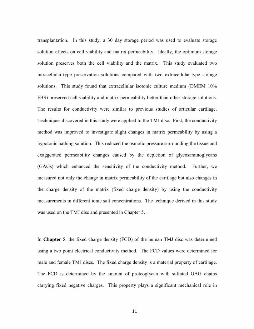
2.1 Anatomy and Biomechanical Function
Similar to other joints in vertebrates, the temporomandibular joint (TMJ) consists
of bone to bone interfaces with cartilage protecting bone surfaces from impact and
friction build-up. Cartilage in the joint provides a unique load dissipating environment
that allows the joint to function through the life of the patient. In the closed jaw position
or central occlusal position, the strongly convex mandible condyle of the mandible bone
rests within the concave surface of the glenoid fossa of the squamous portion of the
temporal bone. A thin layer articular cartilage lines both the mandible condyle and
glenoid fossa; and unique to the joint is the articular disc or TMJ disc that is positioned
between the two boney surfaces and divides the joint into an upper and lower joint
compartment (Figure 1).
Figure 1 Cross-sectional view of temporomandibular joint and overhead view
of TMJ disc outlining specific regions. [1]

17
The articular disc is ovid shaped in dorsal view and biconcaved shaped in the
parasagital section [35]. Both inferior and superior joint compartments are filled with
synovial fluid that aid in lubrication of the condyle and disc and supply surrounding
avascular cartilage and TMJ disc with nutrients [2, 35].
During initial jaw motion, the mandible condyle first rotates in central occlusal
position within the inferior joint compartment [2]. As the mandible elevates, the
temporalis, masseter, and medial pterygoid muscles contract. The mandible condyle
further translates out of the resting glenoid fossa concavity and down the posterior slope
of the fossa eminence. The ability of the joint to rotate (diarthroid joint) and translate
(ginglyo joint) gives rise to the classification of ginglymoarthroidal joint [2]. The TMJ
disc translates with the condyle through lateral and medial pole attachments through
robust insertion of capsular ligaments. The posterior attachment to the TMJ disc is
through a massive pad of fibrous retrodiscal tissue with inner clusters of adipose cells
[35]. The posterior band of the TMJ disc is attached to the surrounding ligaments
through large, thick fibrous tissues with interspersed adipose cell, with insertion into the
condyle and temporal bone [35]. The posterior attachment is believed to assist the disc’s
recovery in retrusion back to central intercuspal positon [35, 72]. The disc has anterior
attachment to the lateral pterygoid muscle and anterior to the condyle. As the mandible
closes or depresses, the masseter and inferior lateral pterygoid muscles act, as the disc is
positioned back to central intercuspal position [2].
Thin layers of articular cartilage protect the mandible condyle (0.2-0.5 mm) and
fossa along with eminence (0.1-0.3 mm) [2, 28]. The articular disc on the other hand is
18
much thicker (2-4 mm) along the posterior and anterior zones and (1-2 mm) along the
intermediate zones. It is believed that the disc supplies the majority of joint load
dissipation, lubrication and protection of articular surfaces. The disc is believed to act to
fill joint areas of low areas of contact, in order to reduce high contact stresses and
distribute them through the disc.
Another key aspect of the TMJ anatomy and function is the unique loading
environment to the joint. The joint is said to operate as a 1) a rotating hinge joint with 2)
secondary gliding motion [2]. However, the joint motion must be further examined. For
example, the joint has two condylar poles with muscle attachments. As the joint
functions, muscles contract and release bilaterally. This caused protrusive and retrusive
movement, opening and closing with translation and rotation within the joint. However,
during mastication the mandible also undergoes lateral shifts and orbital motions, which
create grinding motion and rotation of the condyles about multiple axes. Therefore, the
joint not only rotates and translates, but when analyzing both condylar poles, the joint is
allowed complex six degrees of freedom (6 DOF) about its condylar axis [6, 37, 73, 74].
Finite element studies have determined the TM joint is a load bearing joint [37]. This
gives rise to a very unique mechanical function in the TMJ disc. The disc is highly
anisotropic functioning under tensile, compressive, shear, as well as frictional and
traction forces often during the same masticatory stroke.
19
2.2 TMJ Disc Cell Characterization and Extracellular Matrix Composition
Fibroblasts
In porcine TMJ disc, fibroblasts were found to comprise 70% of the cell
population. In the anterior posterior axis, the anterior and posterior regions of the disc
had higher fibroblast content than the intermediate portion of the disc, 3.5% and 6.3%
greater respectively. The overall cell density from histology was 681±197 cells/mm2.
Fibroblasts appeared to exhibit uniform ultrastructural characteristics between regions of
the disc. The ultrastructural characteristics included a large nuclei, minimally visible
rough endoplasmic reticulum and even less visible smooth endoplasmic reticulum. Golgi
apparatus and mitochondria were rarely observed. Similarly, ribosomes were observed in
scarce amounts; however few fibroblasts had an abundant amount of ribosomes.
Fibroblasts were also found in greater amounts along the periphery of the disc,
specifically at the anterior and posterior attachment sights [19, 75]. Fibroblasts appeared
to align with surrounding collagen fibrils.
Chondrocytes, Chondrocyte-like cells, and Fibrochondrocytes
Chondrocytes are generally identified in articular cartilage and the nucleus
pulposis of IVD. During embryonic development, chondrocytes provide major functional
growth of the skeleton through endrochondral ossification mainly due to the remarkable
ECM synthesizing ability of chondrocytes. The flexibility of cartilage as opposed to
mineralized bone also helps in the development. As the growth tapers and the skeleton
becomes mature, chondrocytes are mainly found in cartilage at the end of long bones,

20
IVD, and nasal septum for the purpose of distributing mechanical loads, providing
flexibility, and protecting boney surfaces [76].
Chondrocytes in the TMJ disc were distinct from other designated chondrocyte-
like cells found in the disc. Chondrocytes exhibited well-organized rough and smooth
endoplasmic reticulum, Golgi apparatus and many ribosomes. While the chondrocytes
exhibited a well-defined electron-lucent zone or pericellular matrix, the pericellular
capsule was not found [26].
Chondrocyte-like cells on the other hand were found to have very few organelles
with a lack of pseudopodia and pericellular matrix, including a small amount of rough
endoplasmic reticulum and lesser amounts of smooth endoplasmic reticulum [26]. Few
cells had identifiable Golgi apparatus. However, the chondrocyte-like cells did have an
abundant amount of mitochondria and large nucleus, with moderate amounts of
ribosomes [1, 26, 77-79]. Chondrocyte-like cells were found in abundant concentrations
in the center of the TMJ disc which corresponds well to the greater amounts of
chondroitin sulfate and higher compressive modulus found for this region [32, 80].
Berkovitz et al described the human TMJ disc as a dense fibrous connective tissue with
chondrocyte-like cells scattered sparsely. Overall, the ratio of fibroblast to chondrocyte-
like cells was 2.35 to 1 [26].
Often cells of the TMJ disc are referred to as fibrochondrocytes since these cells
lack a pericellular matrix but still retain chondrocyte markers [49]. Overall, the cells
within the disc appear to exhibit more fibroblast like characteristics resembling synovial
fibroblasts due to proteinase and proteinase inhibitors [81]. Among the cell types in the
21
TMJ disc, fibroblasts, chondrocytes and fibrochondrocytes have been shown to express
different markers based on normal and pathologic discs.
Collagen
Collagens are considered a main structural protein of connective tissue. In
humans, collagens account for one third of the total proteins, being the most prevalent
protein in the ECM. Collagen is known for its triple helix formation of three parallel,
staggered polypeptide chains. The triple helixes interweave to form fibrils; fibrils align
and bind together to become microfibrils in fibrillar collagens. Of the types of collagen,
the first five are fibrillary and the most common is type I collagen , which makes up 90%
of the collagen found the body and can be found in dermis, bone, tendon and ligaments
[56].
22
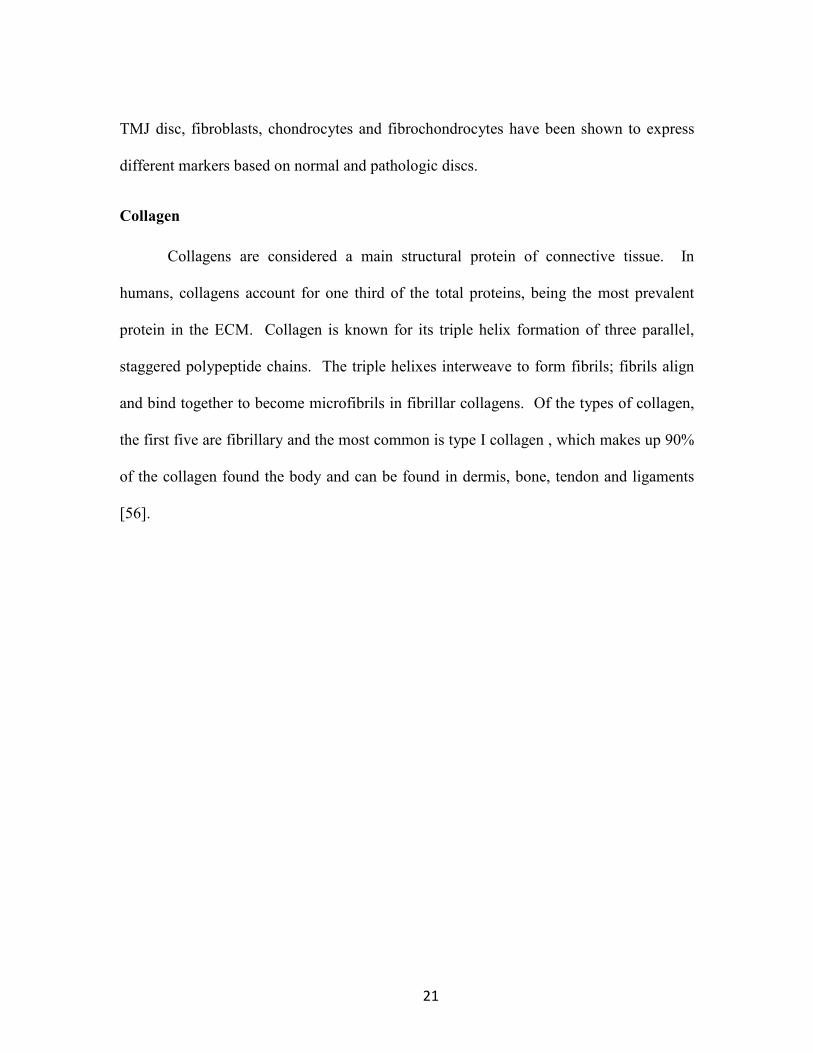
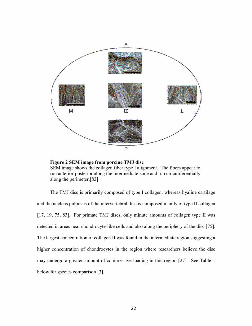
Figure 2 SEM image from porcine TMJ disc
SEM image shows the collagen fiber type I alignment. The fibers appear to run anterior-posterior along the intermediate zone and run circumferentially along the perimeter.[82]
The TMJ disc is primarily composed of type I collagen, whereas hyaline cartilage
and the nucleus pulposus of the intervertebral disc is composed mainly of type II collagen
[17, 19, 75, 83]. For primate TMJ discs, only minute amounts of collagen type II was
detected in areas near chondrocyte-like cells and also along the periphery of the disc [75].
The largest concentration of collagen II was found in the intermediate region suggesting a
higher concentration of chondrocytes in the region where researchers believe the disc
may undergo a greater amount of compressive loading in this region [27]. See Table 1
below for species comparison [3].
23
Type I collagen is aligned in a ring like fashion around the perimeter of the disc
and runs anterior-posterior along the intermediate region of the disc (see Figure 2 above).
Biomechanical studies have correlated anisotropy in tensile strength to collagen
alignment. Collagen accounts for 80-95% of the TMJ disc dry weight and up to 50% of
the volume by wet weight [84-87]. Average collagen fiber diameter has been reported as
~18 μm ± 9 μm [27]. Collagen fibers have been observed to have a wavy and crimped
pattern across the entire thickness of the disc [23, 88-90].
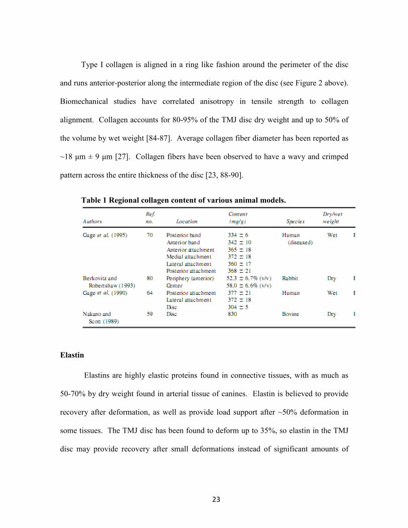
Table 1 Regional collagen content of various animal models.
Elastin
Elastins are highly elastic proteins found in connective tissues, with as much as
50-70% by dry weight found in arterial tissue of canines. Elastin is believed to provide
recovery after deformation, as well as provide load support after ~50% deformation in
some tissues. The TMJ disc has been found to deform up to 35%, so elastin in the TMJ
disc may provide recovery after small deformations instead of significant amounts of
24
loading. Elastin content in TMJ disc has been reported to be very low by dry weight, in
bovine TMJ disc 3-7% [91, 92]. Although elastin has been found to be cross-linked with
collagen in the TMJ disc, due to the scarce amount it is unlikely that elastin contributes to
the mechanical loading of the tissue [25, 75, 89]. Additionally, the presence of elastin
has varied between regions of the disc and between inferior and superior surfaces [22,
92].
GAG/Proteoglycans
Proteoglycans (PGs) are synthesized by almost all types of mammalian cells. PGs
have been found within cells, on the cell surface, and in the ECM. PGs contribute to
material properties in tissues as well as carry out roles in cell adhesion and cell signaling
[93].
There are a wide variety of PGs defined by their construction of polysaccharide
units. PGs are composed of a core protein covalently connected to linear polymers of
disaccharides, known as glycosominoglycans (GAG). PGs can have one GAG chain as
present in decorin or as many as a hundred in aggrecan. PGs are named according to the
type of GAG chains, such as chondroitin sulfate, dermatan sulfate, keratin sulfate, and
heparan sulfate [93].
It is common in tissues for PGs to form large aggregates where PGs are connected
to hyaluronan (HA) through non-covalent interaction with LINK protein. Additionally,
these large PG aggregates are contained within a collagen matrix. The
interconnectedness between the ECM components, like the hyaluronan interaction with
large PG aggregates, give tissues unique biomechanical properties especially in cartilage
25
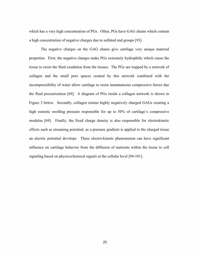
which has a very high concentration of PGs. Often, PGs have GAG chains which contain
a high concentration of negative charges due to sulfated end groups [93].
The negative charges on the GAG chains give cartilage very unique material
properties. First, the negative charges make PGs extremely hydrophilic which cause the
tissue to resist the fluid exudation from the tissues. The PGs are trapped by a network of
collagen and the small pore spaces created by this network combined with the
incompressibility of water allow cartilage to resist instantaneous compressive forces due
the fluid pressurization [69]. A diagram of PGs inside a collagen network is shown in
Figure 3 below. Secondly, collagen retains highly negatively charged GAGs creating a
high osmotic swelling pressure responsible for up to 50% of cartilage’s compressive
modulus [69]. Finally, the fixed charge density is also responsible for electrokinetic
effects such as streaming potential, as a pressure gradient is applied to the charged tissue
an electric potential develops. These electro-kinetic phenomenon can have significant
influence on cartilage behavior from the diffusion of nutrients within the tissue to cell
signaling based on physicochemical signals at the cellular level [94-101].
26
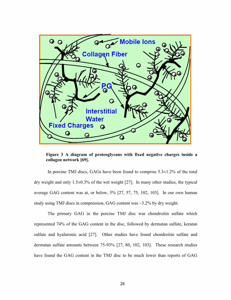
Figure 3 A diagram of proteoglycans with fixed negative charges inside a
collagen network [69].
In porcine TMJ discs, GAGs have been found to comprise 5.3±1.2% of the total
dry weight and only 1.5±0.3% of the wet weight [27]. In many other studies, the typical
average GAG content was at, or below, 5% [27, 57, 75, 102, 103]. In our own human
study using TMJ discs in compression, GAG content was ~3.2% by dry weight.
The primary GAG in the porcine TMJ disc was chondroitin sulfate which
represented 74% of the GAG content in the disc, followed by dermatan sulfate, keratan
sulfate and hyaluronic acid [27]. Other studies have found chondroitin sulfate and
dermatan sulfate amounts between 75-93% [27, 80, 102, 103]. These research studies
have found the GAG content in the TMJ disc to be much lower than reports of GAG

27
content in hyaline cartilage [3]. The low GAG content in the TMJ disc may indicate the
disc plays a much less significant role in compressive loading and may have a distinct
mechanical role from other types of cartilages that rely on a high PG content and high
fixed charge density [49, 57, 104]. Kalpakci et al. has conducted a GAG, collagen type I
and DNA for cell count comparison between species shown in Figure 4.
Figure 4 Biochemistry comparison of the TMJ disc between species
A comparison of biochemistry from different species by Kalpakci et al. Collagen compared is type I. Regional comparison within species noted by: PBC-Posterior Region, ABC-Anterior Region, IZM- Medial Region, IZC-Intermediated Region, and IZL-Lateral Region [141].

28
Water content
Water content is the major component in cartilage as well as the TMJ disc. Water
content is also considered a significant mechanical component of cartilaginous tissues,
affecting the diffusivity of the tissue and the compressive strength as water exudes from
the tissue. Water content has also been used as a marker for tissue degeneration as
proteoglycans and collagens are cleaved during enzymatic degradation resulting in loss of
matrix retention and tissue swelling [69, 80].
The average water content found in human TMJ discs from our studies were
between 76-80% involving subjects with an average age of 78 years [62]. In previous
studies using porcine tissues, the water content value was somewhat lower around
71±2%. This could be due to the water content increasing with age. Regional
differences were found in the porcine study; however, they were not found in our human
studies [27].
2.3 Temporomandibular Joint Disorder
Temporomandibular Joint Disorders is a broad term used to describe a group of
over 20 pathological conditions and collectively represents the most common type of
chronic orofacial pain. Symptoms include orofacial pain, muscle pain in the head and
neck, periauricular pain, clicking and locking of the jaw with limited jaw opening [2]. In
the US population, five percent of adults were reported to suffer from TMD like pain,
with 6% women and 3% men. In 2011, the NIDCR released the results of the first ever
prospective study, Orofacial Pain: Prospective Evaluation and Risk Assessment

29
(OPPERA), which began in 2005 and sought to elucidate the associations and risks for
progression of TMD [9-13].
Risk Factors and OPPERA Study
The OPPERA study found that psychosocial factors lead to an increased risk in
developing TMDs, such as higher levels of psychological distress, and greater levels of
perceived stress and catastrophizing, along with somatic awareness and greater levels of
preexisting pain sensitivity [9, 10]. Pain amplification was the most significant indicator
for increased risk of development and progression of chronic TMD pain [11]. Pain
amplification refers to changes in the peripheral and central nervous system which cause
increased perception and response to nociceptive stimuli. The response could be
generated by genetic predisposition or it can be developed as a neurological response to
biological processes or environmental stimuli. In the most conclusive finding in the
prospective study, patients with a preexisting chronic pain condition, for example
fibromyalgia and low back pain, were most likely to develop chronic TMD pain [9]. The
study further reported that there were clear indications that genetics did play a role in the
development of TMDs as several genes were identified as potential markers of risk for
TMD [13]. Of the population sampled, the gender disparity confirmed previous levels as
women to men ratio was 3:1; however, socioeconomic status was not a significant factor
in the development of TMD. Race was indicated as a factor with Caucasians having an
increased odds ratio for development as compared to African Americans. Finally, age
did have an effect on the increased risk of development. The sampled age group was
30
from 18-44 years of age, with an increased risk of development highest amongst the 36-
44 age group [12].
Gender Differences
TMJ disorders are primarily found in female patients between the ages of 20 and
40 years of age [8, 105]. This discrepancy in treatment by gender is believed to be
attributed to hormonal differences because these symptoms primarily appear during
childbearing ages in females. While the mechanistic role of hormones remains to be
elucidated, many studies have indicated that estrogen and progesterone could act on the
nervous system to enhance nociceptive processing. In one rat study, estrogen appears to
increase the number of localized nociceptors and also enhance signaling of those
receptors in parts of the hippocampus [106].
Studies have been conducted to determine effects of hormones on connective
tissues such as cartilage [7, 105]. These hormones have been found to have a significant
effect on differentiation and metabolism. In a study on female rats, collagen was
produced at significantly lower levels than male controls but when the females were
ovariectomized the differences were removed [107]. This may indicate significant
differences in biochemical composition and thus affect biomechanical properties
providing the rationale to investigate both male and female tissue in our biomechanical
studies.
In our electrical conductivity study on male and female TMJ discs, we found
differences in conductivity and porosity between male and female tissues and these
differences were magnified by mechanical loading [62]. At the present time, the exact

31
pathways and specific receptors responsible for the gender paradox are unknown.
However, studies have demonstrated clinically and experimentally that gender
differences exist, indicating the need for further research to examine fundamental
differences in tissue composition and structure along with comparing male and female
tissue response to hormones such as 17β estradiol [51].
Mechanical Dysfunction
Studies have indicated that changing biomechanical stresses can cause TMJ disc
tissue to quickly adapt to maintain efficiency of the joint. Studies have shown that the
TMJ disc requires normal physiological loading to maintain tissue growth and function,
while excessive hydrostatic stresses result in enzymatic degradation of ECM and
apoptosis [108]. These stresses damage cartilage tissue resulting in the production and
accumulation of free radicals [21, 109, 110]. Free radicals are produced by degenerative
cell responses that reduce oxygen in situ resulting in reactive oxygen species. Reactive
oxidative species such as superoxide, hydrogen peroxide and hydroyl radicals can result
in ECM and cell damage [21, 109, 111]. The appearance of free radicals has been
observed during normal masticatory function as well as during abnormal movements
such as clenching [21]. An example of free radical damage to the TMJ synovial joint
cavity is illustrated by increased articular pressure as a result of inflammation which
induces localized hypoxia. Hypoxic conditions arise due to the upset of balance of
pressure between synovial capsule chamber and the capillary perfusion pressure [54].
Temporary hypoxia leads to the free radical production. The reduction environment
cleaves hyaluronan chains in hyaluronic acid leading to the degradation of synovial fluid

32
[54, 112]. The fluid becomes ineffective, increasing joint friction and in-turn causing
accelerated joint degradation.
Figure 5 Possible effects of mechanical dysfunction in the TMJ disc
Abnormal mechanical loading leading to TM Joint dysfunction and tissue degradation [112].
These factors are usually concomitant with increasing joint load. Inflammation
occurs as a response to the localized hypoxia and reduction to available nutrients. The
result of inflammation causes the release of pro-inflammatory cytokines upregulating
MMP9, specifically interleukins (IL- 1, 3, 6, 8, 12, and 17), along with tumor necrosis
factor-α (TNF-α) [112].
Another possible cause of TMD is the loss of physiological nutrition transport
within the disc. The living cells of the TMJ disc require nutrients to maintain growth and
development and waste products must be removed to keep the environment from
becoming toxic. Since the TMJ disc is avascular, solutes must be transported passively

33
via diffusion to all of the cells. Studies have shown that sustained loading decreases the
rate of diffusion and transport properties of the cartilage significantly, thus decreasing the
nutrient levels deep within the tissue. Increasing joint load causes decreased diffusivity
of oxygen and glucose into the joint, leading to a further degenerative state. The local pH
can also significantly drop as lactic acid builds up and the cells begin to die at low
nutrient levels (i.e. oxygen and glucose) within 24 hours [113].
Changes in the tissue as a result of inflammation or response to mechanical
dysfunction over time can result in degeneration of the disc. With degeneration, the
mechanical properties of the tissue are affected altering the physiological function of the
TMJ disc. The TMJ disc becomes displaced and no longer travels in smooth congruent
motion protecting the condyle and fossa [37]. As disc displacement or disc derangement
worsens, the disc can limit the jaw’s range of motion and in some cases block the jaw
from opening or closing (known as derangement without reduction) [48]. Disc
displacement and derangement are common events for patients who seek treatment for
TMDs [15].
Disc Displacement and Other Derangement
In patients who seek treatment for TMDs, disc displacement is present in nearly
70% of cases [5]. This emphasizes the importance of the TMJ disc’s biomechanical role
in TMDs. The disc exhibits a highly biconcave shape in humans and appears to exhibit
some anisotropy based on regions within the tissue. The TMJ disc translates with the
condyle during function because it is supported by an anterior attachment to the lateral
pterygoid and anterior insertion into the condyle and a posterior attachment to the

34
retrodiscal tissue [114]. During opening and closing of the jaw the disc translates
anteriorly with the condyle [2]. The translation requires the disc to support tension as it is
stretched anteriorly and posteriorly over the condyle during motion. Alterations in
normal TMJ disc translation can cause pathological loadings resulting in damage to the
disc and surrounding boney surfaces. One of the first symptoms of disc displacement is
clicking and popping in the joint when the mouth is opened and closed. This is caused by
the disc being displaced anteriorly or posteriorly until the discal attachments force the
disc back into normal position over the condyle. The joint is said to be reduced or
restored to normal position, clinically know as disc displacement with reduction. If the
disc is displaced anteriorly or posteriorly and prevents the joint from opening or closing
without moving into normal position, the symptom is disc displacement without
reduction [48]. Disc displacement has been found to create high stress distributions and
increase friction between articular surfaces resulting in osteoarthritis in patients with
internal disc derangement [29, 42, 43, 115].
TMDs are generally classified by stages of TMJ disc displacement and
derangement. In early stage TMDs, the TMJ disc appears healthy but mild displacement
is present indicated by possible clicking. In the intermediate stage, pain is more prevalent
and the disc begins to show signs of deformation through MRI/x-rays. This can lead to
progressive development of joint locking and hardening of disc tissue. In late
intermediate stage progression, more pain is present accompanied by severe joint
dysfunction, including locking and morphologic changes in bone structure and

35
surrounding tissues. In late stage progression, severe pathology of TMJ disc tissues and
surrounding bone along with severe loss of joint function is present [45, 46].
Treatments
Current practice uses medications in 90% of TMD related disorders. Commonly
used agents are corticosteroids, muscle relaxants, antioxidants, opiates and anti-
inflammatory drugs. The effectiveness of the prescribed medications has come under
considerable debate due to their ability to treat the symptoms and not the underlying
mechanism of pathophysiology [116]. In the meantime, even though considerable
progress has been made toward discoveries on the molecular mechanism of TMJ
disorder, decisive treatments and delivery methods have been extremely complex in
dealing with the individual patient. One of the main reasons for lack of empirical
evidence in support of drugs is the lack of randomized clinical studies [116].
The majority of TMD patients can be successfully treated by non-surgical
therapies and surgical interventions may be required for only a small portion of the TMD
population. Some of the ways that patients can personally alleviate pain and keep
symptoms from worsening are performing jaw exercises, avoiding extreme or rapid
movements, applying hot and cold compresses to the region, and eating soft foods [2].
Patients may also be given splint devices to fit over the teeth to prevent the effects of
clenching and grinding. Grinding of the teeth or bruxism is believed to cause TMD
through tooth erosion, muscular strain, and inflammation of the tissue within the joint
space [36]. Splints are therefore prescribed in the hopes of controlling the consequences
of bruxism and slowing the onset of TMDs [117, 118].

36
Surgical repair covers a range of invasive from arthrocentesis and arthroscopy, to
finally total joint replacement, arthroplasty, if minimally invasive treatments fail.
Arthrocentesis has become a very popular choice among surgical interventions due to the
minimal invasiveness of the procedure, time of operation of one hour, and its cost
effectiveness.
Arthrocentesis
Arthrocentesis is prescribed for acute TMJ disorder, including internal
derangement, OA, chronic closed lock and open lock, synovitis, rheumatoid arthritis and
adhesions [119]. Arthrocentesis is an office outpatient procedure taking less than 1 hour
with treatment and recovery (the procedure is performed with intra-venous deep
sedation). Two 18 gauge needles are atraumatically inserted in the superior joint space
and the space is insufflated and irrigated with 150-300cc of lactated ringer’s solution,
washing out inflammatory mediators and relieving pain. An inlet and outlet is required
for this process to irrigate the joint space removing boney/cellular particles causing
inflammation and inflammatory mediators. The mandible is manipulated to ensure return
of maximum opening. A steroid (Dexamethasone 6mg) and local anesthetic (Marcaine
.5% with epinephrine 1:200,0001cc) are administered by flushing through the
inflow/outflow needle ports. Finally, a sodium hyaluronate is applied through injection
which replaces synovial fluid that was previously washed from the joint. The sodium
hyaluronate aids in the lubrication of the joint and also reduces inflammation and pain
[119-121].

37
Nitzan et al. first documented treatment of TMJ dysfunction using arthrocentesis
in 1991 [119]. Since then numerous clinical treatments have been carried out. Treatment
success has been evaluated by visual analog scale (VAS) pain scales and a comparison of
jaw mobility after surgery measured by maximal interincisal opening (MIO) [121].
Arthrocentisis have been shown to improve immediate symptoms of TMDs such as pain
relief, however the long-term effects are debatable. This is often a reoccurring follow-up
procedure, with temporary relief of pain which seems to restore joint function but could
be causing damage to the TMJ tissues further since the symptoms are masked but the
underlying causes are still present.
Arthroscopy
During arthroscopy, a small incision (6.35 mm) is made anterior to the ear and an
athroscope is inserted into the superior joint space. The arthroscope allows the oral
surgeon to inspect the tissue, remove damaged tissue, and realign the disc and condyle if
necessary. The arthroscope is connected to a video screen. The surgeon can then
examine the joint, remove pathologic tissue, or position the disc back normal position
through the use of anchors (see Mitek anchor in the next section). [36]. Afterwards, the
disc is lavaged with saline and treated with drugs described above in the same way as
athrocentesis.
With arthroscopy, a larger incision is required for the scope, disc inspection, and
attachment. Athrocentesis requires less than 1 hour in the operating room, while
arthroscopy can take longer given the procedures of inspecting the superior joint
compartment. Success of the procedure is highly dependent on the concise placement of

38
the lavage needles and how well the inflammatory mediators can be removed in the
process [119].
Success rates for arthroscopy have been reported between 79%-91% compared to
success rates for arthrocentisis of 75%-91.8% [121]. A 6 to 30 month follow-up study of
arthrocentisis with lavage indicated that only 1 out of the 46 patients did not report a
significant improvement in pain, function, and maximum interincisal opening. While the
results for the improvement of pain and jaw range of motion are generally accepted,
questions on the sustainable restoration of TMJ tissue and sounding joint cavity tissue
remain. The controversy lies in the follow-up after treatment. Over the long term,
successful outcomes for pathologies such as disc degeneration are highly debatable and
the question remains if the treatment simply relieves pain or if it accelerates disc
degeneration.
Mitek Anchor
To help treat anterior disc displacement, common in TMDs, physicians implant a
device called the Mitek mini anchor into the bone of the condyle [122, 123]. The device
is a small titanium anchor with nitinol wings that expand when reaching medullary bone.
Once the anchor is set in the condyle, the sutures insert into the posterior band to secure
the disc. The Mitek anchor has been reported to have success rates of up to 90%. The
anchor has shown favorable osseointegration and biocompativility through a 59 month
follow-up. The Mitek anchor offers many advantages. It is applied minimally invasively
through arthroscopy, it provides occlusal stability by anchoring the anterior portion of the

39
TMJ disc which is usually displaced, and it has been shown to reduce pain and improve
joint function. [122, 124].
Joint Replacements
TMJ implants have had a history of recalls and complications due to the complex
loading environment’s effect on the implant. Evidence for recalls has come mainly from
the immune reaction from wear debris. The investigation of biochemical, biomechanical,
and degeneration studies were launched due in large part to the previous failure in TMJ
implants.
The Christensen implant was in effect since the 1960’s. The implant was
comprised of a cobalt-chrome alloy for fossa and condyle head, with a molded
polymethylmethylacrilate (PMMA) head on the alloy framework. For total TMJ joint
replacement, the procedure creates metal wear debris. Patients developed metallosis,
causing an increase in stress concentration at the fossa implant site, and resulted in
hypersensitivity in the joint. In July 1993, the FDA halted use of implants made before
the 1976 medical device law due to safety concerns and lack of supporting information
demonstrating efficacy of those implants.
Techmedica developed a TMJ total joint prosthesis in 1989. A 5-year follow-up
study was performed in 1993 at the request of the FDA. The study found a 90% success
rate for occlusal and skeletal stability and an 89% reduction in pain. The product was re-
admitted for approval in 1997. The fossa component of the device is made from pure
titanium mesh with an articular surface of ultra-high-weight-polyethylene. The condyle
component is made from medical grade titanium alloy with a head of cobalt-chromium
40
molybdenum alloy. Fit for the device is determined by CT imaging that generates a
stereolithographic plastic model of the patient’s jaw and condyle that is anatomically
exact. Only four TMJ implants have been approved by the FDA since Dec. 30, 1998.
The Vitek Implant: A Need to Understand TMJ disc biomechanics.
The Vitek Proplast TMJ implant was recalled by the FDA in January 1991. A
chemical engineer named Dr. Charles Homsy developed a soft Proplast material of
silicon embedded in a wire mesh to reconstruct the glenoid fossa of the temporal bone. A
Teflon disc, from DuPont, was reinforced with graphite and used between the condyle
and fossa. The porous Proplast was believed to adhere to superior joint surfaces of the
glenoid fossa combined with Teflon’s ability to facilitate movement and withstand wear
[125].
Vitek notified the FDA of plans to market the “interpositional implant” (IPA) in
March 1983. Proplast had been used favorably to stabilize the femoral head and
acetabulum in hip prosthesis. In turn Vitek sought 510 Premarket Notification citing
substantial equivalence to existing silicon sheeting devices. To justify safety of the
device, Vitek cited the “scarcity of probative scientific material” and cited Homsy’s own
research in related dental applications which were not related to the TMJ disc. When
questioned about the Teflon fragmentation that had been discovered with hip implants,
the team claimed the TM joint was “largely non-load bearing” [52, 125].
Oral surgeons began implanting the device in 1984. By 1986 a large patient
group across North America was suffering. The surgical community was slow to report
surgical failures and follow-up symptoms. Emphasis was placed on conservative

41
recovery of soft foods and the historical difficulty of recovery for this joint was cited.
But during this time, a much deeper understanding of the problem emerged. MRI’s
showed inflammation of surrounding tissues and fragmentation of the implants.
Emergency surgeries were conducted which found Giant cell reactions in response to
wear particles. These were found throughout the joint and migrated to local lymph nodes
[2, 52, 125].
In 1988, news of implant failure had reached the FDA. Vitek issued a voluntary
safety alert under FDA guidance. In January of 1991, the FDA ordered a recall of all
Vitek implants. During the recall the FDA issued a statement that failure of the
prosthesis was mainly due to the lack of understanding TMJ disorders, specifically TMJ
joint mechanics, and tissue function. It concluded that a prosthetic designed without
clear understanding of the working joint would inevitably fail [52, 123, 125].
In total, over 26,000 implants were administered between 1984 and 1991, mostly
in women. Often emergency surgeries were conducted to remove the implants to resolve
immune response from implant fragmentation and wear debris. As many as six surgeries
were required in some cases to rebuild the mandible and craniofacial tissues affected by
Giant cell reactions, which resulted in chronic limited jaw opening. Patients reported loss
of eyesight, itching and burning during chewing, tiredness and debilitating pain in the
craniofacial region [52, 123, 125]. As a result of the delay between first reports of
implant failure and FDA recall, new laws and regulations were enacted such as the
Medical Device Reporting Regulations (1984) and Safe Medical Device Act (1990).

42
TMJ Disc Mechanical Properties
Mandibular Condyle Cartilage Mechanical Properties
The articular cartilage of the mandible condyle and glenoid fossa are primarily
fibrocartilage with mainly type I collagen, unlike the articular cartilage of the knee which
is hyaline cartilage and type II collagen. The fluid phase consists of water and dissolved
electrolytes, which is over 80% of the tissue by volume. The intrinsic stiffness measured
signified by the compressive modulus depends on Donnan swelling pressure generated by
the fixed charged density (i.e. proteoglycan (PG) concentration) and the solid mesh. In
hyaline cartilage the apparent stiffness is given as 30-50% by the Donnon swelling
pressure [69].
To examine the mandibular condyle cartilage mechanical properties, a
microindentation test was performed by Singh et al. 2009 [28]. The tests were completed
under two conditions, an isotonic (0.15M PBS) condition that measured apparent
compressive modulus (matrix solid and Donnan pressure) and intrinsic compressive
modulus under hypertonic conditions (2.0M) that measured only the compressive
modulus of the solid matrix. The mechanical response from the tissue in two different
ionic concentrations was analyzed by curve fitting the load response. The two curves
were used to determine the contribution of the solid phase versus the ion/fluid phase of
the cartilage. The apparent aggregate, shear modulus and permeability were on average
about 20% higher than the intrinsic mechanical properties. This indicates that although
significant, the apparent mechanical properties are less dependent on the PG content than
the solid matrix. In other joints, such as the knee joint with significant amounts of PG in
43
the hyaline cartilage, obtain as much as 30-50% of their mechanical compressive
modulus from Donnan osmotic pressure resulting from PG content.
TMJ Disc Mechanical Properties
The TMJ disc experiences a complex range of forces under normal physiologic
conditions such as mastication. These forces can be compression, tension and shear.
Cells respond by producing matrix proteins and growth factors for the synthesis of
collagen and aggrecan, along with maintaining cell phenotype and proliferation in
response to mechanical loading. Intermittent mechanical loading within the physiologic
range has been shown to maintain homeostasis of TMJ cartilage, while high sustained
stress may introduce catabolic changes [37, 56, 69]. It is generally believed that a
breakdown in this interrelated balance between mechanical loading and tissue
homeostasis causes a cascade of biological signaling processes that could lead to
degeneration of the disc [50].
The transfer of energy through a biologic material depends on the primary
structure and function of the tissue [25, 37, 50]. In the case of the fibrocartilaginous TMJ
disc, the collagen fibers primarily absorb tensile loads, such as stretching, while
compressive loads are absorbed by fluid flow through the disc. The resistance of fluid
flow increases significantly as the disc is compressed, since the porosity is significantly
reduced under mechanical load. These properties are strain dependent. The
physiological level of strain varies between tissues; some examples include skin at 40%,
tendons at 2-5%, and around 4% for canine TMJ discs [56, 126].
44
Cartilaginous tissues are generally characterized as viscoelastic materials in which
load response is dependent on load rate and load response changes with respect to time.
The mechanical load response is usually highly related to fluid flow within the tissue,
known as the viscous response. This is modeled as a dash pot in phenomenological
models as the material supplies viscous stresses in response to loading. Also, collagen
crimping and elastin levels give the tissue the elastic properties. Together the viscous
and elastic response is highly rate dependent. Therefore, in order to compare tissue
mechanical properties, the load rate and test protocol must be given consideration [69].
Mechanical tests on viscoelastic materials are rarely load-to-failure or dynamic
load-to-failure tests, since maximum stress would be dictated by load rate. Instead,
viscoelastic tests seek to characterize the material under different load rates or strain
rates. These tests involve stress-relaxation, creep, or recovery experiments which provide
information on the mechanical properties of load rate. Often a series of load rates are
used or a series of increasing strains are used with a measured load response within the
tissue. The load response characteristics are measured from the tissue and indicate how
well the tissue absorbs stress and dissipates energy [37].

45
Figure 6 Example plots of creep test and stress relaxation test
In the creep test, stress is held constant while stain varies with respect to time, requiring displacement. While in the stress relaxation test, strain is held constant and the stress reaction is recorded with respect to time. The stress relaxation test was chosen in this dissertation to model the tensile properties of the human TMJ disc [22].
In a stress-relaxation test, the material is loaded by applying a set strain or set
displacement which deforms the material invoking a stress response, see Figure 6 above.
This stress response dissipates over time until a steady state level is achieved. The load
response is measured over the time the material reaches steady state. The relaxation
modulus is obtained by dividing the stress at steady state with the relaxation time. In a
creep test, an instantaneous stress is applied to the material and kept constant while time-
dependent strain levels are recorded. The response of the tissue is measured by strain,
until the strain level becomes constant, reaching steady state. Finally, general recovery
tests involve applying a load, then characterizing the response of the material after
loading is removed. Numerous models for the behavior of TMJ discs have been
proposed by assuming the material to be linearly elastic, but the time-dependent response

46
of cartilage suggests that more complicated constitutive models are required [53, 70, 127,
128].
Tensile
Since the tissue is a viscoelastic material, strain rate is highly correlated with
stress response; therefore, the strain rate needs to match physiologic level simulated in
vivo. In the case of the TMJ disc, finite element models have determined physiologic
strain levels range from 0-30%, as most tissue tests use this as range for testing. This
measures the energy absorptive capacity of the viscoelastic material which is more
physiologically relevant, since the tissue operates underneath these conditions during
everyday use.
Figure 7 TMJ disc with anteroposterior and mediolateral sample orientations
for tensile test
The TMJ disc is labeled with anteroposterior and mediolateral strips used for tensile tests. Note the diagram shows the orientation as an overlay, since in one TMJ disc samples can be extracted in either anteroposterior or mediolateral orientation [1].
During normal joint function, as the condyle translates anteriorly, the disc stretches
anterior-posteriorly across the head of the condyle to protect articulating surfaces. Due to

47
the anisotropic range of forces the disc undergoes, the disc has adapted highly regional
anisotropic material properties. As a result, the testing direction has a significant impact
on tensile properties of the TMJ disc (see Figure 7 above for directional orientation). To
illustrate, the porcine tensile modulus in the intermediate zone was found to be 37.4 MPa
in the anteroposterior direction and 1.6 MPa in the mediolateral direction [129]. Further,
in comparing the anterior, intermediate, and posterior zones, the canine central zone was
found to have an ultimate tensile modulus of 14.7 MPa compared to the anterior (46.7
MPa) and posterior (69.7 MPa) zones [126]. The general overall trend is the
anteriorposterior direction has a higher tensile modulus than the mediolateral direction.
However, studies have produced results that vary greatly between species. This has been
attributed to biologic variation as well as differences in models chosen to analyze
material response. Many review papers have cited the need for consistent protocols to
establish the mechanical properties of human TMJ disc tissue.
The anisotropic properties were gathered from creep and relaxation stress tests. A
good example is supplied by Tanaka et al. 2002 using a creep test to evaluate the
influence of creep duration on residual stress in bovine TMJ disc tissue in the
mediolateral direction [130]. A 1.5 MPa stress was applied to the specimen for 20
minutes and the tissue response was measured for 20 minutes. The creep test showed a
dramatic increase in stress within the first 5 seconds tapering to a constant stress at the 3
minute mark. A Burger’s model was used to fit the stress relaxation curve. The model
consisted of Maxwell model that accounted for residual strain after load removal and
several Voight models to characterize creep features. The findings indicated that the disc
48
is able to absorb a significant amount of energy within the first 5 seconds of tensile
loading, similar to that found in compression. This would indicate that the articular disc
is responsible for absorbing joint forces and changing shape in-order to dissipate energy
within the joint. Without this residual stress absorption, a stress concentration could
occur to the surrounding surfaces of the mandible condyle and fossa of the temporal bone
that would result in catabolic changes of injury.
M. Beek et al. 2001 analyzed human TMJ discs using dynamic indentation on the
anterior, intermediate, and poster regions [32]. The study found the intermediate region
to be 2 to 3 times stiffer, withstanding a significant amount more energy dissipation than
the anterior and posterior regions. These findings may indicate the disc’s ability to
transfer stress from the stiffer intermediate region through hydrostatic pressure and into
the anterior and posterior regions. In the process the disc can deform, increasing contact
area that would decrease the stress concentration in the joint. The findings are in
agreement with histological data that indicate a higher proteoglycan concentration along
the intermediate region of the disc along with a greater collagen density found by Mills
et. al 1994 [75]. The collagen alignment along the interior region in the anterior posterior
direction would direct fluid flow to the peripheral anterior poster bands, which have a
mediolateral alignment. In the process, proteoglycans would be trapped in the collage
matrix further reducing fluid flow [37]. This accounts for high energy dissipation upon
impact and the slow recovery after loading.

49
Compression
One of the original investigations into the viscoelastic properties of the TMJ disc
was demonstrated on human TMJ discs under unconfined compression tests at two
different load rates. The study concluded that the material did exhibit viscoelastic
response under compression by finding two different compression moduli corresponding
to two different load rates [131].
Additionally, several other studies assessed the regional dependency on
compressive properties. Allen et al. investigated the five disc regions, as well as the
superior and inferior surfaces of the disc using an unconfined compression incremental
stress relaxation test [132]. The instantaneous modulus (500 kPa) was higher than the
relaxed modulus (80 kPa) for combined samples, while regional differences in stiffness
were found across the disc’s surface regions. [132] The compressive moduli were
significantly lower (100-1000 times) than the tensile moduli found in Detamore et al.
2003 porcine tensile study [24, 132]. The intermediate and medial regions of the disc had
the highest relaxation modulus, while the anterior and posterior portion of the disc had
the highest instantaneous modulus. Surface differences were only found in the relaxed
modulus, where the inferior surface was more resistant to compressive forces than the
superior surface [132]. Further studies also confirmed regional differences in
compressive moduli [32, 133]
Along with finding regional variation of compressive moduli within the disc,
studies have found a marked difference in compressive properties depending on how the
compressive load was applied. This would indicate significant material reactions at work

50
and gives insight as to how the disc functions under physiologic loading. Increasing
frequency of dynamic compressive load caused the disc to become stiffer [31, 134].
After preconditioning, repeatable stress strain curves were reached with clear hysteresis
under low strains. This would indicate, under physiologic conditions, that hydrostatic
pressure may be responsible for significant load dissipation within the joint, without
significant dependency of the collagen network. Upon significant cyclic loading, the disc
cannot keep the same levels of fluid pressurization and after several cycles at high strains,
the relaxed compressive modulus was 1/2 to 1/7th that of the original instantaneous
modulus [134].
The general finding in regional differences was the central region possessed a
greater dynamic compressive modulus and more rapidly dissipating energy than other
regions. This trend has been correlated to motion studies and finite element models that
predict that the central region, specifically the intermediate region experiences the
greatest amount of compressive forces from the mandible condyle during normal joint
movement [39, 104]. Biochemical analysis by region has confirmed the greatest levels of
proteoglycans and few observed chondrocytes in the intermediate region. Finally,
Willard et al. 2012 compared control regional samples to samples with GAG depleted
through Chondroitinase ABS. The study found through unconfined compression stress
relaxation tests that only the intermediate region had compressive moduli affected
through the removal of GAGs. This would indicate not only a significant level of GAG
present in the intermediate region but compressive moduli in the region dependent on
51
GAG to dissipate compressive forces. The particular mechanism of biphasic action is
more similar to other types of cartilage such as hyaline in articular cartilage.
Shear
Shear forces in the TMJ disc are believed to be caused by incongruent articular
surfaces acting on the disc during compressive loading. In addition, the disc is
bioconcave with anisotropic material properties which could cause further non-uniform
compressive loads across the disc, resulting in shear. Shear stresses are believed to cause
mechanical injury such as fatigue and perforations along the disc [34, 135]. The shear
modulus of the disc was found to be dependent on frequency and direction of the shear
load [31]. Further studies revealed dynamic shear modulus increased with dynamic
frequency which could indicate the shear properties of the disc are dependent on
interstitial fluid flow (i.e. fluid exudation from the disc could increase shear stiffness).
The relationship of shear with compression in the disc is consistent with results
found in bovine meniscus and articular cartilage [135, 136]. This correlation may be a
result of fluid pressurization during compression that decreases the size of the pores in
the ECM and decreases the permeability of fluid effectively stiffening the material to
shear forces [137].
In studies in articular cartilage, shear stress has been found to cause mechanical
injury such as fatigue with damage being irreparable [34, 136]. Injury to the disc as a
result of shear stresses has been modeled extensively in finite element models of the TMJ
disc [33, 38, 73, 74, 115, 138]. These models depend on accurate material properties
from biomechanics studies.
52
Friction and Tractional Forces
The primary lubrication mechanism of the TMJ disc is a boundary layer lubricant,
surface-active phospholipids secreted by synovites, fibrochondrocytes and osteoblasts
[139]. The surface-active phospholipids bond to articular surfaces through hydrogen
bonding on their polar end. The nonpolar moieties face opposite the articulating surface,
being hydrophobic create a very low surface energy and much less conductive to surface
friction. A lipid solvent applied to articulating surfaces was shown to increase friction
150%. Hyaluronic acid and lubricin are important lubricants also found in the synovial
fluid of the inferior and superior joint capsules [139]. Another aspect of lubrication
mechanism, particularly in the TMJ disc is the concept of weeping lubrication. A study
by Nickel et al. showed a weeping lubrication mechanism was a significant method of
joint lubrication, as the disc is compressed, interstitial water is compressed to the
boundary of the disc surface for lubrication [140]. The study showed by measuring static
friction on the surfaces of discs with a range of thicknesses that weeping lubrication had
positive correlation of thickness to joint lubrication. The study also reported that impact
loading due to trauma caused surface damage on fresh porcine discs that had a
logarithmic effect on surface friction. The in vivo implications are that sustained
clenching or bruxism may have a significant negative impact on the TMJ disc’s ability to
maintain lubrication. The results of the disc impact or trauma indicate that the level of
impact may introduce surface damage to the disc surfaces that predispose the disc to
degenerative changes similar to osteoarthiritis [140].
53
Large shear and compressive forces were observed in FEM in these regions which
correlates well with the TMJ perforation findings in cadaverous tissues [33, 42, 141,
142]. Translational forces applied mediolaterally across the TMJ disc may be one cause
of tissue failure because traction forces are applied at the surface where the disc is
relatively weak [41]. Traction forces in the TMJ are composed of friction and plowing
forces, which result from movement and fluid pressurization due to loading [143].
Studies have shown that plowing forces are approximately 10 times greater than frictional
forces [144, 145].
Biomechanical property differences between species
The differences in mechanical properties between species have been noted
abundantly within review papers that seek to establish baseline TMJ disc mechanical
properties [25, 50, 71]. The rationale for establishing baseline material properties arises
from the desire to set design criteria for tissue engineering replacements and reach a
consensus on mechanical function.

54
Figure 8 Morphological comparison between species
TMJ discs from different species. Notice the size and biconcavity similarities between the Human and Pig TMJ disc [71].
Establishing these properties has proven difficult due to differences between
studies sample load rates, non-uniformity in sample preparation, different regional
sampling, different testing protocols in different temperatures, the use of different ages in
humans as well as variations in tissue quality. Figure 8 above shows an example of the
differences in morphology of the TMJ disc between species. Though differences exist on
the level of 10 to 100 fold, they do not entirely account for biological variations. The
variation in species qualities have been justified by the structure meets function criterion,
with herbivores (cows, goats, and rabbits) exhibiting highest compressive modulus across
all disc regions. Animals tested exhibiting different food types, different masticatory
patterns for the food quality, as well as the size and morphological development of the

55
animal. Therefore, each species resulting mechanical property has to be taken into
consideration under the testing configuration that produced the results.
The overall elastic moduli for bovine samples were much lower than for human
discs (25-65 MPa) which may be due to differences in chewing motion and diet [146]. In
tensile tests, the overall strain rates varied from 6mm/min to 500 mm/min. The resulting
variance between the porcine group central region had an anteriorposterior modulus of
18.5 MPa (6mm/min strain rate) versus 76.4 MPa (500 min/min). Between species the
modulus varied between 14.3 MPa for porcine to 101 MPa for canine for anterioposter
modulus, however strain rate varied between species tests. The mediolateral modulus
was much lower than the anterioposterior orientation, when tested, regardless of species.
In general, information on human TMJ disc was scarce. And in order to properly
compare differences between species, such as the most commonly accepted equivalent
animal model, the porcine to the human, the tests should be conducted under the same
strain load rate, sample extraction, and test protocol.
The compressive properties of other animal models were found to be significantly
higher than those of porcine samples. Unconfined compression tests on canine samples
were found to have instantaneous modulus values of around 31 MPa while stress
relaxation tests on bovine discs yielded an instantaneous modulus of ~15 MPa [128, 147].
Specific differences in protocols such as strain rate, frequency rate, and amplitude have
all been found to have a significant impact on the compressive properties of TMJ disc
tissue, but it is assumed that most of the variation is due to interspecies differences in disc
composition [30, 132, 134, 148]. Regional and topographical variation during

56
compression has been observed for TMJ discs of several species. In tensile loading,
variation between species and region is shown in Figure 9 below. The porcine sample
had significantly lower moduli in the lateral region and large instantaneous modulus in
the posterior region while these differences were not seen in the bovine model [132, 147,
148]. The general assumption is that the compressive properties vary between the
mediolateral and anteroposterior directions, but the exact values and relationship are
currently debated in the literature [49].
Figure 9 Comparison of the tensile modulus by species
The left figures A&B show a comparison of the tensile modulus by species by Kalapaki et al. The right figures A&B show a comparison of the compressive modulus by species at 20% strain. Regional moduli were

57
compared with PBC-Posterior Region, ABC-Anterior Region, IZM- Medial Region, IZC-Intermediated Region, and IZL-Lateral Region [141].
2.4 Transport Properties of the TMJ Disc
The nutrient transport in TMJ discs is believed to play a significant role in matrix
remodeling. Currently studies from other cartilages must be relied upon like the
intervertebral discs (IVD) to draw comparisons and analyze methods of diffusion studies
due to the lack of TMJ disc transport properties. The TMJ disc and the IVD are similar
in that both are large avascular tissues that require nutrient delivery through diffusion
from the nutrient rich environment around the periphery of the tissue. The driving
transport mechanism is the diffusive flux driven by high concentrations of nutrients
around the periphery of the disc (synovial fluid) to areas of low nutrient concentration in
the center of the avascular disc. As nutrients are being transported to the center of the
disc, cells consume nutrients at a rate dependent on the concentration of the substrates
and pH [149]. For solutes, such as oxygen which is consumed rapidly, a steep
concentration gradient may occur that results in low concentrations at the center of the
disc (less than 1%) [150, 151]. The concentration gradient can be described as a change
in value of a quantity (nutrients/molecules) with a change over a distance. The
concentration gradient depends on the diffusivity of the disc, a material property, as well
as the consumption rate within the disc. Results from our lab have discovered a high
nutrient gradient through the TMJ disc. Our transport study using the electrical
conductivity method has found that the TMJ disc has very low diffusivity when
compared to other cartilages such as hyaline and IVD [152]. Not only was the diffusivity
58
low, the material property was negatively affected by mechanical loading. With this
understanding, there may exist a steep nutrient concentration gradient within the TMJ
disc that is uniquely venerable to pathologic loading. Therefore the transport properties
of the disc must be analyzed with the mechanical properties of the disc so that a clear
understanding of the nutrient environment may be understood. Since direct
measurements are not possible in vivo, only in vitro measurements are collected and with
the use of modeling techniques such as finite element models we are able to study the
unique nutrient environment based on the model.
Diffusion Chamber
The most basic measurement of the diffusivity of solutes within a tissue specimen
is through a diffusion chamber. The chamber is designed to hold a tissue specimen
between an upstream and downstream well containing different concentrations of solute.
The diffusivity is measured by keeping the upstream concentration constant while
measuring the concentration change over time in the downstream well. In cartilage, the
diffusivity of solutes is very low and the time needed to reach steady state would take
days, so more effective techniques have been made available.
Electrical Conductivity
Kuo et al. 2010 investigated porcine TMJ discs by using the electrical
conductivity method [61]. The TMJ disc has been found to have a very low proteoglycan
content, which would indicate a very low fixed charged density. In the case of uncharged
tissues, the diffusivity of small solutes in the tissue is proportional to the electrical
59
conductivity. Instead of using a concentration gradient to drive the flux of solutes
through the tissue, an electric potential was used as the source flux for the small ions of
sodium and chloride in PBS. The measurement of electrical conductivity for the sample
was based on the 4-wire resistance method. A cylindrical sample (~5mm diameter) was
placed in a sealed chamber with PBS. The sample was enclosed by two source
cylindrical electrodes on each face of the sample, with coaxial Ag/AgCl sintered wires
through the electrodes. The cylindrical electrodes sourced a very low current density
while the sensing Ag/AgCl wires measure the voltage drop across the tissue. The
resistance ( Ω ) values across the specimens were measured at a very low, constant
current density of 0.015 mA/cm2. The height of the specimen was measured with an
electrical current sensing micrometer. The electrical conductivity (χ) values of the
specimens were calculated by:
A
h
Ω=χ
1
where h and A are the height and cross-sectional areas of the specimens, respectively.
The electrical conductivity of the specimen was measured at 0%, 10%, and 20%
compression levels. For uncharged tissue in NaCl, the relative diffusivity (D/D0) of NaCl
is simply related to the relative conductivity by (Gu and Yao et al., 2002) [153]:

60
00 χφχ
wD
D =
2
where D is the mean ion diffusivity of Na+ and Cl- in tissue, D0 is the mean ion
diffusivity in the bathing solution, χ is the electrical conductivity of the tissue, and χ0 is
the conductivity of the bathing solution. In our analysis, Na+ and Cl− were assumed to
carry the electrical current because these ions are the primary ionic components of PBS.
The conductivity of porcine TMJ discs and human TMJ discs have been studied using
this method [152].
FRAP
Shi et al used fluorescence recovery after photobleaching (FRAP) to measure the TMJ
disc diffusivity tensor across regions porcine TMJ discs [58]. FITC dextran was allowed
to diffuse into the TMJ disc sample until a uniform concentration was reached (48 hour
period). An argon laser (488nm) was used to bleach a fluorescent probe. Images of the
fluorescence recovery after photobleaching were collected through a microscope with
high speed camera. The frames were then analyzed to reconstruct the recovery profile
and calculate the anisotropic diffusivities in two directions for each sample. Results were
then analyzed for noise and a comparison was made for diffusivities between regions
[58].

61
Regional Variation, Differences Between Species, and Gender differences
In the electrical conductivity study, the anterior region was found to have a
significantly higher diffusivity than the posterior region in porcine TMJ discs. However,
no regional differences were detected in human TMJ discs. Using the FRAP method, the
anterior region again was found to have a higher diffusivity than the posterior region for
porcine TMJ discs. One possible reason regional differences were detected in the porcine
and not in human disc, could be related to the age of the sample. Samples were tested
from relatively young adult pigs (8-10 months) and relatively older human subjects (76-
92 years) due to the availability of samples. Previous studies have shown that
proteoglycan content increases with age. The presence of charged tissue in human
samples may have outweighed regional response.
Using the electrical conductivity method, the samples were tested under
physiologic mechanical strains (0%, 10%, and 20%). Increasing strain rates caused the
diffusivity of samples to drop significantly. This may indicate that mechanical load has a
significant detrimental effect on diffusivity. In the human conductivity study by Wright
et al., male and female groups were tested. The study found that female conductivity was
higher than male conductivity, indicating a difference in transport material properties
between the two groups. The study also found that female conductivity decrease more
under mechanical load than male conductivity [62].
2.5 Modeling the TMJ Disc
In vivo TMJ disc measurements of stress and effects on tissue remodeling are
impractical due to spatial placement of measuring instruments [28, 33]. A few studies
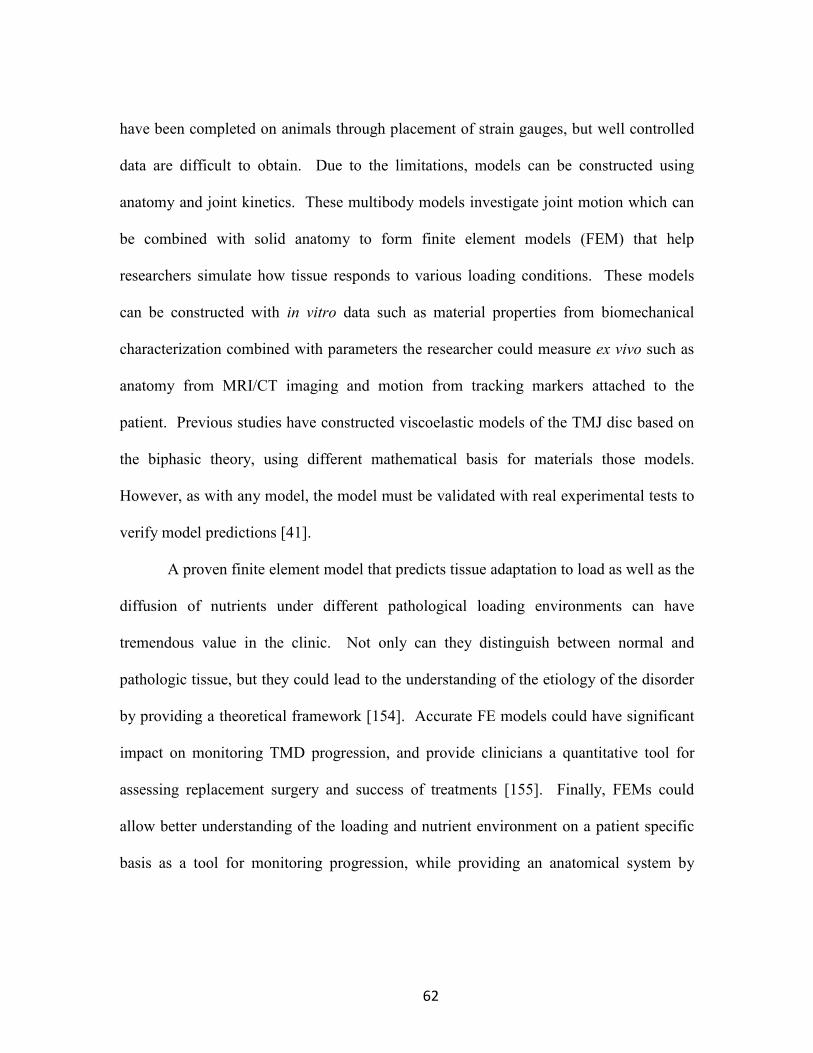
62
have been completed on animals through placement of strain gauges, but well controlled
data are difficult to obtain. Due to the limitations, models can be constructed using
anatomy and joint kinetics. These multibody models investigate joint motion which can
be combined with solid anatomy to form finite element models (FEM) that help
researchers simulate how tissue responds to various loading conditions. These models
can be constructed with in vitro data such as material properties from biomechanical
characterization combined with parameters the researcher could measure ex vivo such as
anatomy from MRI/CT imaging and motion from tracking markers attached to the
patient. Previous studies have constructed viscoelastic models of the TMJ disc based on
the biphasic theory, using different mathematical basis for materials those models.
However, as with any model, the model must be validated with real experimental tests to
verify model predictions [41].
A proven finite element model that predicts tissue adaptation to load as well as the
diffusion of nutrients under different pathological loading environments can have
tremendous value in the clinic. Not only can they distinguish between normal and
pathologic tissue, but they could lead to the understanding of the etiology of the disorder
by providing a theoretical framework [154]. Accurate FE models could have significant
impact on monitoring TMD progression, and provide clinicians a quantitative tool for
assessing replacement surgery and success of treatments [155]. Finally, FEMs could
allow better understanding of the loading and nutrient environment on a patient specific
basis as a tool for monitoring progression, while providing an anatomical system by

63
which to design implants [28]. But before beginning the merits of a model of the TMJ
disc, the theoretical background must be examined first.
Biphasic Theory
In biphasic mixture theory, soft tissues can be assumed to be a mixture with a
solid phase consisting of ECM components collagen and PG and a fluid phase consisting
of the interstitial fluid within the solid matrix.
The compressive response to loading is related to fluid flow within the tissue. If
the tissue has low permeability high frictional drag forces could occur, additionally the
solid matrix responds relative to Hooke’s generalized law. The fluid phase is assumed to
carry a major portion of loading in the TMJ disc due to low permeability ~10-16 m4·N-1·s-
1. As a result, biomechanical tests typically apply a stress (stress-relaxation) or strain
(creep test) over time to measure the time-dependent response of the tissue [56, 156].
Triphasic Theory
The triphasic theory builds upon the biphasic theory by adding an ion phase to the
mixture of solid and fluid phase [157]. The ion phase stems from the charged sulfate
(SO3-) and carboxyl (COO-) groups on the GAGs in the ECM. The highly negatively
charged side chains give cartilage a fixed charge density (FCD), which is the charge of
the solid matrix (0.04 to 0.2 mEq·mL-1 in normal cartilage) [66, 67, 157]. The negative
charges due to the solid matrix must be balanced by mobile positive charges (e.g., Na+) to
maintain electroneutrality [66]. An imbalance in ion concentration is caused by the
mobile ions and fixed negative charges inside the tissue becoming greater than the ion

64
concentration outside the tissue. The resulting osmotic pressure caused by the imbalance
of ion concentration inside and outside the tissue is known as the Donnan osmotic
pressure. Donnan osmotic pressure gives cartilage the unique swelling pressure
responsible for as much as 30-50% of tissue response to loading, nearly 0.2 MPa. Further
as the tissue is compressed the tissue hydration decreases, increasing fixed charge density
and increasing Donnan osmotic pressure [67, 157].
The addition of a charged solid phase generates load support by 1) Donnan
osmotic swelling pressure 2) the charged hydrophilic GAG chains further resist fluid flow
through the solid matrix, and 3) as the tissue is compressed, the negative side chains
resist load through steric repulsion. The triphasic theory has been used to model
viscoelasticity, swelling, and electrokinetic behavior found in various charged, hydrated
soft tissues [156-158].
Finite Element Models
Finite element models (FEM) have been used by a variety of research groups to
predict mechanical reactions in vivo. The FE models make fundamental assumptions
about the behavior of joint tissues under mechanical stress (linear/non-linear elastic) and
about the boundary conditions between anatomical structures. Static three dimensional
FE models have been used to evaluate condyle position relative to the mandible at
multiple static occlusal positions. A static 3D linear-elastic FE model used by Beek et al.
found TMJ disc deformation prevented small contact areas and high peak stresses
between articular surfaces of the joint [33]. A 3D model proposed by Tanaka et al.
examined variation in stress profiles between the TMJ disc and condyle with and without

65
displacement. The model analyzed stress profiles upon jaw opening and closing, finding
higher stress profiles between the TMJ disc and condyle in models with TMJ disc
displacement [42, 44]. Further Tanaka et al. used MRI and joint motion to examine
friction between articulating surfaces, discovering that friction concentrations between
articulating surfaces may result in injury to the disc and could be a primary cause of disc
displacement [115]. Tanaka et al. along with Gallo et al. used the FE models to examine
not only peak principal stresses on the disc but the direction of the stresses in relation to
collagen fiber alignment found previously in the disc. The FE model has allowed Gallo
et al. to simulate and understand condyle translation and the effect of condyle loading on
the TMJ disc, such as mediolateral shear stress across the intermediate region of the disc
with anterorposterior collagen alignment [44, 73].
In a similar model to Gallo’s, Koolstra and van Eijden investigated the effect of
stress distribution from the TMJ disc upon articulating surfaces [38]. The FE models
showed the TMJ disc distributed local contact stresses over a large area of incongruent
surfaces, therefore reducing high stress concentrations between articular surfaces. The
study also showed the disc transfers compressive principle stresses into shear stresses
across the disc [38].
Another model by Tanaka et al. and more recent model developed by Palomar et
al. added the main connective ligaments and major masticatory muscles to generate
biomechanical behavior of soft tissues during nonsymmetrical movement [44, 159]. In
an associated model Koolstra et al. used human anatomic information retrieved from a
cadaver with mechanical properties input into the model from previous porcine studies

66
[38]. Both models used electromyography (EMG) data to verify muscle forces. Palomar
el al found higher shear stresses in the lateral portion of the posterior band. In addition
Palomar et al. predicted from the FEM that asymmetric bruxism may affect lateral
regions of both discs, and both discs are more prone to higher loads and more likely to
suffer perforations [42, 44, 137].
Jaw Tracking
The motivation for jaw tracking originated from the desire to move beyond joint
range of motion measurements to understand the complete mechanics of mandibular
motion. Motion measuring tools have been used for a considerable amount of time in
dental practice from pantographs to photographics, along with articulators which measure
maximum incisional opening, Bennet’s angle and Posselt’s curve [160]. Earlier efforts
sought to identify TMD risk factors associated with the morphology of the condyle and
articulating surfaces through radiographs [161]. But advances in jaw tracking have
sought not only to describe articulating surfaces but to define joint motion and stress
profiles through the use of instantaneous accelerations combined with joint morphology
to make assessments on patient’s risk toward TMD symptoms. Jaw tracking has a variety
of levels based on the technological system. Most systems track one point on the
mandible, relating jaw motion to one index range of motion, much like Posselt’s
envelope of motion. However, six degrees of freedom (6 DOF) jaw tracking has been
used by several research groups to track the mandible as a rigid body relative to the
temporal bone and retrieve patient specific anatomy based on MRI to fit with the tracking
data.
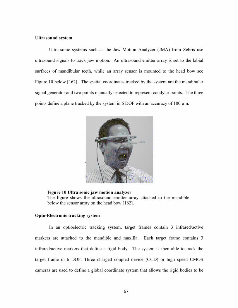
67
Ultrasound system
Ultra-sonic systems such as the Jaw Motion Analyzer (JMA) from Zebris use
ultrasound signals to track jaw motion. An ultrasound emitter array is set to the labial
surfaces of mandibular teeth, while an array sensor is mounted to the head bow see
Figure 10 below [162]. The spatial coordinates tracked by the system are the mandibular
signal generator and two points manually selected to represent condylar points. The three
points define a plane tracked by the system in 6 DOF with an accuracy of 100 µm.
Figure 10 Ultra sonic jaw motion analyzer
The figure shows the ultrasound emitter array attached to the mandible below the sensor array on the head bow [162].
Opto-Electronic tracking system
In an optioelectric tracking system, target frames contain 3 infrared/active
markers are attached to the mandible and maxilla. Each target frame contains 3
infrared/active markers that define a rigid body. The system is then able to track the
target frame in 6 DOF. Three charged coupled device (CCD) or high speed CMOS
cameras are used to define a global coordinate system that allows the rigid bodies to be
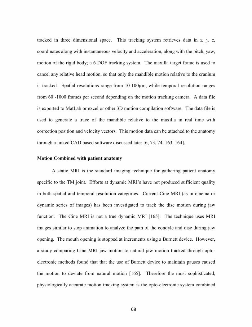
68
tracked in three dimensional space. This tracking system retrieves data in x, y, z,
coordinates along with instantaneous velocity and acceleration, along with the pitch, yaw,
motion of the rigid body; a 6 DOF tracking system. The maxilla target frame is used to
cancel any relative head motion, so that only the mandible motion relative to the cranium
is tracked. Spatial resolutions range from 10-100µm, while temporal resolution ranges
from 60 -1000 frames per second depending on the motion tracking camera. A data file
is exported to MatLab or excel or other 3D motion compilation software. The data file is
used to generate a trace of the mandible relative to the maxilla in real time with
correction position and velocity vectors. This motion data can be attached to the anatomy
through a linked CAD based software discussed later [6, 73, 74, 163, 164].
Motion Combined with patient anatomy
A static MRI is the standard imaging technique for gathering patient anatomy
specific to the TM joint. Efforts at dynamic MRI’s have not produced sufficient quality
in both spatial and temporal resolution categories. Current Cine MRI (as in cinema or
dynamic series of images) has been investigated to track the disc motion during jaw
function. The Cine MRI is not a true dynamic MRI [165]. The technique uses MRI
images similar to stop animation to analyze the path of the condyle and disc during jaw
opening. The mouth opening is stopped at increments using a Burnett device. However,
a study comparing Cine MRI jaw motion to natural jaw motion tracked through opto-
electronic methods found that that the use of Burnett device to maintain pauses caused
the motion to deviate from natural motion [165]. Therefore the most sophisticated,
physiologically accurate motion tracking system is the opto-electronic system combined
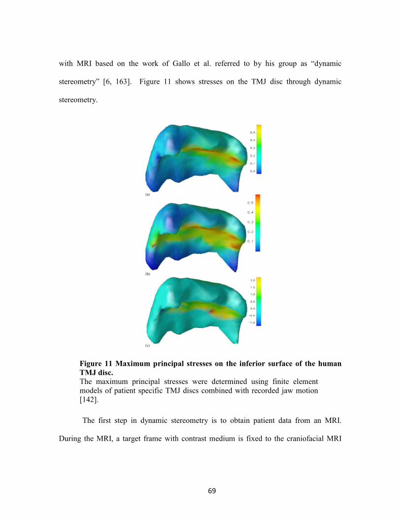
69
with MRI based on the work of Gallo et al. referred to by his group as “dynamic
stereometry” [6, 163]. Figure 11 shows stresses on the TMJ disc through dynamic
stereometry.
Figure 11 Maximum principal stresses on the inferior surface of the human
TMJ disc.
The maximum principal stresses were determined using finite element models of patient specific TMJ discs combined with recorded jaw motion [142].
The first step in dynamic stereometry is to obtain patient data from an MRI.
During the MRI, a target frame with contrast medium is fixed to the craniofacial MRI

70
arm to establish a coordinate basis for transfer between MRI and jaw motion tracking.
After the MRI, the contrast agent target frame is measured to the maxilla jaw tracking
target frame. Now the coordinate systems can be merged when the MRI is imported to a
CAD/CAM software such as SolidWorks. The MRI anatomy file is meshed and
imported into a finite element program. The motion tracking data is recorded by software
developed by Gallo et al. and processed into a rigid body trace of the motion of the
mandible. The motion tracking data is transferred to the patient specific finite element
model for analysis, where the data can be analyzed. Gallo et al. performed stress profile
analysis using material properties gathered from animal studies. With the combined MRI
anatomy of the disc, material properties, and motion data, many finite element models
were constructed to simulate joint loading conditions and TMJ disc damage that
mimicked the in vivo environment [74, 163, 164, 166].
Gallo et al. used the MRI combined with jaw tracking to determine aspect ratios,
instantaneous velocities and distances of stress field translations. These data were used to
evaluate the amount of work the TMJ disc was subjected to during opening/closing along
with laterotrusive movements. The studies show laterotrusive movement generated over
fifty percent more work than previously thought to the contralateral side, while results in
opening and closing set a baseline exposure for work during cyclic loading that could
potentially lead to fatigue failure. In a similar study Nickel and Iwasaki et al. compared
patients with TMD to patients without history of the disorder. The study found that
patients with TMD sustained TMJ loads 9.5-65% higher than subjects with normal disc
position. A range of biting forces were compared during the study, with TMD patients

71
experiencing higher joint loads, indicating that pathological loading to the TMJ disc is
present in TMD patient and that continued overloading could lead to progression [144].
Proposed Research and Scope of Project
Recent studies in our lab have shown that levels of oxygen and glucose
significantly affect cell proliferation and matrix synthesis of porcine TMJ disc cells in
vitro. This suggests that transport of nutrients through the extracellular matrix (ECM)
plays a key role in maintaining the physiological concentration of nutrients, further
implying that deviations from physiological levels of nutrient or mechanical loading may
initiate tissue remodeling and matrix degradation. We have also found that the human
TMJ disc has very low diffusivity compared to other fibrocartilages (hyaline and IVD).
Our preliminary data suggest that a steep nutrient concentration gradient might exist in
the TMJ disc and that this nutrient environment is uniquely vulnerable to pathological
mechanical loadings. Therefore, the goal of this research study is to characterize the
human mechanical properties of the TMJ disc and determine the human transport
properties of the disc under mechanical strain.
Hypothesis
Our central hypothesis is that mechanical properties will vary regionally across
the TMJ disc and that sustained mechanical loading can alter solute transport and nutrient
levels in the TMJ disc as well as mechanical function resulting in disc derangement and
degeneration.

72
3 TRANSPORT PROPERTIES IN HUMAN TMJ DISCS
3.1 Abstract
This study investigated the effect of mechanical strain on solute diffusion in
human TMJ discs (mean cadaver age 77.8) using the electrical conductivity method. The
electrical conductivity, as well as small ion diffusivity, of male and female TMJ discs
was determined under three compressive strains. In the male group, the average disc
electrical conductivity (mean ± sd) at 0% strain was 5.14±0.97 mS/cm, decreased to
4.50±0.91 mS/cm (-12.3%) at 10% strain, and 3.93±0.81 mS/cm (-23.5%) at 20%
compressive strain. Correspondingly, the average disc relative ion diffusivity at 0%
strain was 0.44±0.08, decreased to 0.40±0.08 (-8.9%) at 10% strain, and 0.36±0.08 (-
16.7%) at 20% compressive strain. In the female group, the average disc electrical
conductivity at 0% strain was 5.84±0.59 mS/cm, decreased to 5.01±0.50 mS/cm (-14.2%)
at 10% strain, and 4.33±0.46 mS/cm (-25.8%) at 20% compressive strain.
Correspondingly, the average disc relative ion diffusivity at 0% strain was 0.49±0.05,
decreased to 0.43±0.04 (-11.3%) at 10% strain, and 0.39±0.04 (-19.9%) at 20%
compressive strain. The results indicated that mechanical strain significantly impeded
solute diffusion through the disc. This mechanical strain effect was larger in the female
than in the male human TMJ disc. This study may provide new insights into TMJ
pathophysiology.
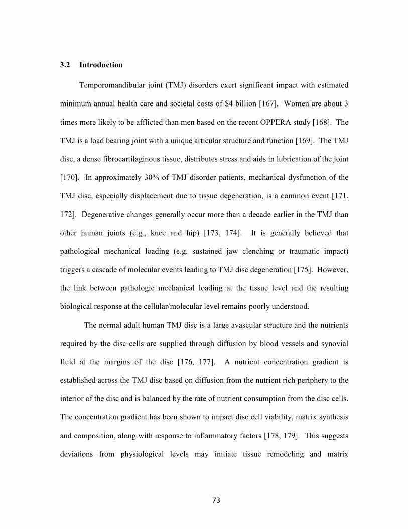
73
3.2 Introduction
Temporomandibular joint (TMJ) disorders exert significant impact with estimated
minimum annual health care and societal costs of $4 billion [167]. Women are about 3
times more likely to be afflicted than men based on the recent OPPERA study [168]. The
TMJ is a load bearing joint with a unique articular structure and function [169]. The TMJ
disc, a dense fibrocartilaginous tissue, distributes stress and aids in lubrication of the joint
[170]. In approximately 30% of TMJ disorder patients, mechanical dysfunction of the
TMJ disc, especially displacement due to tissue degeneration, is a common event [171,
172]. Degenerative changes generally occur more than a decade earlier in the TMJ than
other human joints (e.g., knee and hip) [173, 174]. It is generally believed that
pathological mechanical loading (e.g. sustained jaw clenching or traumatic impact)
triggers a cascade of molecular events leading to TMJ disc degeneration [175]. However,
the link between pathologic mechanical loading at the tissue level and the resulting
biological response at the cellular/molecular level remains poorly understood.
The normal adult human TMJ disc is a large avascular structure and the nutrients
required by the disc cells are supplied through diffusion by blood vessels and synovial
fluid at the margins of the disc [176, 177]. A nutrient concentration gradient is
established across the TMJ disc based on diffusion from the nutrient rich periphery to the
interior of the disc and is balanced by the rate of nutrient consumption from the disc cells.
The concentration gradient has been shown to impact disc cell viability, matrix synthesis
and composition, along with response to inflammatory factors [178, 179]. This suggests
deviations from physiological levels may initiate tissue remodeling and matrix

74
degeneration. The rate of solute diffusion in tissue is governed by solute diffusivities
which are affected by the composition and structure of the matrix, as well as mechanical
strains on the tissue. Recently, we examined the effect of mechanical loading on small
ion diffusivity in porcine TMJ discs using the electrical conductivity method. The results
indicated that solute diffusivities in the TMJ disc are much lower than the values in other
cartilaginous tissues, and compressive strain significantly impeded solute transport in the
porcine TMJ disc [61]. Moreover, our cell metabolic studies have shown that porcine
TMJ discs have higher cell densities and nutrient consumption rates than articular
cartilage and the intervertebral disc (IVD) [180]. Therefore, it is likely that a steeper
nutrient gradient may exist in TMJ discs and is vulnerable to pathological events which
impede nutrient supply, including sustained joint loading due to jaw clenching. Although
pigs have been considered the best TMJ biomechanical experimental model [181, 182],
the mechanical and transport properties of TMJ discs have not been fully compared due
to the lack of human material property data. Especially, to our knowledge, the transport
properties (e.g., solute diffusivity) and the effect of mechanical loading on these
properties, in the human TMJ disc have not been determined. Such knowledge is
important for translating the research findings from an animal model to human.
The electrical conductivity method has been employed to determine the effect of
mechanical strains on ion diffusivities in hydrogels, IVD, and porcine TMJ discs [61,
183, 184]. In these studies, a relationship between ion diffusivities and the porosity of
the material were also determined. In this study, we adopted this method to study the
impact of mechanical loading on the solute transport in human TMJ discs. We
75
hypothesized that the electrical conductivity of the human TMJ disc was mechanical
strain-dependent due to changes in tissue porosity caused by tissue compression. In
addition, we sought to determine the electrical conductivity in both male and female
subjects to investigate if there were sex differences. Therefore, the objective of this study
was to measure the electrical conductivity of male and female human TMJ discs under
three compressive strains in five disc regions.
3.3 Materials and Methods
Specimen preparation
Human TMJ discs were harvested from the left TMJ of fresh cadavers in the
MUSC gross anatomy laboratory with institutional approval. Discs with visible signs of
degeneration or trauma were removed, leaving 12 male and 12 female discs for the study.
The mean cadaver age was 76.0±7.0 for males and 79.6±10.5 for females. The extracted
TMJ disc was wrapped in cellophane and placed into plastic bags. Saline moistened
gauze was added to the bag to maintain humidity and prevent specimen dehydration. The
disc sample was immediately used for porosity and conductivity measurement. Care was
taken to prevent specimen dehydration during specimen preparation. Cylindrical plugs
were punched from five disc regions (anterior, lateral, intermediate, medial, and
posterior) using a 5mm corneal trephine (Figure 12a below). The plugs were microtomed
on a freezing stage to remove the natural concave tissue shape and to allow for a flat
surface during electrical conductivity measurements. Each time, one plug was punched
and microtomed in a moisture-controlled dissection hood. The rest of the disc was

76
wrapped and placed in the same sealed plastic bag. Prior to the water content
measurement, the specimen was thawed at room temperature for 20 minutes in a sealed
plastic vial. The initial height of the specimen was measured by a custom-designed
electrical current sensing micrometer to avoid sample compression. The prepared
specimens (n=60 for male, n=60 for female) had an average height of 1.49±0.46 mm.
Three conductivity measurements were made on each specimen corresponding to the
three levels of compressive strain (0%, 10%, and 20%) based on the initial height. Both
finite element analysis and MRI imaging data have shown that the change of human TMJ
disc thickness during normal joint motion is about 15-20% [185]. The sustained
mechanical loading, such as bruxism, may induce 20-30% change of the disc thickness.
Porosity (water volume fraction) measurement
The porosity or water volume fraction of specimens at the undeformed state (w
0φ)
was determined using a buoyancy method [186]. Briefly, the weight of the specimens in
air (Wwet) and the weight in phosphate buffered saline (PBS) solution (WPBS) were
measured using the density-determination kit of a Sartorius analytical balance (Sartorius
YDK01, Germany). After measuring electrical conductivity, specimens were lyophilized
and the dry weights (Wdry) were recorded. According to Archimedes’ principle and the
biphasic assumption, the water volume fraction was determined by:

77
w
PBS
PBSwet
drywetw
WW
WW
ρρφ
−−
=0
3
where PBSρ is the mass density of PBS (1.005 g/ml), and wρ
is the mass density of
water. The TMJ disc tissue was considered as a biphasic mixture, consisting of solid
phase and free water phase. Both phases are assumed to be incompressible. Thus, the
water volume fraction of the specimen at different compression levels (wφ ) can be
calculated by [157]:
e
eww
++=
1
0φφ
4
where e is the tissue dilatation. Considering one-dimensional confined compression in
this study, the tissue dilatation is equal to the compressive strain.
Electrical conductivity measurement
In this study, the impact of mechanical strain on electrical conductivity of human
TMJ disc was examined in the superior-inferior direction only. This was achieved by
using a confined chamber to ensure that the applied mechanical strain and measured
electrical conductivity were aligned in the same direction see Figure 12b below. The
superior-inferior direction is the main transport route in the TMJ disc due to its short
diffusion distance compared to the anterior-posterior and medial-lateral directions. The
electrical conductivity was measured based on the principle of a four wire resistance test
using a Keithley Sourcemeter (Model 2400, Keithley Instruments, Inc., Cleveland, OH)

78
and a custom designed conductivity chamber reported previously [61, 187]. Briefly, the
conductivity apparatus consists of two stainless steel current electrodes coaxial to two
Teflon-coated Ag/AgCl voltage electrodes placed on the top and bottom of a cylindrical
nonconductive Plexiglass chamber (5mm diameter). The specimen was placed inside the
chamber for measurement and compression (Figure 12b below).
(a)
(b)
Figure 12 (a) Schematic of specimen preparation. (b) Schematic of apparatus
for measuring electrical conductivity
The region and size of test specimens were removed from five regions of each disc, see Figure 11 (a). The conductivity meter shown in Figure 11 (b) consisted of two stainless steel current electrodes, two Ag/AgCl voltage-sensing electrodes, a nonconductive Plexiglass chamber, a current sensing micrometer, and a sourcemeter.
Lateral Medial
Anterior
Intermediate
Human TMJ disc
5mm
1.5
mm
Disc specimen
Posterior V
Current sensing
I
Sourcemeter
Tissue
Current
Voltage (Ag/AgCl) electrodes
Cylindrical
chamber

79
The resistance (R) values across the specimens were measured at a low, constant current
density of 0.015 mA/cm2. The height of the specimen was measured with an electrical
current sensing micrometer. The electrical conductivity (χ) values of the specimens were
calculated by:
AR
h
⋅=χ
5
where h and A are the height and cross-sectional area of the specimens, respectively. The
precision for the resistance measurements was 0.5 Ω while the height measured with an
accuracy of ±1.0 µm. The electrical conductivity of the specimen was measured at 0%,
10%, and 20% compression levels. The confined compression of the tissue specimen
was achieved by lowering the micrometer to the desired height (Figure 12b above) and
fluid was allowed to exude from the sample through a small clearance between the
chamber and the current electrodes. A resistance measurement was taken at a 15 minute
interval after the specimen was compressed to ensure the sample had reached equilibrium
with no fluid exudation. The influence of the order of the strain level on the electrical
conductivity was not examined in this study, since the order of the strain level is not
expected to affect the equilibrium response. All conductivity tests were performed in a
bathing solution of PBS at a physiological concentration of 0.15M (pH 7.4) at room
temperature (22°C).

80
Ion diffusivity calculation
Under a zero fluid flow condition, the measured electrical conductivity (χ) of a
tissue in NaCl solution is related to intrinsic cation and anion diffusivities (Di, i = +, - )
by [188, 189]:
RTDcDcF w
c /)(2 −−++ += φχ
6
where Fc is the Faraday constant, R is the gas constant, T is the temperature, c+ is the
cation ion concentration, and c- is the anion concentration. The TMJ disc was considered
as uncharged in this study, since biochemical studies have shown that the GAG content
of human and porcine TMJ discs is very low (< 4% dry weight) compared to hyaline
cartilage and the IVD [190, 191]. More importantly, the high ionic concentration of
bathing solution (0.15 M) assured a small osmotic effect on the diffusion process. For
uncharged tissues in NaCl solution, the relative diffusivity (D/D0) of NaCl is simply
related to the relative conductivity by [183]:
00 χφχ
wD
D =
7
where D is the mean ion diffusivity of Na+ and Cl- in tissue, D0 is the mean ion
diffusivity in the bathing solution, χ is the electrical conductivity of the tissue, and χ0 is
the conductivity of the bathing solution. In our analysis, Na+ and Cl− were assumed to
carry the electrical current because these ions are the primary ionic components of PBS.

81
Statistical analysis
The electrical conductivity, porosity, and relative ion diffusivity were determined
for each specimen (5 regions per disc) under three compressive strains. Mixed effects
analysis of variance (ANOVA) was used to investigate the main and interaction effects
on each outcome of disc region, strain, sex, and age. The models accommodated
correlations among measurements from the same disc both within and between the three
strains, allowing that measurements on a disc from the same region may be more highly
correlated than measurements from different regions. SAS/STAT® software version 9.3
(SAS Institute Inc., Cary, NC) was used for statistical analyses and statistical significance
was determined at p-values < 0.05. Pilot data analyses showed that none of the main and
interaction effects involving region or age emerged as statistically significant. Hence
subsequent analyses were performed using the average response among the 5 regions for
each disc at each strain. Two-way mixed effects ANOVA incorporating a random effect
for disc was used to assess the effects of strain and sex on this average outcome.
3.4 Results
Electrical conductivity
The effects of compressive strain on electrical conductivity in the five disc
regions are shown in Figure 13 below. No significant regional variation was detected at
all strain levels in both the male and female groups. However, there was a significant
decrease in electrical conductivity with increases of compressive strain in all five disc
regions in both the male and female groups (p<0.0001). In the male group, the average

82
disc electrical conductivity (mean ± sd) at 0% strain was 5.14±0.97 mS/cm, decreased to
4.50±0.91 mS/cm (-12.3%) at 10% strain, and 3.93±0.81 mS/cm (-23.5%) at 20% strain
(Figure 13 below).

83
Anterior Lateral Intermediate Medial Posterior
0
2
4
6
8
10
Ele
ctr
ica
l C
on
du
cti
vit
y (
mS
/cm
)
Disc Region
0% strain
10% strain
20% strain
Male
Anterior Lateral Intermediate Medial Posterior
0
2
4
6
8
10
Female 0% strain
10% strain
20% strain
Ele
ctr
ica
l C
on
du
cti
vit
y (
mS
/cm
)
Disc Region
Figure 13 Effect of compressive strains on regional distribution of electrical
(a)
(b)

84
conductivity by gender
(a) Male electrical conductivity by region (b), Female electrical conductivity by region. Mixed effects ANOVA as described in the Statistical Analysis section confirmed the lack of significant difference in outcomes among the disc regions and among two-factor interactions between disc region and strain or sex or age of disc donor.
In the female group, the average disc electrical conductivity at 0% strain was
5.84±0.59 mS/cm, decreased to 5.01±0.50 mS/cm (-14.2%) at 10% strain, and 4.33±0.46
mS/cm (-25.8%) at 20% strain (Figure 13 above). The electrical conductivity was higher
in the female than in the male TMJ disc, although the differences were not statistically
significant (p=0.08). Interestingly, the interaction between strain and gender was found
to be statistically significant (p=0.0004). The impact of the mechanical strain on the
electrical conductivity was higher in the female TMJ disc compared to the male.
Porosity (water volume fraction)
The effect of compressive strain on tissue porosity (water volume fraction) in the
five disc regions is shown in Figure 14 below. No significant regional variation was
detected at all strain levels in both the male and female groups. However, there was a
significant decrease in tissue porosity with increases of compressive strain in all five disc
regions in both the male and female groups (p<0.0001).

85
Anterior Lateral Intermediate Medial Posterior
0.0
0.1
0.2
0.3
0.4
0.5
0.6
0.7
0.8
0.9
1.0
Po
ros
ity
Disc Region
0% strain
10% strain
20% strainMale
Anterior Lateral Intermediate Medial Posterior
0.0
0.1
0.2
0.3
0.4
0.5
0.6
0.7
0.8
0.9
1.0
Female 0% strain
10% strain
20% strain
Po
ros
ity
Disc Region
(a)
(b)

86
Figure 14 Effect of compressive strains on regional distribution of porosity by
gender
(a),Male porosity by region (b), Female porosity by region. Mixed effects ANOVA as described in the Statistical Analysis section confirmed the lack of significant difference in outcomes among the disc regions and among two-factor interactions between disc region and strain or sex or age of disc donor.
In the male group, the average disc porosity at 0% strain was 0.760±0.011,
decreased to 0.733±0.012 (-3.6%) at 10% strain, and 0.700±0.014 (-7.9%) at 20% strain
(Figure 14 above). In the female group, the average disc porosity at 0% strain was
0.770±0.014, decreased to 0.745±0.016 (-3.2%) at 10% strain, and 0.713±0.018 (-7.4%)
at 20% strain (Figure 14 above). The porosity was higher in the female than in the male
TMJ disc, although the differences were not statistically significant (p=0.0533). The
interaction between strain and sex was found statistically significant (p=0.0218), even
though the magnitude of this effect appears very small.
Ion diffusivity
Electrical conductivities of PBS at 22C were measured using an Orion
conductivity meter (Model 150Aplus, Beverly, MA). The value of 15.5 mS/cm for
solution conductivity was used to normalize the measured conductivity data of tissues to
calculate the relative ion diffusivity. The effect of compressive strain on relative ion
diffusivity in the five disc regions is shown in Figure 15 below. No significant regional
variation was detected at all strain levels in both the male and female groups. However,
there was a significant decrease in ion diffusivity with increases of compressive strain in
all five disc regions (p<0.0001).

87
Anterior Lateral Intermediate Medial Posterior
0.0
0.2
0.4
0.6
0.8
Re
lati
ve D
iffu
siv
ity
Disc Region
0% strain
10% strain
20% strain
Male
Anterior Lateral Intermediate Medial Posterior
0.0
0.2
0.4
0.6
0.8
Female 0% strain
10% strain
20% strain
Re
lati
ve D
iffu
siv
ity
Disc Region
(b)
(a)

88
Figure 15 Effect of compressive strains on regional distribution of ion
diffusivity by sex
(a), Male ion diffusivity by region (b), Female ion diffusivity by region. Mixed effects ANOVA as described in the Statistical Analysis section confirmed the lack of significant difference in outcomes among the disc regions and among two-factor interactions between disc region and strain or sex or age of disc donor.
In the male group, the average disc relative ion diffusivity at 0% strain was
0.44±0.08, decreased to 0.40±0.08 (-8.9%) at 10% strain, and 0.36±0.08 (-16.7%) at 20%
strain (Figure 15 above). In the female group, the average disc relative ion diffusivity at
0% strain was 0.49±0.05, decreased to 0.43±0.04 (-11.3%) at 10% strain, and 0.39±0.04
(-19.9%) at 20% strain (Figure 15 above). The relative ion diffusivity was higher in the
female than in the male TMJ disc, although the differences were not statistically
significant (p=0.1502). The interaction between strain and sex was found statistically
significant (p=0.0004). The impact of the mechanical strain on the relative ion diffusivity
was higher in the female TMJ disc compared to the male.
Finally conductivity, porosity and diffusivity were analyzed with respect to %
stran compression as shown in Figure 16 below. Mechanical strain is shown to decrease
electrical conductivity and porosity. The association between conductivity and porosity
is shown in Figure 17 below.
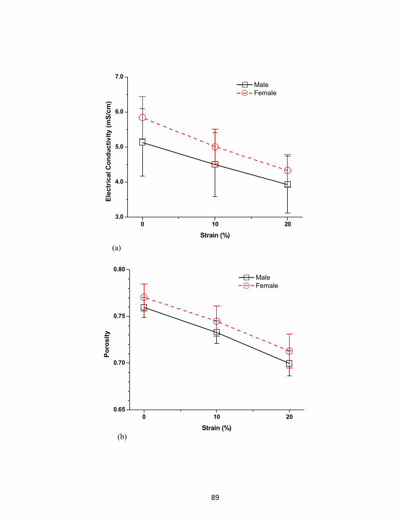
89
0 10 20
3.0
4.0
5.0
6.0
7.0
Ele
ctr
ica
l C
on
du
cti
vit
y (
mS
/cm
)
Strain (%)
Male
Female
0 10 20
0.65
0.70
0.75
0.80
Po
ros
ity
Strain (%)
Male
Female
(a)
(b)

90
0 10 20
0.2
0.3
0.4
0.5
0.6
Re
lati
ve D
iffu
siv
ity
Strain (%)
Male
Female
Figure 16 The effect of sex on the strain-dependent electrical conductivity
(a), porosity (water volume fraction) (b), and relative ion diffusivity of human TMJ disc (c). Note that the average value of the five regions from the same disc (male n=12, female n=12) were used to represent the outcomes of that disc since the regional variations were found not statistically significant in this study.
(c)

91
0.68 0.70 0.72 0.74 0.76 0.78
2.0
3.0
4.0
5.0
6.0
7.0
Ele
ctr
ica
l C
on
du
cti
vit
y (
mS
/cm
)
Porosity
Male
0.68 0.70 0.72 0.74 0.76 0.78
Female
Figure 17 Association between the electrical conductivity and the porosity
(water volume fraction).
Each dashed line depicts the data for one disc.
3.5 Discussion
The objective of this study was to investigate the effect of mechanical strain on
small ion diffusivity in human TMJ discs using the electrical conductivity method. The
measured electrical conductivity and calculated ion diffusivity from this study indicated
that compressive mechanical strain significantly impeded solute diffusion through the
disc. Our results further indicated that this mechanical strain effect was larger in the
female than in the male human TMJ disc.

92
The decrease in electrical conductivity with increasing mechanical strain in the
human TMJ disc was mainly due to the porosity (water volume fraction) reduction
caused by increasing compression (see Figure 14 and Figure 16b above). Tissue
compression caused fluid exudation and a corresponding decrease in tissue porosity. The
reduction in porosity resulted in decreased ion diffusivity in the tissue and subsequently
decreased electrical conductivity [183, 184]. This is in agreement with previous studies
reporting decreasing diffusivity of solutes with increasing static compressive strain in
articular cartilage [192, 193], IVD tissues [194, 195], and porcine TMJ discs [61]. The
strain-dependent electrical conductivity and ion diffusivity indicated that mechanical
strain impeded solute transport in human TMJ discs. Moreover, the impact of
mechanical strain on ion diffusivity was larger in the female than in the male TMJ disc.
Figure 17 above showed the association between the electrical conductivity and the
porosity for the male and female human TMJ discs. It seems that the electrical
conductivity of the female disc is more sensitive to the change of the tissue porosity due
to the mechanical strain. This suggested that a subtle tissue structure difference might
exist between the male and female TMJ discs. However, to our knowledge, there is no
study to quantitatively compare the biochemical composition (e.g., water, GAG, and
collagen contents) and tissue structure (e.g., fiber density and orientation) in human male
and female TMJ discs. This will be a very interesting thing to look at in our next study.
The pathological implication of this finding needs to be further explored. Although the
recent OPPERA study confirmed a long standing report of 3 to1 incidence of females to
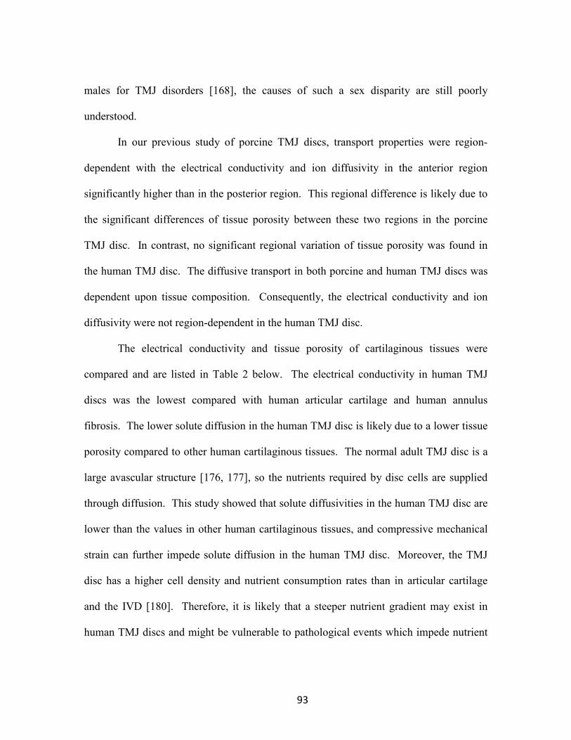
93
males for TMJ disorders [168], the causes of such a sex disparity are still poorly
understood.
In our previous study of porcine TMJ discs, transport properties were region-
dependent with the electrical conductivity and ion diffusivity in the anterior region
significantly higher than in the posterior region. This regional difference is likely due to
the significant differences of tissue porosity between these two regions in the porcine
TMJ disc. In contrast, no significant regional variation of tissue porosity was found in
the human TMJ disc. The diffusive transport in both porcine and human TMJ discs was
dependent upon tissue composition. Consequently, the electrical conductivity and ion
diffusivity were not region-dependent in the human TMJ disc.
The electrical conductivity and tissue porosity of cartilaginous tissues were
compared and are listed in Table 2 below. The electrical conductivity in human TMJ
discs was the lowest compared with human articular cartilage and human annulus
fibrosis. The lower solute diffusion in the human TMJ disc is likely due to a lower tissue
porosity compared to other human cartilaginous tissues. The normal adult TMJ disc is a
large avascular structure [176, 177], so the nutrients required by disc cells are supplied
through diffusion. This study showed that solute diffusivities in the human TMJ disc are
lower than the values in other human cartilaginous tissues, and compressive mechanical
strain can further impede solute diffusion in the human TMJ disc. Moreover, the TMJ
disc has a higher cell density and nutrient consumption rates than in articular cartilage
and the IVD [180]. Therefore, it is likely that a steeper nutrient gradient may exist in
human TMJ discs and might be vulnerable to pathological events which impede nutrient
94
supply, including sustained joint loading due to jaw clenching. The strain distribution in
the TMJ disc in vivo is complex and can be determined through finite element analysis
[185]. The strain-dependent diffusivity established in this study needs to be incorporated
into finite element models to assess the overall impact of physiological/pathological
loading on solute transport in human TMJ discs.
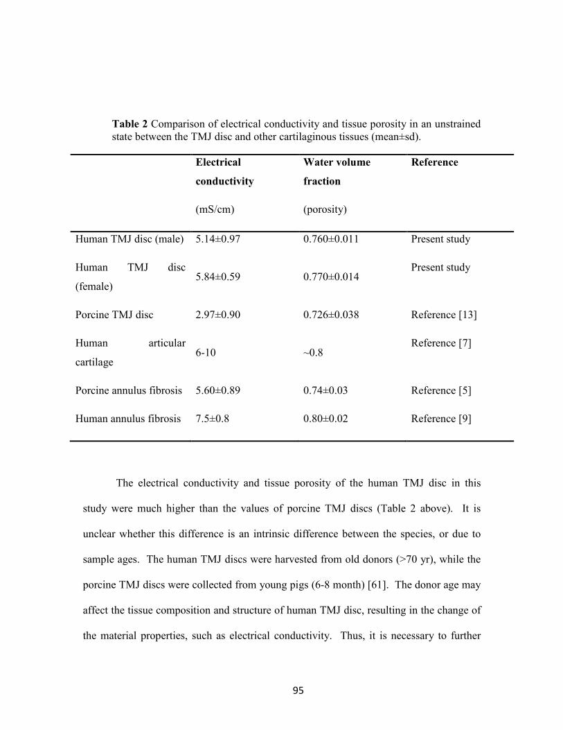
95
Table 2 Comparison of electrical conductivity and tissue porosity in an unstrained state between the TMJ disc and other cartilaginous tissues (mean±sd).
Electrical
conductivity
(mS/cm)
Water volume
fraction
(porosity)
Reference
Human TMJ disc (male) 5.14±0.97 0.760±0.011 Present study
Human TMJ disc
(female) 5.84±0.59 0.770±0.014
Present study
Porcine TMJ disc 2.97±0.90 0.726±0.038 Reference [13]
Human articular
cartilage 6-10 ~0.8
Reference [7]
Porcine annulus fibrosis 5.60±0.89 0.74±0.03 Reference [5]
Human annulus fibrosis 7.5±0.8 0.80±0.02 Reference [9]
The electrical conductivity and tissue porosity of the human TMJ disc in this
study were much higher than the values of porcine TMJ discs (Table 2 above). It is
unclear whether this difference is an intrinsic difference between the species, or due to
sample ages. The human TMJ discs were harvested from old donors (>70 yr), while the
porcine TMJ discs were collected from young pigs (6-8 month) [61]. The donor age may
affect the tissue composition and structure of human TMJ disc, resulting in the change of
the material properties, such as electrical conductivity. Thus, it is necessary to further

96
examine the age effect in future studies. In this study, the electrical conductivity was
measured in the superior-inferior direction. However, considering the fiber structure of
the TMJ disc, the diffusion in the TMJ disc is significantly anisotropic [58]. Although
the superior-inferior direction is the main transport route in TMJ disc, the impact of
mechanical strain on solute diffusion in the anterior-posterior and medial-lateral
directions still needs to be examined. It is also necessary to determine the impact of
mechanical strain on the diffusivities of other essential solutes, such as oxygen and
glucose. In summary, the effect of mechanical strain on small ion diffusivity in human
TMJ discs was investigated using electrical conductivity methods. The results indicated
that mechanical strain significantly impeded solute diffusion through the disc. This
mechanical strain effect was larger in the female than in the male human TMJ disc.
Although pigs have been considered as a good animal model to study human TMJ disc
mechanics, the similarity in terms of tissue material properties still needs further
validation.

97
4 Transport Properties in Porcine Articular Cartilage
4.1 Abstract
Various preservation solutions have been evaluated for longer hypothermic
cartilage storage for tissue transplantation, however, the results are mixed. This research
was to determine whether phosphate buffered saline (PBS) or organ preservation
solutions would preserve both the extracellular matrix and chondrocytes of articular
cartilage better than culture medium during refrigerated storage in the time frame that
cartilage is stored for clinical use. Porcine cartilage plugs were stored, without the
underlying bone, in culture medium with and without fetal bovine serum (FBS), PBS,
Belzer’s and Unisol solutions for 1 month at 4ºC. Metabolic activity was tested using a
resazurin reduction method and matrix permeability was evaluated by measuring
electrical conductivity. Storage in culture medium with 10% FBS was shown to provide
good cartilage metabolic function for 7 days decreasing to about 36% after 1 month of
storage. There was no significant difference between samples stored in culture medium
with and without FBS after 1 month of storage (p=0.5005). Cartilage plugs assessed after
1 month of storage demonstrated less viability and higher permeability in alternative test
solutions (PBS, Belzer’s and Unisol) compared with culture medium (p<0.05).
Refrigerated storage of cartilage in PBS and two solutions (Belzer’s and Unisol) designed
for optimal refrigerated tissue and organ storage results in loss of chondrocyte function
and retention of matrix permeability. In contrast, significantly better retention of
chondrocyte function and loss of matrix permeability was observed in culture medium.

98
Future research is focused on combining retention of chondrocyte function and matrix
permeability by storage solution formulation.
List of Abbreviations
DMEM Dulbecco’s modified Eagle’s medium
ECM extracellular matrix
GAG Glycosaminoglycan
FBS fetal bovine serum
PBS phosphate buffered saline
4.2 Introduction
Donated donor-derived osteochondral tissue grafts are typically harvested within
24 hours of donor death and banked at 4ºC for up to 42 days for repair of clinical
cartilage defects. Cartilage plugs are employed in mosaicplasty procedures for smaller
defects less than 3 cm wide and less than 1cm deep. Osteochondral allograft
transplantation has been an effective treatment option with promising long-term clinical
outcomes for larger focal posttraumatic defects in the knee for young, active individuals
[196]. There were 15,797 allogeneic human articular cartilage procedures performed in
the United States of America during 2006 [197]. It is anticipated that many more
procedures would be performed if better cartilage preservation methods were available.
99
The duration of hypothermic refrigerated storage of osteochondral grafts is rather
unusual. Although the cells in heart valve leaflets persist for weeks during hypothermic
refrigeration in culture medium [198, 199], most tissues are refrigerated for only hours
before significant loss of cell viability and tissue function occur. There are, however,
reports supporting chondrocyte survival for days or weeks of hypothermic storage in their
natural extracellular matrix in humans [200-203] and several animal species [203-212].
Because such survival was considered unusual, compared with other tissue types, we
previously initiated research to assess the impact of 4ºC storage in DMEM culture
medium on cartilage cell viability and establish whether or not cartilage plugs were an
acceptable model compared with bisected femoral heads with both cartilage and bone
tissue present [213]. We also included extracellular matrix (ECM) permeability
evaluation during storage because prolonged storage of cartilage with viable cells might
promote the release of enzymes that impact tissue material properties [213]. We found
that storage in DMEM culture medium with 10% FBS provides good cartilage viability
for 7 days. In addition, there was a marked tendency for cartilage plugs to demonstrate
higher viability values than the femoral heads. However, 1 month storage of cartilage
resulted in loss of both chondrocyte viability and ECM permeability [213].
Various preservation solutions have been evaluated for longer hypothermic
cartilage storage, however, the results are mixed. Onuma et al. [205] compared DMEM,
saline, EuroCollins solution, and UW solution to determine which provided the best
hypothermic preservation of rat osteochondral tissues. They concluded that UW solution,
an intracellular type solution, was the most suitable. In contrast, Teng et al. [212] clearly
100
demonstrated the positive impact of more complex culture media formulation upon
chondrocyte survival such as DMEM, an extracellular type solution. There are other
indications in the literature that media supplementation or modification, such as FBS
supplement, might promote chondrocyte survival. However FBS supplement is also an
issue for tissue bank products due to FBS batch variation and the associated health risks
[212, 214]. To date, extensive research is still needed to determine what type of solution
is best for chondrocyte preservation in cartilage and to define the optimal solution
composition.
Therefore, the purpose of the research presented in this manuscript was further
investigation of cartilage cell and biomaterial properties during storage in a common
culture medium, hypothermic solutions designed for cell, tissue and organ storage and a
commonly used salt solution. Moreover, the effect of FBS supplement on cartilage cell
viability was examined during storage in common culture medium. Improved storage
solutions could result in increased utilization of banked allogeneic cartilage for
reconstruction of articular cartilage defects and possibly storage and distribution of tissue
engineered cartilage.
4.3 Materials and Methods
Specimen Preparation
Articular cartilage, 2-3 mm thick, was obtained from the femoral weight bearing
condyles of animals after they were sacrificed for other experimental studies (bona fide
excess tissues). Bona fide excess tissue is a term used to describe animal-derived
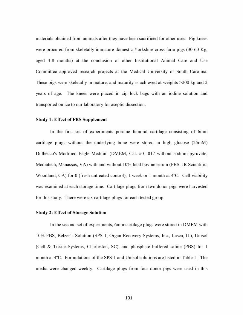
101
materials obtained from animals after they have been sacrificed for other uses. Pig knees
were procured from skeletally immature domestic Yorkshire cross farm pigs (30-60 Kg,
aged 4-8 months) at the conclusion of other Institutional Animal Care and Use
Committee approved research projects at the Medical University of South Carolina.
These pigs were skeletally immature, and maturity is achieved at weights >200 kg and 2
years of age. The knees were placed in zip lock bags with an iodine solution and
transported on ice to our laboratory for aseptic dissection.
Study 1: Effect of FBS Supplement
In the first set of experiments porcine femoral cartilage consisting of 6mm
cartilage plugs without the underlying bone were stored in high glucose (25mM)
Dulbecco's Modified Eagle Medium (DMEM, Cat. #01-017 without sodium pyruvate,
Mediatech, Manassas, VA) with and without 10% fetal bovine serum (FBS, JR Scientific,
Woodland, CA) for 0 (fresh untreated control), 1 week or 1 month at 4ºC. Cell viability
was examined at each storage time. Cartilage plugs from two donor pigs were harvested
for this study. There were six cartilage plugs for each tested group.
Study 2: Effect of Storage Solution
In the second set of experiments, 6mm cartilage plugs were stored in DMEM with
10% FBS, Belzer’s Solution (SPS-1, Organ Recovery Systems, Inc., Itasca, IL), Unisol
(Cell & Tissue Systems, Charleston, SC), and phosphate buffered saline (PBS) for 1
month at 4ºC. Formulations of the SPS-1 and Unisol solutions are listed in Table 1. The
media were changed weekly. Cartilage plugs from four donor pigs were used in this

102
study and were mixed and randomly assigned to each tested group. The cell viability of
cartilage plugs was examined during a 4-day recovery period after 1 month of
hypothermic storage. The cartilage plugs were recovered in DMEM with 10% FBS at 37
ºC. There were twenty cartilage plugs for each tested group in each experiment. The
tissue matrix permeability before and after 1 month hypothermic storage was examined
using the electrical conductivity method. There were twenty cartilage plugs for each
tested group.
Viability Assessment
Chondrocyte metabolic activity was assessed using the resazurin reduction
method. The resazurin reduction assay incorporates a water soluble fluorometric viability
oxidation-reduction (REDOX) indicator which detects metabolic activity by both
fluorescing and changing color in response to chemical reduction of the growth medium.
Metabolically active cells reduce resazurin to fluorescing resorufin [215]. Fresh control
and hypothermically stored tissue samples were placed in 37°C culture conditions for 1
hour to permit adjustment to tissue culture conditions in DMEM plus 10% FBS. The
tissues were then incubated for three hours with resazurin working solution, after which
aliquots of medium were placed in microtiter plate wells and read on a microtiter plate
spectrofluorometer at a wavelength of 590 nm. The data is expressed as the mean ±
standard deviation relative fluorescent units after subtraction of baseline florescence from
incubation controls containing the same amount of culture medium and resazurin reagent
without tissue.

103
Biomaterial Testing
Cartilage plugs were also evaluated for permeability by measuring their electrical
conductivity to determine if cartilage matrix characteristics were being altered during
storage. Specimens were prepared by cutting a 5mm cylindrical plug using a corneal
trephine from the stored 8mm diameter cartilage discs. The samples were tested after 0
and 1 month of storage, the cartilage surfaces were trimmed manually using a sharp
blade. The average height after trimming was 1.53 ± 0.27 mm. Then conductivity was
tested, first in isotonic PBS and then after swelling in hypotonic saline (0.2xPBS). The
electrical conductivity was measured based on the principle of a four wire resistance test
using a Keithley Sourcemeter (Model 2400, Keithley Instruments, Inc., Cleveland, OH)
and a custom designed conductivity chamber reported previously [153, 213]. Briefly, the
conductivity apparatus consists of two stainless steel current electrodes coaxial to two
Teflon-coated Ag/AgCl voltage electrodes placed on the top and bottom of a cylindrical
nonconductive Plexiglass chamber (5mm diameter). The specimen was placed inside the
chamber for measurement. The resistance (R) values across the specimens were
measured at a low, constant DC current density of 0.015 mA/cm2. The height of the
specimen was measured with an electrical current sensing micrometer. The electrical
conductivity (χ) values of the specimens were calculated by:

104
AR
h
⋅=χ
8
where h and A are the height and cross-sectional area of the specimens, respectively. The
precision for the resistance measurements was 0.5 Ω while the height measured with an
accuracy of ±1.0 µm. All electrical conductivity measurements were performed at room
temperature (22°C).
Electrical conductivity is a material property of biological tissues. Its value is
related to the diffusivity of small ions in the tissue, which depend upon tissue
composition and structure [216, 217]. Using an electrical conductivity method, the effect
of matrix composition on solute permeability has previously been studied in hydrogels
and cartilaginous tissues [218, 219]. In this study, we adopted this method to study the
impact of 4ºC storage on cartilage ECM solute permeability. It could be considered that
the measured electrical conductivity mainly represents the tissue property since
chondrocytes normally occupy 1-10% volume of articular cartilage [220]. Moreover, the
transport of small solutes (e.g., ions, oxygen, and glucose) within avascular cartilage
tissues mainly depends on diffusion [221]. Therefore, electrical conductivity was
selected in addition to cell viability in order to evaluate tissue ECM changes that may
affect nutrient transport ability as well as mechanical function in vivo.
Statistical Analysis
The measurements were presented as mean ± standard deviation. One-way
ANOVA with Tukey’s post hoc analysis (p<0.05) was conducted to determine

105
differences in mean values of cell fluorescence units and electrical conductivity. In Study
1, the effect of FBS supplementation on cell viability (n=6) was examined. In Study 2,
the effect of storage solution on cell viability (n=20), as well as on electrical conductivity
(n=20), were examined. SPSS 16.0 software (SPSS Inc., Chicago, IL) was used for all
statistical analyses and significant differences were reported at p-values < 0.05.
4.4 Results
In Study 1, the effect of FBS supplementation on chondrocyte metabolic activity
was assessed using the resazurin reduction method and results are shown in Figure 18
below. After 7 days storage, the chondrocyte metabolic activity of cartilage plugs stored
in DMEM medium with 10% FBS was not significantly different from the value of fresh
controls (p=0.3879), while the metabolic activity of cartilage plugs stored in DMEM
medium without 10% FBS was significantly lower than the value of fresh controls
(p=0.0339). Extended storage reduced the cartilage metabolic activity. The metabolic
activity of cartilage samples dropped to about 36% of the value of the fresh controls after
1 month of storage. There was no significant difference between samples stored with and
without FBS after 1 month of storage (p=0.5005).

106
0
200
400
600
#
#
28 Day7 Day
No FBSNo FBS FBSFBS
RF
U/m
g t
iss
ue
Fresh
Figure 18 Chondrocyte viability assessed by resazurin reduction reaction
Chondrocyte viability during cartilage plug hypothermic storage in culture medium with (open bars) and without FBS (cross hatched bars) assessed by the resazurin reduction metabolic assay (n=6). The data shown are means ± standard deviations. # indicates significant difference compared to fresh controls at p<0.05.
In Study 2, cartilage samples were stored at 4ºC for 1 month in three solutions and
culture medium with 10% FBS followed by cell metabolic activity and matrix
permeability evaluations. All solutions resulted in decreased chondrocyte viability after 1
month of storage (<21% of fresh controls), however, during 4-day follow-up evaluations
under cell culture conditions culture medium supplemented with 10% FBS resulted in
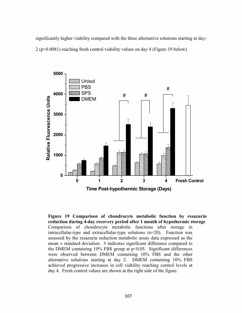
107
significantly higher viability compared with the three alternative solutions starting at day-
2 (p<0.0081) reaching fresh control viability values on day 4 (Figure 19 below).
0 1 2 3 4 5
0
1000
2000
3000
4000
5000
#
#
Re
lati
ve
Flu
ore
sc
en
ce
Un
its
Time Post-hypothermic Storage (Days)
Unisol
PBS
SPS
DMEM
Fresh Control
#
Figure 19 Comparison of chondrocyte metabolic function by resazurin
reduction during 4-day recovery period after 1 month of hypothermic storage
Comparison of chondrocyte metabolic functions after storage in intracellular-type and extracellular-type solutions (n=20). Function was assessed by the resazurin reduction metabolic assay data expressed as the mean ± standard deviation. # indicates significant difference compared to the DMEM containing 10% FBS group at p<0.05. Significant differences were observed between DMEM containing 10% FBS and the other alternative solutions starting at day 2. DMEM containing 10% FBS achieved progressive increases in cell viability reaching control levels at day 4. Fresh control values are shown at the right side of the figure.
108
Simultaneously, samples were prepared for matrix permeability assessment using
the electrical conductivity method. When the plugs were tested in the isotonic saline,
significant differences in electrical conductivity were observed between the control and
plugs stored in culture medium with FBS (p=0.0078), Unisol (p=0.0034), and SPS
(p=0.0068) (Figure 20a below). The differences in isotonic solution were small, however
they were statistically significant and were magnified when the samples were
subsequently swollen in hypotonic solution for testing (Figure 20b below). The electrical
conductivity of samples stored in culture medium was lower than the values of the
samples stored in the other storage solutions (p<0.0001) and only about 60% of the fresh
control (Figure 20b below).

109
Fresh DMEM PBS Unisol SPS
0
2
4
6
8
10
12
14
16
p=0.0068p=0.0034
###
Ele
ctr
ica
l C
on
du
cti
vit
y (
mS
/cm
)
Storage Method
p=0.0078
(a) Isotonic conductivity
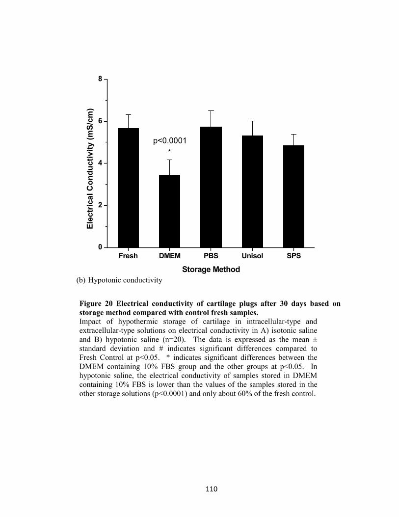
110
Fresh DMEM PBS Unisol SPS
0
2
4
6
8
p<0.0001
*
Ele
ctr
ica
l C
on
du
cti
vit
y (
mS
/cm
)
Storage Method
Figure 20 Electrical conductivity of cartilage plugs after 30 days based on
storage method compared with control fresh samples.
Impact of hypothermic storage of cartilage in intracellular-type and extracellular-type solutions on electrical conductivity in A) isotonic saline and B) hypotonic saline (n=20). The data is expressed as the mean ± standard deviation and # indicates significant differences compared to Fresh Control at p<0.05. * indicates significant differences between the DMEM containing 10% FBS group and the other groups at p<0.05. In hypotonic saline, the electrical conductivity of samples stored in DMEM containing 10% FBS is lower than the values of the samples stored in the other storage solutions (p<0.0001) and only about 60% of the fresh control.
(b) Hypotonic conductivity

111
4.5 Discussion
Chondrocyte viability at the time of implantation is an important factor in
ensuring long-term allograft survival in vivo [222, 223]. Commercially available fresh
osteoarticular allografts are stored for at least seventeen days to allow serologic and
microbiologic testing prior to implantation because of concerns about potential infection
[200, 224]. We found, in agreement with our prior study [213], that storage in DMEM
culture medium with 10% FBS provides good cartilage viability for 7 days. In that study
[213], we compared the assay employed in the present study with Trypan blue. The
metabolic assay provided higher cell viability values at 7 days than Trypan blue, by 28
days the results using the two assays were similar with less than 30% cell viability. Our
interpretation of the results was that many of the chondrocytes may have been damaged
at 7 days, resulting in loss during the cell isolation procedures employing collagenase
required prior to the determination of cell viability using Trypan blue. This may also
explain concerns previously expressed regarding the use of fluorescent assays for
cartilage storage studies [225]. It is possible that the metabolic assay at this time point is
an accurate measure of potential viability if the tissues were implanted rather than
subjected to collagenase digestion. The Trypan blue-excluding cells from fresh and both
7- and 28-day-stored tissues were also able to proliferate. Qualitatively similar appearing
cultures were obtained after 1 week under physiological conditions provided that
consistent numbers of Trypan blue excluding cells were plated.
The presence of FBS improved cell survival after the first week of storage, but
there was no difference in cartilage viability between samples stored with and without

112
FBS after 1 month storage (Figure 18). This last observation leads us to believe that with
further culture medium supplementation, it may be possible to remove FBS from
osteochondral storage solutions reducing concerns about FBS batch variation and the
associated health risks [212, 214].
Comparison of tissue metabolic activity after hypothermic storage in solutions
employed for organ and tissue storage (SPS-1 and Unisol), complex DMEM culture
media supplemented with FBS, and simple PBS yielded unanticipated results (Figure 19).
Belzer’s solution (SPS-1) and Unisol are both examples of “intracellular-type”
preservation solutions which are typically hypertonic and formulated to restrict the
passive exchange of water and ions during hypothermia-induced inhibition of cell
membrane pumps (reviewed, [226]). An intracellular-type solution usually includes a
non-permeating anion such as lactobionate or gluconate to partially replace chloride ions
in the extracellular space. This provides osmotic support to balance the intracellular
oncotic pressure generated by cytosolic macromolecules and their associated counter-ions
locked inside the cell. In contrast, saline and culture media are “extracellular-type”
solutions [226]. They are isotonic with a plasma-like complement of ions that mimics the
normal extracellular environment of cells. Culture media contain a more complete
complement of ions, amino acids and other metabolites that mimic the extracellular
composition of plasma while providing nutritional support. PBS is a simple formulation
consisting of salts and a buffer without nutritional components. Prolonged hypothermic
storage of cells and tissue in extracellular media usually results in cell swelling, due to
cold inactivation of membrane pumps, and cell death [226].

113
Most published studies on cartilage hypothermic storage have employed
extracellular type solutions. However, Onuma et al. [205] compared DMEM, saline,
EuroCollins and Belzer’s solutions to determine which provided the best hypothermic
preservation of rat osteochondral tissues. They concluded on the basis of two assays and
histology that Belzer’s solution (SPS-1) was the most suitable. In contrast, Teng et al.
[212] clearly demonstrated the positive impact of more complex culture media
formulation upon chondrocyte survival, such as DMEM, especially when supplemented
with insulin growth factor-1 or an apoptosis inhibitor (Z-VAD-fmk). Our results confirm
that complex extracellular-type culture media are best for maintenance of chondrocyte
functions, which we have previously shown to correlate with cell survival by trypan blue
[213]. The three alternative solutions, compared to DMEM, resulted in less metabolic
activity after one month that achieved statistical significance during follow-up recovery
incubation under physiologic tissue culture conditions. The resazurin metabolic assay
has an advantage over other more commonly employed assays of being non-cytotoxic, so
the same piece of tissue can be assayed several times as we have done in this study
(Figure 19). Single time point use of the resazurin assay may be hard to interpret due to
fluxes in metabolic activity during recovery from experimental insults or delayed cell
death. This could lead to high readouts compared to assays such as Trypan blue
[Brockbank et al., 2011b]. Alternatively, as already discussed, the differences observed
could be due to reversibly damaged cells that are being further damaged by the isolation
procedure required prior to Trypan blue evaluation. A deficiency of the resazurin assay
in contrast with intracellular viability indicators using florescent microscopy methods is

114
that the assay does not discriminate between cartilage zones, so the results obtained with
this assay are the mean of the entire sample. An alternative way to express our data is to
measure DNA content, after the tissue digestion, and estimate the number of cells present
using the conversion 7.7 pg DNA per cell [Kim et al. 1988] and then extrapolate the
relative metabolic activity per cell. We have expressed the resazurin results in relative
florescent units per mg dry weight of tissue. Future experiments are planned to
determine whether the recovery observed in the culture medium group is due to cells
recovering from reversible injuries or proliferation.
However, when cartilage permeability was assessed the three alternative solutions
all resulted in better retention of permeability in marked contrast with the significant
permeability decrease observed for complex extracellular-type media stored cartilage
(Figure 20b) [213]. The values of the electrical conductivity in this study are comparable
to those in the literature for articular cartilage [Hasegawa et al. 1983]. The electrical
conductivity (11.51±0.73 mS/cm) of porcine articular cartilage measure in this study is
higher than the values (6-10 mS/cm) of human articular cartilage [Hasegawa et al. 1983].
The higher electrical conductivity in young porcine articular cartilage is likely due to
higher tissue porosity (water content) compared with that in human articular cartilage.
The value of electrical conductivity is related to the diffusivity of small ions in the tissue,
which depends upon tissue composition and structure [216, 217]. Using an electrical
conductivity method, the effect of matrix composition on solute permeability has
previously been studied in hydrogels and cartilaginous tissues [152, 218, 219]. These
studies show that the electrical conductivity is positively correlated with tissue porosity

115
(i.e., water volume fraction) in cartilaginous tissues (e.g., articular cartilage,
intervertebral disc, and temporomandibular joint disc). This may be attributed to an
increase of ion diffusivities with porosity. The hydration/porosity of articular cartilage is
maintained by the GAG content through the osmotic swelling mechanism. The fixed
negative charge on the GAGs attracts counter-ions and gives rise to Donnan osmotic
pressure that favors tissue hydration [216, 217]. This fixed charge induced Donnan
osmotic swelling is more significant in hypotonic conditions. A decrease of fixed charge
density due to a decrease of GAG content will reduce tissue porosity, resulting in a
decrease of electrical conductivity. It has been shown that the trypsin treated porcine
annulus fibrosus has a lower porosity and electrical conductivity compared to the control
group [Gu et al, 2002]. In this study, for complex extracellular-type media stored
cartilage, the decrease of the electrical conductivity may be due to the decrease of the
porosity caused by the decrease of the GAG content. Maintenance of living metabolizing
cells in this group of cartilage may lead to production of functional enzymes that
subsequently degrade the ECM, decreasing its GAG content. In the future, the change of
tissue porosity and GAG contents in stored cartilage needs to be further determined.
Cartilage tissue is comprised of a solid and fluid phase and it may be treated as a
biphasic material [156]. Cartilage executes its biomechanical role in the body to absorb
and distribute joint stresses based on the mechanism of fluid pressurization [227].
Therefore, the permeability characteristic is an important mechanical property of
cartilage that is often overlooked in contrast to more commonly studied tensile or
compressive parameters. Studies of such biomechanics parameters in the literature have
116
consistently failed to demonstrate any changes in cold stored cartilage. We previously
observed extracellular matrix damage in frozen articular cartilage using laser scanning
microscopy [228] and permeability deterioration during hypothermic storage of cartilage
[213]. Permeability changes were observed during both isotonic and hypotonic testing,
and the differences were greater in hypotonic saline than in isotonic saline. Hypotonic
solution made the tests more sensitive due to increased tissue swelling which magnified
changes in the highly charged ECM [153].
Our working hypothesis to explain the new permeability data is that maintenance
of living metabolizing cells in the cartilage leads to production of functional enzymes that
subsequently degrade the ECM, decreasing its permeability. The rationale for this is that
none of the three solutions that resulted in retention of permeability supported cell
functions to the same degree as culture medium. Experimental enzyme treatment with
trypsin has been previously shown to alter the permeability of intervertebral discs [153,
229]. Further experiments designed to determine whether release of endogenous
enzymes, metalloproteinases, results in cartilage permeability changes are in progress.
Biochemical assays are also included to directly measure biochemical composition (e.g.,
water, collagen, and GAG content) changes. Laser scanning microscopy [228] and
RAMAN spectroscopy [230] may also provide useful information on changes in collagen
structure during hypothermic storage.

117
Table 3 Formulations of intracellular-type solutions used for cartilage
storage.
Components (mmol/L) Unisol I a SPS-1
Ionic
Na+
62.5
30
K+ 70.0 125
Ca++ 0.05 -
Mg++ 15.0 5.0
Clˉ 30.1 -
SO4ˉ - 5.0
pH buffers
H2PO4ˉ
2.5
25
HCO3ˉ 5.0 -
HEPES 35.0 -
Impermeants
Lactobionateˉ
30.0
100
Sucrose 25.0 -
Mannitol 25.0 -
Glucose 5.0 -
Gluconate 70.0 -

118
Raffinose
Colloids
Hydroxyethyl Starch
-
-
30
50g/L
Pharmacologics
Adenosine
2.0
5.0
Glutathione
Allopurinol
3.0
-
3.0
0.136g/L
Osmolality (mOsm/Kg)
pH
350
7.6
320
7.4
119
5 Fixed Charge Density in Human TMJ Discs
5.1 Abstract
The TMJ disc has been found to have roughly 5% ~ glycosaminoglycan (GAG) by
dry weight with chondroitin sulfate and dermatan sulfate being the primary GAGs. In
cartilage, fixed charge density (FCD) results from the negative charges on the end of
GAG chains. It has been shown during tissue degeneration that GAGs are depleted
resulting in the loss of mechanical function in cartilage. In the TMJ disorders (TMDs),
tissue degeneration causes the disc to become displaced resulting in mechanical
dysfunction of the jaw. The aim of this research was to determine the amount of FCD
present in the human TMJ disc and compare the significance to other types of cartilage
which have been well characterized.
Human postmortem tissues were used for all TMJ disc specimens, six female and
six male. The fixed charge density was determined using a two conductivity method
developed by Jackson et al. [231]. A blyscan biofluorescence assay was used to
determine glycosaminoglycan (GAG) content by dry weight.
The total average FCD (mean ± SD) of the human TMJ disc was 0.050 ± 0.018
(mEq charge/g wet tissue), while male FCD was 0.053 ± 0.017 (mEq/g) and female FCD
was 0.046 ± 0.018 (mEq/g) with no significant difference (p=0.325). Even though the
intermediate region had the lowest FCD 0.041 ± 0.015 (mEq/g), no significant difference
was found between regions (ANOVA with Tukey’s Post Hoc, p=0.673). The total GAG
content as % dry weight was 1.65% ± 0.64, where male GAG content was 1.66% ± 0.59

120
and female GAG content was 1.63% ± 0.72 (with no regional significant difference,
p=0.91).
Our results were comparable to FCD values of other fibrocartilaginous tissues.
Previous reports indicate the value of bovine lumbar annulus fibrosus (AF) FCD as 0.060
mEq/g compared to 0.050 mEq/g for the TMJ disc in this study and 0.09 mEq/g for
porcine articular cartilage tested in this study. The corresponding GAG contents by dry
weight were 5.4% for annulus fibrosus, 1.65% for TMJ disc and 21.8 % for articular
cartilage control sample from the femoral head of porcine knee. The role of FCD in the
TMJ disc may not be as significant as many other cartilages such as hyaline which are
involved in primarily compressive load support.
5.2 Introduction
TMJ disorders (TMDs) have an age of onset between 25-45 years, much earlier
than degenerative changes in other joints (~55 years). In addition, TMDs affect twice as
many women as men. Symptoms include clicking and popping of the jaw and pain in the
craniofacial region (NIDCR). Severe cases of TMJ disorder can result in disc
displacement. In over 70% of these cases, disc displacement arises due to disc
degeneration brought on by a change in the material properties of the disc which inhibit
the disc from carrying out normal biomechanical function [5, 16]. These changes can
involve enzymatic degradation of collagen and glycosaminoglycans (GAGs), major
extracellular matrix proteins responsible for mechanical function.
Cartilage has a unique concentration of proteoglycans with long GAG chains
containing a high density of negative charges. This is responsible for high fixed charge
121
density per unit volume of tissue in some types of cartilage, such as articular cartilage and
nucleus pulposus (NP) of the intervertebral disc (IVD). The fixed charge density gives
cartilage a high osmotic swelling pressure, which allows cartilage to absorb and distribute
large forces without injury to cells within cartilage and also the articular surfaces of joints
[68, 101, 232, 233]. The fixed charge density is also responsible for electrokinetic effects
such as streaming potential. These electrokinetic phenomenon can have significant
influence on cartilage behavior from the diffusion of nutrients within the tissue to cell
signaling based on physicochemical signals at the cellular level [94-101]. These unique
properties have been demonstrated in other types of cartilages. This has led to a better
understanding of how articular cartilage maintains its ECM in response to load and while
elucidating cell response to macroscopic ECM generation. In some cases, the depletion
of FCD has been used as a marker for tissue degeneration [68]. In this study, the FCD is
investigated in order to better understand the TMJ disc behavior as cartilage. By
understanding the role FCD plays in TMJ disc function and behavior, we can better
understand the nature of TMJ disc degeneration in TMJ disorders.
The TMJ disc is a dense, avascular fibrocartilage which receives nutrients by
diffusion. Bruxism or other mechanical insult to the tissue could result in a significant
reduction of available nutrients within the tissue. In the past, we have measured the
diffusivity of small ions and nutrients (Glucose and Oxygen) under mechanical strain
within the tissue while considering the TMJ disc to be an uncharged tissue [61, 62].
Recently, however, we have begun to investigate the effect of the charged matrix on disc
mechanical behavior as well as response to nutrient diffusion under load. Other types of
122
cartilage have been shown to contain a highly charged matrix owing to the large
proteoglycan content within the tissue. This material property has provided key insights
into tissue behavior [96, 101, 234].
The TMJ disc acts to distribute contact stresses between the mandible condyle and
glenoid fossa while lubricating the joint. The motion and function of the TMJ disc is
unique compared to other joints which give rise to the TMJ disc's unique biochemical
composition. The TMJ disc is composed of 85-95% (dry weight) type I collagen with a
significant amount of proteoglycan 3-5% (dry weight) [234]. This is significantly
different from articular cartilage which has been well characterized and contains mainly
Type II collagen ~60% (dry weight) and proteoglycans, 25-35% (dry weight) [69].
In triphasic models of cartilage, the tissue is comprised of a solid (collagen and
proteoglycan), fluid and ion phase (fixed charges in the GAG matrix along with small
ions suspended in the interstitial fluid). The role of mechanical loading support is not
only defined by the solid-interstitial fluid reaction, but must be considered as an ion
exchange medium in which fixed negative charges within the matrix resist fluid
exudation and support load through high osmotic swelling pressure [59, 68, 95-99, 101,
153, 232, 235]. Collagen fibers retain the proteoglycans and resist the cartilage's natural
swelling pressure. Many cartilage degeneration models investigate the loss of
proteoglycan content or loss of collagen content and its effect on tissue function and
further degeneration. The FCD is responsible for as much as 50% of the loading in the
joint, with the loss of fixed charge density being a key bio indicator of degeneration [98].

123
The FCD is due to proteoglycan proteins which are heavily glycosylated. These
large proteins are synthesized by chondrocytes and in the case of TMJ tissue unique
fibrochondrocyte-like cells [26]. Proteoglycans are composed of a core protein
covalently connected to one or more linear polymers of disaccharides known as
glycosaminoglycans (GAGs). The GAG chains have a sulfated end group which gives
the proteoglycan the fixed negative charges at the end of the brush like structures.
Chondroitin sulfate and dermatan sulfate are the primary sulfated groups that have been
measured in the TMJ disc [234]. Research has shown a higher concentration of
proteoglycans within the intermediate region of the TMJ disc, where the disc is believed
to maintain a role in compressive load support.

124
Figure 21 The effect of fixed charge density on the solute
permeabilities in cartilage This figure is taken from Maroudas et al. seminal work investigating cartilage as an ion exchange material where charged ions balance a charged matrix [101].
The interaction between the solid, fluid and charged solid matrix of cartilage has
been well studied in hyaline cartilage of the knee, the intervertebral disc IVD in the spine
as well many other articular surfaces in the hip and shoulder [101, 232, 235-238]. But to
our knowledge, the role that the fixed negative charges play in the TMJ disc has not been
investigated. The objective of this study was to determine the fixed charge density of
human TMJ disc in order to understand the role the FCD plays in TMJ disc
biomechanical function. In addition, the FCD will be used in our triphasic model of the
TMJ disc in order to simulate the influence of FCD on mechanical function and transport

125
behavior of the tissue (see Figure 21 above [94, 97, 232]. This will allow us to predict
the in vivo mechanical and nutrient environment within the disc [95, 99].
Theoretical Background:
In the past, a radioisotope method was used to track the attraction of isotopes to
the charged proteoglycan matrix. But given the increased accuracy of sintered electrodes
and high impedance sourcemeters, a proven conductivity approach was used in place of
radioactive materials [231].
An electrical conductivity approach was taken to measure the fixed charge density
(FCD) of the human TMJ disc [61, 62, 101, 153, 218, 231, 239, 240]. The method used
in this study employed a two point conductivity measurement approach developed by
Jackson et al. [231]. This approach used the rationale that the fixed charge density of a
tissue specimen will remain constant in two bathing solutions with different salt
concentration. The conductivity of the tissue specimen was measured in isotonic bathing
solution, then equilibrated in hypotonic solution and measured again in a hypotonic
bathing solution. The measured conductivities in each bathing solution along with the
known ionic concentrations of bathing solution were used to calculate the FCD described
below.

126
Conductivity of a specimen
The conductivity of a tissue specimen is given by:
AR
h
⋅=χ
9
where specimen height and area are given by h and A, respectively and Ω is the measured
resistance of the tissue. The conductivity of the tissue (χ) is related to the solute
diffusivities (Di, i= D+, D-) within the tissue by (Helfferich, 1962):
RTDcDcF w
c /)(2 −−++ += φχ
10
where Fc is the Faraday constant, R is the gas constant, c+ is the cation ion concentration
and c- is the anion ion concentration. For negatively charged tissues to maintain
equilibrium with the KCl solution, the following electroneutrality condition must be
satisfied within the tissue:
Fccc += −+
11
where c+ is the cation concentration, c- is the anion concentration and cF is the absolute
value of the fixed charge density assuming a negatively charged tissue containing
proteoglycans. In addition to electroneutrality condition, Donnan equilibrium must be
satisfied between charged ions within the tissue (c+ and c- ) and the external bathing
solution ion concentration (c*):

127
−+ ⋅= ccc 2*)(
12
From electroneutrality and the ideal Donnan equation, the ion concentrations can
be calculated by:
2
)(4)( 2*2 cccc
FF ++=+
2
)(4)( 2*2 cccc
FF ++−=−
13
The equation for the ion concentrations can be substituted in the conductivity
equation with respect to diffusivity (Helfferich, Ion Exchange 1962):
2*
2
22
2 )(4)( ccDRT
F Fw
c += φχ 2*
1
22
1 )(4)( ccDRT
F Fw
c += φχ
14
where χ1 is the conductivity of the specimen in isotonic bathing solution (0.1M KCl) and
χ2 is the conductivity of the specimen in hypotonic bathing solution (0.03M KCl). The
bathing solution concentrations are c1* for isotonic solution and c2
* for hypotonic
concentration (molar). Although conductivities are different in two bathing solutions, the
fixed charge density, cF, remains the same provided there is no swelling. Using these two
equations and Di, i= D+, D- for KCL allowing the diffusivites to cancel, therefore cF can
be expressed as:
128
2*
12
2
2
1
2
2
2
1
2
1 )(][
)]()[(2 c
cccF −
−−=χχ
χ
15
The fixed charge density was calculated from Equation 15 above and compared to the
GAG content for each region [231].
5.3 Materials and Methods
Sample preparation
Human postmortem tissues were used for all TMJ disc specimens. A total of 6
female and 6 male joints were dissected in accordance to the MUSC anatomy guidelines
with IRB approval. The right TM joint was removed while leaving the joint
compartments intact. Specimens were wrapped in cellophane with KCL soaked gauze,
placed into a zip lock bag, and stored at -20 for later dissection. Discs were visually
inspected for signs of trauma or degeneration; only healthy discs were used in the
experiments. Each region of the disc (Anterior, Posterior, Lateral, Intermediate and
Medial) were sampled from the disc using a 5 mm diameter trephine, see Figure 22. The
cylindrical samples were then microtomed (Leica, Germany) to remove natural
biconcavity of the sample and to remove the superficial layer of tissue. This ensured flat,
concentric surfaces on the ends of the cylindrical specimen for conductivity
measurements. After the samples were microtomed, they were immediately measured for
height using a custom designed current sensing micrometer to prevent compressing the
sample while measuring. The height was recorded before the samples were measured for

129
water content. In addition to TMJ discs, articular cartilage from the porcine femoral
heads (n=6) was used as a control sample
Water Content
The water content was measured as water volume fraction reported previously,
(Yao and Gu et al. 2002). The weight of the specimens was recorded in air Wwet and then
KCl (WKCl) using a density determination kit and analytical balance (Satorious YDK01,
Germany). After the electrical conductivity measurement, the specimens were
lyophilized and the dry weights were recorded, Wdry. Using Archimedes principle and the
biphasic assumption, the water volume fraction was determined by (Gu et. al 1996):
w
KCL
KCLwet
drywetw
WW
WW
ρρφ
−−
=
16
where ρKCl is the mass density of KCl (1.007 g/ml), and ρw is the mass density of water.
Conductivity measurement
Conductivity of the samples was measured using a custom designed conductivity
chamber based on the 4-wire method. All samples were equilibrated in 0.15 KCL 4
hours prior to measuring conductivity. The chamber was made of two 5 mm diameter
stainless steel reference electrodes and two Ag/AgCl sintered wires (0.52 mm diameter)
as measuring electrodes coaxial to the reference electrodes (Figure 22b below). A
Keithley 2400vSourcemeter (Kansas City) was used to deliver a 0.015 mA/cm2 current
density while measuring the resistance of the tissue in Ohms. After measurements of the

130
tissue were recorded in isotonic solution, the samples were placed in hypotonic KCL
(0.03M KCL) and allowed to equilibrate for 24 hours. The samples were then measured
for height, air and KCL weight to determine if swelling had occurred. Statistical
comparison (t-test) indicated no significant swelling. A second conductivity reading was
taken under 0.015 mA/cm2 current density in hypotonic bathing solution (see Table 4 for
conductivity steps). Samples were then lyphilized and dry weight was measured for
water content calculation.
Biochemistry
A blyscan biofluorescence assay was used to determine glycosaminoglycan
(GAG) content by dry weight. A 2 ± 0.1 mg sample was removed from each lyphilized
sample. The 2 mg dry tissue was digested in 1.5ml of Papain solution in a 1.5 ml
centrifuge tube at 65C⁰ for 48 hours. Blyscan dye was added to the solution to bind with
available GAG. The samples were centrifuged at 12,000 rmp to remove GAG from
suspension. The supernatant dye was poured off the GAG pellet from the centrifuge tube
and 1.0 ml Blyscan reagent was added to suspend the GAG again. A 200μL sample was
removed from the centrifuge tube and added to a 96 well plate. The well plate was read
using FLUOstar Optima fluorescent plate reader (Isogen, Netherlands) at 660λ. GAG
concentrations for each sample were determined using a standard calibration curve to
correlate fluorescence units to GAG content (dry weight).
131
Statistical analysis
A one-way ANOVA statistical analysis was conducted using SPSS statistics
software to determine if significant differences existed between regions of the disc,
groups (male and female). A Shapiro-Wilk test for normality was run before conducting
ANOVA for regional group, with normality being determined. A confidence interval of
95% was used to report significant differences (p<0.05). A linear regression was also
performed to determine if there was a correlation between FCD, GAG, Age and Water
Content.

132
Anterior
Medial
Posterior
Lateral
1.5mm
5mm
Intermediate
Human TMJ Disc
(a)
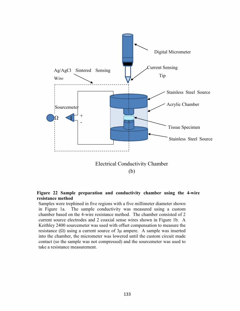
133
Sourcemeter
+ I Ω-
Digital Micrometer
Current Sensing
Tip
Acrylic Chamber
Stainless Steel Source
Ag/AgCl Sintered Sensing
Wire
Tissue Specimen
Stainless Steel Source
Electrical Conductivity Chamber
(b)
Figure 22 Sample preparation and conductivity chamber using the 4-wire
resistance method
Samples were trephined in five regions with a five millimeter diameter shown in Figure 1a. The sample conductivity was measured using a custom chamber based on the 4-wire resistance method. The chamber consisted of 2 current source electrodes and 2 coaxial sense wires shown in Figure 1b. A Keithley 2400 sourcemeter was used with offset compensation to measure the resistance (Ω) using a current source of 3μ ampere. A sample was inserted into the chamber, the micrometer was lowered until the custom circuit made contact (so the sample was not compressed) and the sourcemeter was used to take a resistance measurement.

134
Table 4 Conductivity measurement steps
Step 1 Sample preparation, height and water content measurement
Isotonic (0.015M
PBS)
Step 2 Sample placed in conductivity meter with isotonic (0.15M PBS) bathing solution, resistance read.
Isotonic (0.015M
PBS)
Step 3 Sample equilibrated in hypotonic (0.03M PBS) for 24 hrs.
Hypotonic (0.03M
PBS)
Step 4 Sample placed in conductivity meter with hypotonic bathing solution, resistance read.
Hypotonic (0.03M
PBS)
5.4 Results
Fixed Charge Density
The total average FCD (mean ± SD) of the human TMJ disc was 0.050 ± 0.018
mEq charge/g wet tissue. Regional FCD is shown in Figure 23 below. Even though the
intermediate region had the lowest FCD 0.041 ± 0.015 mEq charge/ g wet tissue, no
significant difference was found between regions (ANOVA with Tukey’s Post Hoc,
p=0.673). The FCD content for the articular cartilage (AC) control was 0.09 mEq/g wet
tissue with a corresponding GAG content of 21% by dry weight.

135
Figure 23 Fixed charge density by region (male and female combined, n=12).
The intermediate region had the lowest fixed charge density but this value was not statistically significant.
FCD was then analyzed by gender. Male FCD was 0.053 ± 0.017 mEq/g wet
tissue, while female FCD was 0.046 ± 0.018 mEq/g wet tissue with no significant
difference (p=0.325). A regional comparison of FCD between male and female TMJ
samples is shown in Figure 24 below. There were no regional differences found within
male and female groups. A two-way ANOVA was used to investigate gender and
regional interaction with no interaction found (p=0.561).

136
Figure 24 Male and female comparison of FCD by region (n=6).
Biochemical Composition
The total GAG content as % dry weight was 1.65% ± 0.64, where male GAG
content was 1.66% ± 0.59 and female GAG content was 1.63% ± 0.72 (with p<<0.05).
There was no regional significant difference based on GAG content (p=0.91). Volume
water content was 83.1% ± 2.9% for males and 82.8% ± 3.8% for females, with no
statistical significance (p=0.941). Mean volume water content did not vary between
regions (p=0.916).

137
Linear Correlations Between FCD, GAG, and Wc
A linear regression plot of GAG vs FCD is shown in Figure 25 below. Articular
cartilage control samples were also measured in this study and plotted with the TMJ disc
samples to further investigate FCD and GAG correlation shown in Figure 26 below. No
linear correlation was found between FCD and GAG (R2 =0.007). Next, a plot of age and
FCD is shown in Figure 27 below.
Additionally, there was no linear correlation between FCD and age (R2 =0.001).
Finally, a linear regression plot was conducted between water volume fraction and FCD,
shown in Figure 28. Only a slight linear correlation was found between FCD and water
content (R2 =0.208).
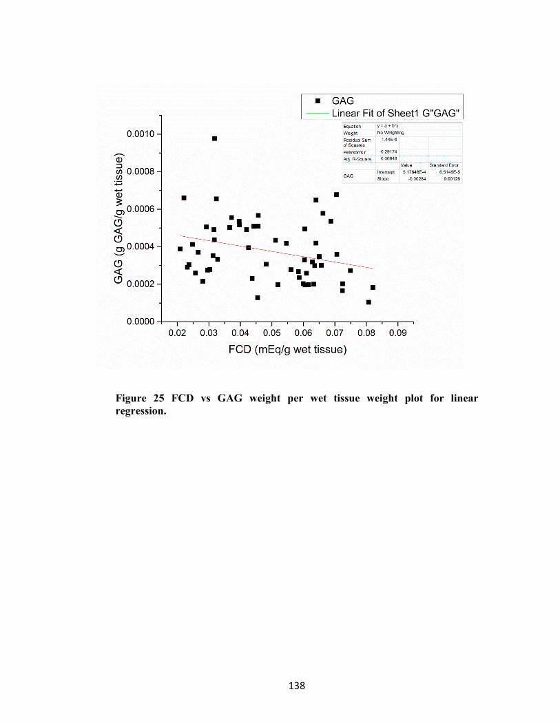
138
Figure 25 FCD vs GAG weight per wet tissue weight plot for linear
regression.

139
Figure 26 Association between fixed charge density and GAG content per wet
weight of tissue.
Articular cartilage samples were plotted with the TMJ sample to investigate a linear correlation between FCD and GAG. Since the TMJ disc sample had low GAG content to FCD signal, they were tightly grouped, whereas the AC control samples had a much larger GAG content and associated FCD signal.

140
Figure 27 Association between fixed charge density and age
There was no correlation between age and depletion of FCD. The general assumption was as age increased and degeneration likely increased therefore reducing GAG and FCD. But this trend was not shown within the samples and age of this population tested.

141
Figure 28 Association between fixed charge density and % water content
The correlation was stronger with water content than GAG content. But the R value of 0.47 was still low likely due to the small range of FCD (.02-0.09) and small range of water content (0.76-0.90).

142
5.5 Discussion
The purpose of this study was to determine the FCD in human TMJ discs and
understand its importance in tissue behavior. The FCD of the samples were measured by
a two point conductivity method [231]. Results were first compared with other cartilage
types reported previously for both FCD and GAG contents. Finally, FCD values for the
TMJ disc were compared by assessing regional variation, male and female FCD, aging
effects and the relationship to the tissue’s biochemical structure and function. All FCD
values are reported as milliequivalent charges per gram of wet tissue (assuming
monovalent GAG where 1mEq=1mMol).
Our results were comparable to FCD values of other fibrocartilaginous tissues.
Previous reports indicate the value of bovine lumbar annulus fibrosus (AF) FCD as 0.060
mEq/g compared to 0.050 mEq/g for the TMJ disc in this study. The comparison
between other cartilage types is shown in Figure 29 below. The FCD value for the TMJ
disc is lower than AF tissues. The glycosaminoglycan (GAG) found in the bovine
annulus fibrosis was 5.4% compared to the GAG content of 1.65% the for human TMJ
disc found in this study. The lower GAG content in the TMJ disc could account for the
lower FCD compared to AF tissue [231]. A GAG content comparison between tissues is
shown in Figure 30 below.

143
Figure 29 Fixed charge density graphed in comparison to other cartilage
types.
The cartilages listed are TMJ disc, Articular Cartilage from porcine femoral head from this study, Annulus Fibrosus from bovine IVD, and Nucleus Pulposus from bovine IVD [231]. The TMJ disc FCD was most similar to the AF tissue of the IVD.

144
Figure 30 GAG content graphed in comparison to other cartilage types.
The cartilages listed are TMJ disc, Articular Cartilage from pig femoral head from this study, Annulus Fibrosus from bovine lumbar IVD, and Nucleus Pulposus from bovine lumbar [231]. The TMJ disc GAG content is again most similar to the AF GAG content of the IVD, which is consistent with the FCD findings.
Also, articular cartilage porcine femoral heads were used as a control for this
study. The FCD was found to be 0.09 mEq/g wet tissue with a corresponding GAG
content of 21.8% dry weight (both FCD and GAG were statistically significant from TMJ
discs (ANOVA p<<0.05)). This value was expected to be higher given articular
cartilage’s higher proteoglycan content. This value also was within a reasonable upper
limit for the protocol used to determine TMJ disc FCD and GAG. The value for articular
145
cartilage is proportional with the nucleus pulposus with a 0.19 mEq/g wet tissue and
corresponding GAG content of 49%. The fact that the FCD is lower with respect to other
cartilages could indicate a more biphasic loading environment in the disc. This loading
environment could be required to support a large amount of tension and shear-traction
forces through the dense collagen I matrix, while compressive forces could be supported
by fluid pressurization as interstitial fluid tries to exude from the tissue with low
hydraulic permeability.
A significant weakness of this study was the age range 72-91 of the discs (overall
average 80.75 ± 6.11, female 86.42 ± 3.46 and male 76.85 ± 4.17, p>0.05 between female
and male). Ideally this study would have included human discs from age of onset 20-25
through old age ~80 but tissues were only available within this age range. In addition, we
tried to sample discs with respect to age to keep the average age of male and female
similar. The difficulty arose from finding female tissue that had no signs of degeneration.
Over twice as many female discs were sampled compared to male to arrive at the same
number of healthy discs, yet the age was still statistically higher. It could be possible
with older tissues that large matrix proteins such as collagen bundles are preserved
through storage, whereas sulfated GAG chains are cleaved by enzymatic degradation.
However, other studies have stored tissues in a similar manner with comparable FCD
values found in this study [101, 231]. Additionally, when age correlation to GAG and
FCD content were investigated for these tissues, no correlation was found. Future work
should investigate differences in GAG and FCD in normal compared to degenerated TMJ
discs. It may be possible that even though the FCD may not play a large role in

146
mechanical loading support, the FCD and GAG could play a vital role in the extracellular
detection of the loading environment, and therefore crucial for cell response to matrix
generation.
As mentioned previously, no GAG to FCD correlation was found in the TMJ
samples. This may have been due to the low signal of FCD and corresponding low GAG
content. With signals this low, a clear linear correlation was difficult due to the cluster of
results at similar levels of GAG~ 1.65% ± 0.64 and FCD 0.050 ± 0.018. When articular
cartilage samples were analyzed with the TMJ samples, a linear correlation began to
appear (R2 =0.59). However, TMJ samples had to be removed due to the large sample
number disparity (TMJ disc n=60, Articular cartilage n=6).
The proteoglycan content of the TMJ disc is relatively small compared to other
cartilages. In effect, the FCD was small but within an order of magnitude. This value
was still very important to measure in the human TMJ disc. This material property will
allow us to understand the role FCD plays in nutrient transport and function, while
helping us understand the similarities and differences to other types of cartilage.
In conclusion, the FCD content of human TMJ discs were most similar to annulus
fibrosus tissues, with a FCD value of 0.050 ± 0.018 mEq charge/g wet tissue compared to
0.060 ± 0.027 mEq charge/g wet tissue. The TMJ disc may rely more on the collagen
matrix for tensile and traction load support and less on GAG content for a role in
compressive loading. The FCD for TMJ disc tissue appears to be significantly lower than
knee articular cartilage in porcine femoral heads, which could maintain a different
loading environment. However, based solely on the FCD values it is too general to

147
conclude how the tissue behaves. More research is needed to understand how much the
tissue deforms during loading. The loss of water content and high deformation would
significantly increase the FCD in the tissue. In addition, finite element models can
effectively use the FCD property values to simulate the FCD effect on energy dissipation,
material response, and changes in diffusivity under joint loading within a charged tissue
environment. Finally, if GAG content and FCD content does not play a major role in
mechanical function, it could impact on the physicochemical signaling environment
within the disc. More advanced finite element models combined with accurate patient
anatomy are needed to bridge these biomaterial properties with biologic processes.
148
6 Tensile Properties of the Human TMJ Disc
6.1 Abstract
The temporomandibular joint disc is a large fibrocartilage responsible for the stress
dissipation and lubrication of the jaw. In over 70% of cases of diagnosed TMD, the TMJ
disc has been found to be displaced resulting from disc degeneration. Previous studies
have sought to characterize the mechanical properties of the TMJ disc in an effort to
determine the disc’s mechanical function specifically in tension as it is believed the disc
could carry a significant amount of joint loading through tension during opening and
closing of the jaw.
Animal models have been used extensively in order to characterize the
biomechanical function of the TMJ disc. But few studies have investigated human TMJ
disc biomechanical function owing to the scarcity of human tissues. In this research
study, TMJ discs were extracted from twelve human cadavers (six male and six female).
The tensile properties of the TMJ disc were investigated under incremental stress
relaxation. The TMJ disc samples were tested at a rate of 1% per second for physiologic
0-30% strain in a 37o PBS filled chamber.
The initial instantaneous stress response was plotted for each incremental strain
while the relaxed modulus was plotted at equilibrium, with the slope yielding the
instantaneous and relaxed modulus. The overall instantaneous modulus was 7.59 ± 3.26
MPa and relaxed modulus was 3.27 ±1.39 MPa. Female instantaneous modulus 8.87±
3.34 MPa was greater than male instantaneous modulus 6.46 ± 2.77 MPa (p=0.003, two
149
sample t-test). Female relaxed modulus 3.94± 1.54 MPa was greater than male relaxed
modulus 2.67 ± 0.93 MPa (p=0.002, two sample t-test).
The results indicate the TMJ disc instantaneous and relaxed modulus was lower
than previous studies investigating porcine TMJ discs [24]. In addition, anisotropy was
not found between the anteroposterior and mediolateral sections of the disc as anisotropy
was found in previous studies. The lower modulus could be attributed to the age of the
TMJ discs samples.
6.2 Introduction
TMJ disorder is a broad term used to describe many symptoms and pathologies
related to craniofacial pain, jaw pain and function (NIDCR). The exact etiology appears
to be multifactorial. As many as 20-30% of the population experience clicking, popping,
and pain in the TM joint with about 3-4% developing TMDs [7]. However, for those that
experience progression of the disorder, symptoms can be debilitating including TMJ disc
derangement. In 70% of these cases, the TMJ disc loses biomechanical function resulting
in disc displacement [5, 14, 16]. Animal models have been used extensively in order to
characterize the biomechanical function of the TMJ disc [24, 26, 30, 130, 134]. But few
studies have investigated human TMJ disc biomechanical function owing to the scarcity
of human tissues. In this study, human TMJ disc were used to measure the tensile
modulus under incremental stress relaxation tests. Studies show the tensile properties of
the disc are significant for jaw opening and closing. The results were interpreted with
respect to previous pig models. The biomechanical function was also analyzed using
current quasilinear viscoelastic theory [14, 24, 114, 241-243].
150
The large fibrocartilaginous TMJ disc plays a central role in stress dissipation and
lubrication between the incongruent surfaces of the glenoid fossa and condyle of the
mandible [24, 114]. The disc shields the TM joint from high stress concentrations and
dissipates energy throughout the tissue [114]. The dense collagen matrix along with the
proteoglycan content absorbs and distributes joint loads [24]. In the event of mechanical
injury in cartilage or degeneration of tissue, the cartilage loses its ability to support the
mechanical load which further stresses the tissue while cellular function ceases to
maintain regeneration of a healthy tissue matrix that can withstand mechanical load [14,
21]. The result is a joint degenerative disease such as osteorarthritis as well as internal
derangement of the TMJ disc [241, 244].
The TMJ disc is unique from other types of cartilages because it is made up of
80% to 90% of collagen I [24]. Comparatively, hyaline cartilages of the knee are made
up of ~60% collagen II [69]. The disc exhibits a highly biconcave shape in humans and
appears to exhibit some anisotropy based on regions within the tissue. The TMJ disc
translates with the condyle during function because it is supported by an anterior
attachment to the lateral pterygoid and anterior insertion into the condyle and a posterior
attachment to the retrodiscal tissue [114]. Through these attachments the disc divides the
glenoid fossa and the condyle into the upper and lower compartments. During opening
and closing of the jaw the disc translates anteriorly with the condyle [2]. The translation
requires the disc to support tension as it is stretched anteriorly and posteriorly over the
condyle during motion [37]. Therefore, the tensile properties are important to the disc to
maintain mechanical function. Better research and understanding of this mechanical

151
function would allow researchers to better model changes in mechanical function, as well
as provide benchmark material property data for tissue engineered replacements [14, 73,
242, 245].
There are a range of joint loading conditions the TMJ disc must support during
everyday function, including support through tension in opening and closing, tractional
shear forces through articular surfaces, and also compressive forces [24, 114, 246]. The
disc allows anterior-posterior translation of the jaw during opening and mediolateral
translation during chewing. The disc executes a complex role in the six degrees of
freedom of motion of the mandible and TMJ disc.
Type I collagen is the main extracellular matrix protein in the TMJ disc between
80-90% by dry weight. Collagen fibers run in an anterior-posterior direction throughout
the intermediate region of the disc. The fibers run in a random-circumferential direction
around the outside edges of the disc [27]. A variety of joint loads have been analyzed by
FE models. These models indicate that loading conditions such as stress field translation
running perpendicular to collagen fiber alignment could provide a unique pathological
loading environment that may promote injury to the disc. Stress from the condyle
running mediolaterally and perpendicular to collagen fibers could provide mechanical
injury to the disc. Therefore a primary goal of this study is to understand the tensile
anisotropy that may exist in the TMJ by measuring the tensile properties in the
anteroposterior verses mediolateral direction [24, 73, 114, 245].
In addition to exploring anisotropy in the TMJ disc, this research study seeks
investigate TMJ disc’s viscoelastic response under tensile strain using the quasi-linear

152
viscoelastic model. This model has been well documented in other soft tissues but
remains to be applied to TMJ disc tissues [56, 247, 248].
Previous tensile studies have investigated the TMJ disc under incremental stress
relaxation on cyclic loading in tension. Detamore used porcine tissues with different
regional orientation to determine incremental stress relaxation properties of the tissue
[24]. The disc was shown to have an instantaneous linear elastic response to increments
of tensile stress. The disc exhibited anisotropy between mediolateral and anterior
posterior orientation [249]. Beatty et al analyzed pig discs under a 0.5, 50 and 500
mm/sec initial load with an AP modulus of 76.4 MPa at 500 mm/min and 27 MPa at 0.5
mm/min and a ML modulus of 3.2 MPa at 500 mm/min [250]. These studies vary widely
on the reports of the TMJ disc tensile modulus, from 1 MPa to 500 MPa. This is in large
part due to the tensile test setup and different load rates of 1 mm/sec for Detamore, 3
mm/min for Tanaka, and 500 mm/sec for Beatty [244, 249, 250]. In addition, few human
tissues have been tested in tension except for Tanaka et al and Kalpakci et al [70, 71]. As
a result, most FE models and tissue engineering studies rely solely on either porcine or
canine tissue properties.
The goal of this study was to characterize human TMJ discs sampled from male
and female subjects. The protocol developed for this study was determined through
careful consideration of previous tensile studies as well as analysis of other soft tissues.
The main objectives of this study were to determine the tensile modulus under consistent,
relevant loading protocol, investigate viscoelastic response and anisotropic variation, and
compare male and female moduli. This understanding of human mechanical

153
characteristics will be valuable to models that evaluate dynamic joint function as well as
patient specific models of anatomy which monitor progression of TMJ disc tissue
degeneration.
6.3 Materials and Methods
Sample Preparation
Human TM joints were dissected from 6 male (67.5±8.4 years) and 6 female
(70.33±8.69 years) subjects in accordance with IRB guidelines. Donors were fresh frozen
and not formaline fixed. After the joints were removed, they were wrapped in cellophane
and PBS soaked gauze and placed in a specimen bag and frozen until use. Joints were
thawed 12 hours and the TMJ disc removed. Discs were visually inspected for signs of
trauma or degeneration. Only healthy TMJ discs were used in the study. The left joint
were used for mediolateral tensile strips while the right TMJ disc was used for anterior
posterior specimens see
Figure 31 below for strip orientation. Tissue tensile strips were prepared using a
2mm spacer and parallel razor blades. For the mediolateral strips, three specimens were
taken from each disc in the mediolateral central, anterior, and posterior regions of the
disc. Likewise, specimens from the anterior posterior orientation were taken in central,
lateral and medial portions of the disc. Care was taken to remove specimens in equal
spacing and avoid peripheral attachments on the outer edges of the disc.
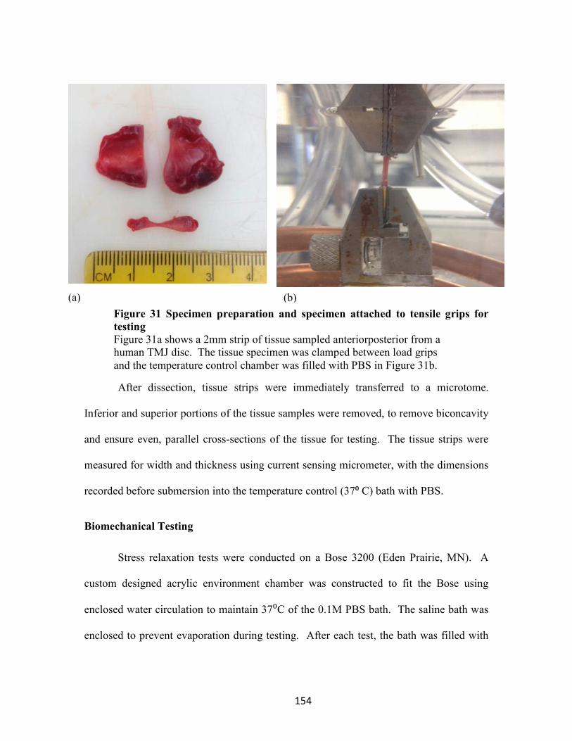
154
Figure 31 Specimen preparation and specimen attached to tensile grips for
testing
Figure 31a shows a 2mm strip of tissue sampled anteriorposterior from a human TMJ disc. The tissue specimen was clamped between load grips and the temperature control chamber was filled with PBS in Figure 31b.
After dissection, tissue strips were immediately transferred to a microtome.
Inferior and superior portions of the tissue samples were removed, to remove biconcavity
and ensure even, parallel cross-sections of the tissue for testing. The tissue strips were
measured for width and thickness using current sensing micrometer, with the dimensions
recorded before submersion into the temperature control (37⁰ C) bath with PBS.
Biomechanical Testing
Stress relaxation tests were conducted on a Bose 3200 (Eden Prairie, MN). A
custom designed acrylic environment chamber was constructed to fit the Bose using
enclosed water circulation to maintain 37⁰C of the 0.1M PBS bath. The saline bath was
enclosed to prevent evaporation during testing. After each test, the bath was filled with
(a) (b)

155
fresh PBS. Stress relaxation tests were programed using WinTest software. Specimens
were attached to the Bose tensile clamps using cyanolacrylic. The chamber was filled
with PBS and allowed to equilibrate for 10 minutes see Figure 32 below.
Figure 32 A temperature controlled PBS chamber and a Bose 3200 axial
tester
Cameras were initially used to measure the change in cross sectional area of the specimen under load.
A 0.1 Newton (N) tare load was applied the specimen and the initial height of the
specimen was recorded though the load arm height. Using the specimen height at tare
load, a rate of 1% per second was calculated and used as the ramp loading speed between
dwell times. The specimen was loaded at 5%, 10%, 15%, 20% and 30% stain increments
with dwell times for relaxation of 15, 15, 15, 20, and 30 minutes, respectively to ensure

156
equilibrium was reached, see Figure 33 below The times were based on preliminary tests
results that ensured the tissue had reached equilibrium. The load rate of 1% per second
was chosen to maintain a uniform loading rate for each specimen regardless of length.
Therefore, each specimen was loaded at a consistent rate as a ratio to length.
Figure 33 An example of the stress-relaxation response to incremental
increases in strain.
For each strain increment, the tissue response is a peak instantaneous response followed by a decrease in stress as the tissue is held at constant strain, until the tissue reaches the relaxed/equilibrium stress. After a designated time increment, the strain is increased and the stress response follows a similar pattern of relaxation. The peak instantaneous response and relaxation stress at equilibrium were plotted to determine the
Instantaneous Modulus
Relaxed Modulus

157
instantaneous modulus and relaxed modulus as indicated on the figure above.
Quasilinear Viscoelastic Theory
The quasilinear viscoelastic theory was based on YC Fung’s theory for non-linear
elasticity which accounts for the previous strain history which affects the stress in
biologic materials [56]. The theory was formulated based on continuous spectrum of
relaxation rather than combining many Kelvin or Maxwell models. The reduced
relaxation function derived from the QVT is used to fit stress data that has been
normalized with respect to the peak stress and initial time. The reduced relation function
is fitted with an exponential integral and resolved using a short time constant, τ1 and long
time constant τ2 based on a logarithmic scale, while a material constant C is used as the
final fitting parameter to account for the load dissipating property of the biologic material
shown in Equation 17 [56]. See Figure 34 below for τ1, and τ2
Quasilinear Viscoelastic Theory Curve fitting of reduced relaxed function was
completed using Origin Pro 9.1 software (Cambridge, Massachusetts). All samples were
normalized by stress and time so that at t=0 the initial time began at 0 and the initial peak
stress began at 1.
Each relaxation curve was then fit using the reduced relaxation function yielding
parameters C, τ1, τ2 for each curve Figure 35 below. Figure 36 below shows the
relaxation curves for 5%, 10%, 15%, 20% and 30% strains which have been normalized
in order to be fit with the reduced relaxation function in Equation 17:

158
17
dtt
ezE
z
t
∫∞ −
=)(1
18
where E1 is the exponential integral function in Equation 18. An exponential
approximation was used to fit peak instantaneous response for each level of incremental
strain (σε(ε)), where A and B were material constants:
( )1)( −= εε εσ BeA
19
However, when the instantaneous peak response was fit for the samples, the fit was
linear.
∗+
−
∗+
=
1
2
1
1
2
1
ln1
1
)(
ττ
ττ
C
tE
tEC
tG

159
Figure 34 The reduced relaxation function G(t) of a solid with a continuous
relaxation spectrum S(τ)=c/τ for τ1<τ<τ2 from Neubert 1963, reprinted Fung
[56].

160
Figure 35 Example plot of stress relaxation test with each relaxation curve
divided into their respective strain increments before normalizing and curve
fitting with quasilinear viscoelastic theory.
In order to plot the reduced relaxation function for each strain increment, the maximum stress was normalized to σmax/σ and the time was normalize to to=0, therefore G(0)=1. The reduced relaxation function (Equation 17) was used to fit the relaxation curve for each strain increment, yielding the material constant, C and time constants τ1, τ2.
5% 10% 15% 20% 30%

161
Figure 36 Quasilinear Viscoelastic Theory Curve fitting of reduced
relaxed function for a single specimen. Note the curve for each strain level was fit individually with the reduced relaxation function. Before curve fitting with the reduced relaxation function proposed by Fung, relaxation curve from each increment of strain were normalized by stress (G(t)=1) and time (t(0)=0). Then each relaxation curve was fit using the reduced relaxation function yielding parameters C, τ1, τ2 for each curve.
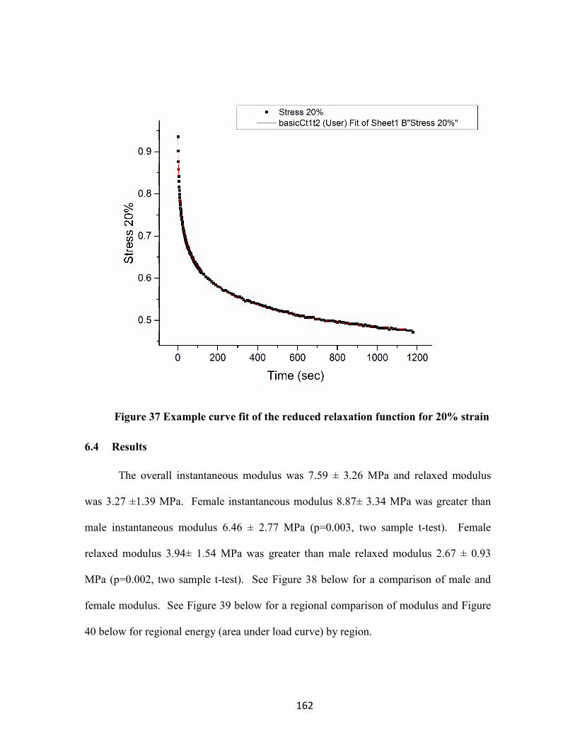
162
Figure 37 Example curve fit of the reduced relaxation function for 20% strain
6.4 Results
The overall instantaneous modulus was 7.59 ± 3.26 MPa and relaxed modulus
was 3.27 ±1.39 MPa. Female instantaneous modulus 8.87± 3.34 MPa was greater than
male instantaneous modulus 6.46 ± 2.77 MPa (p=0.003, two sample t-test). Female
relaxed modulus 3.94± 1.54 MPa was greater than male relaxed modulus 2.67 ± 0.93
MPa (p=0.002, two sample t-test). See Figure 38 below for a comparison of male and
female modulus. See Figure 39 below for a regional comparison of modulus and Figure
40 below for regional energy (area under load curve) by region.

163
Figure 38 Bar plot of the instantaneous and relaxed modulus according to
gender
Using two sample t-test, at the 005 level, the population means of both instantaneous and relaxed modulus are significantly different between male and female, p<0.05. Male and female groups n=36.

164
Figure 39 Regional instantaneous and relaxed modulus for combined male
and female groups
No regional differences were detected. A Shapiro-Wilk test was first conducted to determine normality for the sample set ML Cent through AP-Med with n=11. Normality was confirmed, p<0.05. A One-way ANOVA was run with 6 groups and found that the means were not statistically significant, p<0.05 for both the instantaneous and relaxed data set. At the 0.05 level, the means between region (n=11) are not significantly different.

165
Figure 40 Results of human tensile instantaneous and relaxed energy by
region
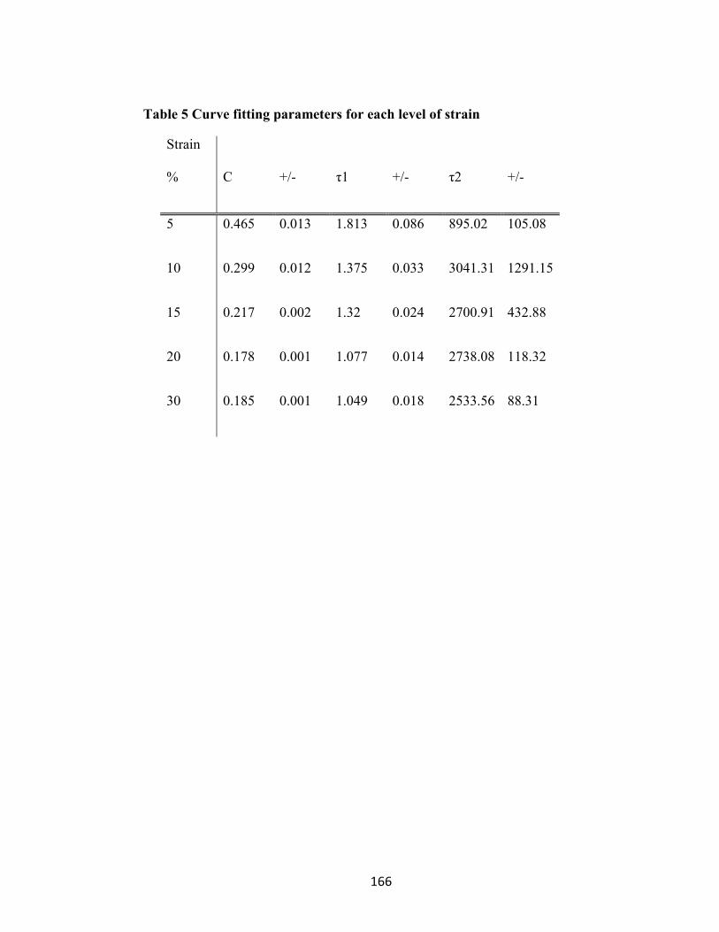
166
Table 5 Curve fitting parameters for each level of strain
Strain
% C +/- τ1 +/- τ2 +/-
5 0.465 0.013 1.813 0.086 895.02 105.08
10 0.299 0.012 1.375 0.033 3041.31 1291.15
15 0.217 0.002 1.32 0.024 2700.91 432.88
20 0.178 0.001 1.077 0.014 2738.08 118.32
30 0.185 0.001 1.049 0.018 2533.56 88.31

167
Results of previous studies for comparison:
Figure 41 Detamore et. al. results from porcine testing for comparison [24]
Significant anisotropy between ML-Cent region and AP regions were detected.

168
Figure 42 Compilation of results from previous tensile studies emphasizing
the variation in tensile modulus [23, 70, 128, 129]

169
Figure 43 Results of Detamore et al. porcine tensile instantaneous and relaxed
energy for comparison [24]

170
6.5 Discussion
The modulus values for the TMJ disc were less than other fibrous musculoskeletal
tissues such as the human patellar tendon 504-660 MPa, human supraspinatus tendon 86
MPa, articular cartilage 13.6 MPa [248, 251]. The modulus was lower than previous
human TMJ disc elastic modulus tested previously by Tanaka as 44.0 MPa [70]. By
comparison, the instantaneous modulus of the human TMJ disc was found to be between
6-10 MPa. The value was much less than moduli found for tendon in previous studies
and much closer to the value for articular cartilage. See Figures 41-43 above for
comparison of previous studies. However, the instantaneous modulus found for human
TMJ disc in this study was significantly different from porcine TMJ discs (14-30 MPa
excluding the mediolateral intermediate oriented samples) [24].
The modulus value for female TMJ discs was higher than male TMJ discs. This
was somewhat surprising given the amount of biologic variation between material
properties in subjects overall. In addition, this material property value was independent
of sample length or thickness size since all ramp loads were conducted at 1% per second
constant strain rate. Modulus values for female were greater both in the AP and ML
direction. Similarly, the relaxed modulus was consistently greater in females. More
research is needed in order to understand the direct relationship the increased modulus
has with the structure, function in human TMJ discs. During dissection, the female discs
were consistently smaller and had a much more convex shape over a small condyle. The
slope of the eminence was generally steep. The differences in anatomical morphology

171
should be matched with normal joint function to better understand the mechanical role of
the TMJ disc.
Although sex differences in modulus were determined, no AP to ML oriented
differences were found. The finding was consistent between the male and female groups.
The lack of differences between AP to ML oriented samples does not agree with previous
studies that found large variation between 5-25 times greater strength in the AP direction
than ML direction [249, 250, 252]. In this study, there was a greater average modulus for
AP oriented samples, though the value was not statistically significant. Animal tissues by
comparison may exhibit more anisotropy due to the tissue being well defined in terms of
matrix. Human tissues that were tested in this study averaged ~67 years of age. Due to
age of subjects, older tissue may lose some anisotropy and become more uniform. The
age of tissues may also explain why the values for modulus were generally lower.
The overall modulus compared to porcine tissues were much greater in pigs 14-30
MPa compared to 7.59 MPa in humans [249]. Pig tissues could have an overall larger
strength based on the structure of anatomy. These were young tissues compared to older
tissues in adult humans. In the pig study, the ML Center oriented samples of the TMJ
disc was extremely weak in comparison to other regions. In this study the ML center
oriented samples had the lowest average modulus between oriented samples, however
this value was not significant based on the biologic variation. An interesting observation
in this study was the value for ML center modulus in females had both the largest and
smallest values for modulus within all the groups and greatest standard deviation among
tissues. This phenomenon should be investigated further to understand if this region

172
shows signs of degeneration or changes in tissue composition and structure after injury or
age. Finally, a morphological comparison between porcine tissues may help understand
differences with regard to the human. The porcine animal model has been shown to be
the most relevant to the human [25, 35]. In order for this model to translate well to the
human, fundamental differences between the pig biconcave shape and size of the disc
would have to be investigated. For instance, the morphology between human and pig
TMJ discs must be compared with relation to jaw function between species.
In comparison to other human studies, Kalpakci et al found the instantaneous
modulus to be significantly higher 55 MPa compared to 7.59 MPa in this study [252].
This modulus value was significantly greater than the other animal modulus values
concluded in the study. In addition, the disc showed significant anisotropy with the AP
Cent modulus being much larger than the ML Cent regions. The age range and load rate
for human samples were not listed. Therefore, it was difficult to make a comparison
between studies on that basis.
In our experience with mechanical testing in this study, we found the stress strain
response to be highly dependent on load rate which is characteristic of viscoelastic
materials. It is likely that such anisotropy exists between the AP and ML groups due to
the many studies of SEM evidence of collagen fibers running in the AP direction.
However, with regard to tensile testing, if a constant load rate was applied to each sample
as in the case of Detamore and Kalpakci of ~1mm/sec then the length of sample may
have an influence in the stress strain response [249, 252]. The AP length is generally 10
mm while the ML width of the TMJ dis is usually 18-25 mm, with the longest section
173
being in the ML center. If a uniform tensile load was applied to an AP and ML sample at
a rate of 1mm/sec samples, then the rate at which the load was applied could affect the
sample response, thus causing a larger modulus for the shorter sample (AP) and a small
modulus for the long sample (ML). In this case, the AP sample could be loaded under
twice the load rate. This may account for some of the large differences in anisotropy;
however, it should be true for the ML Anterior and ML Posterior cases, which much
more isotropic compared to AP. In our case, the AP and ML showed little anisotropy
under a uniform 1 %/sec load rate but due to a much lower modulus and larger standard
deviation, other factors with the biologic samples could have been a factor. Therefore, in
the future the gage length of the samples tested along with the load rate should be listed
or sample should be loaded at a uniform rate, so that results from these test can be
compared acknowledging the load rate.
In this study, a linear response to strain was consistent throughout all regions
(R2~0.997). In determining both the instantaneous and relaxed modulus through linear
fit, the stress response was linear for all strains, in this case strains through 30%. In other
tissues, such as tendon, the nonlinear response both instantaneous and relaxed modulus
became apparent with strains over 30% [56]. This was similar to other tensile studies on
the TMJ disc [249, 252].
The differences between the instantaneous modulus (7.59 MPa) and relaxed
modulus (3.27 MPa) were significant. This was also true for the instantaneous and
relaxed areas under the stress-strain plots, since they were essentially calculated from the
same values. Detamore et al. referred to the area under these plots to indicate the strain

174
energy absorbed by the disc [24]. The large difference ~50% between strain energies of
the instantaneous and relaxed energy may indicate the disc’s ability to dissipate large
strain energies upon initial loading. These large strain energies may be absorbed through
viscous response while the linear elastic response appears to be maintained through 30%
strain. An analysis of the individual relaxation curves for each sample is needed to
further understand the viscoelastic response between the initial loading and relaxed
equilibrium.
In order to model individual stress relaxation response with respect to time, a
reduced relaxation function proposed by Fung was used [56]. This model was proposed
based on the quasilinear viscoelastic theory (QLV). The theory was chosen early on
instead of Maxwell, Voigt and Kelvin models due to the nonlinear behavior of biologic
tissue which often depends on strain history. The QLV theory appears to account for
strain history for finite deformations and has been used to model many types of
cartilaginous and other soft tissues.
For individual strain response, the time and stress was normalized to time=0,
stress =1. Each stress relaxation curve began at the peak instantaneous response and
ended at equilibrium for each strain increment. For each sample that was tested, five
relaxation curves (according to strain %) were curve fit by the reduced relaxation
function. The reduced relaxation curve fit involved a logarithmic fit with an exponential
integral. The curve fit yielded three relaxation parameters: C, τ1, τ2. The physical
meaning of the curve fitting parameters corresponded to a short time constant, τ1 to
model the initial instantaneous drop in stress and a long time constant; and τ2, to model
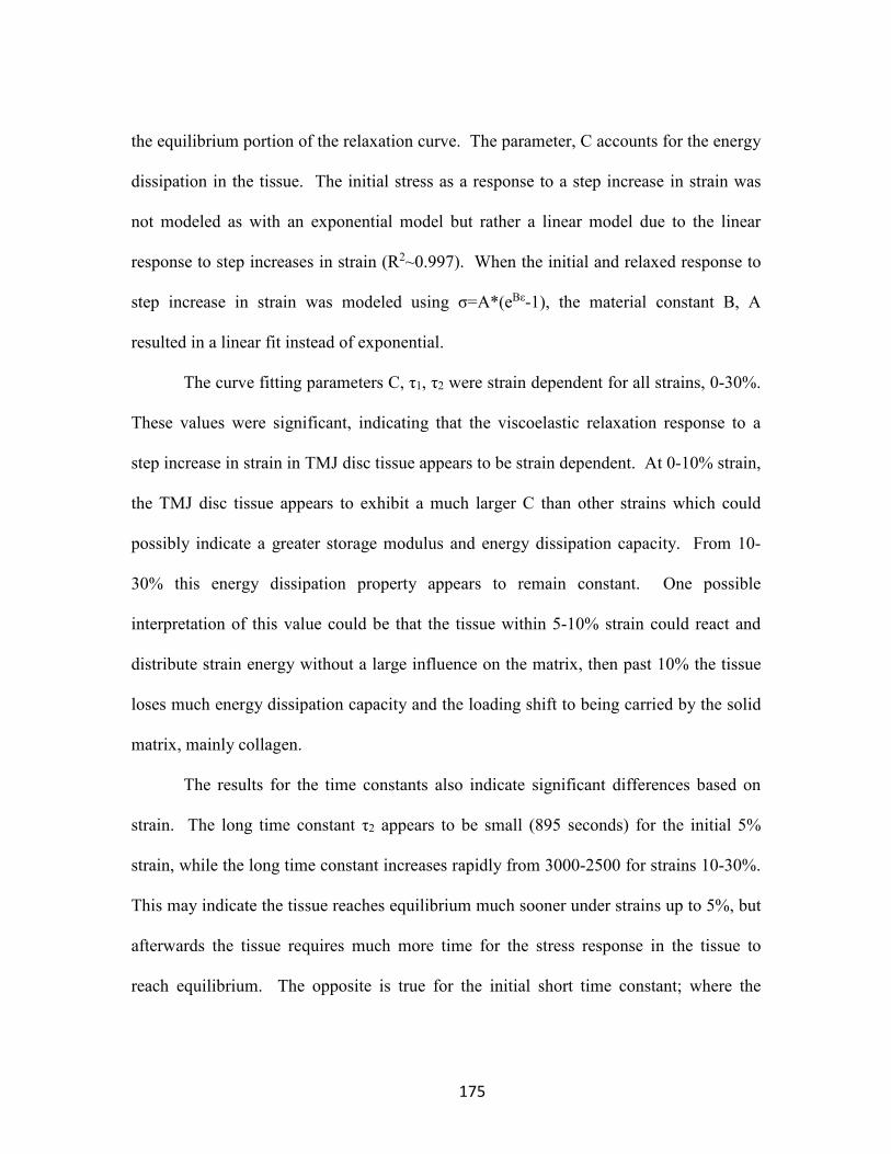
175
the equilibrium portion of the relaxation curve. The parameter, C accounts for the energy
dissipation in the tissue. The initial stress as a response to a step increase in strain was
not modeled as with an exponential model but rather a linear model due to the linear
response to step increases in strain (R2~0.997). When the initial and relaxed response to
step increase in strain was modeled using σ=A*(eBε-1), the material constant B, A
resulted in a linear fit instead of exponential.
The curve fitting parameters C, τ1, τ2 were strain dependent for all strains, 0-30%.
These values were significant, indicating that the viscoelastic relaxation response to a
step increase in strain in TMJ disc tissue appears to be strain dependent. At 0-10% strain,
the TMJ disc tissue appears to exhibit a much larger C than other strains which could
possibly indicate a greater storage modulus and energy dissipation capacity. From 10-
30% this energy dissipation property appears to remain constant. One possible
interpretation of this value could be that the tissue within 5-10% strain could react and
distribute strain energy without a large influence on the matrix, then past 10% the tissue
loses much energy dissipation capacity and the loading shift to being carried by the solid
matrix, mainly collagen.
The results for the time constants also indicate significant differences based on
strain. The long time constant τ2 appears to be small (895 seconds) for the initial 5%
strain, while the long time constant increases rapidly from 3000-2500 for strains 10-30%.
This may indicate the tissue reaches equilibrium much sooner under strains up to 5%, but
afterwards the tissue requires much more time for the stress response in the tissue to
reach equilibrium. The opposite is true for the initial short time constant; where the
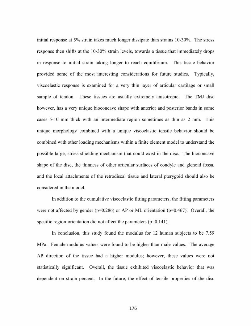
176
initial response at 5% strain takes much longer dissipate than strains 10-30%. The stress
response then shifts at the 10-30% strain levels, towards a tissue that immediately drops
in response to initial strain taking longer to reach equilibrium. This tissue behavior
provided some of the most interesting considerations for future studies. Typically,
viscoelastic response is examined for a very thin layer of articular cartilage or small
sample of tendon. These tissues are usually extremely anisotropic. The TMJ disc
however, has a very unique bioconcave shape with anterior and posterior bands in some
cases 5-10 mm thick with an intermediate region sometimes as thin as 2 mm. This
unique morphology combined with a unique viscoelastic tensile behavior should be
combined with other loading mechanisms within a finite element model to understand the
possible large, stress shielding mechanism that could exist in the disc. The bioconcave
shape of the disc, the thinness of other articular surfaces of condyle and glenoid fossa,
and the local attachments of the retrodiscal tissue and lateral pterygoid should also be
considered in the model.
In addition to the cumulative viscoelastic fitting parameters, the fitting parameters
were not affected by gender (p=0.286) or AP or ML orientation (p=0.467). Overall, the
specific region-orientation did not affect the parameters (p=0.141).
In conclusion, this study found the modulus for 12 human subjects to be 7.59
MPa. Female modulus values were found to be higher than male values. The average
AP direction of the tissue had a higher modulus; however, these values were not
statistically significant. Overall, the tissue exhibited viscoelastic behavior that was
dependent on strain percent. In the future, the effect of tensile properties of the disc

177
should be modeled with finite element models. In addition, consideration should be
given for regional behavior of the disc as well as discal attachments that may affect disc
behavior in response to loading.

178
7 GENERAL CONCLUSIONS AND FUTURE WORK
The purpose of this research was to conduct a biomechanical characterization of
the human TMJ disc. Through experimental methods the biomechanical environment of
the disc was investigated through strain dependent diffusivity and fixed charge density
measured by the conductivity method and the viscoelastic tensile properties of the disc
measured by incremental stress relaxation. These important properties will have a
bearing on the TMJ disc biomechanical function under mastication and will give us
insight on the nutrient concentration gradient across the disc as well as the
electrochemical signaling environment within the disc.
Conductivity, Diffusivity, and Nutrient Transport within the Human TMJ Disc
From the electrical conductivity study, we found mechanical strain decreases the
porosity of the disc and thereby decreases conductivity of the disc, which is proportional
to diffusivity. This finding supports our overall hypothesis that mechanical strain
significantly affects the transport behavior of the disc and may play an important role in
altering the nutrient environment within the tissue leading to tissue degeneration. With
strains of 10% and 20% the diffusivity of small ions Na+ and Cl- decreased by -12.3%
and -23.5%, respectively. In studies conducted since, we have found the diffusivity of
larger solutes such as glucose decreased by as much as 40% under a 20% strain. This
would indicate a much steeper nutrient concentration gradient than indicated by the
electrical conductivity study; further, the impact of mechanical loading may have an even
greater impact on diffusion with much larger growth factors and cytokines. Our FRAP

179
studies support similar findings [58]. This provides further rationale for the need to
develop an accurate 3D finite element model to investigate the effect of mechanical strain
on the diffusion of different solutes within the disc and their effect on cell behavior and
matrix remodeling.
The conductivity of female TMJ discs (5.84 mS/cm) were slightly greater and
affected more by mechanical strain than the conductivity of male TMJ discs (5.14
mS/cm). This may indicate slight differences in the extracellular matrix of TMJ discs of
males and females. We found the human TMJ disc conductivity (5.45 mS/cm) to be
much greater than the conductivity we found in our porcine study (3.10 mS/cm). This
should be taken into account when working with young porcine tissues to investigate
treatments and tissue engineering strategies for human TMJ discs. Although the
conductivity in porcine TMJ discs was much lower corresponding to a lower porosity,
they were affected by mechanical strain in a similar manner. This observation is
important to keep in mind when comparing or translating models. Overall conductivity
of the TMJ disc (5.5 mS/cm) is ~25% lower than IVD and hyaline cartilage (7.5 mS/cm).
However, when moving to oxygen and glucose diffusion, the diffusivity of TMJ disc
tissue was much lower (~50%) than IVD and hyaline cartilage. It must be emphasized
that our results for conductivity can only be interpreted for small solutes investigated in
the experimental tests. When moving to large solutes, the effect of mechanical loading
may have more effect on solute diffusivity. In addition, when the conductivity
comparisons are made between other types of cartilages, the conductivity values do not

180
account for the fixed charge density that may further affect diffusivity especially in
cartilages that have a much greater concentration of fixed charge density.
Fixed Charge Density in Human TMJ Discs
The fixed charge density is a material property of cartilage which results from
highly charged proteoglycans present in cartilaginous tissues. This material property is
unique to cartilage and is responsible for high osmotic swelling pressure which maintains
load and also for unique streaming potentials and electrokinetic affects within cartilage.
The fixed charge density (FCD) found in human TMJ discs was 0.05 mEq/g wet tissue
and was comparable to previous studies that found the FCD for annulus fibrosus tissue of
porcine to be 0.06 mEq/g wet tissue.
The fixed charge density must be taken in to account when modeling tissue
especially since the TMJ disc is believed to incur large deformations under mechanical
load which would effectively increase the fixed charge density concentration. Inside the
TMJ disc the electric potentials generated by fluid pressure moving through the charged
matrix may alter electrochemical signals within the disc. Further, the alignment of matrix
fibers has been shown to be anisotropic through SEM analysis, due to this observation the
streaming potentials based on direction of fluid flow may be anisotropic in the TMJ disc.
This physicochemical signaling environment will need further investigation in the TMJ
disc model.
Finally, the human specimens measured for FCD were relatively old (81 years)
and had been stored previously in refrigeration. The value measured in the study
provides an overall basis for FCD, arguably on the low end of what could be expected in

181
persons within the age of onset of TMDs (25-45). The role of FCD and streaming
potential within the disc must be investigated further from the standpoint of the large
deformations the TMJ disc undergoes to provide stress shielding and lubrication of the
joint. The FCD should be investigated for concentrations through the inferior to superior
directions, with emphasis given to the surface charges along the outer surface of the disc.
A double layer effect could be present on the surface of the disc which is dependent on
the TMJ disc fixed charge density and allows for extreme lubrication of the disc in
addition to the hyaluronan within the synovial fluid. This effect should be studied in
more detail and compared to shear-tractional studies by Nickel et al. [144].
TMJ Disc Mechanical Behavior
To determine the tensile properties of the TMJ disc, an incremental strain was
placed on the tissue and the viscoelastic stress relaxation response was recorded. In
determining both the instantaneous and relaxed modulus through linear fit, the stress
response was linear for all strains. In other tissues such as tendon, the nonlinear response
to both instantaneous and relaxed modulus became apparent with strains over 30% [56,
69]. In previous work, the TMJ disc was shown to exhibit anisotropic tensile responses
between the anteroposterior and mediolateral directions [24]. In our case, the TMJ disc
did not appear to exhibit anisotropy between regions and exhibited a tensile modulus
lower than previous studies likely do the age of donors in this study.
When analyzing the viscoelastic stress relaxation we found significant differences
to load response based on strain. We found that the tissue reaches equilibrium much
sooner under strains up to 5% but afterwards the tissue requires much more time for the

182
stress response in the tissue to reach equilibrium with 10-30% strain. This could indicate
a shift load in response under physiological loading conditions, for instance an elastic
response within 5% strain followed by fluid pressurization response. The results of
viscoelastic response must analyzed with respect to the highly bioconcave anatomy of the
TMJ disc. It is likely the TMJ disc exhibits complex biomechanical function through its
viscoelastic behavior and anatomical structure which can only be analyzed with through
FE modeling.
7.1 Future Work
The body of this research has focused on the direct measurement of key biomaterial
properties of the human TMJ disc. Understanding these properties has allowed us to
model and understand the disc in relation to other types of cartilages. But for the
experimental properties measured in this study to apply to the human patient, a 3D model
of patient specific anatomy, joint loads, and material reaction must be constructed. This
3D finite element model will allow us to not only investigate how each parameter
influences disc behavior under loading, but it will also allow us to investigate how the
parameters influence each other during joint function, a multiphasic true material
characterization. (i.e. fixed charge density effects on diffusivity). This model can also be
constructed with key cell metabolic data from our explant studies that will allow us to
predict the physicochemical environment of the disc in response to physiologic and
pathologic loading (i.e. how does the change in disc diffusivity through sustained loading
alter the available oxygen content thereby affect local cell metabolism?)

183
Developing a Multibody Model through Dynamic Stereometry: Patient Anatomy
Combined with Jaw Kinematics
To generate a patient specific model, the TMJ disc’s anatomy and motion must
first be measured during function. A multibody model is generated to model and predict
inner joint loads acting on the disc which will be used later in the finite element stress
analysis. A multibody model alone is a useful diagnostic tool that traces condylar motion
and allows researcher to analyze condyle, disc and fossa motion in real time. This tool
can be used to classify patient pathology given enough patient numbers in our predictive
study. One main difficulty we have encountered using the multibody model to classify
patient pathology is the condylar motion path is so complex during mastication combined
with many different classifications of TMD’s.
We have implemented a system in our lab first pioneered by Gallo and co-
workers to combine high-resolution MRI with real-time 3D motion tracking during jaw
function (Fig. 2). This technique uses high-speed infrared cameras to track target frames
on the mandible and maxilla in 6 degrees of freedom (DOF). The motion data is
combined with three-dimensional TMJ anatomies obtained from high-resolution MRI
using a computer CAD program then analyzed using a finite element program (COMSOL
(Comsol, Burlington, MA) [4].

184
Figure 44 The dynamic stereometry system consists of (A) high resolution
MRI, (B) 6 DOF jaw tracking, and (C) Algorithms to reconstruct dynamic
TMJ anatomy into a finite element model.
MRI Imaging: Dicom images from MRI were segmented in Amira (Amira, Burlington,
MA) to generate at 3D solid mesh of the patient TMJ, including condyle, fossa, and disc.
The solid mesh was imported to SolidWorks (Waltam, MA) where trajectories were
applied to run the motion analysis. The anatomy of the TMJ bony and disc structures in
the different jaw positions was obtained by recording a series of 14 magnetic resonance
parasagittal slices through the TMJ using a 1.5 T scanner and 12-cm ∅ surface coils.
Human TMJ Mechanical Characterization: In order to define TMJ disc’s multiphasic
properties in the model, we will measure the compressive modulus, tensile modulus as
well as the strain dependent transport properties of human cadaver TMJ discs.
Fossa
Disc
Condyl

185
D: Simplified TMJ Model C: FEM Model of TMJ
Foss
Disc
Condyle
Displacement
in Y&Z
directions
B: Left TMJ Section A: Reconstructed TMJ from CT & MRI
Figure 45 Showing the MRI/CT reconstruction of a TM joint MRI/CT to
meshed solid Finite Element model
Patient specific anatomy was constructed from (a) MRI/CT geometries, (b) section view of solid, (c) Solid geometries were formulated into a tetrahedral mesh and (d) evaluated with patient motion data in simplified
model.

186
7.2 Finite Element Model
Because of the lack of appropriate animal models and the difficulties of in vivo
measurements, finite element modeling is the only means possible to examine the
relationships between coupled physical properties of the disc. This 3D multiphasic model
of the human TMJ disc will allow us to determine the effect of mechanical loading on the
nutrient diffusivities within the tissue. In addition, attempts at dynamic MRI to analyze
jaw function have been ineffective due to lack of resolution and absence of true
masticatory motion. As a result, the dynamic stereometry of the TMJ developed by Gallo
et al is the only validated technique to provide 3D dynamic TMJ anatomy during jaw
function [14]. This study will incorporate dynamic stereometry to obtain patient specific
geometries and jaw function to build a 3D model of the TMJ disc from the validated 2D
model.
Develop and validate a 2D axial symmetric finite element model of cylindrical TMJ
disc explants using material properties.
The 2D model will be based on the triphasic theory developed by Lai et al.
combined with our extended multiphasic mechano-electrochemical theory [20]. Our
model considers charged hydrated tissue as a mixture of distinct phases (charged solid,
interstitial water, ions and neutral solutes). Material properties such as region-dependent
aggregate and tensile modulus, strain-dependent fixed charge density, hydraulic
permeability, and solute diffusivities in five disc regions will be inputs that define the
material [46]. The weak form of the governing equations will be formulated from
Galerkin residual method and solved using implicit Euler backward scheme with

187
software, COMSOL (Comsol, Burlington, MA) [45]. The model will calculate changes
in streaming current and solute diffusivities under compressive load. The finite element
model of the TMJ explants will be validated in a well-controlled unconfined stress
relaxation compression test on cylindrical samples from five regions of both human and
porcine discs (n=10). The disc will be subjected to a stress relaxation test (TA Q800
Dynamic Mechanical Analyzer) and the responses of axial load and streaming current
recorded (Model 2400, Keithley Instruments Inc., Cleveland, OH.)
A 2D dynamic multiphasic finite element model of TMJ disc explants will be
obtained for the human model and the porcine model. The responses of axial load and
streaming current (I) under the relaxation tests will be recorded in real-time. The
precision of the finite element model will be evaluated by comparing the outputs of
model prediction to recorded axial load and streaming current measured at the center of
the specimen.
Integrate the dynamic stereometry and the multiphasic finite element model to
establish the 3D patient specific model.
TMJ anatomy (High resolution MRI)
TMJ kinematics (6 DoF jaw tracking)
Finite Element Model
of TMJ disc Signal analyzer
Shape
Motion
Tissue properties: Mechanical/electrical/transport
Measurements:
Mechanical signals:
Strain/Stress/Pressure
Electrical signals:
Ionic environments
Chemical signals:
Oxygen/glucose/lactate
Biological signals: Energy metabolic rates
Dynamic Stereometry
Cell properties: Cell density and metabolic rates
Figure 46 Schematic of integrating the dynamic measuring system

188
The validated 2D finite element model can be mathematically extended to a 3D
model of the TMJ disc without significant difficulties. This material model will be
developed into a patient specific model through the MRI (Siemens Trio 3T) acquired
TMJ disc anatomy. Dicom images from the MRI will be segmented in Amira (Amira,
Burlington, MA) to generate at 3D solid mesh of the patient TMJ, including condyle,
fossa, and disc [48]. The nutrient supply at the margin of the disc from the synovial fluid
and capillary will be considered by the concentration boundary conditions. The
segmented mesh will be exported for analysis in Hyperworks (Hyperworks, Troy, MI).
In Hyperworks, trajectories recorded using 6 DOF jaw tracking will be applied to the
patient anatomy to run the motion analysis. We will transfer stress strain profiles from
Hyperworks to (Comsol, Burlington, MA) to analyze the mechanical, electrical,
chemical, and biological signals.
Results and Interpretation: The patient specific model will be analyzed based on
individual kinematics to determine stress field translation [49], nutrient concentrations,
and metabolic rates within the disc. Patient specific models will be analyzed individually
and across patient populations to develop bio-indicators (e.g., nutrient levels and energy
metabolic rates) and evaluate risk among patient groups. We anticipate that the
mechanical loading will dramatically impede the nutrient transport, resulting in extremely
low oxygen/glucose concentration profiles within the TMJ disc which will significantly
affect cell matrix synthesis and even cell death. The effect is mainly due to the strain-
dependent oxygen/glucose diffusivities and hydraulic permeability.

189
Potential Difficulties and Alternative Strategies: Because the nutrient concentrations
and metabolic rate are interrelated, these measurements are expected to be correlated
among themselves within disc region, among disc regions within a disc, and across the
loading settings. Thus, it may be possible to gain efficiency by jointly modeling these
outcomes rather than evaluating them separately, although at a cost of greater model
complexity. If statistically significant bio-indicators do not emerge from analyses of each
outcome, we will investigate joint models that incorporate both known relationships and
flexibly permit both positive and negative correlations among the outcomes.
Innovation: The innovation of this study lies in the project’s unique approach to use a
patient specific model to build a pathway between biomechanics and pathobiology while
bringing the clinical field a crucial, non-invasive method to detect early indicators of
TMJ disorders.
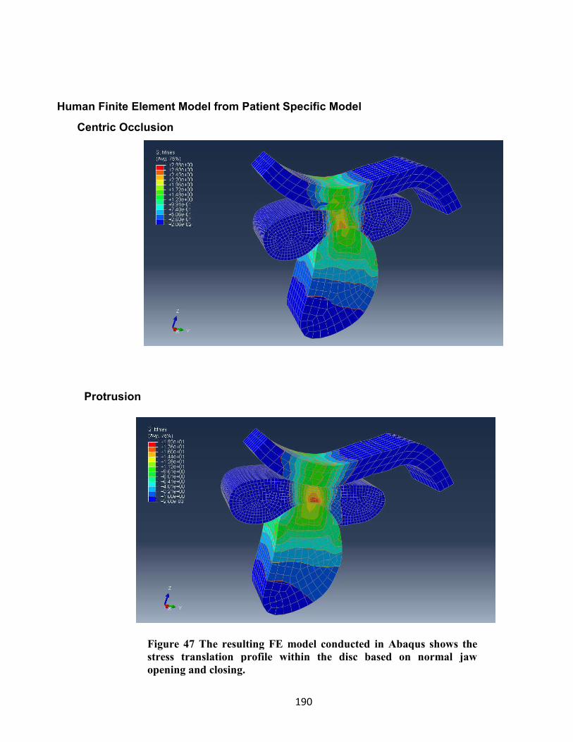
190
Human Finite Element Model from Patient Specific Model
Centric Occlusion
Protrusion
Figure 47 The resulting FE model conducted in Abaqus shows the
stress translation profile within the disc based on normal jaw
opening and closing.

191
APPENDIX
192
8 APPENDIX
A biomechanical investigation of thoracic hyperkyphoscoliosis in EOS treated with rib
fixation
8.1 Abstract
Study Design: Biomechanical comparison in an immature porcine model of different
anchorage techniques, pedicle screw fixation and a newly developed rib hook construct.
Objective: The goal of this research study was to examine kyphotic deformity effects on
upper thoracic fixation, specifically pedicle screws and laminar hooks placed on the ribs.
Summary of Background Data: After a successful clinical outcome in treating over 50
patients with severe kyphosis, a biomechanical validation of a 4 rib fixation in the upper
thoracic region is investigated.
Methods: Twelve full-immature pig spines (8 weeks) were instrumented with growing
rods bilaterally. In two test groups, 6 spines were instrumented with pedicle screws and 6
spines were instrumented with lamina hooks to the ribs in proximal fixation. All spines
were potted distally. The spines were loaded with a pure bending load at the potted
proximal connection (MTS, Eden Prairie, MN).
Results: For the pedicle screw group, fixation failure was recorded on all six tested
spines. Failure was pedicle screw pull out. The average deflection angle at the failure
point was 35.8±1.3 degrees with average maximum bending force of 118.6±25.7N. For
the rib construct group, no fixation failure was observed on all six tested spines through
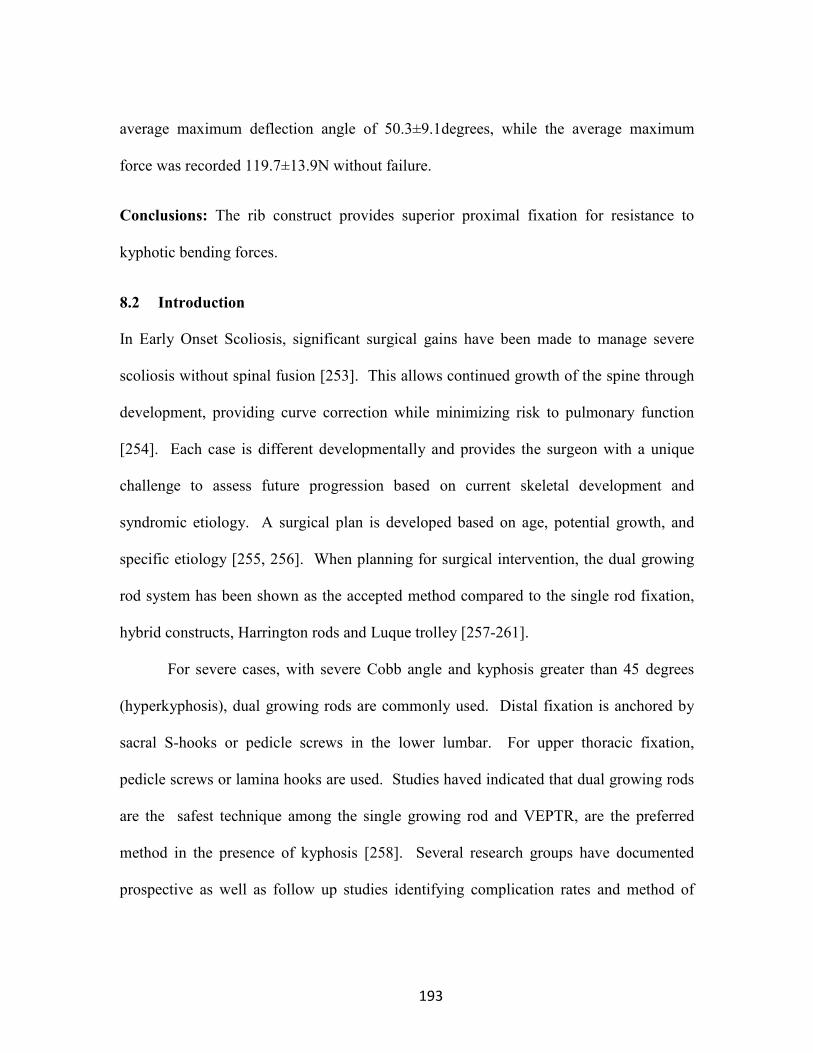
193
average maximum deflection angle of 50.3±9.1degrees, while the average maximum
force was recorded 119.7±13.9N without failure.
Conclusions: The rib construct provides superior proximal fixation for resistance to
kyphotic bending forces.
8.2 Introduction
In Early Onset Scoliosis, significant surgical gains have been made to manage severe
scoliosis without spinal fusion [253]. This allows continued growth of the spine through
development, providing curve correction while minimizing risk to pulmonary function
[254]. Each case is different developmentally and provides the surgeon with a unique
challenge to assess future progression based on current skeletal development and
syndromic etiology. A surgical plan is developed based on age, potential growth, and
specific etiology [255, 256]. When planning for surgical intervention, the dual growing
rod system has been shown as the accepted method compared to the single rod fixation,
hybrid constructs, Harrington rods and Luque trolley [257-261].
For severe cases, with severe Cobb angle and kyphosis greater than 45 degrees
(hyperkyphosis), dual growing rods are commonly used. Distal fixation is anchored by
sacral S-hooks or pedicle screws in the lower lumbar. For upper thoracic fixation,
pedicle screws or lamina hooks are used. Studies haved indicated that dual growing rods
are the safest technique among the single growing rod and VEPTR, are the preferred
method in the presence of kyphosis [258]. Several research groups have documented
prospective as well as follow up studies identifying complication rates and method of

194
implant failure in patients [257, 262-265]. The studies indicated a complication rate
between 48-52%. A common direct implant related complication was at the proximal
fixation, either through pedicle screw pullout or lamina hook failure. Although Sankar et.
al did not find a correlation with preoperative levels of kyphosis and complication rate
[265], a recent study by Watanabe has indicated that kyphosis level increases for every
20 degrees was an independent risk factor for failure of the proximal fixation [266].
Increasingly, in both studies and conference proceedings, significant kyphosis has been a
concern for effective dual growing rod treatment, especially in upper thoracic fixation
[267].
The difficulty with controlling kyphosis with growing rods comes with the
inherent pullout forces generated in the upper thoracic curve. The problem is
compounded by deformed vertebra and often osteoporosis in this region. Adult cadaver
studies have shown that reduction in bone mineral density due to osteoporosis
significantly reduces pedicle screw pull out strength [268-270]. The studies show the
significant risk with pedicle screws in osteoporotic spine. Further, in the review of early
onset scoliosis complications, lamina hooks with lamina anchorage in this region are also
reported with the same incidence of failure, if not higher [266]. From a biomechanical
perspective, an inherent risk is involved in the placement of rigid titanium rods, securely
anchored in the sacrum or lower lumbar, to a proximal foundation with questionable bone
density and size with distraction forces directly opposing the anchor in the presence of
kyphosis.

195
The clinical basis of this study came as a result of experiencing this problem in
two cases with kyphosis angles of greater than 50 degrees. In each case, neither pedicle
screw fixation or the VEPTR was recommended. Since the patients had a kyphosis angle
greater than 40 degrees, the VEPTR was excluded. Since the patients were considered
osteoporotic by the WHO criteria, pedicle screw fixation was excluded. The initial case
was a revised surgery where pedicle anchorage in place had already failed. In order to
move fixation away from the pedicle, the surgeon developed a 4 rib construct which
anchored lamina hooks to the ribs [271]. In both cases, the patients had satisfactory
outcome with no complications with 3 year follow up [271]. The procedure has been
proven effective in over 50 cases involving severe kyphosis over 50 degrees, including 9
cases of kyphosis over 100 degrees. With an average of 2 year follow up, the construct
has provided significant improvement in both Cobb (24 degrees) and thoracic kyphosis
(34 degree improvement pre vs post op). In all cases, only two indications of proximal
dislodgement or failure have been indicated. Only one construct was removed due to
infection.
Rather than recommend the 4 rib construct for wide spread use based on clinical
observation, this research group developed a controlled biomechanical model by which to
test the upper thoracic fixation strength of the 4 rib construct with the currently used
pedicle screw fixation. In the biomechanical model, immature pigs spines were used
(~21.3 kg) to simulate the spine of a 31.75 kg pediatric patient. To simulate kyphosis
acting against the proximal fixation, a pure bending test was conducted across the full
length of immature porcine spines. Two groups were used for the study with anchorage

196
to the proximal fixation, a control group with pedicle screw fixation and an experimental
group consisting of the 4 rib construct. Although, many pullout studies have investigated
pedicle screws and lamina hooks on individual vertebra in animal and cadaver models
[272] [273], this group sought to examine full bending of the construct to investigate the
mode of fixation failure between the novel 4 rib construct and the control pedicle screw
group.
8.3 Materials and Methods
Twelve pig spines (6 for the pedicle group, 6 rib for the construct group) were
obtained from the Strom Thurmond research center (Medical University of South
Carolina, Charleston, SC). Spines were harvested from Yorkshire domestic male pigs (8
weeks, weight 21.3 kg (+/- 1.3 kg) by the surgeon from T1 to L5 with thoraces intact in
the animal OR under IACUUK guidelines. The size and weight of both spine, rib cage
and pedicles were equivalent to the skeletal size of a 31.75 kg patient as determine by the
surgeon as representative of the patient population. Titanium growing rods (5.5 mm)
were instrumented bilaterally in both groups. No instrumentation was included distally,
since the distal foundation was potted for biomechanical testing. In the proximal
fixation, for the pedicle screw group, pedicle screws were placed in T3 and T4 bilaterally
in accordance with previous methods, see Figure 48a below and Figure 49a below [258].
When setting the pedicle screw group in the animal OR, a fluoroscope was used for
placement, see Figure 49 below. In the 4 rib construct group, lamina hooks were placed
on ribs 3-6 by the same surgeon relating the porcine anatomy to the pediatric patient.
Lamina hooks (wide blade for 5.5mm rod, Medtronic Memphis, Tennessee) were used on
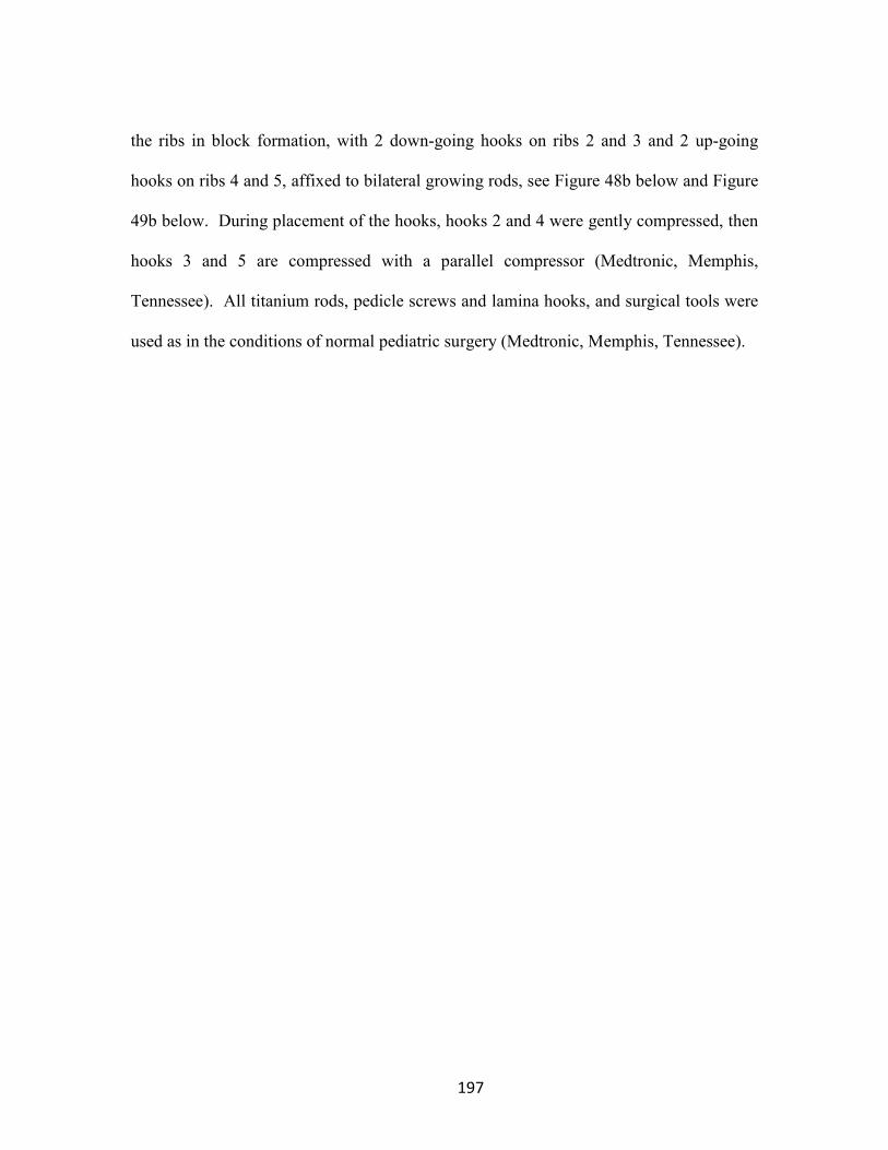
197
the ribs in block formation, with 2 down-going hooks on ribs 2 and 3 and 2 up-going
hooks on ribs 4 and 5, affixed to bilateral growing rods, see Figure 48b below and Figure
49b below. During placement of the hooks, hooks 2 and 4 were gently compressed, then
hooks 3 and 5 are compressed with a parallel compressor (Medtronic, Memphis,
Tennessee). All titanium rods, pedicle screws and lamina hooks, and surgical tools were
used as in the conditions of normal pediatric surgery (Medtronic, Memphis, Tennessee).
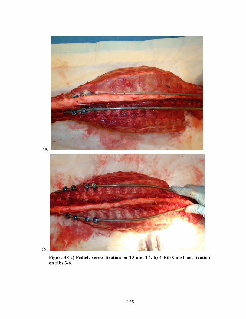
198
(a)
(b)
Figure 48 a) Pedicle screw fixation on T3 and T4. b) 4-Rib Construct fixation
on ribs 3-6.

199
(a)
(b)
Figure 49 showing placement of pedicle screw and hook constructs by
fluoroscopy in the animal OR
a) Pedicle screw placement on T3 and T4, b) Rib construct placement on ribs 3-6.

200
After the pig spines were instrumented and imaged, they were refrigerated (4
degrees C) until mechanical testing. Studies have shown musculoskeletal that samples
retain biomechanical strength for up to 5 freeze thaw cycles [148]. The samples were
potted (Bondo-Marhyde, Atlanta, GA) proximally and distally in custom designed potting
fixtures for mounting, see Figure 50 below. In the distal pot, growing rods extended past
the lower lumbar and were used to align the base of the spine within the pot. Similarly,
the proximal end of the specimen was potted to T1 with small acrylic rods (1mm
diameter) extending past T1 for alignment. Thus, all spines were consistently aligned
within both proximal and distal pots along the horizontal and vertical axis before testing.
Figure 50 Test configuration for full spine bending
Inline load cell with actuator measured bending force at potted proximal connection at T3

201
The distal potted end of the spine was anchored to a custom built mount which
securely anchored the base of the spine perpendicular to the MTS instrument (MTS,
Minneapolis, Minnesota), see Figure 50 above. A pure bending force, which simulated
kyphosis, was applied to the spine by the MTS at the proximal potted fixation (see Figure
50 above) [274]. The force was applied by using the MTS load arm which was able to
generate bending from 0 to 90 degrees for each specimen. The bending force was
measured by a load cell in line with the actuator. A pin-pin connection was used to attach
the actuator to the load arm of the MTS along with a pin-pin connection to the proximal
pot, in order to apply a pure bending force without generating a moment at the proximal
pot.
All spines were loaded from 0 to 90 degrees deflection angle, with the resulting
load measured. MTS load data were synched with video data. ImageJ was used to
determine deflection angle and displacement. Maximum bending force was determined
as a 20% decrease in load as a result of fixation failure or maximum force at the end of
the experiment when no failure had occurred.
8.4 Results
For the pedicle screw group, fixation failure was recorded on all six tested spines,
see Figure 51 below. Failure was pedicle screw pull out. The average deflection angle at
the failure point was 35.8±1.3 degrees. Angular deflection was reported as normalized,
with each specimen beginning at zero degrees. The average bending force at the failure
point was 118.6±25.7N.
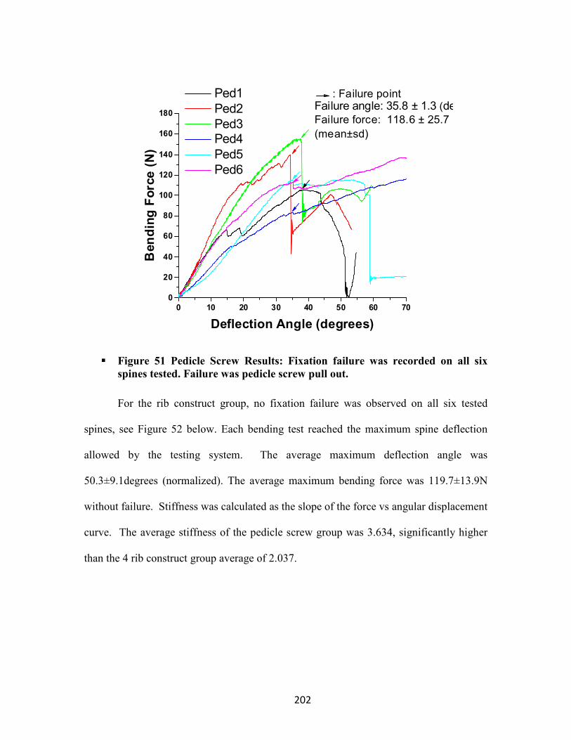
202
0 10 20 30 40 50 60 70
0
20
40
60
80
100
120
140
160
180
Ben
din
g F
orc
e (
N)
Deflection Angle (degrees)
Ped1
Ped2
Ped3 Ped4
Ped5
Ped6
: Failure pointFailure angle: 35.8 ± 1.3 (degreeFailure force: 118.6 ± 25.7 (N)
(mean±sd)
Figure 51 Pedicle Screw Results: Fixation failure was recorded on all six
spines tested. Failure was pedicle screw pull out.
For the rib construct group, no fixation failure was observed on all six tested
spines, see Figure 52 below. Each bending test reached the maximum spine deflection
allowed by the testing system. The average maximum deflection angle was
50.3±9.1degrees (normalized). The average maximum bending force was 119.7±13.9N
without failure. Stiffness was calculated as the slope of the force vs angular displacement
curve. The average stiffness of the pedicle screw group was 3.634, significantly higher
than the 4 rib construct group average of 2.037.

203
0 10 20 30 40 50 60 70
0
20
40
60
80
100
120
140
160
180 Maximum angle: 50.3 ± 9.1(degreeMaxium force: 119.7 ± 13.9 (N)
(mean±sd)
Be
nd
ing
Fo
rce (
N)
Deflection Angle (degrees)
Rib1 Rib2
Rib3
Rib4
Rib5 Rib6
No implant failure
Figure 52 Rib Construct Results: No fixation failure was observed on all six
spines tested. Each bending test reached the maximum spine deflection
allowed by the testing system
Figure 53 below shows an example failure curve a pedicle screw sample
compared to the 4 rib construct.
Figure 54 below shows a comparison of the pedicle screw and 4 rib construct
results.

204
Figure 53 Example failure curves of pedicle screw (red) and 4 rib construct
(black).
Pedicle screw constructs consistently failed at 35.8 +/- 1.3 degrees while 4 rib construct did not reach failure through the same angle of deflection.

205
(a)

206
(b)
Figure 54 showing maximum force and maximum deflection for the pedicle
screw and 4 rib construct group
Average maximum force (N) and deflection (degrees) for the pedicle screw (n=6) and 4 rib construct groups (n=6) tested. a) Maximum force with n=6 for each group, b) Maximum deflection with n=6 for each group.
Table 6 Maximum force and deflection
Maximum Force
(Newtons)
Maximum Deflection
(degrees)
Pedicle Screw 118.6±25.7 35.8±1.3
4 Rib Construct 119.7±13.9* 50.3±9.1*
*Denotes maximum force/deflection at end of testing without failure

207
8.5 Discussion
The objective of this study was to compare upper thoracic fixation methods in
early onset scoliosis in the presence of severe kyphosis. For this study, a novel 4 rib
construct was compared to a control group with standard pedicle screw instrumentation.
A well-controlled spine bending mechanical model was developed using bilateral
titanium growth rods instrumented to intact immature porcine spines.
The spine was loaded through 90 degrees angular deflection to simulate severe
kyphosis before load frame stroke length of the MTS was reached. Both the pedicle
screw group and 4 rib construct group were loaded through the same deflection angle.
However, each pedicle screw specimen failed within this range whereas no specimen in
the 4 rib construct group failed. The average deflection angle at failure for the pedicle
screw group was 35.8±1.3 degrees. The 4 rib construct group was able to deflect through
a mean of 50.3±9.1 degrees before the test was stopped, without reaching failure. These
results indicate that the 4 rib construct can withstand much higher levels of angular
deflection, and as a result, indicate that the 4 rib construct provides fixation through
greater kyphosis angles than pedicle screws. The reason the results are stated in angular
displacement instead of linear displacement is to keep to a physiologically relevant
measure of displacement, with the angular displacement being equivalent to kyphosis
angles.
When comparing maximum bending force reached by the implant recorded during
angular deflection, the pedicle screw group obtained 118.6±25.7 N and the 4 rib construct
group obtained 119.7±13.9 N. However, in the pedicle screw group, the maximum force

208
was recorded at failure and represents the maximum level of force and corresponding
angle (35.8±1.3 degrees) the construct can withstand (see Figure 51 above for failure
curve). In the 4 rib construct group, the average force of 119.7±13.9N was the maximum
force recorded at the end of the test and therefore was a representation of the maximum
force experience by the implant without failure at the end of the MTS load limitations at
an average angle of ~50.3±9.1 degrees (see Figure 52above for failure curve). These
results indicate both constructs experience similar maximum loads through the
application of bending/kyphotic force but the associative force-displacement response is
different between groups. In order to explain why the pedicle screw group failed and the
4 rib construct did not, the force vs angular displacement curves must be analyzed to
understand the loading response in regard to the stiffness of implant.
From observation of the experiments and analysis of the data, the pedicle screw
group resisted bending at the start of the test, incurring a large initial force from the onset
of bending. This was due to the rods being rigidly attached to vertebral bodies 3 and 4,
which resulted in close alignment with little contour. Once 30 degrees bending had
occurred in the spine, a peak in loading occurred with the implant failing through pedicle
screw pull out. Stiffness was considered for the entire construct as a ratio of force to
angular displacement. The overall stiffness was measured from the slope of the load vs
angular displacement curve was 3.634 which was significantly larger than the 4 rib
construct stiffness of 2.037. The lower stiffness value resulting from the 4 rib construct
group was likely due the contour of the rod during rib placement, flexure allowed by the
lamina hooks, and also slight flexure of the ribs. This resulted in a construct that allowed

209
much more flexure without the load-up in bending force that produced failure of the
implant. First, the rods were contoured over the ribs, which resulted in an initial kyphotic
contour. Rods were attached to lamina hooks placed at the tubercle, with abduction of
the rods and a kyphotic contour along the ribs. The combination of the 4 rib construct’s a
“wide stance” in contour with the ribs and slight flexure of the ribs allowed more flexure
in the construct. From observation of the tests, the 4 rib construct specimens flexed
evenly through bending. The resulting force response was a gradual increase in load,
even though deflection angles were increasing. This produced a force vs angular
displacement groups without a sharp spike in load response unlike the pedicle screw
group, which demonstrated a sharp rise in force response, with catastrophic failure of the
screw implant.
In addition to a lower stiffness, the connection of the 4 rib construct proved
superior. The opposite facing lamina hooks resisted the large distraction forces from
kyphotic bending much better than the pedicle screw group. The inferior facing lamina
hook on rib 3 was crucial at the beginning of the block fixation, which represent the
anatomical location of rib 2 for the most superior rib in human fixation. Interestingly,
biomechanical studies have shown the pedicle screw pullout strength was much larger
than pullout strength of the lamina hooks on the lamina, usually from failure of the
lamina. We found in this study that lamina hooks placed at the tubercle resulted in higher
fixation strength than the pedicle screw. One of the weaknesses of this study, however,
was the exclusion of a lamina hook group with fixation to the lamina. The pedicle screw
group was compared as the currently accepted standard between the growing rods and the

210
VEPTR. All ribs were examined for dislocation and signs of rib failure at the end of the
study, but none were observed from failure analysis of force data. This would indicate a
much larger range of implant loading and 4 rib fixation strength.
The full spine bending test in this study was a load by deflection until failure or
maximum deflection was reached. The limitations were based on using an MTS with
limited travel on which we used the load are to load samples. In the future, a testing
configuration of cyclic loading would be more physiologic under these conditions, as
most active patients see repetitive loading and likely not extreme deflection. Contour of
the rods were based on surgical preference and not uniform over all the sample groups.
We observed that titanium rods can flex through 90 degrees without implant failure. We
also observed that the ribs can stay intact through extreme loading, much larger than
physiologic conditions. But overall this study presents more questions in pediatric spine
surgery that must be addressed. Specifically, it raises the question of when do the
tradeoffs of stiffness and rod rigidity cease when compared to flexation in an active
patient. From observing this study, clearly there is a payoff to having flexation in the
construct in order not have to failure at the fixation site. A pediatric spine is active and
yet still developing. Therefore, a crucial design parameter to determine in future studies
is the amount of flexure necessary to the implant rigidity for support in both kyphosis and
scoliosis curves yet withstand transverse pullout forces. It will be important to answer
how stiff the implant needs to be relative to the bone density and flexural strength of the
pediatric spine will help in the management of these severe cases.

211
In the future, research must be expanded to determine if fixation to the ribs could
be a viable alternative to vertebral body fixation altogether. First, the prediction of bone
strength at the fixation site will greatly enhance the outcome of surgery and allow for
critical assessment of anchorage success in planning. In order to accurately pair the bone
quality fixation predictions with biomechanical anchorage strength, an animal model that
creates scoliosis through tethering must be developed. The animal model will not only
provide a better biomechanical basis for our construct testing, but will allow a better
understanding of construct performance while in vivo. The analysis includes not only the
strength of construct from the animal spine, but also the amount of fusion and well as
developmental changes the construct may either promote or inhibit. Once the constructs
can be compared in a living animal model, research can progress into better fusionless
constructs that will aid the development of the pediatric spine.

212
9 REFERENCES
1. Benjamin, M. and J.R. Ralphs, Biology of fibrocartilage cells. Int Rev Cytol, 2004. 233: p.
1-45.
2. Okeson, J.P., Management of temporomandibular disorders and occlusion. 4th ed1998,
St. Louis: Mosby. x, 638 p.
3. Almarza, A.J. and K.A. Athanasiou, Design characteristics for the tissue engineering of
cartilaginous tissues. Ann Biomed Eng, 2004. 32(1): p. 2-17.
4. de Bont, L.G. and B. Stegenga, Pathology of temporomandibular joint internal
derangement and osteoarthrosis. Int J Oral Maxillofac Surg, 1993. 22(2): p. 71-4.
5. Farrar, W.B. and W.L. McCarty, INFERIOR JOINT SPACE ARTHROGRAPHY AND
CHARACTERISTICS OF CONDYLAR PATHS IN INTERNAL DERANGEMENTS OF THE TMJ.
Journal of Prosthetic Dentistry, 1979. 41(5): p. 548-555.
6. Gallo, L.M., et al., Description of mandibular finite helical axis pathways in asymptomatic
subjects. J Dent Res, 1997. 76(2): p. 704-13.
7. Solberg, W.K., M.W. Woo, and J.B. Houston, Prevalence of mandibular dysfunction in
young adults. J Am Dent Assoc, 1979. 98(1): p. 25-34.
8. Warren, M.P. and J.L. Fried, Temporomandibular disorders and hormones in women.
Cells Tissues Organs, 2001. 169(3): p. 187-92.
9. Fillingim, R.B., et al., Potential psychosocial risk factors for chronic TMD: descriptive data
and empirically identified domains from the OPPERA case-control study. J Pain, 2011.
12(11 Suppl): p. T46-60.
10. Fillingim, R.B., et al., Summary of findings from the OPPERA baseline case-control study:
implications and future directions. J Pain, 2011. 12(11 Suppl): p. T102-7.
11. Maixner, W., et al., Orofacial Pain Prospective Evaluation and Risk Assessment study--
the OPPERA study. J Pain, 2011. 12(11 Suppl): p. T4-11 e1-2.
12. Slade, G.D., et al., Study methods, recruitment, sociodemographic findings, and
demographic representativeness in the OPPERA study. J Pain, 2011. 12(11 Suppl): p. T12-
26.
13. Smith, S.B., et al., Potential genetic risk factors for chronic TMD: genetic associations
from the OPPERA case control study. J Pain, 2011. 12(11 Suppl): p. T92-101.
14. Detamore, M.S. and K.A. Athanasiou, Motivation, characterization, and strategy for
tissue engineering the temporomandibular joint disc. Tissue Engineering, 2003. 9(6): p.
1065-1087.
15. Farrar, W.B. and W.L. McCarty, Jr., The TMJ dilemma. J Ala Dent Assoc, 1979. 63(1): p.
19-26.
16. McCarty, W.L. and W.B. Farrar, SURGERY FOR INTERNAL DERANGEMENTS OF THE
TEMPOROMANDIBULAR-JOINT. Journal of Prosthetic Dentistry, 1979. 42(2): p. 191-196.
17. Landesberg, R., E. Takeuchi, and J.E. Puzas, Cellular, biochemical and molecular
characterization of the bovine temporomandibular joint disc. Arch Oral Biol, 1996. 41(8-
9): p. 761-7.
18. Haskin, C.L., S.B. Milam, and I.L. Cameron, Pathogenesis of degenerative joint disease in
the human temporomandibular joint. Crit Rev Oral Biol Med, 1995. 6(3): p. 248-77.

213
19. Milam, S.B., et al., Characterization of the extracellular matrix of the primate
temporomandibular joint. J Oral Maxillofac Surg, 1991. 49(4): p. 381-91.
20. Milam, S.B. and J.P. Schmitz, Molecular biology of temporomandibular joint disorders:
proposed mechanisms of disease. J Oral Maxillofac Surg, 1995. 53(12): p. 1448-54.
21. Milam, S.B., G. Zardeneta, and J.P. Schmitz, Oxidative stress and degenerative
temporomandibular joint disease: a proposed hypothesis. J Oral Maxillofac Surg, 1998.
56(2): p. 214-23.
22. O'Dell, N.L., et al., Morphological and biochemical evidence for elastic fibres in the Syrian
hamster temporomandibular joint disc. Arch Oral Biol, 1990. 35(10): p. 807-11.
23. Shengyi, T. and Y. Xu, Biomechanical properties and collagen fiber orientation of TMJ
discs in dogs: Part 1. Gross anatomy and collagen fiber orientation of the discs. J
Craniomandib Disord, 1991. 5(1): p. 28-34.
24. Detamore, M.S. and K.A. Athanasiou, Tensile properties of the porcine
temporomandibular joint disc. J Biomech Eng, 2003. 125(4): p. 558-65.
25. Detamore, M.S. and K.A. Athanasiou, Structure and function of the temporomandibular
joint disc: implications for tissue engineering. J Oral Maxillofac Surg, 2003. 61(4): p. 494-
506.
26. Detamore, M.S., et al., Cell type and distribution in the porcine temporomandibular joint
disc. J Oral Maxillofac Surg, 2006. 64(2): p. 243-8.
27. Detamore, M.S., et al., Quantitative analysis and comparative regional investigation of
the extracellular matrix of the porcine temporomandibular joint disc. Matrix Biol, 2005.
24(1): p. 45-57.
28. Singh, M. and M.S. Detamore, Biomechanical properties of the mandibular condylar
cartilage and their relevance to the TMJ disc. J Biomech, 2009. 42(4): p. 405-17.
29. Tanaka, E., et al., Lubrication of the temporomandibular joint. Ann Biomed Eng, 2008.
36(1): p. 14-29.
30. Tanaka, E., et al., Strain-rate effect on the biomechanical response of bovine
temporomandibular joint disk under compression. J Biomed Mater Res A, 2003. 67(3): p.
761-5.
31. Tanaka, E., et al., Dynamic shear properties of the temporomandibular joint disc. J Dent
Res, 2003. 82(3): p. 228-31.
32. Beek, M., et al., Dynamic properties of the human temporomandibular joint disc. J Dent
Res, 2001. 80(3): p. 876-80.
33. Beek, M., et al., Three-dimensional finite element analysis of the cartilaginous structures
in the human temporomandibular joint. J Dent Res, 2001. 80(10): p. 1913-8.
34. Spirt, A.A., A.F. Mak, and R.P. Wassell, Nonlinear viscoelastic properties of articular
cartilage in shear. J Orthop Res, 1989. 7(1): p. 43-9.
35. Herring, S.W., et al., Temporomandibular joint in miniature pigs: anatomy, cell
replication, and relation to loading. Anat Rec, 2002. 266(3): p. 152-66.
36. Ingawale, S. and T. Goswami, Temporomandibular joint: disorders, treatments, and
biomechanics. Ann Biomed Eng, 2009. 37(5): p. 976-96.
37. Tanaka, E. and T. van Eijden, Biomechanical behavior of the temporomandibular joint
disc. Crit Rev Oral Biol Med, 2003. 14(2): p. 138-50.
38. Koolstra, J.H. and T.M. van Eijden, Combined finite-element and rigid-body analysis of
human jaw joint dynamics. J Biomech, 2005. 38(12): p. 2431-9.

214
39. Nagahara, K., et al., Displacement and stress distribution in the temporomandibular joint
during clenching. Angle Orthod, 1999. 69(4): p. 372-9.
40. Pileicikiene, G., et al., A three-dimensional model of the human masticatory system,
including the mandible, the dentition and the temporomandibular joints. Stomatologija,
2007. 9(1): p. 27-32.
41. Spilker, R.L., J.C. Nickel, and L.R. Iwasaki, A biphasic finite element model of in vitro
plowing tests of the temporomandibular joint disc. Ann Biomed Eng, 2009. 37(6): p.
1152-64.
42. Tanaka, E., et al., Three-dimensional finite element analysis of human
temporomandibular joint with and without disc displacement during jaw opening. Med
Eng Phys, 2004. 26(6): p. 503-11.
43. Tanaka, E., et al., Stress distribution in the temporomandibular joint affected by anterior
disc displacement: a three-dimensional analytic approach with the finite-element
method. J Oral Rehabil, 2000. 27(9): p. 754-9.
44. Tanaka, E., et al., Stress analysis in the TMJ during jaw opening by use of a three-
dimensional finite element model based on magnetic resonance images. Int J Oral
Maxillofac Surg, 2001. 30(5): p. 421-30.
45. Wilkes, C.H., Arthrography of the temporomandibular joint in patients with the TMJ
pain-dysfunction syndrome. Minn Med, 1978. 61(11): p. 645-52.
46. Wilkes, C.H., Internal derangements of the temporomandibular joint. Pathological
variations. Arch Otolaryngol Head Neck Surg, 1989. 115(4): p. 469-77.
47. Zarb, G.A. and G.E. Carlsson, Temporomandibular disorders: osteoarthritis. J Orofac Pain,
1999. 13(4): p. 295-306.
48. Perez del Palomar, A. and M. Doblare, An accurate simulation model of anteriorly
displaced TMJ discs with and without reduction. Med Eng Phys, 2007. 29(2): p. 216-26.
49. Allen, K.D. and K.A. Athanasiou, Tissue Engineering of the TMJ disc: a review. Tissue Eng,
2006. 12(5): p. 1183-96.
50. Detamore, M.S. and K.A. Athanasiou, Motivation, characterization, and strategy for
tissue engineering the temporomandibular joint disc. Tissue Eng, 2003. 9(6): p. 1065-87.
51. Detamore, M.S., K.A. Athanasiou, and J. Mao, A call to action for bioengineers and
dental professionals: directives for the future of TMJ bioengineering. Ann Biomed Eng,
2007. 35(8): p. 1301-11.
52. Abramowicz, S., et al., Temporomandibular joint reconstruction after failed teflon-
proplast implant: case report and literature review. Int J Oral Maxillofac Surg, 2008.
37(8): p. 763-7.
53. Mow, V.C., M.H. Holmes, and W.M. Lai, Fluid transport and mechanical properties of
articular cartilage: a review. J Biomech, 1984. 17(5): p. 377-94.
54. Shirakura, M., et al., Activation of the hypoxia-inducible factor-1 in overloaded
temporomandibular joint, and induction of osteoclastogenesis. Biochem Biophys Res
Commun, 2010. 393(4): p. 800-5.
55. de Bont, L.G., L.C. Dijkgraaf, and B. Stegenga, Epidemiology and natural progression of
articular temporomandibular disorders. Oral Surg Oral Med Oral Pathol Oral Radiol
Endod, 1997. 83(1): p. 72-6.
56. Fung, Y., Biomechanical properties of living tissue1981, NY: Springer-Verlag. pp. 196-260.

215
57. Almarza, A.J., et al., Biochemical analysis of the porcine temporomandibular joint disc. Br
J Oral Maxillofac Surg, 2006. 44(2): p. 124-8.
58. Shi, C., et al., Anisotropic solute diffusion tensor in porcine TMJ discs measured by FRAP
with spatial Fourier analysis. Ann Biomed Eng, 2010. 38(11): p. 3398-408.
59. Gu, W.Y., W.M. Lai, and V.C. Mow, TRANSPORT OF FLUID AND IONS THROUGH A
POROUS-PERMEABLE CHARGED-HYDRATED TISSUE, AND STREAMING POTENTIAL DATA
ON NORMAL BOVINE ARTICULAR-CARTILAGE. Journal of Biomechanics, 1993. 26(6): p.
709-723.
60. Gu, W.Y., W.M. Lai, and V.C. Mow, A mixture theory for charged-hydrated soft tissues
containing multi-electrolytes: passive transport and swelling behaviors. J Biomech Eng,
1998. 120(2): p. 169-80.
61. Kuo, J., et al., Effect of Mechanical Loading on Electrical Conductivity in Porcine TMJ
Discs. Journal of Dental Research, 2011. 90(10): p. 1216-1220.
62. Wright, G.J., et al., Effect of Mechanical Strain on Solute Diffusion in Human TMJ Discs:
An Electrical Conductivity Study. Annals of Biomedical Engineering, 2013. 41(11): p.
2349-2357.
63. Jackson, G.W. and D.F. James, THE PERMEABILITY OF FIBROUS POROUS-MEDIA.
Canadian Journal of Chemical Engineering, 1986. 64(3): p. 364-374.
64. Kuo, J., et al., Regional cell density distribution and oxygen consumption rates in porcine
TMJ discs: an explant study. Osteoarthritis and Cartilage, 2011. 19(7): p. 911-918.
65. Milam, S.B., Pathogenesis of degenerative temporomandibular joint arthritides.
Odontology, 2005. 93(1): p. 7-15.
66. Maroudas, A., Physicochemical properties of cartilage in the light of ion exchange
theory. Biophys J, 1968. 8(5): p. 575-95.
67. Maroudas, A., H. Muir, and J. Wingham, The correlation of fixed negative charge with
glycosaminoglycan content of human articular cartilage. Biochim Biophys Acta, 1969.
177(3): p. 492-500.
68. Mow, V.C., et al., Effects of fixed charges on the stress-relaxation behavior of hydrated
soft tissues in a confined compression problem. International Journal of Solids and
Structures, 1998. 35(34-35): p. 4945-4962.
69. Mow, V.C. and W.C. Hayes, BASIC ORTHOPEDIC BIOMECHANICS. Mow, V. C. And W. C.
Hayes. Basic Orthopaedic Biomechanics. X+453p. Raven Press: New York, New York,
USA. Illus1991. X+453P.
70. Tanaka, E., et al., Viscoelastic properties of the human temporomandibular joint disc in
patients with internal derangement. J Oral Maxillofac Surg, 2000. 58(9): p. 997-1002.
71. Kalpakci, K.N., et al., An interspecies comparison of the temporomandibular joint disc. J
Dent Res, 2011. 90(2): p. 193-8.
72. Sun, Z., Z.J. Liu, and S.W. Herring, Movement of temporomandibular joint tissues during
mastication and passive manipulation in miniature pigs. Arch Oral Biol, 2002. 47(4): p.
293-305.
73. Gallo, L.M., et al., Mechanical work during stress-field translation in the human TMJ. J
Dent Res, 2006. 85(11): p. 1006-10.
74. Gallo, L.M., et al., Stress-field translation in the healthy human temporomandibular joint.
J Dent Res, 2000. 79(10): p. 1740-6.

216
75. Mills, D.K., D.J. Fiandaca, and R.P. Scapino, Morphologic, microscopic, and
immunohistochemical investigations into the function of the primate TMJ disc. J Orofac
Pain, 1994. 8(2): p. 136-54.
76. Archer, C.W. and P. Francis-West, The chondrocyte. Int J Biochem Cell Biol, 2003. 35(4):
p. 401-4.
77. Poole, C.A., M.H. Flint, and B.W. Beaumont, Chondrons in cartilage: ultrastructural
analysis of the pericellular microenvironment in adult human articular cartilages. J
Orthop Res, 1987. 5(4): p. 509-22.
78. Okuda, Y., J.P. Gorski, and P.C. Amadio, Effect of postnatal age on the ultrastructure of
six anatomical areas of canine flexor digitorum profundus tendon. J Orthop Res, 1987.
5(2): p. 231-41.
79. Rufai, A., M. Benjamin, and J.R. Ralphs, The development of fibrocartilage in the rat
intervertebral disc. Anat Embryol (Berl), 1995. 192(1): p. 53-62.
80. Nakano, T. and P.G. Scott, Changes in the chemical composition of the bovine
temporomandibular joint disc with age. Arch Oral Biol, 1996. 41(8-9): p. 845-53.
81. Kapila, S., C. Lee, and D.W. Richards, Characterization and identification of proteinases
and proteinase inhibitors synthesized by temporomandibular joint disc cells. J Dent Res,
1995. 74(6): p. 1328-36.
82. Shi, C., et al., Relationship between anisotropic diffusion properties and tissue
morphology in porcine TMJ disc. Osteoarthritis and Cartilage, 2013. 21(4): p. 625-633.
83. Minarelli, A.M. and E.A. Liberti, A microscopic survey of the human temporomandibular
joint disc. J Oral Rehabil, 1997. 24(11): p. 835-40.
84. Cheung, H.S., Distribution of type I, II, III and V in the pepsin solubilized collagens in
bovine menisci. Connect Tissue Res, 1987. 16(4): p. 343-56.
85. Ghadially, F.N., et al., Ultrastructure of rabbit semilunar cartilages. J Anat, 1978. 125(Pt
3): p. 499-517.
86. Girdler, N.M., In vitro synthesis and characterization of a cartilaginous meniscus grown
from isolated temporomandibular chondroprogenitor cells. Scand J Rheumatol, 1998.
27(6): p. 446-53.
87. Hasler, E.M., et al., Articular cartilage biomechanics: theoretical models, material
properties, and biosynthetic response. Crit Rev Biomed Eng, 1999. 27(6): p. 415-88.
88. Gage, J.P., R.M. Shaw, and F.B. Moloney, Collagen type in dysfunctional
temporomandibular joint disks. J Prosthet Dent, 1995. 74(5): p. 517-20.
89. Scapino, R.P., et al., The behaviour of collagen fibres in stress relaxation and stress
distribution in the jaw-joint disc of rabbits. Arch Oral Biol, 1996. 41(11): p. 1039-52.
90. Berkovitz, B.K., Crimping of collagen in the intra-articular disc of the temporomandibular
joint: a comparative study. J Oral Rehabil, 2000. 27(7): p. 608-13.
91. Keith, D.A., Elastin in the bovine mandibular joint. Arch Oral Biol, 1979. 24(3): p. 211-5.
92. Gross, A., A. Bumann, and B. Hoffmeister, Elastic fibers in the human temporo-
mandibular joint disc. Int J Oral Maxillofac Surg, 1999. 28(6): p. 464-8.
93. Esko JD, K.K., Lindahl U, Proteoglycans and Sulfated Glycosaminoglycans, in Essentials of
Glycobiology2009, Cold Spring Harbor Laboratory Press: NCBI.
94. Chammas, P., W.J. Federspiel, and S.R. Eisenberg, A MICROCONTINUUM MODEL OF
ELECTROKINETIC COUPLING IN THE EXTRACELLULAR-MATRIX - PERTURBATION

217
FORMULATION AND SOLUTION. Journal of Colloid and Interface Science, 1994. 168(2):
p. 526-538.
95. Eisenberg, S.R. and A.J. Grodzinsky, ELECTROKINETIC MICROMODEL OF EXTRACELLULAR-
MATRIX AND OTHER POLY-ELECTROLYTE NETWORKS. Physicochemical Hydrodynamics,
1988. 10(4): p. 517-539.
96. Frank, E.H. and A.J. Grodzinsky, CARTILAGE ELECTROMECHANICS .2. A CONTINUUM
MODEL OF CARTILAGE ELECTROKINETICS AND CORRELATION WITH EXPERIMENTS.
Journal of Biomechanics, 1987. 20(6): p. 629-639.
97. Frank, E.H. and A.J. Grodzinsky, CARTILAGE ELECTROMECHANICS .1. ELECTROKINETIC
TRANSDUCTION AND THE EFFECTS OF ELECTROLYTE PH AND IONIC-STRENGTH. Journal
of Biomechanics, 1987. 20(6): p. 615-&.
98. Frank, E.H., et al., STREAMING POTENTIALS - A SENSITIVE INDEX OF ENZYMATIC
DEGRADATION IN ARTICULAR-CARTILAGE. Journal of Orthopaedic Research, 1987. 5(4):
p. 497-508.
99. Grodzinsky, A.J., ELECTROMECHANICAL AND PHYSICOCHEMICAL PROPERTIES OF
CONNECTIVE-TISSUE. Crc Critical Reviews in Biomedical Engineering, 1983. 9(2): p. 133-
199.
100. Lai, W.M., et al., On the electric potentials inside a charged soft hydrated biological
tissue: Streaming potential versus diffusion potential. Journal of Biomechanical
Engineering-Transactions of the Asme, 2000. 122(4): p. 336-346.
101. Maroudas, A., PHYSICOCHEMICAL PROPERTIES OF CARTILAGE IN LIGHT OF ION
EXCHANGE THEORY. Biophysical Journal, 1968. 8(5): p. 575-&.
102. Nakano, T. and P.G. Scott, A quantitative chemical study of glycosaminoglycans in the
articular disc of the bovine temporomandibular joint. Arch Oral Biol, 1989. 34(9): p. 749-
57.
103. Axelsson, S., A. Holmlund, and A. Hjerpe, Glycosaminoglycans in normal and
osteoarthrotic human temporomandibular joint disks. Acta Odontol Scand, 1992. 50(2):
p. 113-9.
104. Lumpkins, S.B. and P.S. McFetridge, Regional variations in the viscoelastic compressive
properties of the temporomandibular joint disc and implications toward tissue
engineering. J Biomed Mater Res A, 2008.
105. Campbell, J.H., et al., Estrogen receptor analysis of human temporomandibular disc. J
Oral Maxillofac Surg, 1993. 51(10): p. 1101-5.
106. Wu, Y.W., et al., 17-Beta-estradiol enhanced allodynia of inflammatory
temporomandibular joint through upregulation of hippocampal TRPV1 in ovariectomized
rats. J Neurosci, 2010. 30(26): p. 8710-9.
107. Abubaker, A.O., P.C. Hebda, and J.N. Gunsolley, Effects of sex hormones on protein and
collagen content of the temporomandibular joint disc of the rat. J Oral Maxillofac Surg,
1996. 54(6): p. 721-7; discussion 727-8.
108. Burger, E.H., J. Klein-Nulend, and J.P. Veldhuijzen, Mechanical stress and osteogenesis in
vitro. J Bone Miner Res, 1992. 7 Suppl 2: p. S397-401.
109. Nitzan, D.W., The process of lubrication impairment and its involvement in
temporomandibular joint disc displacement: a theoretical concept. J Oral Maxillofac
Surg, 2001. 59(1): p. 36-45.

218
110. Nitzan, D.W., Intraarticular pressure in the functioning human temporomandibular joint
and its alteration by uniform elevation of the occlusal plane. J Oral Maxillofac Surg,
1994. 52(7): p. 671-9; discussion 679-80.
111. Zardeneta, G., S.B. Milam, and J.P. Schmitz, Iron-dependent generation of free radicals:
plausible mechanisms in the progressive deterioration of the temporomandibular joint. J
Oral Maxillofac Surg, 2000. 58(3): p. 302-8; discussion 309.
112. Cairns, B.E., Pathophysiology of TMD pain--basic mechanisms and their implications for
pharmacotherapy. J Oral Rehabil, 2010. 37(6): p. 391-410.
113. Ohshima, H. and J.P. Urban, The effect of lactate and pH on proteoglycan and protein
synthesis rates in the intervertebral disc. Spine, 1992. 17(9): p. 1079-82.
114. Tanaka, E. and T. van Eijden, Biomechanical behavior of the temporomandibular joint
disc. Critical Reviews in Oral Biology & Medicine, 2003. 14(2): p. 138-150.
115. Tanaka, E., et al., Modeling of the effect of friction in the temporomandibular joint on
displacement of its disc during prolonged clenching. J Oral Maxillofac Surg, 2008. 66(3):
p. 462-8.
116. Ethunandan, M. and A.W. Wilson, Temporomandibular joint arthrocentesis -more
questions than answers? J Oral Maxillofac Surg, 2006. 64(6): p. 952-5.
117. Chung, S.C., Y.K. Kim, and H.S. Kim, Prevalence and patterns of nocturnal bruxofacets on
stabilization splints in temporomandibular disorder patients. Cranio, 2000. 18(2): p. 92-
7.
118. Glass, E.G., A.G. Glaros, and F.D. McGlynn, Myofascial pain dysfunction: treatments used
by ADA members. Cranio, 1993. 11(1): p. 25-9.
119. Nitzan, D.W., M.F. Dolwick, and G.A. Martinez, Temporomandibular joint arthrocentesis:
a simplified treatment for severe, limited mouth opening. J Oral Maxillofac Surg, 1991.
49(11): p. 1163-7; discussion 1168-70.
120. Al-Belasy, F.A. and M.F. Dolwick, Arthrocentesis for the treatment of
temporomandibular joint closed lock: a review article. Int J Oral Maxillofac Surg, 2007.
36(9): p. 773-82.
121. Guo, C., Z. Shi, and P. Revington, Arthrocentesis and lavage for treating
temporomandibular joint disorders. Cochrane Database Syst Rev, 2009(4): p. CD004973.
122. Wolford, L.M., M.C. Pitta, and P. Mehra, Mitek anchors for treatment of chronic
mandibular dislocation. Oral Surg Oral Med Oral Pathol Oral Radiol Endod, 2001. 92(5):
p. 495-8.
123. Wolford, L.M., Temporomandibular joint devices: treatment factors and outcomes. Oral
Surg Oral Med Oral Pathol Oral Radiol Endod, 1997. 83(1): p. 143-9.
124. Fields, R.T., Jr. and L.M. Wolford, The osseointegration of Mitek mini anchors in the
mandibular condyle. J Oral Maxillofac Surg, 2001. 59(12): p. 1402-6; discussion 1407.
125. Kearns, G.J., D.H. Perrott, and L.B. Kaban, A protocol for the management of failed
alloplastic temporomandibular joint disc implants. J Oral Maxillofac Surg, 1995. 53(11):
p. 1240-7; discussion 1248-9.
126. Teng, S., et al., Biomechanical properties and collagen fiber orientation of
temporomandibular joint discs in dogs: 2. Tensile mechanical properties of the discs. J
Craniomandib Disord, 1991. 5(2): p. 107-14.
127. Lai, W.F., J. Bowley, and J.G. Burch, Evaluation of shear stress of the human
temporomandibular joint disc. J Orofac Pain, 1998. 12(2): p. 153-9.

219
128. Tanne, K., E. Tanaka, and M. Sakuda, The elastic modulus of the temporomandibular
joint disc from adult dogs. J Dent Res, 1991. 70(12): p. 1545-8.
129. Beatty, M.W., et al., Strain rate dependent orthotropic properties of pristine and
impulsively loaded porcine temporomandibular joint disk. J Biomed Mater Res, 2001.
57(1): p. 25-34.
130. Tanaka, E., et al., Dynamic properties of bovine temporomandibular joint disks change
with age. J Dent Res, 2002. 81(9): p. 618-22.
131. Chin, L.P., F.D. Aker, and K. Zarrinnia, The viscoelastic properties of the human
temporomandibular joint disc. J Oral Maxillofac Surg, 1996. 54(3): p. 315-8; discussion
318-9.
132. Allen, K.D. and K.A. Athanasiou, Viscoelastic characterization of the porcine
temporomandibular joint disc under unconfined compression. J Biomech, 2006. 39(2): p.
312-22.
133. Lumpkins, S.B. and P.S. McFetridge, Regional variations in the viscoelastic compressive
properties of the temporomandibular joint disc and implications toward tissue
engineering. J Biomed Mater Res A, 2009. 90(3): p. 784-91.
134. Tanaka, E., et al., Dynamic compressive properties of porcine temporomandibular joint
disc. Eur J Oral Sci, 2003. 111(5): p. 434-9.
135. Zhu, W., et al., Viscoelastic shear properties of articular cartilage and the effects of
glycosidase treatments. J Orthop Res, 1993. 11(6): p. 771-81.
136. Zhu, W., K.Y. Chern, and V.C. Mow, Anisotropic viscoelastic shear properties of bovine
meniscus. Clin Orthop Relat Res, 1994(306): p. 34-45.
137. Tanaka, E., et al., Shear properties of the temporomandibular joint disc in relation to
compressive and shear strain. J Dent Res, 2004. 83(6): p. 476-9.
138. Tanaka, E., et al., Biomechanical response of condylar cartilage-on-bone to dynamic
shear. J Biomed Mater Res A, 2008. 85(1): p. 127-32.
139. Nitzan, D.W., 'Friction and adhesive forces'--possible underlying causes for
temporomandibular joint internal derangement. Cells Tissues Organs, 2003. 174(1-2): p.
6-16.
140. Nickel, J.C., et al., The effect of disc thickness and trauma on disc surface friction in the
porcine temporomandibular joint. Arch Oral Biol, 2001. 46(2): p. 155-62.
141. Werner, J.A., B. Tillmann, and A. Schleicher, Functional anatomy of the
temporomandibular joint. A morphologic study on human autopsy material. Anat
Embryol (Berl), 1991. 183(1): p. 89-95.
142. Donzelli, P.S., et al., Biphasic finite element simulation of the TMJ disc from in vivo
kinematic and geometric measurements. J Biomech, 2004. 37(11): p. 1787-91.
143. Dunbar, W.L., Jr., et al., An evaluation of three-dimensional diarthrodial joint contact
using penetration data and the finite element method. J Biomech Eng, 2001. 123(4): p.
333-40.
144. Nickel, J.C., et al., Laboratory stresses and tractional forces on the TMJ disc surface. J
Dent Res, 2004. 83(8): p. 650-4.
145. Nickel, J.C., et al., Static and dynamic loading effects on temporomandibular joint disc
tractional forces. J Dent Res, 2006. 85(9): p. 809-13.
146. Tanaka, E., et al., Biomechanical behaviour of bovine temporomandibular articular discs
with age. Arch Oral Biol, 2001. 46(11): p. 997-1003.
220
147. del Pozo, R., et al., The regional difference of viscoelastic property of bovine
temporomandibular joint disc in compressive stress-relaxation. Med Eng Phys, 2002.
24(3): p. 165-71.
148. Allen, K.D. and K.A. Athanasiou, A surface-regional and freeze-thaw characterization of
the porcine temporomandibular joint disc. Ann Biomed Eng, 2005. 33(7): p. 951-62.
149. Stairmand, J.W., S. Holm, and J.P. Urban, Factors influencing oxygen concentration
gradients in the intervertebral disc. A theoretical analysis. Spine, 1991. 16(4): p. 444-9.
150. Holm, S., et al., Nutrition of the intervertebral disc: solute transport and metabolism.
Connect Tissue Res, 1981. 8(2): p. 101-19.
151. Bartels, E.M., et al., Oxygen and lactate concentrations measured in vivo in the
intervertebral discs of patients with scoliosis and back pain. Spine, 1998. 23(1): p. 1-7;
discussion 8.
152. Kuo, J., et al., Effect of mechanical loading on electrical conductivity in porcine TMJ discs.
J Dent Res, 2011. 90(10): p. 1216-20.
153. Gu, W.Y., M.A. Justiz, and H. Yao, Electrical conductivity of lumbar anulus fibrosis: Effects
of porosity and fixed charge density. Spine, 2002. 27(21): p. 2390-2395.
154. Shen, G. and M.A. Darendeliler, The adaptive remodeling of condylar cartilage---a
transition from chondrogenesis to osteogenesis. J Dent Res, 2005. 84(8): p. 691-9.
155. Moses, J.J. and D.C. Topper, A functional approach to the treatment of
temporomandibular joint internal derangement. J Craniomandib Disord, 1991. 5(1): p.
19-27.
156. Mow, V.C., et al., Biphasic creep and stress relaxation of articular cartilage in
compression? Theory and experiments. J Biomech Eng, 1980. 102(1): p. 73-84.
157. Lai, W.M., J.S. Hou, and V.C. Mow, A triphasic theory for the swelling and deformation
behaviors of articular cartilage. J Biomech Eng, 1991. 113(3): p. 245-58.
158. Maroudas, A.I., Balance between swelling pressure and collagen tension in normal and
degenerate cartilage. Nature, 1976. 260(5554): p. 808-9.
159. del Palomar, A.P., et al., Clenching TMJs-loads increases in partial edentates: a 3D finite
element study. Ann Biomed Eng, 2008. 36(6): p. 1014-23.
160. Soboleva, U., L. Laurina, and A. Slaidina, Jaw tracking devices--historical review of
methods development. Part I. Stomatologija, 2005. 7(3): p. 67-71.
161. Lemoine, J.J., et al., Radiographic analysis for jaw motion normalization. J Oral
Maxillofac Surg, 2005. 63(7): p. 961-7.
162. Enciso, R., et al., The virtual craniofacial patient: 3D jaw modeling and animation. Stud
Health Technol Inform, 2003. 94: p. 65-71.
163. Krebs, M., et al., A new method for three-dimensional reconstruction and animation of
the temporomandibular joint. Ann Acad Med Singapore, 1995. 24(1): p. 11-6.
164. Woltring, H.J. and E.B. Marsolais, Optoelectric (Selspot) gait measurement in two- and
three-dimensional space--a preliminary report. Bull Prosthet Res, 1980. 10-34: p. 46-52.
165. Seemann, R., et al., Pseudodynamic MRI differs from natural opening of the
temporomandibular joint. Oral Surg Oral Med Oral Pathol Oral Radiol Endod, 2008.
105(3): p. 371-8.
166. Fushima, K., et al., Analysis of the TMJ intraarticular space variation: a non-invasive
insight during mastication. Med Eng Phys, 2003. 25(3): p. 181-90.

221
167. Stowell, A.W., R.J. Gatchel, and L. Wildenstein, Cost-effectiveness of treatments for
temporomandibular disorders: biopsychosocial intervention versus treatment as usual. J
Am Dent Assoc, 2007. 138(2): p. 202-8.
168. Slade, G.D., et al., Study methods, recruitment, sociodemographic findings, and
demographic representativeness in the OPPERA study. The journal of pain : official
journal of the American Pain Society, 2011. 12(11 Suppl): p. T12-26.
169. Werner, J.A., B. Tillmann, and A. Schleicher, Functional anatomy of the
temporomandibular joint. A morphologic study on human autopsy material.
Anat.Embryol.(Berl), 1991. 183(1): p. 89-95.
170. Nickel, J.C. and K.R. McLachlan, In vitro measurement of the frictional properties of the
temporomandibular joint disc. Arch.Oral Biol., 1994. 39(4): p. 323-331.
171. Ahmad, M., et al., Research diagnostic criteria for temporomandibular disorders
(RDC/TMD): development of image analysis criteria and examiner reliability for image
analysis. Oral surgery, oral medicine, oral pathology, oral radiology, and endodontics,
2009. 107(6): p. 844-60.
172. Schiffman, E.L., et al., The Research Diagnostic Criteria for Temporomandibular
Disorders. I: overview and methodology for assessment of validity. Journal of orofacial
pain, 2010. 24(1): p. 7-24.
173. Solberg, W.K., T.L. Hansson, and B. Nordstrom, The temporomandibular joint in young
adults at autopsy: a morphologic classification and evaluation. J.Oral Rehabil., 1985.
12(4): p. 303-321.
174. Pullinger, A.G., D.A. Seligman, and W.K. Solberg, Temporomandibular disorders. Part I:
Functional status, dentomorphologic features, and sex differences in a nonpatient
population. The Journal of prosthetic dentistry, 1988. 59(2): p. 228-35.
175. Milam, S.B., Pathogenesis of degenerative temporomandibular joint arthritides.
Odontology., 2005. 93(1): p. 7-15.
176. Rees, L.A., The structure and function of the mandibular joint. Br.Dent.J., 1954. 96: p.
125-133.
177. Leonardi, R., et al., Expression of vascular endothelial growth factor in human
dysfunctional temporomandibular joint discs. Arch.Oral Biol., 2003. 48(3): p. 185-192.
178. Yamaguchi, A., et al., Role of hypoxia and interleukin-1beta in gene expressions of matrix
metalloproteinases in temporomandibular joint disc cells. Arch.Oral Biol., 2005. 50(1): p.
81-87.
179. Tojyo, I., et al., Effect of hypoxia and interleukin-1beta on expression of tenascin-C in
temporomandibular joint. Oral Dis., 2008. 14(1): p. 45-50.
180. Kuo, J., et al., Regional Cell Density Distribution and Oxygen Consumption Rates in
Porcine TMJ Discs: An Explant Study. Osteoarthritis Cartilage, 2011.
181. Mills, D.K., et al., An animal model for studying mechanisms in human
temporomandibular joint disc derangement. J.Oral Maxillofac.Surg., 1994. 52(12): p.
1279-1292.
182. Herring, S.W., et al., Temporomandibular joint in miniature pigs: anatomy, cell
replication, and relation to loading. Anat.Rec., 2002. 266(3): p. 152-166.
183. Gu, W.Y., et al., Diffusivity of ions in agarose gels and intervertebral disc: Effect of
porosity Annals of Biomedical Engineering, 2004. 32: p. 1710-1717.
222
184. Jackson, A.R., F. Travascio, and W.Y. Gu, Effect of mechanical loading on electrical
conductivity in human intervertebral disk. J Biomech Eng, 2009. 131(5): p. 054505.
185. Koolstra, J.H. and T.M. van Eijden, Combined finite-element and rigid-body analysis of
human jaw joint dynamics. J.Biomech., 2005. 38(12): p. 2431-2439.
186. Yao, H., et al., Effects of Swelling Pressure and Hydraulic Permeability on Dynamic
Compressive Behavior of Lumbar Annulus Fibrosus. Annals of Biomed Engng, 2002. 30: p.
1234-1241.
187. Gu, W.Y. and M.A. Justiz, Apparatus for measuring the swelling dependent electrical
conductivity of charged hydrated soft tissues. J Biomech Engng, 2002. 124: p. 790-793.
188. Frank, E.H., et al., Physiochemical and bioelectrical determinants of cartilage material
properties, in Biomechanics of Diarthrodial Joints, V.C. Mow, D.O. Wood, and S.L. Woo,
Editors. 1990, Spring-Verlag: New York. p. 261-282.
189. Maroudas, A., Physicochemical properties of cartilage in the light of ion exchange
theory. Biophys.J, 1968. 8(5): p. 575-595.
190. Almarza, A.J., et al., Biochemical analysis of the porcine temporomandibular joint disc.
Br.J.Oral Maxillofac.Surg., 2006. 44(2): p. 124-128.
191. Kuo, J., et al., The region-dependent biphasic viscoelastic properties of human
temporomandibular joint discs under confined compression. J Biomech, 2010. 43(7): p.
1316-21.
192. Quinn, T.M., P. Kocian, and J.J. Meister, Static compression is associated with decreased
diffusivity of dextrans in cartilage explants. Arch Biochem Biophys, 2000. 384: p. 327-
334.
193. Quinn, T.M., V. Morel, and J.J. Meister, Static compression of articular cartilage can
reduce solute diffusivity and partitioning: implications for the chondrocyte biological
response. J Biomech, 2001. 34(11): p. 1463-1469.
194. Jackson, A.R., et al., Effect of compression and anisotropy on the diffusion of glucose in
annulus fibrosus. Spine (Phila Pa 1976.), 2008. 33(1): p. 1-7.
195. Yuan, T.Y., et al., Strain-dependent oxygen diffusivity in bovine annulus fibrosus. J
Biomech Eng, 2009. 131(7): p. 074503.
196. Gross, A.E., et al., Fresh osteochondral allografts for posttraumatic knee defects: long-
term followup. Clinical orthopaedics and related research, 2008. 466(8): p. 1863-70.
197. Worldwide markets and emerging technologies for tissue engineering and regenerative
medicine, in Marketing and Technology Reports2009, InteLab Corporation.
198. Taylor, M.J. and K.G.M. Brockbank, Frontiers in biopreservation technology: Challenges
for the storage of living tissues and engineered constructs, in Anti-aging Medical
Therapeutics, R.M. Klatz and R. Goldman, Editors. 2003, Health Quest Publications:
Marina Del Rey. p. 515-26.
199. Brockbank, K.G., J.F. Carpenter, and P.E. Dawson, Effects of storage temperature on
viable bioprosthetic heart valves. Cryobiology, 1992. 29(5): p. 537-42.
200. Williams, S.K., et al., Prolonged storage effects on the articular cartilage of fresh human
osteochondral allografts. The Journal of bone and joint surgery. American volume, 2003.
85-A(11): p. 2111-20.
201. Ball, S.T., et al., The effects of storage on fresh human osteochondral allografts. Clinical
orthopaedics and related research, 2004(418): p. 246-52.
223
202. Allen, R.T., et al., Analysis of stored osteochondral allografts at the time of surgical
implantation. The American journal of sports medicine, 2005. 33(10): p. 1479-84.
203. Malinin, T., H.T. Temple, and B.E. Buck, Transplantation of osteochondral allografts after
cold storage. The Journal of bone and joint surgery. American volume, 2006. 88(4): p.
762-70.
204. Black, J., et al., Articular cartilage preservation and storage. II. Mechanical indentation
testing of viable, stored articular cartilage. Arthritis and rheumatism, 1979. 22(10): p.
1102-8.
205. Onuma, K., et al., Cold preservation of rat osteochondral tissues in two types of solid
organ preservation solution, culture medium and saline. Cell and tissue banking, 2009.
10(1): p. 1-9.
206. Rodrigo, J., E. Thompson, and C. Travis, 4 degree C preservation of avascular
osteocartilaginous shell allografts in rats. Trans Orthop Res Soc, 1980. 5: p. 72.
207. Wayne, J.S., et al., Long-term storage effects on canine osteochondral allografts. Acta
orthopaedica Scandinavica, 1990. 61(6): p. 539-45.
208. Oates, K.M., et al., Effect of tissue culture storage on the in vivo survival of canine
osteochondral allografts. Journal of orthopaedic research : official publication of the
Orthopaedic Research Society, 1995. 13(4): p. 562-9.
209. Rohde, R.S., R.K. Studer, and C.R. Chu, Mini-pig fresh osteochondral allografts
deteriorate after 1 week of cold storage. Clinical orthopaedics and related research,
2004(427): p. 226-33.
210. Williams, R.J., 3rd, J.C. Dreese, and C.T. Chen, Chondrocyte survival and material
properties of hypothermically stored cartilage: an evaluation of tissue used for
osteochondral allograft transplantation. The American journal of sports medicine, 2004.
32(1): p. 132-9.
211. Kim, W., et al., Functional Viability of Chondrocytes Stored at 4 degrees C. Tissue
engineering, 1996. 2(1): p. 75-81.
212. Teng, M.S., A.S. Yuen, and H.T. Kim, Enhancing osteochondral allograft viability: effects
of storage media composition. Clinical orthopaedics and related research, 2008. 466(8):
p. 1804-9.
213. Brockbank, K.G., et al., Impact of Hypothermia upon Chondrocyte Viability and Cartilage
Matrix Permeability after 1 Month of Refrigerated Storage. Transfusion medicine and
hemotherapy : offizielles Organ der Deutschen Gesellschaft fur Transfusionsmedizin und
Immunhamatologie, 2011. 38(6): p. 387-392.
214. Brockbank, K.G., A.E. Heacox, and K. Schenke-Layland, Guidance for removal of fetal
bovine serum from cryopreserved heart valve processing. Cells, tissues, organs, 2011.
193(4): p. 264-73.
215. O'Brien, J., et al., Investigation of the Alamar Blue (resazurin) fluorescent dye for the
assessment of mammalian cell cytotoxicity. European journal of biochemistry / FEBS,
2000. 267(17): p. 5421-6.
216. Maroudas, A., Physicochemical properties of cartilage in the light of ion exchange
theory. Biophysical journal, 1968. 8(5): p. 575-95.
217. Frank, E.H., et al., Physiochemical and bioelectrical determinants of cartilage material
properties, in Biomechanics of Diarthrodial Joints, V.C. Mow, D.O. Wood, and S.L. Woo,
Editors. 1990, Springer-Verlag: New York. p. 261-82.

224
218. Gu, W.Y., et al., Diffusivity of ions in agarose gels and intervertebral disc: Effect of
porosity. Annals of Biomedical Engineering, 2004. 32(12): p. 1710-1717.
219. Jackson, A. and W. Gu, Transport Properties of Cartilaginous Tissues. Current
rheumatology reviews, 2009. 5(1): p. 40-50.
220. Stockwell, R.A., Chondrocytes. J Clin Pathol Suppl (R Coll Pathol), 1978. 12: p. 7-13.
221. Yao, H. and W.Y. Gu, Convection and diffusion in charged hydrated soft tissues: a
mixture theory approach. Biomechanics and modeling in mechanobiology, 2007. 6(1-2):
p. 63-72.
222. Beaver, R.J., et al., Fresh osteochondral allografts for post-traumatic defects in the knee.
A survivorship analysis. The Journal of bone and joint surgery. British volume, 1992.
74(1): p. 105-10.
223. Bakay, A., et al., Osteochondral resurfacing of the knee joint with allograft. Clinical
analysis of 33 cases. International orthopaedics, 1998. 22(5): p. 277-81.
224. LaPrade, R.F., et al., Refrigerated osteoarticular allografts to treat articular cartilage
defects of the femoral condyles. A prospective outcomes study. The Journal of bone and
joint surgery. American volume, 2009. 91(4): p. 805-11.
225. Lightfoot, A., J. Martin, and A. Amendola, Fluorescent viability stains overestimate
chondrocyte viability in osteoarticular allografts. Am J Sports Med, 2007. 35(11): p.
1817-23.
226. Brockbank, K.G.M., A.E. Heacox, and K. Schenke-Layland, Tissue Preservation, in
Advances in Biopreservation, J.G. Baust and J.M. Baust, Editors. 2007, CRC Press/Taylor
& Francis: Boca Raton. p. 157-96.
227. Soltz, M.A. and G.A. Ateshian, Interstitial fluid pressurization during confined
compression cyclical loading of articular cartilage. Annals of biomedical engineering,
2000. 28(2): p. 150-9.
228. Brockbank, K.G., et al., Quantitative second harmonic generation imaging of cartilage
damage. Cell and tissue banking, 2008. 9(4): p. 299-307.
229. Gu, W.Y. and H. Yao, Effects of hydration and fixed charge density on fluid transport in
charged hydrated soft tissues. Annals of Biomedical Engineering, 2003. 31(10): p. 1162-
1170.
230. Votteler, M., et al., Raman spectroscopy for the non-contact and non-destructive
monitoring of collagen damage within tissues. Journal of biophotonics, 2012. 5(1): p. 47-
56.
231. Jackson, A.R., et al., A Conductivity Approach to Measuring Fixed Charge Density in
Intervertebral Disc Tissue. Annals of Biomedical Engineering, 2009. 37(12): p. 2566-2573.
232. Lai, W.M., J.S. Hou, and V.C. Mow, A TRIPHASIC THEORY FOR THE SWELLING AND
DEFORMATION BEHAVIORS OF ARTICULAR-CARTILAGE. Journal of Biomechanical
Engineering-Transactions of the Asme, 1991. 113(3): p. 245-258.
233. Yao, H., et al., Effects of swelling pressure and hydraulic permeability on dynamic
compressive behavior of lumbar annulus fibrosus. Annals of Biomedical Engineering,
2002. 30(10): p. 1234-1241.
234. Detamore, M.S. and K.A. Athanasiou, Structure and function of the temporomandibular
joint disc: Implications for tissue engineering. Journal of Oral and Maxillofacial Surgery,
2003. 61(4): p. 494-506.

225
235. Urban, J.P.G. and J.F. McMullin, SWELLING PRESSURE OF THE INTERVERTEBRAL DISK -
INFLUENCE OF PROTEOGLYCAN AND COLLAGEN CONTENTS. Biorheology, 1985. 22(2): p.
145-157.
236. Best, B.A., et al., COMPRESSIVE MECHANICAL-PROPERTIES OF THE HUMAN ANULUS
FIBROSUS AND THEIR RELATIONSHIP TO BIOCHEMICAL-COMPOSITION. Spine, 1994.
19(2): p. 212-221.
237. Urban, J.P.G., S. Holm, and A. Maroudas, DIFFUSION OF SMALL SOLUTES INTO THE
INTERVERTEBRAL-DISK - AS INVIVO STUDY. Biorheology, 1978. 15(3-4): p. 203-223.
238. Urban, J.P.G., et al., NUTRITION OF INTERVERTEBRAL-DISK - INVIVO STUDY OF SOLUTE
TRANSPORT. Clinical Orthopaedics and Related Research, 1977(129): p. 101-114.
239. Gu, W.Y. and M.A. Justiz, Apparatus for measuring the swelling dependent electrical
conductivity of charged hydrated soft tissues. Journal of Biomechanical Engineering-
Transactions of the Asme, 2002. 124(6): p. 790-793.
240. Gu, W.Y., W.M. Lai, and V.C. Mow, A mixture theory for charged-hydrated soft tissues
containing multi-electrolytes: Passive transport and swelling behaviors. Journal of
Biomechanical Engineering-Transactions of the Asme, 1998. 120(2): p. 169-180.
241. Allen, K.D. and K.A. Athanasiou, Tissue engineering of the TMJ disc: A review. Tissue
Engineering, 2006. 12(5): p. 1183-1196.
242. Almarza, A.J. and K.A. Athanasiou, Design characteristics for the tissue engineering of
cartilaginous tissues. Annals of Biomedical Engineering, 2004. 32(1): p. 2-17.
243. Johns, D.E. and K.A. Athanasiou, Design characteristics for temporomandibular joint disc
tissue engineering: learning from tendon and articular cartilage. Proceedings of the
Institution of Mechanical Engineers Part H-Journal of Engineering in Medicine, 2007.
221(H5): p. 509-526.
244. Tanaka, E., et al., Viscoelastic properties of the human temporomandibular joint disc in
patients with internal derangement. Journal of Oral and Maxillofacial Surgery, 2000.
58(9): p. 997-1002.
245. Gallo, L.M., et al., Stress-field translation in the healthy human temporomandibular joint.
Journal of Dental Research, 2000. 79(10): p. 1740-1746.
246. Nickel, J.C. and K.R. McLachlan, IN-VITRO MEASUREMENT OF THE FRICTIONAL-
PROPERTIES OF THE TEMPOROMANDIBULAR-JOINT DISC. Archives of Oral Biology, 1994.
39(4): p. 323-331.
247. Fields, A.J., et al., Influence of Biochemical Composition on Endplate Cartilage Tensile
Properties in the Human Lumbar Spine. Journal of Orthopaedic Research, 2014. 32(2): p.
245-252.
248. Kwan, M.K., T.H.C. Lin, and S.L.Y. Woo, ON THE VISCOELASTIC PROPERTIES OF THE
ANTEROMEDIAL BUNDLE OF THE ANTERIOR CRUCIATE LIGAMENT. J Biomech, 1993.
26(4-5): p. 447-452.
249. Detamore, M.S. and K.A. Athanasiou, Tensile properties of the porcine
temporomandibular joint disc. Journal of Biomechanical Engineering-Transactions of the
Asme, 2003. 125(4): p. 558-565.
250. Beatty, M.W., et al., Strain rate dependent orthotropic properties of pristine and
impulsively loaded porcine temporomandibular joint disk. Journal of Biomedical
Materials Research, 2001. 57(1): p. 25-34.

226
251. Johnson, G.A., et al., TENSILE AND VISCOELASTIC PROPERTIES OF HUMAN PATELLAR
TENDON. Journal of Orthopaedic Research, 1994. 12(6): p. 796-803.
252. Kalpakci, K.N., et al., An Interspecies Comparison of the Temporomandibular Joint Disc.
Journal of Dental Research, 2011. 90(2): p. 193-198.
253. PR, H., Treatment of scoliosis. Correction and internal fixation by spine instrumentation. J
Bone Joint Surg Am, 1962. 44: p. 591-610.
254. Moe, J.H., et al., Harrington instrumentation without fusion plus external orthotic
support for the treatment of difficult curvature problems in young children. Clin Orthop
Relat Res, 1984(185): p. 35-45.
255. Campbell, R.M., Jr., Spine deformities in rare congenital syndromes: clinical issues. Spine
(Phila Pa 1976), 2009. 34(17): p. 1815-27.
256. Oetgen, M.E. and L.C. Blakemore, Growing Rods in Early-Onset Scoliosis. Seminars in
Spine Surgery, 2012. 24(3): p. 155-163.
257. Akbarnia, B.A., et al., Dual growing rod technique for the treatment of progressive early-
onset scoliosis: a multicenter study. Spine (Phila Pa 1976), 2005. 30(17 Suppl): p. S46-57.
258. Thompson, G.H., B.A. Akbarnia, and R.M. Campbell, Jr., Growing rod techniques in early-
onset scoliosis. J Pediatr Orthop, 2007. 27(3): p. 354-61.
259. Thompson, G.H., et al., Comparison of single and dual growing rod techniques followed
through definitive surgery: a preliminary study. Spine (Phila Pa 1976), 2005. 30(18): p.
2039-44.
260. Wang, S., et al., Dual growing rods technique for congenital scoliosis: more than 2 years
outcomes: preliminary results of a single center. Spine (Phila Pa 1976), 2012. 37(26): p.
E1639-44.
261. Akbarnia, B.A., et al., Dual growing rod technique followed for three to eleven years until
final fusion: the effect of frequency of lengthening. Spine (Phila Pa 1976), 2008. 33(9): p.
984-90.
262. Bess, S., et al., Complications of growing-rod treatment for early-onset scoliosis: analysis
of one hundred and forty patients. J Bone Joint Surg Am, 2010. 92(15): p. 2533-43.
263. Yang, J.S., et al., Growing rod fractures: risk factors and opportunities for prevention.
Spine (Phila Pa 1976), 2011. 36(20): p. 1639-44.
264. Hicks, J.M., et al., Complications of pedicle screw fixation in scoliosis surgery: a
systematic review. Spine (Phila Pa 1976), 2010. 35(11): p. E465-70.
265. Sankar, W.N., D.C. Acevedo, and D.L. Skaggs, Comparison of complications among
growing spinal implants. Spine (Phila Pa 1976), 2010. 35(23): p. 2091-6.
266. Watanabe, K., et al., Risk factors for complications associated with growing-rod surgery
for early-onset scoliosis. Spine (Phila Pa 1976), 2013. 38(8): p. E464-8.
267. Schroerlucke, S.R., et al., How does thoracic kyphosis affect patient outcomes in growing
rod surgery? Spine (Phila Pa 1976), 2012. 37(15): p. 1303-9.
268. Coe, J.D., et al., Influence of bone mineral density on the fixation of thoracolumbar
implants. A comparative study of transpedicular screws, laminar hooks, and spinous
process wires. Spine (Phila Pa 1976), 1990. 15(9): p. 902-7.
269. Halvorson, T.L., et al., Effects of Bone Mineral Density on Pedicle Screw Fixation. Spine
(Phila Pa 1976), 1994. 19(21): p. 2415-2420.
270. Butler, T.E., Jr., et al., The strength and stiffness of thoracic implant anchors in
osteoporotic spines. Spine (Phila Pa 1976), 1994. 19(17): p. 1956-62.
227
271. Gross, R.H., An alternate method of fixation for management of early-onset deformity
with thoracic kyphosis. J Pediatr Orthop, 2012. 32(6): p. e30-4.
272. Mahar, A.T., et al., Biomechanical comparison of different anchors (foundations) for the
pediatric dual growing rod technique. Spine J, 2008. 8(6): p. 933-9.
273. Liljenqvist, U., et al., Pullout strength of pedicle screws versus pedicle and laminar hooks
in the thoracic spine. Acta Orthop Belg, 2001. 67(2): p. 157-63.
274. DiAngelo, D.J. and K.T. Foley, An improved biomechanical testing protocol for evaluating
spinal arthroplasty and motion preservation devices in a multilevel human cadaveric
cervical model. Neurosurg Focus, 2004. 17(3): p. E4.