biopsy, curettage and electrocautery...biopsy, curettage and electrocautery professor rodney...
TRANSCRIPT

Biopsy, Curettage and Electrocautery
Professor Rodney Sinclair MBBS, MD, FACD
St Vincent’s HospitalEpworth HospitalUniversity of MelbourneSkin and Cancer Foundation, Melbourne
Informed consent
• Obtaining written informed consent is advisable before any surgical procedure
• The patient should be informed about the reasons for the procedure, possible risks of adverse effects, and possible complications
5
Handling sharps
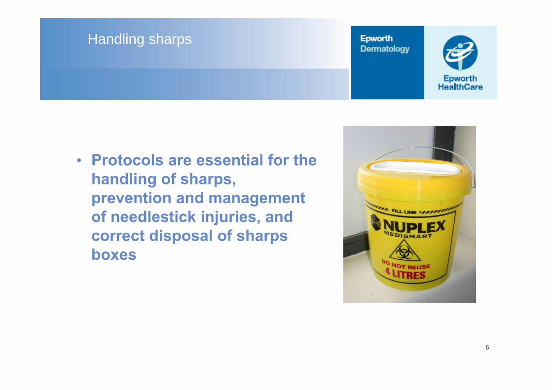
• Protocols are essential for the handling of sharps, prevention and management of needlestick injuries, and correct disposal of sharps boxes
6
Preparation
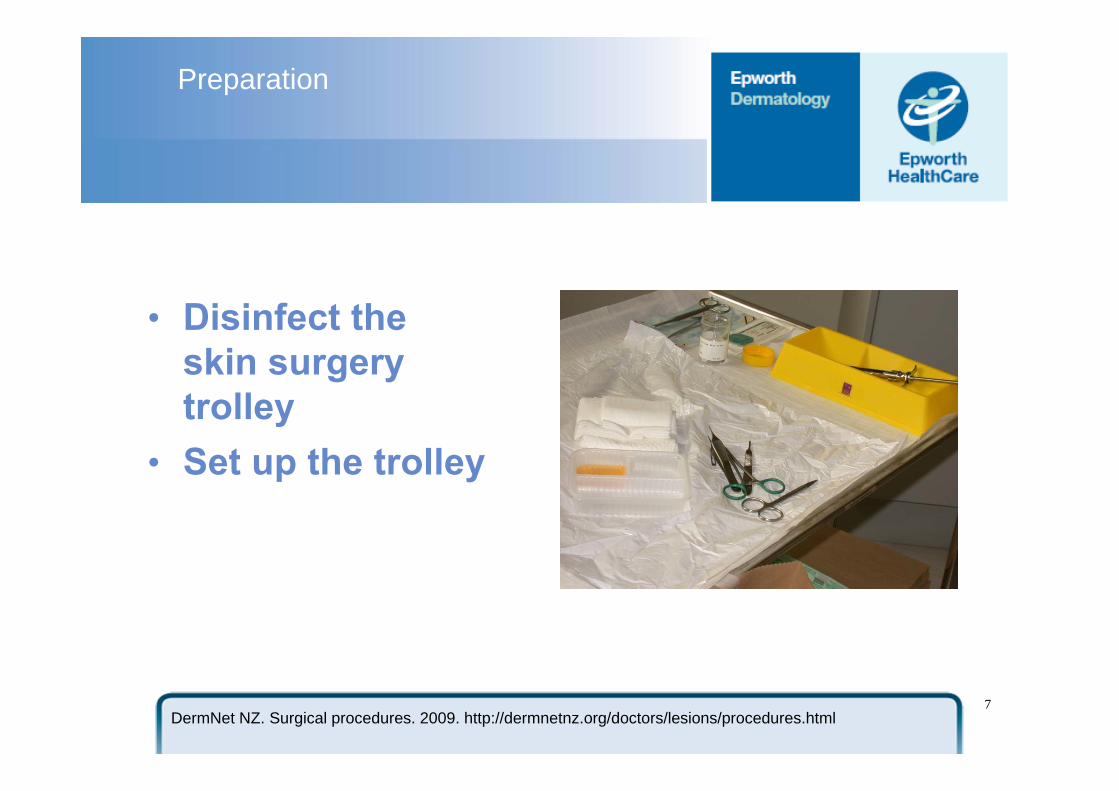
• Disinfect the skin surgery trolley
• Set up the trolley
7DermNet NZ. Surgical procedures. 2009. http://dermnetnz.org/doctors/lesions/procedures.html
Local anaesthesia for skin biopsy
• Lignocaine is the most commonly used local anaesthetic agent for skin infiltration
• Adding adrenaline (epinephrine)– prolongs the duration of anaesthesia– restricts blood loss – decreases the rate of absorption and therefore:
o reduces peak concentration in the bloodo decreases systemic toxicity; and o increases the safety margin
8
Using adrenaline with lignocaine
• There is a risk of necrosis secondary to vasoconstriction of end-arterioles if adrenaline is used when anaesthetising fingers, toes, the tip of the nose, ears, and penis
• However, supplemental adrenaline has been used safely when anesthetising the nose and periphery of the ear
9
Using adrenaline with lignocaine
• Using adrenaline for digital block is controversial. However, evidence suggests that lignocaine with adrenaline may be used for digital anaesthesia except for patients with:
– peripheral vascular disease– connective tissue disease– Raynaud’s disease– antiphospholipid syndrome
10
Topical anaesthesia
• Anaesthesia may be achieved by topical eutectic mixture of local anaesthetics (EMLA)
• Depth of anaesthesia is approx 5 mm after application of EMLA under occlusion (after 2 hours). This is sufficient when performing skin biopsy on the knees, elbows, chest, abdomen, face and genitals
• Topical anaesthesia may be less effective in areasof thick epidermis and dermis, e.g. back, palmsand soles
11
Adverse events
• Although rare, hypersensitivity reactions to local anaesthetic agents may be due to additives (e.g. preservatives)
12
Pain/discomfort
• Pain or discomfort associated with administration of local anaesthetics maybe due to:
o trauma of needle penetrating the skino sudden stretching of tissue due to local anaesthetic o the local anaesthetic agent itself
13
Minimising discomfort
– Pain can be minimised by:o using a small-gauge needle o slowly administering the anaesthetic to reduce
sudden expansion of tissueo avoiding injecting the area with an excess of the
anaesthetic agento warming the agent to body temperature before
administrationo pre-cooling the skin with ice cubes o using a topical anaesthetico buffering the anaesthetic with bicarbonate
14
Minimising discomfort
• Pain can be minimised by:o distracting the patiento pinching the skin, which stimulates local sensory
nerves, partially blocking transmission of other painful stimuli
o counter-irritating the skin by very gently scratching the skin approximately 1–2 cm from the injection site while injecting
o vibration of the skin
15
Minimising discomfort
• Injections on the palmoplantar aspect are very painful. If the lesion is close to the side of the palm/sole, the needle can be introduced through the dorsal skin
• When injecting on the palmoplantar surface,it is better to inject a small amount of local anaesthetic, wait for the area to be anaesthetised, and then push the needlein further
16
Prior to injection
• Check for underlying vessels and nerves in the biopsy area in order to avoid them
• Disinfect the relevant skin area and vial (e.g. using alcohol wipes)
• Scrub for 10 seconds with 70% isopropyl alcohol
• Draw anaesthetic solution using a large-gauge needle, then change to a small-gauge needle before injection
17
Injection technique
• Infuse into the intralesional area slowly, then move slowly from the treated to untreated areas to reduce the pain of reinsertion
18
Shave biopsy
• In a shave excision, the elevated part of a cutaneous growth is shaved off
• Common indications include seborrhoeic keratoses and skin tags
• Shave biopsies are also taken of superficial lesions where depth is not required to provide the pathologist with maximum surface area for examination
20
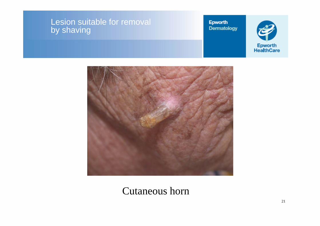
Lesion suitable for removalby shaving
Cutaneous horn21
Punch biopsy
• A disposable biopsy punch is used to remove a cylinder of skin tissue, including the epidermis, dermis, and sometimes the subcutaneous fat
• Can be used for any solid lesion or small vesicle that can be contained within the punch
• A 2 mm punch is adequate for non-facial lesions; however, in granulomatous conditions or those with atypical features, ≥3 mm biopsies are preferable 23
Punch biopsy techique
– After anaesthetising, tighten the skin around the biopsy site by stretching it in a direction perpendicular to the resting skin lines
– Punch biopsy of the scalp should be performed parallel to the direction of emergence of hairs fromthe scalp
– The punch is inserted using rotational movements until a “give” is felt where it enters the subcutaneous tissue
24
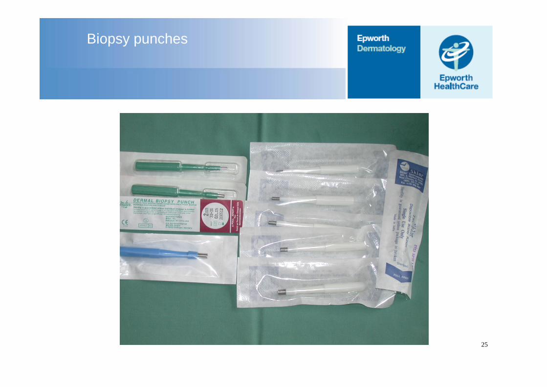
Biopsy punches
25
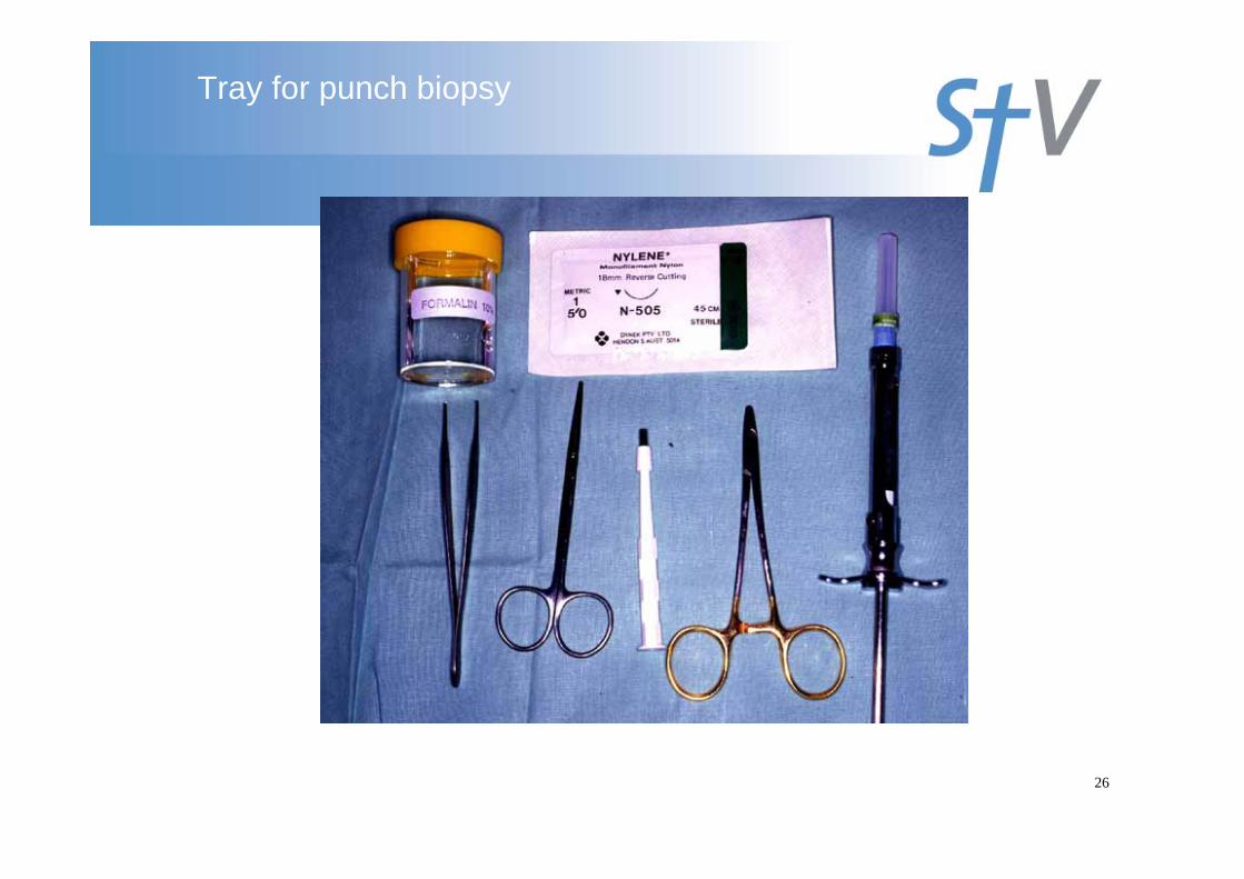
Tray for punch biopsy
26
Punch biopsy
Advantages:• Ease of performance
• Obtaining uniformly shaped tissue
Disadvantages:
• The material obtained may be insufficient
• Often biopsy may not include deeper tissue
27
Incisional biopsy
• Involves taking part of the tissue to confirm the diagnosis
• Commonly used when an inflammatory dermatosis of deeper tissue is suspectedand where excisional biopsies cannot be conducted because of the size or location of the lesion
• The incision may extend into the surrounding normal skin
29
Non-excisional biopsies
• For a non-excisional biopsy it is best toobtain normal skin, part of the lesion, andthe intervening transition zone
• If the centre of the lesion appears to bemost severe or malignant, the centre canbe biopsied
30
Excisional biopsy
– The whole lesion is removed via an elliptical excision, with a margin of normal skin, downto the subcutis
– Recommended excision margins:• 3 mm for BCC• 4 mm for SCC• 1 mm initially for suspected melanomas• Definitive excision margins of confirmed melanoma
depend on the histological depth of the tumour
– Excision is the preferred method for a suspected melanoma 31
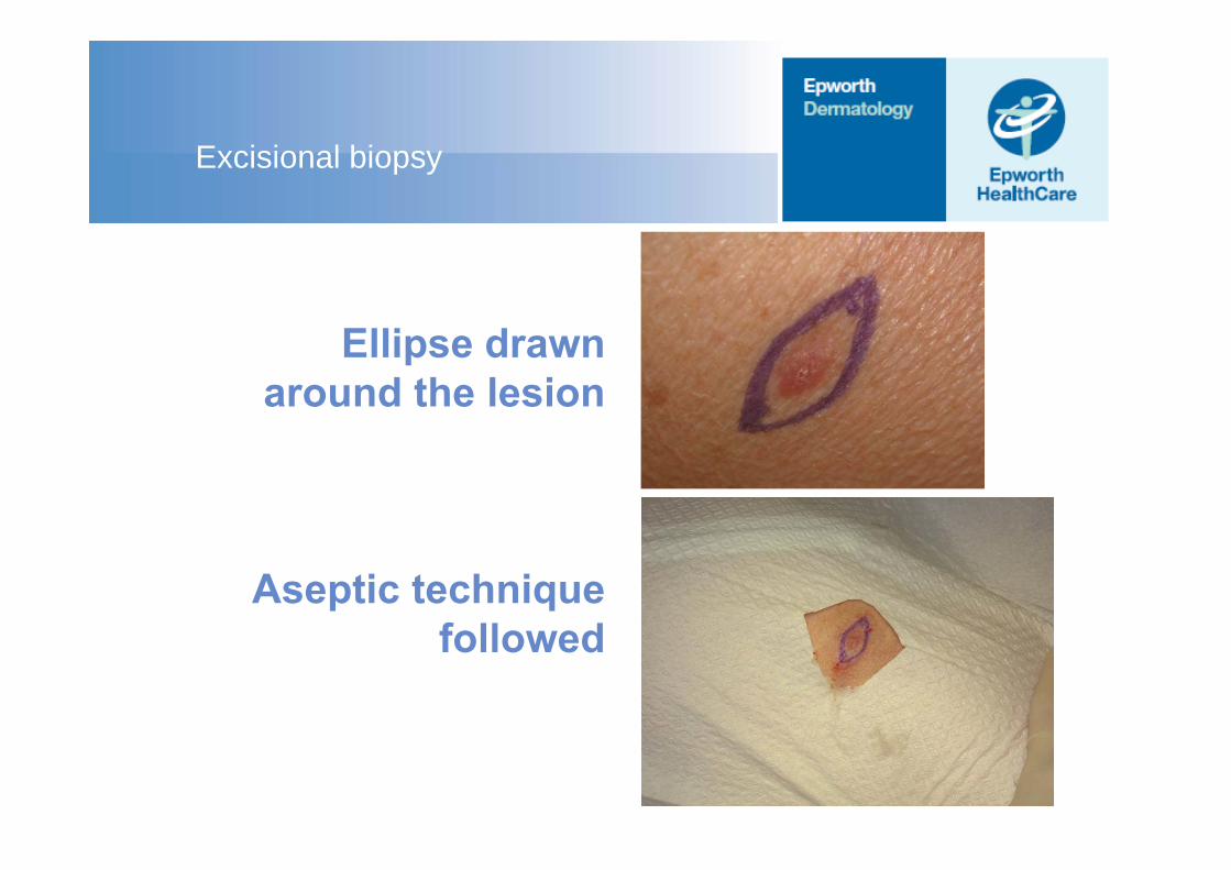
Excisional biopsy
Ellipse drawnaround the lesion
Aseptic technique followed
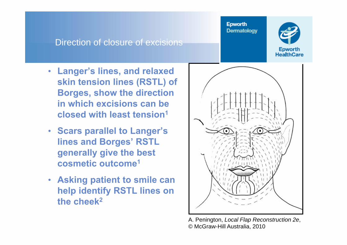
Direction of closure of excisions
• Langer’s lines, and relaxed skin tension lines (RSTL) of Borges, show the direction in which excisions can be closed with least tension1
• Scars parallel to Langer’s lines and Borges’ RSTL generally give the best cosmetic outcome1
• Asking patient to smile can help identify RSTL lines on the cheek2
A. Penington, Local Flap Reconstruction 2e,© McGraw-Hill Australia, 2010
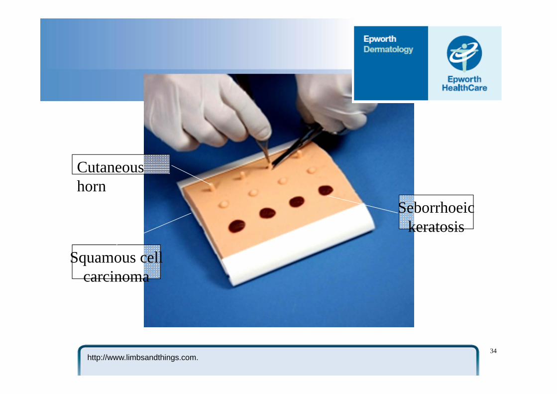
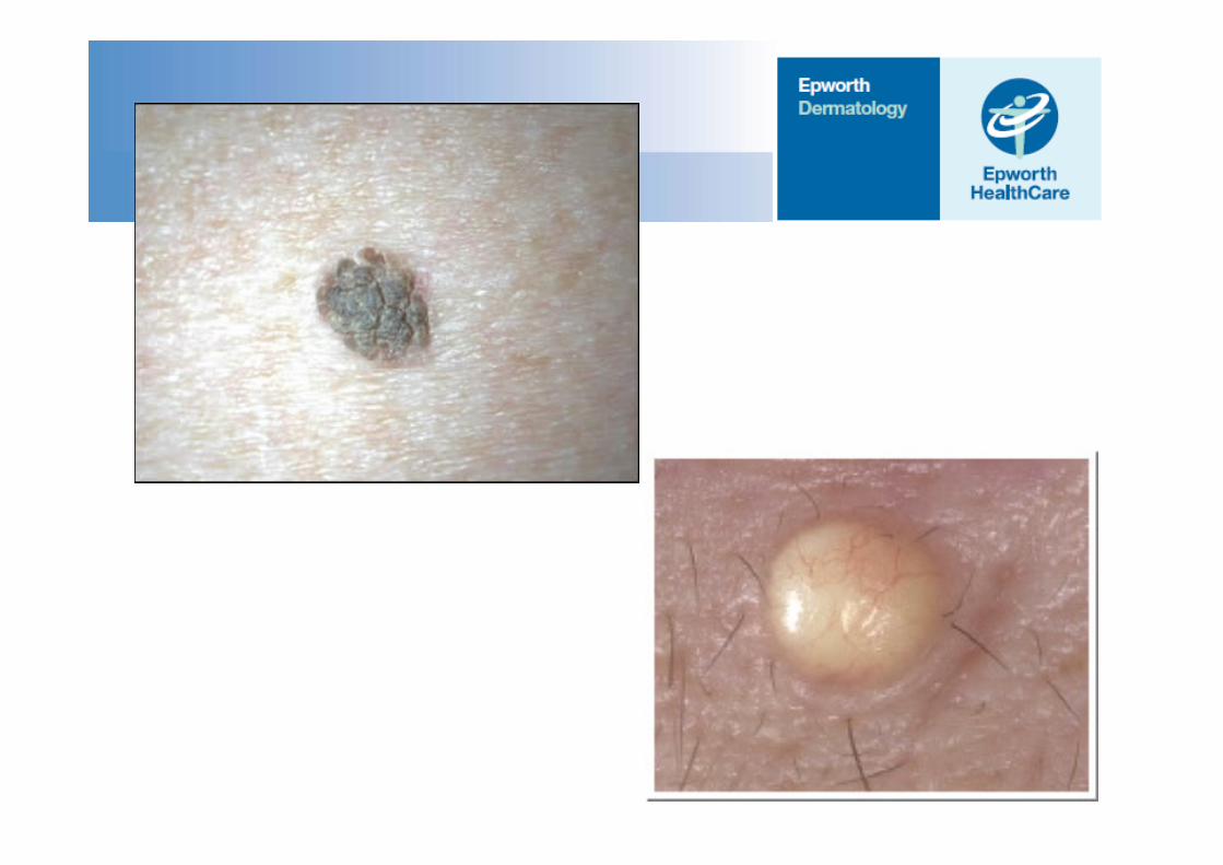
Seborrhoeickeratosis
Cutaneous horn
Squamous cellcarcinoma
34http://www.limbsandthings.com.
Possible complications
• Bleeding
• Infection
• Scarring
• Slow healing
• Wound dehiscence
35
Bleeding
In the following cases, incisions may carry an increased risk of bleeding:
• On the scalp, face or genitals• In elderly patients with atrophic skin• In patients taking medications that affect
clotting(e.g. anticoagulants, antiplatelet agents, PLAVIX)
• In patients with bleeding disorders
36
Controlling bleeding
• Application of pressure for about 2–3 minutes usually stops oozing
• Electrocautery/hyfrecation• Fibrous absorber (e.g. calcium/sodium
alginate dressing) helps reduce bleeding and promotes wound healing
• On the scalp, apply the ring of a large artery forceps around the biopsy site, with pressure
37
Reducing risk of scarring
• Note any history of hypertrophic scars or keloidal tendency
• Areas with good vasculature (e.g. the face, genitals, mucosa) usually heal quickly, with little scarring
• Some sites have higher rates of keloidal scarring (e.g. sternum, deltoid region and upper back)
• Using fine sutures reduces scarring• Occlusive dressings for at least 4 days promote
healing of sutured wounds• Uncovered wounds have more scab formation,
more infection and worse scarring38
Reducing risk of infection
• The chances of secondary infections are low, if aseptic precautions are taken
• Systemic antibiotics may be considered for patients: • with diabetes mellitus• with extensive eczema• who are debilitated• with artificial or abnormal heart valves• on immunosuppressants
• Prophylaxis could be considered for all procedures below the knee, for wedge excisions of the lip and ear, and lesions in the groin
• Apply antiseptic ointment on a wound before an occlusive dressing
39
Specimen handling
• Volume of formalin required for optimal fixation is approximately 10 times the volume of the biopsy specimen
• Ensure minimal handling of tissue when transferring to the formalin container. Take care not to crush the specimen with forceps.
• Beware using a skin hook or needle• When removing or sampling many lesions,
photographing and numbering the lesions and removing/sampling in numbered order assists in matching them accurately to the histology report
40
Suturing
• When choosing sutures and needles, consider:
• the location of the lesion• the amount of tension exerted on the wound
• Absorbable sutures lose most of their tensile strength in less than 60 days. They are generally used for buried sutures and do not require removal
• Non-absorbable sutures maintain most of their tensile strength for more than 60 days. They are generally used for skin surface sutures
41
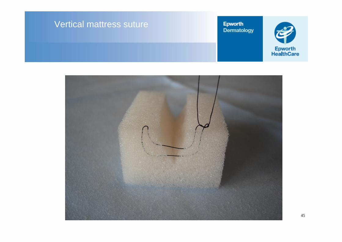
Vertical mattress suture
45
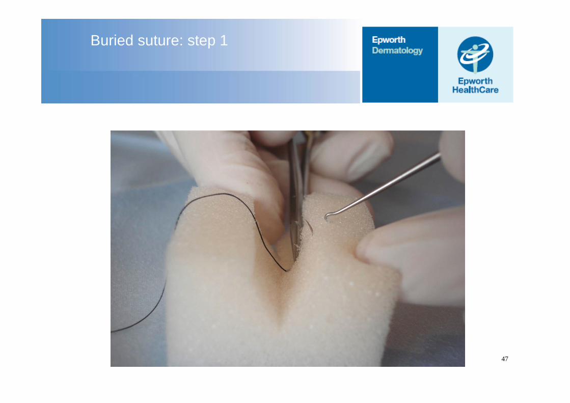
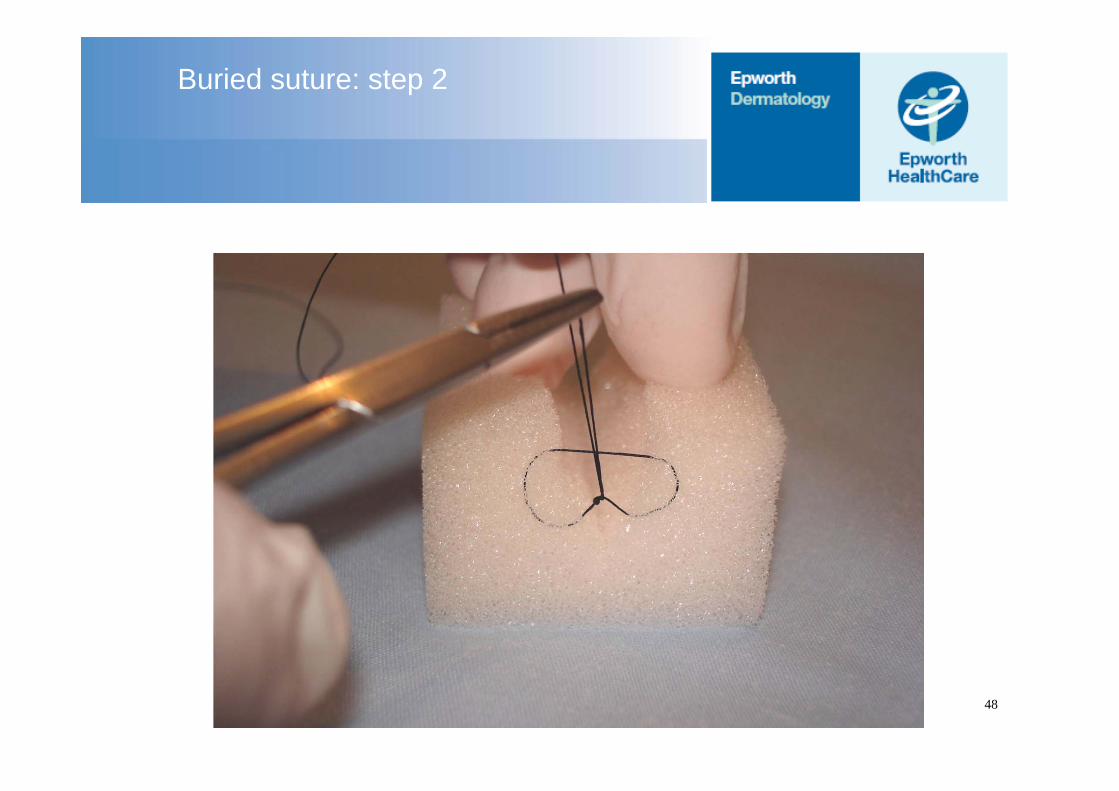
Buried suture: step 1
47
Buried suture: step 2
48
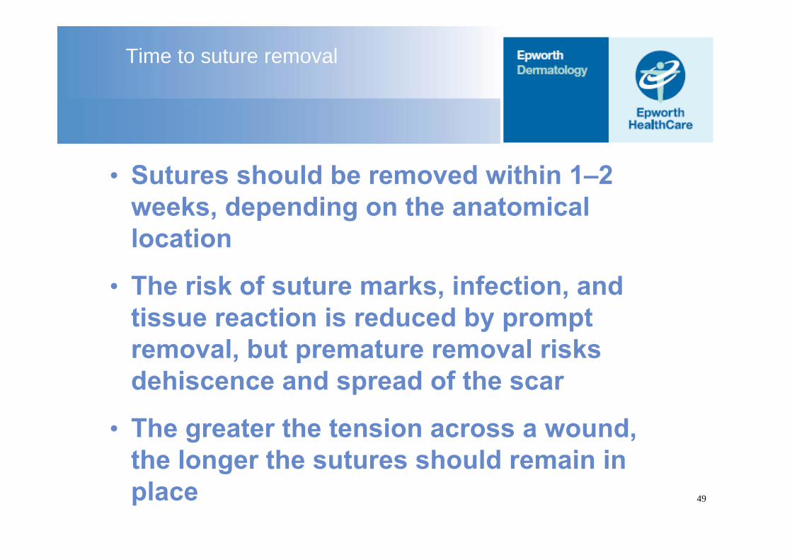
Time to suture removal
• Sutures should be removed within 1–2 weeks, depending on the anatomical location
• The risk of suture marks, infection, and tissue reaction is reduced by prompt removal, but premature removal risks dehiscence and spread of the scar
• The greater the tension across a wound, the longer the sutures should remain in place 49
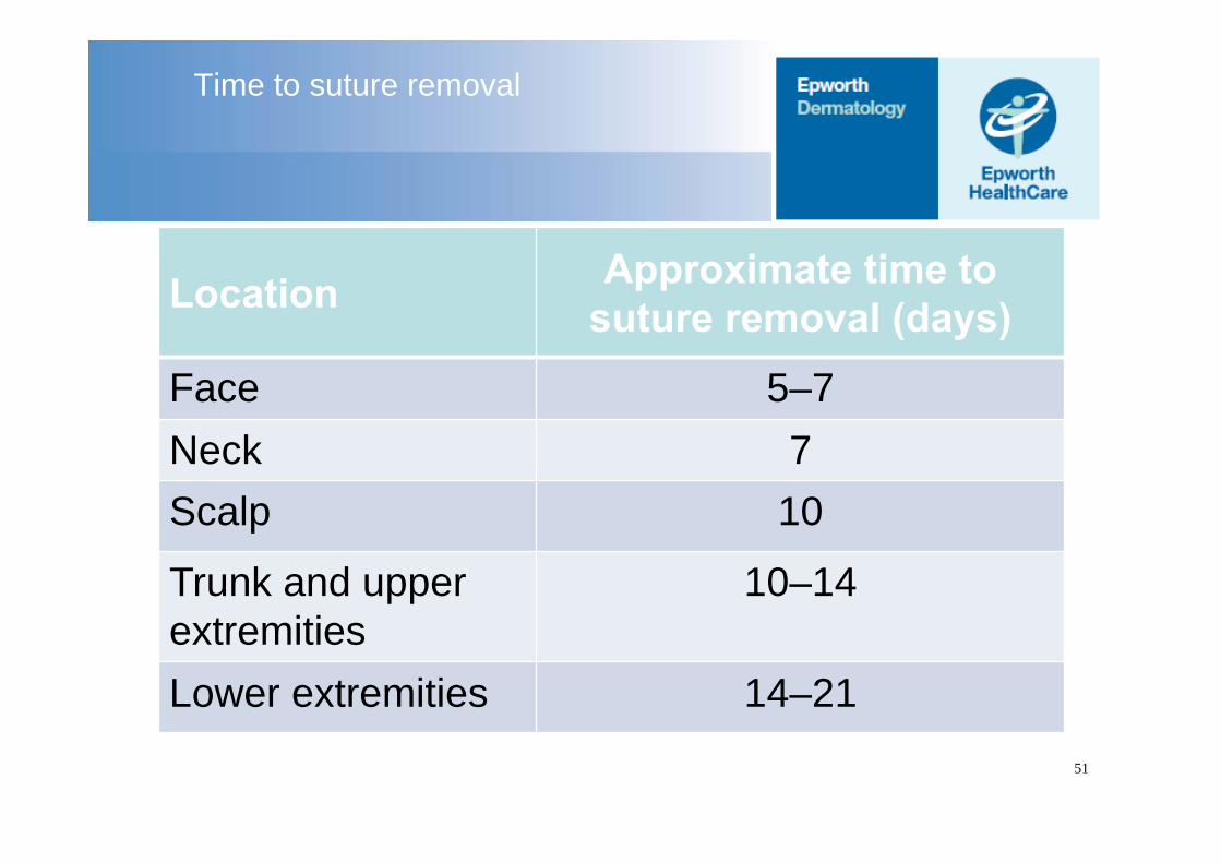
Time to suture removal
Location Approximate time to suture removal (days)
Face 5–7Neck 7Scalp 10
Trunk and upper extremities
10–14
Lower extremities 14–2151
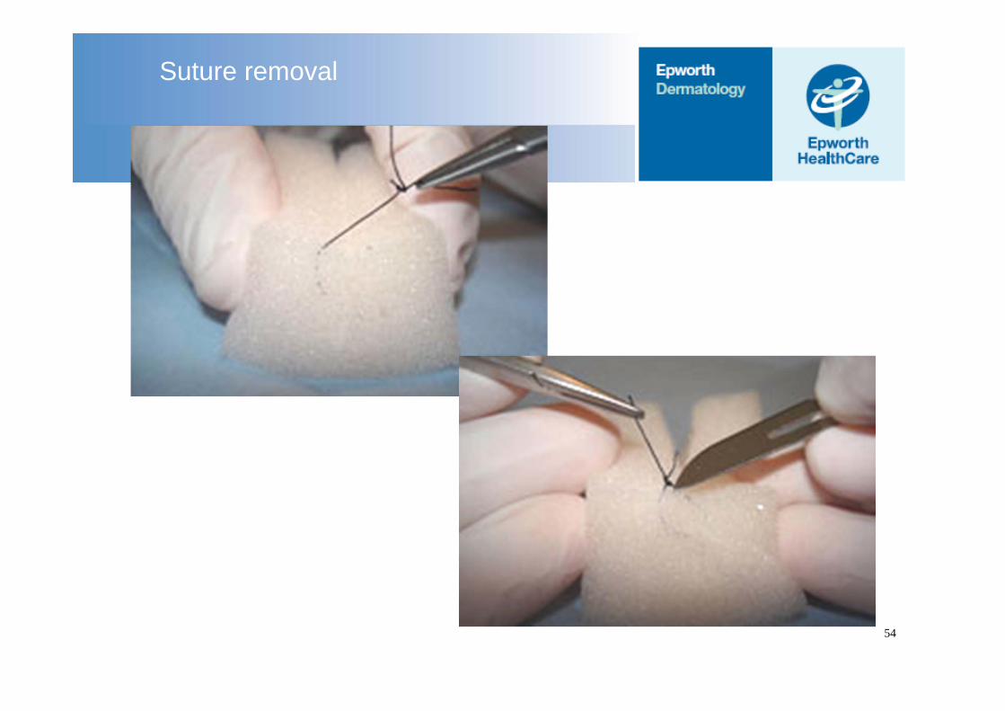
Suture removal
54
Use of tape to prevent scarring
• After suture removal, scars are susceptible to skin tension, which may be the trigger for hypertrophic scarring
• A study found that paper tape, applied to Caesarian section scars after suture removal and left in place for 12 weeks, prevented hypertrophic scar formation
55
Vitamin E and aloe vera creams
• There is little evidence to support the use of topical vitamin E cream to reduce scar formation1
• Effects of aloe vera on wound healing are mixed. Some studies report positive results; others show no benefit or potential negative effects2
56
Curettage
Technique of tissue removal using a curette
Purpose – obtain biopsy – debulk lesion– remove lesion
Methods– Simple– Serial– With diathermy– With cryotherapy
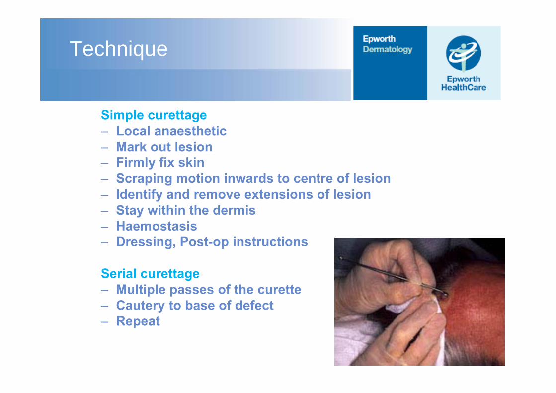
Technique
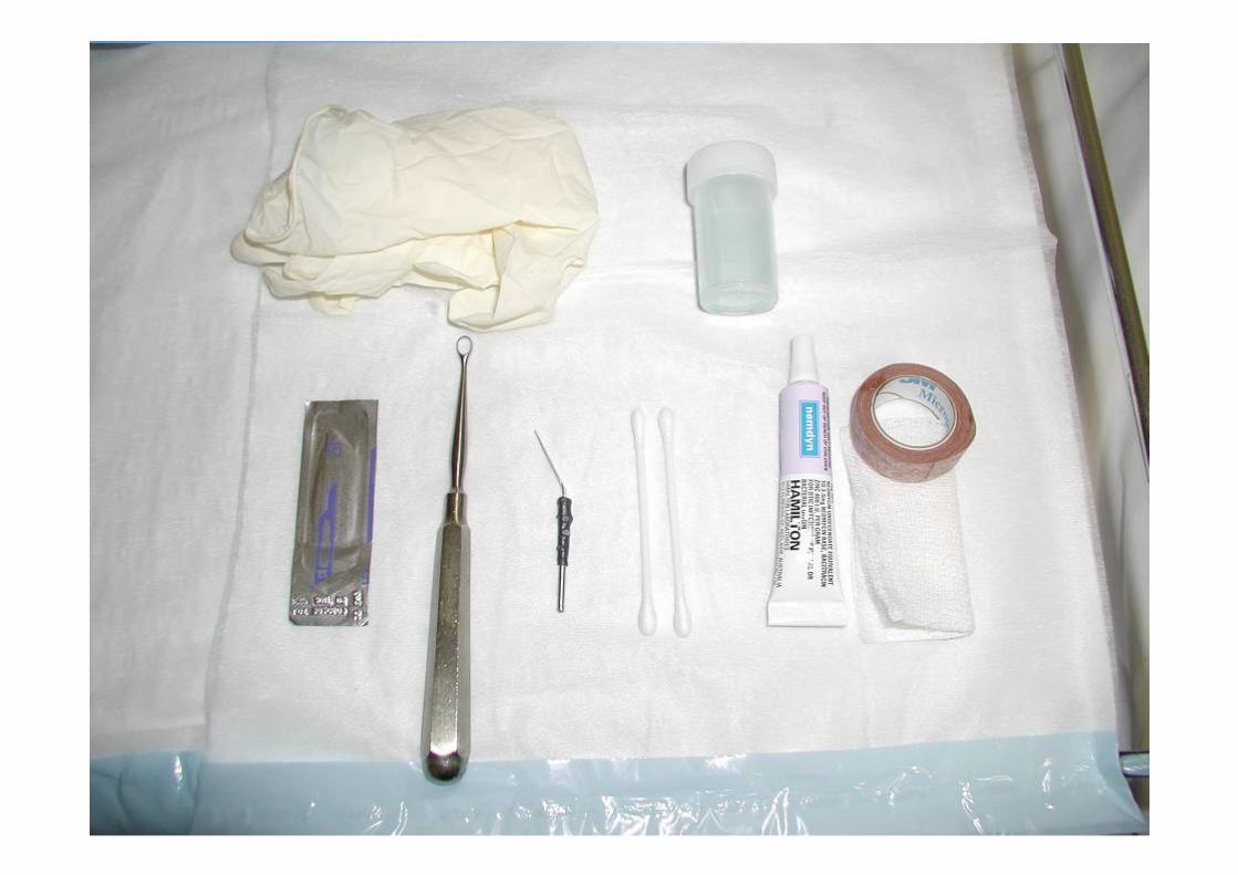
Simple curettage– Local anaesthetic– Mark out lesion– Firmly fix skin– Scraping motion inwards to centre of lesion– Identify and remove extensions of lesion– Stay within the dermis– Haemostasis– Dressing, Post-op instructions
Serial curettage– Multiple passes of the curette– Cautery to base of defect– Repeat
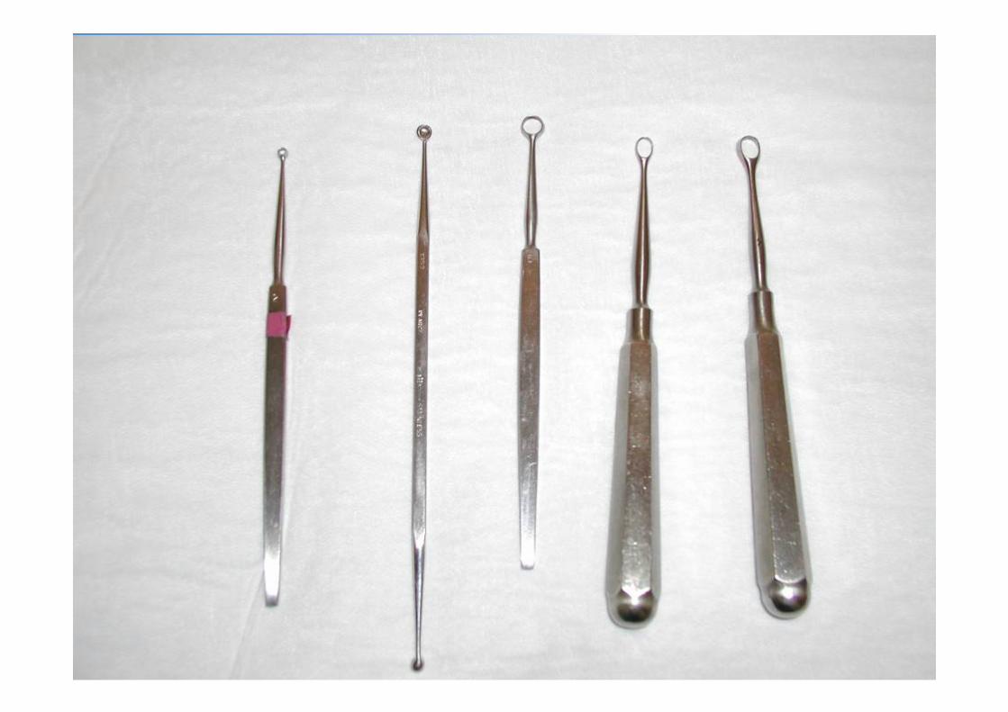
Blunt vs disposable curettes
– Blunt curette – does not create false planes – finds plane of natural cleavage eg. for seb ks, BCCs
– Sharp curette – sharp, cutting
Curettage
Lesion selection– Suitable pathology– Easily distinguished from normal skin– Size (usually < 1cm)
Site considerations– Skin thickness – not for thin areas– Ability to fix skin – scalp, back, forehead– Resultant scar – Implications of recurrence
Indications
Benign lesions– Seb keratoses – Solar keratoses– Molluscum contagiosum– Pyogenic granuloma– Milia– Warts– Sebaceous hyperplasia
Malignant lesions– BCC
(<1cm, sBCC or nBCC, not previously treated, non risk sites)
– Bowens– SCC’s (in general not suitable)
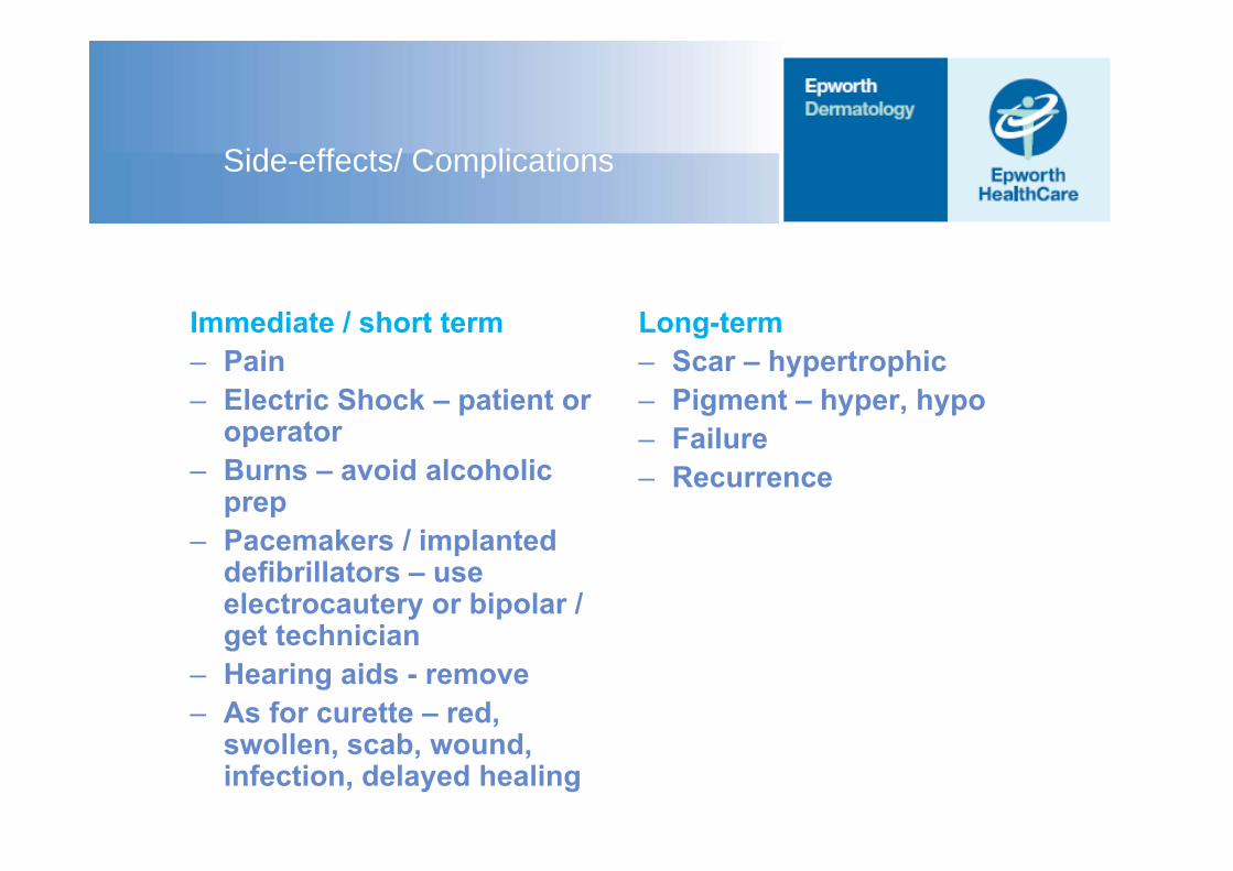
Side-effects/ Complications
Short-term– Pain– Bleeding– Delayed healing– Infection
Medium-long term– Scar –
hypertrophic– Hypopigmentation – Recurrence
“Cautery”
“An agent or instrument used to destroy abnormal tissue by burning, searing, or scarring, including caustic substances, electric currents, lasers, and very hot or very cold instruments. “
– Electrical – Electrocautery = hot wire – “Diathermy” = electrosurgery (Electrocoagulation,
Electrodessication, Electrofulguration, Electrosection, Electrolysis)
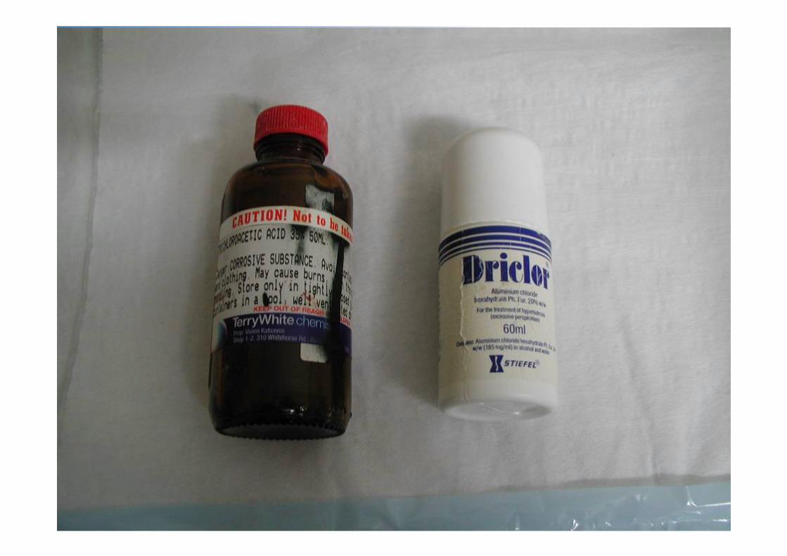
– Chemical– TCA 35 – 50%– Aluminium chloride hexahydrate 20% DRICLOR– Ferric subsulphate (Monsel’s solution)– Silver nitrate
Electrosurgery
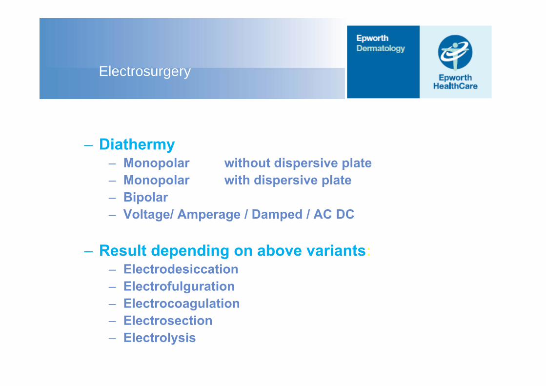
– Diathermy – Monopolar without dispersive plate– Monopolar with dispersive plate– Bipolar– Voltage/ Amperage / Damped / AC DC
– Result depending on above variants:– Electrodesiccation – Electrofulguration– Electrocoagulation– Electrosection– Electrolysis
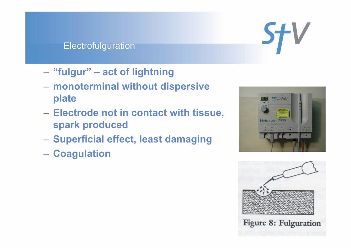
Electrofulguration
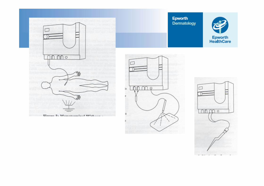
– “fulgur” – act of lightning– monoterminal without dispersive
plate– Electrode not in contact with tissue,
spark produced– Superficial effect, least damaging– Coagulation
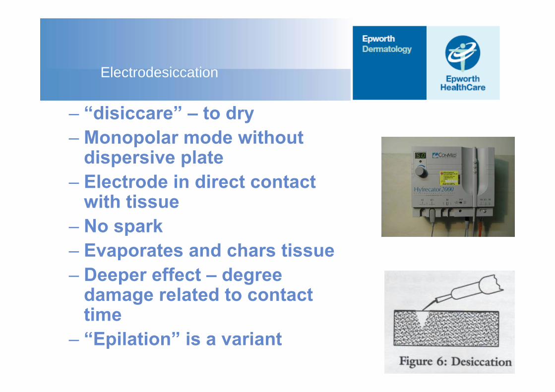
Electrodesiccation
– “disiccare” – to dry– Monopolar mode without
dispersive plate– Electrode in direct contact
with tissue– No spark– Evaporates and chars tissue– Deeper effect – degree
damage related to contact time
– “Epilation” is a variant
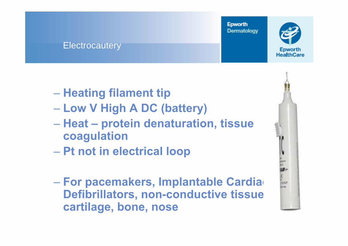
Electrocautery
– Heating filament tip– Low V High A DC (battery)– Heat – protein denaturation, tissue
coagulation– Pt not in electrical loop
– For pacemakers, Implantable Cardiac Defibrillators, non-conductive tissue –cartilage, bone, nose
Indications
– As for curettage plus– Skin tags– Dermatosis papulosa nigra– Small seb ks– Sebaceous hyperplasia– Comedones – closed & open
– Spider naevi– Cherry angiomas– Telangiectasia– Syringoma
Side-effects/ Complications
Immediate / short term– Pain– Electric Shock – patient or
operator– Burns – avoid alcoholic
prep– Pacemakers / implanted
defibrillators – use electrocautery or bipolar / get technician
– Hearing aids - remove– As for curette – red,
swollen, scab, wound, infection, delayed healing
Long-term– Scar – hypertrophic– Pigment – hyper, hypo– Failure– Recurrence
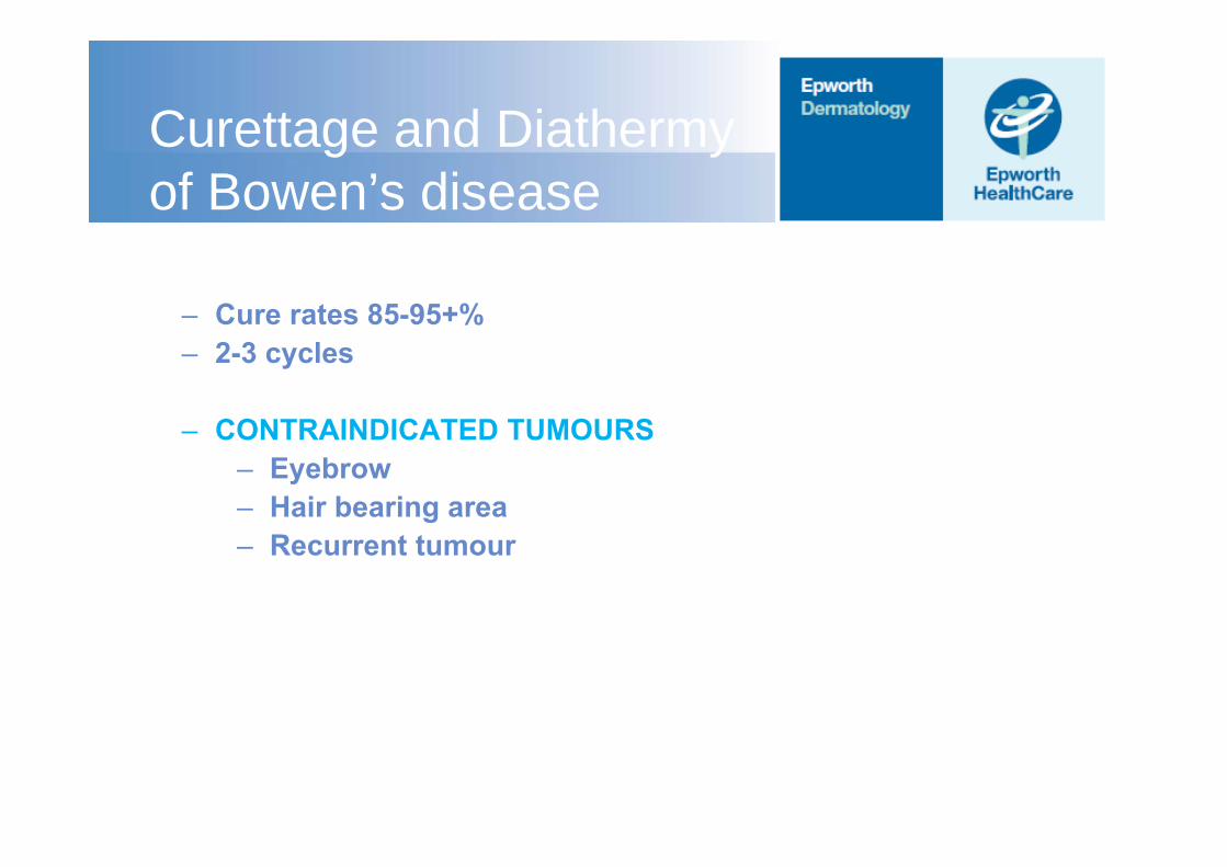
Curettage and Diathermy of Bowen’s disease
– Cure rates 85-95+%– 2-3 cycles
– CONTRAINDICATED TUMOURS – Eyebrow– Hair bearing area– Recurrent tumour
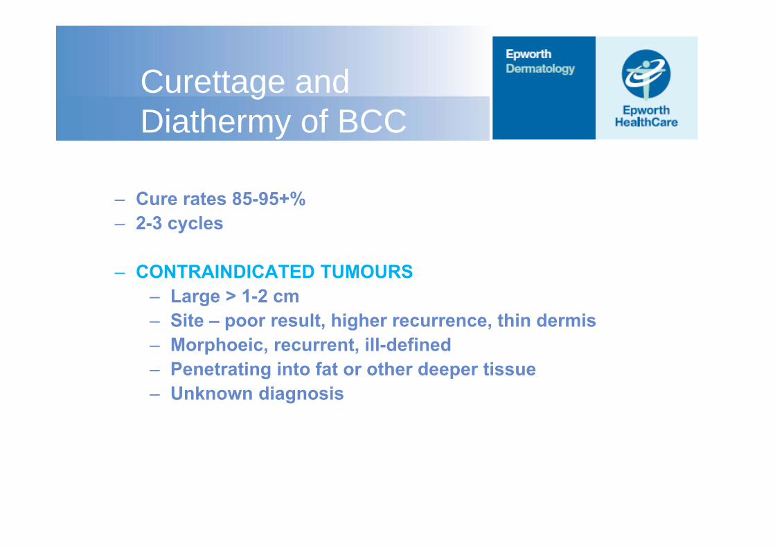
Curettage and Diathermy of BCC
– Cure rates 85-95+%– 2-3 cycles
– CONTRAINDICATED TUMOURS – Large > 1-2 cm– Site – poor result, higher recurrence, thin dermis– Morphoeic, recurrent, ill-defined– Penetrating into fat or other deeper tissue– Unknown diagnosis
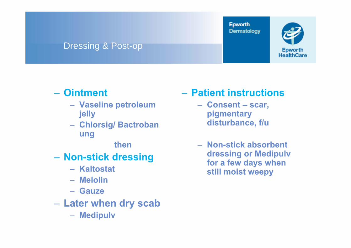
Dressing & Post-op
– Ointment – Vaseline petroleum
jelly– Chlorsig/ Bactroban
ungthen
– Non-stick dressing– Kaltostat– Melolin – Gauze
– Later when dry scab– Medipulv
– Patient instructions– Consent – scar,
pigmentary disturbance, f/u
– Non-stick absorbent dressing or Medipulv for a few days when still moist weepy