black adrenal adenoma causing cushing's syndrome: 40 years
TRANSCRIPT
466 CARTAS CIENTÍFICAS
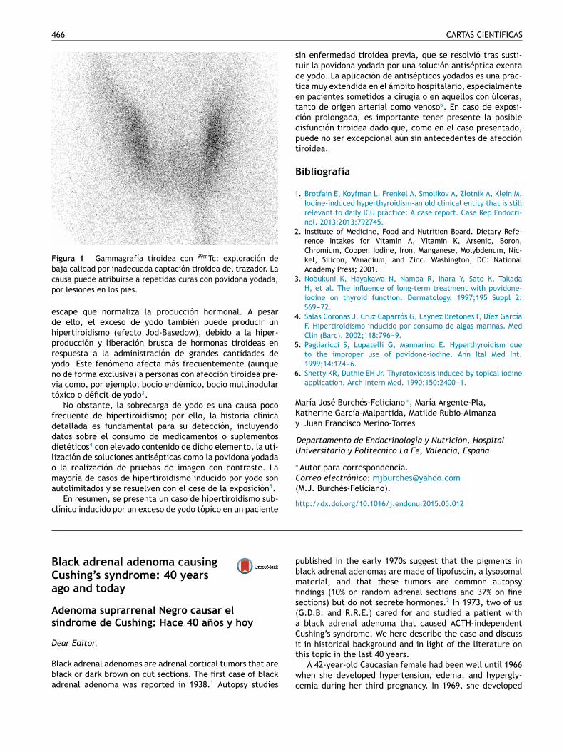
Figura 1 Gammagrafía tiroidea con 99mTc: exploración debaja calidad por inadecuada captación tiroidea del trazador. Lacausa puede atribuirse a repetidas curas con povidona yodada,por lesiones en los pies.
escape que normaliza la producción hormonal. A pesarde ello, el exceso de yodo también puede producir unhipertiroidismo (efecto Jod-Basedow), debido a la hiper-producción y liberación brusca de hormonas tiroideas enrespuesta a la administración de grandes cantidades deyodo. Este fenómeno afecta más frecuentemente (aunqueno de forma exclusiva) a personas con afección tiroidea pre-via como, por ejemplo, bocio endémico, bocio multinodulartóxico o déficit de yodo3.
No obstante, la sobrecarga de yodo es una causa pocofrecuente de hipertiroidismo; por ello, la historia clínicadetallada es fundamental para su detección, incluyendodatos sobre el consumo de medicamentos o suplementosdietéticos4 con elevado contenido de dicho elemento, la uti-lización de soluciones antisépticas como la povidona yodadao la realización de pruebas de imagen con contraste. Lamayoría de casos de hipertiroidismo inducido por yodo sonautolimitados y se resuelven con el cese de la exposición5.
En resumen, se presenta un caso de hipertiroidismo sub-clínico inducido por un exceso de yodo tópico en un paciente
sin enfermedad tiroidea previa, que se resolvió tras susti-tuir la povidona yodada por una solución antiséptica exentade yodo. La aplicación de antisépticos yodados es una prác-tica muy extendida en el ámbito hospitalario, especialmenteen pacientes sometidos a cirugía o en aquellos con úlceras,tanto de origen arterial como venoso6. En caso de exposi-ción prolongada, es importante tener presente la posibledisfunción tiroidea dado que, como en el caso presentado,puede no ser excepcional aún sin antecedentes de afeccióntiroidea.
Bibliografía
1. Brotfain E, Koyfman L, Frenkel A, Smolikov A, Zlotnik A, Klein M.Iodine-induced hyperthyroidism-an old clinical entity that is stillrelevant to daily ICU practice: A case report. Case Rep Endocri-nol. 2013;2013:792745.
2. Institute of Medicine, Food and Nutrition Board. Dietary Refe-rence Intakes for Vitamin A, Vitamin K, Arsenic, Boron,Chromium, Copper, Iodine, Iron, Manganese, Molybdenum, Nic-kel, Silicon, Vanadium, and Zinc. Washington, DC: NationalAcademy Press; 2001.
3. Nobukuni K, Hayakawa N, Namba R, Ihara Y, Sato K, TakadaH, et al. The influence of long-term treatment with povidone-iodine on thyroid function. Dermatology. 1997;195 Suppl 2:S69---72.
4. Salas Coronas J, Cruz Caparrós G, Laynez Bretones F, Díez GarcíaF. Hipertiroidismo inducido por consumo de algas marinas. MedClin (Barc). 2002;118:796---9.
5. Pagliaricci S, Lupatelli G, Mannarino E. Hyperthyroidism dueto the improper use of povidone-iodine. Ann Ital Med Int.1999;14:124---6.
6. Shetty KR, Duthie EH Jr. Thyrotoxicosis induced by topical iodineapplication. Arch Intern Med. 1990;150:2400---1.
María José Burchés-Feliciano ∗, María Argente-Pla,Katherine García-Malpartida, Matilde Rubio-Almanzay Juan Francisco Merino-Torres
Departamento de Endocrinología y Nutrición, Hospital
Universitario y Politécnico La Fe, Valencia, Espana
∗ Autor para correspondencia.Correo electrónico: [email protected](M.J. Burchés-Feliciano).
http://dx.doi.org/10.1016/j.endonu.2015.05.012
Black adrenal adenoma causingCushing’s syndrome: 40 yearsago and today
Adenoma suprarrenal Negro causar elsíndrome de Cushing: Hace 40 anos y hoy
Dear Editor,
Black adrenal adenomas are adrenal cortical tumors that areblack or dark brown on cut sections. The first case of blackadrenal adenoma was reported in 1938.1 Autopsy studies
published in the early 1970s suggest that the pigments inblack adrenal adenomas are made of lipofuscin, a lysosomalmaterial, and that these tumors are common autopsyfindings (10% on random adrenal sections and 37% on finesections) but do not secrete hormones.2 In 1973, two of us(G.D.B. and R.R.E.) cared for and studied a patient witha black adrenal adenoma that caused ACTH-independentCushing’s syndrome. We here describe the case and discussit in historical background and in light of the literature onthis topic in the last 40 years.
A 42-year-old Caucasian female had been well until 1966when she developed hypertension, edema, and hypergly-cemia during her third pregnancy. In 1969, she developed
CARTAS CIENTÍFICAS 467
right femoral head aseptic necrosis. She also noted a40-pound weight gain, rounding of face, the developmentof a dorsal fat pad, ruddy complexion, facial hair, weak-ness, easy fatigability, emotional liability, irregular menses,and easy bruising. In January 1973, she was seen at HarborGeneral Hospital (now Harbor-UCLA Medical Center). Shedenied skin darkening, exogenous steroid ingestion, orfamily history of endocrine diseases. Physical examinationrevealed a hypertensive, Cushingoid female. Endocrine eva-luation revealed absence of suppression of plasma or urinary17-hydroxysteroids by both low (2 mg) and high (8 and12 mg) doses of dexamethasone administration. Her plasma17-hydroxysteroids failed to increase after metopirone(more commonly called ‘‘metyrapone’’ now) administration
or synthetic ACTH (Cortrosyn) infusion. ACTH-independentCushing’s syndrome was diagnosed and the presenceof a functional adrenal lesion deemed probable. Adre-nal androgen levels were not measured. The patientunderwent an exploratory laparotomy through which theright adrenal gland was found to be grossly normal butthe left adrenal to harbor a mass. A left adrenalectomywas performed. Her post-operative course was uncompli-cated and the Cushingoid features gradually regressed inthe ensuing 2 months. She developed adrenal insufficiencypostoperatively and was treated with corticosteroids withtapered doses.
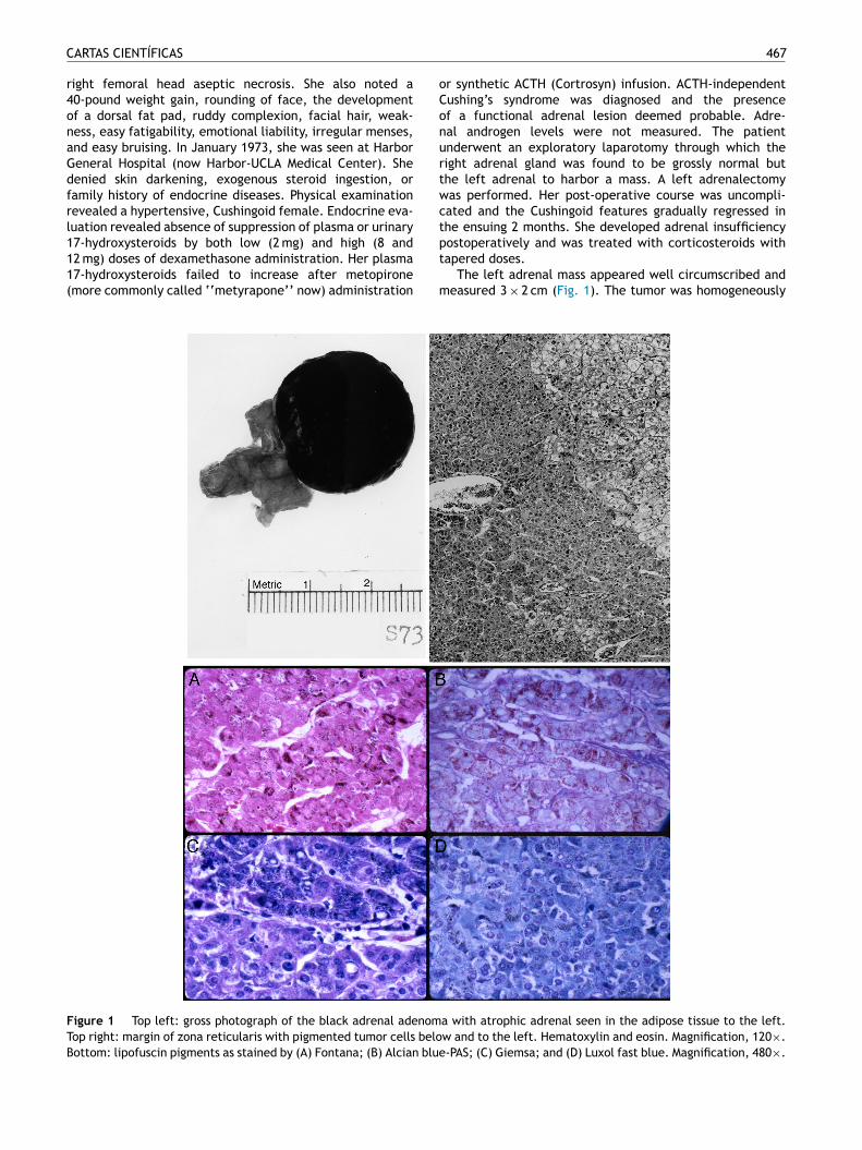
The left adrenal mass appeared well circumscribed andmeasured 3 × 2 cm (Fig. 1). The tumor was homogeneously
Figure 1 Top left: gross photograph of the black adrenal adenoma with atrophic adrenal seen in the adipose tissue to the left.Top right: margin of zona reticularis with pigmented tumor cells below and to the left. Hematoxylin and eosin. Magnification, 120×.Bottom: lipofuscin pigments as stained by (A) Fontana; (B) Alcian blue-PAS; (C) Giemsa; and (D) Luxol fast blue. Magnification, 480×.
468 CARTAS CIENTÍFICAS
dark brown to black throughout (Fig. 1, top left). Microsco-pically, the mass consisted of large, polygonal, eosinophi-lic cells resembling those of the zona reticularis. The tumorcells abutted directly on but did not invade into the non-pigmented cells of the gland (Fig. 1, top right). The majorityof these cells contained heavy deposits of golden brown,slightly refractile, granular, pigments which were localizedpredominantly at the cell periphery. The pigments werevisible also with Congo Red, Gomori iron, and in even inunstained slides (Fig. 1, bottom). It reacted weakly withSudan black and negatively with acid fast stains. Fontanastain was strongly positive and the pigment assumed a red-dish coloration with the periodic acid-Schiff’s technique.The pigments appeared green with Giemsa stain and gree-nish blue with Luxol fast blue stain, thus indicating that theyconsisted of lipofuscin. Mitotic figures were very rare. Thetumor cells did not exhibit nuclear atypia, necrosis, or aty-pical mitotic figures.
To assess how frequent the black adrenal adenoma is,we examined our pathology database between 1998 and2014. One hundred and fourteen adrenal cortical tumorswere found. The average age of patients was 53 years (range23---70). Forty-one of the adenomas were aldosterone-secreting, 23 cortisol-secreting, 1 androgen-secreting, and49 nonfunctional. The average adenoma size was 3.1 cm(range 0.2---27). Twenty-three of the adenomas were≤1.5 cm and 91 larger. The tumor color ranged from yellow,orange, tan, red, to brown. None of the 114 adenomas waspredominantly black or dark brown.
Our study of the frequency of black adrenal adenomahere and the work of others in the last 40 years advanceour understanding of this interesting tumor. Black adrenaladenomas may be more common on post-mortem adrenalsbut they are certainly rare in surgical adrenal samples. Inour own series of 115 adrenal cortical tumors from surgi-cal adrenal samples, not a single black adrenal adenomawas encountered. Although the incidence of black adrenaladenomas has not been formally addressed in other surgicalseries, the mostly single-case reports of this unique-coloredtumor even recently suggest that they are indeed rare in sur-gical adrenal samples.3 The discrepancy between the highfrequency of incidental black adrenal adenomas in autopsyfindings and their exceptionally low incidence in surgicalseries may be due to the small size and non-functionalnature of the adenomas which avoid the surgery in spiteof the tumors’ radiological features. Most of black adrenaladenomas, like in this case, cause ACTH-independent Cus-hing’s syndrome, some cause primary hyperaldosteronism,and a few even result in masculinization.3---5 Less frequentlyreported are nonfunctional black adrenal adenomas whichpresent as incidentalomas.6 With the introduction of CT,MRI, and FDG-PET, the imaging characteristics of adrenaladenomas in general and the distinct imaging features ofblack adrenal adenomas in particular have now been welldescribed. Unlike most other adrenal adenomas, the blackones exhibit high Hounsfield units (>30) on CT, high T2 sig-nal and lack of drop of signal on out-of-phase imaging onMRI, and high standard uptake value (higher than thatof liver) on FDG-PET, which all indicate less tumor lipidcontent but higher tissue density and blood supply, featuressuspicious of pheochromocytoma, interstitial tumors, andmalignancy.6---8 Furthermore, the black adrenal adenomas
are often not visualized by radiocholesterol scintigraphy.9,10
Biologically, however, the black adrenal adenomas arebenign without histological evidence of aggressivenessor invasiveness, as our patient’s tumor. Black adrenaladenomas should now be considered as one subtype of adre-nal adenomas with atypical imaging characteristics. Theclinical significance of lipofuscin pigments in black adrenaladenomas remains unclear.11 Black adrenal adenomas areunilateral, solitary adrenal cortical tumors; they are in con-trast to primary pigmented nodular adrenocortical disease(PPNAD) which involves diffuse nodular enlargement of bothadrenal glands.12 PPNAD can occur as part of the Carneycomplex and is associated with a genetic defect, PRKAR1Amutation. The pigments in PPNAD are also due to lipofuscin.Patients with PPNAD can exhibit a paradoxical increase ofcortisol levels after administration of dexamethasone.
In summary, black adrenal adenomas appear to derivefrom the zona reticularis and their black color is due to lyso-somal lipofuscin. Clinically very rare tumors, they mainlypresent as Cushing’s syndrome or other syndromes of adre-nocortical hormone hypersecretion. Although biologicallybenign, they exhibit atypical imaging characteristics sus-picious of malignancy. The main difference between blackadrenal adenomas and other adrenal cortical tumors is justthe appearance to the naked or aided eye.
References
1. Baker MR. A pigmented adenoma of the adrenal. Arch Path.1938;26:845---52.
2. Robinson MJ, Pardo V, Rywlin AM. Pigmented nodules (blackadenomas) of the adrenal An autopsy study of incidence, morp-hology, and function. Hum Pathol. 1972;3:317---25.
3. Ueda Y, Tanaka H, Murakami H, Ninomiya T, Yamashita Y, Ichi-kawa M, et al. A functioning black adenoma of the adrenalgland. Intern Med. 1997;36:398---402.
4. Cohen RJ, Brits R, Phillips JI, Botha JR. Primary hyperaldoste-ronism due to a functional black (pigmented) adenoma of theadrenal cortex. Arch Pathol Lab Med. 1991;115:813---5.
5. Tanaka S, Tanabe A, Aiba M, Hizuka N, Takano K, Zhang J,et al. Glucocorticoid- and androgen-secreting black adreno-cortical adenomas: unique cause of corticotropin-independentCushing syndrome. Endocr Pract. 2011;17:e73---8.
6. Prince EA, Yoo DC, DeLellis RA, Mayo-Smith WW. CT and PETappearance of a pigmented black adrenal adenoma in a patientwith lung cancer. Clin Radiol. 2007;62:1229---31.
7. Nakajo M, Nakajo M, Kajiya Y, Tani A, Tsuruta M, Sugita S,et al. A black adrenal adenoma difficult to be differentia-ted from a malignant adrenal tumor by CT, MRI, scintigraphyand FDG PET/CT examinations. Ann Nucl Med. 2011;25:812---7.
8. Pitts A, Ih G, Wei M, Dhall D, Nissen NN, Waxman A, et al.Clinical utility of FDG-PET for diagnosis of adrenal mass: alarge single-center experience. Hormones (Athens). 2013;12:417---27.
9. Ambrosi B, Colombo P, Faglia G. Cushing’s syndrome dueto a black adenoma of the adrenal gland: lack of tumourvisualization by radiocholesterol scintigraphy. Eur J Nucl Med.1994;21:1367---8.
10. Reschini E1, Baldini M, Cantalamessa L. A black adrenocorticaladenoma causing Cushing’s syndrome not imaged by radiocho-lesterol scintigraphy. Eur J Nucl Med. 1990;17:185---7.
11. Jung T, Bader N, Grune T. Lipofuscin: formation, distri-bution, and metabolic consequences. Ann NY Acad Sci.2007;1119:97---111.

CARTAS CIENTÍFICAS 469
12. Stratakis CA. Adrenocortical tumors, primary pigmentedadrenocortical disease (PPNAD)/Carney complex, and otherbilateral hyperplasias: the NIH studies. Horm Metab Res.2007;39:467---73.
Run Yu a,∗, Meng Weib, Xuemo Fan c, Richard R. Ellisd,Glenn D. Braunstein a
a Department of Medicine, Cedars-Sinai Medical Center,
Los Angeles, CA 90048, United States
b Cedars-Sinai Medical Care Foundation, Beverly Hills, CA
90211, United Statesc Department of Pathology, Cedars-Sinai Medical Center,
Los Angeles, CA 90048, United Statesd Providence Little Company of Mary Medical Center, San
Pedro, CA 90732, United States∗ Corresponding author.E-mail address: [email protected] (R. Yu).
http://dx.doi.org/10.1016/j.endonu.2015.05.002
Síndrome de insensibilidadparcial a andrógenos con restosmüllerianos. Descripción de uncaso
Partial androgen insensitivity syndrome withpersistent müllerian remnants. A case report
El síndrome de insensibilidad parcial a andrógenos (PAIS) esun trastorno de la diferenciación sexual 46 XY en el queexiste una pérdida de función del receptor de andrógenos(RA). Se trata de una enfermedad causada por mutacionesen el gen AR (Xq11-12) que codifica para el RA.
Presentamos el caso de un recién nacido a términoproducto de un embarazo controlado sin complicaciones,segundo hijo de padre de origen italiano y madre domi-nicana, sanos y no consanguíneos. Ecografías prenatalesnormales con diagnóstico de sexo femenino. Peso al naci-miento 2.862 g (−0,12 DE), longitud 46,5 cm (−0,62 DE),perímetro cefálico 35 cm (1,16 DE), con Apgar 8-9. Pre-sentaba genitales ambiguos, hiperpigmentados, con untubérculo genital de 1,5 cm con meato urinario hipospádico,rodetes de labios escrotales simétricos, fusionados en suporción posterior, sin abertura vaginal ni testes a la pal-pación, correspondiente a Prader 4-5 (fig. 1). Presentó undistrés respiratorio leve, secundario a una hipertensión pul-monar con una miocardiopatía hipertrófica no obstructiva,que se resolvió en 2 semanas. El equilibrio ácido-base ylos niveles de sodio y potasio fueron normales. La ecogra-fía abdominal realizada el primer día de vida demostró unaestructura compatible con útero y una cavidad endometrialrellena de líquido, no visualizándose ovarios ni testes.
El estudio hormonal en la primera semana de vida mostróLH < 0,1 UI/ml, FSH 0,4 mUI/ml, testosterona 152 ng/dl, cor-tisol 7 �g/ml, ACTH 23,3 pg/ml, androstendiona 5,5 ng/ml,DHEAS 159 �g/ml, aldosterona 1.428 pg/ml, 17-hidroxi-progesterona 9 ng/ml, renina 12,2 ng/ml/h y estradiol17-beta < 12 pg/ml, dihidrotestosterona 28,2 ng/dl, todosdentro de valores normales. Posteriormente se solicitó:11-desoxicortisol 6,15 ng/ml, desoxicorticosterona 3 ng/ml,inhibina B 99 pg/ml y hormona antimülleriana 18,8 ng/ml,también en valores normales para la edad.
El cariotipo fue 46 XY, compatible con trastorno de ladiferenciación sexual 46 XY con genitales ambiguos y elestudio de la microdeleción del gen SRY resultó normal. Sesolicitó estudio del gen del receptor de los andrógenos y del
gen SRD5A2, asociado al déficit de 5-alfa-reductasa, ya quela madre nació en la comarca de la República Dominicanadonde se describió este síndrome.
La cistoureterografía miccional seriada evidenció unauretra de morfología masculina con un seno urogenital yun nuevo control ecográfico a los 10 días de vida revelóla presencia de estructuras paravesicales compatibles congónadas sin folículos en su interior, de 0,8 y 1 cm. Ante elhallazgo del sexo genético 46 XY, la producción de testos-terona en niveles normales por una estructura ajena a lasuprarrenal (en un probable tejido testicular posiblementeen la gónada), uretra de longitud y trayecto de caracte-rísticas masculinas, y la existencia de un pene de tamanoaceptable y tras el acuerdo multidisciplinar entre endocri-nología infantil, cirugía infantil, neonatología y la familia, alos 18 días de vida se le asignó sexo masculino.
A los 36 días de vida fue sometido a cirugía por una her-nia inguino-escrotal izquierda, realizándose orquidopexia ybiopsia gonadal. La biopsia testicular evidenció hipoplasiagerminal marcada (tipo II de Nistal et al.1) y cariotipo gona-dal 46 XY.
Ante el hallazgo de un recién nacido con genitales ambi-guos y una estructura compatible con útero en ecografíade urgencia, el diagnóstico más probable es una viriliza-ción de una mujer 46 XX, siendo la causa más frecuente lahiperplasia suprarrenal congénita por el déficit de 21-hidroxilasa. Al demostrase un sexo genético 46 XY, eltrastorno de la diferenciación sexual 46 XY es más complejode filiar2. El estudio del gen SRY, principal gen implicado
Figura 1 Genitales ambiguos al nacimiento.