blindness eclampsia: ct mr imaging - jnnp.bmj.com · 902...
TRANSCRIPT

Journal ofNeurology, Neurosurgery, and Psychiatry 1989;52:899-902
Blindness in eclampsia: CT and MR imagingRODERICK DUNCAN, DONALD HADLEY, IAN BONE, E M SYMONDS,B S WORTHINGTON, P C RUBIN
From the Institute ofNeurological Sciences, Southern General Hospital, Glasgow and University Hospital,Nottingham NG7 2UH
SUMMARY Three cases of cortical blindness complicating eclampsia are described, with magneticresonance imaging (MRI) and X-ray computed tomography (CT). The correspondence of MRIlesions (hyperintense on T2 weighted, and hypointense on T, weighted sections) and low attenuationlesions on CT scan indicated ischaemia rather than haemorrhage as the pathological mechanism.
Reversible focal neurological lesions are a rare featureof eclampsia, most reports being of patients withcortical blindness. Where X-ray computed tomogra-phy (CT) has been carried out, this has either shownlow attenuation lesions in the occipital cortex,'1"7multiple low attenuation lesions,67 or has been nor-mal.'2-15 We present three cases ofblindness complicat-ing eclampsia, and discuss CT and magnetic resonanceimaging (MRI) findings.
Methods
Unenhanced CT was carried out using a Philips 310 Tomos-can, producing 6 mm axial cuts parallel to the orbito-meatalline. Corresponding T2 weighted (SE 2000/120) and T,weighted (IR 1600/400/40) 8 mm thick axial MRI sectionswere obtained using a Picker 0-15 Tesla resistive imager.
Case 1A 38 year old para 1 + 0 in the 38th week of pregnancy wasadmitted to hospital with a history of acute visual loss andfrontal headache. She had been hypertensive during her firstpregnancy and had no past history of migraine. The visualloss had woken her in the morning, and had progressed overtwo hours until she was blind. On admission to hospital, shewas hypertensive with a blood pressure of 165/120 mm Hg,and proteinuric. She had a tonic/clonic seizure, eclampsiawas diagnosed and a caesarean section was performed.On transfer to the Regional Neurological Unit, she was
drowsy, but oriented. She could perceive only strong light,but pupillary responses were normal. There was a conjugatepalsy of upward and left lateral gaze. Tendon reflexes wereexaggerated with clonus in the left leg. Plantars weredowngoing. Vibration sense was absent below the waist, butsensory testing was otherwise normal. There were no rashes
Address for reprint requests: Dr Duncan, Institute of NeurologicalSciences, Southern General Hospital, Glasgow G51 4TF, UK.
Received 17 November 1988 and in revised form 3 February 1989.Accepted 6 February 1989
Fig 1 Case 1: axial T2 weightedMR image showing highsignal lesions at both occipital poles (arrows).
or other clinical evidence of systemic vasculitis. Bloodpressure was 138/95 mm Hg.ESR was raised to 86 mm in the first hour, she had a
platelet count of 137 x 109/mm3, and a urea of 7 4 mmol/l.Fibrin degradation products, and a clotting screen werenonnal. Autoantibody studies were negative. Cranial CTperformed on admission showed no abnormality.Her vision and abnormal neurological signs recovered to
normal over 24 hours, despite continuing poor control ofherblood pressure. MRI was performed on day 3 and showed
899
by copyright. on 2 M
ay 2019 by guest. Protected
http://jnnp.bmj.com
/J N
eurol Neurosurg P
sychiatry: first published as 10.1136/jnnp.52.7.899 on 1 July 1989. Dow
nloaded from
Duncan, Hadley, Bone, Symonds, Worthington, Rubin..i. ii......
Fig 2 Case 2: axial unenhanced CT imnage showing lowdensity in the right occipital lobe. A higher section showed asimilar lesion on the left (arrow).
focal lesions in both occipital poles. These were non-spaceoccupying and gave a hyperintense signal on the T2 weightedimages, and a hypointense signal on the T, weighted images(fig 1). Follow up MRI performed 2 months later showedcomplete resolution of the occipital lesions.
Case 2A 20 year old para 0 + 0 had a caesarean section at 34 weeksfor severe pre-eclampsia, diagnosed on the basis of hyperten-sion, proteinuria and headache. She had no past history ofhypertension or migraine. Post partum her blood pressureremained high despite treatment with atenolol, intermittentlyreaching levels as high as 200/100 mmHg. The fourth eveningpost partum she became confused and complained of severefrontal headache. The next morning she was alert andoriented, but her headache persisted. Over three hours hevision deteriorated until she was blind. She was transferred tothe Regional Neurological Unit, where she suffered 2 tonic/clonic seizures. On examination, blood pressure was 165/110mmHg. The fundi were normal, as were eye movements andpupillary reflexes. Vision had improved to perception ofbright light throughout the visual fields. Neurologicalexamination was otherwise normal.
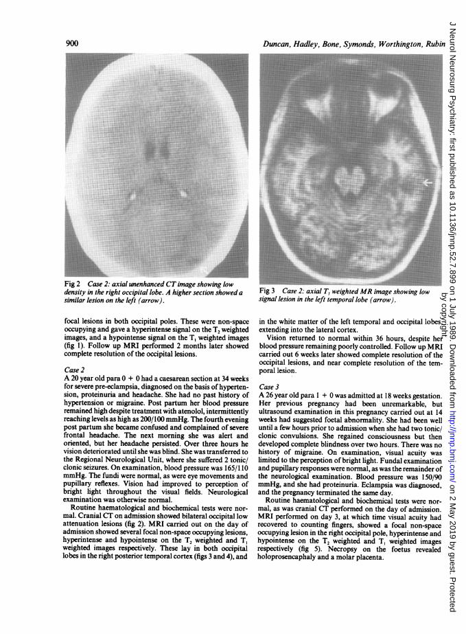
Routine haematological and biochemical tests were nor-mal. Cranial CT on admission showed bilateral occipital lowattenuation lesions (fig 2). MRI carried out on the day ofadmission showed several focal non-space occupying lesions,hyperintense and hypointense on the T2 weighted and T,weighted images respectively. These lay in both occipitallobes in the right posterior temporal cortex (figs 3 and 4), and
Fig 3 Case 2: axial T, weightedMR image showing lowsignal lesion in the left temporal lobe (arrow).
in the white matter of the left temporal and occipital lobes,extending into the lateral cortex.
Vision returned to normal within 36 hours, despite herblood pressure remaining poorly controlled. Follow up MRIcarried out 6 weeks later showed complete resolution of theoccipital lesions, and near complete resolution of the tem-poral lesion.
Case 3A 26 year old para 1 + 0 was admitted at 18 weeks gestation.Her previous pregnancy had been unremarkable, butultrasound examination in this pregnancy carried out at 14weeks had suggested foetal abnormality. She had been welluntil a few hours prior to admission when she had two tonic/clonic convulsions. She regained consciousness but thendeveloped complete blindness over two hours. There was nohistory of migraine. On examination, visual acuity waslimited to the perception of bright light. Fundal examinationand pupillary responses were normal, as was the remainder ofthe neurological examination. Blood pressure was 150/90mmHg, and she had proteinuria. Eclampsia was diagnosed,and the pregnancy terminated the same day.
Routine haematological and biochemical tests were nor-mal, as was cranial CT performed on the day of admission.MRI performed on day 3, at which time visual acuity hadrecovered to counting fingers, showed a focal non-spaceoccupying lesion in the right occipital pole, hyperintense andhypointense on the T2 weighted and T, weighted imagesrespectively (fig 5). Necropsy on the foetus revealedholoprosencaphaly and a molar placenta.
900
by copyright. on 2 M
ay 2019 by guest. Protected
http://jnnp.bmj.com
/J N
eurol Neurosurg P
sychiatry: first published as 10.1136/jnnp.52.7.899 on 1 July 1989. Dow
nloaded from

Blindness in eclampsia: CTand MR imaging.. j,_ .....~~~~~~~~~~~~~~~~~~~~~~~~~~~~~~~~~~~~~~.......Fig 4 Case 2: axial T2 weighted MR image showing highsignal lesion in the white matter ofthe left temporal lobecorresponding to the lesion seen infig 3 and in the leftoccipital lobe (arrows).
Fig 5 Case 3: axial T2 weighted MR image showing a highsignal lesion at the right occipital pole (arrow).
Her vision returned to normal over 3 days, despite poorblood pressure control. Follow up MRI done 3 months laterwas normal.
901Discussion
In all three cases the diagnosis of eclampsia seemscertain, with hypertension, proteinuria and seizuresoccurring in association with pregnancy. AlthoughCase 3 occurred at 18 weeks, this was in associationwith molar degeneration, an established risk factor forearly eclampsia.'6 Despite the statistical associationbetween the two conditions," none ofour patients hada history of migraine.
Blindness due to eclampsia may be due to lesions atany site along the visual pathway, but the majority ofcase reports are of cortical blindness. Although therewere minor retinal changes in Case 1, intact pupillaryreflexes and the radiological finding of occipital cor-tical lesions in our cases confirm that they too were ofcortical origin. The majority of patients with corticalblindness due to eclampsia recover vision, over aperiod varying from 2 hours to 21 days'-15 1122 alth-ough there is one reported case ofpersistent deficit.5 InCase I blindness was the presenting feature ofeclamp-sia, while in Case 2 it occurred 5 days post partum.Approximately 50% of the cases in the literature haveoccurred post partum,48912'3202' by up to 7 days.'3Blindness was the presenting feature of eclampsia in
27121113 15six cases.Clinically, there also were lesions at other sites in
Case 1. The finding of gaze palsies indicated a lowmidbrain/high pontine lesion. The sensory level tovibration sense suggested the possibility of a spinalcord lesion, as did the findings of increased reflexesand clonus. In the other two cases there was no clinicalevidence of focal lesions other than those in theoccipital cortex. In cases in the literature, increasedtendon reflexes are common,346712152122 and are notalways associated with seizures. More definiteevidence of pyramidal dysfunction such as ankleclonus, upgoing plantars and hemiparesis is alsofound,34'42' as are gaze palsies,37 nystagmus7 andfluent dysphasia.'3 In one case cortical lesions on CTcoexisted with bilateral retinal detachments.9Most recent reports feature the results of CT, and
this either was normal,7121315 showed focal low atten-uation occipital lesions,'-5 9" or more widespread lowattenuation lesions.67 The lesions were non-enhanc-ing, and were ascribed to localised oedema or toinfarction. Repeat CT has shown partial resolution oflesions in 3-5 days237 and complete resolution in 9-14days.'69'0 A recent report24 describes the results ofMRI in a single case of eclampsia. Although clinicallythe patient had no focal neurological features, lesionswith similar signal characteristics to those seen in ourcases were shown in the right occipital lobe and leftparietal lobe. They had resolved by the time a furtherscan was carried out 3 weeks post partum, and wereascribed to localised oedema. They were not detectedby CT. CT and MRI findings in hypertensive ence-phalopathy are similar.24Our MRI findings are consistent with those in
by copyright. on 2 M
ay 2019 by guest. Protected
http://jnnp.bmj.com
/J N
eurol Neurosurg P
sychiatry: first published as 10.1136/jnnp.52.7.899 on 1 July 1989. Dow
nloaded from

902transient ischaemic lesions,25 and reflect focal increasesin brain water content,26 which may persist for severaldays after the resolution of neurological signs.27 Thisresults in increases in T, and T2 relaxation times, givinghypointensity on T, weighted images and hyperinten-sity on T2 weighted images. In contrast, acute focalhaemorrhage gives a hypointense signal on T2 weight-ed images or, at the subacute stage, a hyperintensesignal on T, weighted images.
In the large necropsy series of Sheehan and Lynch28and Govan1' gross haemorrhage was the mostfrequent cause of death, but the most common findingwas of cortical groups of small (0 3-1 mm) haemorr-hages associated with infarcts of similar size. It seemslikely that our findings represent such lesions, and thatthey are primarily ischaemic rather than haemorr-hagic. Small haemorrhages are seen in the placentalvascular bed in eclampsia, and ultrastructural studiessuggest that they are secondary to ischaemic vasculardamage.30
Angiographic studies in eclampsia show generalisedlarge vessel spasm,31 and tend to support the view thatischaemia is the cause of focal neurological dysfunc-tion. However, angiographically demonstrated spasmdoes not always relate well to cerebral blood flow,32and spasm may occur without ischaemia and viceversa.The finding of ischaemic lesions in eclampsia sug-
gests that aggressive treatment of hypertension islikely to exacerbate neurological damage. It wouldappear that the most appropriate treatment in suchcases would be a drug with cerebral vasodilatoractivity such as nifedipine. Alternatively, a morespecific cerebral vasodilator agent such as nimodipinemight be used in conjunction with other antihyperten-sive agents.
We thank the obstetricians and neurologists wholooked after our three patients for allowing us toreport them. The MRI Unit was funded by grantsfrom the Medical Research Council and other bodies.
References
I Beeson JH, Duda EE. Computed axial tomography scan demon-stration ofcerebral oedema in eclampsia preceded by blindness.Obstet Gynaecol 1982;60:529-32.
2 Goodlin RC, Streib E, Sun SF, Cox TA, Williams NA. Corticalblindness as the initial symptom in severe pre-eclampsia. Am JObstet Gynaecol 1983;147:841-2.
3 Grimes DA, Ekbladh LE, McCartney WH. Cortical blindness inpre-eclampsia. Int J Obstet 1980;17:601-3.
4 Gyr T, Tamzin MS, Zimmerli W. Postpartum amaurosis inpatients with pre-eclampsia. Z Geburtzhilfe Perinatol1983;187:293-5.
5 Ferrando M, Gil-Vemet S, Sabater R, Romero R, Poveda R.Occipital infarct after an episode of eclampsia. Importance ofcomputerised axial tomography. Rev Clin Esp 1984;173:183-4.
6 Hill JA, Devoe LD, Elgammal TA. Central haemodynamicfindings associated with cortical blindness in severe eclampsia.J Reprod Med 1985;30:435-8.
Duncan, Hadley, Bone, Symonds, Worthington, Rubin7 Liebowitz HA, Hall PE. Cortical blindness as a complication of
eclampsia. Ann Emer Med 1984;13:365-7.8 McNamee PT, McComb JM, O'Connor FA, Adgey AAJ. Com-
plete recovery from late puerperal eclampsia with associatedblindness. Int J Cardiol 1982;1:327-8.
9 Moodley J, Pillay M, Pillay R. Temporary blindness and eclamp-sia. A report of 2 cases. S Afr MedJ 1985;68:677-8.
10 Tongyai T, Virutamasen P, Sawanwela N. Transient blindness inpre-eclamptic patient: a case report. J Med Assoc Thai1984;67:629-33.
11 Lau SPC, Chan FL, Yu YL, Woo E, Huang CY. Corticalblindness in toxaemia of pregnancy; findings of computedtomography. Br J Radiol 1987;60:347-9.
12 Arulkumaran S, Gibb DMF, Rauff M, Kek LP, Ratnam SS.Transient blindness associated with pregnancy induced hyper-tension. Case reports. Br J Obstet Gynaecol 1985;92:847-9.
13 Beal MF, Chapman PH. Cortical blindness and homonymoushemianopia in the postpartum period. JAMA 1980;244:2085-7.
14 Levavi H, Neri A, Zoldan J, Segal J, Ovadia J. Pre-eclampsia,"HELLP" syndrome and postictal cortical blindness. ActaObstet Gynaecol Scand 1987;66:91-2.
15 Nishimura RN, Koller R. Isolated cortical blindness in pregnancy.West J Med 1982;137:335-7.
16 Scott JS. Pregnancy toxaemia associated with hydrops fetalis,hydatidiform mole and hydramnios. J Obst Gynaecol Brit Emp1958;65:689.
17 Rotton WN, Sachtheben MR, Friedman EA. Migraine andeclampsia. Obstet Gynaecol 1959;14:322-30.
18 Rubin PC, McCabe R. Postpartum migraine and severe pre-eclampsia. Lancet 1984;u:286.
19 Chew SY, Tay D. Temporary blindness in severe pre-eclampsia.Sing J Obstet Gynaecol 1981;12:55-7.
20 Singh BM, Morris LJ, Strobos RJ. Cortical blindness in puer-perium. JAMA 1989;243:1 134.
21 Hauswald M. Cortical blindness and late postpartum eclampsia.Am J Emer Med 1987;5:130-2.
22 Neuntefel W, Riss W. Amaurosis in EPH gestosis. Z GeburtzhilfePerinatol 1986;190:95-7.
23 Crawford S, Varner MW, Digre KB, Servais G, Corbett JJ.Cranial magnetic resonance imaging in eclampsia. ObstetGynaecol 1987;70:474-7.
24 Hauser RA, Lacey DM, Knight MR. Hypertensive ence-phalopathy-magnetic resonance demonstration of reversiblecortical and white matter lesions. Arch Neurol 1988;45:1078-83.
25 Brant-Zawadski M, Kucharczyk W. Vascular disease--ischaemia. In: Brant-Zawadski M, Norman D, eds. MagneticResonance Imaging ofthe Central Nervous System. New York:Raven Press, 1987:221-34.
26 Gotoh 0, Asano T, Koide T, Takakura K. Ischaemic brainoedema following occlusion ofthe middle cerebral artery in therat. I. The time courses of the brain water, sodium andpotassium contents and blood-brain barrier permeability to I-125 albumin. Stroke 1985;16:101-9.
27 Kucharczyk W, Brant-Zawadski M. Magnetic resonance imagingof cerebral ischaemia and infarction. In: Kressel HY, ed.Magnetic Resonance Annual 1987. New York: Raven Press,1987:49-69.
28 Sheehan HL, Lynch JB. Pathology of Toxaemia of Pregnancy.Edinburgh: Churchill Livingston, 1973.
29 Govan ADT. The pathogenesis of eclamptic lesions. PatholMicrobiol 1961;24:561-75.
30 DeWolf F, Robertson WB, Brosen I. The ultrastructure of acuteatherosis in hypertensive pregnancy. Am J Obstet Gynaecol1975;123:164-74.
31 Lewis LK, Hinshaw DB, Will AD, Hasso AN, Thompson JR. CTand angiographic correlation of severe neurological disease intoxaemia of pregnancy. Neuroradiology 1988;30:59-64.
32 Bergvall V, Steiner L, Forster DMC. Early pattern of cerebralcirculatory disturbances following subarachnoid haemorr-hage. Neuroradiology 1973;5:24-32.
by copyright. on 2 M
ay 2019 by guest. Protected
http://jnnp.bmj.com
/J N
eurol Neurosurg P
sychiatry: first published as 10.1136/jnnp.52.7.899 on 1 July 1989. Dow
nloaded from