pneumoniaksumsc.com/download_center/1st/3.respiratory block... · · 2018-01-29pathophysiology 1....
TRANSCRIPT

Pneumonia
Community acquired pneumonia
(CAP)

Objectives• Discuss the epidemiology and pathophysiology of
pneumonia and CAP
• Explain the different classifications of pneumonia
• Recognize clinical presentations associated with CAP
• Discuss the diagnosis and treatment of CAP
• Identify common etiological agents causing CAP and discuss their laboratory work up
• Discuss virulence factors and prevention of Streptococcus pneumoniae

Definition
• Pneumonia is an infection that leads to inflammation
of the parenchyma of the lung (the alveoli)
(consolidation and exudation)
• It may present as acute, fulminant clinical disease or
as a chronic disease with a more prolonged course

Epidemiology
• Overall the rate of CAP 5-6 cases per 1000 persons per year
• Mortality 23%– High, especially in old people
• Almost 1 million annual episodes of CAP in adults > 65 yrs in the US
Risk factors – Age < 2 yrs, > 65 yrs
– Alcoholism
– Smoking
– Asthma and COPD
– Aspiration
– Dementia
– Prior influenza
– HIV
– Immunosuppression
– Institutionalization
– Recent hotel : Legionella
– Travel, pets, occupational exposures- birds (C. psittaci)
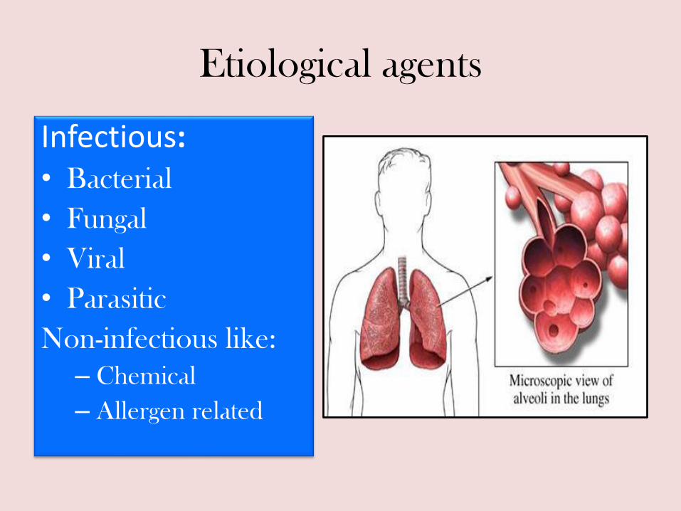
Etiological agents
Infectious:• Bacterial
• Fungal
• Viral
• Parasitic
Non-infectious like:
– Chemical
– Allergen related
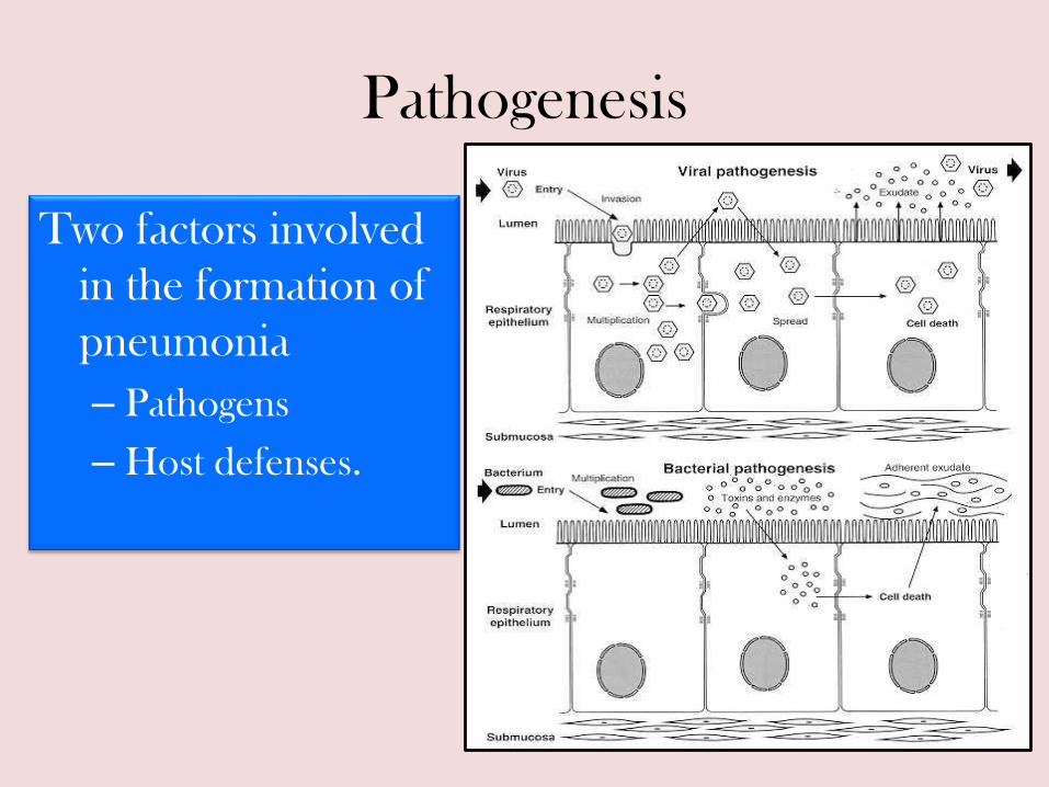
Pathogenesis
Two factors involved
in the formation of
pneumonia
– Pathogens
– Host defenses.

Defense mechanism of respiratory tract
• Filtration and deposition of environmental
pathogens in the upper airways
• Cough reflux
• Mucociliary clearance
• Alveolar macrophages
• Humoral and cellular immunity
• Oxidative metabolism of neutrophils

Pathophysiology
1. Inhalation or aspiration of pulmonary pathogenic organisms into a lung segment or lobe.
2. Results from secondary bacteraemia from a distant source, such as Escherichia coli urinary tract infection and/or bacteraemia (less commonly).
3. Aspiration of oropharyngeal contents (multiple pathogens).
Classification
• Pneumonia classified according to:
1. Pathogen
• Bacterial
– Typical
– Atypical
• Viral
• Fungal
• Parasite
2. Anatomy
3. Acquired environment

Classification by anatomy
1. Lobar: entire lobe
2. Lobular: (bronchopneumonia).
3. Interstitial

Lobar pneumonia

Classification by acquired environment
Community acquired pneumonia (CAP)
Hospital acquired pneumonia (HAP)
Nursing home acquired pneumonia (NHAP)

CAP- fever+ productive cough + infiltrate
• CAP : pneumonia acquired outside of
hospitals or extended-care facilities
Typical
• Strept. pneumoniae
– (lobar pneumonia)
• Haemophilus influenzae
• Moraxella catarrhalis
• S. aureus
• Gram-negative organisms
Atypical
• Atypical: not detectable
on gram stain; won’t
grow on standard media
• Mycoplasma
pneumoniae
• Chlamydia pneumoniae
• Legionella pneumophila

Community acquired pneumonia
• Strep pneumonia 48%
• Viral 23%
• Atypical orgs (MP,LG,CP) 22%
• Haemophilus influenza 7%
• Moraxella catharralis 2%
• Staph aureus 1.5%
• Gram –ive orgs 1.4%
• Anaerobes
Typical pneumonia
Clinical manifestation
• The onset is acute
• Prior viral upper respiratory infection
• Respiratory symptoms
– Fever
– Shaking chills
– Cough with sputum production (rusty-sputum)
– Chest pain- or pleurisy
– Shortness of breath

Diagnosis
• Clinical– History & physical
• X-ray examination
• Laboratory– CBC- leukocytosis
– Sputum• Gram stain- 15%• Culture
– Blood culture- 5-14%
– Pleural effusion gram + culture
Pneumococcal pneumonia

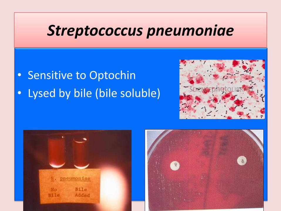
Streptococcus pneumoniae
• Gram positive diplococci
• Alpha hemolytic streptococci
• Catalase negative
• Normal flora of upper respiratory tract in 20-40% of people
• Causes: – Resp infections
• pneumonia, sinusitis, otitis,
– Non resp infections• bacteremia, meningitis

• Virulence factors:
– Capsule
• More than 90 capsular types
– Pneumolysin
– Autolysin
– Neuraminidase
• Prevention: vaccination
Streptococcus pneumoniae
Streptococcus pneumoniae
• Sensitive to Optochin
• Lysed by bile (bile soluble)

Atypical pneumonia
• Chlamydia pneumonia
• Mycoplasmapneumonia
• Legionella spp
• Psittacosis (Chlamydia psittaci)
• Q fever (Coxiellaburnettii)
• Approximately 15% of all CAP
• Not detectable on gram stain
• Won’t grow on standard media
• Most don’t have a bacterial cell wall Don’t respond to β-lactams

Symptoms
• Insidious onset
• Mild to severe
• Headache
• Malaise
• Fever
• Dry cough
• Arthralgia / myalgia
Signs
• Minimal
• Low grade fever
• Few crackles
• Rhonchi
Atypical pneumonia

Diagnosis & Treatment • Diagnosis:
– X-ray
– CBC
• Mild elevation WBC
– U&Es
• Low serum Na (Legionalla)
– LFTs• ↑ ALT• ↑ Alk Phos
– Sputum Culture on special media (BCYE) for Legionella
– Urine antigen for Legionella
– Serology for detecting antibodies
– DNA detection
• Treatment:
• Macrolide
• Quinolones
• Tetracycline
❖ B lactams have no
activity
• Treat for 10-14 days

Mycoplasma pneumonia
• Eaton’s agent (1944)
• No cell wall
• Common
• Rare in children and in > 65
• People younger than 40.
• Crowded places like schools,
homeless shelters, prisons.• Can cause URT symptoms
• Usually mild and responds well to antibiotics.
• Can be very serious
• May be associated with extra
pulmonary findings:
– skin rash, hemolysis,
myocarditis, pancreatitis,
encephalitis
• Diagnosis:
– Serology
– NAAT
– Culture can be done but
requires special media and
slow grower (weeks)

Mycoplasma
pneumonia
Cx-ray

Chlamydia pneumonia
• Obligate intracellular organism
• 50% of adults sero-positive
• Mild disease
• Sub clinical infections common
• 5-10% of community acquired pneumonia
• Diagnosis:– Serology
– NAAT

Psittacosis
• Chlamydia psittaci
• Exposure to birds
• Bird owners, pet shop
employees, vets
• Parrots, pigeons and
poultry
• Birds often
asymptomatic

• Exposure to farm animals mainly sheep
• Spread by inhalation of infected animal birth products
• Pneumonia is acute form of infection
• Diagnosis: serology
Q fever (Coxiella burnetti)

Legionella pneumophila
• Can cause– Hyponatraemia common
• (<130mMol)
– Bradycardia
– WBC < 15,000
– Abnormal LFTs
– Raised CPK
– Acute Renal failure
• Legionnaire's disease
• Serious outbreaks linked
to exposure to cooling
towers
• Can be very severe and
lead to ICU admission.
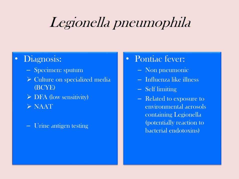
Legionella pneumophila
• Pontiac fever:
– Non pneumonic
– Influenza like illness
– Self limiting
– Related to exposure to
environmental aerosols
containing Legionella
(potentially reaction to
bacterial endotoxins)
• Diagnosis:
– Specimen: sputum
➢ Culture on specialized media
(BCYE)
➢ DFA (low sensitivity)
➢ NAAT
– Urine antigen testing

Legionnaires in ICU

• Factors to consider in selection of antibiotic:
– Co morbidities
– Previous antibiotic exposure in last 3 months
– Severity
• Out patient management vs requiring inpatient
admission vs requiring ICU
Antibiotic Treatment of CAP

Macro
lid
es
Doxy
cycl
ine
Lev
ofl
oxaci
n
B-l
acta
m
An
d
Macro
lid
e
B-l
acta
mA
nd
Lev
o
Outpatient, healthy
patient with no exposure
to antibiotics in the last 3
months
-S. pneumoniae
-Atypical
pathogens
-Viral
Outpatient, patient with
comorbidity or exposure
to antibiotics in the last 3
months
As above +
Anaerobes
S. aureus
Inpatient : Not ICU Same as above +
coliforms
Inpatient : ICU Same as above +
Pseudomonas