blood coagulation thrombophilia - johns hopkins hospital course... · blood coagulation and...
TRANSCRIPT

Blood Coagulationand
Thrombophilia
Cliff Takemoto M.D.
Pediatric Hematology
The Johns Hopkins University

Disclosure Information
Cliff Takemoto MD
NONE

Objectives
Develop a framework to understand coagulation
Know how to approach coagulopathies

What is in a clot?
www.junebughunter.net
www.uphs.upenn.edu
Platelets
Fibrin(coagulation proteins)
VENOUSSlow flowRED-(rbc’s)
ARTERIALFast flowWHITE (plts)

collagenTF
How we clotCut in the endothelium
Exposes collagen for platelet binding sitesExposes tissue factor for activating coagulation
collagencollagen
TF TFcollagenvWF
vWF
vWF fibrinogen
fibrinogenvWFTF TF
TF
TF
Platelet ActivationPlatelet granule secretion
Activate fibrinogen receptorProvides site for prothrombinase complex
ThrombinActivationAnd fibrindeposition
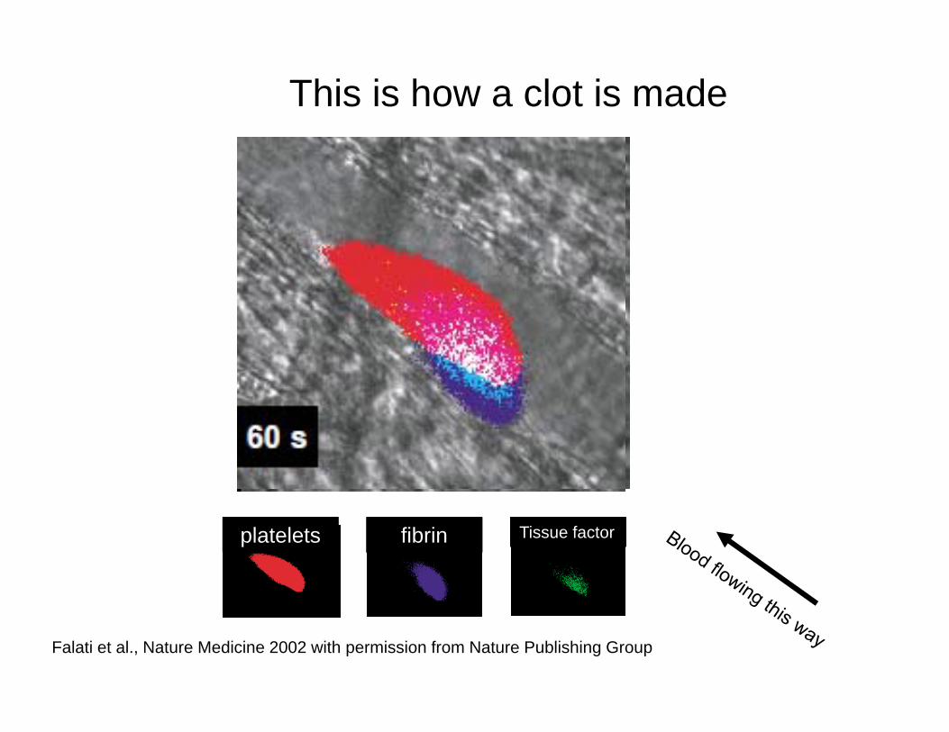
This is how a clot is made
Falati et al., Nature Medicine 2002 with permission from Nature Publishing Group
platelets fibrin Tissue factor

how a clot is made in vivo
Thrombin activation INITIATED by TISSUE FACTOR (microparticles?)
TWO pathways to PLATLET ACTIVATION:TF (thrombin)
Collagen (vWF)

Coagulation Cascades… the often-asked question: “Why is coagulation so complicated?” resolves itself into the question: “Why are there so many stages?”
R.G. Macfarlane, Nature 1964
John Hagemen
Rosenthal syndrome
Stephen Christmas
Rufus Stuart
Antti-hemophilc factor proconvertin
proaccelerin
prothrombin
fibrinogen

Coagulation CascadesSome concepts to remember…
•Highly regulated complexes of serine proteases and co-factors
•All paths lead to thrombin activation
•Goal is to make a fibrin clot
•They are not just a number—•they have a personality too!!!
www.uphs.upenn.edu
thrombin
fibrin

Serine Proteases
FVIII
FV
TF
Co-Factors
FII
PC
TM
PS
FIX
FX
FVII
procoagulant
anticoagulant

IIprothrombin
IIathrombin Fibrin
polymers
Fibrinogen
Xa
VaCa++,
PL
“Prothrombinase”
IXa
VIIIaCa++, PL
intrinsic
tenase
CONTACT FACTORS
(prekallikrein,HMWK, XII,XI)
Clot formation--physiology
VIIa
TF
extrinsic
tenase
SERINE PROTEASE
COFACTOR
Know the functions and mechanism of activation of factor___ in coagulation

II IIa
Xa
VaCa++,
PL
“Prothrombinase”
IXa
VIIIaCa++, PL
intrinsic
tenase
CONTACT FACTORS
(prekallikrein,HMWK, XII,XI)
VIIa
TF
extrinsic
tenase
fibrinogen Fibrin clot
PTPTT
Know the consequences of deficiency of factor___ on the laboratory assessment of hemostasis
Clot formation--physiology

II IIa
Xa
Va
“Prothrombinase”
IXa
VIIIa
CONTACT FACTORS
(prekallikrein,HMWK, XII,XI)
Clot formation--physiology
VIIa
TF
fibrinogen Fibrin clot
INITIATION
AMPLIFICATIONPROPAGATION

II IIa
Xa
Va
IXa
VIIIa
Clot formation--physiology
VIIa
TF
fibrinogen Fibrin clot
X
initi
atio
npr
opag
atio
n

enzyme complexes : protease cofactor
Prothrombinase Xa Va
Intrinsic tenase IXa VIIIa
Extrinsic tenase VIIa TF
Also need Calcium and a phosholipid surface
Activation of thrombin: 3 complexes

A few words about contact activation…
•Regulate Inflammation
•HK, PK, FXII deficiencydo not bleed
•FXI deficiency associatedwith bleeding
CofactorHigh Molecular-Weight Kininogen (HK)
ProteasePrekallikrein (PK)FXIIFXI
InibitorC1 esterase inhibitor
C1inh

ThrombinIIa
procoagulant anticoagulant
FibrinogenFV, FVIII, FXI
FXIIITAFI (fibrinolysis inhibitor)
Platelet Activation
Protein C/Sbound to Thrombomodulin
Thrombin has both pro- and anti-coagulant functions

Fibrinogen Structure
Gorkun O. et al., Blood 1997; 89:4407-44. With permission from the American Society of Hematology
Know basic structure of fibrinogen and its gene control
Fibrinogen Genes
6 polypeptide chains3 genes:
Fibrinogen (soluble) Fibrin (insoluble)

Fibrin Clot formation and fibrinolysis
D DE D DE D DE
D DE D DE D DE D DE
D DE D DE D DE
D DEfibrinogen
FXIII plasmin
Thrombin (IIa) ABfibrinopeptides
D-dimer
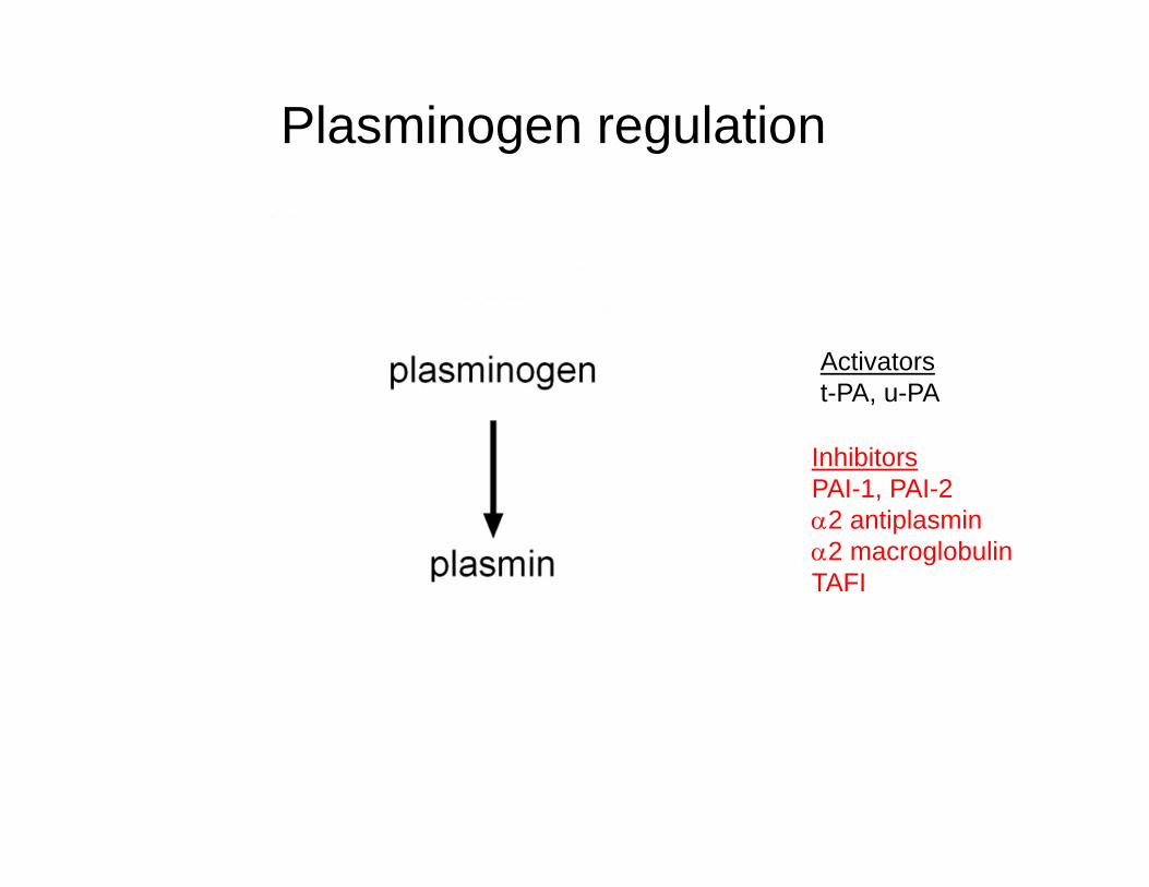
Activatorst-PA, u-PA
InhibitorsPAI-1, PAI-22 antiplasmin2 macroglobulinTAFI
Plasminogen regulation

Endogenous Anticoagulants:Turning off the clot
Protein C VIIIa, Va (the cofactors)Protein S
Antithrombin IIa, Xa (serine proteases)
TFPI VIIa/TF

II IIa
VIIa
Xa
Va
IXa
VIIIa
Protein C
Protein SAT III
thrombomodulin
IIa
TFPI
How to stop the clot

Heparin—physical characteristicsIt binds to Antithrombin
heparin pentasaccharide
binding to antithrombin Activation loop
binds to reactiveSite in serine
protease

Heparin-- physical characteristics
Pentasacchride Antithrombinbinding sequence
From
the
Wal
lstre
etjo
unal

Heparin binds and activates Antithrombin
Pentasacchride sequence of heparin binds antithrombin
ATIII IIa
ATIII
Xa
Coagulation Factors
Suicide inhibitor
Heparin accelerates this reaction >1000fold
IIaXa

Antithrombin Deficiency
• Prevalence 1/5000• Risk of thrombosis
15-20X• Acquired forms-
nephrotic syndrome, liver disease
• Lab- Antithrombin activity
IIa
IXa
Xa XIa
AT IIIheparin
IIi XiIXi
XIi

Protein C and Protein S
C4b binding protein
Thrombomodulin
IIa
VIIIaVa
APC
S S
C
cofactors
Vitamin K dependentProtein C-serine proteaseProtein S-cofactorActivated by thrombin/TM

Protein C and S deficiency
• Protein C- 1/250-500• Protein S- 1/1000 • Increase risk 5-10X• Acquired causes:
Both: Vit. K deficiency Protein S: estrogens, pregnancy
• Homozygous protein C presents with neonatal purpura fulminans
• Lab- Protein C and S activity
TTM
C APCS
S
VIIIaVa
VIIIi
Vi
C4bBP
S

Thromboembolic events with other prothrombotic risk factors
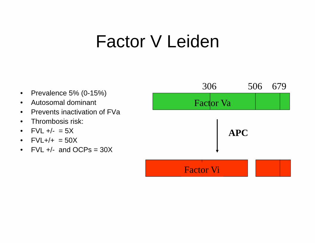
Factor V Leiden
• Prevalence 5% (0-15%)• Autosomal dominant• Prevents inactivation of FVa• Thrombosis risk: • FVL +/- = 5X• FVL+/+ = 50X • FVL +/- and OCPs = 30X
306 506 679
APC
Factor Va
Factor Vi

Arg 506Arg 306 Arg 679
FVa
39 sec30 sec
R506Q is the FV leiden mutation
What is APC resistance?
FVL:
PTT (APC)PTT
75sec30sec
= 1.3
= 2.5normal:

Factor Invivo T1/2 Synthesis Function vitKFibrinogen (I) 2-4 days liver -Prothrombin (II) 3 days liver SP +V 36 hrs liver/(mega) CoF -VII 3-6 hrs liver SP +VIII 8-12 hrs liver/EC CoF -IX 22 hrs liver SP + X 40 hrs liver SP +XI 80 hrs liver SP -XII 50-70 hrs liver SP -XIII 10 days liver/M TG -VWF 12 hrs EC/(mega)
Characteristics of Clotting Factors
mega—megakaryocyte; M—macrophage; EC—endothelial cell; SP—serine protease; CoF—Cofactor;TG--transglutaminaseColeman Hemostasis and Thrombosis 3rd Edition

Factor Incidence InheritanceFibrinogen (I) 1:1 million afibrinogen-recessive
hypofibrinogen-dominantProthrombin (II) 1:2 million recessiveV 1:1 million recessiveVII 1:300,000 recessiveVIII 1:5,000 x-linkedIX 1:30,000 x-linkedX 1:1 million recessiveXI 1:1 million recessive
1:12 (het) Ashekenazi JewishXII 1:50 (het) recessiveXIII 1:1 million recessiveVWF 1:100 Type 1 dominant
1:1 million Type 3 recessive
Characteristics of Clotting Factors
Bolton-Maggs et. al., Haemophilia 2004

Approach to abnormal laboratory screens
Elevated aPTT Elevated PT
Elevated aPTT and PT
Elevated Thrombin Time

Elevated aPTT
Mixing study 1:1 ratiocontrol + patient plasma
correction no correction
BleedingFVIIIFIXFXI
No BleedingContact factors--FXII--PK--HMWK
Heparin Circulating Inhibitor
Phospholipid dependentLupus Anticoagulant--DRVVT--phospholipid neutralization--platelet neutralization
Non-specific
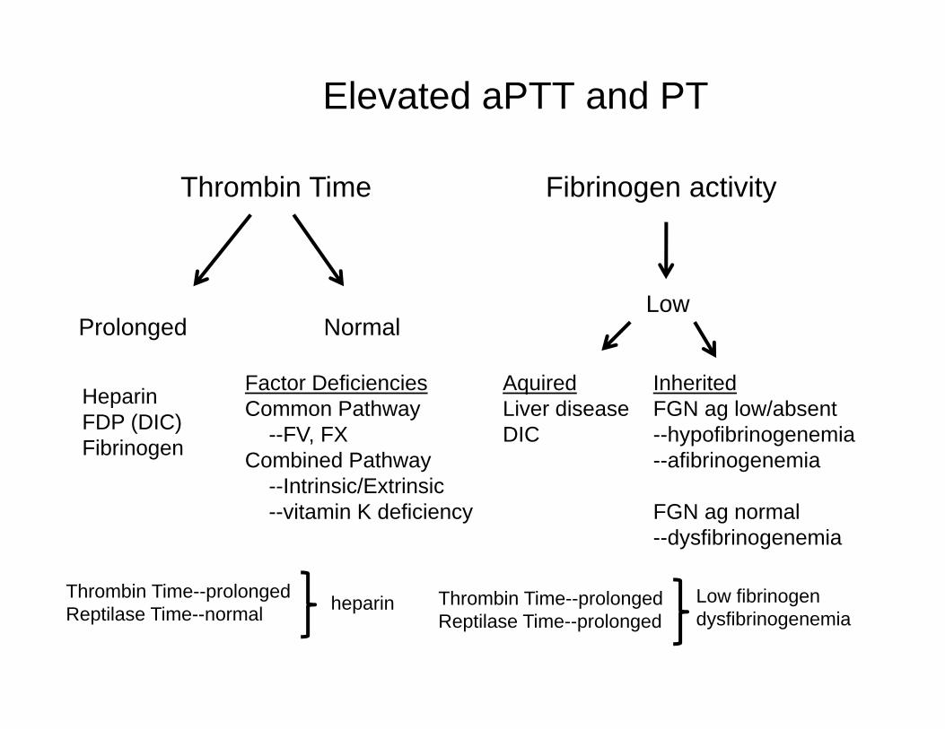
Elevated aPTT and PT
Thrombin Time
Low
HeparinFDP (DIC)Fibrinogen
Prolonged Normal
Factor DeficienciesCommon Pathway
--FV, FXCombined Pathway
--Intrinsic/Extrinsic--vitamin K deficiency
Fibrinogen activity
AquiredLiver diseaseDIC
InheritedFGN ag low/absent--hypofibrinogenemia--afibrinogenemia
FGN ag normal--dysfibrinogenemia
Thrombin Time--prolongedReptilase Time--normal heparin Thrombin Time--prolonged
Reptilase Time--prolongedLow fibrinogendysfibrinogenemia

Case
23 year old male with hemoptysis for 3 weeks
Seen at Eastern Shore ER and spiral CT shows large PE
Transport Team calls you—asks for management advice and recommendation for bloodwork before treatment?

Case
Antithrombin, protein C/S, APC, prothrombin 20210,
Homocysteine, etc—all normal.
One test you were not able to get was the antiphospholipid antibodies. He is already on heparin. When can you do the test? Does it matter?

Case
DRVVT was tested on coumadin and prolonged. The “confirm ratio” was high, suggesting
an antiphospholipid antibody
RVVT: 74 (27-45)1:1 mix 56.2 (27-45)4:1 mix 65.3 (27-45)
aPTT 30 (24-34)PT 10.9 (10.4-12.0)
RVVT:confirm 1.8 (1-1.4)ratio

Antiphospholipid antibodies (APA)
LAACA
B2GPI
Lupus Anticoagulantprolong PTT test AND is phospholipid dependent(mixing study with plt neutralization, DRVVT)
--usually benign and transient--can be seen in association with thrombosis--bleeding when directed against specific factor(FII, FV, FVIII)
--can be acquired transaplacentally in neonates
Anticardiolipin AntibodiesELISA based detection
Anti-Beta2 Glycoprotein I AntibodiesELISA based detection

Dilute Russell viper venom time(DRVVT—like aPTT)
X
Xa Va II IIa
dRVVT (sec)
dRVVT + PL (sec)
Confirm Ratio
60 sec.30 sec.
= 2
(1 - 1.4)Normal
•Activate X•Sensitive to inhibition by Antiphospholipid Ab
If the confirm ratio is highAn a phospholipid ab is present
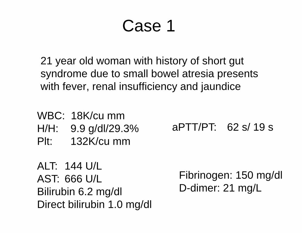
Case 1
21 year old woman with history of short gut syndrome due to small bowel atresia presents with fever, renal insufficiency and jaundice
WBC: 18K/cu mmH/H: 9.9 g/dl/29.3%Plt: 132K/cu mm
ALT: 144 U/LAST: 666 U/L Bilirubin 6.2 mg/dlDirect bilirubin 1.0 mg/dl
aPTT/PT: 62 s/ 19 s
Fibrinogen: 150 mg/dlD-dimer: 21 mg/L

FV 14%FVII 29%FVIII 138%
Coagulopathy of Liver Disease
Case 1
Diagnosis?

Case 2
6 week infant with presents with lethargy, bulging fontelle
On examination, extensive bruising, bleeding from the mouth

Case 2
WBC: 10.9K/cu mmH/H: 7.8 g/dl 22.2%Plt: 407K/cu mm
aPTT/PT: >100 s/ 65 s
Fibrinogen: 360 mg/dlD-dimer: > 2.6 mg/L

FII 3%FV 108%FVII 4%FVIII 132%FIX 2%FX 1% Management?
Diagnosis?
Case 2
Late Vitamin K Deficiency Bleeding(VKDB)
FFPIV vitamin K

Vitamin K metabolism
Active FactorsBind to Phopholipid
surface
Ca++
Vitamin K Dependent Factors
II Protein C
VII Protein S
IX
X Gla proteins
osteocalcin
(bone)

Case 3
17 old male with presents with bruising andPetechia
WBC:10.9K/cu mmH/H: 7.8 g/dl 22.2%Plt: 17K/cu mmBlasts on peripheral smear
aPTT/PT: 40s/19 s
Fibrinogen: 112 mg/dlD-dimer: > 54.7 mg/L


Flow
normal
APML

Case 3
Diagnosis:
Acute Promyelocytic LeukemiaWith DIC

Concepts about the pathophysiology of DIC:microvascular clotting
Consumption of Coagulation factors
and platelets
Underlying disease
Severe Bleeding
Depression of Anticoagulant proteinsimpaired fibrinolysis
Fibrin deposition Microvascular thrombosis
Activation of coagulation
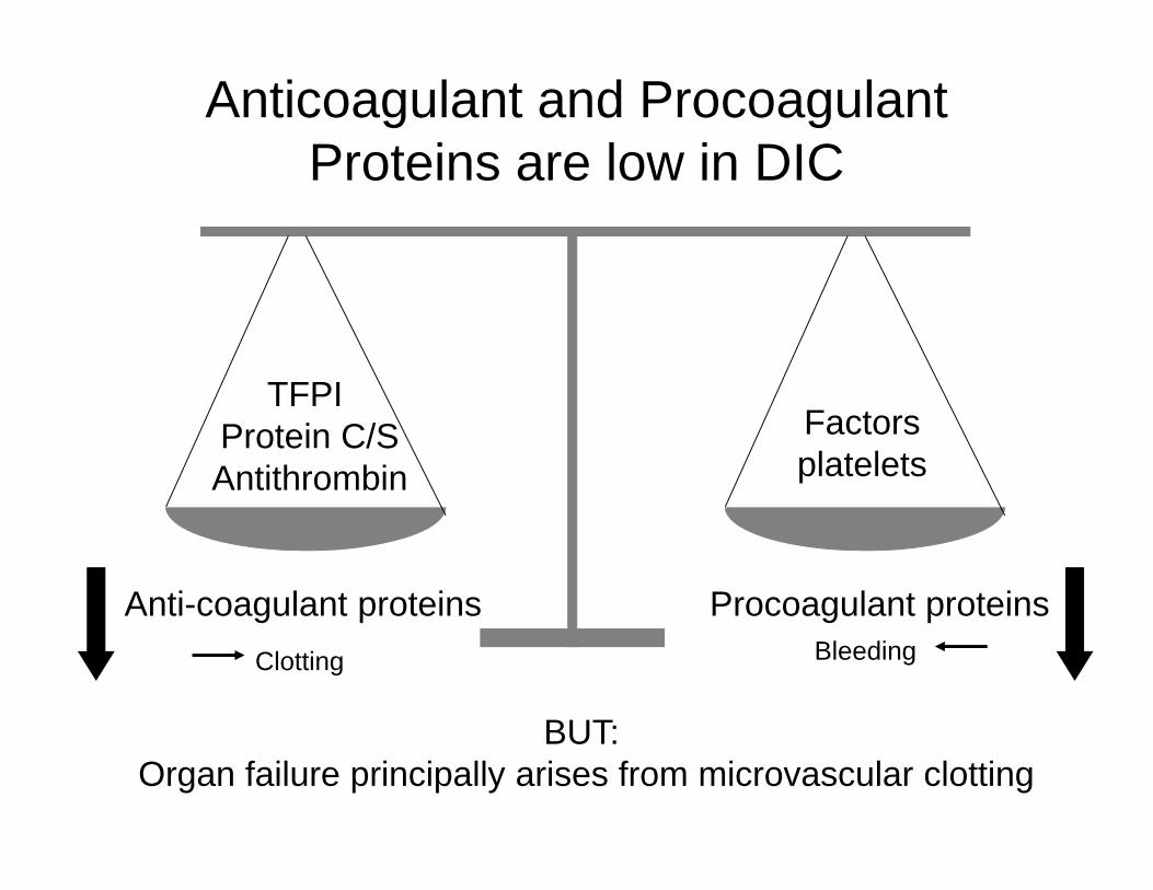
Anticoagulant and Procoagulant Proteins are low in DIC
BUT: Organ failure principally arises from microvascular clotting
Anti-coagulant proteins Procoagulant proteins
TFPI Protein C/SAntithrombin
Factorsplatelets
Clotting Bleeding

Diagnosing DIC
• Thrombocytopenia• Elevated D-dimers/FSP (sensitive)• Low fibrinogen (late finding)• Elevated PT (PT more sensitive than aPTT)
No single test
Scoring Criteria for diagnosis and managment

Pathogenesis of DIC
1. Tissue Factor initiation of coagulationfrom monocytes, other cells
2. Decreased anticoagulants leading to uncontrolled amplification of thrombin
3. Fibrinolysis accompanies DIC but is impaired due to upregulation of PAI with fibrin propagation
Microvascular fibirn deposition Leads to organ dysfunction

Treatment of DIC
• Treat underlying disease
• Coagulopathy? Treat bleeding/bleeding risk• No consensus guidelines for transfusion with platelets,
FFP, cryoprecipitate
• Role for anticoagulants?
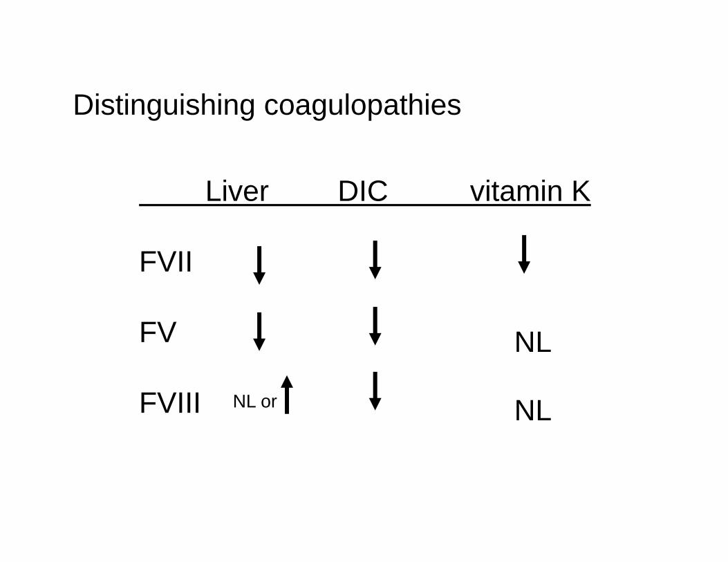
Liver DIC vitamin K
FVII
FV
FVIII
Distinguishing coagulopathies
NL or NL
NL

Coagulopathy in other acquired disorders
Hemophagocytic syndromes—DIC with hypofibrinogenemia
Leukemia—DIC (APML); platelet dysfunction (M7, M5 AML)
L-Asparaginase—impaired protein synthesis with hemorrhage and thrombosis; AT deficiency
Nephrotic Syndrome—loss of coagulation factors with thrombosis; AT deficiency

Bleeding in Renal Disease
Platelet dysfunction--Uremia--Increased nitric oxide production--Anemia
Treatment--dialysis--RBC Txn; plt Txn--DDAVP; cryoprecipitate--conjugated estrogens

Summary
Function: Serine Protease or CoFactorExcept FXIII transglutaminase
Site of synthesis: LiverFVIII liver+endothelium; vWF endotheliumFXIII liver+Macrophage; FV liver+megakaryocyte
Consequence of deficiency: Bleeding Except for most Contact Factors
Half life: FVII 2hrs; FXIII 2 weeks

Summary
Work up for PTT/PT elevation:
Liver coagulopathy—FVIII preserved
Vitamin K deficiency
DIC—anticoagulant and procoagulant low