blood gases: pathophysiology and interpretation tintinalli chapter 26 sept. 1, 2005
TRANSCRIPT

Blood Gases: Pathophysiology and Interpretation
TintinalliChapter 26
Sept. 1, 2005
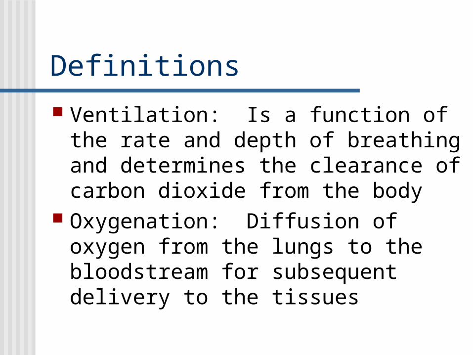
Definitions Ventilation: Is a function of the rate
and depth of breathing and determines the clearance of carbon dioxide from the body
Oxygenation: Diffusion of oxygen from the lungs to the bloodstream for subsequent delivery to the tissues

Minute Ventilation Total amount of new air moved in and out
of the airways and lungs each minute Equals tidal volume multiplied by the
respiratory rate Normal tidal volume is 7mL/kg, or 500mL
in adult Normal rate is 12 breaths/minute Normal minute ventilation is 6L/min

Dead Space Anatomic dead space occurs in the trachea,
bronchi, and bronchioles Alveolar dead space (high V/Q mismatch)
occurs when ventilation of the alveolar-capillary is normal but perfusion is absent
The combined dead space is physiologic dead space and is about 30% of the tidal volume
ARDS and COPD can increase dead space to 60%
Dead space over 60% typically requires intubation

Partial Pressures Normal atmospheric pressure is 760mm
Hg Partial pressure of H20 is 47mm Hg and is
subtracted from atmospheric pressure (760-47=713)
Remaining gases are: Nitrogen 79% (563mm Hg) Oxygen 21% (149mm Hg) CO2 0.04% (0.3mm Hg)

Alveolar Gas Equation
For each mL of O2 leaving the alveolus, 0.8 to 1.0 mL of CO2 enters it
This is the respiratory quotient (RQ)

A-a Gradient Determines the degree of lung
function impairment The A-a gradient is the partial
pressure of alveolar oxygen minus the partial pressure of arterial oxygen (PAO2-PaO2)
Normal is 2-10mm Hg or 10 plus one tenth the person’s age

A-a Gradient PAO2=(PB-PH2O)(FIO2)-PaCO2/RQ
PAO2=(760-47)(0.21)-40/0.08
PAO2=100mm Hg at sea level in room air
PaO2 in a normal, healthy adult in room air at sea level is 90-100mm Hg
So, the PAO2-PaO2 is 100 minus 90, or about 10mm Hg

A-a Gradient PAO2-PaO2 of 20-30mm Hg on room
air indicates mild pulmonary dysfunction, and greater than 50mm Hg on room air indicates severe pulmonary dysfunction
The causes of increased gradient include intrapulmonary shunt, intracardiac shunt, and diffusion abnormalities

PaO2
Factors affecting the PaO2 include alveolar ventilation, FIO2, altitude, age, and the oxyhemoglobin dissociation curve
Relation between PaO2 and SaO2:PaO2 corresponds to SaO2
60mm Hg 90%
50mm Hg 80%
40mm Hg 70%
30mm Hg 60%
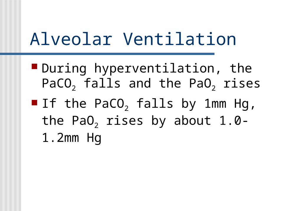
Alveolar Ventilation
During hyperventilation, the PaCO2 falls and the PaO2 rises
If the PaCO2 falls by 1mm Hg, the PaO2 rises by about 1.0-1.2mm Hg

Factors Affecting Oxyhemoglobin Dissociation pH:
The more acidic the blood, the more readily hemoglobin gives up oxygen and the higher the PaO2
With alkalosis, hemoglobin binds more tightly to oxygen
A rise or fall in pH of 0.10 causes a fall or rise in the PaO2 of about 10%, respectively

Factors Affecting Oxyhemoglobin Dissociation
Partial pressure of CO2
CO2 entering blood from tissues shifts the curve to the right
Oxygen is displaced from the hemoglobin and delivers oxygen at a higher PO2 than normal

Factor Affecting Oxyhemoglobin Dissociation Temperature
With a rise in blood temperature, hemoglobin releases oxygen more readily, which increases the PO2 in the plasma

Factors Affecting Oxyhemoglobin Dissociation Exercise
With exercise, muscles release large amounts of CO2 and acids
Muscle temperature can rise 3-4oC Combined, these shift the
oxyhemoglobin dissociation curve to the right, which releases O2 more readily

Factors Affecting Oxyhemoglobin Dissociation 2,3-Diphosphoglycerate (2,3-DPG)
With prolonged hypoxia over several hours, 2,3-DPG quantities increase, which shifts the dissociation curve to the right
If the concentration of 2,3-DPG falls, such as in banked blood or sepsis, the curve shifts left and the PaO2 falls

PaO2/FIO2 Ratio To estimate the impairment of
oxygenation, calculate the PaO2/FIO2 ratio Normally, this ratio is 500-600 Below 300 is acute lung injury* Below 200 is ARDS*
*Along with diffuse infiltrates, normal PCWP, and appropriate mechanism

Pulse Oximetry Factors that affect the pulse ox
effectiveness Impaired local perfusion (hypothermia,
vasopressors) Ambient light (fluorescent) Nail polish (particularly blue) Abnormal hemoglobin Very high PO2
Carboxyhemoglobin falsely raises readings Methemoglobin falsely lowers the readings

Questions1. A SaO2 of 90% corresponds to a PaO2 of:
A. 40%B. 50%C. 60%D. 70%E. 80%
2. Which of the following affects the pulse oximetry readings?A. Blue nail polishB. Ambient fluorescent lightingC. HypothermiaD. CarboxyhemoglobinE. All will adversely affect the pulse ox readings
3. Which is false regarding oxyhemoglobin dissociation?A. As temperature rises, hemoglobin binds oxygen with more affinityB. Exercise shifts the dissociation curve to the right, releasing oxygen more readilyC. Hemoglobin binds oxygen with more affinity in an alkylotic stateD. During sepsis, the curve shifts left, causing a decrease in the PaO2

Questions4. ARDS is defined as bilateral diffuse infiltrates, normal PCWP, appropriate mechanism, and a
PaO2/FIO2 ratio of what?A. Below 500B. Below 400C. Below 300D. Below 200E. Below 100
5. True or FalseMinute ventilation is the respiratory rate multiplied times the tidal volume
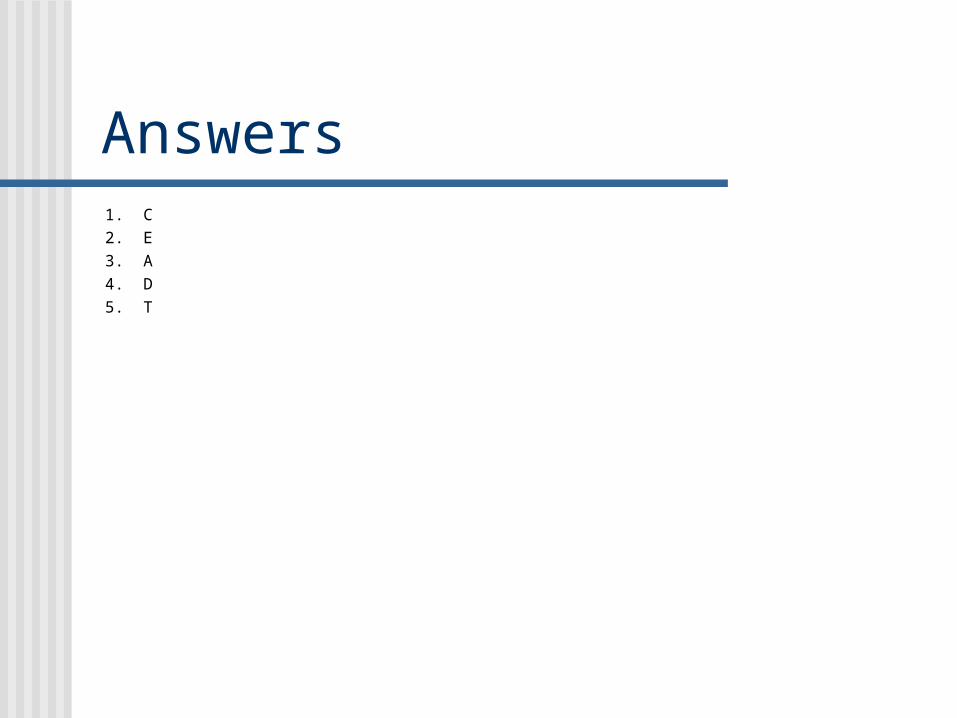
Answers1. C2. E3. A4. D5. T