blood pressure control mike clark, m.d.. map = co x svr co = hr x sv sv = edv – esv (edv concerned...
TRANSCRIPT
Blood Pressure Control
Mike Clark, M.D.
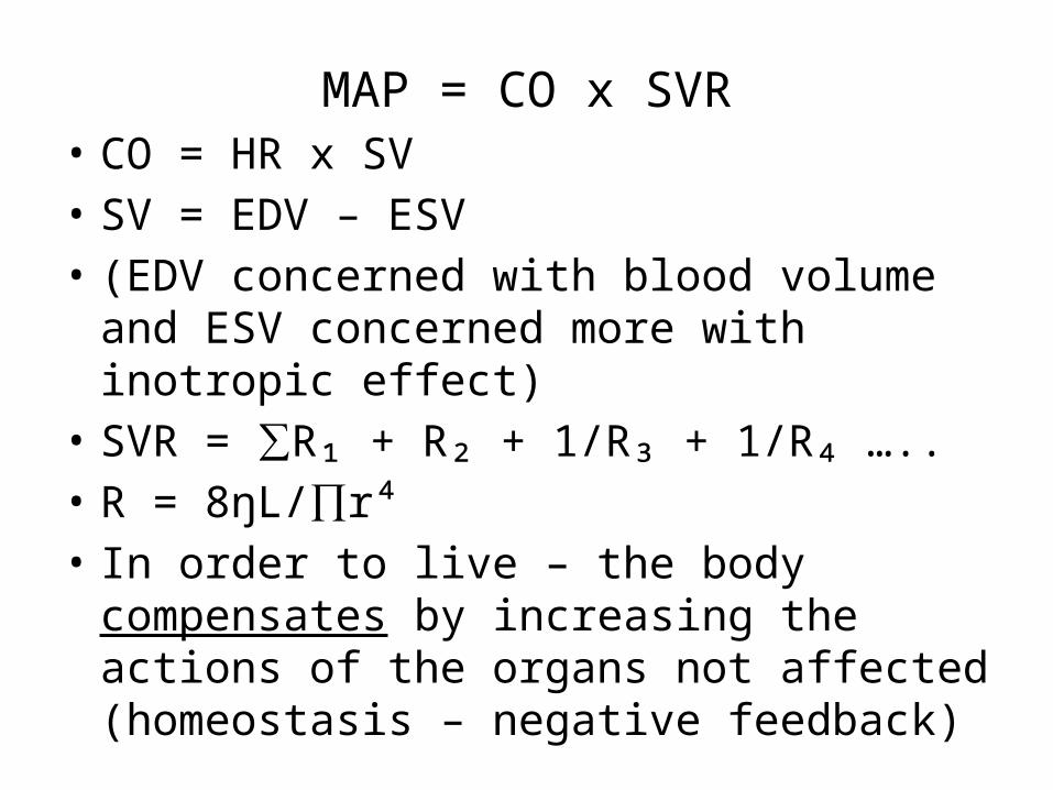
MAP = CO x SVR• CO = HR x SV• SV = EDV – ESV • (EDV concerned with blood volume and ESV
concerned more with inotropic effect)• SVR = ∑R₁ + R₂ + 1/R₃ + 1/R₄ …..• R = 8ŋL/∏r⁴• In order to live – the body compensates by
increasing the actions of the organs not affected (homeostasis – negative feedback)
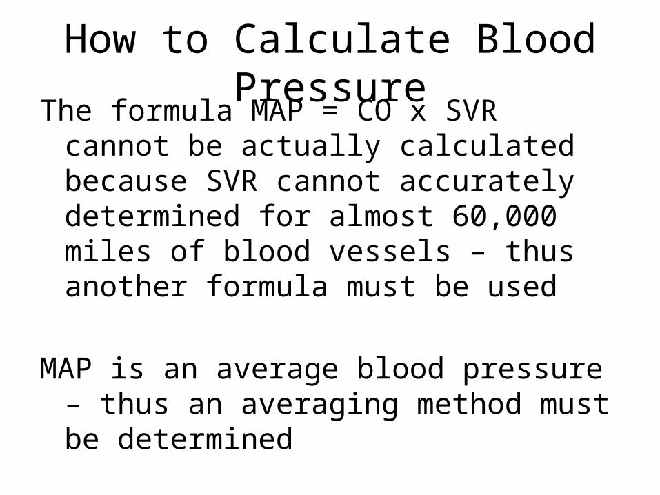
How to Calculate Blood PressureThe formula MAP = CO x SVR cannot be actually
calculated because SVR cannot accurately determined for almost 60,000 miles of blood vessels – thus another formula must be used
MAP is an average blood pressure – thus an averaging method must be determined
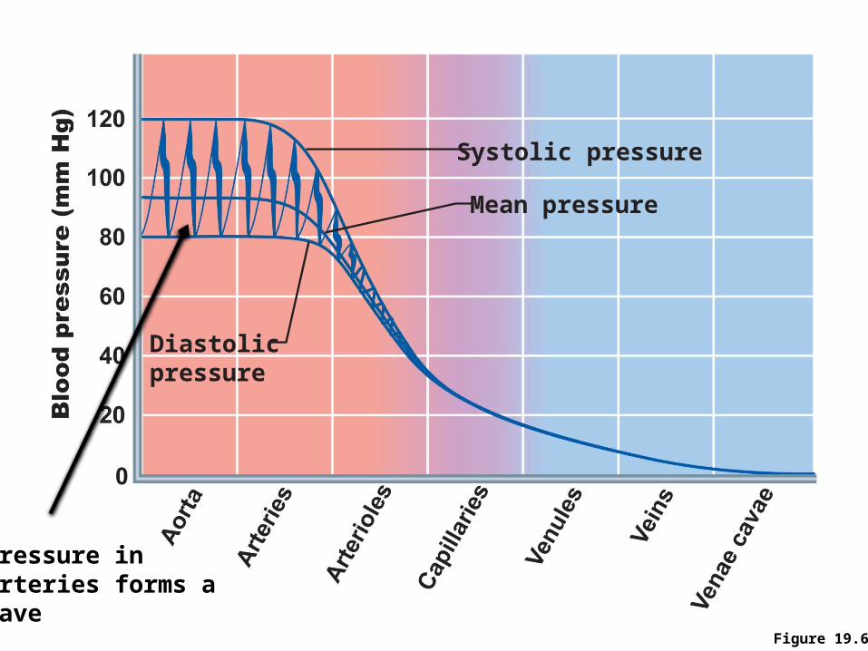
Figure 19.6
Systolic pressure
Mean pressure
Diastolic pressure
Pressure inArteries forms a Wave
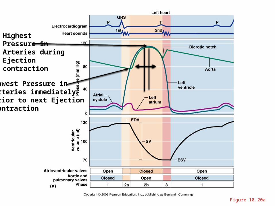
Figure 18.20a
Highest Pressure inArteries duringEjection contraction
Lowest Pressure inArteries immediately Prior to next Ejection contraction
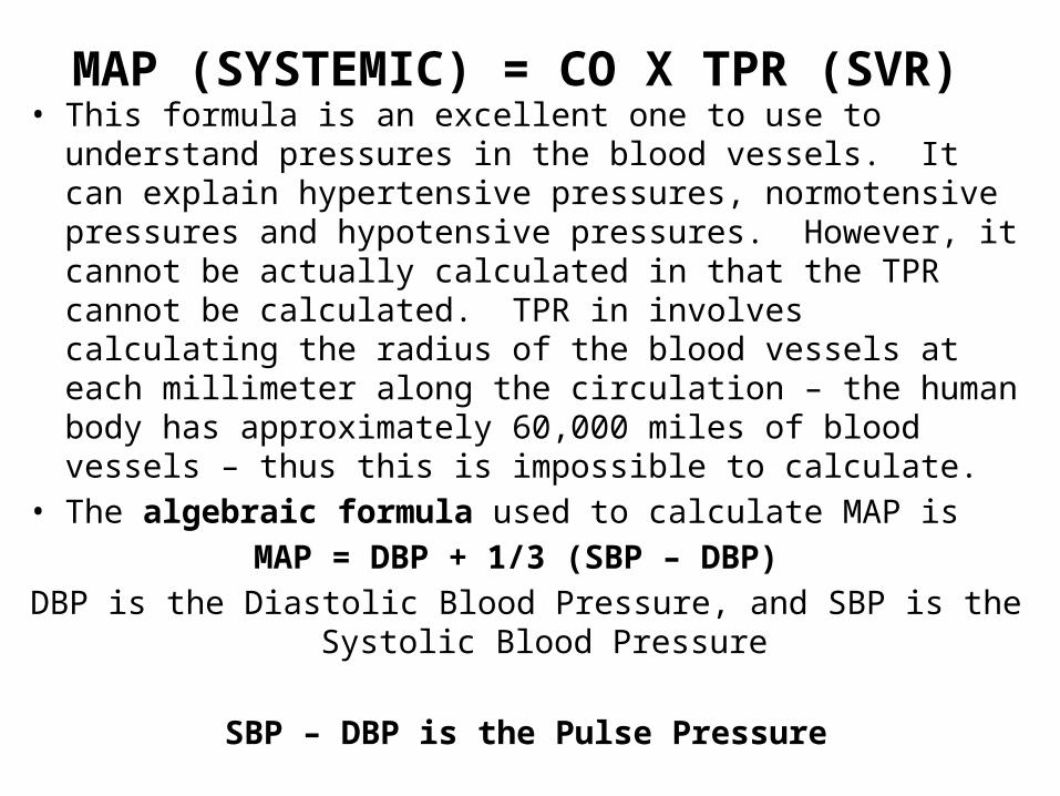
MAP (SYSTEMIC) = CO X TPR (SVR) • This formula is an excellent one to use to understand pressures in
the blood vessels. It can explain hypertensive pressures, normotensive pressures and hypotensive pressures. However, it cannot be actually calculated in that the TPR cannot be calculated. TPR in involves calculating the radius of the blood vessels at each millimeter along the circulation – the human body has approximately 60,000 miles of blood vessels – thus this is impossible to calculate.
• The algebraic formula used to calculate MAP is MAP = DBP + 1/3 (SBP – DBP)
DBP is the Diastolic Blood Pressure, and SBP is the Systolic Blood Pressure
SBP – DBP is the Pulse Pressure
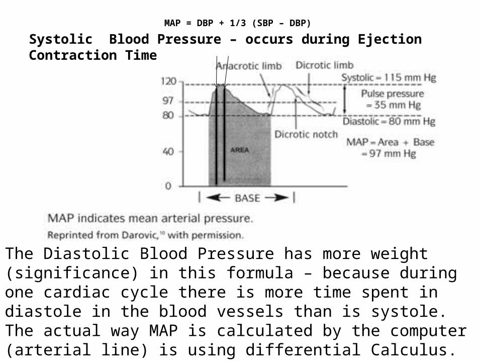
MAP = DBP + 1/3 (SBP – DBP)
Systolic Blood Pressure – occurs during Ejection Contraction Time
The Diastolic Blood Pressure has more weight (significance) in this formula – because during one cardiac cycle there is more time spent in diastole in the blood vessels than is systole. The actual way MAP is calculated by the computer (arterial line) is using differential Calculus. Differential calculus exactly calculates the area under a curve.
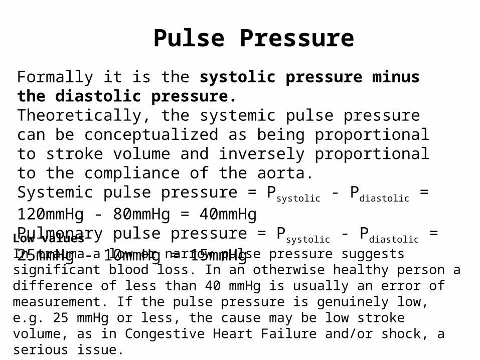
Pulse PressureFormally it is the systolic pressure minus the diastolic pressure.Theoretically, the systemic pulse pressure can be conceptualized as being proportional to stroke volume and inversely proportional to the compliance of the aorta.Systemic pulse pressure = Psystolic - Pdiastolic = 120mmHg - 80mmHg = 40mmHg Pulmonary pulse pressure = Psystolic - Pdiastolic = 25mmHg - 10mmHg = 15mmHg
Low valuesIn trauma a low or narrow pulse pressure suggests significant blood loss. In an otherwise healthy person a difference of less than 40 mmHg is usually an error of measurement. If the pulse pressure is genuinely low, e.g. 25 mmHg or less, the cause may be low stroke volume, as in Congestive Heart Failure and/or shock, a serious issue.
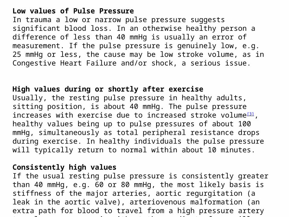
Low values of Pulse PressureIn trauma a low or narrow pulse pressure suggests significant blood loss. In an otherwise healthy person a difference of less than 40 mmHg is usually an error of measurement. If the pulse pressure is genuinely low, e.g. 25 mmHg or less, the cause may be low stroke volume, as in Congestive Heart Failure and/or shock, a serious issue.
High values during or shortly after exerciseUsually, the resting pulse pressure in healthy adults, sitting position, is about 40 mmHg. The pulse pressure increases with exercise due to increased stroke volume[3], healthy values being up to pulse pressures of about 100 mmHg, simultaneously as total peripheral resistance drops during exercise. In healthy individuals the pulse pressure will typically return to normal within about 10 minutes.
Consistently high valuesIf the usual resting pulse pressure is consistently greater than 40 mmHg, e.g. 60 or 80 mmHg, the most likely basis is stiffness of the major arteries, aortic regurgitation (a leak in the aortic valve), arteriovenous malformation (an extra path for blood to travel from a high pressure artery to a low pressure vein without the gradient of a capillary bed), hyperthyroidism or some combination. (A chronically increased stroke volume is also a technical possibility,
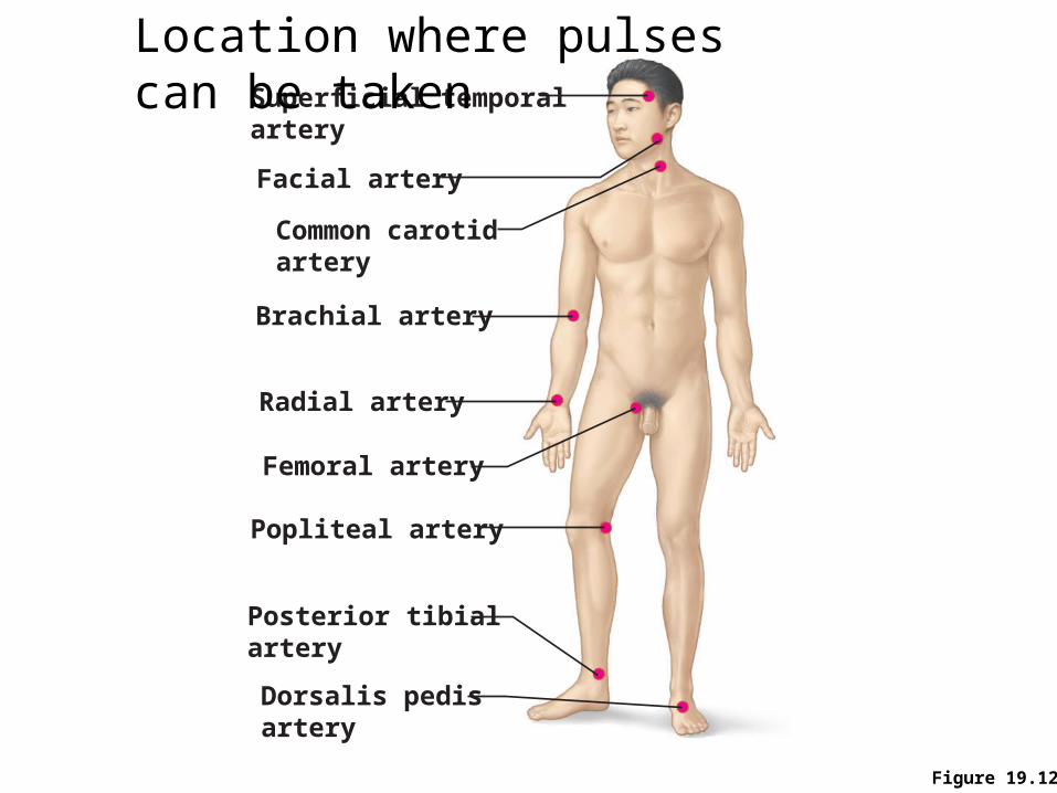
Figure 19.12
Common carotidartery
Brachial artery
Radial artery
Femoral artery
Popliteal artery
Posterior tibialartery
Dorsalis pedisartery
Superficial temporalartery
Facial artery
Location where pulses can be taken

MAP = CO x SVR• CO = HR x SV• SV = EDV – ESV • (EDV concerned with blood volume and ESV
concerned more with inotropic effect)• SVR = ∑R₁ + R₂ + 1/R₃ + 1/R₄ …..• R = 8ŋL/∏r⁴• In order to live – the body compensates by
increasing the actions of the organs not affected (homeostasis – negative feedback)

Widespread versus Local Control
• Widespread control affects Mean Arterial Pressure (MAP) in the entire Systemic Circulation- this control is mediated through the Nervous and Hormonal Systems
• Local Control affects MAP in localized tissues and organs – it generally does not affect overall blood pressure – organs possessing good local control mechanisms are the brain, heart, skin, lungs, skeletal muscles
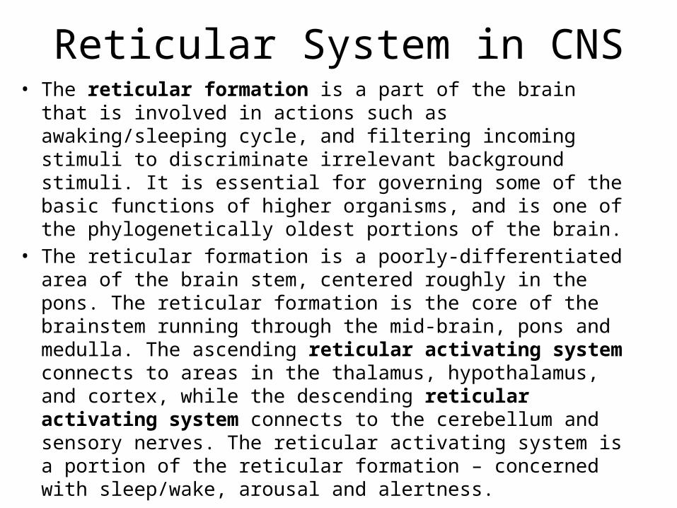
Reticular System in CNS• The reticular formation is a part of the brain that is involved in
actions such as awaking/sleeping cycle, and filtering incoming stimuli to discriminate irrelevant background stimuli. It is essential for governing some of the basic functions of higher organisms, and is one of the phylogenetically oldest portions of the brain.
• The reticular formation is a poorly-differentiated area of the brain stem, centered roughly in the pons. The reticular formation is the core of the brainstem running through the mid-brain, pons and medulla. The ascending reticular activating system connects to areas in the thalamus, hypothalamus, and cortex, while the descending reticular activating system connects to the cerebellum and sensory nerves. The reticular activating system is a portion of the reticular formation – concerned with sleep/wake, arousal and alertness.
Reticular System (Functions) 1. Somatic motor control - Some motor neurons send
their axons to the reticular formation nuclei, giving rise to the reticulospinal tracts of the spinal cord. These tracts function in maintaining tone, balance, and posture--especially during body movements.
Other motor nuclei include gaze centers, which enable the eyes to track and fixate objects, and central pattern generators, which produce rhythmic signals to the muscles of breathing and swallowing
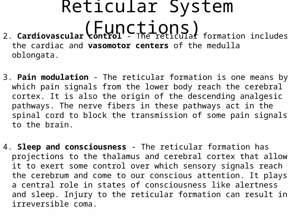
Reticular System (Functions) 2. Cardiovascular control - The reticular formation includes the cardiac
and vasomotor centers of the medulla oblongata.
3. Pain modulation - The reticular formation is one means by which pain signals from the lower body reach the cerebral cortex. It is also the origin of the descending analgesic pathways. The nerve fibers in these pathways act in the spinal cord to block the transmission of some pain signals to the brain.
4. Sleep and consciousness - The reticular formation has projections to the thalamus and cerebral cortex that allow it to exert some control over which sensory signals reach the cerebrum and come to our conscious attention. It plays a central role in states of consciousness like alertness and sleep. Injury to the reticular formation can result in irreversible coma.
Reticular System (Functions)
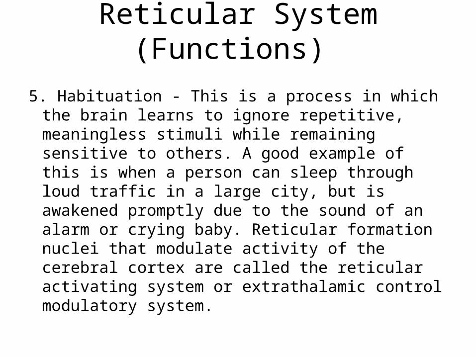
5. Habituation - This is a process in which the brain learns to ignore repetitive, meaningless stimuli while remaining sensitive to others. A good example of this is when a person can sleep through loud traffic in a large city, but is awakened promptly due to the sound of an alarm or crying baby. Reticular formation nuclei that modulate activity of the cerebral cortex are called the reticular activating system or extrathalamic control modulatory system.

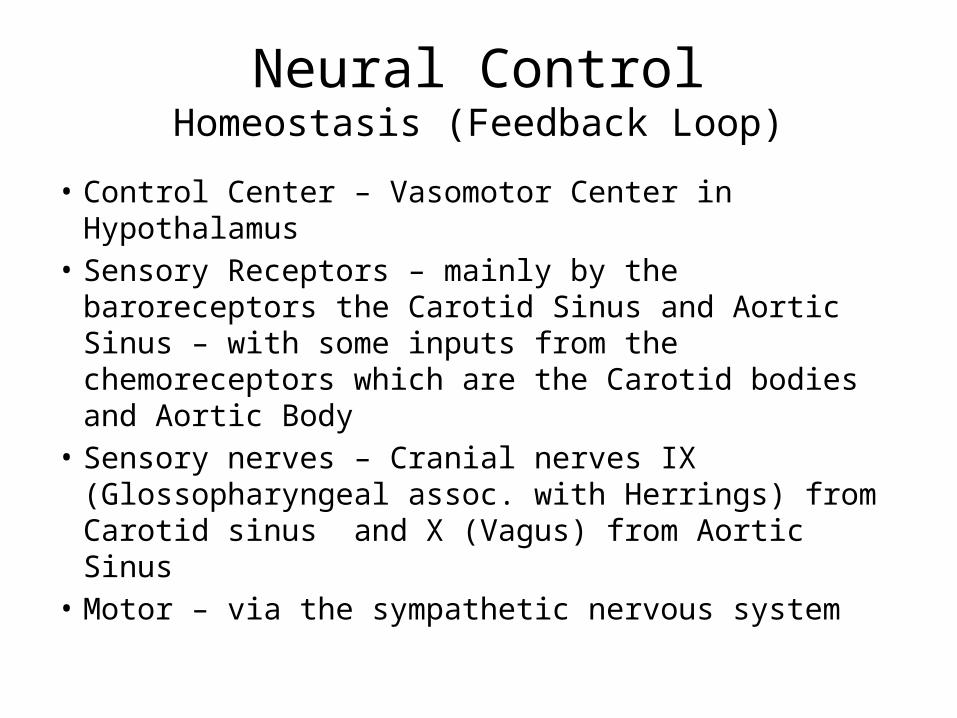
Neural ControlHomeostasis (Feedback Loop)
• Control Center – Vasomotor Center in Hypothalamus
• Sensory Receptors – mainly by the baroreceptors the Carotid Sinus and Aortic Sinus – with some inputs from the chemoreceptors which are the Carotid bodies and Aortic Body
• Sensory nerves – Cranial nerves IX (Glossopharyngeal assoc. with Herrings) from Carotid sinus and X (Vagus) from Aortic Sinus
• Motor – via the sympathetic nervous system
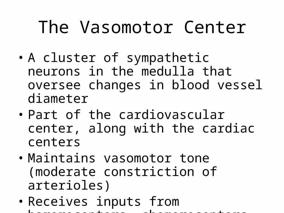
The Vasomotor Center
• A cluster of sympathetic neurons in the medulla that oversee changes in blood vessel diameter
• Part of the cardiovascular center, along with the cardiac centers
• Maintains vasomotor tone (moderate constriction of arterioles)
• Receives inputs from baroreceptors, chemoreceptors, and higher brain centers
Short-Term Mechanisms: Baroreceptor-Initiated Reflexes
• Baroreceptors are located in– Carotid sinuses– Aortic arch– Walls of large arteries of the neck and thorax
Short-Term Mechanisms: Baroreceptor-Initiated Reflexes
• Baroreceptors taking part in the carotid sinus reflex protect the blood supply to the brain
• Baroreceptors taking part in the aortic reflex help maintain adequate blood pressure in the systemic circuit
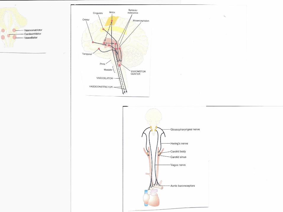
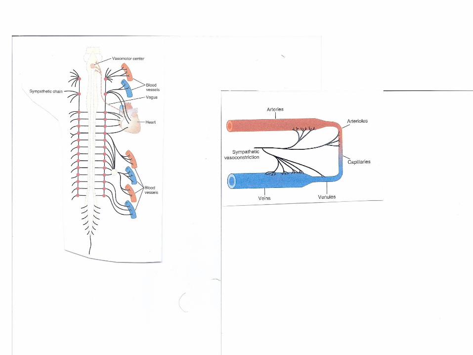
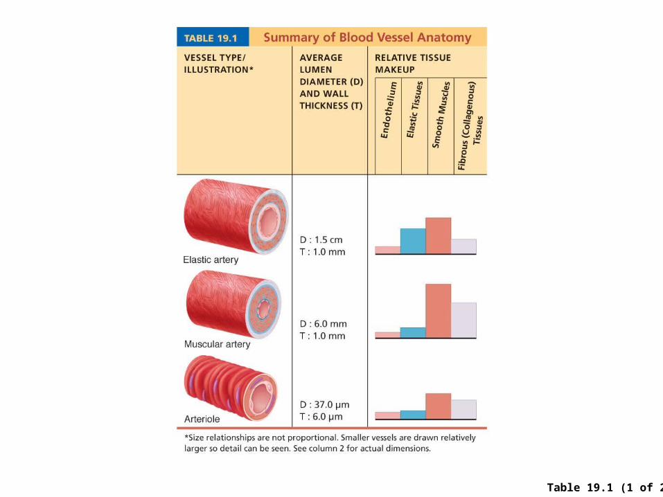
Table 19.1 (1 of 2)
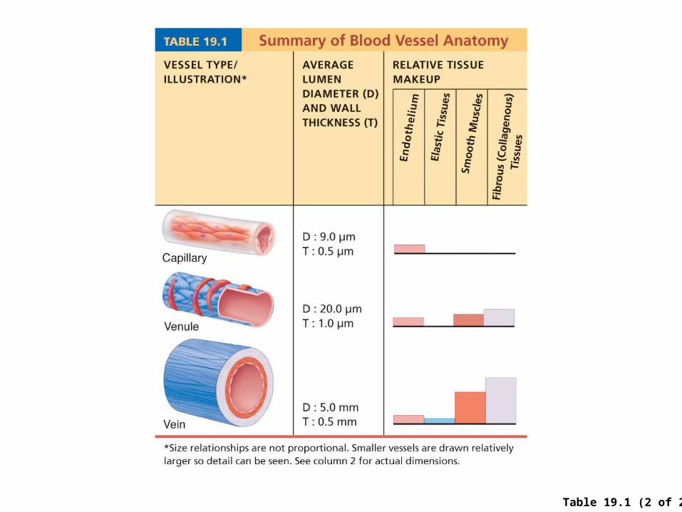
Table 19.1 (2 of 2)
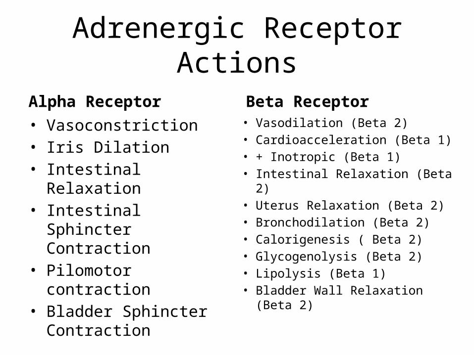
Adrenergic Receptor Actions
Alpha Receptor• Vasoconstriction• Iris Dilation• Intestinal Relaxation• Intestinal Sphincter
Contraction• Pilomotor contraction• Bladder Sphincter
Contraction
Beta Receptor• Vasodilation (Beta 2)• Cardioacceleration (Beta 1)• + Inotropic (Beta 1)• Intestinal Relaxation (Beta 2)• Uterus Relaxation (Beta 2)• Bronchodilation (Beta 2)• Calorigenesis ( Beta 2)• Glycogenolysis (Beta 2)• Lipolysis (Beta 1)• Bladder Wall Relaxation (Beta 2)
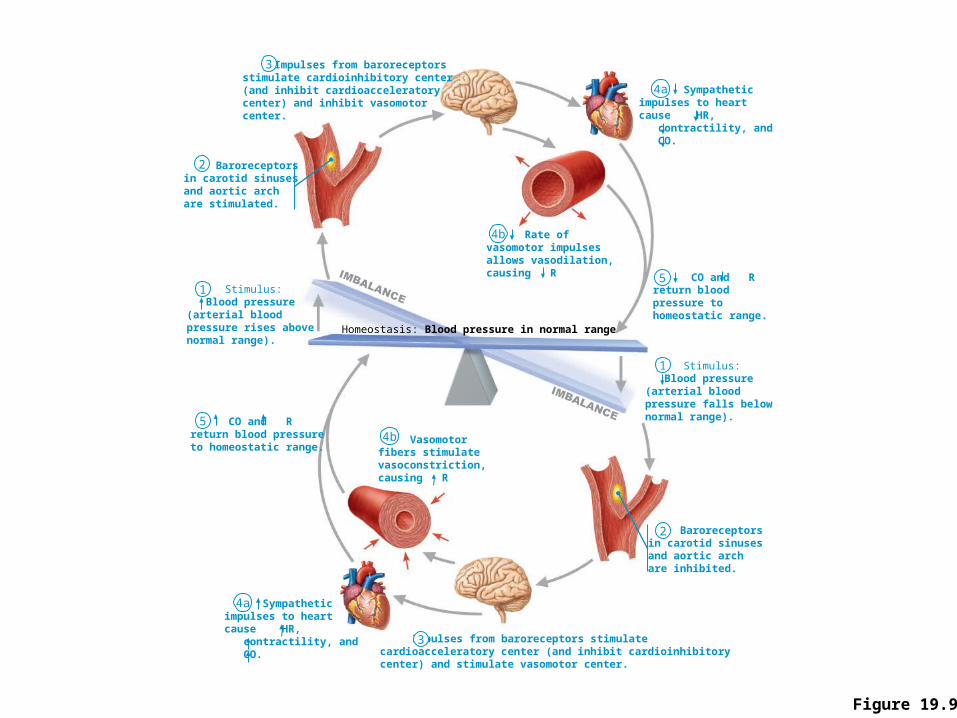
Figure 19.9
Baroreceptors in carotid sinusesand aortic archare stimulated.
Baroreceptorsin carotid sinusesand aortic archare inhibited.
Impulses from baroreceptorsstimulate cardioinhibitory center(and inhibit cardioacceleratorycenter) and inhibit vasomotorcenter.
Impulses from baroreceptors stimulatecardioacceleratory center (and inhibit cardioinhibitorycenter) and stimulate vasomotor center.
CO and Rreturn bloodpressure tohomeostatic range.
CO and Rreturn blood pressureto homeostatic range.
Rate ofvasomotor impulsesallows vasodilation,causing R
Vasomotorfibers stimulatevasoconstriction,causing R
Sympatheticimpulses to heartcause HR, contractility, and CO.
Sympatheticimpulses to heartcause HR, contractility, and CO.
Stimulus: Blood pressure(arterial bloodpressure falls belownormal range).
Stimulus: Blood pressure(arterial bloodpressure rises abovenormal range).
3
2
1
5
4a
4b
Homeostasis: Blood pressure in normal range
4b
3
2
1
5
4a
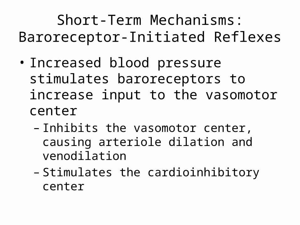
Short-Term Mechanisms: Baroreceptor-Initiated Reflexes
• Increased blood pressure stimulates baroreceptors to increase input to the vasomotor center– Inhibits the vasomotor center, causing arteriole
dilation and venodilation– Stimulates the cardioinhibitory center
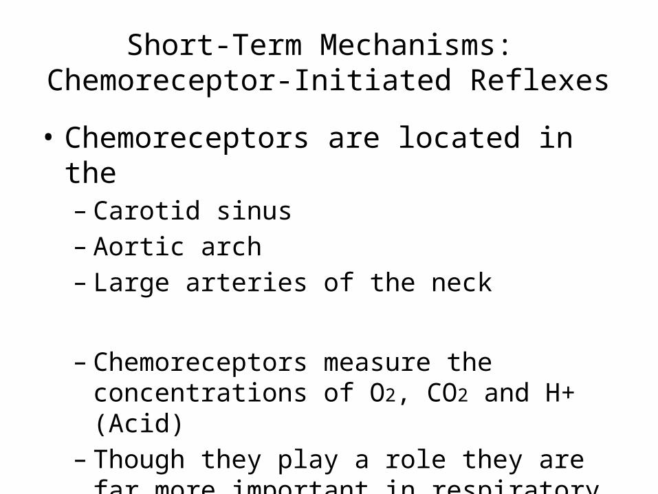
Short-Term Mechanisms: Chemoreceptor-Initiated Reflexes
• Chemoreceptors are located in the– Carotid sinus– Aortic arch– Large arteries of the neck
– Chemoreceptors measure the concentrations of O2, CO2 and H+ (Acid)
– Though they play a role they are far more important in respiratory control
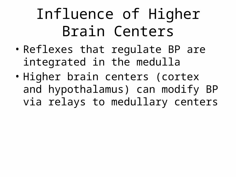
Influence of Higher Brain Centers
• Reflexes that regulate BP are integrated in the medulla
• Higher brain centers (cortex and hypothalamus) can modify BP via relays to medullary centers
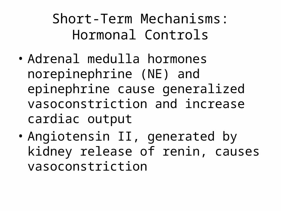
Short-Term Mechanisms: Hormonal Controls
• Adrenal medulla hormones norepinephrine (NE) and epinephrine cause generalized vasoconstriction and increase cardiac output
• Angiotensin II, generated by kidney release of renin, causes vasoconstriction
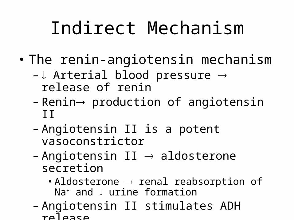
Indirect Mechanism
• The renin-angiotensin mechanism– Arterial blood pressure release of renin– Renin production of angiotensin II – Angiotensin II is a potent vasoconstrictor– Angiotensin II aldosterone secretion
• Aldosterone renal reabsorption of Na+ and urine formation
– Angiotensin II stimulates ADH release
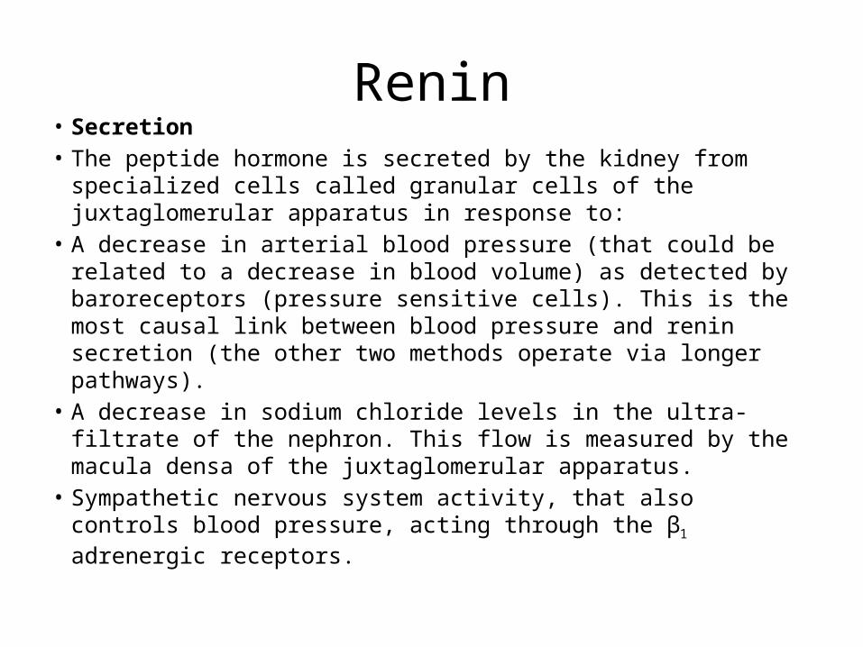
Renin• Secretion• The peptide hormone is secreted by the kidney from specialized
cells called granular cells of the juxtaglomerular apparatus in response to:
• A decrease in arterial blood pressure (that could be related to a decrease in blood volume) as detected by baroreceptors (pressure sensitive cells). This is the most causal link between blood pressure and renin secretion (the other two methods operate via longer pathways).
• A decrease in sodium chloride levels in the ultra-filtrate of the nephron. This flow is measured by the macula densa of the juxtaglomerular apparatus.
• Sympathetic nervous system activity, that also controls blood pressure, acting through the β1 adrenergic receptors.
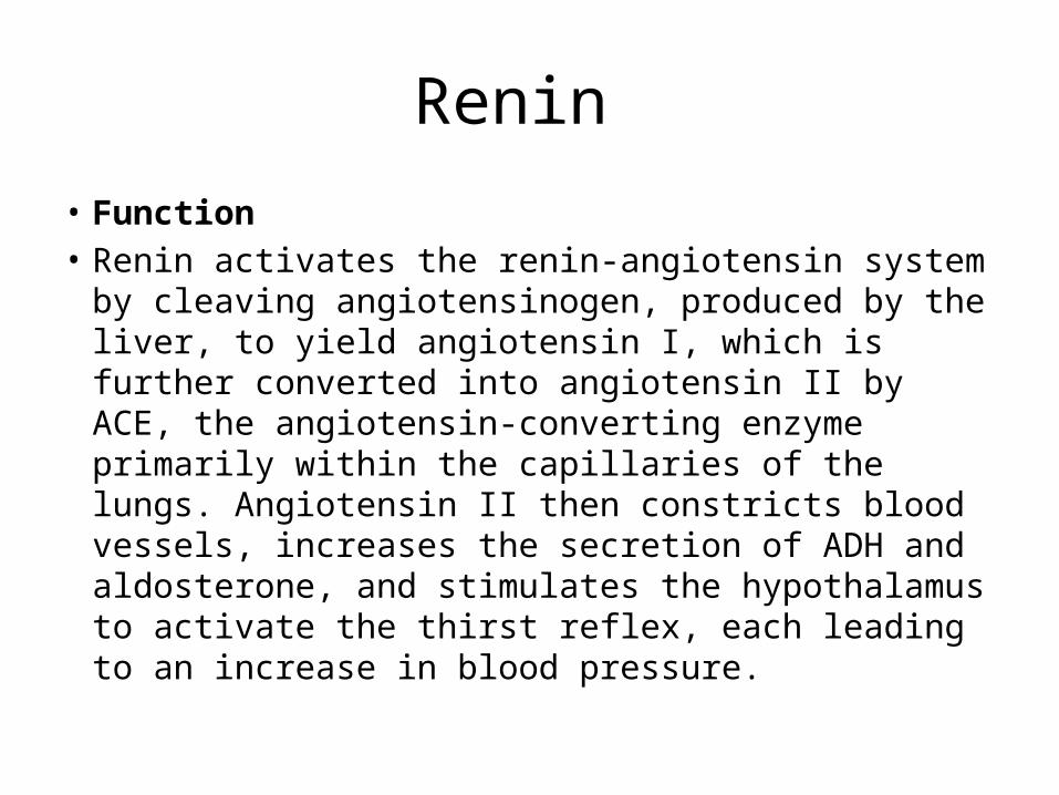
Renin
• Function• Renin activates the renin-angiotensin system by
cleaving angiotensinogen, produced by the liver, to yield angiotensin I, which is further converted into angiotensin II by ACE, the angiotensin-converting enzyme primarily within the capillaries of the lungs. Angiotensin II then constricts blood vessels, increases the secretion of ADH and aldosterone, and stimulates the hypothalamus to activate the thirst reflex, each leading to an increase in blood pressure.
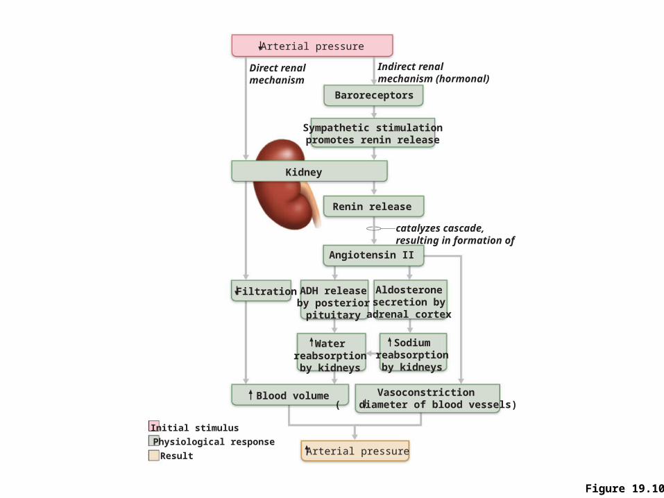
Figure 19.10
Arterial pressure
Baroreceptors
Indirect renalmechanism (hormonal)
Direct renalmechanism
Sympathetic stimulationpromotes renin release
Kidney
Renin release
catalyzes cascade,resulting in formation of
ADH releaseby posterior
pituitary
Aldosteronesecretion by
adrenal cortex
Waterreabsorptionby kidneys
Blood volume
Filtration
Arterial pressure
Angiotensin II
Vasoconstriction( diameter of blood vessels)
Sodiumreabsorptionby kidneys
Initial stimulus
Physiological response
Result
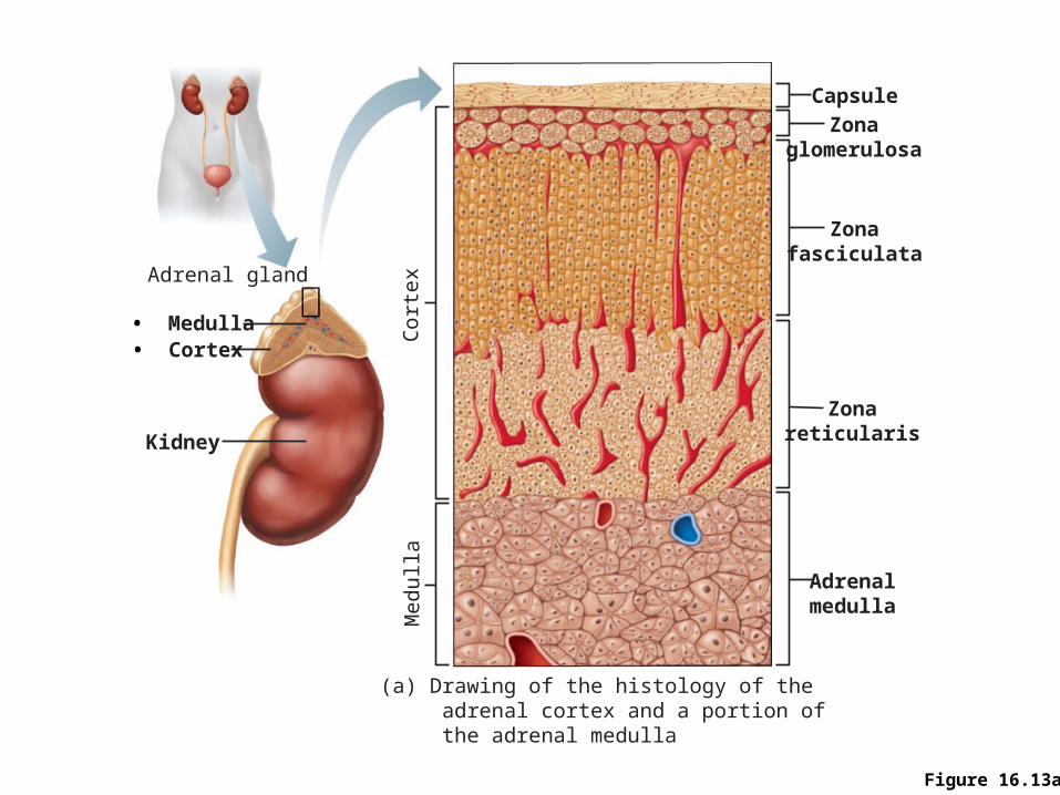
Figure 16.13a
• Cortex
Kidney
• Medulla
Adrenal gland
Capsule
Zonaglomerulosa
Zonafasciculata
Zonareticularis
Adrenalmedulla
(a) Drawing of the histology of the adrenal cortex and a portion of the adrenal medulla
Med
ulla
Cort
ex
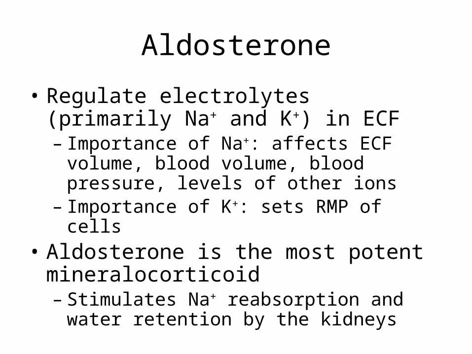
Aldosterone
• Regulate electrolytes (primarily Na+ and K+) in ECF– Importance of Na+: affects ECF volume, blood
volume, blood pressure, levels of other ions– Importance of K+: sets RMP of cells
• Aldosterone is the most potent mineralocorticoid – Stimulates Na+ reabsorption and water retention
by the kidneys
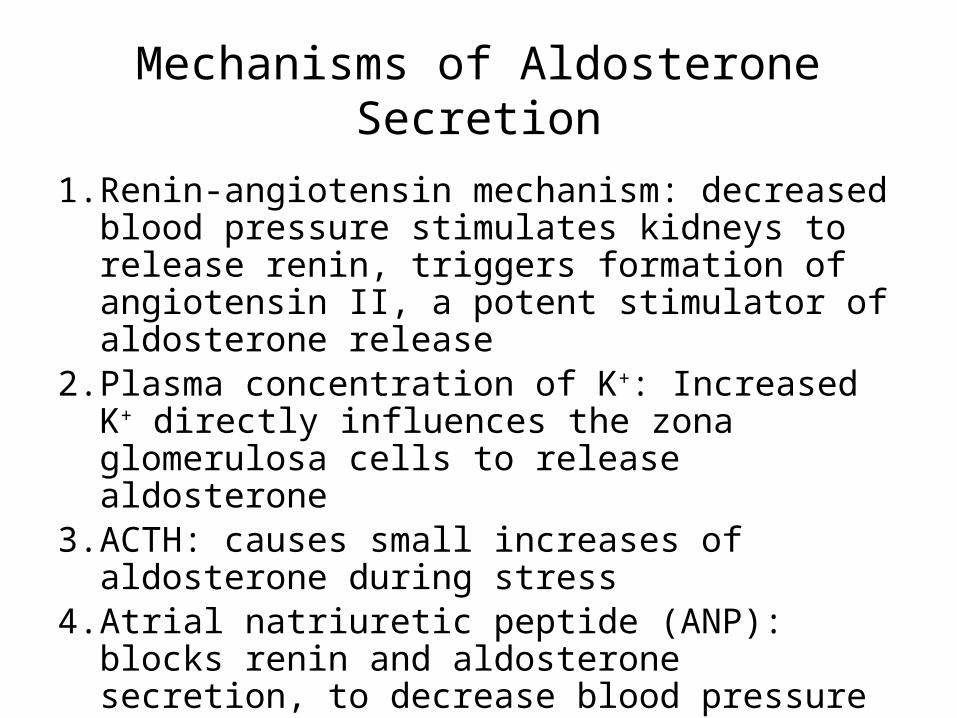
Mechanisms of Aldosterone Secretion
1. Renin-angiotensin mechanism: decreased blood pressure stimulates kidneys to release renin, triggers formation of angiotensin II, a potent stimulator of aldosterone release
2. Plasma concentration of K+: Increased K+ directly influences the zona glomerulosa cells to release aldosterone
3. ACTH: causes small increases of aldosterone during stress
4. Atrial natriuretic peptide (ANP): blocks renin and aldosterone secretion, to decrease blood pressure
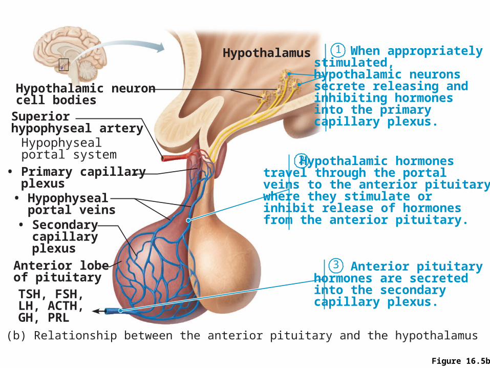
Figure 16.5b
1
2
3
When appropriatelystimulated, hypothalamic neurons secrete releasing and inhibiting hormones into the primary capillary plexus.
Hypothalamic hormones travel through the portal veins to the anterior pituitary where they stimulate or inhibit release of hormones from the anterior pituitary.
Anterior pituitaryhormones are secreted into the secondary capillary plexus.
Hypothalamus
Hypothalamic neuroncell bodies
Hypophysealportal system
Superiorhypophyseal artery
(b) Relationship between the anterior pituitary and the hypothalamus
Anterior lobeof pituitaryTSH, FSH, LH, ACTH, GH, PRL
• Primary capillary plexus• Hypophyseal portal veins• Secondary capillary plexus
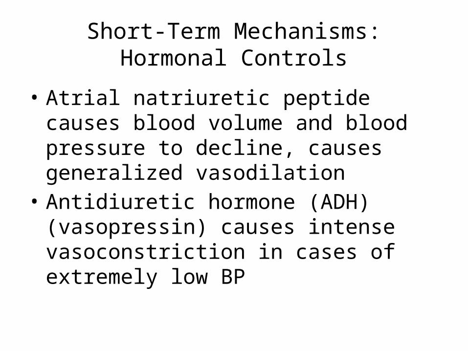
Short-Term Mechanisms: Hormonal Controls
• Atrial natriuretic peptide causes blood volume and blood pressure to decline, causes generalized vasodilation
• Antidiuretic hormone (ADH)(vasopressin) causes intense vasoconstriction in cases of extremely low BP
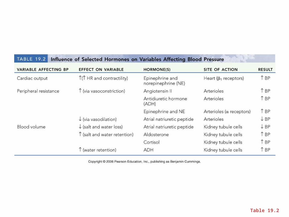
Table 19.2
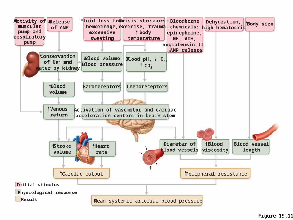
Figure 19.11
Activity ofmuscularpump andrespiratory
pump
Releaseof ANP
Fluid loss fromhemorrhage,
excessivesweating
Crisis stressors:exercise, trauma,
bodytemperature
Bloodbornechemicals:
epinephrine,NE, ADH,
angiotensin II; ANP release
Body size
Conservationof Na+ and
water by kidney
Blood volumeBlood pressure
Blood pH, O2, CO2
Dehydration,high hematocrit
Bloodvolume
Baroreceptors Chemoreceptors
Venousreturn
Activation of vasomotor and cardiacacceleration centers in brain stem
Heartrate
Strokevolume
Diameter ofblood vessels
Cardiac output
Initial stimulus
Result
Physiological response
Mean systemic arterial blood pressure
Bloodviscosity
Peripheral resistance
Blood vessellength
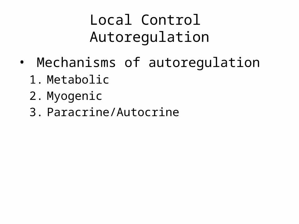
Local Control Autoregulation
• Mechanisms of autoregulation1. Metabolic2. Myogenic3. Paracrine/Autocrine
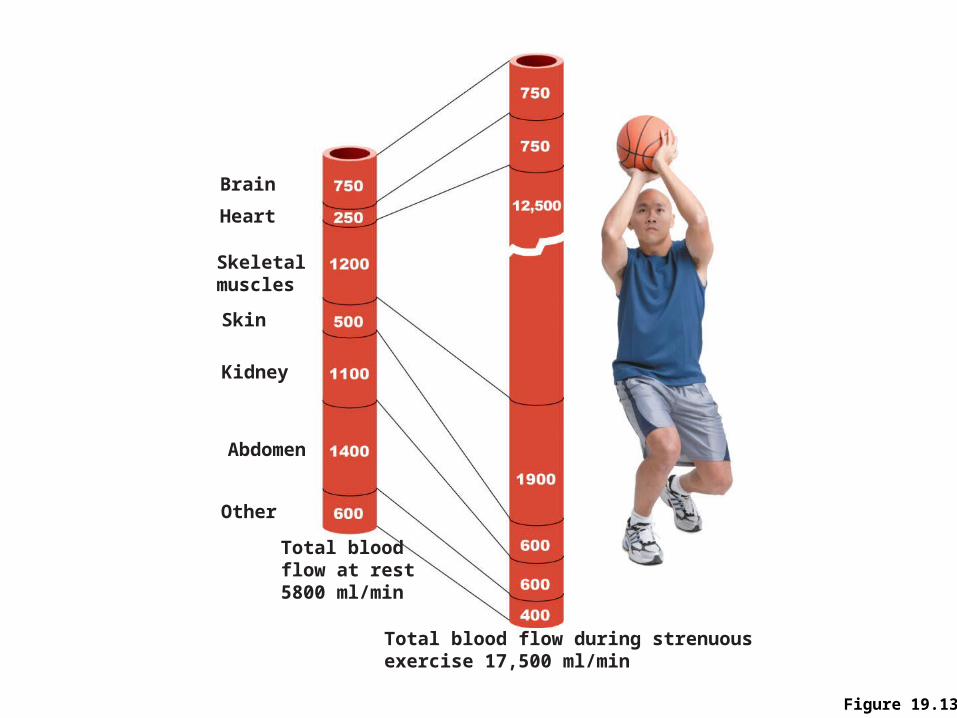
Figure 19.13
Brain
Heart
Skeletalmuscles
Skin
Kidney
Abdomen
Other
Total blood flow during strenuousexercise 17,500 ml/min
Total bloodflow at rest5800 ml/min
Autoregulation
• Automatic adjustment of blood flow to each tissue in proportion to its requirements at any given point in time
• Is controlled intrinsically by modifying the diameter of local arterioles feeding the capillaries
• Is independent of MAP, which is controlled as needed to maintain constant pressure
Blood Flow Through Body Tissues
• Blood flow (tissue perfusion) is involved in– Delivery of O2 and nutrients to, and removal of
wastes from, tissue cells – Gas exchange (lungs)– Absorption of nutrients (digestive tract)– Urine formation (kidneys)
• Rate of flow is precisely the right amount to provide for proper function
Active versus Reactive Hyperemia• Functional hyperemia, or active hyperemia, is the increased blood
flow that occurs when tissue is active.• When cells within the body are active in one way or another, they use
more oxygen and fuel, such as glucose or fatty acids, than when they are not. The blood vessels compensate for this metabolism by dilatation, allowing more blood to reach the tissue. This prevents deprivation of the tissue.
• Since most of the common nutrients in the body are converted to carbon dioxide when they are metabolized, smooth muscle around blood vessels relax in response to increased concentrations of carbon dioxide within the blood and surrounding interstitial fluid. The relaxation of this smooth muscle results in vascular dilation and increased blood flow.
• Reactive hyperemia is the transient increase in organ blood flow that occurs following a brief period of ischemia . Following Ischemia there will be a shortage of oxygen and a build-up of metabolic waste.
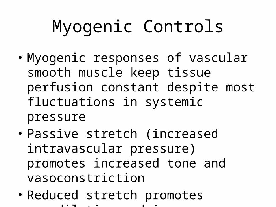
Myogenic Controls
• Myogenic responses of vascular smooth muscle keep tissue perfusion constant despite most fluctuations in systemic pressure
• Passive stretch (increased intravascular pressure) promotes increased tone and vasoconstriction
• Reduced stretch promotes vasodilation and increases blood flow to the tissue
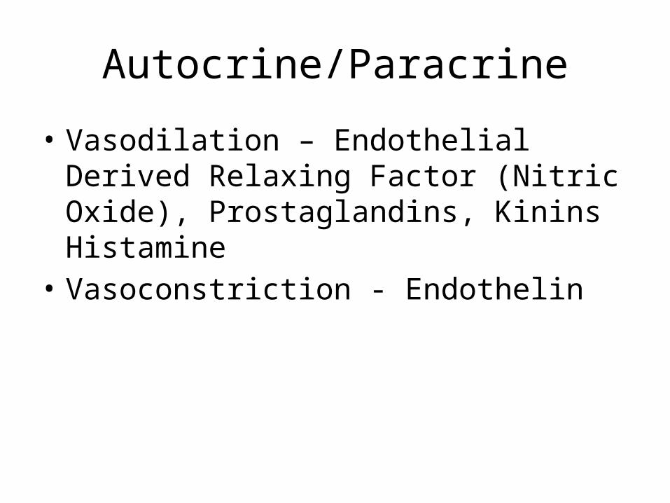
Autocrine/Paracrine
• Vasodilation – Endothelial Derived Relaxing Factor (Nitric Oxide), Prostaglandins, Kinins Histamine
• Vasoconstriction - Endothelin
Metabolites
• H+, CO2, Adenosine, K+
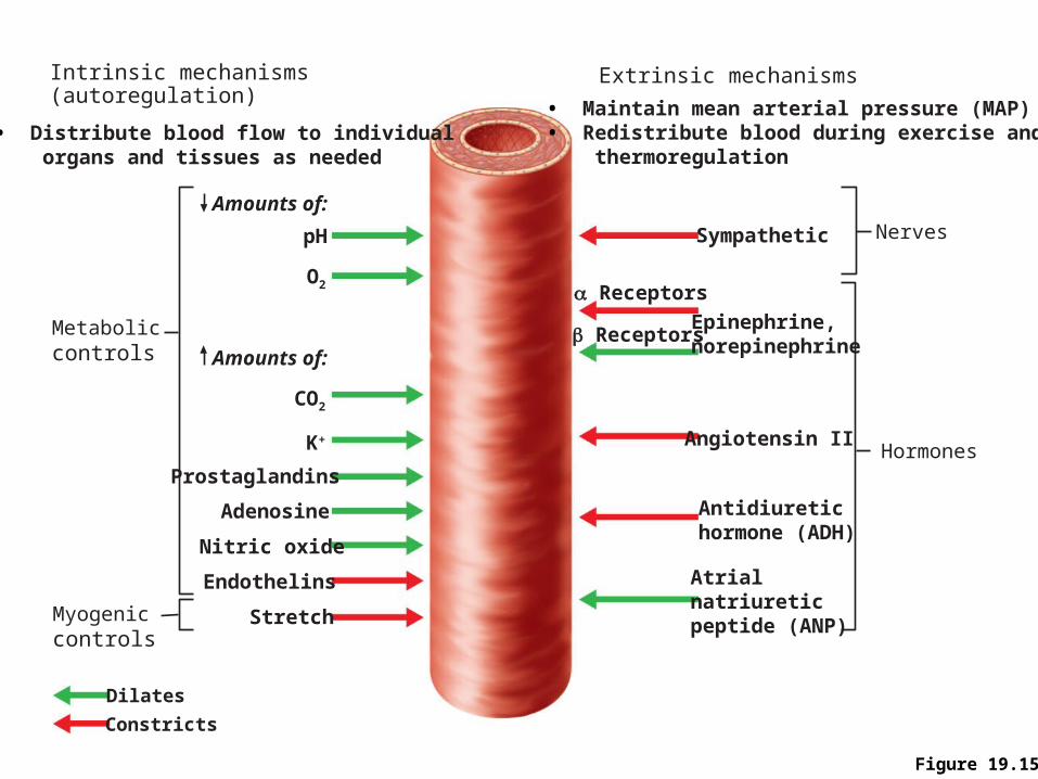
Figure 19.15
Metaboliccontrols
pH Sympathetic
a Receptors
b ReceptorsEpinephrine,norepinephrine
Angiotensin II
Antidiuretichormone (ADH)
Atrialnatriureticpeptide (ANP)
Dilates
Constricts
Prostaglandins
Adenosine
Nitric oxide
Endothelins
Stretch
O2
CO2
K+
Amounts of:
Amounts of:
Nerves
Hormones
Myogeniccontrols
Intrinsic mechanisms(autoregulation)
• Distribute blood flow to individual organs and tissues as needed
Extrinsic mechanisms
• Maintain mean arterial pressure (MAP)• Redistribute blood during exercise and thermoregulation
Long-Term Autoregulation
• Angiogenesis– Occurs when short-term autoregulation cannot
meet tissue nutrient requirements– The number of vessels to a region increases and
existing vessels enlarge – Common in the heart when a coronary vessel is
occluded, or throughout the body in people in high-altitude areas

Blood Flow: Skeletal Muscles
• At rest, myogenic and general neural mechanisms predominate
• During muscle activity– Blood flow increases in direct proportion to the
metabolic activity (active or exercise hyperemia)– Local controls override sympathetic vasoconstriction
• Muscle blood flow can increase 10 or more during physical activity
Blood Flow: Brain
• Blood flow to the brain is constant, as neurons are intolerant of ischemia
• Metabolic controls– Declines in pH, and increased carbon dioxide cause
marked vasodilation• Myogenic controls
– Decreases in MAP cause cerebral vessels to dilate – Increases in MAP cause cerebral vessels to constrict
Blood Flow: Brain
• The brain is vulnerable under extreme systemic pressure changes – MAP below 60 mm Hg can cause syncope
(fainting)– MAP above 160 can result in cerebral edema
Blood Flow: Skin
• Blood flow through the skin– Supplies nutrients to cells (autoregulation in
response to O2 need)– Helps maintain body temperature (neurally
controlled) – Provides a blood reservoir (neurally controlled)
Blood Flow: Skin
• Blood flow to venous plexuses below the skin surface– Varies from 50 ml/min to 2500 ml/min, depending
on body temperature– Is controlled by sympathetic nervous system
reflexes initiated by temperature receptors and the central nervous system

Temperature Regulation
• As temperature rises (e.g., heat exposure, fever, vigorous exercise)– Hypothalamic signals reduce vasomotor
stimulation of the skin vessels– Heat radiates from the skin
Temperature Regulation
• Sweat also causes vasodilation via bradykinin in perspiration– Bradykinin stimulates the release of NO
• As temperature decreases, blood is shunted to deeper, more vital organs
Blood Flow: Lungs
• Pulmonary circuit is unusual in that– The pathway is short– Arteries/arterioles are more like veins/venules
(thin walled, with large lumens)– Arterial resistance and pressure are low (24/8 mm
Hg)
Blood Flow: Lungs
• Autoregulatory mechanism is opposite of that in most tissues– Low O2 levels cause vasoconstriction; high levels
promote vasodilation– Allows for proper O2 loading in the lungs
Blood Flow: Heart
• During ventricular systole– Coronary vessels are compressed– Myocardial blood flow ceases– Stored myoglobin supplies sufficient oxygen
• At rest, control is probably myogenic
Blood Flow: Heart
• During strenuous exercise– Coronary vessels dilate in response to local
accumulation of vasodilators– Blood flow may increase three to four times