bmc bioinformatics - laboratoire d'informatique,...
TRANSCRIPT

This Provisional PDF corresponds to the article as it appeared upon acceptance. Thefully-formatted PDF version will become available shortly after the date of publication, from the
URL listed below.
Epigenetic acquisition of inducibility of type III cytotoxicity in P. aeruginosa
BMC Bioinformatics 2006, 7:272 doi:10.1186/1471-2105-7-272
Didier Filopon ([email protected])Annabelle Merieau ([email protected])
Gilles Bernot ([email protected])Jean Paul Comet ([email protected])
Rozenne Leberre ([email protected])Benoit Guery ([email protected])
Benoit Polack ([email protected])Janine Guespin-Michel ([email protected])
ISSN 1471-2105
Article type Research article
Submission date 25 November 2005
Acceptance date 30 May 2006
Publication date 30 May 2006
Article URL http://www.biomedcentral.com/1471-2105/7/272
Like all articles in BMC journals, this peer-reviewed article was published immediately uponacceptance. It can be downloaded, printed and distributed freely for any purposes (see copyright
notice below).
Articles in BMC journals are listed in PubMed and archived at PubMed Central.
For information about publishing your research in BMC journals or any BioMed Central journal, go to
http://www.biomedcentral.com/info/authors/
BMC Bioinformatics
© 2006 Filopon et al., licensee BioMed Central Ltd.This is an Open Access article distributed under the terms of the Creative Commons Attribution License (http://creativecommons.org/licenses/by/2.0),
which permits unrestricted use, distribution, and reproduction in any medium, provided the original work is properly cited.

1
Epigenetic acquisition of inducibility of type III cytotoxicity in P.
aeruginosa
Didier Filopon1, Annabelle Mérieau2, Gilles Bernot3, Jean-Paul Comet3, Rozenne LeBerre4, Benoit Guery4, Benoit Polack1§, Janine Guespin-Michel2 1 GREPI EA 2938 CHU de Grenoble, BP 217, 38043 Grenoble cedex 9, France. 2 Laboratoire de microbiologie du froid, EA 2123, Université de Rouen, F-76 821 Mt St Aignan, France 3 LaMI, CNRS UMR 8042, Université d'Évry-Val-d'Essonne, Boulevard François Mitterrand, 91025 Évry, France 4 EA 2689, Faculté de Médecine, Pole Recherche, CHRU de Lille, 1 Place de Verdun, 59045 Lille, France
§ Corresponding author Email addresses:
DF: [email protected] AM: [email protected] GB: [email protected] JPC: [email protected] RL: [email protected] BG: [email protected] BP: [email protected] JGM: [email protected]

2
Abstract
Background
Pseudomonas aeruginosa, an opportunistic pathogen, is often encountered in chronic
lung diseases such as cystic fibrosis or chronic obstructive pneumonia, as well as acute
settings like mechanical ventilation acquired pneumonia or neutropenic patients. It is a
major cause of mortality and morbidity in these diseases. In lungs, P. aeruginosa settles
in a biofilm mode of growth with the secretion of exopolysaccharides in which it is
encapsulated, enhancing its antibiotic resistance and contributing to the respiratory
deficiency of patients. However, bacteria must first multiply to a high density and
display a cytotoxic phenotype to avoid the host's defences. A virulence determinant
implicated in this step of infection is the type III secretion system (TTSS), allowing
toxin injection directly into host cells. At the beginning of the infection, most strains
isolated from patients’ lungs possess an inducible TTSS allowing toxins injection or
secretion upon in vivo or in vitro activation signals. As the infection persists most of the
bacteria permanently loose this capacity, although no mutations have been evidenced.
We name “non inducible” this phenotype. As suggested by the presence of a positive
feedback circuit in the regulatory network controlling TTSS expression, it may be due
to an epigenetic switch allowing heritable phenotypic modifications without genotype’s
mutations.
Result
Using the generalised logical method, we designed a minimal model of the TTSS
regulatory network that could support the epigenetic hypothesis, and studied its
dynamics which helped to define a discriminating experimental scenario sufficient to
validate the epigenetic hypothesis. A mathematical framework based on formal methods

3
from computer science allowed a rigorous validation and certification of parameters of
this model leading to epigenetic behaviour. Then, we demonstrated that a non inducible
strain of P. aeruginosa can stably acquire the capacity to be induced by calcium
depletion for the TTSS after a short pulse of a regulatory protein. Finally, the increased
cytotoxicity of a strain after this epigenetic switch was demonstrated in vivo in an acute
pulmonary infection model.
Conclusion
These results may offer new perspectives for therapeutic strategies to prevent lethal
infections by P. aeruginosa by reverting the epigenetic inducibility of type III
cytotoxicity.
Background
Pseudomonas aeruginosa is a Gram-negative opportunistic pathogen associated with
sepsis in burned, neutropenic, and intensive care patients as well as with severe chronic
lung injury in cystic fibrosis and chronic obstrusive pneumonia disease[1]. Cytotoxic P.
aeruginosa inject toxins from their cytoplasm into eukaryotic target cells through a
protein secretory apparatus, the type III secretion system (TTSS)[2]. Activation of this
system (especially toxins production and secretion) is dependant on the contact between
the bacteria and the host cells in vivo and could be triggered by calcium depletion of the
growth medium in vitro. TTSS is encoded by three classes of genes coding the secretion
apparatus, the toxins and the regulators. Four TTSS toxins are known in P. aeruginosa
to be secreted through the TTTS: ExoS, ExoT, ExoY and ExoU. All three classes of
TTSS genes are co-ordinately controlled by a common transcriptional activator ExsA
encoded by the exsCEBA operon, which controls its own synthesis[3]. The first gene of

4
the TTSS exsD-pscBL operon encodes ExsD which inhibits ExsA activity by forming
an inactive complex[4]. Fixation of ExsD to ExsA is prevented by ExsC, encoded by
the first gene of the exsCEBA operon, which interacts with ExsD[5,6]. Finally, ExsE,
encoded in the exsCEBA operon interacts with ExsC and prevents its binding to ExsD.
Upon TTSS activation by calcium depletion, ExsE is secreted through it, thus releasing
ExsC and the inhibition of ExsA by ExsD[6,7]. This complex regulatory network is
described in figure 1A. However, numerous strains possessing the entire set of genes
required for type III cytotoxicity cannot be induced by host contact or calcium depletion
unless submitted to exsA overexpression[8,9]. These strains will be named non
inducible for type III cytotoxicity to distinguish them from truly non cytotoxic ones,
such as mutants in TTSS. It has recently been shown that they accumulate in the lungs
of cystic fibrosis patients during long term infection.
Epigenetic phenotypic modifications arise and can be transmitted from a cell to its
progeny in the absence of any genetic modifications. As a consequence, several
phenotypes may arise from the same genome in the same conditions, which is
equivalent to the existence of multiple steady states according to physicist’s
terminology. Such a phenomenon has been under investigations for example in the
lactose metabolic network of Escherichia coli since 1957 [10-12]. Since all the genes
necessary for cytotoxic secretion are present and can be activated in non inducible P.
aeruginosa strains, this is consistent with the hypothesis that the transition from
inducibility to non inducibility of type III cytotoxicity is an epigenetic switch. Thomas
conjectured that a positive feedback circuit (comprising interacting elements, each of
which exerts, directly or indirectly, a positive action on itself) in a non linear dynamical
system, such as a regulatory network controlling a biological process, is a necessary

5
condition (although not sufficient) for the existence of multiple steady states[13,14].
This hypothesis has now been formally demonstrated[15,16]. Thus, the presence of the
positive feedback circuit in the TTSS regulatory network where ExsA positively
regulates it’s own synthesis, could lead to a bistable behaviour for the gene exsA (either
on or off) corresponding to an epigenetic switch of the TTSS (inducible or not)[17].
However, at least two intertwined feedback circuits control the production of ExsA: a
positive feedback circuit at the transcriptional level (ExsA is required for its own
synthesis) and a negative feedback circuit (ExsA is required for the synthesis of its
inhibitor) (Figure 1B). The existence of the positive circuit is a necessary, not sufficient,
condition for multistationarity and more particularly epigenetic bistable switches[13].
Consequently, one has to check if the system actually displays multistationarity.
Molecular techniques can not by themselves prove or refute this hypothesis. Indeed, the
absence of a mutation can be very difficult to demonstrate, since all possible genes in
which a mutation could induce such a phenotypic change may not be known.
Furthermore, the presence of a mutation in a putatively involved gene in inducible
strains may not mean that it is the initial cause of the phenotypic change: an epigenetic
switch could be the first event, followed by a mutation that may further stabilize it.
Mathematical analysis of the network’s dynamic is therefore necessary to determine if
the epigenetic hypothesis is coherent: this means if a reasonable model, which exhibits
two stable solutions, can be drawn. As the parameters of the system are not known, we
used generalized logics, a method derived from a Boolean approach[18,19].
The actual regulatory network to analyse is the one in figure 1A. However, proteins
ExsC and ExsE are modulators of protein ExsD activity, and can be omitted from the
model (Figure 1B) as far as the aim of this model is not to describe the whole regulatory

6
network, but only to address the question of the possibility of an epigenetic switch
between inducibility and non inducibility of the TTSS. Figure 1C shows the graph that
models the minimal regulatory network: vertices represent the biological entities (x=
ExsA, and y= ExsD) and lines their interactions (“–” for negative action and “+” for
positive action). The output (z) is the production of the secretory devices and toxins
secretion under induction by calcium depletion, i.e. inducibility. This graph is similar to
the one worked out for the regulation of mucoidy in the same bacteria[20]. This model,
a formal computer science approach, using model checking Computation Tree Logic
(CTL) and a dedicated framework SMBioNet[21], allowed us to fully establish the
consistency of the epigenetic hypothesis for the acquisition of a type III cytotoxicity and
to propose experimental evidences that are sufficient to prove it. We thus could design
in vivo and in vitro experiments to test it.
Results
Computer modelling
Generalised logical analysis.
Logical analysis relies on a simplification of the sigmoid curve representing the action
of a transcriptional activator as a step function, defining a threshold of concentration
above which the activation takes place[19]. In the case of the network shown in figure
1C, x has two distinct actions and it is very unlikely that the concentration thresholds
above which protein ExsA is active on the promoter of gene exsD and on the promoter
of its own gene are identical. Consequently, we associate two threshold values with x
and treat it as a three-level logical variable (0, 1, 2). Variable y has one action only and
is treated as a two-level logical variable (0, 1). Therefore, two different graphs must be
7
drawn depending on which promoter is the most sensitive to ExsA (Figure 2). For
simplicity, we consider only the two master elements, x and y.
Another aspect of the generalised logical method is that it allows analysis of the
dynamic properties of a network in terms of the "functionality" of its constitutive
feedback circuits. The effect of a circuit does not only depend on the mere existence of
the relevant interactions, but also on their relative strengths, which are noted as discrete
parameters (K). For a given regulatory network, the number of dynamics depends on the
number of thresholds and parameters[17]. In the case of the two graphs depicted in
figure 2, there are 324 different combinations of parameters for each graph. Each set of
parameters defines a specific temporal behaviour. Different set of values of the
parameters can lead or not to epigenetic behaviour.
The use of a Formal computer science method allows to select parameter set
that lead to a model exhibiting the hypothesised behaviour.
Finding suitable valuations for parameters constitutes a major issue for the modelling.
We runned the whole corpus of formal methods from computer science to include the
dynamical knowledge or hypotheses on the system[21]. Our method starts by
expressing the biological hypothesis (i.e. the epigenetic behaviour) formally, as formal
sentences which can be manipulated automatically by computer. Here we applied a
widely used temporal logic called Computation Tree Logic, because time plays a central
role in behavioural properties. Moreover, formal sentences in CTL can be automatically
checked against the 648 models by using a Model Checking algorithm. Technically,
behaviour is represented by a transition system (state graph), which can be
automatically computed from the regulatory network and the values of parameters.
Then, it becomes possible to extract (via the ‘brut force technique’) the sets of

8
parameters, if any, which lead to a behaviour that satisfies the CTL sentences. Model
Checking thereby classifies the set of possible dynamic behaviours into two groups:
those which satisfy the property and those which do not. This methodology is
instrumented by the software SMBioNet [21] which, for the given regulatory network,
automatically returns all sets of parameters which make the hypothesis coherent with
the model.
In the present case, the dynamical hypothesis (epigenetic hypothesis) can be translated
into two CTL. In the first, the non inducible phenotype is stable in most of the bacteria.
The sets of parameters to be considered for consistency must therefore induce a
behaviour where a non-cytotoxic bacterium (z=0) cannot subsequently become
cytotoxic, in the same conditions. This is formally expressed in temporal logic as: (z=0)
=> always (z=0). In the second CTL, the cytotoxic phenotype is stable. Thus, even in
the presence of the inhibitor (y) there is a state in which inducibility of the type III
cytotoxicity (z=1) is activated recurrently (i.e. z is activated in a recurrent way even if
the y�x inhibition is functionally active). This means that if at a given time the
bacterium has acquired the new phenotype, then later on, it will be again in the same
state. This is formally expressed in temporal logic as: (z=1) => Fs (z =1), where Fs
means “in a strict future”. The epigenetic hypothesis is consistent, if and only if, at least
one set of parameters leads to a behaviour satisfying these two formal properties. From
figure 2, z=1 requires that x=2. Consequently the epigenetic hypothesis means that it is
possible to make recurrent (x=2) and the previous formulae are equivalent to
[((x=2)=>Fs(x=2)) and ((x=0)=>always(not(x=2)))] as well as to [((x=2)=>Fs(z=1)) and
((x=0)=>always(z=0))]. SMBioNet formally proves that the epigenetic hypothesis is
consistent because 8 models satisfy these formulae, 2 of which are in agreement with

9
the known biological facts. The state graph, for one of these 2 models, is displayed in
figure 3.
Design of a discriminating experimental scenario.
In addition to proving the consistency of the epigenetic hypothesis, formal methods can
establish the discriminating power of the experimental scenario. The goal is to prove
both formal sentences in vivo. The two previous formulae reflect the existence of two
steady states. As indicated above, the first formula states that the basal level of x is a
steady state and is consequently satisfied in vivo which is true for a non inducible strain.
Thus, only the second formula remains to be experimentally validated. The second
formal sentence ((x=2) => Fs (z=1)) is always true when x ≠ 2 because, according to
the truth table, the implication (false => anything) is always true. Consequently it is
unnecessary to conduct an experiment where x ≠ 2. Therefore any experiment must start
by pushing the concentration of x to 2 which means to pulse x to saturation with an
external signal. Moreover Fs(z=1) signifies “wait a lapse of time to allow the system to
settle down and check if the bacterium has acquired a cytotoxic phenotype”. If the
bacterium has not changed its phenotype, then the experiment a priori fails.
Consequently the second part of the experimental scenario is rigorously sufficient to
check the second formal sentence. The length of this “lapse of time” must be
determined experimentally; here it stands for “as many generations later as possible”. If
the bacterium has acquired an inducible phenotype, that lasts at least several generations
after the external signal has been removed, then epigenesis is proven.
This formal approach was useful to suggest in vitro and in vivo experiments to test our
hypothesis. It showed that the two stable attractors differ only by the amount of ExsA

10
protein, and that, if the hypothesis is correct, no differences in the concentration of the
inhibitor ExsD can lead to a change in phenotype (Figure 3B). Thus, if a transient
increase in the amount of ExsA protein can shift the system from a non cytotoxic to a
stable cytotoxic state, the existence of the epigenetic switch would be demonstrated.
Experimental results
An ExsA pulse allows to recover a stable Type III Secretory phenotype
Experiments suggested by the formal method require pulsing ExsA by application of an
external stimulus and observing the change in phenotype (if any) and its stability when
the stimulus is removed. In order to pulse ExsA, we added an additional exsA gene,
under the control of an inducible promoter, to a non inducible P. aeruginosa strain
PAO1. Thus, we constructed plasmid pexsAind. In this construction, transcription of
exsA is under the control of the ptac promoter repressed by the LacI protein produced in
large amounts because of the presence of the lacIq gene[22]. Under inducing conditions
(presence of isopropyl-beta-D-thiogalactopyranoside, IPTG), transcriptional repression
by LacI is inhibited, permitting over-expression of the exsA gene from the ptac
promoter. This induction is immediately released when the medium is depleted of the
inducer. The most straightforward test to determine the resulting phenotype is the
electrophoretic detection of the type III toxins secreted in vitro after bacteria have been
submitted to calcium depletion.
Strain PAO1 used in this study was described as unable to induce death of human
polymorphonuclear neutrophils through the TTSS[23], and is non inducible (does not
produce type III toxins). This strain was transformed with pexsAind. In the absence of
inducer it produced no, or very little, toxin after 3 hours of culture in calcium-depleted

11
medium (Figure 4A, lane 3). Strain CHA, a cytotoxic strain that secretes large amounts
of toxins ExoS and ExoT[23], was used as a positive control (Figure 4A, lane 1). The
presence of IPTG during the 3 h of growth (not shown) or for a 20 min pulse before the
3 h culture (Figure 4A, lane 4) results in substantial toxin production. We observed this
epigenetic switch from a culture grown up to 6 hours (about 7 generations), in Luria
Bertani medium (LB) without EGTA after the ExsA pulse, before the leakiness of the
construct became a drawback (Figure 4B, lane 1 vs. 3). The secretory phenotype, under
calcium depletion, acquired by PAO1 (pexsAind) is therefore stable for about 10
generations, after the pulse of ExsA production from the additional gene on pexsAind.
In contrast, a CHA ExsA– mutant (CHA-D1) transformed with pexsAind did not
produce toxins in absence of IPTG nor after a 20 min pulse (Figure 4A, lane 6-7).
Secretion of type III toxins from this strain was only obtained when IPTG was present
in the medium all along the calcium depletion (Figure 4A, lane 8). This shows that
induction of toxin secretion by a 20 min pulse in strain PAO1 required the presence of
an active feedback loop on exsA. Transient production of ExsA is not able to promote
inducibility in the absence of an active exsA gene under the regulation of its own
autoactivated promoter. It also shows that this result was not an artefact. Indeed,
leakiness of the construct or noise, high production of protein ExsA upon a 20 min
IPTG pulse (the exsA gene is on a high-copy-number plasmid and under the control of a
strong promoter), or even persistence of some IPTG in the culture medium after 3 h of
growth cannot be responsible for toxin secretion. Whatever the remaining production or
presence of ExsA protein in the bacteria after four generations without IPTG, it is not
high enough to promote toxin secretion by itself. To confirm that TTSS inducibility
observed was correlated to ExsA expression we determined relative exsA mRNA level

12
using real time PCR under the same experimental conditions used above. Results are
indicated under corresponding lanes in figures 4A and 4B. Relative expression levels of
exsA in strains CHA, PAO1 (pexsAind) none pulsed or pulsed were respectively
14.3±2.3, 2.2±0.5 and 17.2±7.4 times higher than in strain PAO1 (Figure 4A). In
addition, after 10 generations following IPTG pulse, exsA level in strain PAO1
(pexsAind) was still 12.6±4.1 times higher than in strain PAO1 under calcium depletion
(Figure 4B). These results can be compared to that obtained using toxins secreted
through the TTSS in the same experimental conditions and show that the level of
secreted toxins is correlated to exsA expression in these conditions.
PAO1 (pexsAind) acquires an in vivo type III cytotoxicity after a transient
increase of ExsA.
A clinical correlation between type III secretory protein phenotype and lung injury
severity has previously been shown[24,25]. Indeed, unlike the wild type strain CHA, an
exsA null mutant induces no increase of protein concentration in the bronchoalveolar
lavage fluid (BALF) during the infection[26]. Therefore, we examined, through an acute
pulmonary infection model, the cytotoxicity of PAO1 carrying or not pexsAind. Strains
were pulsed or not with IPTG during 20 min, washed and then cultivated in tryptic soy
broth before infection. The injury of the alveolar capillary barrier was estimated by the
amount of proteins recovered in the BALF (Figure 5). A bacterial inoculum of 108 CFU/
ml, strain PAO1 (pexsAind) treated with an IPTG pulse, led to a higher quantity of
proteins (1.6±0.2 g/dL) compared to the wild type strain (0.4±0.03 g/dL) even if the
latter was injected at a ten times higher inoculum (1.04±0.21 g/dL for PAO1 at 2.109
CFU/ml, data not shown). The non pulsed PAO1 (pexsAind) strain showed protein level
13
ranging between the pulsed and the wild type strain which was significantly different.
But this strain showed no significant difference with the wild type strain injected at a
ten times higher inoculum. Although results obtained with the non pulsed strain PAO1
(pexsAind) remained unclear, this experiment illustrated the stable acquisition of
cytotoxicity by a transient increase of the main transcriptional regulator, ExsA.
Discussion
Despite a functional wild-type genomic background, some P.aeruginosa strains are
unable to develop a type III dependent cytotoxicity except with an over-expression, in
trans, of the transcriptional activator of this virulence system, ExsA[8]. We proposed to
call these strains non-inducible for type III cytotoxicity. This observation and the
presence of a positive feedback loop regulating the expression of exsA led us to
hypothesize the possibility of an epigenetic switch between inducibility/non inducibility
of the type III dependant cytotoxicity in P.aeruginosa. With the help of generalised
logic and a formal computer approach we designed a minimal model of P.aeruginosa
type III basic regulation under calcium depletion and established the consistency of the
hypothesis of an epigenetic acquisition of an inducible type III cytotoxicity. This model
also showed that in this case, and in this case only, an artificial (exogenous) transient
increase in protein ExsA would suffice to permanently switch a non inducible strain to
inducibility. Next, we engineered an exsA inducible gene. With this construction, we
demonstrated that a stable acquisition of the secretory phenotype after a transient signal
relies on the presence of the feedback loop of the auto regulated exsA gene in agreement
with the model. These non inducible strains thus switched to inducibility, also showed
the acquisition of in vivo inducible type III cytotoxicity. Since the exsA gene added in

14
trans was under the control of the inducible promoter, ptac, we have experienced
leakiness or noise. This phenomenon, ubiquitous in biological systems, is responsible
for cell-to-cell variation in gene expression[27]. It has been shown to be the cause of
population bimodal heterogeneity, when coupled with a feedback circuit responsible for
bistability, for instance in the repartition of lytic/temperate phages in the population
upon infection of E.coli by bacteriophage-λ[28]. In some individuals in the population
bearing pexsAind such a noise-dependant switch could be responsible for the slight
activity observed with non induced PAO1(pexsAind) strain in in vitro experiments
(Figure 4B, lane 3) and the in vivo increase of cytotoxicity of this strain. Moreover it
has been shown in vitro that metabolic state, which is probably modified in lungs,
regulates the percentage of cells able to induce type III secretion gene expression under
inducing conditions evidencing that only a subpopulation could induce type III gene
expression under inducing conditions[29]. Although this does not prove that this is the
mechanism involved to acquire or lose type III cytotoxicity during infection, the results
obtained in vivo make the epigenetic hypothesis very likely. However, this hypothesis
does not necessarily apply to the cytotoxicity of other bacteria, nor to mucoidy in P.
aeruginosa for which an epigenetic acquisition had been proposed[20]. In each case, the
hypothesis must be tested, first by modelling then experimentally.
Conclusion
These in vitro and in vivo experiments, in accordance to the predictions of the formal
method, indicate that a stable acquisition (or loss) of a phenotypic trait involved in the
pathogenicity of P. aeruginosa can arise from an epigenetic switch. Direct therapeutic
consequences could arise for P. aeruginosa infections especially in cystic fibrosis. Any

15
therapy inhibiting the type III regulon would reduce the pathogenicity of the bacteria
and the severity of the infection. Since the inducibility of type III secretory phenotype is
a bistable phenotype, a reduction of ExsA activity below the triggering threshold would
be sufficient to impede the positive feedback circuit leading to reversion of the type III
secretory phenotype and reduction of the pathogenicity of the bacteria. To do so, we
may investigate the use of quorum sensing signals as the Rhl quorum sensing system
represses TTSS expression[30,31] or the effect of small molecules able to block TTSS
toxins secretion as shown in Yersinia which could lead to a repression of the system due
to the absence of the secretion of the inhibitor ExsE[32]. According to the hypothesis
that planctonic type III secreting form of P. aeruginosa is responsible for early and
invasive phases of the infection[33-35], such a therapy would improve antibiotic
performance and other treatments to eradicate the infection or limit spreading.
Methods
Bacterial strains and growth conditions
Pseudomonas aeruginosa strains used in this study were: CHA, a cytotoxic isolate from
a cystic fibrosis patient[36] which secretes toxins under inducing condition for the type
III secretion system[23]; PAO1, a noncytotoxic strain widely used in laboratory studies;
PAO1 ExsA–, an isogenic mutant of PAO1 unable to synthesize ExsA. PAO1 ExsA–
was obtained using a mutation method based on sacB negative selection and cre-lox
antibiotic marker recycling[37]. Strains were routinely grown in Luria-Bertani broth
(LB), Tryptic Soy Broth (TSB) or plated on PIA (Pseudomonas Isolation Agar, Difco,
France). The pexsAind plasmid was introduced into PAO1 and its isogenic mutant by
electroporation and maintained with 300 µg of carbenicilin/ml.

16
Construction of an inducible exsA gene
The exsA gene was amplified by PCR using the high fidelity polymerase PfuUltra
(Stratagene) with P. aeruginosa chromosomal DNA as the template and primers
EXSAS and EXSAR (see table 1). Their 5’ termini contain EcoRI and HindIII
restriction sites, respectively. PCR products were ligated into pCR®-Blunt II-TOPO®
vector (Invitrogen). The EcoRI-HindIII exsA fragment from this plasmid was inserted
into pTTQ18[22], digested by the same enzymes. This inducible system was amplified
from pTTQ18exsA with the Xbaptac and KpnlacIq primers (see table 1), carrying XbaI
and KpnI restriction sites, respectively, and inserted into the vector pCR®-Blunt II-
TOPO®. For the final construct, pexsAind, the XbaI-KpnI fragment from the previous
vector containing exsA was ligated into pUCP20[38], a high-copy-number shuttle vector
for P. aeruginosa.
Reverse transcription
Total RNA were extracted after calcium depletion using HighPure RNA Extraction kit
(Roche) according to the manufacturer’s instructions. Reverse transcription was realized
using the Transcriptor reverse transcriptase (Roche) with 1,33µg total RNA, 1mM
dNTP and 1mM of primer 16sas or RTExsA (Table 1). Primer and RNA were mixed,
heated for 10 min at 65°C then dNTP, buffer and enzyme were added. cDNA were
obtained after a reaction conducted for 30 min at 55°C, followed by 5 min at 85°C.
Real time PCR
Real-time PCR assays with hydrolysis probes were conducted on a LightCycler
apparatus (Roche), using FastStart DNA Master Hybridization Probe kit (Roche) by

17
following the manufacturer’s instructions. Sequences of exsA gene and 16s rRNA gene
used has housekeeping gene were obtained from the P. aeruginosa genome sequence
from Pseudomonas Genome Project[39]. Primers and probes (table 1) were designed
and obtained from Proligo® (France) and were used at final concentrations of 0.75µM
and 0.2 µM, respectively. Final MgCl2 concentration was adjusted to 4mM. Volume of
cDNA from reverse transcription was 5 µl per assay. Thermal cycling conditions were
10 min at 95°C, followed by 45 cycles of 10 s at 95°C and 10s at 58°C. Relative
expression level of the exsA gene to the 16s rRNA gene was calculated using the
Lightcycler software 4.0 for each strain and reported to exsA relative expression of the
strain PAO1. Values are the mean (± standard error) of three experiments.
SDS PAGE
Bacteria were cultivated in LB broth overnight, diluted to 1.2x10⁸ cfu/ml in LB
supplemented or not supplemented with 1mM ITPG and incubated with aeration for 20
min at 37°C. Then, bacteria were spun down, washed twice in LB and grown with
aeration for 3 h at 37°C in the presence or absence of 1mM IPTG in conditions of
calcium depletion: LB medium supplemented with 5mM EGTA and 20mM MgCl2
(inducing conditions known to induce secretion of type III toxins in vitro[40]). Bacterial
densities were determined by optical density measurement at 600nm. Cultures were
spun down, and proteins in the supernatant were precipitated by perchloric acid
(precipitated volume was normalised to 9x10⁸ cfu) and washed with acetone. Proteins
were separated on a 12% SDS-PAGE, and stained with Coomassie blue. In the secretion
experiment with a delay between the IPTG pulse and calcium depletion (Figure 4B),
bacteria were pulsed with 1mM ITPG for 20 min then washed with LB. Next, they were

18
maintained in exponential phase of growth by serial dilution with fresh medium during
a definite time before calcium depletion.
Animals and infection model
Specific pathogen-free Sprague Dawley rats (n=40) (280-320g), (Charles River
Laboratoires France, St Germain/l’Arbresle, France) were housed in the Lille University
Animal Care Facility and allowed food and water ad lib. All experiments were
performed with approval of the Lille Institutional Animal Care and Use Committee. P.
aeruginosa strains were cultivated in Triptic Soy Broth (TSB) broth overnight, diluted
to 1.2x10⁸ cfu/ml in TSB supplemented or not supplemented with 1mM ITPG and
incubated with aeration for 20 min at 37°C. Cultures were then centrifuged, washed
twice and grown with aeration at 37°C in tryptic soy broth medium. After 3 hours,
bacteria were washed and resuspended in physiological serum to reach a final
concentration of 108 or 2x10⁹ cfu/ml evaluated by spectrophotometry. Acute lung injury
was produced according to the method described by Pennington and Ehrie[41]. Under
short anaesthesia, a small midline incision was made on the neck ventral surface after
swabbing it with ethanol. The trachea was exposed by blunt dissection. Using a 28-
gauge needle, 0.5 ml/kg of bacterial suspension was instilled into the trachea, followed
by injection of 0.5 ml of air[42,43]. The animals were studied 24 h after instillation of
the bacteria.
Bronchoalveolar lavage (BAL)
Bronchoalveolar lavage was performed by cannulating the trachea. Lungs from each
experimental group were washed with a total of 20 ml in 5-ml aliquots of PBS with 3
mM EDTA. The returned fluid was pooled and centrifuged (200g for 10 min). BAL
19
fluid (BALF) samples were filtered and immediately frozen at –80°C after collection.
Protein concentration in the BALF was measured with an automated analyzer (Hitachi
917, Japan).
Authors’ contributions
DF participated to the design of the study, carried out the microbiological and animal
experiments and drafted the manuscript. RL carried out animal experiments. BG carried
out animal experiments and drafted the manuscript. JFGM proposed the model while
GB and JPC designed and performed computer studies and draft the manuscript. AM,
BP, JFGM conceived the study and participated to its design and coordination and
drafted the manuscript. All authors read and approved the final manuscript.
Acknowledgements
This work was supported by a grant from the “association Vaincre la Mucoviscidose”.
We thank L. Quénée for technical assistance and Professors F. Morel and F. Molina,
and the members of the epigenomic workshop in genopole for helpful discussions.
References 1. Govan JR, Harris GS: Pseudomonas aeruginosa and cystic fibrosis: unusual
bacterial adaptation and pathogenesis. Microbiol Sci 1986, 3:302-308.
2. Hueck CJ: Type III protein secretion systems in bacterial pathogens of
animals and plants. Microbiol Mol Biol Rev 1998, 62:379-433.
3. Frank DW: The exoenzyme S regulon of Pseudomonas aeruginosa. Mol
Microbiol 1997, 26:621-629.
4. McCaw ML, Lykken GL, Singh PK, Yahr TL: ExsD is a negative regulator of
the Pseudomonas aeruginosa type III secretion regulon. Mol Microbiol 2002, 46:1123-1133.

20
5. Dasgupta N, Lykken GL, Wolfgang MC, Yahr TL: A novel anti-anti-activator
mechanism regulates expression of the Pseudomonas aeruginosa type III
secretion system. Mol Microbiol 2004, 53:297-308.
6. Rietsch A, Vallet-Gely I, Dove SL, Mekalanos JJ: ExsE, a secreted regulator
of type III secretion genes in Pseudomonas aeruginosa. Proc Natl Acad Sci U
S A 2005, 102:8006-8011.
7. Urbanowski ML, Lykken GL, Yahr TL: A secreted regulatory protein couples
transcription to the secretory activity of the Pseudomonas aeruginosa type
III secretion system. Proc Natl Acad Sci U S A 2005, 102:9930-9935.
8. Dacheux D, Attree I, Toussaint B: Expression of ExsA in trans confers type
III secretion system-dependent cytotoxicity on noncytotoxic Pseudomonas
aeruginosa cystic fibrosis isolates. Infect Immun 2001, 69:538-542.
9. Jain M, Ramirez D, Seshadri R, Cullina JF, Powers CA, Schulert GS, Bar-Meir M, Sullivan CL, McColley SA, Hauser AR: Type III secretion phenotypes of
Pseudomonas aeruginosa strains change during infection of individuals
with cystic fibrosis. J Clin Microbiol 2004, 42:5229-5237.
10. Novick A, Wiener M: Enzyme induction is an all-or-none phenomenon. Proc
Natl Acad Sci USA 1957, 43:553-556.
11. Cohn M, Horibata K: Inhibition by glucose of the induced synthesis of the ββββ-
galactoside-enzyme system of Eschercichia coli. Analysis of maintenance. J
Bacteriol 1959, 78:601-611.
12. Ozbudak EM, Thattai M, Lim HN, Shraiman BI, van Oudenaarden A: Multistability in the lactose utilization network of Escherichia coli. Nature 2004, 19;427:737-740.
13. Thomas R: On the relation between the logical structure of systems and
their ability to generate multiple steady states or sustained oscillations. Springer Series in Synergies 1980, 9:180-193.
14. Thomas R: Laws for the dynamics of regulatory networks. Int J Dev Biol 1998, 42:479-485.
15. Soulé C: Graphic requierement for multistationarity. ComPlexUs 2003, 1:123-133.
16. Angeli D, Ferrell JE, Jr., Sontag ED: Detection of multistability, bifurcations,
and hysteresis in a large class of biological positive-feedback systems. Proc
Natl Acad Sci U S A 2004, 101:1822-1827.
17. Kaufman M, Thomas R: Emergence of complex behaviour from simple
circuit structures. C R Biol 2003, 326:205-214.
21
18. Snoussi EH, Thomas R: Logical identification of all steady states: the concept
of feedback loop characteristics states. Bull Math Biol 1993, 57:277-297.
19. Thomas R, d'Ari R: Biological feedback. CRC Press; 1990.
20. Guespin-Michel JE, Kaufman M: Positive feedback circuits and adaptive
regulations in bacteria. Acta Biotheor 2001, 49:207-218.
21. Bernot G, Comet JP, Richard A, Guespin J: Application of formal methods to
biological regulatory networks: extending Thomas' asynchronous logical
approach with temporal logic. J Theor Biol 2004, 229:339-347.
22. Stark MJ: Multicopy expression vectors carrying the lac repressor gene for
regulated high-level expression of genes in Escherichia coli. Gene 1987, 51:255-267.
23. Dacheux D, Attree I, Schneider C, Toussaint B: Cell death of human
polymorphonuclear neutrophils induced by a Pseudomonas aeruginosa
cystic fibrosis isolate requires a functional type III secretion system. Infect
Immun 1999, 67:6164-6167.
24. Roy-Burman A, Savel RH, Racine S, Swanson BL, Revadigar NS, Fujimoto J, Sawa T, Frank DW, Wiener-Kronish JP: Type III Protein Secretion Is
Associated with Death in Lower Respiratory and Systemic Pseudomonas
aeruginosa Infections. J Infect Dis 2001, 183:1767-1774.
25. Hauser AR, Cobb E, Bodí M, Mariscal D, Vallés J, Engel JN, Rello J: Type III
protein secretion is associated with poor clinical outcomes in patients with
ventilator-associated pneumonia caused by Pseudomonas aeruginosa. Crit
Care Med 2002, 30:521-528.
26. Ader F, Le Berre R, Faure K, Gosset P, Epaulard O, Toussaint B, Polack B, Nowak E, Viget NB, Kipnis E, Guery BP: Alveolar Response to Pseudomonas
aeruginosa: Role of the Type III Secretion System. Infect Immun 2005, 73:4263-4271.
27. Ozbudak EM, Thattai M, Kurtser I, Grossman AD, van Oudenaarden A: Regulation of noise in the expression of a single gene. Nat Genet 2002, 31:69-73.
28. Arkin A, Ross J, McAdams HH: Stochastic kinetic analysis of developmental
pathway bifurcation in phage lambda-infected Escherichia coli cells. Genetics 1998, 149:1633-1648.
29. Rietsch A, Mekalanos JJ: Metabolic regulation of type III secretion gene
expression in Pseudomonas aeruginosa. Mol Microbiol 2006, 59:807-820.
30. Bleves S, Soscia C, Nogueira-Orlandi P, Lazdunski A, Filloux A: Quorum
sensing negatively controls type III secretion regulon expression in
Pseudomonas aeruginosa PAO1. J Bacteriol 2005, 187:3898-3902.

22
31. Hogardt M, Roeder M, Schreff AM, Eberl L, Heesemann J: Expression of
Pseudomonas aeruginosa exoS is controlled by quorum sensing and RpoS. Microbiology 2004, 150:843-851.
32. Nordfelth R, Kauppi AM, Norberg HA, Wolf-Watz H, Elofsson M: Small-
molecule inhibitors specifically targeting type III secretion. Infect Immun 2005, 73:3104-3114.
33. Hassett DJ, Cuppoletti J, Trapnell B, Lymar SV, Rowe JJ, Yoon SS, Hilliard GM, Parvatiyar K, Kamani MC, Wozniak DJ, Hwang SH, McDermott TR, Ochsner UA: Anaerobic metabolism and quorum sensing by Pseudomonas
aeruginosa biofilms in chronically infected cystic fibrosis airways:
rethinking antibiotic treatment strategies and drug targets. Adv Drug Deliv
Rev 2002, 54:1425-1443.
34. Vance RE, Rietsch A, Mekalanos JJ: Role of the type III secreted exoenzymes
S, T, and Y in systemic spread of Pseudomonas aeruginosa PAO1 in vivo. Infect Immun 2005, 73:1706-1713.
35. Corech R, Rao A, Laxova A, Moss J, Rock MJ, Li Z, Kosorok MR, Splaingard ML, Farrell PM, Barbieri JT: Early immune response to the components of
the type III system of Pseudomonas aeruginosa in children with cystic
fibrosis. J Clin Microbiol 2005, 43:3956-3962.
36. Toussaint B, Delic-Attree I, Vignais PM: Pseudomonas aeruginosa contains
an IHF-like protein that binds to the algD promoter. Biochem Biophys Res
Commun 1993, 196:416-421.
37. Quenee L, Lamotte D, Polack B: Combined sacB-based negative selection
and cre-lox antibiotic marker recycling for efficient gene deletion in
pseudomonas aeruginosa. Biotechniques 2005, 38:63-67.
38. West SEH, Schweizer HP, Dall C, Sample AK, Runyen-Janecky LJ: Construction of improved Escherichia-Pseudomonas shuttle vectors derived
from pUC18/19 and sequence of the region required for their replication in
Pseudomonas aeruginosa. Gene 1994, 128:81-86.
39. Pseudomonas Genome Project [http://www.pseudomonas.com]
40. Hovey AK, Frank DW: Analyses of the DNA-binding and transcriptional
activation properties of ExsA, the transcriptional activator of the
Pseudomonas aeruginosa exoenzyme S regulon. J Bacteriol 1995, 177:4427-4436.
41. Pennington JE, Ehrie MG: Pathogenesis of Pseudomonas aeruginosa
pneumonia during immunosuppression. J Infect Dis 1978, 137:764-774.
42. Viget NB, Guery BP, Ader F, Neviere R, Alfandari S, Creuzy C, Roussel-Delvallez M, Foucher C, Mason CM, Beaucaire G, Pittet JF: Keratinocyte

23
growth factor protects against Pseudomonas aeruginosa-induced lung
injury. Am J Physiol Lung Cell Mol Physiol 2000, 279:L1199-L1209.
43. Le Berre R, Faure K, Fauvel H, Viget NB, Ader F, Prangere T, Thomas AM, Leroy X, Pittet JF, Marchetti P, Guery BP: Apoptosis inhibition in P.
aeruginosa-induced lung injury influences lung fluid balance. Intensive Care
Med 2004, 30:1204-1211.
Figures
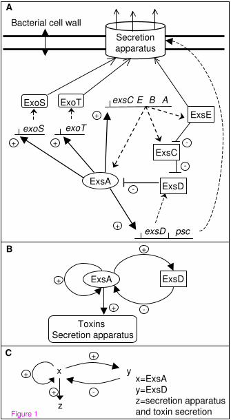
Figure 1 - Models of the Regulation of the TTSS.
(A) The regulatory network of the type III secretion system. Regulators encoded outside
of the ExsA regulon are not shown. Filled arrows and dashed lines represent positive or
negative interactions respectively, dotted lines stand for transcription and translation,
opened arrows represent the secretion of proteins. (B) The molecular sub-network
drawn here only shows interactions involved in feedback circuits: autoregulation of
exsA, activation by ExsA of the operons involved in the cytotoxic response, and the
inhibition of ExsA by ExsD. Induction of secretion and expression of cytotoxicity by
the target, or by calcium depletion, is considered constant and is therefore not indicated.
Other regulations of the exsA gene are not represented. (C) The minimal regulatory
graph extracted from the molecular graph. The three variables are x=ExsA, y=ExsD (the
ExsA inhibitor), and z=type III secretory apparatus and toxin secretion. The four arrows
represent autoregulation of the exsA gene (x→x), transcriptional activation of the exsD
gene by protein ExsA (x→y), transcriptional activation of the genes involved in type III
secretory system (x→z), and inhibition of ExsA by ExsD (y→x).
Figure 2 - Underlying dynamics depend on the values of interactions thresholds
and on the parameters values.

24
The labels of the vertices indicate the sign of the interaction and its threshold. As these
thresholds are almost always unknown, value 1 means only that the corresponding
threshold is the lowest one, and value 2 that it is the second lowest threshold. Two
different graphs must then be drawn depending on which promoter is more sensitive to
ExsA. (A) In this case, the threshold above which x is active on x is higher (level 2)
than that above which it is active on y (level 1). (B) Represents the reverse case. z is an
output element whose level is determined by the value of x: low values of x will lead to
negligible amounts of z, while high values of x will lead to high levels of z.
Figure 3 - One result from SMBioNet in accordance with the epigenetic
hypothesis of the regulatory graph.
SMBioNet provides a graphical interface that allows the user to define a regulatory
graph and to edit temporal properties. SMBioNet exhibits all sets of parameters which
satisfy the properties. Consistency is thus established if and only if at least one set of
parameters is selected by SMBioNet. (A) Parameter table. The two first columns list all
possible states of the network according to genes x and y. The third column gives the
Kx,w parameters which define the expression level towards which x tends to evolve.
Note that w represents all the positive regulatory effects that are active on x (including
the lack of active negative effects). Here, w reflects the effects of x, if its value is higher
than the activity thresholds indicated in the graph, and the effects of y, if its value is
lower than the activity threshold. The fourth column similarly gives the Ky,w
parameters which define the expression level towards which y tends to evolve. (B) State
transition graph. The dynamics of the model are deduced from the values of the
parameters via a desynchronisation algorithm[21].
25
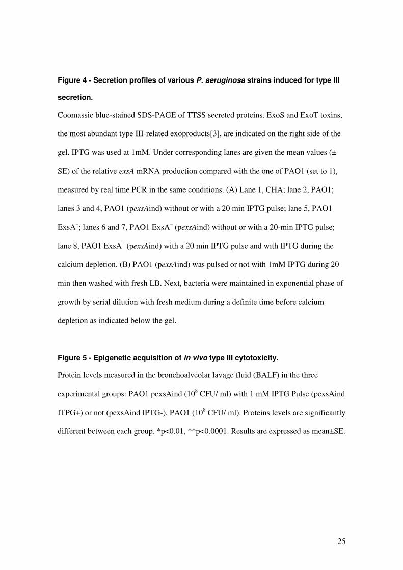
Figure 4 - Secretion profiles of various P. aeruginosa strains induced for type III
secretion.
Coomassie blue-stained SDS-PAGE of TTSS secreted proteins. ExoS and ExoT toxins,
the most abundant type III-related exoproducts[3], are indicated on the right side of the
gel. IPTG was used at 1mM. Under corresponding lanes are given the mean values (±
SE) of the relative exsA mRNA production compared with the one of PAO1 (set to 1),
measured by real time PCR in the same conditions. (A) Lane 1, CHA; lane 2, PAO1;
lanes 3 and 4, PAO1 (pexsAind) without or with a 20 min IPTG pulse; lane 5, PAO1
ExsA–; lanes 6 and 7, PAO1 ExsA– (pexsAind) without or with a 20-min IPTG pulse;
lane 8, PAO1 ExsA– (pexsAind) with a 20 min IPTG pulse and with IPTG during the
calcium depletion. (B) PAO1 (pexsAind) was pulsed or not with 1mM IPTG during 20
min then washed with fresh LB. Next, bacteria were maintained in exponential phase of
growth by serial dilution with fresh medium during a definite time before calcium
depletion as indicated below the gel.
Figure 5 - Epigenetic acquisition of in vivo type III cytotoxicity.
Protein levels measured in the bronchoalveolar lavage fluid (BALF) in the three
experimental groups: PAO1 pexsAind (108 CFU/ ml) with 1 mM IPTG Pulse (pexsAind
ITPG+) or not (pexsAind IPTG-), PAO1 (108 CFU/ ml). Proteins levels are significantly
different between each group. *p<0.01, **p<0.0001. Results are expressed as mean±SE.

26
Tables
Table 1 - PCR primers and probes used, restriction sites are in bold (6-FAM, 6-carboxyfluorescein; TAMRA, 6-carboxytetramethylrhodamine).
Name Sequences (5’-3’) Description
EXSAS CCGAATTCTTATAATATGCAAGGAG
EXSAR GGAAGCTTTCAAAAAACGTCAGTTA
Amplification of exsA
Xbaptaq CGTCTAGATTGACAATTCATCGCCTCG
KpnlacIq GGGTACCTCACTGCCCGC TTTCCAGT
Amplification of exsAind
RTExsA CAGCGAACGCGATGATGCC Reverse transcription of exsA
ExsAQS GAACTGACCGTCCAGGACAT
ExsAQR CAACGCTCGACTTCACTCAA
ExsADL (6-Fam)CCTGGCGAGTTGCTTTTCGTCCGC(Tamra)
Amplification and detection of exsA
cDNA in RT PCR
16s sens AAGCAACGCGAAGAACCTTA
16as CACCGGCAGTCTCCTTAGAG
16sDL (6-Fam)ACGAAGGGACGCACGAG(Tamra)
Amplification and detection of 16S
rRNA cDNA in RT PCR
ExsA
exoTexoS
exsD psc
ExsD
A
Bacterial cell wall
ExoT
+
exsC B AE
ExsE
ExsC
+
-
-
-+
B
ExsA ExsD+
+
-
Toxins
Secretion apparatus
+
+
Secretion
apparatus
ExoS
x=ExsA
y=ExsD
z=secretion apparatus
and toxin secretion
C
y
z
x+
+
-+
Figure 1

A
x y
z
2+
2+1-
1+ B
x y
z
1+
2+1-
2+
Figure 2

y
x
(0,0) (1,0) (2,0)
(0,1) (1,1) (2,1)
B
x y Kx Ky
0 0 Kx{y}=0 Ky{}=0
0 1 Kx{}=0 Ky{}=0
1 0 Kx{xy}=2 Ky{}=0
1 1 Kx1=1 Ky{}=0
2 0 Kx{xy}=2 Ky2=1
2 1 Kx3=1 Ky4=1
A
Figure 3

Time laps before
Ca2+ depletion
66
43
1 2 3 4
8H
- + IPTG Pulse
6H
+ -
MW(kDa)
ExoT
ExoS
B
ExoT
ExoS
1 2 3 4 5 6 7 8MW(kDa)
A
CHA PAO1 PAO1
(pexsAind)
- +- - - ++
Strains
IPTG pulseIPTG duringCa2+depletion
+
-
43
66
CHA-D1 CHA-D1 (pexsAind)
12.6 ±±±±4.1
14.3 ±±±±2.3 2.2 ±±±±0.5 17.2 ±±±±7.4(1)
Figure 4

pexsAind IPTG+ PAO1 pexsAind IPTG-
BA
LF
Pro
tein
(g
/dl)
0.0
0.2
0.4
0.6
0.8
1.0
1.2
1.4
1.6
1.8
2.0 *
***
Figure 5