board of studies and programme coordinator
TRANSCRIPT


Board of Studies and Programme Coordinator
Unit writing and Editing Editing Prof. P. S. Bisht Department of Physics, Kumaun University Soban Singh Jeena Campus, Almora -263601
Programme Coordinator Dr. Kamal Deolal Department of Physics School of Sciences, Uttarakhand Open University
Writing Prof. H M Agrawal, Department of Physics, CBSH, G.B.P.U.A.&T. Pantnagar, India Dr. Mahipal Singh Department Physics RH GPGC Kashipur, US Nagar, Uttarakhand Dr. Tara Bhatt Department of Physics, MBPG College, Haldwani Nainital Dr. Gagan Dixit Department of Physics, CBSH, G.B.P.U.A.&T. Pantnagar, India Dr.S.Abdul Khader Department of P.G studies in Physics Govt.Science College, Chitradurga-577501. Dr. Kamal Deolal Department of Physics School of Sciences, Uttarakhand Open University Haldwani
Course Title and code : Modern Physics (BSCPH302) ISBN No. : Copyright : Uttarakhand Open University Edition : 2019 Published by : Uttarakhand Open University, Haldwani, Nainital- 263139 Printed by :
Board of Studies Prof. P. D. Pant Director School of Sciences Uttarakhand Open University, Haldwani Prof. P. S. Bisht, SSJ Campus, Kumaun University, Almora. Dr. Kamal Deolal Department of Physics School of Sciences, Uttarakhand Open University
Prof. S.R. Jha, School of Sciences, I.G.N.O.U., Maidan Garhi, New Delhi Prof. R. C. Shrivastva, Professor and Head, Department of Physics, CBSH, G.B.P.U.A.&T. Pantnagar, India

BSCPH-302
Modern Physics
DEPARTMENT OF PHYSICS
SCHOOL OF SCIENCES
UTTARAKHAND OPEN UNIVERSITY Phone No. 05946-261122, 261123
Toll free No. 18001804025 Fax No. 05946-264232, E. mail [email protected]
htpp://uou.ac.in

Contents
Course 10: Modern Physics Course code: BSCPH302
Credit: 3
Unit
number
Block and Unit title Page
number
Block 1
1 ATOMIC MODELS 1 2 BASICS OF ATOMIC SPECTRA I 28 3 BASICS OF ATOMIC SPECTRA II 53 4 X-RAY 74 5 PHOTOELECTRIC EFFECT AND COMPTON EFFECT 102 Block 2 : 6 MOLECULAR SPECTRA 123 7 RAMAN SPECTRA 164 8 LASER 181 Block 3:- 9 PROPERIS OF NUCLEUS 204 10 NUCLEUS MODELS 225 11 NUCLEAR REACTION
246
12 NUCLEAR FISSION AND NUCLEAR FUSION 268 13 RADIOACTIVITY 290 14 ELEMENTARY PARTICLES 312

BSCPH302 Modern Physics
Page 1
UNIT 1 ATOMIC MODELS
Structure
1.1 Introduction
1.2 Objectives
1.3 Electronic Specific Charge
1.4 Atomic Models
1.5 Thomson’s Atomic Model
1.5.1 Drawbacks of Thomson’s Atomic Model
1.6 Ruther’s Experiment of α- Particle scattering
1.6.1 Assumptions of Rutherford’s Theory of α-Particle Scattering
1.6.2 Theory of α-Particle Scattering Experiment
1.6.3 Conclusions from Rutherford’s Scattering Formula
1.7 Rutherford’s Atomic Model
1.7.1 Shortcomings of Rutherford’s Atomic Model
1.8 Bohr’s Atomic Model
1.8.1 Bohr’s Interpretation of the Hydrogen Spectrum
1.8.2 Spectral Series of Hydrogen Atom
1.8.3 The Energy-Level Diagram
1.8.4 Shortcomings of Bohr’s Atomic Model
1.9 Fine Structure and Sommerfeld’s Atomic Model
1.9.1 Limitations/Drawbacks of Sommerfeld’s Theory
1.10 Correspondence Principle
1.11 Summary
1.12 Glossary
1.13 References
1.14 Suggested Readings
1.15 Terminal Questions
1.16 Answers

BSCPH302 Modern Physics
Page 2
1.1 INTRODUCTION
The eventual constituent of matter is called atom which was considered indivisible for a long-time. Later on, the experiments performed by J . J Thomson, Wilson, Millikan and others showed beyond doubt the existence of negatively charged particles, called the electrons within the atom. Since atom as a whole is neutral, it must have an equal positive charge. J.J. Thomson suggested atomic model that has a large sphere of positive charge with negative electrons embedded in it. But this model was not accepted. After this, Rutherford formulated an atomic model according to which an atom consists of a central positively charged nucleus surrounded by planetary electrons. The number of orbital electrons is equal to the number of protons in the nucleus. In this unit, we shall deal with various atomic models like Thomson model, Rutherford model, Bohr’s model, Sommerfeld’s model.
1.2 OBJECTIVES
After studying this unit, you should be able to understand-
• electronic specific charge • various atomic models • drawbacks of atomic models • correspondence principle
1.3 ELECTRONIC SPECIFIC CHARGE
The ratio of electronic charge (e) to the electronic mass (m) is known as electronic specific charge.
Thus, electronic specific charge = ��
= �.�×��� � �� ���×���� �� = 1.77×10�� Coulomb/ Kg
This value is constant.
1.4 ATOMIC MODELS
Any worthwhile model of the atom must be able to account for the following established properties of the atom-
(i) The atom is electrically neutral (ii) The atom of different elements is stable. (iii) Every atom, under proper conditions, emits a characteristic spectrum. (iv) Atoms of different elements have different chemical properties.

BSCPH302 Modern Physics
Page 3
(v) Atoms are arranged in the periodic table and show ‘periodicity’ in their properties.
We now discuss some of the models of the atom that can account for the most of the above mentioned expectation.
1.5 THOMSON’S ATOMIC MODEL
In 1898, Sir J.J. Thomson, for the first time, suggested a model for the atom which is known as ‘Thomson’s model’. According to this model, an atom is a positively charged sphere of radius 10-10 meter, in which mass and positive charge of the atom are uniformly distributed. The electrons are embedded here and there within this sphere. The number of electrons is such that their negative charge is equal to the positive charge of the atom. Thomson’s atomic model is shown in figure 1.
Electron
Positively charged matter
Figure 1: Thomson’s atomic model
This model is equivalent to water-melon where seeds are embedded here and there (equivalent to electrons) and the rest part (reddish part) is equivalent to the positively charged matter. Hence this atomic model is also called ‘water-melon model’ or ‘plum pudding model’. Various phenomenon like thermionic emission, photo-electric emission and ionization, were explained on the basis of this model.
1.5.1 Drawbacks of Thomson’s Atomic Model
Thomson’s atomic model explained various phenomenon however this model has some drawbacks-
- -
- -
- -
-
-

BSCPH302 Modern Physics
Page 4
(i) In order to explain the emission of light from atom, it was assumed that whenever an atom receives energy from outside, the electrons in it start vibrating and radiate electromagnetic radiation (light waves) of the frequency of their vibration. But, according to this explanation, there should be only one line in the spectrum of hydrogen atom (which has a single electron), whereas, in fact, it has many lines. Thus, this model was unable to explain this point.
(ii) This model could not predict any reason for α-particle scattering.
These were failures of Thomson’s atomic model. Due to these drawbacks, Thomson’s atomic model could not be accepted. This led to the idea of another atomic model, known as the nuclear atom model.
1.6 RUTHERFORD’S EXPERIMENT OF α- PARTICLE
SCATTERING
α- particles are doubly charged helium (He) atoms that have lost both of their electrons. Thus, the mass of an α-particle is four times the mass of a hydrogen atom and a positive charge two times the charge of a proton.
If a beam of α-rays is made to fall upon a photographic plate in vacuum, the shadow image formed there has sharp and clear edges. But, if air or some other gas is introduced or a screen having thin foil of metal is placed in the path of α-rays, the image formed is not sharp but becomes diffused. This spreading out of the stream of α-particles on passing through thin layers of matter is known as scattering.
The schematic diagram of Rutherford’s experiment on scattering of α-particles is shown in following figure 2. α-particles from radioactive source are incident on a thin gold foil of thickness ~ 10-7 m, beyond which is placed a screen coated with zinc sulphide. Α-particle on striking the screen caused tiny flashes which could be viewed and counted. The following observations are noted from this experiment-
(i) Most of the α-particles passing straight through the gold foil indicating thereby that the atoms are hollow.
(ii) Some of the α-particles are scattered through small angles but a few of them were deviated through a large angle and occasionally a particle went back along the directions from which it comes i.e. deviates through about 1800. This large scale scattering is called ‘anomalous scattering’.
Thomson’s atomic model could not explain these observations. Rutherford, therefore, was forced to picture an atom in which the entire positive charge and almost whole of the atomic mass are concentrated in a tiny central core called the nucleus about which the electrons are grouped in some sort of configuration.

BSCPH302 Modern Physics
Page 5
α-particles Nucleus
Figure 2: Schematic diagram of Rutherford’s experiment of α-particles scattering
1.6.1 Assumptions of Rutherford’s Theory of α-Particle Scattering
Rutherford developed his theory of α-particle scattering on the basis of a number of assumptions. These assumptions are mentioned below-
(i) The first and foremost assumption, that was the basis of his entire theory, was the concept of the nucleus. Rutherford assumed that the entire positive charge of the atom was concentrated in a very tiny, very massive part at its centre. The negatively charged electrons were present in a relatively much vaster volume around this nucleus and the major part of the atom was thus just empty space or vacuum.
(ii) α-particle as well as the nucleus

BSCPH302 Modern Physics
Page 6
1.6.2 Theory of α-Particle Scattering Experiment
Let us explain α-particle scattering. Rutherford assumed that when an α-particle approaches the positive nucleus, Coulomb’s repulsive force is given by-
F = ������������ ��!" …..(1)
Here, +Ze and +2e are the charges on nucleus and α-particle respectively and r is the distance between the nucleus and approaching α-particle..
This force comes into play and hence the α-particles are deflected from their path. In this
expression ε0 is the permittivity of free space and ����� = 9 ×10⁹ Newton-m2/Coulomb2
Obviously, F is inversely proportional to r2. If distance r is very large, the α-particle experience a very small electrostatic repulsion and pass almost undeflected. On the other hand, if r is very small i.e. the particle passes close to the nucleus, it experiences strong repulsive force and scattered through a large angle. Occasionally, an α-particle may be aimed just at the nucleus the repulsive force will slow down the particle which will be momentarily stopped and returned almost in the direction from which it came.
Rutherford also derived a mathematical formula describing the scattering of α-particles by thin foil. For the purpose of calculations, following assumptions were made-
(i) The nucleus and the particle, both act as point charges and masses. (ii) The nucleus is considered to be so heavy that its motion during the scattering process may
be ignored. (iii) The scattering is caused by repulsive electrostatic force between the nucleus and α- particle
which obeys Coulomb’s inverse square law.
The calculations show that the number of α-particles received per square cm. of a screen placed at an angle θ to the direction of incident particles as a distance R from the scatter, i.e. thin metal foil is given as-
N(θ) = K’ #�$%&'�("� = K’ N0 cosec4 (θ/2) …..(2)
where K’ is a constant which depends on charge (+Ze) of the nucleus, speed v of the α-particles and geometrical factors of the experiment. The above formula is known as Ruther’s scattering formula.
These theoretical predictions were experimentally tested and verified by Rutherford and his coworkers Geiger, Marsden and Chadwick.

BSCPH302 Modern Physics
Page 7
log N(θ)
O log ( �$%&'("�
Figure 3: Graph between log ()*+,-./� and log N(θ)
A graph is plotted between log ( �$%&'("� and log N(θ) [Figure 3] which came to be a straight line as
was expected from Rutherford scattering formula indicating that-
N(θ) α �$%&'(" …..(3)
The experimental data of Rutherford’s scattering of α-particles also led to an estimate of the size of the nucleus. The velocity of those α-particles which meet the nucleus in head on collision i.e. scattered through 1800, becomes zero at the distance of closet approach to the nucleus.
Let us consider an α-particle travelling initially with a constant velocity v towards the nucleus at a very large distance from it. As the α-particle approaches the nucleus, its velocity goes on decreasing due to electrical repulsion. As a result, the kinetic energy of α-particle goes on decreasing while its potential energy increases. At the distance of closest approach (r0), the α-particle is stopped momentarily and thus whole of its initial kinetic energy is changed into potential energy.
If (+2e) is the charge and m the mass of α-particle, Z the atomic number and (+Ze) the charge on the nucleus, then-
Initial kinetic energy of α-particle, K = (1/2) mv2
The potential energy of α-particle when it is at the distance of closest approach r0 from the
nucleus, U = ������������ ��!�
Now, K = U

BSCPH302 Modern Physics
Page 8
(1/2) mv2 = ������������ ��!�
or r0 = ����� ���"�0" …..(4)
1.6.3 Conclusions from Rutherford’s Scattering Formula
The Rutherford scattering formula tells us that –
(i) For a given foil and for α-particles of a given incident energy, the number N(θ) is
directly proportional to �$%&'(" or cosec4(θ/2)
(ii) For α-particles, detected at a particular angle of scattering θ, the number N(θ) is- (a) directly proportional to the number of foil atoms per unit volume. (b) directly proportional to the square of the atomic number of the foil atoms. (c) directly proportional to the thickness of the foil of a given element and (d) inversely proportional to the square of the kinetic energy of the incident alpha
particles.
1.7 RUTHERFORD’S ATOMIC MODEL
In 1911, Rutherford presented a model of the atom, called ‘Rutherford’s atomic model’. According to this model, the mass of the atom (leaving the mass of its electrons) and its entire positive charge are concentrated at the centre of the atom in a nucleus of radius ~ 10-15 meter. Electrons have no place inside the nucleus. Around the nucleus, electrons are distributed in a hollow sphere of radius ~ 10-10 meter. The dimensions of the radius and of the electron are negligibly small as compared to the overall size of the atom so that most of the volume occupied by an atom is actually an empty space. In this way, the discovery of the nucleus of the atom is due to Rutherford. The total negative charge of the electrons is equal to the positive charge of the nucleus.
Rutherford assumed that the electrons in the atom are not stationary but are revolving around the nucleus in different orbits and the necessary centripetal force is provided by the electrostatic force of attraction between the electron and the nucleus.

BSCPH302 Modern Physics
Page 9
Figure 4: Rutherford’s atomic model
1.7.1 Shortcomings of Rutherford’s Atomic Model
Rutherford’s atomic model was supported by the periodic table of the elements. However, this model suffers from two main defects-
(i) Rutherford proposed that electrons revolve at a high speed in circular orbits around the positively charged nucleus. When a charged particle i. e. electron revolves around positively charge nucleus, it needs to be accelerated so as to keep it moving in circular orbits. However, according to electromagnetic theory, whenever a charged particle such as an electron is accelerated around another charged center (nucleus) which are under force of attraction, there will be continuous radiation of energy. This loss of energy would slow down the speed of the electron. This would reduce the radius of the electron–orbit. Eventually the electron would fall into the nucleus. The result would be that the atom would collapse. But this does not happen. Thus Rutherford’s atom could not explain the stability of the atom. Failure of Rutherford’s model i.e. reduction of radius of orbit is shown below. (ii) Rutherford proposed that electrons revolve around the nucleus in the fixed orbits. However, he did not specify the orbits and the number of electrons in each orbit.

BSCPH302 Modern Physics
Page 10
Figure 5: Revolving electron around the nucleus
1.8 BOHR’S ATOMIC MODEL
There were some shortcomings of Bohr’s atomic model. Rutherford’s model could not explain the stability of the atom and line spectrum. In 1913, Neil Bohr removed these difficulties by the application of Planck’s quantum theory. Let us discuss the postulates as proposed by Bohr. Bohr proposed the following postulates-
(i) Electrons can revolve only in those orbits in which their angular momentum is an integral multiple of h/2π, where ‘h’ is the Planck’s constant (h= 6.64 × 10⁻³⁴ J-sec). If ‘m’ is the mass of the electron and it is revolving with velocity ‘v’ in an orbit of radius ‘r’ , then its angular momentum = mvr. According to this postulate, we have- mvr = n (h/2π) …..(5) where n = 1, 2, 3, ………. (integer). ‘n’ is called ‘principle quantum number’ of the orbit. Thus, according to Bohr’s atomic model, electrons can revolve only in certain discrete orbits of definite radii, not in all. These are called ‘stable orbits’.
(ii) While revolving in stable orbits, the electrons do not radiate energy inspite of their acceleration towards the centre of the orbit. Hence, the atom remains stable and is said to exist in a stationary state.
(iii) An atom radiates energy only when an electron jumps from a stationary orbit of higher energy to one of lower energy.

BSCPH302 Modern Physics
Page 11
If the electron jumps from an initial orbit of energy Ei to a final orbit of energy Ef (Ei > Ef), a photon of frequency ν = (Ei – Ef)/h is emitted. Based on these postulates, Bohr derived the formulae for the radii of the stationary orbits and the total energy of the electron in the orbit. Let us derive Bohr formulae for the radius of nth orbit, velocity of electron in permitted orbits, the radius of nth orbit and the energy of the electron in the nth orbit. Let us consider an atom whose nucleus has a positive charge Ze, where Z is the atomic number. Let an electron of charge (-e) and mass ‘m’ moves round the nucleus in an orbit of radius ‘r’ with velocity ‘v’. v
Electron
Figure 6: Motion of electron around the nucleus in an atom
The electrostatics force of attraction between the nucleus and the electron is-
Fe = ��πε��12��2�3" . …..(6)
The centrifugal force on the electron is-
Fc = �0"! . …..(7)
For the stability of the atom, two forces should be equal i.e. Fe = Fc
r
+Ze Nucleus

BSCPH302 Modern Physics
Page 12
��πε��12��2�3" = �0"!
or mv2 = ��"����! . …..(8)
From the first postulate, we have-
mvr = n 4 � ; n = 1, 2, 3, ………… …..(9)
Squaring both sides, we have-
m2 v2 r2 = n2 ( 4 � )2 . …..(10)
Dividing equation (8) by equation (10), we get-
�0"�"0"!" = 67"'89�:&"; <"8="
or r = &"4"������" . …..(12)
In general, we can write-
rn = &"4"������" . …..(13)
This is the expression for the radius of nth orbit. It is clear that, rn α n2.
Now from equation (9),
v = &4 ��! . …..(14)
Putting for ‘r’ in the above equation (14), we get-
v = ��" 4��& . …..(15)
This is the expression for the velocity of electron in permitted orbits.
Clearly, v α �&; n = 1, 2, 3, …………
In general, we can write-
vn = ��" 4��& . …..(16)

BSCPH302 Modern Physics
Page 13
This is the velocity of electron in nth orbit. For n = 1, the velocity will be maximum. This shows that the velocity of electron is maximum in the lowest orbit (n = 1) and goes on decreasing in higher orbits.
We can generalize this expression for hydrogen atom. For hydrogen atom, Z = 1, equation (13) becomes-
rn = &"4"�����" . …..(17)
For first orbit, n = 1, therefore, the radius of first orbit,
r1 = 4"�����" …..(18)
= ��.�� ����' > $�?�" �@.@A���" BC�D.����.����� EF���.����G�"
= 0.53× 10⁻¹⁰ metre
= 0.53 A0 .
This is called Bohr’s radius. Its value is 0.53 A0.
From equation (17), we have-
rn = n2 ( 4"�����" )
= n2 r1
or rn = (0.53) n2 A0 . …..(19)
This is the expression for the radius of nth orbit.
For second orbit, n = 2,
r2 = (0.53) × (2)2 = 2.12 A0 .
Similarly, the velocity of electron in nth orbit in hydrogen atom is given as-
vn = �" ���& [ putting Z= 1 in equation (16) ] …..(20)
For first orbit, n = 1,
v1 = �" ��� = ��.�×���G�" ×D.��×�@.@A×��"BC � = 2.19 ×10⁶ m/sec

BSCPH302 Modern Physics
Page 14
0? = .����H �/$�?D��J �/$�? = ��DK .
The kinetic energy of the electron, K = (1/2) mv2 = (1/2) � ��"����!) (Using equation 8)
= ��"@���! . …..(21)
The potential energy of the electron in an orbit of radius ‘r’ due to electrostatic attraction by the nucleus is given by-
U = ����������L��!
= - ����� ��"! . …..(22)
The energy E of an electroninan orbit is the sum of kinetic and potential energies. Therefore, the total energy of the electron-
E = K + U
= ��"@���! - ����� ��"! = - ��"@���! .
Putting for r from equation (12), we get-
E = - ��"�'@��"4" ; �&"= . …..(23)
In general, En = - ��"�'@��"4" ; �&"= . …..(24)
This is the expression for the energy of the electron in the nth orbit.
1.8.1 Bohr’s Interpretation of the Hydrogen Spectrum
If an electron jumps from an outer initial orbit n2 of higher energy to an inner orbit n1 of lower energy, the frequency of the radiation emitted is given by-
υ = MN"L MN4 .
But En1 = - ��"�"@��"4" ; �&"=
and En2 = - ��"�"@��"4" ; �&""=

BSCPH302 Modern Physics
Page 15
υ = �4 O− ��"�'@��"4" ; �&""= + ��"�'@��"4" ; �&"=R or υ = ��"�'@��"4� ; �&" − �&""= . …..(25)
The corresponding wavelength is given as-
λ = ?S
= ?C6"7'J9�"<� T N"L N""U .
Therefore �V = ��"�'@��"4�? ; �&" − �&""= …..(26)
1/λ is called wave number i.e. the number of waves per unit length. It is denoted by W.
The quantity ��'@��"?4� is a constant called Rydberg constant (R).
Therefore, Rydberg constant R = ��'@��"?4� . …..(27)
Therefore, equation (26) becomes-
�V = YZ ; �&" − �&""= . …..(28)
For hydrogen or hydrogen- like atoms (He+, Li+, ……….), Z = 1, therefore equation (28) becomes-
�V = Y�1� ; �&" − �&""=
or �V = Y ; �&" − �&""= . …..(29)
Rydberg constant R = ��'@��"?4� = [�.� ×���� EF\×[�.�×���G\'
@�@.@A���"]/��"�D��J �/$�?���.� ����' > $�?��
= 1.097× 10⁷ per m
The energy expression En = - ��"�"@��"4" ; �&"= can be written in terms of Rydberg’s constant (R).
Therefore En = -Z2 ^4?&"
or En = − �"×[�.��K×��_��\×[�.� ×����'>$�?\×[D×��J�$�?�\&"

BSCPH302 Modern Physics
Page 16
= − �"×[�D.�×�.�×���\&" Joule
En = -Z2 �D.�&" eV . …..(30)
For hydrogen, Z = 1
En = - ^4?&" eV
En = - �D.�&" eV . …..(31)
1.8.2 Spectral Series of Hydrogen Atom
In this section, we shall discuss five spectral series of hydrogen atom. These series are as follows-
(i) Lyman series: When an electron jumps from second, third, fourth,………..etc. orbits to the first orbit, the spectral lines are in the ultraviolet region. Here n1 = 1 and n2= 2, 3, 4, 5,……….
�V = Y ; ��" − �&"=; n =2, 3, 4, 5, …………….
This is identified as Lyman series.
(ii) Balmer series: When an electron jumps from outer orbits to the second orbit, n1=2 and n2 = 3, 4, 5,………..etc. �V = Y ; � " − �&"=; n = 3, 4, 5, …………….
This series is called Balmer series and lies in the visible region of the spectrum. The first line in the series (n = 3) is called the Hα line; the second (n=4), the Hβ line; the third (n=5) the Hγ line.
(iii) Paschen series: Paschen series in the infrared region are given by n1= 3 and n2 = 4, 5, 6,…….etc. �V = Y ; �D" − �&"=; n = 4, 5, 6…………….
(iv) Brackett series: If n1= 4 and n2= 5, 6, 7, ……….etc., we get the Brackett series. �V = Y ; ��" − �&"=; n = 5, 6, 7,…………….
This series lies in the very far infrared region of the hydrogen spectrum. (v) Pfund series: If n1= 5 and n2= 6, 7, 8……….etc., we get the Pfund series. �V = Y ; �A" − �&"=; n = 6, 7, 8,…………….
This series also lies in the very far infrared region of hydrogen spectrum. By putting n = ∞ in each one of the series, we get the wavelength of series limit, i.e. the last time in the series.

BSCPH302 Modern Physics
Page 17
1.8.3 The Energy-Level Diagram
An energy level diagram is a sort of one-dimensional scale of energy along which each electron
according to this energy state can be located. Let us represent the equation En = - ��"�'@��"4" ; �&"=
diagrammatically.
En = - ��"�'@��"4" ; �&"= = - [�.����� EF\���"[�.���� G\'
@�@.@A×���" ]/��"��.� ×����' > $�?�" ; �&"= = - �D.�&" eV, n = 1, 2, 3,…………….
The lowest energy level E1 (for n = 1) is called the normal or ground state of the atom and the higher energy levels E2, E3, E4,……… are called the excited states. As n increases, En increases. As n increases, the energy levels become crowded and tend to form continuum.
In the energy-level diagram, the discrete energy states are represented by horizontal lines and the electronic jumps between these states by vertical lines. The following figure shows schematically how spectral lines are related to atomic energy levels.
n = ∞ En (eV)
n = 7
n = 6
n = 5 Pfund series
n = 4 -0.85 eV
Brackett series
n=3 -1.51 eV
Paschen series
n =2 -3.4 eV
Balmer series
n =1 -13.6 eV
Lyman series
Figure 7: The energy-level diagram of hydrogen

BSCPH302 Modern Physics
Page 18
1.8.4 Shortcomings of Bohr’s Atomic Model
Bohr’s theory, although was very successful in explaining the spectrum of hydrogen atom and giving valuable information about atomic structure, has the following drawbacks-
(i) The fine structure i.e. individual line of hydrogen spectrum accompanied by a number of faint lines cannot be explained by Bohr’s theory as such. The fine structure of spectral lines can only be explained when (a) the relativistic variation in the mass of the electron and (b) electron ‘spin’ are taken into account.
(ii) Bohr’s theory fails to explain the variation in intensity of the spectral lines of an element. The intensity of the spectral lines can be explained by quantum mechanics.
(iii) Bohr’s theory fails to explain the spectra of complex atoms. It is only applicable to one-electron atoms such as hydrogen, hydrogen isotopes, ionized helium, etc.
(iv) Bohr’s theory fails to explain satisfactorily the distribution of electrons in atoms. (v) The success of Bohr’s theory in explaining the effect of magnetic field on spectral
lines is only partial, i.e. it cannot explain the ‘anomalous Zeeman effect’.
Example 1: An alpha particle with kinetic energy10 MeV is heading towards a stationary point-nucleus of atomic number 50. Estimate the distance of closest approach.
Solution: Given – kinetic energy K = 10 MeV, atomic number Z = 50
Using formula K = ������������ ��!�
or r0 = ������������ ��` = 9 ×10⁹ ×
[A��.����\[ �.����\����H�.����
= 1.44×10⁻¹⁴ m.
Example 2: The number of particles scattered at 600 is 100 per minute in an alpha particle experiment, using gold foil. Find out the number of particles per minute scattered at 900 angle.
Solution: We know that- N(θ) α �$%&'("
or N1/N2 = $%&';("" =$%&';(" = .
Given, N1 = 100, θ1= 600, θ2= 900
Therefore, 100/N2 = $%&'a��" b$%&'aH��" b
or N2 = 25 .

BSCPH302 Modern Physics
Page 19
Therefore, the number of particles per minute scattered at 900 angle is 25.
Example 3: How many revolutions does an electron in the first Bohr orbit of hydrogen atom make per second?
Solution: According to Bohr’s postulate of quantization of angular momentum, we know
mvr = n (h/2π)
for first orbit, n = 1
mvr = h/(2 π)
or v = h/(2 π mr) .
Number of revolutions per second= orbital frequency = 1/ Time period
= 1/ (distance covered in one revolution/ orbital velocity)
= orbital velocity/ distance covered in one revolution
= v / (2 π r) = h/(4 π2mr2)= �.�×����' > $�?�×�D.���"×��.�×���� EF�×��.AD×������" = 6.55×10¹⁵ per sec .
Self Assessment Question (SAQ) 1: The energy of electron in the first orbit in hydrogen atom is -13.6 eV. Calculate the energy in the second orbit.
Self Assessment Question (SAQ) 2: The Rydberg constant for hydrogen is 10967700 m-1. Calculate the shortest and longest wavelength limits of Lyman series.
Self Assessment Question (SAQ) 3: The energy of an alpha particle is 1.2× 10⁻¹³ Joule. Upto what closest distance it can reach the nucleus of silver (Z =47).
Self Assessment Question (SAQ) 4: Choose the correct option-
(i) According to Bohr’s atomic model, the radius of the stationary orbit characterized by the principal quantum number ‘n’ is proportional to-
(a) n (b) n2 (c) n-2 (d) n-1
(ii) In hydrogen atom, the angular momentum of the electron in the lowest energy state is-
(a) h/2π (b) h/π (c) 2π/h (d) 2h
(iii) The unit of wave number is-
(a) metre (b) metre2 (c) metre-1 (d) none of these
(iv) Which series lies in visible region?

BSCPH302 Modern Physics
Page 20
(a) Lyman (b) Balmer (c) Paschen (d) Pfund
Self Assessment Question (SAQ) 5: Fill in the blanks-
(i) Bohr’s radius is…………..A0
(ii) The value of electronic specific charge is ………….
(iii) Any model of the atomic structure must explain the …………..and the …... of atoms.
Self Assessment Question (SAQ) 6: Mention ‘True’ or ‘False’
(i) If Rydberg constant for hydrogen is R, then the short wavelength limit of Lyman series is (1/R).
(ii) The ratio of wavelengths of Hα and Hβ lines of Paschen series is of the order of 100.
(iii) The value of e/m for electrons is not constant.
(iv) Rutherford’s atomic model is also known as watermelon model.
1.9 FINE STRUCTURE AND SOMMERFELD’S ATOMIC
MODEL
A close examination of lines of Balmer series of hydrogen spectrum shows fine structure, i.e. each line is a group of a number of close lines. To explain this fine structure of the lines in the hydrogen spectrum, Sommerfeld suggested that the orbit of the electron in an hydrogen atom is elliptic and not circular as assumed by Bohr.
The motion of the electron in the Bohr picture of the atom is essentially one dimensional. Obviously, one quantum number is sufficient to specify the state of the atom. An elliptic orbit is two dimensional. Clearly, an electron moving in an elliptic orbit requires two quantum numbers to define its state.
Sommerfeld and Wilson showed that quantization condition, in the case of an elliptic orbit is given by-
∮ d. ef = gℎ …..(32)
where ‘p’ is a momentum and ‘q’ the corresponding position coordinate. They have further showed that Bohr’s quantization rule is a particular case of a more generalized quantum condition given by equation (32).
Using radial coordinates with the elliptic orbit each coordinate has to obey the general condition of quantization given by equation (32). Let pr be the radial momentum and pθ be abgular momentum, then-

BSCPH302 Modern Physics
Page 21
∮ d! . ei = g!ℎ …..(33)
∮ dj. ek = gjℎ …..(34)
where nr and nθ are called the radial quantum number and the angular or azimuthal quantum number respectively. Both these quantum numbers are integers (positive) and nθ + nr = n, where n is the principal quantum number. It can take the integral values1, 2, 3,……… etc. To determine the allowed elliptical orbits, we have to evaluate the integrals in equations (33) and (34).
pθ is a constant (From Kepler’s law), i.e.
pθ= p = constant ( Angular momentum)
Electron
X
Figure 8: Sommerfeld’s atom model
Integrating equation (34) from 0 to 2π, we get-
l djek = gjℎ ��
Or pθ = (2π) = nθ h
Or pθ = nθ h/ (2π) …..(35)
Now, pr = m(dr/dt) (Momentum along the radius)
Pr dr = m(dr/dt) dr = m [ (dr/dθ). (dθ/dt)] (dr/dθ) dθ
= m;m!mj= mjmn ek
But pθ = mr2 (dθ/dr)
r
θ
Nucleus

BSCPH302 Modern Physics
Page 22
Therefore, pr dr = ;�! m!mj= djek …..(36)
The equation of an ellipse in polar coordinates is-
�! = ��� ?o$jp��L�"� …..(37)
Where ‘a’ is the semi-major axis and ‘ε’ is the eccentricity.
Taking loge of above equation, we get-
- loge r = loge ( 1+ε cos θ) – loge a (1-ε2) Differentiating with respect to θ, we get-
-�! m!mj = − � $%&j���� ?o$j� or �! m!mj = � $%&j���� ?o$j� …..(38)
Therefore, pr dr = �"$%&"j���� q r j�" djek .
…..(39) Equation (33) takes the form- l s tug k × djek�1 + s cos k� �
� = g!ℎ
or pθ y �"$%&"jmj���� q r j�" �� = g!ℎ . …..(40)
But we know that y �"$%&"jmj���� q r j�" �� = �√�L�" − 2| .
Therefore, equation (40) takes the form-
pθ O �√�L�" − 2|R = nr h
or �}(√�L�" − 2|dj = nr h
or &(4√�L�" − gjℎ = nr h …..(41)
(since pθ = nθ 4 � )
or nr + nθ = &(√�L�"
or n = &(√�L�" ( since n = nr + nθ)

BSCPH302 Modern Physics
Page 23
or 1- ε2 = &("&" . …..(42)
But for an ellipse, 1- ε2 = ~"p" .
Where ‘a’ and ‘b’ are the semi-major and semi-minor axes respectively.
Hence, ~"p" = 1 − s = &("&"
or ~p = &(& . …..(43)
Equation (43) is the condition that determines the allowed elliptical orbits. The allowed elliptical orbits are those for which the ratio of major and minor axes is that of two integers. When nθ = n, b = a, ε = 0 and the orbit becomes circular. nθ cannot be zero, since the ellipse would then degenerate into a straight line passing through the nucleus. Also nθ cannot be greater than n, since b is always less than a. Hence for a given value of n, nθ can assume only n different values, which means there can be only n elliptical orbits of different eccentricities.
The total energy of a single electron is-
En = Potential Energy + Kinetic Energy
Potential Energy = ������L������! = L��"����!
Kinetic Energy = � � �;m!mn= + ;i mjmn= �
where m!mn = radial component of the velocity and rmjmn = transverse component of the velocity.
Hence, En = L��"����! + � � �;m!mn= + ;i mjmn= � . …..(44)
Sommerfeld found this energy equal to - ��'�"@��"4" �&"
i.e., En = - ���'�"@��"4" � �&" .
This expression is similar to that of Bohr’s theory. This shows that the quantum number ‘n’ in the Sommerfeld model is the principal quantum number of Bohr’s theory of atomic model.
Sommerfeld modified his theory, taking into account the variation of the mass of the electron with velocity. It can be shown that the total energy En in the relativistic theory is-
En = ��'�"@��"4"&" - ��'�"�"@��"4" ; &&( − D�= …..(45)

BSCPH302 Modern Physics
Page 24
where α = �" ��?4 ≈ ��DK
‘α’ is a dimensionless quantity and is called the fine structure constant.
1.9.1 Limitations/Drawbacks of Sommerfeld’s Theory
The shortcomings of Sommerfeld theory are as follows-
(i) The quantum numbers introduced in the theory, are introduced more or less as postulates. The quantum numbers, therefore, do not become a naturally logical part of the theory.
(ii) The theory was applicable only to one electron atoms like hydrogen or to hydrogen-like ions like singly ionized helium or doubly ionized lithium. It could not be used for many electron atoms, not even the two-electron neutral helium atom.
(iii) The distribution and arrangement, of the electrons in the atom, was not covered by the theory.
(iv) The selection rules, introduced for making the Sommerfeld relativistic theory results match with the experimental results, appear ad hoc in nature.
(v) The theory could not explain the variations in the intensity of spectral line. (vi) The theory could not explain the observed anomalous Zeeman Effect.
1.10 CORRESPONDENCE PRINCIPLE
In physics, the correspondence principle states that the behavior of systems described by the theory of quantum mechanics (or by the old quantum theory) reproduces classical physics in the limit of large quantum numbers. In other words, it says that for large orbits and for large energies, quantum calculations must agree with classical calculations. The principle was formulated by Niels Bohr in 1920, though he had previously made use of it as early as 1913 in developing his model of the atom.
The term codifies the idea that a new theory should reproduce under some conditions the results of older well-established theories in those domains where the old theories work. This concept is somewhat different from the requirement of a formal limit under which the new theory reduces to the older, thanks to the existence of a deformation parameter. Classical quantities appear in quantum mechanics in the form of expected values of observables, and as such the Ehrenfest theorem (which predicts the time evolution of the expected values) lends support to the correspondence principle.
1.11 SUMMARY
In this unit, we have learnt about various atomic models- Thomson model, Rutherford’s atomic model, Bohr’s model and Sommerfeld atom model. We have studied Rutherford’s experiment of α- ray scattering and found that for a given foil and for α-particles of a given incident energy, the
BSCPH302 Modern Physics
Page 25
number N(θ) is directly proportional to �$%&'(" or cosec4(θ/2). We have studied Bohr’s
interpretation of the hydrogen spectrum and discussed spectral lines of hydrogen atom. We have studied correspondence principle too. In the unit, we have included solved examples and self assessment questions (SAQs) to check your progress.
1.12 GLOSSARY
Spectral series- a group of spectral lines in the spectrum of a substance
Scattering- dispersion
Stationary- motionless, still
Attraction- lure, hold
1.13 REFERENCES
1. Elementary Text Book of Physics, JP Agarwal, Pragati Prakashan, Meerut
2. A Text Book of Modern Physics, Mahipal Singh, Ram Prasad and Sons, Agra
1.14 SUGGESTED READINGS
1. Fundamentals of Physics; Halliday, Resnick and Walker; John Wiley and Sons (Asia) Pte Ltd, Singapore
2. Modern Physics, Beiser, Tata McGraw Hill
1.15 TERMINAL QUESTIONS
1. Describe Rutherford’s atomic model and evidence that led to it. Give its shortcomings.
2. State the postulates of Bohr’s atomic model. Obtain expressions for the radius and electron energy of the nth orbit.
3. Describe Sommerfeld’s model. Also give the shortcomings of Sommerfeld model.
4. Write notes on-
(i) Spectral lines of hydrogen atom
(ii) Electronic specific charge
5. Estimate the wavelength of Hα line of hydrogen, assuming that the nucleus has infinite mass. Calculate the wavelength of Balmer series limit.

BSCPH302 Modern Physics
Page 26
6. Find out the shortest wavelength of the Balmer series (limit of the Balmer series) and the largest wavelength of the Lyman series.
7. Calculate the radius of the third Bohr orbit.
8. Describe Rutherford’s α-particle scattering experiment. How was nucleus discovered from it?
9. What are the differences between Rutherford’s atomic model and Bohr’s atomic model of an atom? Explain.
10. Why was gold foil used in α- particle scattering experiment?
1.16 ANSWERS
Self Assessment Questions:
1. We know, En = - �D.�&" eV
For second orbit, n = 2, therefore, En = - �D.� " eV = En = - �D.�� eV = -3.4 eV .
2. Given, R = 10967700 m-1
For Lyman series, �V = Y ; ��" − �&"=; n =2, 3, 4, 5, ……………∞
For shortest wavelength, n = ∞ .
Therefore, �V = Y ; ��" − ��"= = R
or λ = 1/R = 1/ 109677 m = 911.6 A0
For longest wavelength, n = 2 .
Therefore, �V = Y ; ��" − � "= = 10967700×; ��" − � "= = 10967700× (3/4)
or λ = 4/( 10967700×3) = 1215× 10-10 m = 1215 A0
3. We know, r0 = ������������ ��` = 9 ×10⁹ ×
[�K×�.�×���\[ ×�.�×���\�. ×���� = 1.8× 10¹³ m
4. (i) - (b), (ii)-(a), (iii)- (c), iv- (b)
5. (i) 0.53 (ii) 1.77× 10¹¹ coulomb/kg (iii) stability, spectra
6. (i) True, (ii) True, (iii) False, (iv) False
Terminal Questions:

BSCPH302 Modern Physics
Page 27
5. For Hα line, n = 3
Therefore, using �V = Y ; � " − �&"=; n = 3, 4, 5, …………….∞
=1.097× 10⁷ ×; � " − �D"= = 1.097× 10⁷×;�� − ��= = 1.097× 107 × (5/36)
or λ = 36 / (1.097 ×107×5) = 6.56 × 10⁻⁷ m .
For Balmer series limit, n = ∞
�V = 1.097× 10⁷ ×; � " − ��"= = 1.097 × 107 × (1/4)
or λ = 3646 × 10-10 m .
6. For Balmer series, �V = Y ; � " − �&"=; n = 3, 4, 5, …………….∞ .
For shortest wavelength, n = ∞
�V = Y ; � " − ��"= = R (1/4)
λ = 4/R = 4/ (1.097×10⁷) = 3.64× 10⁻⁷ m .
For Lyman series, �V = Y ; ��" − �&"=; n =2, 3, 4, 5, ……………∞ .
For largest wavelength of Lyman series, n = 2 .
Therefore, �V = Y ; ��" − � "== R (1-1/4) = R (3/4)= 1.097× 3/4
or λ = 121.6× 10⁻⁹ m .
7. We know that, rn = (0.53) n2 A0 .
For third Bohr’s orbit, n = 3, therefore, r3 = (0.53) 32 = 4.5 A0
10. The gold nucleus is heavy and hence produces a larger deflection in the path of α- particle. Secondly, extremely this foil can be made of gold.

BSCPH302 Modern Physics
Page 28
UNIT 2 BASICS OF ATOMIC SPECTRA I
Structure
2.1 Introduction
2.2 Objectives
2.3 Space and spin quantization
2.4 Pauli’s exclusion Principle
2.5 Vector Model of atom
2.6 Spin orbit interaction
2.7 Terminology used with atomic energy levels
2.8 Atomic term symbols
2.8.1 Atomic Term symbol with one unpaired electron
2.9 Coupling Scheme
2.9.1 L-S Coupling Scheme
2.9.2 J-J Coupling Scheme
2.10 Ordering of Levels and Terms
2.11 Lande Interval Rule
2.12 Selection rules
2.13 Summary
2.14 Glossary
2.15 References
2.16 Suggested Reading
2.17 Solved Examples
2.18 Review Questions
2.19 Objective Question
2.20 Answers

BSCPH302 Modern Physics
Page 29

BSCPH302 Modern Physics
Page 30
2.1 INTRODUCTION
Before we start this unit and go deeper about the atomic optical spectra let’s review the things that you are supposed to know about atomic structure and spectroscopy.
Figure 1: Experimental arrangement showing emission spectrum
So, what is Spectrum? Spectrum (Spectra is plural of spectrum) means a band of different colors/wavelengths. For example, when white light is passed through a prism it splits in different colors (Figure 1). With the help of more sophisticated spectrometers these colors can be seen more clearly. It was found in the first half of 19th century that certain sources of light when examined by a spectroscope show not a continuous band of light but a number of bright lines separated by dark intervals. A very important discovery was made when it was found that the number and positions of these lines was characteristic of the substance emitting light. Why the spectrum arises? Actually when the atoms of a particular element are excited by any means heating or electrically, the outer/valence electrons jump to the higher excited states. After some time they got de-excited and comes down to the ground state. Now depending upon the energy difference between the ground state and the higher state from where it is being de-excited, the radiation is emitted. Since the emitted radiation is mostly in visible range for atomic spectra it is called optical spectra. In case of molecules there are different types of spectra because of different energy levels rotational energy levels, vibrational energy levels and electronic energy levels. And that’s why molecular spectroscopy is complicated also.
The development of atomic Physics is based on the explanation of atomic spectrum which includes the explanation of occurrence of Fraunhofer lines in the spectrum of sunlight, or Lyman ,

BSCPH302 Modern Physics
Page 31
Balmer, Paschen, Pfund series of Hydrogen spectrum. After the discovery of Nuclear Model of atom give by Rutherford, the successful explanation of atomic structure or atomic spectrum was given by Neil Bohr. There were mainly 3 postulates of Bohr Theory also known as old quantum theory:
1. Electrons in atom revolve in circular orbits.
2. Only those orbits are allowed for which the angular momentum is integral multiple of h/2π
3. When electron jumps from higher orbit to lower one it emits radiation and when it goes from lower to higher orbit it absorbs radiation and hence the emission or absorption spectrum arises.
Using Bohr postulates, the energy of electron in nth orbit of Hydrogen atom comes out to be (this you must have already read)
nE 2
6.13−= .
After Bohr, Sommerfeld made some corrections to the atomic model, but still it was not sufficient enough to explain certain things, such as
1. Fine structure of spectral lines
2. Atomic spectrum of atoms having more than 1 electron in their outer shell
3. About the intensity of spectral lines
4. Why some transitions take place and some are forbidden.
A great help in this regard is given by Quantum Mechanics. Schrodinger equation and discovery of spin together resolved almost the mystery of atomic spectrum. Quantum mechanically the state of an electron in an atom is defined by four quantum numbers.:
1. Principal quantum number ‘n” where n can take the values 1,2,3,4,5,6…..
2. Orbital quantum number “l” and l can take the values 0,1,2,3,…. (n-1). It gives information about the orbital angular momentum L.(Bold letter means vector and without bold means magnitude)
h)1( += llL .
One component of angular momentum (say the z-one) is always quantized such that
hmL lZ =
where “ml” is the magnetic orbital quantum number which can take (2l+1) values starting from –l…0…..to +l
3. Spin quantum number “s” and s= ½ for single electron. Spin angular momentum is given by
h)( 1+= ssS .
One component of spin angular momentum is also quantized such that
hmS sZ =
where ms is the magnetic spin quantum number. There are (2s+1) allowed values for it starting from –s…..0….. to …+s.

BSCPH302 Modern Physics
Page 32
The quantum numbers n, l, ml comes after solving the Schrodinger equation for Hydrogen atom, but spin was discovered separately. Stern Garlech experiment gives experimental proof for the existence of spin.
2.2 OBJECTIVES
After studying this unit you will be able to
1. Find the angle between orbital and spin angular momentum
2. To find spectroscopic terms for different atomic configurations
3. Find different excited states giving rise to atomic spectra
2.3 SPACE AND SPIN QUANTISATION
We have h)1( += llL and hmL lZ = .
Now, if there is an external magnetic field B in the direction of z-axis and θ is the angle between L and B. Then
Figure2: Orientation of S and L with respect to external magnetic field
)( 1+=
=
=
llCos
LCos
LCos
m
LL
l
Z
Z
θ
θ
θ
Since ml and l can have only certain allowed values, θ will also have only some specific values. Thus the angular momentum L can have only certain orientations with respect to external magnetic field. This is called space quantization. Similarly spin quantization is there, and if α is the angle between S and B then

BSCPH302 Modern Physics
Page 33
)( 1+=
ssCos msα .
2.4 PAULI’S EXCLUSION PRINCIPLE
Pauli’s exclusion principle states that no two electrons in an atom may have the same set of quantum numbers or for the two possible states, at least one of these 4 quantum numbers should be different. For example in 1s state two electrons can reside (1s2). The quantum numbers for them are
n1 = n2 = 1,
l1 = l2 = 0,
ml1 = ml2 = 0,
ms1=1/2 but ms2=-1/2.
Thus for both electrons three quantum numbers n, l, ml are same but the fourth (ms) is different. Pauli Exclusion Principle plays an important role in ionic bonding, covalent bonding, nuclear shell model, nuclear binding energy and many other fields.
Actually Pauli’s principle is applicable to not only electrons but to all fermions. Fermions are the particles which have half integer spin (like 1/2 or 3/2).
Let’s consider that there are two identical and indistinguishable particles 1 and 2 and two states ‘a’ and ‘b’. Then the wave function for particle 1 in state ‘a’ and 2 in state ‘b’ can be written as:
)()(21
baab
ψψψ = .
Now if the particles 1 and 2 are interchanged then the wave function can be written as
)()(21
abba
ψψψ = .
Since both Ψab and Ψba are equally probable the complete wave function (Ψ) representing the two particles in states ‘a’ and ‘b’ will be a linear combination of Ψab and Ψba. So,
)(2
1 ψψψbaab
±= ;
)]()()()([2
12121
abba ψψψψψ ±= .
Here 2
1 is the probability amplitude. Square of it i.e.
21
or 50% gives the probability. And ‘+’ sign is
used for symmetric wave function and ‘-’ for anti-symmetric wave functions. Now if both the states are same i.e a=b then in case of anti-symmetric wave function the wave function Ψ will be zero, but in case of symmetric wave functions it will have some value. Thus for the anti-symmetric wave function, two

BSCPH302 Modern Physics
Page 34
particles cannot exist in same state. This is the Pauli’s exclusion principle and such particles represented by anti-symmetric wave functions and following Pauli’s law are called Fermions. Ex- electron, proton, neutron etc. all these have ½ spin.
Pauli’s principle has found application in the build up and modeling of periodic table, band theory of solids and in Astrophysics studying the formation of neutron star and white dwarf.
2.5 VECTOR MODEL OF ATOM
Vector model of atom is that the total angular momentum (J) is the vector sum of orbital angular momentum (L) and spin angular momentum (S).
J= L+S ……………………………..(1)
For single electron small letters are used, hence j = l + s.
The quantum number corresponding to J is called the inner quantum number j, and
j = l ± s
the z component of J is Jz and is given by Jz = mjħ
mj can take (2j+1) values starting from +j……0……to –j.
When there is no interaction between spin and orbital angular momentum, l and s take up

BSCPH302 Modern Physics
Page 35
Figure 3: (a) Orbital l and spin s angular momenta add (couple) to give a resultant j. With no magnetic interaction the vectors remain fixed in space. (b) Spin-orbit coupling results in precessional motion of l and s around their resultant j. (c) and (d) Projections of l and s on z-axis, lz
and sz respectively, are no longer constants of the motion and hence ml and ms are not good quantum numbers in the presence of spin-orbit coupling.
fixed orientations in space. Their projections on the z-axis are ml and ms which are constant. When spin-orbit interaction takes place l and s precess around their mutual resultant j. Their projections on the z-axis, ml and ms are undefined and are no longer constant. See figure 3. If there is spin-orbit interaction as well as an external magnetic field also and both are of comparable strength, then the vector model doesn’t work.
If we square equation 1, then
SLSLJ •++=
+•+=•
2222S)(LS)(LJJ
.
If δ is the angle between L and S then L.S= LSCosδ and
δLSCosSLJ 2222 ++=.
2.6 SPIN ORBIT INTERACTION
An electron revolving around the nucleus is equivalent to a magnetic dipole. It possesses an internal magnetic moment μL and creates an internal magnetic field. Electrons also possess the spin magnetic moment due to its spinning character. The spin magnetic moment interacts with this internal magnetic field and the interaction is called spin-orbit interaction.
Initially, the Hamiltonian to solve the Schrodinger equation was written in terms of electrostatic attractive interaction between the electrons and nucleus, and repulsive interaction between the electrons. But later after the discovery of spin a correction term corresponding to the spin-orbit interaction is also included. This correction term helped to explain the fine structure of spectral lines. It is found by calculation that energy correction corresponding to spin orbit interaction is proportional to L.S. From vector model we have:
h2
2111
2222
2222
)()()(SL
SL
SL
+−+−+=•
−−=•
•++=
sslljj
SLJ
SLJ
.

BSCPH302 Modern Physics
Page 36
2.7 TERMINOLOGY USED WITH ATOMIC ENERGY LEVELS
The terminologies used in atomic spectroscopy are as follows and are represented in Figure4:
State- The state of an atom is the condition of motion of all electrons. To specify a state, one must list four quantum numbers for each electron. If several states have the same energy, they are degenerate. The state with lowest energy is the ground state.
Level- A collection of states having the same energy in the absence of an external electric or magnetic field, all correspond to an energy level. A level is distinguished by a particular value of the total angular momentum, the quantum number J. The level with lowest energy is the ground level.
Sub level- An external field splits an energy level into several sublevels, distinguished by a magnetic quantum number.
Term- In many atoms, the levels cluster in related groups which can be labeled by multiplicity and orbital angular momentum. Such a collection of levels comprises a spectroscopic term. For example, if we say a 3D term (here 3 is the multiplicity), it means the 3D3,3D2 and 3D1 levels.
Configuration- Specification of the quantum numbers n and l for the orbital of each electron defines a configuration. For example- 1s2,2s2,2p2,….
Equivalent orbitals- Orbitals with the same n and l are equivalent. The electrons in equivalent orbitals are sometimes called equivalent electrons.
Statistical weight- The number of distinct states in a specified collection is the statistical weight. The statistical weight of a level is 2J+1, for a term it is (2S+1)(2L+1). For a single electron with a specified n it is 2n2.
A transition between two sublevels is called a component. A transition between two levels is a line. The collection of transitions between two terms is called multiplet. A multiplet consists of from one to a dozen or more lines. The collection of transitions between two configurations is referred to as a transition array. A transition array may comprise hundreds of lines in a dozen or more multiplets.

BSCPH302 Modern Physics
Page 37
Figure 4: Spectroscopic terminologies and their representation
2.8 ATOMIC TERM SYMBOLS
As defined above ‘term’ is cluster of levels. The notation used to write a spectroscopic term is
Note: No of levels in which a multiplet will split due to LS coupling is (2S+1) if L>S or (2L+1) if S>L.
2.8.1 Atomic term symbol with one unpaired electron:
(a) For example –Na atom (1s2,2s2,2p6,3s1) unpaired electron is in ‘s’ orbital
Then S=s=1/2 multiplicity 2S+1= 2

BSCPH302 Modern Physics
Page 38
L=l=0 (corresponding to s orbital), L=0 means S (don’t confuse with the S used for multiplicity and S corresponding to L=0)
J = j = sl ± = 21
0 ± = 21
.
Hence the term for ground state of Na will be written as: 2S1/2 .
Now, it is independent of the value of principle quantum number n, hence for all atoms of such type of configuration (s1) the term will be 2S1/2 .
(b) If unpaired electron is in p orbital – p1
Then S= s = 21
multiplicity 2S+1= 2
L= l= 1 corresponds to P
J = j = sl ± = 21
1± = 23
21
, .
Hence the term is 2P with three levels 2P1/2,3/2. This will be the term for ground state of Boron (1s2, 2s2, 2p1) and excited state of Sodium (1s2, 2s2, 2p6, 3p1).
Figure 5: Representing the levels for ground and excited state of Na
Now if there are more than 1 electron then to write the spectroscopic term certain rules are followed given by different coupling schemes.
2.9 COUPLING SCHEMES
The unpaired electrons in outer shell are also called as optically active electrons because excitation or de-excitation of these only give rise to optical spectrum. So if there is more than 1 unpaired electron then there will be more interaction terms other than the spin orbit interaction of the individual electron. There will be interaction between spin- spin and orbital- orbital angular momenta of all electrons respectively.

BSCPH302 Modern Physics
Page 39
Spin angular momenta of one and orbital angular momenta of other (vice versa) will also interact. Depending upon which interaction is more dominating there are two types of coupling scheme:
L-S coupling and J-J Coupling
Basically these two coupling schemes will provide a method to find the total angular momentum quantum number (j) for the atoms and depending upon the number of allowed values of j, number of terms can be calculated. And using the value of j the energy corresponding to that term can be find out. So first we study the LS coupling scheme.
2.9.1 LS coupling scheme
If the spin orbit interaction is less dominating and the interaction between orbital-orbital and spin –spin angular momentum of all electrons is strong than LS coupling scheme takes place. It is also known as Russel Saunders scheme. They developed it to explain the fine structure of the spectra of stars. Normally, LS coupling takes place in atoms with low atomic number (Z � 10). The reason for this is that the spin-orbit coupling is weak in comparison to the electrostatic effects.
Let the orbital and spin angular momentum quantum number for all electron are l1, l2, l3, l4, ….and s1, s2, s3,s4…..respectively..
Then the orbital angular momentum of all electrons, li couple to yield a total orbital angular momentum L. The possible values of the quantum number L are
L= 21 ll − , 121 +− ll , 221 +− ll ,………….. 21 ll + .
The spins of all electrons, si couple to yield a total spin angular momentum S. The possible values of the quantum number S are
S= 21 ss − , 121 +− ss , 221 +− ss ,……….. 21 ss +
Figure 6: Coupling of l1, l2 to form L and s1, s2 to form S

BSCPH302 Modern Physics
Page 40
Figure 7: L and S couple to give J
L and S can then couple to yield the total angular momentum J. The possible values of the quantum number J are
J = SL − , 1+− SL , 2+− SL ,…………….. SL + .
Ex 1- Spectroscopic term for two non equivalent electrons in ‘s’ and ‘p’ orbital
Here we have one electron in s orbital and one in p orbital. So
l1=0 and l2=1
L= 21 ll − ……. 21 ll +
= 10 − ,… 10 +
= 1.
Now for both electrons
s1=1/2 and s2=1/2
S= 21
21− ………
21
21+
=0, 1.
When L=1, S=0
J= 01− …….. 01+
=1.
And multiplicity 2S+1= 1.
And corresponding to L=1 state is P.

BSCPH302 Modern Physics
Page 41
So the term is 1P1
When L=1, S=1
Figure 8: Vector diagram for the possible combinations of L and S to form J
J= 11− ……. 11+
=0, 1, 2
Multiplicity 2S+1=3
And corresponding L=1 state is P
So the term is 3P with levels 3P0,
3P1, 3P2
Thus there are four levels 1P1, 3P0,
3P1, 3P2 .
Ex 2- Find the spectroscopic terms for the electronic configuration 3d1,4p1
Here l1=2 and l2=1 (corresponding to d and p orbitals)
s1=1/2 and s2=1/2
so, S =21
21− ……
21
21+
= 0, 1.
And L= 12 − …… 12 +
= 1, 2, 3.
Now, when S = 0, L=1 then J=1 So, the term is 1P1.

BSCPH302 Modern Physics
Page 42
And when S=0 L=2, then J=2 , Term 1D2.
When S=0 L=3 , J=3 Term 1F3
Next, when S=1, L=1 then J=0,1,2
So the term is 3P0 ,1,2 are
When S=1 L=2 then J=1,2,3
So the term is 3D1, 2, 3
Next when S=1 L=3 then J=2, 3, 4
Term is 3F2,3, 4
Thus there are total 12 levels (shown in Figre 8) 1P1,1D2
1F3 3P0,
3P1, 3P2
3D1, 3D2,
3D3 3F2,
3F3, 3F4 .
Ex-3 Spectroscopic terms for 3 non equivalent electrons such as 2p3p4d
l1=1 l2=1 l3=2 s1=1/2 s2=1/2 s3=1/2
First two p electrons will combine and give L= 0, 1, 2
Now l3 will combine each of these L values.
So l3=2, L=0 , L’= 2 means D
l3=2, L=1 , L’= 3, 2, 1 represents F, D and L
Figure 9: Terms and levels for 4p4d configuration by LS coupling
So l3=2, L=2. L’= 4, 3, 2, 1, 0 represents G, F, D, L and S

BSCPH302 Modern Physics
Page 43
Similarly first s1 and s2 will combine to give S= 0, 1
Now s3 will combine these two values of S.
s3=1/2 S= 0 so S’= ½
s3=1/2 S= 1 so S’= 3/2 and 1/2
Now all L’ and S’ combinations are to be take to get the values of J.
So there will be 9 terms. One S, two L, three D, two F and one G.
Thus it can be seen that only 3 electrons make the process so complicated. So as the number of electrons increase the spectrum becomes complicated.
Now for the equivalent electron i.e. for which n and l two quantum numbers are already same, we have to take care of Pauli’s exclusion principle in the sense that at least one out of the remaining two quantum numbers should not be same. Therefore in the case of equivalent electrons some of the terms do not appear in comparison to that of equivalent electrons.
Ex-4 Spectroscopic terms for equivalent electrons in completely filled shells
Filled shell such as s2 both the electrons are in s orbital.
So l1 = l2 = 0
ml1 = ml2 = 0
ms1= 1/2 and ms2= - 1/2
ML= ∑ml=0 corresponds to L=0
Ms=∑ms= 0 corresponds to S=0
So by L-S coupling J=0
Therefore the term is 1S0
For all filled orbitals such as p6,d10, f14 there will be single term 1S0
Note:- Thus you observed that for filled orbitals the total angular momentum quantum no is zero. Therefore total angular momentum will also be zero and this results zero magnetic moment. That’s why diamagnetic materials have no unpaired electron.
Ex-5 Spectroscopic terms for 2 equivalent optically active electrons such as 2p2
If maximum number of electrons in an orbital is ‘n’ then the term for ‘x’ electron will be same as that for (n-x) electrons. For example there can be 6 electrons in p orbital. Hence the terms for p2 will be same as that for p4. In d orbital there can be 10 electrons. So d2 and d8 will have same terms d3 and d7 will have same and so on. Now let’s calculate for 2p2:
For p2 kind of configuration, l1 = l2 = 1 and possible values of ml1 and ml2 are -1,0, and 1 and for ms1 and ms2 are 1/2 or -1/2 now for these two electrons already two quantum numbers n and l are same (n1=n2=2 and l1 = l2 = 1 ). According to Pauli’s principle all the four quantum numbers cannot be same. So we have to make all possible combinations of ml1 and ml2 and ms1 and ms2 without violating Pauli’s principle. Below is the table listing all possible sets:

BSCPH302 Modern Physics
Page 44
ml1 ml2 ms1 ms2 Ml = ml1+ ml2 Ms= ms1+ ms2
-1 0 1/2 1/2 -1 1
-1 1 1/2 1/2 0 1
-1 -1 1/2 -1/2 -2 0
-1 0 1/2 -1/2 -1 0
-1 1 1/2 -1/2 0 0
0 1 1/2 1/2 1 1
0 -1 1/2 -1/2 -1 0
0 0 1/2 -1/2 0 0
0 1 1/2 -1/2 1 0
1 -1 1/2 -1/2 0 0
1 0 1/2 -1/2 1 0
1 1 1/2 -1/2 2 0
-1 0 -1/2 -1/2 -1 -1
-1 1 -1/2 -1/2 0 -1
0 1 -1/2 -1/2 1 -1
From the table it can be observed that
Ms=-1, 0, 1 for Ml= -1, 0,1
It means S=1 , L=1 therefore J= 0,1,2
so the term 3P0,1,2
Now Ms=0 for Ml=-2,-1,0,1,2
S=0 and L=2 and this gives J=2
Therefore the term is 1D2
Next Ms=0 and Ml=0
This corresponds to S=0 and L=0 which gives J=0

BSCPH302 Modern Physics
Page 45
Hence, the term is 1S0
Thus the terms for two optically active equivalent electrons in p orbital are 3P0,1,2 , 1D2, 1S0.
Note: There is one more method to calculate the spectroscopic terms for two equivalent electrons. That is Bratt
scheme. That is not discussed here.
2.9.2 J-J Coupling Scheme
In case of jj coupling, the orbital angular momentum, and spin, of each electron are first coupled to form a total angular momentum for that electron. These single electron total angular momenta are then combined into a total angular momentum, for the group of electrons. This is in contrast to LS coupling, where the total orbital angular momentum and total spin of the system are calculated first and then combined to the total angular momentum of the whole system. J-J coupling occurs when the spin orbit interaction is strong.
If l1, l2, l3, l4…. are the orbital angular momentum and s1,s2, s3….. are the spin angular momentum quantum numbers of electrons then in case of J-J coupling first j1, j2, j3….are calculated.
j1= 11 sl + , 1,11 −+ sl , ….….. 11 sl −
j2= 22 sl + , 122 −+ sl ,…….… 22 sl −
j3= 33 sl + , 133 −+ sl ,…….… 33 sl −
and so on…
then total J is calculated as
J= 21 jj + , 121 −+ jj ………. 21 jj −
Ex 6- Find the spectroscopic terms for two nonequivalent electrons p1d1 by J-J coupling.
Sol- l1=1 and l2=2
s1=1/2 and s2=1/2
j1= 21
1+ , 121
1 −+ …..21
1− = 21
,23
j2= 21
2 + , 121
2 −+ ……..21
2 − = 23
,25

BSCPH302 Modern Physics
Page 46
Figure 10: Terms and levels of 4p4d configuration obtained by JJ Coupling
Now we have to take possible combinations of j1 and j2 .
So, if j1 = 23
and j2 =25
J = 25
23+ , 1
25
23
−+ ,…..25
23− = 4, 3,2,1
And if j1 = 23
and j2 =23
then
J=23
23+ , 1
23
23
−+ ,…..23
23− = 3,2,1,0
Next , j1 = 21
, and j2 =25
J=25
21
+ , 125
21
−+ ,…..25
21− = 3,2
j1 = 21
and j2 =23
J=23
21
+ , 123
21
−+ ,…..23
21− = 2,1
The terms are (3/2 , 5/2) 4,3,2,1 , (3/2 , 3/2)3,2,1,0 , (1/2 , 5/2)3,2 , (1/2 , 3/2)2,1 (Figure 9)

BSCPH302 Modern Physics
Page 47
Thus total 12 values of J are obtained. Which is same as obtained by LS Coupling. However, pattern of levels different: in LS coupling we found 3 singlets (1P1,
1D2,1F3 ) and 3 triplets ( 3P0,1,2 ,
3D1,2,3 , 3F2,3,4) while in JJ coupling we had 2 quartet and 2 doublet.
2.10 ORDERING OF LEVELS AND TERMS
1. Both for the equivalent or nonequivalent electrons, the states having different values of S have different energy. One with highest S has lowest energy.
2. For the same value of S, The term with largest L has lowest energy Both for the equivalent or nonequivalent electrons.
3. For nonequivalent electrons, if S and L are same then the level with largest J has highest energy. However there are some cases when largest J has lowest energy. These are called inverted multiplets while the former called normal multiplets.
4. In the multiplets formed from equivalent electrons
a. in a less than half filled sub-shell (such as p2, d4 and so on..) the level with lowest J will lie lowest.
b. more than half field (like p4, d8) the highest J lie lowest.
2.11 LANDE INTERVAL RULE
Under LS coupling, the energy interval between consecutive levels J and J+1 of a fine structure multiplet is proportional to J+1, which is to the largest J value involved. This is called Lande interval rule.
i.e. EJ+1- EJ= A(J+1) where A is constant.
So, energy difference between 3P0 and 3P1 = A (here J+1 is 1)
And 3P1 and 3P2 = 2A (here J+1 is 2)
So the relative separation ratio is 1:2
2.12 SELECTION RULES
The transition between different levels is governed by some rules. Electron cannot jump from any level to any level. These rules are called selection rules and on their basis it is decided which transition is allowed and which not.
For LS coupling
1. If the transition is due to one electron only then only those transitions are allowed for which Δl = ±1. It means s to s transition is not allowed but s to p and p to s or p to d and d to p is allowed.
2. If the transition is due to two electrons then for allowed transition Δl1 = ±1 and Δl2 = 0, ±2. It means two electrons can jump from 3d4d to 4s4p.
3. There is no restriction on change in principal quantum number n.
4. For the atom as a whole

BSCPH302 Modern Physics
Page 48
ΔL = 0, ±1, ΔS = 0 and ΔJ = 0, ±1 but J=0 to J=0 is not allowed.
For J-J coupling
1. In case of one electron transition and two electron transition the selection rules are same as for LS coupling. Along with that Δj = 0, ±1 should also be followed.
2. For the atom as a whole ΔJ = 0, ±1 but J=0 to J=0 is not allowed.
2.13 SUMMARY
We started the unit with a revision of definition of optical spectrum and atomic structure, Postulates of Bohr’s old quantum atomic theory and it’s failures and then switched to new quantum theory. According to quantum concepts electron has both orbital and spin angular momentum and both are quantized. Both of these angular momenta combine to give total angular momentum of the atom as a whole, and this is called Vector model of atom. Then we studied about the Pauli’s principle which helped in writing electronic configuration, periodic table and many more things. According to this principle no two electrons (or more generally fermions) can exist in same state. Then we studied about the interaction between spin and orbital motion of electron which leads to the explanation of fine structure of spectral lines. For atoms with more than 1 electron two coupling schemes LS and JJ are introduced to find the total angular momentum quantum number J. In case of LS coupling spin orbit interaction is poor than that in JJ coupling. In LS coupling first total orbital (L) and total spin (S) angular momentum quantum numbers were calculated and then J is calculated. While in JJ coupling first total quantum numbers for each individual electro (ji) is calculated then all these ji combine to give total J.
2.14 GLOSSARY
• Fraunhofer lines Dark (absorption) lines in the spectrum of the Sun or other star
• Orbial Regions within an atom that the electron will most likely occupy
• Quantum Mechanics Branch of Physics that is important at atomic scale phenomena
• Spin rotation about the own axis
• Quantum number an integer in terms of which the quantized property is expressed
• Transition Transfer of electron from one energy level to another
2.15 REFERENCES
1. Introduction to atomic spectra by Harvey Elliott White
2. Modern Physics by Arthur Beiser
3. Atomic and Molecular Physics by Rajkumar
2.16 SUGGESTED READINGS

BSCPH302 Modern Physics
Page 49
1. Fundamentals of molecular spectroscopy by C. N. Banwell
2.Atomic and Molecular Spectroscopy: Basic Aspects and Practical Applications by Sune Svanberg
2.17 SOLVED EXAMPLES
1. Compute the value of total electronic angular momentum of one electron atom in the state 2D5/2.
Sol Here j=5/2
h
h
235
1
=
+= )( jjJ
Note: Here J is magnitude of the total angular momentum vector J. Don’t get it confused with the total angular momentum quantum number of more than 1 electron atom.
2. Determine the possible terms of a one electron atom corresponding to n=3 and compute the angle between l and s vectors for highest j.
(Here bold l and s means the orbital angular momentum and spin angular momentum vector and l and s
mean the corresponding quantum number)
Sol n=3 corresponds to l= 0, 1 ,2 and for one electron atom s=1/2
j= l ±s 2s+1= 2
l= 0 j= 1.2
l=1 j= 3/2, 1/2
l=2 j= 5/2, 3/2
so, the possible terms are 2S1/2, 2P3/2,1/2 and 2D5/2,3/2
let angle between l and s is θ then
BSCPH302 Modern Physics
Page 50
h2
2111 )()()(
sl+−+−+
=•sslljj
h2
2111 )()()(
Cos ls+−+−+
=sslljj
θ
)()(
)()()(Cos
112
111
++
+−+−+=
ssll
sslljjθ
For the term 2D5/2 l= 2, s=1/2 and j=5/2
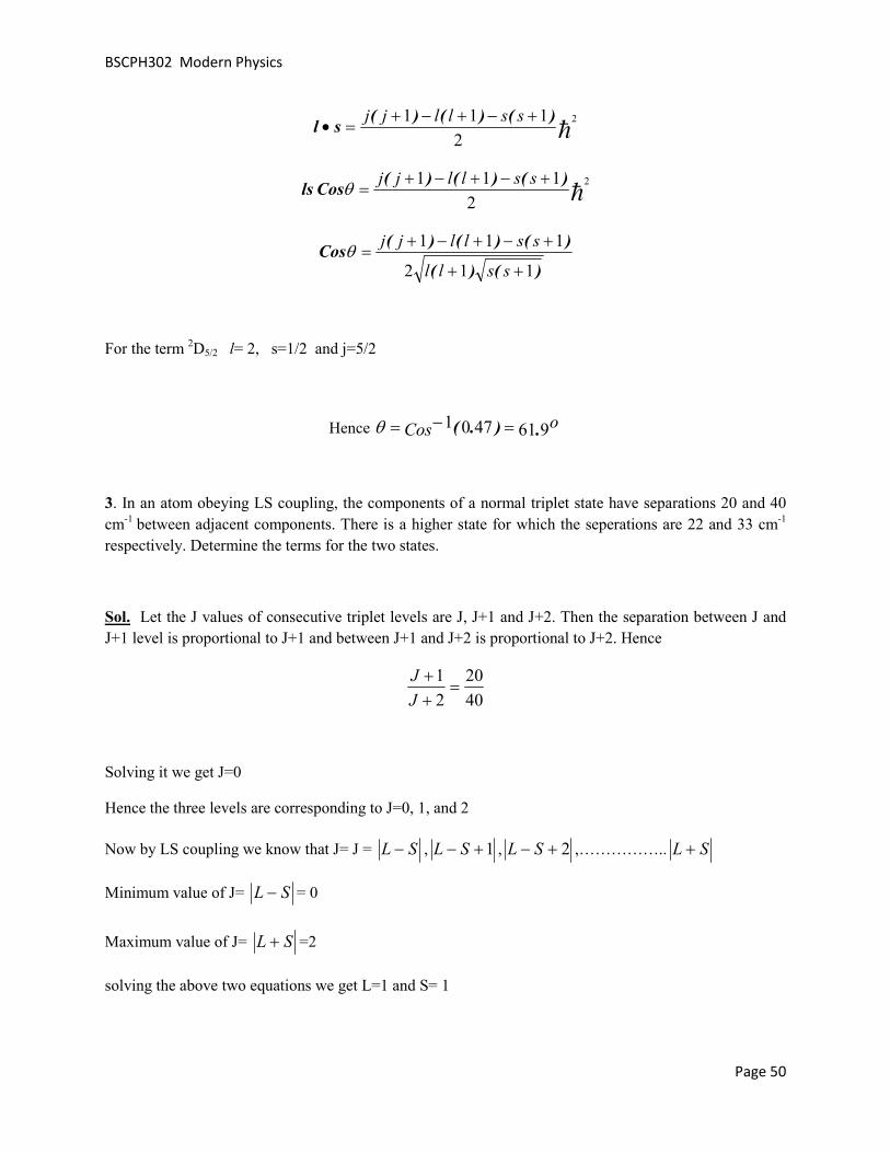
Hence 9614701 .).( oCos =−=θ
3. In an atom obeying LS coupling, the components of a normal triplet state have separations 20 and 40 cm-1
between adjacent components. There is a higher state for which the seperations are 22 and 33 cm-1 respectively. Determine the terms for the two states.
Sol. Let the J values of consecutive triplet levels are J, J+1 and J+2. Then the separation between J and J+1 level is proportional to J+1 and between J+1 and J+2 is proportional to J+2. Hence
4020
21
=++
J
J
Solving it we get J=0
Hence the three levels are corresponding to J=0, 1, and 2
Now by LS coupling we know that J= J = SL − , 1+− SL , 2+− SL ,…………….. SL +
Minimum value of J= SL − = 0
Maximum value of J= SL + =2
solving the above two equations we get L=1 and S= 1

BSCPH302 Modern Physics
Page 51
Hence the term is 3P0, 1, 2
2.18 REVIEW QUESTIONS
1. What do you mean by spin and space quantization. Find the possible orientations for spin vector s with respect to the magnetic field in z axis.
Ans 54.7o and 125.3o
2. Calculate the possible orientations of the total angular momentum vector j corresponding to j=3/2 with respect to a magnetic field along the z-axis.
Ans 39.2o, 75o, 105o,140.8o
3. Is 2D1/2 term possible? Justify your answer.
Ans. No
4. What is Pauli’s exclusion principle? Write it’s significance also.
5. What is the difference between LS and JJ coupling.
6. Write down the electronic configuration of Carbon atom and write the possible spectroscopic terms.
7. Write the spectroscopic terms for two non equivalent electron systems npn’d.
8. Find the ratio of energy separation between levels of the term 3D1,2,3.
Ans 2:3
2.19 OBJECTIVE QUESTIONS
1 An electron whose orbital quantum number is 2 has the angular momentum
a) h6 b) h5 c) 6ħ d)0

BSCPH302 Modern Physics
Page 52
2 All components of orbital and spin angular momentum are quantized.
a) True b)False c) May or may not d) Only in presence of external magnetic field
3 Pauli’s exclusion principle is followed by
a) Electrons only b) All fermions c) All particles d) None
4 A hydrogen atom is in the 4p state. To what state or states can it go by radiating a photon in an allowed transition
a) any of the d and s state b) only to d state c) only to s state d) d and f states
5 If j=3/2 then how many orientations are possible for J with respect to z axis
a)4 b)3 c)2 d) All
6 If j=5/2 then possible values of l are
a)2,5 b)2,3 c)5,3 d) 0
7 For a diamagnetic material, the total angular momentum quantum number is
a)1 b)zero c)±1 d) Any vale
8 Spectroscopic term for d10 type of configuration is
a)3P0 b)1S1 c)1S0 d)2S0
9 For atomic no Z>15 which coupling will dominate
a)LS b)JJ c) Any of the two d) Depends upon magnetic field
10 No of levels for the term 4P3/2 will be
a)4 b)3 c)2 d)1
2.20 ANSWERS
1(a) 2(b) 3(b) 4(a) 5(a) 6(b) 7(b) 8(c) 9(b) 10(b)

BSCPH302 Modern Physics
Page 53
UNIT 3 BASICS OF ATOMIC SPECTRA II
Structure
3.1 Introduction
3.2 Objectives
3.3 Spectra of Alkali Metals
3.4 Explanation of fine structure of Sodium D lines
3.5 Intensity rules
3.6 Zeeman Effect
3.6.1 Classical explanation of Zeeman Effect
3.6.2 Quantum explanation of Zeeman Effect
3.6.3 Frequency position of Zeeman splitted components
3.6.4 Experimental procedure for the observation of Zeeman Effect
3.7 Summary
3.8 Glossary
3.9 References
3.10 Suggested Readings
3.11 Solved Examples
3.12 Review Questions
3.13 Objective Question
3.14 Answers

BSCPH302 Modern Physics
Page 54
3.1 INTRODUCTION
After studying basics of atomic spectra in unit 2, you understand the mechanisms that lead to the origin of different spectral lines. Hydrogen is the simplest atom having only 1 electron, so its spectrum is relatively easier to understand and because of this the initial researches about atomic spectrum start with Hydrogen. Alkali metals have 1 electron in their outer shell. So it is also interesting to study their atomic spectrum and see how it becomes complicated as outer electrons occupy higher orbital.
3.2 OBJECTIVES
The objective of this unit is to make students capable to understand
1. Atomic spectrum and fine structure for alkali metals.
2. Splitting of spectrum lines in presence of exernal magnetic field.
3. Effect of strength of magnetic field over the splitting of spectrum lines which can be used for different applications.
3.3 SPECTRA OF ALKALI METALS
The alkali metals like lithium, Sodium, potassium, rubidium etc have electronic configuration as shown below:
Lithium (Li) 1s2,2s1
Sodium (Na) 1s2,2s2,2p6,3s1
Potassium (K) 1s2,2s2,2p6,3s2,3p6,4s1
Rubidium (Rb) 1s2,2s2,2p6,3s2,3p6,4s2,3d10, 4s2, 4p6,5s1 .
These all have single electron in outer most shell like that of Hydrogen atom. All inner shells are completely filled, so their contribution towards total angular momentum is zero (as you have studied in last unit). Therefore we will deal with the spin and orbital angular momentum of this outer electron only. The spectra emitted by the alkali metals are characterized by the transition of this valence electron.

BSCPH302 Modern Physics
Page 55
Figure 1: The energy levels and spectrum of Sodium atom. The lines from 7p-3s, 6p-3s and so on form the Principal series. The transitions 7s-3p, 6s-3p and so on constitute the Sharp series. The
transitions like 3d-3p, 4d-3p form Diffuse series and Fundamental series is due to 4f-3d, 5f-3d and so on.
Alkali spectra can be grouped into four series as shown in Figure 3.1 for Sodium atom:
1. Principal series: The series arises from the transitions between various P-levels and lowest S-level.
2. Sharp series: This series consists of transition lines from S-level to the lowest of the P-level.
3. Diffuse series: It arises from the transitions between the various D-levels and the lowest P-level.
4. Fundamental series or Bergmann Series: It arises due to transitions from various F-levels and the lowest D-level.
Actually the spectrum is not as simple as is seen in the Figure1. The energy levels except the ‘s’ level, are splitted due to spin orbit interaction and a single transition line has more lines associated with it. This is called fine structure of spectral lines, which you have studied earlier also. Here, we are going to discuss the fine structure of Sodium.

BSCPH302 Modern Physics
Page 56
3.4 EXPLANATION OF FINE STRUCTURE OF SODIUM D
LINES
Figure 2: Fine structure of Sodium showing the D1 and D2 lines arising due to transitions from
3p3/2 to 3s1/2 and 3p1/2 to 3s1/2 respectively. The spin orbit coupling splits 3p level in two sublevels having an energy gap of 0.0021 eV.
Spectrum of Sodium atom arises due to the transition of the outer electron (3s1) to excited states. And the available excited states are 3p, 3d, 4s, 4p and so on. The transitions between energy levels follow the selection rule 1±=∆l and are shown in Figure3.1. The transition line 3p to 3s is called the Sodium D line and the energy change in this transition corresponds to the wavelength of yellow colour (
yellow
sp
hcEE
λ=− 33 , this is the light you see in Sodium Lamps). When this line is observed with a high
resolving power spectrometer it was found to have two components. These two components arise due to spin orbit splitting of the p level. The s state remains unsplitted with j =1/2. The p state splits in two levels corresponding to the j values 1/2 and 3/2 due to spin orbit interaction.
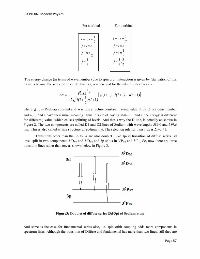
BSCPH302 Modern Physics
Page 57
For s orbital For p orbital
The energy change (in terms of wave number) due to spin orbit interaction is given by (derivation of this formula beyond the scope of this unit. This is given here just for the sake of information)
[ ])()()(
))((
1111
21
2 3
2
+−+−+++
−=∆ ∞ sslljjlll
Z
n
R αε
where R∞ is Rydberg constant and α is fine structure constant having value 1/137, Z is atomic number
and n,l, j and s have their usual meaning. Thus in spite of having same n, l and s, the energy is different for different j value, which causes splitting of levels. And that’s why the D line, is actually as shown in Figure 2. The two components are called D1 and D2 lines of Sodium with wavelengths 589.0 and 589.6 nm. This is also called as fine structure of Sodium line. The selection rule for transition is Δj=0,±1.
Transitions other than the 3p to 3s are also doublet. Like 3p-3d transition of diffuse series. 3d level split in two components 32D5/2 and 32D3/2 and 3p splits in 32P3/2 and 32P1/2.So, now there are three transition lines rather than one as shown below in Figure 3.
Figure3: Doublet of diffuse series (3d-3p) of Sodium atom
And same is the case for fundamental series also, i.e. spin orbit coupling adds more components in spectrum lines. Although the transition of Diffuse and fundamental has more than two lines, still they are
21
21
0
21
,0
=
±=
±=
==
j
j
slj
sl
23
,21
21
1
21
,1
=
±=
±=
==
j
j
slj
sl

BSCPH302 Modern Physics
Page 58
called doublet, because doublet refers to the multiplicity of the energy level (i.e. the number of splitted energy levels) not the number of components of spectrum.
Apart from fine structure splitting there are more factors that cause further splitting of energy levels, like hyperfine splitting- due to nuclear spin angular momentum, Zeeman splitting- because of external magnetic field, Stark splitting-because of external electric field. These you will be studying later. Here, we will discuss the Zeeman effect or Zeeman splitting.
3.5 INTENSITY RULES
What will be the intensity ratio of different components of spectral lines or which will be bright and which will be faint is governed by some intensity rules. General observation shows that the line arising due to the transition in which j and l changes in same way, is strongest. For example
In case of D1 and D2 lines of Sodium,
Figure4: Intensity variation of D1, D2 lines of Sodium
The D1 line is due to transition from 2P1/2 to 2S1/2 Here, change in j, Δj= 0 and change in l, Δl=1
Now The D2 line is due to 2P3/2 – 2S1/2. Here change in j= 1 and change in l=1.So the D2 line is stronger than D1 line.
If there are more than one such line in which j and l changes by same then the one having largest j value will be of highest intensity.
For example- The doublet of diffuse series has transition lines 2D5/2 - 2P3/2 here Δj=1 and Δl=1 And 2D3/2 – 2P1/2 here Δj=1 and Δl=1
But the transition 2D5/2 - 2P3/2 involves higher value of j, hence it will be stronger in intensity.

BSCPH302 Modern Physics
Page 59
Figure 5: Intensity variation of different components of diffuse series of Sodium atom
Now if talk about how intense will be a particular line i.e. the quatitaive value of intensity then following rule will be followed:
Ex-1 D1 , D2 lines of Sodium.
Intensity of D1 line is proportional to(2j+1) value of the starting level (2P1/2)
Intensity of D2 line is proportional to (2j+1) value of it’s starting level 2P3/2
2:1:
42
123
2
121
2
21
2
1
=
=+×
+×=
II
II
DD
D
D
.
Ex-2 Diffuse series
The sum of the intensities of those lines of a multiplet which come from a common initial level is proportional to the quantum weight (2j+1) of that level.

BSCPH302 Modern Physics
Page 60
32
64
)125
2(
)123
2(
==+
+×
+×+
III
I
II
a
cb
a
cb
α
α
(2j+1 value of the starting level of Ia).
And the sum of the intensities of those lines of a multiplet which end on a common level is proportional to the quantum weight of that level.
(2j+1 value of the ending level)
12
24
==+
III
c
ab
So now we have two equations III cab323 −=− and
III cab2=+
On solving these two 5:1:9:: =III cba.
Thus it can be find out by calculation that a particular level will split in hw many levels due to spin orbit interaction and which component will be more intense or how intense? And it can be verified experimentally. In practice we use Sodium lamp in our labs but we are not able to resolve the D1 and D2 lines as the wavelength separation between these lines is only 6Å and our eye cannot resolve such a small value. Therefore Sodium lamps are used as monochromatic light source but in actual it is not monochromatic and that you understand now.
3.6 ZEEMAN EFFECT
Splitting of spectral lines in presence of magnetic field is called Zeeman Effect. It was discovered by Dutch physicist Pieter Zeeman who first observed it in 1896. Zeeman found that when a sodium flame is placed between the poles of a powerful electromagnet the two lines of the first principal doublet are considerable broadened. In his early investigations he was not able to split any line into doublet or triples
)121
2(
)123
2(
+×
+×+
α
α
I
II
c
ab

BSCPH302 Modern Physics
Page 61
but he found that they were widened and their outside edges were polarized. He was later able to photograph the two outer components of lines in a number of elements Zn, Cu, Cd and Sn. A single spectral line is seen splitted in to three polarized components:
1. Central unshifted line- plane polarised (having electric vector* vibrating parallel to the applied external magnetic field ) when viewed perpendicular to the external magnetic field. And this line is not seen when viewed parallel to the magnetic field. This is called π component.
2. Two lines circularly polarized when observed parallel to the magnetic field and linearly polarized (having electric vector* vibrating perpendicular to the applied external magnetic field) when viewed at right angles to the field. Both the lines are equally spaced on both sides of π component. These are called σ component.
Figure 6: Schematic representation of Zeeman effect - Splitting of spectral line in to 3 components
in presence of external magnetic field.
Figure 7: Polarisation of σ and π components of Zeeman Effect

BSCPH302 Modern Physics
Page 62
Zeeman Effect has played an important role in the development of quantum theory. It illustrates the phenomenon of space quantization- that the angular momentum L of the atom can have only a set of discrete orientations with respect to an external magnetic field B. The typical strength of the internal magnetic field of the atom acting on an electron is of the order of 1 Tesla hence the external field is called “weak” if it’s strength is about 0.8 Tesla and lower and if the strength is 1.5 Tesla and higher then it is referred as “strong” field.
3.6.1 Classical explanation of Zeeman Effect
Lorentz explained the Zeeman Effect classically. He already predicted that the magnetically splitted components of spectral lines will be polarized and this was later confirmed by Zeeman, as we discussed above. By simple classical theory of Lorentz it has been shown that if a light source is placed in external magnetic field, the motion of electrons should be modified such as to change their period of motion. The electron moving in circular orbit in a plane perpendicular the external magnetic field , will be speeded up or slowed down by some amount that depends upon the strength of magnetic field. Lets calculate the change in frequency of electron due to the presence of external magnetic field.
When electron is moving in circular orbit of radius r and angular frequency ω then
mr ω2 = F
now suppose due to the external field B, the frequency of revolution changes by Δω then
mr(ω+Δω)2= F+ evB where evB is magnetic force on electron.
mrω2+ mr Δω2+ 2mrωΔω= F+ evB
or mr Δω2+ 2mrωΔω = evB
Δω is very small so Δω2 will be smaller, hence it can be neglected. Therefore
2mrωΔω= evB but v=rω so
m
eBm
eB
πν
ω
4
2
=∆
=∆.
If the field is upward normal to the plane of paper then the electrons moving in counter clockwise direction in the plane of paper are speeded up and those moving in clockwise direction are slowed down by the amount Δυ.
3.6.2 Quantum Explanation of Normal Zeeman Effect:
In an external magnetic field B, a magnetic dipole has an amount of potential energy (Um) that depends upon both the magnitude of magnetic moment (μ) of the dipole and the orientation of this moment with respect to the field.
The torque τ on a magnetic dipole in a magnetic field of flux density B is

BSCPH302 Modern Physics
Page 63
μBSinθτ
Bμτ
=
×=
where θ is the angle between μ and B. The torque is maximum when the dipole is perpendicular to the field and zero when it is parallel or anti-parallel to it.
Figure 8: Precession of orbital angular momentum vector L with respect to the external
magnetic filed B.

BSCPH302 Modern Physics
Page 64
Figure 9: Transition between the energy levels splitted due to Zeeman Effect.
Here ΔE= E2 - E1
And we have studied it earlier that the magnetic moment of the orbital electron depends on it’s angular momentum L. The magnetic moment of electron is
L2m
e−=μ .
The magnetic potential energy of an atom in a magnetic field is
BmUm
Bmm
eUm
LCosmL
LBCosm
eUm
BLm
eUm
BUm
lB
l
lz
µ
θ
θ
µ
=
=
==
=
•=
•=
2
2
2
h
h
.

BSCPH302 Modern Physics
Page 65
In a magnetic field, then the energy of a particular atomic state depends on the value of ml as well as on that of n (the principal quantum number). A state of total quantum number n breaks up into several sub-states when the atom is in a magnetic field, and their energies are slightly more or slightly less than the energy of the state in the absence of the field. This phenomena lead to splitting of individual spectral lines into separate lines when atoms radiate in a magnetic field. The spacing of the lines depends on the magnitude of the field.
Now, because ml can have the (2l+1) values from +l through 0 to –l, a state of given orbital quantum number l is splitted into 2l+1 substates each having energy μB mlB. However, the possible changes in ml for a transition to take place are restricted to Δml=0,±1. Therefore, a spectral line from a transition between two states of different l will split into only three components as shown in figure below:
In the figure 9 you see 9 lines corresponding to the single transition line from l=2 to l=1. But actually in spectrum you see only three components. See the three lines corresponding to the selection rule Δml = +1 have energy ΔE+μBB, so in the spectrum these three will be seen at the same frequency position, so rather than three you will see only 1 line. Similarly the three lines corresponding to Δml = 0 will merge in to single line at the frequency position corresponding to energy ΔE and the lines arising due to the selection rule Δml = -1 will be seen as single line corresponding to energy ΔE- μBB. Out of these three the one for which Δml = 0 is called π component, and Δml = ±1 are called σ component.
Zeeman effect is of two types : Normal Zeeman Effect and Anomalous Zeeman Effect. In Normal Zeeman effect a single spectral line is splitted in to three components while in Anomalous it is more than 3. Normal Zeeman Effect is observed by the singlet states like 1P or 1D etc. The reason behind this is that in the calculation of Normal Zeeman effect we did not include the spin angular momentum (or Spin was not known at that time). If spin angular momentum is also included then there will be spin orbit splitting of spectral lines and rather than L (the orbital angular momentum) it will be J= L+S (total angular momentum) that will interact with the external magnetic field. Hence the spectrum will be more complex having more number of components. You will study later in detail about the Anomalous Zeeman Effect.
So if S=0 the multiplicity 2S+1= 1 and J=L
Hence the singlet levels show only Normal Zeeman effect. For the states having multiplicity greater than 1, you can say that Anomalous Zeeman effect is fine structure of Normal Zeeman Effect.
3.6.3 Frequency position of Zeeman splitted components
For π component, if υo is the frequency of radiation then
hυo= E2 - E1.
For σ components if the frequencies are υ1 and υ2 for the lower and higher energy values respectively, then
h υ1= E2 - E1- μBB

BSCPH302 Modern Physics
Page 66
Bπm
eν
h
Bμνν
h
BμBh
E1E2ν1
oBo 41 −=−=
−−
=.
And similarly,
Bπm
eν
h
Bμνν oBo 42 +=+= .
Thus change in frequency m
eB
πν
4=∆ same as suggested by Lorentz.
If we talk about the intensity then both the σ and π components are equally intense.
3.6.4 Experimental procedure for the observation of Zeeman Effect
Figure 10: Schematic of the experimental set up to observe the Zeeman Effect.

BSCPH302 Modern Physics
Page 67
Figure 11- Fabry Perot Etalon. Here Δ = optical path difference, d = thickness of the etalon, n = refractive index of the glass, and α is the aperture angle. All these parameters help to calculate the wavelength separation between the splitted components.
Figure 12: Fringes of red Cadmium line without magnetic field.

BSCPH302 Modern Physics
Page 68
Figure 13 : Splitted fringes due to the presence of magnetic field.
The Zeeman Effect enables spectroscopic separation of the differently polarized components. To demonstrate the shift, however, we require a spectral apparatus with extremely high resolution. The apparatus is shown schematically in Figure 10. The source in which Zeeman Effect is to be observed is placed between the poles of an electromagnet. Here Cadmium lamp is used. Light emitted by the lamp passes through a Fabry Perot (FP) etalon. A red filter is used to isolate the red Cadmium line. Fabry Perot is a glass plate with both sides being aluminized. The slightly divergent light enters the etalon, and is reflected back and forth several times, whereby part of it emerges each time (see Figure 11). Due to the aluminizing this emerging part is small, i.e., many emerging rays can interfere. The circular fringes are produced by FP due to constructive interference. These circular fringes are imaged as circular rings in the focal plane of the telecopy lens. This intermediate image can either be viewed using an eyepiece or it is viewed by a CCD camera. Figures 12 and 13 show the circular rings with and without magnetic field. Presence of magnetic field causes each ring to split in three components, i.e. confirming the Zeeman Effect.
3.7 SUMMARY
In this unit you studied mainly about the fine structure of spectral lines of Sodium. In the spectrum of Na atom the first line of principal series 3p-3s has actually two components one due to transition from 2P3/2 to 2S1/2 and other due to transition from 2P1/2 to 2S1/2 . These are labeled as D2 and D1 lines and correspond

BSCPH302 Modern Physics
Page 69
to 5890Å and 5896Å wavelengths respectively. This is called the fine structure of Sodium and this splitting of spectral line is due to spin orbit coupling. Next the rules for intensities of these components were discussed.
Apart from spin orbit interaction there is one more type of splitting that is called Zeeman splitting. In presence of external magnetic field there is change in energy of the atom due to the interaction of orbital magnetic dipole moment with the magnetic field. This energy change causes splitting of spectral lines in to 3 polarized components and this effect is known as Zeeman Effect. The effect is observed for the singlet states (S=0) only. The states having multiplicity more than one, show Anomalous Zeeman effect.
3.8 GLOSSARY
• Intensity Power Transferred per unit area. It is a measure of brightness
• Wave number reciprocal to wavelength
• Unpolarized Light The Electric vector of light vibrates in all possible planes
• Plane Polarization Electric vector vibrates only in 1 plane
• Etalon An optical device that has parallel mirrors
• Interfere Superposition of waves moving in same direction
• Fringes Interference pattern
3.9 REFERENCES
1. Atomic and Molecular Physics by Rajkumar
2. Introduction to Quantum Mechanics by David J. Griffiths
2. Modern Physics by Arthur Beiser
3.10 SUGGESTED READINGS
1. Introduction to atomic spectra by Harvey Elliott White
2. Fundamentals of molecular spectroscopy by C. N. Banwell
3.Atomic and Molecular Spectroscopy: Basic Aspects and Practical Applications by Sune Svanberg

BSCPH302 Modern Physics
Page 70
3.11 SOLVED EXAMPLES
Ex-1 A sample of a certain element is placed in a magnetic of flux density 0.3 weber/m2. How far apart are the Zeeman components of a spectral line of wavelength 4500 Å?
Sol: Frequency change due to Zeeman Effect is given by
1092410 31191434
3010 1961
4
×=−×××
×−×=
=∆
...
..
m
eB
πν
( )
Å
mmagnitude
c
c
c
0280
10 1528351083
10 1045002
10924
2
2
.
.)(
=
−×=×
−×××=∆
×∆−=∆
∆−=∆
=
λ
λνλ
λλ
ν
λν
.
Ex2 Show the Zeeman effect in the transition 1F3-1D2.
Sol For F , l= 3 therefore ml= -3,-2,-1,0,1,2,3
For D, l= 2 therefore ml= -2,-1,0,1,2,
Selection rule for transition is Δ ml = 0, ±1

BSCPH302 Modern Physics
Page 71
Ex3 The Zeeman components of a 500 nm spectral line are 0.0116 nm apart when the magnetic field is 1.00T.Find the ratio e/m for the electron.
Sol From Ex1, we have
cλν
λ2×∆
=∆ and m
eB
πν
4=∆
So
λ
πλπ
λλ
24
4
2
B
c
m
emc
eB
×∆=
×=∆
( )kgC
m
e
/.
..
10117481
10 95002
1
1433410 901160 108
×=
−××
××××−×=

BSCPH302 Modern Physics
Page 72
3.12 REVIEW QUESTIONS
1. Find the intensity ratio of different components in the following transition
Ans: Ia: Ib:Ic = 20:1:14
2. The second line of fundamental series is due to transition 5f-3d. This single line will split in how many components due to spin orbit interaction?
Ans: 3
3. Is Sodium light ideally monochromatic? Justify your answer?
4. Distinguish between the spectra of hydrogen and sodium atoms? What makes the two spectra look so different even though both belong to the single valence electron system?
5.Find the minimum magnetic field needed for the Zeeman effect to be observed in a spectral line of 400 nm wavelength when a spectrometer whose resolution is 0.010 nm is used.
Ans- 1.34T
6. For Calcium the transition line (P to S) has wavelength 4226.73Å. Calculate the wavelength of three components of Normal Zeeman effect when the atom is placed in a magnetic field of 4 weber/m2.
Ans 4226.40Å, 4226.73Å and 4227.06Å
3.13 OBJECTIVE QUESTIONS
1. Normal Zeeman effect is observed in singlet sates only
(a) Always True
(b) Always False
(c) Depends upon the atom
(d) May or may not be true
2. Splitting of spectral lines in presence of electric field is called
(a) Stark Effect

BSCPH302 Modern Physics
Page 73
(b) Zeeman Effect
(c) Both (a) and (b)
(d) None
3.The separation between the two components of Sodium D lines is
(a) 6Å
(b) 6.5Å
(c) 0.6 nm
(d) Both (a) and (c)
4. Why don’t we see two separate wavelengths while using Sodium lamp-
(a) Because the two wavelengths are so close that our eye cannot resolve their separation
(6Å)
(b) Because Sodium light is monochromatic
(c) None of the two
5. For sodium D lines, the intensity of
(a) D1 is greater than D2
(b) Intensity of D2 is greater than D1
(c) Both D1 and D2 are of same intensity
(d) None
6. The fine structure of spectral lines is due to
(a) Spin orbit coupling
(b) External magnetic field
(c) External electric field
(d) None
7. 1S0 level will split in how many levels in presence of magnetic field
(a) Zero
(b) 2

BSCPH302 Modern Physics
Page 74
(c) 3
(d) None of the above
8. Selection rule for Zeeman effect is
(a) Δml=±1
(b) Δml=0,±1
(c) Δml=0
(d) None of the above
9. Selection rule for transition between the levels splitted due to spin orbit coupling is
(a) 1±=∆l
(b) 1±≠∆l
(c) 10 ±=∆ ,l
(d) onlyl 1=∆
10. What factor decides that external magnetic field is strong or weak..
(a) Internal magnetic field due to electron’s orbital motion
(b) Spin magnetic moment
(c) Individual atom type
(d) None
11. If magnetic field B1 is sufficient to observe Zeeman effect in atom 1 then, for the same field B1, all atoms will show Normal Zeeman effect
(a) Never
(b) Always
(c) Not necessary, they or may not show
(d) None
3.14 ANSWERS
1(a) 2(a) 3(d) 4 (a) 5 (b) 6 (a) 7(a) 8(b) 9(b) 10(a) 11 (c)

BSCPH302 Modern Physics
Page 75
UNIT 4 X-RAY
Structure
4.1 Introduction
4.2 Objective
4.3 Discovery of X-rays
4.4 Cathode ray tubes
4.5 How X-rays are produced
4.5.1 Modern X-ray tube
4.6 Nature of X-rays
4.7 X-ray diffraction (Bragg’s Law)
4.8 X-ray spectra
4.9 X-ray spectra vs Optical spectra
4.10 Moseley Law
4.10.1 Moseley Law according to Bohr Theory
4.10.2 Significance of Moseley Law
4.11 Absorption of X-rays
4.12 Applications of X-rays
4.13 Summary
4.14 Glossary
4.15 References
4.16 Suggested Readings
4.17 Solved Examples
4.18 Review Questions
4.19 Objective Question
4.20 Answers

BSCPH302 Modern Physics
Page 76
4.1 INTRODUCTION
X-rays are part of electromagnetic spectrum with their wavelength ranging from 0.01-10 nm.
Let’s study how they were discovered and why their study is important?
4.2 OBJECTIVES
The objective of this unit is to make students aware about
1. Important discovery of X-rays and their spectrum.
2. Vast application of X-rays in identification of crystal structure, unknown elements, in the field of
medical etc.
3. Production of X-rays
4.3 DISCOVERY OF X-RAYS
Wilhelm Konard Roentgen on Nov 8, 1895 noticed that when he switched on a cathode ray tube-CRT (which was entirely covered with black cardboard so that no light comes out of it) a nearby sheet of paper coated with barium platinocyanide glowed. Seeing the barium platinocyanide crystals glowing with a shimmering yellow-green fluorescence, Roentgen's keen scientific mind became curious. It was known at that time that cathode rays could make the crystals glow - but the surprising point was that tube was covered with black card board and the paper was not very close to the CRT, so cathode rays were not

BSCPH302 Modern Physics
Page 77
supposed to come out of the card board and glow the sheet. Therefore, he placed the crystal screen at an even greater distance than the range of cathode rays were known to penetrate. Still there was this strange fluorescence. Then he grabbed a book and placed it between the Cathode ray Tube and the screen. The crystals continued to glow. Whatever it was, it was coming through the book! Next, he tried metals - and found that the rays penetrated in varying degrees, although lead and platinum stopped them completely. Then Roentgen exposed his hand, and - his heart must have almost stopped – when he saw the shadows of his bones. As Roentgen made photographs of his findings, it was obvious that he had found something far more exciting than cathode rays. When Roentgen was interviewed about these rays he was asked-
Is it electricity?
He said not in any known form.
Then What is it?
He said I don't know.
He discovered these mysterious new rays by accident during a routine laboratory experiment on the night of November 8, 1895. These new rays were called as "X-ray" by Roentgen. “X” is used as the mathematical symbol for the unknown. Hence the name "X ray," was given because of their unknown nature. Discovery of X- rays were an immediate sensation and only two months later X-rays were being used in medicine. They also stimulated research in new directions. After one year of X-rays, Becquerel discovered radioactivity. Roentgen was awarded the first Nobel Prize in Physics for his discovery of X-rays and sparked a rash of copycat experiments.
4.4 CATHODE RAY TUBE (CRT)
Before we move further, towards the understanding of nature and production of X-rays, you must recall what is Cathode ray tube. Figure 1 shows the diagram of a cathode ray tube. Cathode ray tube is also known as Braun’s tube and was invented by German Physicist Karl Ferdinand Braun. There is a source of electron called electron gun which produces stream of electrons through thermionic emission at the heated cathode. This electron stream is focused in to a beam with the help of control grids. A strong electric field between cathode and anode accelerates the electrons; before they leave the electron gun through a small hole in the anode. The electron beam can be deflected by a capacitor or coils in it’s way which causes it to display an image on the screen. The image may represent electrical waveforms (oscilloscope), pictures (television, computer monitor), echoes of aircraft detected by radar etc. When electrons strike the fluorescent screen, light is emitted. The whole configuration is placed in a glass tube having vacuumed inside to avoid collisions between electrons and gas molecules of the air, which would attenuate the beam.

BSCPH302 Modern Physics
Page 78
Figure 1: Cathode Ray Tube
4.5 HOW X-RAYS ARE PRODUCED
The simple fact is this: X-rays are relatively easy to generate. You simply speed up electrons and let them collide with a target. The electrons cause disturbances within the atoms of the target, releasing X-rays. Any material, even a gas or liquid, will release X-rays when bombarded by high-velocity electrons. Obviously, then, glass can be a target, as is in the tube Roentgen used. In cathode ray tube electrons are accelerated in a vacuum by an electric field and it was the impact of these electrons on the glass end of the tube that produced the penetrating rays which were termed as “X-rays”. The X-ray tube with which Roentgen made his original discovery of X-rays was of the type as shown below in Figure 2 and Figure3. The glass bulb, technically called a Crookes tube, contained a tiny bit of air, about a millionth of normal air pressure. When a high voltage is applied over the tube, the electrons in the gas became free from their atoms and moved towards the high-voltage

BSCPH302 Modern Physics
Page 79
Figure 2: X-ray Machine used by Roentgen
Figure 3: Crookes tube used by Roentgen produced X rays when electrons flowing from its cathode to its anode bombarded the glass tube.
positive anode. The negatively charged electrons, in turn, were drawn toward the highly positive anode. The resultant stream of electrons, actually cathode rays, traveled so fast - about 30,000 miles per second - that most of them could not "turn the corner" to reach the anode (Figure 3). Instead, they smashed into the glass wall of the tube. The glass, therefore, was the target, providing the barrier for the sudden stoppage

BSCPH302 Modern Physics
Page 80
of electrons. The result is radiation of X-rays and glow of fluorescence that Roentgen observed. When the electrons hit the glass walls of the Crookes tube, they lose energy in the form of a green glow and invisible X-rays. Heavy positive ions in the residual gas were drawn to the negative cathode (unlike charges attract), striking with such force that they knocked electrons from the cathode metal. It was this positive-ion bombardment that created and maintained a source of electrons.
A team of physicist, engineers and radiologists revived first generation X-ray device. They found that the antique machine still sparked and glowed and used thousands of times more radiation than it’s modern counter parts to take an image. Old machine took surprisingly clear pictures, but gave the skin a dose of radiation 1,500 times greater than the same image would require today. An exposure that takes 21 milliseconds (thousandths of a second) on a modern machine took up to 90 minutes on the antique system. Many of these early X-ray workers developed cancer, and many of them died untimely, very young.” with all the shielding used today, modern X-ray workers feel less radiation in the hospital than they do at home. Now in all those years, they improved technology so far that you can really neglect what you are receiving when you do normal X-ray scans.”
4.5.1 MODERN X-RAY TUBE:
Figure 4: X-ray tube detail. The focusing cup concentrates electron stream from
cathode and directs it toward tungsten target material
Like Roentgen's original tube, the modern tube also has a cathode and an anode, but with lots of improvements. A basic X-ray unit is comprised of filament, high-voltage transformer and timing circuits - see Figure 4. Now the tube is evacuated to an extremely high vacuum. The cathode structure contains a
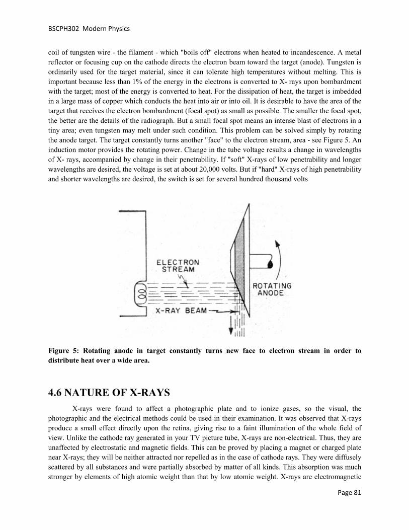
BSCPH302 Modern Physics
Page 81
coil of tungsten wire - the filament - which "boils off" electrons when heated to incandescence. A metal reflector or focusing cup on the cathode directs the electron beam toward the target (anode). Tungsten is ordinarily used for the target material, since it can tolerate high temperatures without melting. This is important because less than 1% of the energy in the electrons is converted to X- rays upon bombardment with the target; most of the energy is converted to heat. For the dissipation of heat, the target is imbedded in a large mass of copper which conducts the heat into air or into oil. It is desirable to have the area of the target that receives the electron bombardment (focal spot) as small as possible. The smaller the focal spot, the better are the details of the radiograph. But a small focal spot means an intense blast of electrons in a tiny area; even tungsten may melt under such condition. This problem can be solved simply by rotating the anode target. The target constantly turns another "face" to the electron stream, area - see Figure 5. An induction motor provides the rotating power. Change in the tube voltage results a change in wavelengths of X- rays, accompanied by change in their penetrability. If "soft" X-rays of low penetrability and longer wavelengths are desired, the voltage is set at about 20,000 volts. But if "hard" X-rays of high penetrability and shorter wavelengths are desired, the switch is set for several hundred thousand volts
Figure 5: Rotating anode in target constantly turns new face to electron stream in order to distribute heat over a wide area.
4.6 NATURE OF X-RAYS X-rays were found to affect a photographic plate and to ionize gases, so the visual, the photographic and the electrical methods could be used in their examination. It was observed that X-rays produce a small effect directly upon the retina, giving rise to a faint illumination of the whole field of view. Unlike the cathode ray generated in your TV picture tube, X-rays are non-electrical. Thus, they are unaffected by electrostatic and magnetic fields. This can be proved by placing a magnet or charged plate near X-rays; they will be neither attracted nor repelled as in the case of cathode rays. They were diffusely scattered by all substances and were partially absorbed by matter of all kinds. This absorption was much stronger by elements of high atomic weight than that by low atomic weight. X-rays are electromagnetic

BSCPH302 Modern Physics
Page 82
rays similar to visible light rays, with this important exception - their wavelength is very small, about 1/10,000th that of light. It is this exceedingly short wavelength of X-rays that makes possible their penetration of matter, and which enables the researcher to look into the vast voids of molecular inner space. Traveling at the same speed as light and radio waves, X- rays can be reflected and refracted only at very small angles. (Roentgen failed to focus X- rays, despite many experiments with lenses of wood, glass, aluminum, and other materials, for this reason.) The darkening of photographic film by X-rays has given them wide application in medicine, research, and industry.
4.7 X-RAY DIFFRACTION (BRAGG’s LAW)
For the confirmation of the wave nature of X-rays Walter and Pohl in 1909 studied the diffraction of X-rays (because diffraction is a wave phenomenon) but could not conclude anything. Then Laue tried in this direction. For diffraction the spacing between the grating elements should be of the order of wavelength radiation. Wavelength of X-rays is of the order of 1Å and it was not possible to have a grating with so small grating element. The spacing between the crystal planes is of this order hence Friedrich and Laue used crystal as diffraction grating and got success in diffracting X-rays. After their experiment Bragg started investigation and he found that in a crystal a set of parallel planes can be drawn as shown in Figure 6. These are called Bragg planes. Suppose the spacing between two consecutive planes is ‘d’ and monochromatic X-rays of wavelength λ are incident at an angle θ with the crystal surface. Incident X-rays are scattered by atoms in all directions and interfere. Constructive interference will take place when the path difference between interfering rays is integral multiple of wavelength.
Figure 6: Diffraction of X-rays by Bragg’s planes. The blue dots represent the lattice points.
From the figure path difference in the rays reflected from point O and G is FG+GH
From the ΔOFG and OHG it can be seen that FG=GH=dSinθ

BSCPH302 Modern Physics
Page 83
for constructive interference path difference=nλ
2dSinθ=nλ.
This is called Bragg’s Law. If the crystal spacing is known, then using Bragg’s law wavelength of X-rays can be determined and if wavelength is known then crystal spacing can be calculated. Bragg’s law is very useful in the determination or confirmation of different crystal structures.
4.8 X-RAY SPECTRA X-rays coming from the X-ray tube do not have single energy but constitute a complex spectrum. X-ray Spectrum has two distinct parts originated because of two different mechanisms:
Continuous spectrum and Line or characteristic spectrum
In an X-ray tube the electrons emitted from the cathode are accelerated towards the metal target anode by an accelerating voltage. These electrons interact with the target atoms and due to the strong field near the nuclei of atoms of target, the incoming electrons are deflected. They may or may not suffer a change in energy. When they suffer a change in energy the lost energy is emitted as the photon of frequency υ.
Figure 7: X-ray spectrum of Molybdenum as function of applied voltage. (schematic). (Line widths not to scale)
According to electromagnetic theory an accelerated charge produces electromagnetic radiation. A fast moving electron suddenly brought to rest by the target, is certainly accelerated. (Actually acceleration means change in velocity per second. So a fast moving electron when comes to rest, its velocity is changed, hence it is supposed to be accelerated). And the radiation emitted in such way is called the Bremsstrahlung (in German language) or the braking radiation. Energy loss due to Bremsstrahlung is more important for electrons than for heavier particles because electrons are more violently accelerated

BSCPH302 Modern Physics
Page 84
when passing near nuclei in their paths. The greater the energy of an electron and the greater the atomic number of the nuclei it encounters, the more energetic the Bremsstrahlung radiation. However, the experiments do not satisfactorily support this, in two aspects. When Tungsten and Molybdenum targets (Figure 7) were bombarded by electrons at several different accelerating potentials, two features were observed that classical electromagnetic theory could not explain:
1. In the case of Molybdenum, intensity peaks occur that indicate the enhanced production of X-rays at certain wavelengths. These peaks occur at specific wavelengths for each target material. Presence of X-rays of specific wavelengths is a non classical effect.
2. The X-rays produced at a given accelerating potential V vary in wavelength but none has a wavelength shorter than a certain value λmin. Increasing V decreases λmin. At a particular V, λmin is the same for both the tungsten and molybdenum targets. Duane and Hunt named Scientists found experimentally that
mV
.1024.1 6
min
−×=λ .
Two observations were explained as:
When the accelerating potential is low, the electrons undergo many collisions with the target and most of their energy is lost in the form of heat, less than 1 percent being transformed in to X-rays. A few electrons lose most of their energy in single collision with target. If electron losses all its energy in the form of X-ray photon, then the photon energy will be maximum.
Kinetic energy of electron =eV
mV
V
eV
hc
eVhc
eVh
6
min
19
834
min
min
min
max
10243.1106.1
1031063.6
−
−
−
×=
××
×××=
=
=
=
λ
λ
λ
λ
ν
.
This shows why λmin is shifted to lower wavelength as the potential is increased. Now if the electron is not completely stopped in one encounter but undergoes a glancing impact which only partially decreases its velocity, then only a fraction of its energy is emitted as radiation and the photon produced has energy less than hυmax or wavelength greater than λmin. The totality of these wavelengths starting from λmin and ranging upward depending upon the number of collisions undergone by the electron constitutes the continuous spectrum (Figure 8). For example if the electron losses some of it’s energy (ɛ) as heat and rest of the energy is converted in to X-ray photon. Then, the frequency of photon will be υ1(< υmax) or wavelength λ (>λmin)
hυ1=eV- ɛ1
now if it gives X-ray photon after 2 collision then hυ2= eV- ɛ2

BSCPH302 Modern Physics
Page 85
Thus depending upon after how many collisions the electron losses its energy as X-ray a continuous train of radiation is emitted having frequencies υ1, υ2 etc. And that’s why it is called continuous spectra of X-rays. Or white spectrum Because of presence of different frequency or wavelength photons.
Figure 8: Continuous X-ray spectra
Now, when the accelerating potential is increased (although how high is required depends upon the target to target), the kinetic energy of the electron becomes quite high. And it can approach to the inner orbit electrons of the target atom as shown n figure 9.

BSCPH302 Modern Physics
Page 86
Figure 9: Formation of Characteristic spectrum
At such a high energy this electron can give its energy to the K shell electron of target atom and the K electron goes out of the orbit. The atom is then called in excited state and the vacancy created in K shell is filled by an electron jumping from the next higher orbit M shell. When M shell electron comes to K shell, it loses its energy of a particular frequency
LK EEh −=ν .
This is called the Kα radiation. If the vacancy of K shell is filled by the second higher orbit M shell then the frequency of X-ray photon is
MK EEh −=ν .
This is called Kβ radiation. Since these radiations have some specific frequency and wavelength, these are called the characteristic X-rays. Figure 10 shows characteristic X-ray spectrum of Molybdenum. Similarly Lα radiation means that the incoming electrons have created vacancy at L shell and it is filled by the M shell electron. Lβ radiation means the L shell vacancy is filled by the N shell electron.

BSCPH302 Modern Physics
Figure
4.9 X-RAY SPECTRA VS OPTICAL SPECTRA
1. X ray spectrum arises due to complete removal of inner orbit electrons while optical spectrum is due to the transition of outermost orbit electrons.
2. The frequencies of x ray spectrum are thousands of times higher than the optical spectrum.
3. X ray spectrum follows Moseley’s law but optical spectrum not.
4. In case of X rays the emission and absorption spectrum are not discrete line but the absorption spectrum has continuous regions bound by sharp edges. But in optical spectra emission and absorption are identical except the fact that emission optical spectrum has more lines than the absorption
4.10 MOSELEY LAW
A systematic study of X-ray spectra of different elements was first made by Moseley in 1913experiments covered a wavelength range from 0.4 to 8 Å, using 38 different elementsray tube. He observed that each element exhibits a spectrum identical with that of the other elements except that the scale of wavelengths is changed.heavier elements, the wavelength of the corresponding lines decreases in gap between the spectra of Calcium and Titanium suggested that there must be some element in between these two. And later Scandium was discovered.
Figure 10: Characterestic X-ray spectrum
RAY SPECTRA VS OPTICAL SPECTRA
1. X ray spectrum arises due to complete removal of inner orbit electrons while optical spectrum is due to outermost orbit electrons.
2. The frequencies of x ray spectrum are thousands of times higher than the optical spectrum.
3. X ray spectrum follows Moseley’s law but optical spectrum not.
4. In case of X rays the emission and absorption spectrum are not identical. X ray emission spectra have discrete line but the absorption spectrum has continuous regions bound by sharp edges. But in optical spectra emission and absorption are identical except the fact that emission optical spectrum has more lines
ray spectra of different elements was first made by Moseley in 1913range from 0.4 to 8 Å, using 38 different elements as targets of his X
that each element exhibits a spectrum identical with that of the other elements except that the scale of wavelengths is changed. It was noticed that on going from the lighter to the heavier elements, the wavelength of the corresponding lines decreases in a regular manner. Thus the large
Calcium and Titanium suggested that there must be some element in between these two. And later Scandium was discovered.
Page 87
1. X ray spectrum arises due to complete removal of inner orbit electrons while optical spectrum is due to
2. The frequencies of x ray spectrum are thousands of times higher than the optical spectrum.
identical. X ray emission spectra have discrete line but the absorption spectrum has continuous regions bound by sharp edges. But in optical spectra emission and absorption are identical except the fact that emission optical spectrum has more lines
ray spectra of different elements was first made by Moseley in 1913-14. His as targets of his X-
that each element exhibits a spectrum identical with that of the other elements from the lighter to the
a regular manner. Thus the large Calcium and Titanium suggested that there must be some element in between

BSCPH302 Modern Physics
Figure 11: Moseley Law- variation of square root of wavelength of K and rays with atomic number
An examination of these spectra revealed the fact that the square root of the frequenproportional to the atomic number of the radiatorby
Here K and σ are the constants that depend upon the material. This is known as Moseley law.
4.10.1 Moseley Law according to Bohr Theory
Bohr has suggested a method of calculating the energy of the electron groups, whiMoseley for the calculation of X-ray frequencies with remarkable results.orbit is given by
Where Z is the atomic number, n is the principal quantum number, e is electron’s charge. Here, Ze is the positive charge on nucleus, but actually this positive charge is screened by the surrounding electrons. For example the electrons in n=2 orbit feels the +Ze charge of nucleus and electrons revolving in the n=1 orbit. Therefore thscreening effect is considered then Z will be replaced by (Zeffect of inner electrons. So,
Or (
2n
ZRhcEn
σ−−=
variation of square root of wavelength of K and L lines of
n examination of these spectra revealed the fact that the square root of the frequenproportional to the atomic number of the radiator (Figure 11) or more exactly that the frequency is given
)( συ −= ZK
Here K and σ are the constants that depend upon the material. This is known as Moseley law.
.1 Moseley Law according to Bohr Theory
Bohr has suggested a method of calculating the energy of the electron groups, which has been adaptray frequencies with remarkable results. Energy of the electron in nth
22
4222hn
emZEn
π−=
Where Z is the atomic number, n is the principal quantum number, e is electron’s charge. Here, Ze is the positive charge on nucleus, but actually this positive charge is screened by the surrounding electrons. For example the electrons in n=2 orbit feels the +Ze charge of nucleus and -2e negative charge of the electrons revolving in the n=1 orbit. Therefore they feel less positive charge of the nucleus. Hence, If screening effect is considered then Z will be replaced by (Z-σ) where σ is a measure of the screening
( )22
4222hn
eZmEn
σπ −−=
)2σ where 3
422ch
meR
π= is called the Rydberg constant.
Page 88
L lines of -X-
n examination of these spectra revealed the fact that the square root of the frequency is nearly or more exactly that the frequency is given
Here K and σ are the constants that depend upon the material. This is known as Moseley law.
ch has been adapted by Energy of the electron in nth
Where Z is the atomic number, n is the principal quantum number, e is electron’s charge. Here, Ze is the positive charge on nucleus, but actually this positive charge is screened by the surrounding electrons. For
2e negative charge of the ey feel less positive charge of the nucleus. Hence, If
σ) where σ is a measure of the screening
is called the Rydberg constant.

BSCPH302 Modern Physics
Page 89
According to Bohr Theory when an electron jumps from one orbit to another orbit of lower energy, it losses (or absorbs) energy in the form of radiation (Figure 12). The energy loss is in a single unit called quantum. If Ei is the energy in initial state and Ef in the final orbit then
Figure 12: Electron transition between different energy levels and corresponding X-ray
the radiated energy
( ) ( )
−−
−=
−=
2
2
2
2
f
f
i
i
if
n
Z
n
ZRhch
EEh
σσυ
υ
;
where σi and σf are screening constants for the initial and final orbits. However For a heavy atom σi=σf. Hence
( )
−−= 22
2 11
fi nnZRc συ .
Now, Kα line of X-ray, arises due to transition of electron from L (n=2) shell to K (n=1) shell, Therefore, the frequency of Kα line is given by

BSCPH302 Modern Physics
Page 90
( )
)(
)(4
3
)(43
21
11
2
222
συ
συ
συ
συ
−=
−=
−=
−−=
ZK
ZRc
ZRc
ZRc
;
where 4
3RcK = and it has been found that for Kα σ=1.
Now, Kβ radiation is due to transition from M (n=3) shell to K (n=1) shell. So, its frequency must be
−−= 222
31
11
)( συ ZRc
or )(9
8συ −= Z
Rc here
98Rc
K = and σ is screening constant.
Table 1 shows the energy values and screening constant for Kα and Kβ lines for different elements. X-ray spectrographs of all elements show that Moseley’s law holds good throughout the periodic table.
Table 1:Various parameters for Kα and Kβ line of different elements.

BSCPH302 Modern Physics
Page 91
4.10.2 Significance of Moseley law
Moseley law is important for the development of modern periodic table. Before Moseley, Mendeleev had arranged the elements in the periodic table in the order of increasing atomic weight. This table had a few anomalies. For example, the properties of Argon, Cobalt and Tellurium were not in agreement with the properties of other elements of their group. Moseley’s work on X-ray spectra indicated that an element is distinguished by the number of positive charges on the nucleus of its atom i.e. by it’s atomic number Z. Rutherford in the same year showed that the atomic number was the fundamental constant which decided the chemical properties of the atom. Moseley then pointed out that the elements in the periodic table must be arranged in the order of increasing atomic number (instead of atomic weight). From this point of view he changed the positions of certain elements in the Mendeleev’s periodic table. For instance, he found that the atomic number of cobalt (27) is less than that of Nickel (28) even though its atomic weight (58.9) is greater than that of Nickel (58.7). Therefore he arranged them in the order Co, Ni instead of Ni, Co. Similarly, he placed Argon (18Ar40) before Potassium (19K39), and Tellurium (52Te127.6) before Iodine (53I127). When these changes were made, the anomalies of Mendeleev’s table disappeared. While arranging the elements in the order of increasing atomic number, Moseley had to left certain gaps such as at Z=43 and 72. He pointed out the corresponding elements could exist but had not been discovered. These elements were subsequently discovered, and they are Technetium and hafnium respectively.
4.11 ABSORPTION OF X-RAYS
It has been noticed that some substances absorb X-rays much more strongly than the others. The intensity of X-rays is reduced as they traverse through matter. Let us suppose that Io is the initial intensity of X-ray falling on a thin layer of matter of thickness x. Let dI is the change in intensity after passing over a thickness dx. Then it has been found that the fraction dI/I of the intensity are proportional to the thickness dx of the layer, i.e.
dxI
dIµ−= ;
where μ is the factor of proportionality and is called absorption coefficient. The negative sign indicates a decrease in intensity. If the factor μ is constant, i.e. independent of x, then on integration, we have
oIxI loglog +−= µ .
Taking log Io as the constant of integration, this may be written
xI
I
o
µ−=
log
or xoeII µ−= ;
where I is the intensity after traversing a layer of matter of thickness x. There is one more coefficient defined as
BSCPH302 Modern Physics
Page 92
dx
dIl −=µ .
It is called liner absorption coefficient. It is the fractional decrease in intensity per unit path through the absorbing medium. If we consider a beam of X-rays having cross section 1cm2 then the linear absorption coefficient is the fraction of the energy of this beam which is absorbed per volume (cm3) of the matter traversed. But for many purposes, instead of the absorption per unit volume, we desire to know the fraction of the energy absorbed when a beam of unit cross section traverses unit mass of the material. This
fraction is ρµµ =m where ρ is the density of the material, and is called the mass absorption coefficient.
The reason for the importance of this quantity is that it is characteristic of the absorbing substance, whereas the absorption per unit volume μl is not. For example the linear absorption coefficient of a given beam of X-rays is much greater in water than in steam, whereas the mass absorption coefficient is the
same in both. The fraction of the beam absorbed by an individual atom is nl
aµµ = , where n is the
number of atoms per cm3. This quantity is called the “atomic absorption coefficient”.
4.12 APPLICATIONS OF X-RAYS 1. Quality control in manufacturing makes extensive use of the non-destructive quality of X-rays in
the inspection of casting and weldments for defects as cracks and gas pockets.
2. X-ray devices in beverage plants check opaque cans moving rapidly on a conveyor line and give the signal for automatic rejection of under-filled cans.
3. Similar kind of devices reveal foreign bodies in food stuffs for example detect and separate pithy from juicy oranges, reveal the improper assembly of electronic tubes, switches, and small electrical assemblies.
4. X rays also gauge the thickness of electroplating, as well as that of hot steel strip racing along at 4000 feet per minute in a rolling mill.
5. Very important application is X-ray diffraction which reveals secrets of inner crystal structure.
6. Art museums use X-rays to examine the authenticity of old paintings. In other applications, X-rays distinguish real diamonds and pearls from their imitations.
7. Biological effects: It is now well known that X-rays as well as gamma rays can change the genes (hereditary units) of our bodies. Excessive X-radiation can also affect flesh, bone, and blood destructively. For these reasons, it is of utmost importance that exposure to radiation is kept at a minimum. Diagnostic voltages now up to 150 kV permit much shorter exposure times, for taking X-rays of body. Moreover, X-ray beams are now focused to the exact area desired. There are tremendous diagnostic and therapeutic benefits of X-rays over a small exposure time and dose. In fact, many a man, woman, and child is alive today because of Roentgen's this discovery. Since the discovery, these strange and unknown rays have done much to alter the nature of the world we live in.

BSCPH302 Modern Physics
Page 93
4.13 SUMMARY
In this unit you studied that X-rays were discovered by chance by Roentgen. They are produced when an accelerated electron is suddenly stopped by some target, then the energy lost by electron is in the form of X-rays. X-rays are electromagnetic radiation having short wavelengths. Their wave nature is confirmed by X-ray diffraction (XRD) from crystal planes. Now XRD is an important tool for getting the information of crystal structure. X-rays of short wavelength or high energy are called hard X-rays while those with low energy are called soft X-rays. X-ray spectrum is of two type-Continuous and Characteristic. Continuous spectrum has X-ray photons of different wavelengths starting from a minimum value λmin. This minimum value of wavelength depends upon the accelerating voltage. Higher the voltage lower is the value of λmin. When kinetic energy of electrons is quite high they interact with the inner shell electrons of the target atom and give them energy to come out of the shell. In this case the inner shell becomes vacant and is filled by higher orbit electrons. The photon thus liberated is called characteristic X-ray photon. Moseley observed that the square root of the frequency of characteristic X-rays is proportional to the atomic number of the target material. On this basis he proposed that the atomic number is more basic than the atomic weight and arranged the periodic table on the basis of atomic number. This leads to the discovery of some new elements.
Since the discovery of X-rays they are being widely used in research, medical and other fields of human life.
4.14 GLOSSARY
• Electromagnetic Radiation Waves produced due to vibration of mutually perpendicular electric and magnetic field
• Fluorescence Light emitted by a substance that has absorbed some radiation
• Monochromatic Single wavelength radiation
• Crystal Solids having long range periodic arrangements
• Weldments When an assembly of pieces are joined by welding
4.15 REFERENCES
1. Introduction to atomic spectra by Harvey Elliott White
2. Elements of X-ray Diffraction by B. D. Cullity
3. Atomic and Molecular Physics by Rajkumar

BSCPH302 Modern Physics
Page 94
4.16 SUGGESTED READINGS
1. Modern Physics by Arthur Beiser
2. Handbook of X-ray spectrometry by Rene Van Grieken, A. Markowicz
4.17 SOLVED EXAMPLES
.
1. What will be the energy of X-ray photon when an L electron in Cu drops into the empty K state? Given R=109737 cm-1
.
From Moseley’s law
( )
−−= 2
221
2 11nn
ZRc συ
Here n1=1 and n2=2, the transition line corresponds to Kα line .
Hence σ=1 and for Cu Z=29
( )
−−×××= 22
210
21
11
129103109737υ
Hz181093.1 ×=υ
2. What is the frequency and energy of X-ray beams of wavelength 1.54 Å.
118
10
8
1095110541
103
−
−
×=
×
×=
=
sec.
.υ
λυ
c
eVE
JE
E
hE
75.7993106.1
1079.12
1079.12
1093.11063.6
19
16
16
1834
=×
×=
×=
×××=
=
−
−
−
−
υ

BSCPH302 Modern Physics
Page 95
J
hE
15
1834
10291
1095110636−
−
×=
×××=
=
.
..
υ
.
3. A sheet of Aluminum 1 mm thick reduces the intensity of a monochromatic x-ray beam to 23.9 percent of its original value. What is the wavelength of the X-rays? (Given that density of Al are
2.70 gm/cc and mass absorption coefficient of Al for wavelengths 0.711 Å, 1.436 Å and 1.542 Å are 5.30,40 and 48.7 cm2/gm respectively.
xI
I
xI
I
eI
I
eII
o
o
x
o
xo
µ
µ
µ
µ
=
−=
=
=
−
−
ln
ln
Here I = 23.9 % of Io i.e. 239.0100
9.23==
oI
I
And x =1 mm. So,
1
1
3.14
43.1
239.01
ln
−
−
=
=
=
cm
mm
µ
.
For Aluminum density ρ = 2.70 gm/cc. So mass absorption coefficient is
gmcm /30.5
70.23.14
2=
=ρµ
.
This value corresponds to λ=0.711Å.

BSCPH302 Modern Physics
Page 96
4 .Calculate the velocity and kinetic energy with which the electrons strike the target of an X-ray tube operated at 50,000 volts. What is the short wavelength limit of the continuous spectrum emitted and the maximum energy per quantum of radiation?
Kinetic energy of electron= 50000 eV
= 50000×1.6×10-19 J
= 8×10-15 J
smv
mv
/1033.1101.91016
10821
831
15
152
×=×
×=
×=
−
−
−
m
V
11
6
6
min
105.250000
10246.1
10246.1
−
−
−
×=
×=
×=λ
Maximum energy per quanta = hυmax
eV
J
hc
4
15
11
834
min
1096.4
1096.7105.2
1031063.6
×=
×=×
×××=
=
−
−
−
λ
.
5. Calculate the K excitation voltage of Copper.
As solved in question 1 the energy of K-α radiation is 7993.75 eV.
Hence the excitation voltage is 7993.75 V.

BSCPH302 Modern Physics
Page 97
6. What should be the value of applied voltage to X-ray tube to emit X-rays with a minimum wavelength of 30 pm.
kV
V
V
V
53.4110153.4
103010246.1
10246.1
10246.1
4
12
6
min
6
6
min
=
×=××
=
×=
×=
−
−
−
−
λ
λ
.
7 Electrons are accelerated in television tubes through potential differences of about 10kV.Find the
highest frequency of the electromagnetic waves emitted when these electrons strike the screen of the tube. What kind of waves are these?
Highest frequency min
max λυ
c=
Hz18
10
8
max
1041.210246.1
103
×=×
×=
−υ
.
This frequency corresponds to X-rays.
m
V
10
6
6
min
10246.110000
10246.1
10246.1
−
−
−
×=
×=
×=λ
BSCPH302 Modern Physics
Page 98
8. The smallest angle of Bragg scattering in potassium chloride (KCl) is 28.4o for0.30 nm x-rays. Find the distance between atomic planes in KCl.
By Bragg’s law
λθ ndSin =2
Here θ= 28.4o and λ=0.30 nm
Considering n=1
nm
Sind
o
3153.0)4.28(2
30.0
=
×=
9. If Kα radiation of Mo(Z=42) has a wavelength of 0.75 Å. Calculate the corresponding wavelength of radiation coming out of Co.
For Kα radiation we have
2)1(4
3−= Z
Rcυ
Å86.1
2641
11
2
2
=
=
−
−=
Co
Mo
Co
Co
Mo
Co
Mo
Z
Z
λ
λλ
υυ
.
4.18 REVIEW QUESTIONS
1. Discuss Moseley’s Law and explain it on the basis of Bohr model for atom.
2. Discuss the characteristic X-ray spectra and pecify why it is called characteristic?

BSCPH302 Modern Physics
Page 99
3. How continuous X-ray spectra arise? And why it is called white spectrum also?
4. What is Moseley’s law? What is it’s importance?
5. What is the difference between X-ray and Optical spectra, as both arises due to transition of electrons in between electronic energy levels.
6. Explain what do you understand by the notations Kα, Kβ Lα and Lβ X-rays?
7. What is Bremsstrahlung radiation? When does it appear?
8. Why X-rays are so important? What are their applications?
9. What do you mean by Soft and Hard X-rays?
10. Discuss Bragg’s law and its importance.
11. What is the frequency and energy of X-ray beams of wavelength 0.71 Å.
Ans. 4.22×10¹⁸ sec-1, 2.79×10⁻⁸ erg;
12. The distance between adjacent atomic planes in Calcite (CaCO3) is 0.30 nm. Find the smallest angle of Bragg scattering for 0.30 nm x-rays.
Ans 2.9o
13. The linear absorption coefficient of copper for 80keV x-rays is 4.7×10⁴ m-1. Find the relative intensity of a beam of 80keV x-rays after it has passed through a 0.10 mm copper foil.
Ans 0.91%
14. Estimate the value of the wavelength of Kα line of silver (Z=47).
Ans. 0.57Å
15. If the K-absorption limit of Uranium is 0.107 Å, find the minimum potential difference required across an X-ray tube to excite the K series.
Ans 116 kV.
4.19 OBJECTIVE QUESTIONS
1 If the X-ray tube is working at 25kV,then the minimum wavelength of X-rays will be
a)0.49Å b) 0.39Å c) 0.29Å d) 0.19Å
2 The wavelength of characteristic X-rays depends upon
a) Temperature of the target
b) Size of target
c) atomic number of target

BSCPH302 Modern Physics
Page 100
d) atomic mass of the target
3 The wavelength of continuous X-rays depends upon
a) the atomic mass of target
b) atomic number of target
c) temperature of target
d) independent of target
4 The intensity of X-rays in a Coolidge tube varies with
a)The potential difference between the cathode and the anode
b)The atomic mass of target
c)The filament current
d)None of these
5 The penetrating power of X-rays depends upon
a) The potential difference between the cathode and the anode
b)The atomic mass of target
c)The filament current
d)None of these
6 X-rays are diffracted by
a) Single slit
b)Double slit
c)Diffraction grating
d) Crystals
7 The structure of different crystals can be studied by
a) Coolidge Tube
b)Bragg’s spectrometer
c) Both Coolidge tube and Bragg spectrometer
d)None of these
8 Kα or Kβ which will have shorter wavelength
a) Kα

BSCPH302 Modern Physics
Page 101
b) Kβ
c)Any of the two
d)Both have same wavelength
9 In case of X-ray absorption, the more basic coefficient is
a) mass absorption coefficient
b) linear absorption coefficient
c)Both
d)None
10 X-rays can be diffracted by crystals because
a) Crystal plan spacing is comparable to X-ray wavelength
b)Grating can be damaged by X-rays
c) X rays are invisible
d)None
4.20 ANSWERS
Ans 1(a) 2(c) 3(d) 4(c) 5(a) 6(d) 7(b) 8(a) 9(a) 10(a)

BSCPH302 Modern Physics
Page 102
UNIT 5 PHOTOELECTRIC EFFECT AND
COMPTON EFFECT
Structure
5.1 Introduction
5.2 Objectives
5.3 Photoelectric Effect
5.4 Experimental Investigation of Photoelectric Effect
5.4.1 Threshold Frequency, Threshold Wavelength, Stopping Potential and Work Function
5.5 Laws of Photoelectric Effect
5.5.1 Failure of Wave Theory
5.5.2 Quantum Theory
5.6 Einstein’s Explanation for Photoelectric Effect
5.6.1 Graphical Representation of Einstein’s Photoelectric Equation
5.7 Photoelectric Cells
5.7.1 Applications of Photoelectric Cells
5.8 Compton Effect
5.9 Summary
5.10 Glossary
5.11 References
5.12 Suggested Readings
5.13 Terminal Questions
5.14 Answers

BSCPH302 Modern Physics
Page 103
5.1 INTRODUCTION
In the previous unit, you have studied about X-rays. In that unit you have studied about Bragg’s law, X-ray spectrometer, Moseley’s rule etc. In this unit, we shall study and learn about Photoelectric effect, its experimental investigation, Einstein’s explanation of Photoelectric effect, laws of Photoelectric effect, photoelectric cells and their applications. You will establish Einstein’s photoelectric equation and know about work function, threshold frequency etc. We shall also discuss Compton Effect in this unit. We shall establish the expression for Compton shift.
5.2 OBJECTIVES
After studying this unit, you should be able to -
• understand photoelectric effect • understand experimental investigation of photoelectric effect • understand Einstein’s explanation of photoelectric effect • compute threshold frequency, work function and Compton shift
5.3 PHOTOELECTRIC EFFECT
In 1898, great scientist and physicist J.J. Thomson established that when light is incident on a metallic surface, the electrons are emitted from the surface. Two years later, in 1900, physicist Lenard observed that when ultraviolet rays are incident on the negative plate, the electrons emitted from the plate are attracted by the positive plate so that the circuit is completed and electric current flows. But if the rays fall on positive plate, the electrons emitted from this plate do not reach the negative plate because the electrons are negatively charged and there is repulsion between electrons and negative plate. As a result, the circuit is not completed and the electric current does not flow.
Ultraviolet rays
Photoelectrons
Photoelectric current
Battery Galvanometer

BSCPH302 Modern Physics
Page 104
Figure 1: Demonstration of photoelectric effect
The phenomenon of emission of electrons from metals under the effect of light rays is called ‘photoelectric effect’. The electrons emitted in this process are called ‘photo-electrons’ and thus the electric current produced is called ‘photo-electric current’. For photoelectric effect, the light of short wavelength (or high frequency) is more effective than the light of long wavelength ( or low frequency). In the case of alkali metals, photoelectric emission occurs even under the action of visible light. Zinc, cadmium etc. are sensitive to only ultraviolet light.
5.4 EXPERIMENTAL INVESTIGATION OF PHOTOELECTRIC
EFFECT
We can study Photoelectric effect in detail with the help of the apparatus as shown in figure 2. It consists of an evacuated glass tube T with a quartz window W. P is a photo-electrically sensitive plate. C is a hollow cylinder and it has a small hole that permits the incident light to fall on the plate P. P is connected to the negative end. C is connected to the positive terminal of a battery B.
W C P
Light
G
T
+ -
B
Figure 2: Experimental investigation of photoelectric effect
When light from a source falls on the plate P, the photoelectrons are emitted from the plate P. These photoelectrons are attracted by the positively charged cylinder C. Therefore, a photoelectric current flows from plate P to cylinder C in the bulb and from C to P outside the bulb. We can measure this electric current from the deflection produced in the galvanometer G. It is found that the strength of the photoelectric current increases as the potential of C is more and more positive with respect to plate P. The deflection in G decreases when the potential of

BSCPH302 Modern Physics
Page 105
cylinder C is negative with respect to plate P. We can summarize the results obtained into four statements, which are known as the laws of photoelectric emission.
5.4.1 Threshold Frequency, Threshold Wavelength, Stopping Potential and Work Function
Let us define some important definitions regarding photoelectric effect-
(i) Threshold Frequency: The minimum frequency of light which can emit photoelectrons from a material is known as ‘threshold frequency’ or ‘cut-off frequency’ of that material. It is denoted by υ0. Its unit is hertz or per second.
(ii) Threshold Wavelength: The maximum wavelength of light which can emit photoelectrons from a material is known as ‘threshold wavelength’ or ‘cut-off wavelength’ of that material. It is denoted by λ0. Its unit is meter or cm. ϑ� = ���, where ’c’ is the velocity of light in vacuum= 3×10⁸ m/sec
(iii) Stopping Potential: The negative potential given to plate at which the photoelectric current becomes zero is called ‘stopping potential’ or ‘cut-off potential’. It is denoted as V0. Its unit is volt. Since the stopping potential stops the maximum-energy electrons, it is a measure of the maximum kinetic energy of the electrons. If the stopping potential be V0, then the maximum kinetic energy EK of the photoelectrons will be given as- EK = e V0, where ‘e’ is the electronic charge Since at the stopping potential, the photoelectric current cannot be obtained by increasing the intensity of the incident light, it is obvious that the stopping potential or the maximum kinetic energy of the photoelectrons does not depend upon the intensity of the incident light.
(iv) Work Function: The minimum energy required for the emission of photoelectron from a metal is called the ‘work function’ of that metal. It is represented by ‘W’. Its unit is Joule. It is different for different metals. Work function W = h υ0, where ‘h’ is Planck’s constant = 6.6× 10⁻³⁴ Joule-sec.
5.5 LAWS OF PHOTOELECTRIC EFFECT
Lenard and Millikan gave the following laws on the basis of experiments on photoelectric effect-
(i) For every metal, there is a particular minimum frequency of the incident light, below which there is no photoelectric emission, whatever is the intensity of the radiation. This minimum frequency which can cause photoelectric emission is called ‘threshold frequency’.
(ii) The strength of photoelectric current or the rate of emission of photoelectrons is directly proportional to the intensity of the incident light, provided the frequency is greater than the threshold frequency.

BSCPH302 Modern Physics
Page 106
(iii) The velocity and hence the energy (maximum kinetic energy) of the emitted photoelectrons is independent of the intensity of the light and depends only on the frequency of the incident light and the nature of the metal. The maximum kinetic energy of the photoelectrons increases as the frequency of the incident light increases.
(iv) Photoelectric emission is an instantaneous process. The time lag, if any, between incidence of radiation and emission of the electrons, is never more than 3×10⁻⁹ sec.
5.5.1 Failure of Wave Theory
The above experimental facts could not be explained on the basis of wave theory of light (electromagnetic theory) due to three main reasons-
(i) We know that the light is in the form of waves. Then with increase in intensity the amplitude of waves and hence the energy accumulated by the waves will increase. These waves will impart more energy to the electrons in the metal and therefore, the energy of the emitted photoelectrons will increase, which is opposite to the experimental fact.
(ii) Whatever be the frequency of the light waves, the photoelectrons must be emitted provided the intensity of light is enough to provide necessary energy to the electrons. But the experimental observation is that if the frequency of light is below a minimum value, then photoelectrons cannot be ejected whatever be the intensity of light.
(iii) The energy transferred by the light waves will not go to a particular electron but it will be dispersed among all the electrons present in the illuminated surface. Hence electrons will take some time in accumulating energy required for emission. But according to experiment, emission of electrons takes place immediately after the light is incident on the metal.
5.5.2 Quantum Theory
In 1905, Photoelectric effect was adequately explained by Einstein on the basis of Planck’s Quantum theory of radiation. According to Planck, the energy of a monochromatic wave with frequency υ can only assume those values which are integral multiples of energy hυ, i.e. En = n hυ, where n is an integer referring to the number of ‘Photons’. Thus the energy of a single photon of frequency υ is E = hυ.
5.6 EINSTEIN’S EXPLANATION FOR PHOTOELECTRIC
EFFECT
When a photon falls on a metal, it transfers entire of its energy hυ to any one of the electrons present in the metal and its own existence is vanished. This energy is used in two ways. A part of this energy is used in ejecting the electron from the metal and the rest part is given to the ejected electron as kinetic energy. We know that all the electrons are not ejected from the surface of the

BSCPH302 Modern Physics
Page 107
metal. The electrons which are ejected from within the metal, they expend some of their acquired energy in collisions with the atoms on their way to the surface. Thus, electrons with different energies are emitted from the metal. The electrons emitted from the surface of the metal have maximum kinetic energy because their energy is not lost by collisions.
Let us suppose that the maximum kinetic energy of photoelectrons emitted from the metal surface EK and W is the energy necessary for ejecting a photoelectron from the metal (work function of the metal), then according to the above explanation, we have-
hυ = W + EK …..(1)
or EK = hυ – W ; …..(2)
where hυ is the energy of the photon absorbed by the electron in the metal.
If the energy of the photon absorbed by the electron is less than the work function W of the metal, then the electron will not be emitted. Therefore, if for the given metal, the threshold frequency of light be υ0, then an amount of energy hυ0 of the photon of light will be spent in ejecting the electron out of the metal i.e. it will be equal to the work function W. Thus
W = hυ0 . …..(3)
Putting the value of W in equation (2), we get-
EK = hυ – hυ0
or EK = h(υ – υ0) . …..(4)
If the maximum velocity of the emitted photoelectrons be υmax, then
EK= � ���p� . …..(5)
Therefore, the above equation (4) can be written as-
� ���p� = h(υ – υ0) . …..(6)
This equation is called ‘Einstein’s photoelectric equation’. This equation explains the laws of photoelectric emission. By this equation, Einstein fully explained the phenomenon of photoelectric effect. For this, he was honoured with Nobel Prize in 1921. In 1915, Millikan tested Einstein’s equation experimentally and found it is in accordance with the results. Photoelectric effect is of great importance in theoretical physics because its successful explanation on the basis of quantum theory is a major proof of the validity of the quantum theory.

BSCPH302 Modern Physics
Page 108
5.6.1 Graphical Representation of Einstein’s Photoelectric Equation
We know Einstein’s photoelectric equation-
� ���p� = h(υ – υ0)
or EK = hυ – hυ0 .
Let us draw a graph between the frequency of light and maximum kinetic energy of ejected electron. We find that this graph a straight line as shown in figure 3. We can compare this equation with standard straight line equation y = mx + c, where m is the gradient (slope) of the line, i.e. m = tan θ
Maximum kinetic energy of photoelectrons (EK)
υ0 θ
O
W C Frequency of light (υ)
A
Figure 3: Graphical representation of Einstein’s photoelectric equation
From the graph, we can conclude that-
(i) The graph between the frequency of light (υ) and maximum kinetic energy of ejected electron (EK) is a straight line.

BSCPH302 Modern Physics
Page 109
(ii) The straight line meets the frequency axis at point C. The value of frequency at this point gives the threshold frequency (υ0).
(iii) If the straight line graph is extended backward, it intersects the energy-axis at a point A. The value of energy (OA) gives the work function of the metal.
(iv) If the straight line makes an angle θ with frequency axis, then the slope of the straight line gives the value of Planck’s constant, i.e. the slope of the straight line tan θ = h.
Thus, by straight line, we can calculate threshold frequency υ0, work function of metal W, Planck’s constant etc.
Example 1: Find out the energy of a photon of wavelength 660 A0. Given h = 6.6×10⁻³⁴ J-sec, c = 3× 10⁸ m/sec.
Solution: Given- λ = 660 A0 = 660×10-10 meter, h = 6.6×10⁻³⁴ J-sec, c = 3× 10⁸ m/sec
We know that the energy of a photon E= 4?V = �.�×����'×D×��J���×����
= 3×10-18 Joule.
Example 2: Which of the metals sodium and copper will be suitable for a photo-electric cell using light of wavelength 4000 A0. The work function of sodium and copper are respectively 2 eV and 4 eV. Given h = 6.6×10⁻³⁴ J-sec, c = 3× 10⁸ m/sec.
Solution: Given that the wavelength of incident light λ = 4000 A0 .
Let us calculate the threshold wavelength for sodium and copper.
We know that W = hυ0 = 4?V�
Or λ0 = 4?� .
For sodium, W = 2 eV = 2×1.6×10-19 Joule and for copper W = 4 eV = 4×1.6×10-19 .
Therefore, ���t�eu��� = 4?� = �.�×����'×D×��J ×�.�×��� = 6.188× 10⁻⁷ meter = 6188 A0 .
Similarly, for copper, �����dd�i� = 4?� = �.�×����'×D×��J�×�.�×��� = 3.094× 10⁻⁷ meter = 3094 A0 .
For ejecting the electrons, the wavelength of the photon incident on the metal should be at least 4000 A 0 i.e. threshold wavelength. Out of two wavelengths, the wavelength for sodium is the longest. Hence, sodium is suitable.

BSCPH302 Modern Physics
Page 110
Example 3: If the wavelength of light incident on a photoelectric cell be reduced from 4000 A0 to 3600 A0, then calculate the change in the cut-off potential. (h = 6.6×10⁻³⁴ J-sec, c = 3× 10⁸ m/sec, e = 1.6× 10⁻¹⁹)
Solution: Given, λ1 =4000 A0 = 4× 10⁻⁷ meter, λ2 = 3600 A0 = 3.6× 10⁻⁷ meter.
We know Einstein’s photoelectric equation, EK = hυ – W
Or eV0 = 4?V – W
Or V0 = 4?V� – �� .
If the wavelength of the incident light be reduced from λ1 to λ2, then the change (increase) in the cut-off potential will be-
Δ V0 = (V0)2 – (V0)1 = ( 4?V"� – �� ) –( 4?V� – �� )
= 4?V"� – �� - 4?V� + �� = 4?V"� - 4?V� = 4?� ; �V" − �V= = 4?� ;VLV"V×V"=.
Putting for h, c, e, λ1 and λ2, we get-
Δ V0 = �.�×����'×D×��J�.�×��� ;�×���_LD.�×���_�×���_×D.�×���_= = 0.34 Volt.
Self Assessment Question (SAQ) 1: The wavelength of a photon is 4000 A0. Find out its energy.
Self Assessment Question (SAQ) 2: The work function of silver is 5.26× 10⁻¹⁹ Joule. Calculate its threshold wavelength.
Self Assessment Question (SAQ) 3: Photoelectrons are emitted from a metal surface with velocity 2× 10⁵ m/sec. If a ray of 6000 A0 be incident on the surface, then calculate the work function.
Self Assessment Question (SAQ) 4: A photon has an energy of 20 eV. Calculate the wavelength of the photon.
Self Assessment Question (SAQ) 5: Energy from Sun is received on earth at the rate of 2 calories per cm2 per minute. If average wavelength of solar light be taken as 5500 A0, then how many photons are received on earth per cm2 per minute? (h = 6.6×10⁻³⁴ J-sec, c = 3× 10⁸ m/sec, 1 calorie = 4.2 Joules)
Self Assessment Question (SAQ) 6: Fill in the blanks-

BSCPH302 Modern Physics
Page 111
(i) The photoelectric effect is the emission of ………..from metals when they are illuminated by………of………….wavelength.
(ii) The photoelectric effect was discovered by.........in............ (iii) The emission of photoelectrons is............... (iv) For radiation of frequency υ, the energy of each quantum is................ (v) Einstein’s photoelectric equation was experimentally verified by...........
5.7 PHOTOELECTRIC CELLS
Photoelectric cell is a device to convert light energy into electric energy. There are many types of photoelectric cells differing in construction but all are based on photoelectric effect. There are three main types of photoelectric cells- (i) photo-emissive cells, (ii) photo-voltaic cells and (iii) photo-conductive cells. Let us discuss these cells one by one.
(i) Photo-emissive cell: A simple form of photo-emissive cell is shown in figure 4. It has a glass, or quartz, bulb whose internal surface (leaving a small part through which light enters) is coated with a thin layer of an alkali metal such as sodium, cesium, etc. This layer is photo-sensitive and acts as the cathode of the cell. At the centre of the bulb is fixed a straight wire which acts as the anode of the cell. The cathode and the anode are connected to Electrons Cathode Light Battery Alkali-metal
- +
R Figure 4: Photo-emissive cell
an external battery, resistance R and the galvanometer G. When light of frequency higher than the threshold is made to fall on the cathode of the cell, the photoelectrons are emitted
Anode
G

BSCPH302 Modern Physics
Page 112
from the cathode. These photoelectrons move towards the anode and so a current flows in the external circuit. In this way, a photo-emissive cell acts as a switch in electrical circuits i.e. flow of electric current can be started or stopped by light. Hence such a cell is also known as photo-resistive cell. Photo-emissive cells are of two types. In one type, the bulb is kept evacuated, while in the other, some inert gas like argon, neon is filled in the bulb. In the vacuum type cell, current starts immediately after the light is incident and it is proportional to the intensity of the incident light. Hence, this cell is most suitable for photometry and in televisions. In gas-filled cell the electric current is somewhat larger due to ionization of the gas but it is not proportional to the intensity of light. This type of cell is used in cinematography and in the recording and reproduction of sound.
(ii) Photo-voltaic cell : A photo-voltaic cell consists of a copper plate coated with a thin layer of cuprous oxide (Cu2O). On this plate is laid a semi-transparent thin film of silver. When external light is made to fall on the oxide layer, the electrons emitted from the layer move towards the silver film. Then the silver film becomes negatively charged and the copper plate becomes positively charged. Therefore, a potential difference is developed between the two and electric current is set up in the external circuit (i.e. resistance R) whose strength is proportional to the intensity of light. This cell supplies electric current without any external source of emf (battery). It is very useful for measuring the intensity of illumination. Light Photoelectrons Ag Cu2O R Cu
Figure 5: Photo-voltaic cell
(iii) Photo-conductive cell: It is based on the principle that the resistance of certain
semiconductors materials as selenium, lead sulphide, etc., decreases when they are exposed to radiations. In others words such material have high dark resistance and low irradiated resistance. The photoconductive cell is found that when radiations of sufficient energy fall on such photosensitive materials they cause the electrons to break away from their covalent

BSCPH302 Modern Physics
Page 113
bonds thereby generating electron hole pairs. These charge carriers are created within the material and reduce its resistance. The four materials normally used in photoconductive cells are cadmium sulphide (Cds), thalmium sulphide (TIS), cadmium salenide (CdSe) and lead sulphide (PbS). In this cell, a thin layer of some semi-conductor is placed below a transparent foil of some metal. This combination is fixed over an iron plate. When light is incident on the transparent foil, the electrical resistance of the semi-conductor layer is reduced i.e. electrical conductance is increased. Hence, a current starts to flow in the battery-circuit connected between the iron-plate and the transparent foil. The electric current changes with change in intensity of light with a time-lag. The time-lag is the main drawback of this cell. Such cells are used in solar batteries.
Light Battery
Transparent foil of metal
Semi-conductor R
Iron plate
Figure 6: Photo-conductive cell
5.7.1 Applications of Photoelectric Cells
There are many applications of photo-electric cells. Let us discuss these applications as below-
(i) The photo-electric cells are used in the reproduction of sound in cinema, television and photo-telegraphy.
(ii) These cells are used to determine the opacity of solids and liquids. (iii) They are used to control temperature in furnaces and chemical reactions. (iv) Using these cells, we can measure the temperature of celestial bodies and can study
their spectra. (v) These cells are used in opening and closing of doors automatically.

BSCPH302 Modern Physics
Page 114
(vi) The photo-electric cells are used for automatic counting of parts manufactured by a machine or of the persons entering a room. When a person enters the room, light falling on the photo-cell is momentarily interrupted and electric current in the cell stops. This momentary stoppage is recorded on a counter.
(vii) These cells are used in automatic switches for street lights. In order to lit on and put- off the street bulbs simultaneously, the bulbs are connected to a switch which is joined to a relay arrangement. The relay is controlled by a photo-electric cell. In the evening, when sun-light stops falling on the photo-electric cell, the cell-current stops and the relay operates switching on all the lights simultaneously. In the morning, as the sun rises, the switch is automatically off and the bulbs are put-off.
(viii) These cells are used in electric eyes. The electric eyes are also called the thief-detector bell. It is used for giving theft-warning in banks, treasuries, etc. When the thief throws torch-light on the cash-box, the light is incident on a photo-cell placed there and a bell at some particular place rings.
(ix) The photo-electric cells are used in instruments measuring light illumination, in exposure-meters measuring the duration of light falling on the photographic plates and in day-light recorders.
(x) The photo-electric cells may be used to compare dark, wheatish and white faces. (xi) These cells are used in photo-electric stores. (xii) Photo-electric cells are also used in obtaining electrical energy from sun-light during
space-travel.
5.8 COMPTON EFFECT
Compton scattering, discovered by Arthur Holly Compton, is the scattering of a photon by a charged particle, usually an electron. It results in a decrease in energy (increase in wavelength) of the photon (which may be an X-ray or gamma ray photon), called the Compton Effect. Part of the energy of the photon is transferred to the recoiling electron. Inverse Compton scattering occurs when a charged particle transfers part of its energy to a photon.
The Compton Effect was observed by Arthur Holly Compton in 1923 at Washington University in St. Louis and further verified by his graduate student Y. H. Woo in the years following. Compton earned the 1927 Nobel Prize in Physics for the discovery.
Y
Scattered photon
E’= hυ’

BSCPH302 Modern Physics
Page 115
Incident photon θ X
E= hυ ϕ
Recoil electron
Figure 7: Compton scattering
Arthur Holly Compton discovered that when X-rays of a sharply defined frequency were incident on a material of low atomic number like carbon, they suffered a change of frequency on scattering. The scattered beam contains two wavelengths. In addition to the expected incident wavelength, there exists a line of longer wavelengths. The change of wavelength is due to loss of energy of the incident X-rays. This elastic interaction is called ‘Compton Effect’.
Compton Effect was explained by Arthur Holly Compton on the basis of quantum theory of radiation. The whole process is treated as a particle collision event between X-ray photon and a loosely bound electron of the scatterer. In this process, both momentum and energy are conserved. In the photon-electron collision, a portion of the energy of the photon is transferred to the electron. As a result, the X-ray proceeds with less than the original energy and therefore has a lower frequency or a higher wavelength.
The incident photon strikes an electron at rest.
Before collision-
Let the energy of incident photon = hυ
Momentum of photon = 4S? , c is the velocity of light in vacuum.
Initial momentum of electron = 0
Initial energy of electron = the rest mass energy = m0c2 .
After scattering/collision-
The energy of scattered photon = hυ’
Momentum of scattered photon = 4S�?
Momentum acquired by electron = mv
The energy of the recoil electron = mc2

BSCPH302 Modern Physics
Page 116
The scattered photon moves off in a direction inclined at an angle θ to the original direction and the recoiled electron moves at angle ϕ to the original direction.
According to the principle of conservation of energy-
Total energy before collision = Total energy after collision
Energy of photon before collision + = Energy of photon after collision +
energy of electron before collision energy of electron after collision
hυ + m0c2 = hυ’+ mc2 . …..(7)
Considering X- and Y- components of the momentum and applying the principle of conservation of momentum,
Along X-axis-
Total momentum before collision = Total momentum after collision
Momentum of photon before collision + = Momentum of photon after collision +
momentum of electron before collision momentum of electron after collision
4S? + 0 = 4S�? cos θ + mv cos ϕ …..(8)
Along Y-axis-
Total momentum before collision = Total momentum after collision
Momentum of photon before collision + = Momentum of photon after collision +
momentum of electron before collision momentum of electron after collision
0 + 0 = 4S�? sin θ - mv sin ϕ . …..(9)
From equation (8), we have-
mvc cos ϕ = h ( υ – υ’ cos θ) . …..(10)
From equation (10), we have-
mvc sinϕ = hυ’ sin θ . …..(11)
Squaring and adding equations (10) and (11), we get-
m2 v2 c2 = h2 (υ2 – 2 υ υ’ cos θ + υ’2 cos2 θ) + h2 υ’2 sin2θ

BSCPH302 Modern Physics
Page 117
= h2 (υ2 - 2υυ’ cos θ) + h2 υ’2
= h2 (υ2 - 2υυ’ cos θ + υ’2) . …..(12)
From equation (7), we get-
mc2 = h (υ – υ’) + m0c2 .
Therefore, m2 c4 = h2 (υ2 – 2 υυ’+ υ’2) + 2 h (υ- υ’) m0 c2 + m02 c4 . …..(13)
Subtracting equation (12) from equation (13), we get-
m2 c2 (c2 –v2) = - 2 h2 υυ’ (1- cos θ) + 2 h (υ-υ’) m0 c2 + m02 c4 . …..(14)
We know from relativistic mass-
m = ����L�"�" .
Squaring both sides, we get-
m2 = ��"�L�"�" = ��"?"?"L0" .
Therefore, m2 c2 (c2 –v2) = m02 c4 . …..(15)
From equations (14) and (15), we get-
m02 c4= - 2 h2 υυ’ (1- cos θ) + 2 h (υ-υ’) m0 c2 + m0
2 c4 .
Therefore, 2 h ( υ – υ’) m0 c2 = 2 h2 υυ’ (1- cos θ)
or SLS�SS� = 4��?" �1 − ��tk�
or �S� − �S = 4��?" �1 − ��tk�
or ?S� − ?S = 4��? �1 − ��tk�
or λ’ – λ = 4��? �1 − ��tk� . …..(16)
Equation (16) is known as ‘Compton formula’.
The change in wavelength dλ = λ’ – λ = 4��? �1 − ��tk�
or simply, dλ = 4��? �1 − ��tk� . …..(17)

BSCPH302 Modern Physics
Page 118
The equation (17) represents the ‘Compton shift’.
From the above formula, we conclude-
(i) Compton shift dλ is independent of the wavelength of the incident radiations (ii) dλ is also independent of the nature of the scattering substance i.e. it has the same
value for all substances containing free electrons. (iii) dλ depends upon the angle of scattering only.
(iv) When θ = 00, then dλ = 4��? �1 − ��tk� = 4��? �1 − ��t0�� = 4��? �1 − 1�
= 0 (Minimum Compton shift).
(v) When θ = 900, then dλ = 4��? �1 − cos 90�� = 4��? �1 − 0� = 4��?
= �.��×����' >L$�?��×�����EF×�D×��J��/$�? = 0.024 × 10⁻¹⁰ meter = 0.024 A0 .
This is known as Compton wavelength.
(vi) When θ = 1800, then dλ = 4��? �1 − cos 180�� = 4��? �1 + 1� = 4��?
= 2× 0.024 A0 = 0.048 A0 . Obviously, dλ has the maximum value at θ = 1800.
Example 4: Monochromatic X-rays of wavelength 0.7078 A0 are scattered by carbon. The X-rays scattered at an angle of 900 with the direction of the incident beam are observed. What is the wavelength of the scattered X-rays?
Solution: Given, λ = 0.7078 A0, θ = 900
We know that- λ’ – λ = 4��? �1 − ��tk� .
The wavelength of the scattered X-rays λ’ = λ + 4��? �1 − ��tk�
= 0.7078× 10⁻¹⁰ meter + �.��×����' >L$�?��×�����EF×�D×��J��/$�? (1- cos 900)
= 0.7078× 10⁻¹⁰ meter + 0.024 × 10⁻¹⁰ meter = 0.7078 A0 + 0.024 A0
= 0.7318 A0 .
Self Assessment Question (SAQ) 7: An X-ray is found to have its wavelength doubled on being scattered through 900. Calculate the wavelength with c = 3× 10⁸ meter/sec, h = 6.62× 10⁻³⁴ Joule-sec and m0= 9.1× 10⁻³¹ Kg.
Self Assessment Question (SAQ) 8: Choose the correct option-
(i) The value of Compton shift is-

BSCPH302 Modern Physics
Page 119
(a) 0.024 A0 (b) 0.024 meter (c) 0.024 mm (d) 0.048 A0
(ii) The Compton shift is-
(a) Shift in charges (b) shift in frequency (c) shift in wavelength (d) none of these
(iii) Compton shift depends on-
(a) Amplitude of frequency (b) incident radiation (c) nature of scattering substance
(d) angle of scattering
5.9 SUMMARY
In this unit, we have studied photoelectric effect and its experimental investigation. We have learnt that the phenomenon of emission of electrons from metals under the effect of light rays is called ‘photoelectric effect’. The electrons emitted in this process are called ‘photo-electrons’ and thus the electric current produced is called ‘photo-electric current’. We have learnt about threshold frequency, threshold wavelength, work function, cut off potential etc. In the unit, we have studied the laws of photoelectric emission of electrons and established Einstein’s photoelectric equation and hence explained its significance. We have also studied about various photoelectric cells- their construction, working and applications in various fields. In the present unit, we have studied and analyzed Compton Effect and established the expression for Compton shift. From the Compton shift, we have concluded that Compton shift dλ is independent of the wavelength of the incident radiations as well as the nature of the scattering substance i.e. it has the same value for all substances containing free electrons. It depends upon the angle of scattering only. In the unit, we have included solved examples and self assessment questions (SAQs) to check your progress.
5.10 GLOSSARY
Impart - pass on, communicate
Opacity- cloudiness, dullness
Scattered – spread, sprinkled
Intensity - strength
5.11 REFERENCES
1. Modern Physics, R. Murugeshan, S. Chand and Company Ltd, New Delhi
2. Modern Physics, Sehgal, Chopra, Sehgal, Sultan Chand and Sons, New Delhi

BSCPH302 Modern Physics
Page 120
5.12 SUGGESTED READINGS
1. Fundamentals of Physics; Halliday, Resnick and Walker; John Wiley and Sons (Asia) Ptv. Ltd, Singapore
2. Modern Physics, Beiser, Tata McGraw Hill
3. Elementary Text Book of Physics, J. P. Agarwal, Pragati Prakashan, Meerut
5.13 TERMINAL QUESTIONS
1. What is photoelectric effect? How does the emission of photoelectrons depend on the intensity and frequency of the incident radiation?
2. Establish Einstein’s photoelectric equation and explain with the help of a graph.
3. Write laws of photoelectric effect.
4. Explain the following-
(a) Threshold wavelength (b) work function (c) cut off potential
5. What is the energy in electron volt of a photon of light of frequency 7.45×10¹⁴ Hz.
6.Ultraviolet light of wavelength 800 A0 and 700 A0 when allowed to fall on hydrogen atoms in their ground states to liberate electrons with kinetic energy 1.8 eV and 4 eV respectively. Find the value of Planck’s constant.
7. The work function of cesium is 2 eV . Explain this statement.
8. What do you mean by photoelectric cells? Discuss various types of photoelectric cells.
9. Write the applications of photoelectric cells.
10. What is the use of photoelectric cell in cinema?
11. Explain Compton Effect. Derive the expression for Compton shift.
12. Explain the significance of Compton Effect.
13. Give the conclusion of Compton shift.
5.14 ANSWERS
Self Assessment Questions:
1. Given, λ = 4000 A0 = 4000×10-10 meter

BSCPH302 Modern Physics
Page 121
We know that E = 4?V = �.�×����'×D×��J����×���� = 4.95× 10⁻¹⁹ Joule
2. Given, work function W = 5.26× 10⁻¹⁹ Joule
We know that W = 4?V�
or λ0 = 4?� = �.�×����'×D×��J�.�A×��� = 3764×10-10 meter = 3764 A0
3. Given, velocity of electrons vmax = 2×10⁵ m/sec, λ= 6000 A0 = 6000×10-10 meter
We know Einstein’s photoelectric equation-
� ���p� = h(υ – υ0)
or � ���p� = hυ – W
or W = hυ - � ���p� = 4?V -� ���p� = �.�×����'×D×��J����×���� - � × 9.1 × 10LD� × �2 × 10A�
Here m = mass of electron = 9.1 ×10⁻³¹ Kg
W = 1.95× 1.6×10-19 Joule = 1.95 eV
4. Given, Energy of photon E = 20 eV = 20× 1.6×10-19 Joule = 32× 10-19 Joule
We know, E = 4?V
or λ = 4?M = �.�×����'×D×��JD ×��� = 618.75× 10⁻¹⁰ meter = 618.75 A0
5. Given, energy received from the sun = 2 cal. Per cm2 per minute
= 2×4.2 Joule/cm2-minute =8.4 Joule/cm2-minute
Energy of one photon received from the sun is-
E = 4?V = �.�×����'×D×��JAA��×���� = 3.6× 10⁻¹⁹ Joule
If the number of photons reaching the earth per cm2 per minute be n, then their energy will be 3.6× 10⁻¹⁹ n Joule. Then,
3.6× 10⁻¹⁹ n = 8.4
or n = 8.4/(3.6×10-19) = 2.3×10¹⁹
6. (i) electrons, light, appropriate (ii) Hertz, 1887 (iii) instantaneous (iv) hυ (v) Millikan

BSCPH302 Modern Physics
Page 122
7. (i) – (a), (ii)- (c), (iii)- (d)
Terminal Questions:
5. Given, frequency υ = 7.45×10¹⁴ Hz
We know that, E = hυ = 6.6 × 10LD� × 7.45 × 10�� = 49.17× 10⁻²⁰ Joule
= 49.17× 10⁻²⁰/ (1.6×10⁻¹⁹) = 3 eV
6. We know that- E = 4?V
We can write, E1 = 4?V and E2 = 4?V"
or E1 –E2 = hc ; �V − �V"=
or h = �MLM"�VV"G �V"LV� =
��L�.@�×�.�×���×K��×����×@��×����D×��J×���×���� = 6.57×10⁻³⁴ Joule-sec.

BSCPH302 Modern Physics
Page 123
UNIT 6: MOLECULAR SPECTRA
STRUCTURE
6.1 Objectives
6.2 Introduction
6.3 Types of molecular spectra
6.4 Pure rotational spectra
6.5 Vibrational spectra
6.6 Vibrational rotational spectra
6.7 Electronic spectra of molecules
6.8 Selection rules
6.9 Luminescence
6.10 Fluorescence and phosphorescence
6.11 Summary
6.12 Glossary
6.13 Model examination questions
6.14 Reference books

BSCPH302 Modern Physics
Page 124
6.1 OBJECTIVES
After studying this unit, we will come to know about:
i. Molecular spectra and its kinds. ii. Pure rotational spectra of diatomic molecules. The transitions between rotational levels
give rise to rotational spectra. This transition provides method for evaluation of molecular parameters.
iii. Vibrational rotational spectra of diatomic molecules. Transition between the vibrational levels results in the vibrational spectra which gives information regarding molecular structure, bond length, inter-atomic distance, etc.
iv. Electronic spectra of diatomic molecules. During an electronic transition, vibrational and rotational energy changes also occur.
v. The selection rules for all the above transitions. vi. The difference between fluorescence, phosphorescence and luminescence and their time
scales.
6.2 INTRODUCTION
Molecular spectroscopy is defined as the study of the interaction of electromagnetic waves and matter. So far we have studied about the interaction of electromagnetic waves with atoms, i.e. when the emitting substance is in atomic state. However, here the emitting substance is a molecule, which may contain two (diatomic) or more atoms (polyatomic). Molecular spectra are band spectra which appear as a continuous band when observed with a low resolving power instrument. With high resolving power instruments, molecular spectra disclose a three-fold structure:
i. Each band is composed of a large number of lines which are arranged with great regularity which are crowded together at the long wavelength side, i.e. at the band head and as they recede from the band head, the lines become more widely separated and also weaker.
ii. There is a regular sequence in which the several bands follow one another, and thus constitute a group of bands.
iii. There is a close and regular arrangement of different groups of bands which form a band system.

BSCPH302 Modern Physics
Page 125
6.3 TYPES OF MOLECULAR SPECTRA
Many substances are known to produce band systems in different spectral regions, viz. the far infrared (about 150µ to 30µ), the near infrared (5µ to 1µ) and the visible and ultraviolet (7000Å to 1000Å). These three systems of bands, according to their mechanism of production are called:
i. Pure rotation spectra in the far infrared region are caused by the rotation of molecules. ii. Rotation vibration bands in the near infrared arising from vibrations of the atoms inside
the molecules, upon which are superposed the rotational motions. iii. Electronic bands in the visible and ultraviolet region, connected with electronic
transitions, over which are superposed molecular vibrations and rotations.
The explanation for three types of band spectra demands that the internal energy of a molecule, i.e. the energy other than the translational is essentially of three kinds, namely electronic, vibrational and rotational which are all quantised and thus molecule possesses only certain discrete energy states. The transition between two energy states of a molecule, as a result of either absorption or emission of energy, will give rise to a spectral line in the spectrum of that molecule. Suppose energy difference of two states is (E – E'') then the wave number (frequency in cm-1) corresponding to the line, arising due to transition between these two states will be:
� = M′LM′′
?? cm-1,
where c is the velocity of light.
The three types of bands, discussed above, are correlated with
i. Change in the rotational energy ii. Simultaneous change of the rotational and vibrational energies, and
iii. Simultaneous change in the rotational, vibrational and electronic energies.

BSCPH302 Modern Physics
Page 126
Fig. 6.1Diagram representing electronic, rotational and vibrational energylevels.
The diagram shows that:
i. A molecule has a number of discrete electronic levels with separation of the same order as in atoms. A transition between two electronic levels results in a radiation that falls in visible or ultraviolet region.
ii. With each electronic level are associated a number of vibrational energy levels whose spacing decreases with increasing quantum number, ν. A transition between two vibrational levels results in a radiation which falls in near infrared region.
iii. With each vibrational level is associated a set of rotational levels whose spacing increases with increasing quantum number, J. A transition between two rotational energy levels results in a radiation that falls in far infrared or microwave region. Fig. 6.1, shows transition from: Electronic level A to electronic level B Vibrational level v'' = 0to vibrational level v' = 0 Rotational level J'' = 1 to rotational level J' = 3
6.4 Pure Rotational Spectra
The bands which appear in far infrared region at wavelength 200 104 Å or more are due to transition involving very small energy changes, about 0.005eV. With such a small quantum of energy, electronic and vibrational energy states will not be excited, or in other words only transitions that are purely rotational in character will appear. Only those molecules which have permanent electric dipole moment can give rise to pure rotational spectra. Thus, homo-nuclear diatomic molecules such as H2, O2, N2, etc. do not exhibit pure rotational spectra while hetero-nuclear diatomic molecules such as HF, HCl, HBr etc. do exhibit. The far infrared spectra are

BSCPH302 Modern Physics
Page 127
known only in absorption spectra and only for molecules HCl, HBr, HI, H2O and NH3 have been studied in detail. The rotational spectra of these molecules consist of a simple series of absorption maxima which are very nearly equidistant on a number scale. A measurement of frequencies of these maxima helps to determine the moment of inertia and the inter-nuclear distance.
6.4.1 The molecule as a rigid rotator:
If there are only two mass points in a molecule, the line joining the two in an axis of symmetry, and rotation about an axis perpendicular to the line has significance; also the moment of inertia about all such perpendicular axes will have the same value. As a first approximation, rotating diatomic molecule, whose nuclei are considered as being separated by a definite mean distance, may be treated as a rigid rotator with free axis. Suppose masses m1and m2are joined by a rigid bar (the bond) whose length is r0 = r1 + r2 (Fig 6.2). The molecule rotates about the centre of gravity C.
Fig. 6.2 A rigid diatomic molecule treated as two masses m1 and m2 joined by a rigid base of length ro = r1 + r2.
We shall calculate rotational energy levels, frequency of spectral lines arising due to transition between two energy levels, selection rule for transitions for the case of diatomic molecules. In this analysis we shall assume that bond between the atoms is stiff and do not change in length (rigid rotator).
Rotational Energy Levels: Let us consider a diatomic molecule with an inter-nuclear distance r0. The molecule has a natural rotation about the axis through its centre of gravity C as shown in fig. 6.2.
The moment of inertia of an assembly of i particles, such as the atoms of a molecule, is expressed as

BSCPH302 Modern Physics
Page 128
I = m1r12 + m2r2
2+m3r32+…
=Σmiri2r
where ri refers to the distance of particles from the axis of rotation. In the present case
I = m1r12 + m2r2
2….(1)
where r1 and r2 are the distances of m1 and m2 from the axis of rotation through C. As the system is balanced about its centre of gravity, we can have
m1r1 = m2r2 ….(2)
Further, r0 = r1 + r2….(3)
From equations (2) and (3), we find that
r1 = � �� � � r0
r2 = ���� � � r0
Putting these values of r1 and r2 in equation 1, we arrive at
I = ��""����� �"r0
2 + �"�"����� �" r0
2
=��"���� r0
2 = m'ro2.…(4)
where the reduced mass, m’, is defined as
m' = ��"����
If the molecule is rotating with angular velocity�, then classically it would have an energy � I� .
We have discussed in atomic spectra that an electron rotating with angular velocity � about the nucleus has an angular momentum Iω which can assume the values that are integral multiple of ? � (Bohr quantum condition), it is easy to write
Iω = J ? �, where J is 0,1,2,…
Therefore, allowed rotational energies will be given by
Er = � I� = � � (I2ω2)
= >"?"@�"�′….(5)

BSCPH302 Modern Physics
Page 129
with J = 0,1,2,… , termed as rotational quantum number.
Now according to quantum mechanics, the angular momentum of a single atom is
L = ���� + 1� ?2|
where l is the orbital quantum number (l=0,1,2…). Therefore, for a diatomic molecule, the angular momentum is
= � � + 1� ?2|…(6)
Thus, substituting this in equation 5-
Er = ?"@�"�J(J+1), where J=0,1,2..…(7)
The above equation obviously relates the allowed rotational energies to a molecular property I and a quantum number J. If this energy Er is converted to the units of rotational term values, it becomes
F(J) = M:??=
4@�"�GJ(J+1), …(8)
where F(J) is called rotational term (in m-1). The above equation can be further written as
F(J) = BJ(J+1) m-1, J=0,1,2……(9)
where B is called rotational constant and is given by
B = 4@�"�Gm-1…(10)
If we account for the multiplicity of rotational energy level, the degeneracy and the population of the various rotational levels can be calculated from Boltzmann’s distribution
NJ = N0�LM¡ E¢⁄ …(11)
In which for the sake of representation an Avogadro’s number of molecules has been considered. This equation states that out of molecules N0, the Avogadro’s number, NJ molecules occupy Jth state of energy equal to EJ at the absolute temperature T (K). k is the Boltzmann constant. The degeneracy (which implies the existence of more than one energy state with the same energy) of a rotational level is found to be (2J+1). Therefore Boltzmann distribution can be written as
NJ = (2J+1) N0�LM¡ E¢⁄ …(12)
And shows population maximum at a value other than J=0.

BSCPH302 Modern Physics
Page 130
Frequency of Spectral line: If a rotational transition occurs from an upper level with a quantum number J’ to lower level of quantum number J’’ then frequency of spectral line, expressed in wave numbers, is given by
�! = M:′LM:′′
??
= ?@�"�G[J'(J'+1) – J''(J''+1)]
B[J'(J'+1) – J''(J''+1)] . …(13)
If a rotating molecule possesses a dipole moment then it can interact with oscillating electromagnetic radiation. That is, the molecule can withdraw from or give up energy to the radiation and consequently transference of energy from radiation to the molecule or vice versa takes place. In other words, a molecule must have a dipole moment in order to give a rise to rotational spectra.
6.4.2 Observed frequency of radiation:
If the transformation occurs between (J+1) level to J energy level, then the energy difference is given by
ΔEJ = hν…(14)
From equation – (13)
EJ+1 – EJ = 4"@�"�[(J+1)(J+2) – J(J+1)]
= 4"@�"�[(J+1)(J+2-J)]
= 4"��"�(J+1).
From equation – (14)
hν = ?"��"�(J+1)
ν = ?��"�(J+1).

BSCPH302 Modern Physics
Page 131
6.4.3 Observed rotational spectra and energy levels:
From equation – (9) EJ = BJ(J+1) m-1 .
Where B is the rotational constant given by
B = ?@�"�Gm-1 .
We can show the allowed energy levels diagrammatically as in fig 6.3.
Fig. 6.3 The allowed rotational energies of a rigid diatomic molecule.
For J = 0, we have EJ = 0 and we would say that the molecule is not rotating at all.
For J=1, the rotational energy EJ = 2B and the rotating molecule has its lowest angular momentum. We may continue to calculate EJ with increasing J. The fig 6.3 shows the allowed rotational energies of a rigid diatomic molecule.
Now to get the spectrum we need to consider the differences between the levels. If the molecule is imagined to be in J=0 (ground rotational state), we can let incident radiation be absorbed to raise it to the J=1 state. Thus, the energy absorbed will be:
EJ’ – EJ = EJ=1 – EJ=0 = 2B – 0 = 2B m-1 .

BSCPH302 Modern Physics
Page 132
Therefore, νJ=0 → J=1 = 2B m-1
In other words, an absorption line will appear at 2B m-1. If now the molecule is raised from the J=1 to the J=2 level by the absorption of more energy, we see
ν J=1 → J=2 = EJ=2 – EJ=1
= 6B – 2B = 4B m-1
In general, to raise the molecule from the state J to state J+1, we would have:
Ν J → J+1 = 2B(J+1) m-1
Fig. 6.4 Allowed transitions between the energy levels of a rigid diatomic molecule and the spectrum which arises from them.
Thus, a stepwise rising of the rotational energy results in an absorption spectrum consisting of lines at 2B, 4B, 6B….. m-1.
6.4.4 Selection Rule:

BSCPH302 Modern Physics
Page 133
In deriving the above spectral pattern, we assumed that a transition can occur from a particular level only to its immediate neighbour, either above or below: we have not, for instance, considered the sequence of transitions J=0 → J=2 →J=4 … In fact, a rather sophisticated application of the Schrodinger wave equation shows that, for this molecule, we need only consider transitions in which J changes by one unit – all other transitions being spectroscopically forbidden. Such a result is called a selection rule, and we may formulate it for the rigid diatomic rotator as:
Selection rule: ΔJ = ±1
6.4.5 Isotope effect in rotational spectra:
When a particular atom in a molecule is replaced by its isotope, there is change in mass and hence in moment of inertia but not in inter-nuclear distance in a diatomic molecule. Denoting the frequency of the isotopically substituted molecule by single prime, we have
ν' = 2B'(J+1)
Δν = ν – ν' = 2(J+1)(B-B')
= 2(J+1) 0 ?8|2¤¥(1 - ��′).
Now ��′ = ¦¦′
= e2
where e2 is constant value of the ratio.
Δν = 2B(J+1)(1-e2) .
As e 1 in most of the cases or we can say that it differs only slightly from unity due to which resulting isotopic shift is small. The isotopic shift increases with the value of J as seen in the above equation.

BSCPH302 Modern Physics
Page 134
Fig. 6.5 The effect of isotopic substitution on the energy levels and rotational spectrum of a diatomic molecule such as carbon monoxide.
Fig 6.5 shows the effect of 13C isotope substitution on the rotational spectrum of diatomic molecule 12C, 16O. Measurement of isotopic shift allows precise determination of atomic weight of atom.
Numerical questions
Q1- The infrared spectrum of H1Br79 consists of a series of lines spaced 17cm-1 apart. Find
the internuclear distance of H1Br79. (h = 6.62 X 10⁻²⁷ erg – sec, c = 3 X 1010cm s-1, N0 = 6.023
X 1023)
Sol.- Separation between two successive lines is 2B, so that
2B = 17 cm-1
or B = 8.5 cm-1 .
The moment of inertia of the molecule is
I = ?@�"§G= 3.29 X 10-40 gm cm2 .

BSCPH302 Modern Physics
Page 135
And reduced mass is m'= �¨C©:�¨� �©: . �#� = � ª K���K� . ��.� D ª��"�
= 1.64 X 10⁻²⁴ gm.
Internuclear distance will be
r2 = ��′
= D. � ª���'��.�� ª���"'
or r = 1.42 Å .
Q2- The J = 1←0 transition in HCl occurs at 10.68 cm-1. Regarding the molecule to be
rigid rotator, calculate the wavelength of the transition J = 15←14.
Sol- The frequency of rotational line is given by
ν r = BJ (J'+1) – BJ'' (J''+1)
Here, J' =1, J'' = 0
So that 10.68 = 2B .
Now ν r = B[15 (15+1) – 14(14+1)] for J= 15 ← 14 transition
= 2B X 15
= 10.68 X 15
λ =���.�@ ª �A = 64 μ.
Self Assessment Questions –
1- What are the salient features of rotational spectra? 2- Why homonuclear diatomic molecules do not exhibit pure rotation spectra? 3- What are the selection rules for transition in a diatomic molecule?
6.5 Vibrational Spectra
The atoms in a molecule do not remain in fixed relative position but vibrate about some mean position. The internuclear distance changes when the nuclei of these molecules vibrate relative to

BSCPH302 Modern Physics
Page 136
each other. Thus, these molecules possess an oscillating dipole, which according to classical electrodynamics, emits radiations of frequency that fall in near infrared region. These spectra are also observed only for molecules which have permanent dipole moment like HCl, HCN, HF – the hetero nuclear diatomic molecules.
Now, we will deal with vibrational spectra of diatomic molecules.
6.5.1 Vibrating diatomic molecule as a harmonic oscillator
Frequency of vibration: A vibrating diatomic molecule can be approximated to a linear harmonic oscillator, whose frequency of vibration can be calculated on the classical lines using Newton’s equation of motion. Suppose the bond distorted from its equilibrium length re to a new length r, then restoring forces on each atom of a diatomic molecule are
m1
m"!mn" = -k(r - re)*
or m2
m"!"mn" = -k(r - re)
where k is known as force constant and is measure of stiffness of the bond, r1 and r2 are the positions of atoms 1 and 2 relative to the centre of mass of the molecule. We know that
r1 =�"!���"
r2 =�!���" .
Putting the value of r1 in the first equation of motion, we get
��"�� �" m"!mn"= -k ( r - re) . ...(15)
Since re is constant, its differentiation with respect to t will be zero, so that we can write
m"!mn" = m"�!L!7�mn" .
Putting in equation (15)
��"�� �" m"�!L!7�mn" = -k (r - re) …(16)

BSCPH302 Modern Physics
Page 137
Let us put r –re = x and ��"�� �" = m'
where x would then represent the displacement of the bond length from its equilibrium position.
Therefore, equation (16) gives
m'm"�mn" = -kx
m"�mn" +
E�′x = 0
m"�mn" + ω2x = 0
which is the equation of simple harmonic motion with frequency of vibration, expressed in cm-1 unit (wave number),
= �? � E�′
= � �? � E�′cm-1 .
Energy levels
Vibrational energies, like other molecular energies are quantised and the permitted vibrational energies for any particular system can be calculated from Schrodinger equation. The eigen values for the energy of a linear harmonic oscillator are of the type
Eν = (ν + � )hcω
where ν is the vibrational quantum number, equal to zero or an integer, and ω is the vibrational frequency of the oscillator expressed in wave numbers.
The above equation gives the allowed energies for the harmonic oscillator. Significance of above equation lies in predicting the existence of zero point energy, equal to ½ hcω (ν=0).
If we transform energy value to term value (on dividing by hc), we obtain for vibrational terms
G(ν) = M«?? = ω (ν +
� )…(17)
Putting ν = 0,1,2,… we get
G(ν) = ¬ ,
D ω ,A ω,
K ω, …..

BSCPH302 Modern Physics
Page 138
Thus, we have a series of equispaced discrete vibrational levels (Fig 6.6), the common separation being ω cm-1. The spacing between vibrational levels is considerably larger than the spacing between rotational levels of a molecule.
Fig. 6.6 Equispaced discrete vibrational levels.
Spectrum: Suppose a transition occurs from an upper vibrational level, in which the quantum number is ν', to a lower state, with quantum number ν''. The change in vibrational energy will be
Eν’ – Eν
'' = (ν' – ν'') hcω .
The frequency νν of the radiation, in wave numbers, accompanying the vibrational transition, would be
νν=M«′ LM«′′
?? = (ν' – ν'')ω
= G(ν') – G (ν'').
Suppose a transition occurs between ν' = 1 and ν'' = 0; then
(ΔE)1,0 = E1'– E0''
= hcω.
The frequency of this transition will be
(νν)1,0 =�M��,�
?? = ω cm-1

BSCPH302 Modern Physics
Page 139
= � �? � E�′
cm-1 .
Therefore, we can write
(ΔE)1,0 = hc (νν)1,0 .
Thus vibrational spectrum is expected to consist of a single band at ω cm-1. Thus an intense band in infrared spectrum is to be concluded as vibrational spectrum, owing its origin to harmonic vibrations of the nuclei along internuclear axis. However, infrared spectrum also consists of some weak bands (called overtones) at frequencies slightly lesser than 2ω, 3ω,.. etc. Their appearance suggests that the vibrations deviate from being harmonic and analysis should be made by treating the vibrating diatomic molecule as anharmonic oscillator.
Selection rule: The selection rule for the harmonic oscillator undergoing vibrational changes is given by
Δν = ±1
Here, we add a condition that vibrational energy changes will only give rise to an observable spectrum if the vibration can interact with radiation. Thus as already stated in the beginning, the vibrational spectra will be observable only in heteronuclear diatomic molecules since heteronuclear molecules have no dipole moment.
6.5.2 Vibrating diatomic molecule as anharmonic oscillator
It is clear from selection rules, for an ideal harmonic oscillator, change in vibrational quantum number is restricted to units, i.e. Δν = ±1 and hence each mode of vibration yields only one band. But actual investigation of the vibration rotation spectrum shows that there is one strong band with one or two other bands called overtones or harmonics. Appearance of these latter bands corresponds to the frequencies which are possible if the change in vibrational quantum number is 2, 3, etc.
Real molecules do not obey exactly the laws of simple harmonic motion; real bonds, although elastic, are not homogenous as to obey Hooke’s law. Fig. 6.7 shows the energy curve for a typical diatomic molecule undergoing anharmonic extensions and compressions together with (dotted) the ideal simple harmonic parabola.

BSCPH302 Modern Physics
Page 140
Fig 6.7 The Morse Curve.
To explain this curve, P.M. Morse gave a purely empirical expression called Morse function as;
E = Deq [1 – exp {a (req – r)}]2…(18)
where Deq is the dissociation energy, a is constant for a particular molecule, req is the value of internuclear distance r that corresponds to the minimum of Morse curve (potential energy curve).
Energy Levels (Eigen values for the energy) ;
When equation (18) is substituted in the Schroedinger equation eigen- values for the energy, i.e., allowed vibrational energy levels are found to be
Ev = (v + � )hcωe - (v +
� )2hcxωe + (v + � )3hcyωe +…, …(19)
where x, y etc. are the anharmonicity constants; ωe is called the equilibrium frequency of the molecule expressed in wave numbers and is the value for small displacements. In term values, we obtain for vibrational terms
G(v)= (v + � )ωe - (v +
� )2 xωe + (v + � )3yωe +… …(20)
The selection rules for the anharmonic oscillator are found to be
Δν = ±1, ±2, ±3,…
For transitions

BSCPH302 Modern Physics
Page 141
ν = 1 to ν = 0 gives fundamental band,
ν = 2 to ν = 0 gives first overtone (second harmonic),
ν = 3 to ν = 0 gives second overtone (third harmonic), etc.
Spectrum
Now suppose a transition occurs from the upper state ν’ to the lower state ν’’; then the energy change will be
Eν' – Eν'' = (ν' – ν'') hcωe – {ν'(ν' + 1) – ν''(ν'' + 1)} hcxωe
Or the corresponding frequency of radiation resulting from the transition will be
vν = M«′ LM«′′
?? = G(ν') – G(ν'')
= (ν' – ν'')ωe – {ν'(ν' + 1) – ν''(ν'' + 1)}xωe
The vibrational quantum number in the final state is always zero, it follows that
νν→0 = νωe – {ν (ν+1)}xωe
= {1 – (ν + 1) x}νωe
where ν is the vibrational quantum number in the initial state.
Thus
For fundamental band: ν = 1 to ν = 0
ν1 = (1 – 2x) ωe
For first overtone band: ν = 2 to ν = 0
ν2 = (1 – 3x)2ωe
For second overtone band: ν = 3 to ν = 0
ν3 = (1 – 4x) 3ωe
where ν1, ν2, ν3 are frequencies of the origins (or centres) of the fundamental, first and second overtone bands respectively. Since the frequencies of first and second overtone bands are approximately 2 to 3 times the frequency of the fundamental band, they appear in regions of the shorter wavelengths than do the fundamental. The intensity of bands falls off with increasing value of Δν due to which it is difficult to detect the overtone band beyond the third. Overtone

BSCPH302 Modern Physics
Page 142
bands are sufficiently weak in intensity but even then they can be studied experimentally due to their appearance in shorter wavelength region.
Vibration bands are almost invariably observed in absorption. The equation….. would also represent the frequency for absorption of fundamental and overtone bands with the difference that now transition will occur from ν = 0 to ν = 1,v = 0 to v = 2 and v = 0 to v = 3, i.e., they are reversed. The three absorption transitions are shown in fig.6.8. At room temperature nearly all the molecules in a particular sample have only the zero point vibrational energy and exist in ν=0 state. The absorption of radiation must, therefore, result in transitions starting from ν = 0.
Fig. 6.8 Allowed vibrational energy levels and some transitions between them.
6.6. Vibrational rotational spectra
Fine structure of vibration rotation bands:
(Diatomic molecule as a vibrating – rotator or rotating oscillator)
Diatomic molecule as a rigid rotator and harmonic oscillator:
So far we have considered rotation and vibration quite separately. Observed fine structure of the rotation bands gives ample evidence that a simultaneous rotation and vibration do occur. As a first approximation, we consider that a diatomic molecule can execute rotations and vibrations quite independently i.e. there are no interaction between rotational and vibrational energies. We

BSCPH302 Modern Physics
Page 143
can then take the combined rotational vibrational energy to be simply the sum of the separate energies, i.e.,
Etotal = Erot + Evib(erg) …(21)
If we first assume the diatomic molecule to be behaving as a linear harmonic oscillator and a rigid rotator, the combined energy will be
Evr = (v + � )hcω+
4"@�"� J (J + 1)
Suppose a simultaneous transition from the vibrational level v' to the level v'' and from the rotational level J' to the level J'' occurs. Then change in energy, on using equation (21).
Evr' - Evr'' = (v' - v'') hcω+ 4"@�"� {J' (J' + 1) - J'' (J'' + 1)}
and the corresponding frequency of radiation arising due to this transition will be
v = M�:¯L M�:¯¯4?
= (v' - v'') ω+B{J' (J' + 1) - J'' (J'' + 1)}, …(22)
where B= 4@�"�?.
Diatomic molecule as a non – rigid rotator and anharmonic oscillator:
If we consider the diatomic molecule as the non – rigid rotator and anharmonic oscillator then the combined energy will be
Erv = hc[ BJ (J + 1) – DJ2(J + 1 )2 + HJ3(J + 1)3 + …
(v + � )ωe - x (v +
� )2ωe].
Initially we ignore the small centrifugal distortion constants D, H, etc.; therefore
Erv=hc[ BJ (J + 1) + (v + � )ωe - x (v +
� )2ωe]. …(23)
By neglecting D, we are again treating here the molecule as rigid yet vibrating. We have mentioned earlier that B is about 10 cm-1 or less, while D is only some 0.01% of B. Since a good infrared spectrometer has a resolving power of about 0.5 cm-1, it is obvious that D is negligible to a very high degree of accuracy.
It may be shown that the selection rules for the combined motions are the same as those for each separately. Therefore,

BSCPH302 Modern Physics
Page 144
∆� = 1, 2, etc.,
∆ = 1.
In fig. 6.9 are shown relevant energy levels and transitions, designating rotational quantum numbers in v = 0 state as J'' and in v = 0 state as J' (single prime is used for upper state and double for lower state).
Fig. 6.9 Some transitions between the rotational – vibrational energy levels of a diatomic molecule together with the spectrum arising from them (absorption spectrum)
An analytical expression for the spectrum can be obtained by applying above selection rules to the energy levels (equation 23). Considering only v = 0 to v = 1 transition we have, general, the frequency of transition as
v = M :,�±� L M :,�±²��4?
= BJ' (J' + 1) + D ωe-��xωe– {BJ'' (J'' + 1) + � ωe -
��xωe}

BSCPH302 Modern Physics
Page 145
= vo + B (J' - J'') (J'+ J''+1) cm-1, …(24)
where for ωe(1 – 2x) we have written vo.
It is to be noted that because we have taken rotation to be independent of vibrational changes, B will remain identical in the upper and lower vibrational states.
Now we can have
(i) ∆ = +1 ; i.e., J' = J'' + 1 or J' - J'' = +1 ; hence v(R) = vo +2B (J'' + 1) cm-1, J'' = 0,1,2,……(25)
(ii) ∆ = -1 ; i.e., J'' = J' + 1 or J' - J'' = -1 ; hence v(P) = vo -2B (J' + 1) cm-1, J'= 0,1,2. …(26) =vo -2BJ'', J'' = 1,2,3 Equations (25) and (26) can be combined in the form v = vo + 2Bm cm-1, …(27)
with m = 1, 2, 3,…
m cannot be zero since this would imply values of J' or J'' to be -1. The frequency vo is usually called the band origin.
Equation (27) represents the combined vibration – rotation spectrum. Such a spectrum will obviously consist of equally spaced lines, with a spacing 2B, on each side of the band origin vo but, since m 0, the line at vo itself will not appear (absence of Q branch).
Equation (27) yields a series of lines, with a constant frequency separation of 2Bcm-1, lying on the lower frequency (longer wavelength) side of the centre of the band (of frequency vo) when m has negative values. These lines constitute the fine structure of what is known as the P branch of the vibration – rotation band.
Numerical
Q1- The fundamental band for CO is centred at 2143.3 cm-1 and first overtone at 4259.7
cm-1. Calculate ωe and ωex.
Sol.- We know that frequencies of fundamental and first overtone bands are given by
ν1 = (1 – 2x) ωe
ν2 = (1 – 3x )2 ωe
Dividing, ³³" = �� – µ � ω¶�� – Dµ � ω¶

BSCPH302 Modern Physics
Page 146
2ν1 – 6xν1 = ν2 – 2xν1
X (2ν1 – 6ν1) = ν2 – 2ν1
X = ³"L ³ ³"L� ³ = � A�.KL� @�.�@A��.�L� @A�.@
X = �.��D��.� = 0.006
Also,
2ν1 – ν2 = 2ωex
ωex = ³L³" = � @�.�L� A�.K
ωex = �.� = 13.45 cm-1
Q2- The values of ωe and ωex are 1580.36 and 12.073 cm-1 respectively for the ground state
of molecular oxygen. Calculate its zero point energy (1eV = 8068 cm-1).
Sol- Vibrational energy of a diatomic molecule is given by
G(ν) = ωe(ν + � ) – ωex(ν + � )2
The zero point energy is obtained by putting ν = 0.
G(0) = � ωe -
��ωex
= (� X 1580.36) – (
� X 12.073)
= 790.18 – 3.02
= 787.16 cm-1
= 0.097 eV
Self Assessment Questions –
1- Give the expression for zero point energy of a vibrating diatomic molecule. Is the zero point energy equal to zero?

BSCPH302 Modern Physics
Page 147
2- Why a vibrating diatomic molecule is considered as an anharmonic oscillator? 3- Which physical quantities can we calculate from vibration rotation spectra?
6.7 Electronic spectra of molecules
We have already studied that when electrons of an atom are excited to higher energy states, the radiation that are absorbed in this process appear as the absorption spectra of atoms. Similarly, when the electrons of a molecule are excited to higher energy states, we get the electronic spectra of molecules. The energy involved in this process is generally large and electronic spectra of molecules are usually found in visible and ultraviolet region. Therefore, electronic spectra arise due to the changes in the arrangement of molecular electrons.
The electronic transition is the result of change in electronic energy. A small change in the electronic energy is accompanied by a large change in the vibrational energy of the molecule. These vibrational energy changes give rise to vibrational transitions which form a group of various bands. Further, a small change in the vibrational energy is accompanied by a large change in the rotational energy of the molecule. These rotational energy changes give rise to rotational transitions forming a series of lines in each band called fine structure of each band.
6.7.1 Electronic transition: Franck Condon Principle
According to Born-Oppenheimer, the total energy E of the molecule in a given quantum state may be supposed as made up of the electronic energy Ee, the vibrational energy Eν and the rotational energy Er. Thus
E = Ee + Eν + Er…(28)
Or in the form of energy changes,
ΔE = ΔEe + ΔEν + ΔEr , ergs
Or Δ·?? = M7
?? + M«?? + M:
?? cm-1
The approximate orders of magnitude of these changes are
M7?? ≈
M«?? X 103cm-1
≈ M:?? X 106cm-1 .
Which clearly shows that vibrational energy changes will produce a ‘coarse structure’ and rotational changes a ‘fine structure’ in the spectra of electronic transition? It may be pointed out that whereas a pure rotation spectrum is given by molecules having a permanent electric dipole

BSCPH302 Modern Physics
Page 148
moment during the motion and vibration spectra require a change of dipole during the motion, electronic spectra are given by all molecules since changes in the electron distribution are always accompanied by a dipole change.
Vibrational energy may be expressed as
Eν = (ν + � ) hcω –(ν + � )2hcxω + (ν + � )3hcyω ….+ …(29)
Where, for convenience, ω is written in place of ωe. Anharmonicity constants x,y etc. refer to the values in the particular electronic state under consideration and vary from one level to another.
Similarly, we know that rotational energy may be expressed as
Er = BνJ(J + 1)hc – Dν J2(J + 1)2hc + …..…(30)
allowing the interaction for vibrational and rotational motions. Bν and Dν refer to the particular vibrational level and electronic state of the molecule.
Fig. 6.10
In fig6.10, solid horizontal line in each curve for an electronic state refers to vibrational energy levels. The probability of transition between two given vibrational levels of two electronic states A and B is determined by Franck-Condon Principle. The principle suggests that:
I. The observed transitions between two states should start from extreme positions of vibrational levels.

BSCPH302 Modern Physics
Page 149
II. They should be represented by vertical lines.
We shall now discuss how the above two predictions result from Franck-Condon Principle.
The principle stems from the idea that electrons move and rearrange themselves much faster than the nuclei of molecules. For example, if one calculates the time for an electron to circle a hydrogen nucleus (10-16 sec) then it comes out to be a thousand times shorter than the typical period of vibration of a molecule (10-13 sec). It means an electronic configuration will change in a time so short that the nuclei will not change their positions. As the internuclear distance of a vibrating molecule, in the course of transition between two electronic states, does not change appreciably, such a transition should then be represented by a vertical line.
Further, Franck-Condon principle states that transitions from one state to another are most probable when nuclei are in their mean positions; for example, at L and N for the level LN. This is because the nuclei spend at these positions the longest time because of vibrational kinetic energy being zero there; also, quantum mechanically, square of the vibrational eigen function is greatest at the extreme positions implying that the probability of finding the nuclei is greatest there. However, for the lowest vibrational energy levels (ν=0), quantum mechanics predicts that most probable position for the nuclei to remain, corresponds to equilibrium position (i.e., minima of the curve, re). Thus most probable internuclear distance for the vibrational levels other than ν = 0, corresponds to the extreme positions and to mid position for ν = 0. It means transitions would start from extreme positions for levels other than ν=0 as for the latter they would start from the midpoint. Since these transitions are most probable, the corresponding vibrational bands will be most intense.

BSCPH302 Modern Physics
Page 150
Fig. 6.11
As per fig 6.11,in absorption experiment the sample is at room temperature and at ordinary temperature most of the molecules are in the lowest vibrational energy level (ν'' = 0) of the ground electronic state. Hence absorption transitions from ν'' = 0 level of the ground electronic state to the different vibrational levels ν' = 0,1,2,3,.. of the excited electronic state are intense. Thus, in absorption spectrum, only a single progression of bands (ν'' = 0 to ν' = 0,1,2,3) corresponding to ν'' = 0 appear. But in emission band spectrum, as the emission source of band spectra are usually at temperatures much higher than the room temperature, molecules in the excited electronic state will occupy several vibrational levels that is ν' = 0,1,2,3,… will be appreciably populated. Hence the transitions from each of these ν' levels to the various ν'' levels of the lower electronic state will occur, resulting in a large number of ν'' progressions (corresponding to ν' = 0,1,2,3,…) of emission bands appear.
6.7.2 Vibrational Coarse Structure
(Sequence and Progression)
Here we shall discuss first vibrational coarse structure ignoring rotational energy changes and then rotational fine structure of electronic spectra.
Ignoring rotational energy changes or taking them as zero, the energy changes accompanying an electronic transition will be
E' - E'' = (Ee' - Ee'') + (Ev' - Ev'')…(31)
The frequency of the spectrum arising due to the electronic transition is then
v0 = M7′ LM7′′?? + M�′ LM�′′
??
Using equations derived before
Bv = (v' + � ) hcω' – (v' +
� )2hcx'ω' + …
and E'' = (v''+ � ) hcω'' – (v'' +
� )2hcx''ω'' +…
Therefore,
v0 = ve + {(v' + � ) ω' – (v' +
� )2x'ω' +…}
- {(v'' + 1
2) ω'' – (v'' +
1
2)2x''ω'' +…} …(32)
where v' and ω' are the vibrational quantum number and equilibrium vibration frequency of the molecule in the initial state, (in absorption spectra, it would denote the upper state), while v'' and

BSCPH302 Modern Physics
Page 151
ω'' refer to the corresponding quantities in the final state (lower state in absorption spectra). Since the rotational energy change is taken zero, equation (32) gives the frequencies v0 of the centres of the series of vibrational bands building the electronic spectrum.
There is essentially no selection rule for v when a molecule undergoes an electronic transition, i.e., every transition v→v'' has some probability. It means v may have any positive or negative integral value, or it may be zero and a great many spectral lines would, therefore, be expected, i.e., a large number of bands are observed in the band system of any molecule. Merely for convenience in classification, a series of bands having a constant value of (v→v'') is called a sequence, and a set of bands having a different value of either v' or v'' while the other varies regularly, for example, v' = 0 while v'' = 0,1,2, etc., is called progression.
Fig. 6.12 The vibrational coarse structure of the band formed during electronic absorption from the ground (v' = 0) state to a higher state (absorption spectra).
It is interesting to note that in absorption the electronic spectrum generally consists of a single progression only, with v'' equal to zero – a situation in which absorption spectrum is considered from the electronic ground state. The reason is quite simple. We know that at room temperature, most molecules exist in the lowest vibrational state i.e., v''= 0 and so the only transitions to be observed with appreciable intensity are those indicated in fig. 6.12. If the temperature of the gas is high or the vibrational quantum is small (i.e., v is small), sufficient number of molecules in the ground state will be in the v'' =1 level. Under such circumstances, the progression v'' =1 will appear in the absorption spectrum along with v'' = 0 progression.

BSCPH302 Modern Physics
Page 152
Let us consider a transition v' = 0 to v'' = 0. The frequency of the origin of resulting band, denoted by v00, on using equation (32), will be
v00 = v0 + ( � ω' -
��x'ω' +…) – (� ω'' -
��x''ω'' +…)…(33)
Putting this equation (33) into equation (32), we get
v0 = v00 + {(1-x') ω' - v'x'ω +…}v'-{(1-x'') ω'' - v''x''ω'' +…}v''
= v00 + (a' - b'v' +…) v' – (a'' - b''v'' +…) v'' , …(34)
where a has been written for (1-x) ω and b for xω. If values of a', b' and a', b'' are known, it is possible to derive the equilibrium vibration frequencies (ωe here being denoted by ω) of that molecule.
6.7.3 Rotational fine structure of electronic – vibration transitions
We have discussed earlier that electronic spectrum consists of one more series of convergent lines constituting the vibrational coarse structure of each electronic transition. Normally each of these lines is broad and under high resolution appears as a cluster of many close lines which, of course, is rotational fine structure.
Energy levels:
The energy levels of a rotating diatomic molecule, ignoring centrifugal distortion, can be represented by
Er = hc.BJ (J + 1), where B = 4@�"�?
where I = moment of inertia, B is the rotational constant and J is the rotational quantum number.
Regarding that the electronic, vibrational and rotational energies of a molecule are completely independent of each other (called Born – Oppenheimer approximation Refer to Molecular Binding), the total energy (excluding kinetic energy of translation) of a diatomic molecule is
Etotal = Ee + Ev + hc.BJ (J + 1) …(35)
Changes in the total energy can be represented as
Etotal = ∆¸ + ∆¸0 + hc. {B (J + 1)}

BSCPH302 Modern Physics
Page 153
The frequency of radiation (spectroscopic line) corresponding to such a change can be expressed as
v = �4? ∆Etotal
= � ∆M7� ∆M��4? + {BJ (J + 1)}
= v0 + {BJ (J + 1)} …(36)
where v0represents the wave number of an electronic vibrational transition or simply it is the frequency (in wave numbers) of the origin of the particular band as discussed earlier.
Self Assessment Questions –
1- What are sequences and progressions? 2- Is electronic spectra exhibited by all molecules? 3- What is Born – Oppenheimer approximation?
6.8 Selection rules:
The selection rule for J depends upon the type of electronic transition undergone by the molecule.
If both the upper and lower electronic states are 1¹ states, (i.e., states in which there is no electronic angular momentum about the internuclear axis) the selection rule is
J = 1 only for 1¹ → 1¹ transitions …(37)
Thus, for each 1¹ → 1¹ transitions, only P and R branches will occur.
For all other transitions i.e. when either the upper or lower series (of both) have angular momentum about the bond axis, selection rule is
J = 0 or 1 …(38)
Thus, for each transition, in addition to P and R branches, Q branch will also occur (because J
= 0).
For both these cases, restriction is that a transition between the states which correspond to J = 0 cannot occur, i.e.
J = 0 ← / →J = 0.
Spectrum:

BSCPH302 Modern Physics
Page 154
If B' and J' refer to the upper electronic state, B'' and J'' to the lower electronic state, then equation (36) representing the frequency of radiation (spectroscopic line) can be written as
v = vo + B'J' (J'+ 1) - B''J'' (J'' + 1) cm-1. …(39)
It should be noted here that B' and B'' now refer to different electronic states, as well as generally, to different vibrational states and so they are sufficiently different from one another quite unlike to case of vibrational rotational spectra where difference (B' — B'') in different vibrational levels was small enough and could be ignored.
Taking P, R and Q branches in turn,
(i) P branch: J = -1, J'' = J' + 1
v (P) = vo–(B' + B'') (J' +1) + (B' - B'') (J'+1)2 , where J' = 0,1,2… …(40)
(ii) R branch: J = +1, J' = J''+1
v (R) = vo+ (B' + B'') (J' + 1) + (B' - B'') (J'' + 1)2, where J'' = 0,1,2,……(41)
Both these equations (40) and (41) can be combined in the form
v (PR) = vo + (B' + B'') m + (B' - B'') m2where m = ±1, ±2, ……(42)
positive m values giving rise to R branch (corresponding to J = +1) and negative values to P branch (corresponding to J = -1). It is to be noted that m 0, since it would correspond in, e.g. P branch, to J = -1 which is impossible. It means that no line from P and R branch appears at the band origin vo.
(iii) Q branch: J = 0, J' = J'', where J'' = 1,2,3,……(43)
Since transitions J = 0→J = 0 is not possible, here J'' = J 0. It means again one line will appear at the band origin.
We shall now discuss the appearance of all these branches, i.e. rotational fine structure of a particular vibrational electronic band, with reference to the difference (B'—B'') being positive or negative.

BSCPH302 Modern Physics
Page 155
Fig. 6.13 The rotational fine structure of a particular vibrational-electronic transition for a diatomic molecule.
Case 1B' B'' : We draw R,P and Q branches separately in fig.6.13 (a); (b) and (c) respectively, taking a 10% difference between the upper and lower B values and choosing B' B''. We note that;
(i) P branch lines appear on the low wave number side of the band origin and the spacing between the lines increases with m.
(ii) R branch line appear on high wave number side of the band origin and the spacing between them decreases rapidly with m; the point at which the R branch separation decreases to zero is termed as band head.
(iii) Q branch lines appear close to low wave number of the origin and their spacing increases with the value of J''.
(iv) Band head appears in R branch on the high wave number side of the origin. Such a band is said to be degraded (or shaded) towards the red, i.e. the tail of the band where intensity falls off points towards the red (low frequency) end of the spectrum.
Case 2 When B' B'' : All previous arguments are reversed i.e.,
(i) P branch lines crowd together to form a band head at the red end of the band. Thus, band head appears in P branch to low frequency side of the origin. Such a band is shaded towards the violet.
(ii) R branch lines, though still appear on the high wave number side of the band origin, show an increase in their separation.
(iii) Q branch spreads to high wave number side.

BSCPH302 Modern Physics
Page 156
The Fortrat Diagram:
Let us express equation (42) for PR and equation (43) for Q lines in terms of variable parameters p and q:
v (PR) = vo+ (B' + B'')p + (B'- B'') p2…(44)
v (Q) = vo + (B' - B'') q + (B' - B'') q2…(45)
We see that each of these two equations represents a parabola p taking both positive and negative values, while q only positive values. We sketch these parabolas, called Fortrat parabola (Fig. 6.14) taking a 10% difference between the upper and lower B values and choosing B' B''. The regions of positive p are labelled as v (R) and those of negative p as v (P).p and q may take only integral values (but not zero), shown by drawing circles around the allowed points on the parabola. This fact enables us to read off the v – value of spectral lines directly from the graph.
Fig. 6.14 Fortrat diagram.
A useful property of Fortrat diagram is that the band head is at the vertex of P, R parabola. The position of vertex can be calculated simply by differentiating equation (44);

BSCPH302 Modern Physics
Page 157
m0 �¼^�m} = B' + B'' + 2 (B'- B'') p = 0
Or p = - §��§¯¯ �§�L §��� for band head. …(46)
Thus (i) if B' B'', the band head occurs at positive p values i.e. in R branch.
(ii) If B' B”, the band head occurs in the region of p negative, i.e. in P branch.
Putting this value of p in eq. (44), we get
v(PR) – vo = - �§�� §��� �§�L§��� (B'+ B'') +
�§�� §���"� �§�L§���" (B'-B'')
= - �§�� §���"��§�L §¯¯�
which is wave number separation between band head and band origin. [v (PR) – vo] is positive for a band degraded to the red (B' B'') while negative for a band degraded to the violet (B'
B'').
The value of J'' at which the vertex of the Q – parabola (band – head) lies is obtained by
differentiating eq. (43) and putting m0½m>¯¯ = 0. That is, from eq. (43),
m0½m>¯¯ = (B'- B'') + 2 (B' - B'') J'' = 0
or J'' = -� ,
whatever be the values of B' and B''. It implies that whereas P and R heads may lie anywhere
depending upon the values of B' and B'', the Q head always lies corresponding to J'' = -� .
Problems:
1.) The zero point energy of the ground state of N2 is 1176 cm-1 and that of its lowest excited state is 727 cm-1. The energy difference between the minima of the two potential energy curves is 50,206 cm 1. What is the energy of the v' = 0 →v'' = 0 transition in cm-1? What is the corresponding wavelength? Solution: The wave numbers of the band origins of a system are given by
vo = ve +(M��LM���4? �
In our case: Zero point energy of ground state = Eo''
Zero point energy of excited state = Eo'

BSCPH302 Modern Physics
Page 158
Therefore,
vo = ve +(M¾� LM¾��4? �
Given ve = 50,206 cm-1
M¾�4? = 727 cm-1 M¾��4? = 1176 cm-1
So that frequency of (O—O) band is
voo= 50,206 + (727 – 1176)
= 50,206 – 449
= 49,757 cm-1
Corresponding wavelength of (O—O) band is given by
λoo = �0¾¾ = ���,KAKcm = 2010 Å
2.) Rotational analysis of one band system is given by
v = 24762 + 25 m – 2.1 m2cm
-1.
Deduce the position of band head, the values of B' and B'' and the degradation of
the band.
Solution: Relation that gives the lines of P and R branches of a band is v = vo + (B' + B'') m + (B' - B'') m2, where vo is the band origin. Comparing it with v =vo + 25m - 2.1m2, …(A) we get vo = 24762 cm-1
B' + B'' = 25 cm-1
B' - B'' = -2.1 cm-1 Solving them we get B' = 11.45 cm-1
B'' = 13.55 cm-1 For the position of the vertex of Fortrat parabola (which is band head) m is given by
m = - §¯ � §¯¯§¯ L §¯¯ = - A ×�L .�� = 5.9
The actual band head lies at the nearest whole number value of m. Thus taking m = 6, position of band head from relation (A) is

BSCPH302 Modern Physics
Page 159
vhead= vo + (25 6) – 2.1 (6)2 = 24762 + (25 6) – (2.1 36) = 24836.4 cm-1 Thus, band head lies towards the high frequency side of the band origin; that is, band head appears in R branch. Such a band is degraded towards the red.
6.9 Luminescence Hot bodies that are self – luminous solely because of their high temperature are said to emit incandescence. All other forms of light emission are called luminescence, or it can also be defined as the emission of light that has not been heated. A system emitting luminescence is losing energy and, if the light emission is to continue indefinitely, some form of energy must be supplied from elsewhere. Most kinds of luminescence are classified according to the source from which this energy is derived. Thus, the light from a gas discharge lamp or from a gallium arsenide laser is electroluminescence produced by the passage of an electric current through the ionised gas or the semiconductor p-n junction. The luminous dial of a clock emits radioluminescence excited by the high energy particles from the radioactive material incorporated in the phosphor coating. Energy from a chemical reaction may excite chemiluminescence, and chemiluminescent reactions that take place in living organisms give rise to bioluminescence, e.g., the light emitted by glow – worms, fireflies and the socalled “phosphorescence” of the sea. Thermoluminescence is a special form of chemiluminescence arising from the chemical reaction between reactive species trapped in a rigid matrix and released by raising the temperature. Some other kinds of luminescence are triboluminescence, observed when certain crystals are crushed, and sonoluminescence, produced in liquids exposed to intense sound waves. For photoluminescence, the energy is provided by the absorption of infra-red visible or ultra-violet light. 6.10 Fluorescence and Phosphorescence
When electromagnetic radiation is absorbed by a system, one or more of several phenomena may occur. Since light is a form of energy, the absorption of light means absorption of energy. Hence the primary effect is (i) Either an increase in the internal energy of the system, or (ii) To raise the electrons in the atoms or molecules of the system to higher energy
levels. This process is known as activation or electronic excitation.
When light energy is absorbed by a system and the energy is not sufficiently high to eject an electron or ionise an atom, then it will raise the electron to a higher energy level. From

BSCPH302 Modern Physics
Page 160
this level it may return to its normal position either directly or in steps with the emission of light energy. When this emission is instantaneous, the phenomenon is known as fluorescence. At room temperature, most molecules are in the lowest vibrational level of the ground electronic state as shown in Jablonski diagram (Fig. 6.15) and from here transition takes place upwards to upper excited singlet state S1 and S2 etc. The absorption of photon takes a time of 10-15 sec. From the excited state, depending upon the environment, the molecule can either emit a photon from the same vibrational level to which it was excited or may relax non – radiatively to the lower vibrational level before emitting a photon.
Thus the process of florescence consists of photon absorption by a molecule to go to an excited singlet state, relaxation from higher vibrational level of that state to its lowest vibrational level and then photon emission to a vibrationally excited level of the ground state and again relaxation of the molecule to the lowest vibrational level of the ground state.
Besides florescence, several other processes also take place like internal conversion, intersystem crossing etc. The non – radiative deactivation between two subsequent states without any change of spin multiplicity is called internal conversion. This process can be fast or slow and takes a time of 10-11 to 10-14 sec. or 10-5 to 10-7 sec. The second process is that in which a change in spin multiplicity takes place and is termed as intersystem crossing. In this the deactivation takes place from singlet to triplet or vice versa. It takes a time of 10-8 sec. Once a triplet is occupied, phosphorescence or de – excitation to the ground state or both can occur. It is a slow process and takes 10 – 10-3 sec. It is also defined as delayed emission.
There is a fundamental difference between the mechanism of the two emission processes, florescence represents the direct return of the excited state, attained as a result of absorption, to the ground state with the emission of radiation in 10-7 to 10-9 sec. and without change of spin multiplicity while in phosphorescence, the excited state reached by absorption possesses non – radiatively a long lived metastable state of lower energy with a change of spin multiplicity and then slowly reverts to the normal state with the emission of radiation in a time 10-3 sec.

BSCPH302 Modern Physics
Page 161
Fig. 6.15 Jablonski Diagram
6.11 Summary
In this unit we have studied about the electronic, vibrational and rotational spectra of diatomic molecules in details. Transitions between different rotational levels give rise to pure rotational spectra of diatomic molecules. Transitions between different rotational levels give rise to pure rotational spectra in the microwave or far infrared region. This study helps in evaluation of molecular parameters. The transition between vibrational levels results in the vibrational spectra which are observed in the near infrared region. This gives information about molecular structure, bond length, interatomic distances etc. In electronic spectra which are obtained in visible and ultraviolet region, transition from one electronic level to the other level takes place. This is accompanied by change in vibrational and rotational energy. This gives rise to vibrational coarse structure and rotational fine structure. We are able to get information about the rotational constants and vibrational frequencies of both ground and excited electronic states.

BSCPH302 Modern Physics
Page 162
6.12 Glossary
Rigid – hard, not flexible.
Transition – Conversion, change over.
Discrete – discontinuous.
Multiplicity – A large number.
Selection rule – Rule designating allowed transition between quantum states.
Overtones – A part of oscillation whose frequency is an integral multiple of the fundamental frequency.
Degraded – reduced in quality.
Instantaneous – Occurring or done without delay.
Radiative – Producing energy from breaking of atoms or jumping of electrons.
Relaxation – The state of being free or at rest.
Metastable – Excited state of an atom that has a longer lifetime than ordinary excited state.
6.13 Model Examination Questions
1) What is the effect of isotropic substitution on the rotational spectra of molecules?
2) Discuss the fine structure of infra – red bands of diatomic molecules. Why are they called degraded towards red (long wavelength side)?
3) Discuss the principal features of rotational band spectrum of diatomic molecules.
4) Give the theory of rotation vibration spectra of diatomic molecules.
5) The band head of the Q branch is always at J = - � , while that of the P or R branch
may be anywhere. Explain.
6) Discuss rotational fine structure of electronic vibrational transition. What is Fortrat diagram?
7) Define Franck Condon Principle.

BSCPH302 Modern Physics
Page 163
Numerical Problems
1) HCl has a B value of 10.593 cm-1 and a centrifugal distortion constant D of 5.3 10-4 cm-1. Estimate the vibrational frequency and force constant of the molecule. The observed vibrational frequency is 2991 cm-1; explain quantitatively, the discrepancy. [Ans- ω = 2995.16 cm-1,k = 5.1 105 dyne/cm]
2) The values of ωe and ωex for upper and lower states of CO are 1515.61, 17.25, 2170.21, 13.46 cm 1. Calculate the energy difference between the minima of the two curves. [Ans- 1.3 10-11 ergs]
3) The force constant of the HCl molecule is 4.8 105 dyne cm 1. Find the energy required to increase the nuclear separation by 1 Å. [Ans- 15 eV]
4) The infrared spectrum of H1Br79 consists of a series of lines spaced 17 cm-1 apart.
Find the internuclear distance of H1Br79. (h = 6.62 10-27 erg sec.,c = 3 1010 cm sec-1, No = 6.023 1023). [Ans- 1.42 Å]
6.14 Reference Books
• Gupta, Kumar and Sharma – Elements of Spectroscopy, Pragati Prakashan. • Colin N. Banwell and Elaine M. McCash – Fundamentals of Molecular Spectroscopy,
Tata McGraw Hill Publications Company. • C. A. Parker – Photoluminescence of Solution, Elsevier Publishing Company. • J. B. Rajam – Atomic Physics, S. Chand and Co.

BSCPH302 Modern Physics
Page 164
UNIT 7: RAMAN SPECTRA
7.1 Introduction
7.2 Objective
7.3 Raman effect
7.4 Experimental Arrangement of Raman Effect
7.5 Quantum theory of Raman Effect
7.6 Intensity of Raman lines
7.7 Vibrational Raman spectra
7.8 Pure rotational Raman spectra
7.9 Raman shift
7.10 Comparison of Raman and Infrared spectra
7.11 Application of Raman spectra
7.12 Summary
7.13 Glossary
7.14 References
7.15 Suggested reading
7.16 Terminal questions
7.17 Answers

BSCPH302 Modern Physics
Page 165
7.1 Introduction
Rayleigh theoretically study the scattering and shows that in scattering of light, the intensity of
scattered light is inversely proportional to the forth power of wavelength ;¤ = �V'=. This is called
Rayleigh scattering. We know the colour of sky is blue due to greater scattering of blue light as it is shorter wavelength. The scattering of lights takes place due to dust particles suspended in atmosphere. In Rayleigh scattering, only incident wavelengths are present in scattered light. No other wavelengths are present in scattered light therefore Rayleigh scattering is also called coherent scattering.
While studying the Rayleigh scattering, Raman found that when a beam of light was passing through some mediums like benzene or toluene, the scattered light contained some additional frequencies along with incident frequency (Rayleigh frequency). This effect was observed by CV Raman in 1928, and called Raman Effect.
7.2. Objective:
After studying this unit we will able to understand:
I. Raman effect and its discovery II. Quantum theory of Raman effect
III. Pure rotational Raman effect IV. Infra-red spectra and Raman spectra V. Applications of Raman spectra
7.3 Raman Effect:
When a monochromatic beam of light passes through a gas, organic liquids like benzene or toluene, or a transparent solid, a small part of light scattered in all directions. The scattered light contains some other frequencies along with frequency of incident light (Rayleigh lines). The spectral lines of additional frequencies are called Raman lines. The frequency of incident light is called Rayleigh line or exciting line. The Raman lines corresponding to each Rayleigh line occur symmetrically on both ride of Rayleigh line as shown in figure 7.1. The lines on the low frequency side of Rayleigh line is called stoke line, while the lines on the higher frequency side is called anti-stoke lines. The intensity of anti-stoke lines are much weaker than stoke lines. It is also observed that the displacement of Raman Stoke lines from the Rayleigh line do not depend on the Rayleigh frequency.

BSCPH302 Modern Physics
Page 166
Rayleigh line (exciting lines)
Stoks lines Anti-stoks lines
ν∆ ν∆
ν (cm-1)
Figure 7.1: Raman Spectra of diatomic gas
If we change the frequency of incident light or used another Rayleigh line, we get other Raman lines for the same medium. However, the displacements of Raman lines from Rayleigh line are same. Thus for a particular material the displacement of Raman lines from Rayleigh line remains same. Thus the Raman displacements are characteristics of a scattering material.
Raman displacement is denoted by ∆ν (in cm-1) unit which is wave number and proportional to energy
ν = λÁ = ·Âq = �V = Ã? ;
where λÁ or ν is wave number, E is energy and ν′ is frequency of incident light, h and c have its usual meanings ∆ν denotes the Raman displacement from Rayleigh line.
Under low resolution we observe one stoke Raman line and one anti-stoke Raman line each having same displacement ∆v. The Raman displacement ∆v is corresponding to main vibrational rotational absorption band near infrared band. On the other hand at high resolution, approximately equidistance lines are found on both side of Rayleigh line. This is pure rotational Raman spectrum as shown in figure 7.1.
7.4 Experimental Arrangement of Raman Experiment:

BSCPH302 Modern Physics
Page 167
The basic arrangement for obtaining the Raman spectra is shown in figure 7.2. In this Experiment a liquid for which Raman spectra is to be obtained is filled in a tube which is called Raman tube figure 7.2. Raman tube is a thin walled glass tube about 15 cm long and 2 cm diameter. One end is closed with a optically plane glass from which scattered light can be emerged towards spectrometer and other is drawn out into the horn shape covered by black tape. This black colour horns shape end causes reflection of backward scattered light. The Raman tube is illuminated by a large spiral shaped lamp L wrapped around the Raman tube T. The outer surface of lamp L is covered by a reflector R. Raman tube is surrounded by a jacket J, in which a filtering solution is used to filter one of the spectral line of mercury lamp.
The mercury arc lamp light source has three prominent lines at 2536, 4047Å and 4358Å. By using noviol glass and CCl4 solution, the 4047Å line may be filtered. Similarly by using pale noviol and quinine, we can filter 4358Å line and finally we have only one prominent 2536Å prominent line for this experiment.
The scattered light passing through plane end of tube and a stop S and finally focused on the slit of a spectrometer G. Spectrograph or photograph of Raman spectra can be obtained by exposing the spectrograph plate for several hours.
SSpectrometer
GStop
HORN SHAPED END
Lamp Reflector Jacket Tube
Figure 7.2: Experimental arrangement of Raman Spectra
7.5 Quantum Theory of Raman Effect:
According to quantum theory of light, a light wave consists of photons, and the energy of
photons is given by ¸ = ℎ�′ = ℎ�� where �′ is frequency of light and � is wave number �V .
When these light photons fall on a molecule of a liquid gas or solid material, the photon collides with molecules. There are two cases of collision called elastic and inelastic collusion.

BSCPH302 Modern Physics
Page 168
I. If the collision is elastic, there is no transfer of energy from photon to molecule or molecule to photon. In this case the light is scattered without any modification in energy or frequency. This scattered light is corresponding to Rayleigh scattering line presence in Raman spectrum.
II. If the collision is inelastic, there is an exchange of energy from photon to molecule or molecule to photon. If the molecule is initially at energy state (say E0), takes energy from the photon and goes to higher energy state of molecule say E1, then photon is scattered with a smaller energy �ℎ�� − ∆¸� where ∆¸ = ¸′ − ¸� as shown in figure 7.3. In this case the frequency of Raman line is less than incident frequency. The scattered light has
frequency 4?³L∆M4 and in term of wave number shift = ∆·Âq such Raman lines are called Stoke
line.
There is another possibility called inelastic collision. If the molecule is initially at higher energy state E1 and after collision it shifted to lower energy state E0, now the scattered photon emerges with increased energy �ℎ�� + ∆¸� after gaining an addition energy ∆E. In this case the frequency of scattered light is greater than incident light. The frequency of scattered light is 4?³�∆M4 and the wave number shift is again ∆·Âq but now this line is in another side to Rayleigh
line. Such Raman lines are called anti-stoke lines. The stoke line and anti-stoke lines are symmetrically situated on both side of Rayleigh (indent) line.

BSCPH302 Modern Physics
Page 169
7.6 Intensity of Raman lines:
At normal temperature, the number of molecule in lower energy state is more than molecules in higher energy state E1. When radiation falls on molecule there is more transition from lower to higher energy state. In this process molecules absorb energy from photons. However, the transition from higher to lower energy state in which photon absorbs energy from molecule is very few. Since Stoke lines are corresponding to transition from lower energy state to higher energy state therefore Stoke lines are more intense than anti-stoke line. In some case the anti-stoke lines are so faint or weak that they are not observed.
If the temperature increases, more molecules are available at higher energy state E1 and transition from higher energy state to lower energy state also increase and consequently anti-stokes lines becomes brighter.
7.8 Pure Rotational Raman Spectra:
Pure rotational Raman spectra arises when the transition from one rotational energy state to another rotational energy state of a molecule.
hcν
hcν-∆
Ehc
ν
+∆E
hcν
∆E
Stokes line Anti-stokes line
E'
E0
Figure 7.3: Quantum Theory of Raman Scattering

BSCPH302 Modern Physics
Page 170
In case of diatomic molecule, the energy involved in the different motion of molecules can be divided into three categories as translational, rotational, and vibrational energies. The rotational energy of a diatomic molecule is given by
Å = � I ω where I is moment of inertia and � is angular frequency.
If J is angular momentum then we know È = Iω
or Å = � É"Ê .
Angular momentum can be given as È = �J�J + 1� ħ where J is rotational quantum number and have values J = 0, 1, 2, 3………………..
thus Å = J�J + 1� ħ" Ê . (1)
The selection rule for pure rotational transition is ∆J = 0, + 2
∆J = 0 is corresponding to Rayleigh line. The transition from J→J+2 (absorption) gives stoke rotational Raman line and J+2→J gives the Anti Stoke Raman rotational Raman lines as shown in figure 7.4.
From eq. 1 the rotational energy in terms of wave number can be expressed as
1� = � = ÅÍ� = ħ 2Ihc J�J + 1�
or � = Â@ Ï"Êq J�J + 1�
Â@ Ï"Êq = B is another constant known as rotational constant. Then the wave number for rotational
energy is can be expressed as
� = Ñ J�J + 1� where J=0, 1, 2, 3……….
For rotational Raman lines the selection rule is ∆J = + 2, therefore the wave number of the rotational Raman lines are given by
�!p�p& = �!pÒÓ�%F4n ± Ô�Å� − �ÅÕ = �!pÒÓ�%F4n ± ÖÑ �J + 2��J + 3� − Ñ J�J + 1�Ø = �!pÒÓ�%F4n ± ÑÖ4 J + 6Ø = �!pÒÓ�%F4n ± 4Ñ� J + 3/2�.

BSCPH302 Modern Physics
Page 171
Thus rotation Raman Shift is
∆� = �!p�p& − �!pÒÓ�%F4n = 4Ñ� J + 3/2�
∆� = 6Ñ, 10Ñ, 14Ñ, 18Ñ … … … … … ….
Thus in pure rotational Raman spectrums, a series of equidistance lines corresponding to different Raman shift are situated on both side of Rayleigh line. The separation between two successive lines is 4B as shown in figure 7.4.
Pure rotational molecular spectra can be shown by molecules that have permanent electric dipole moment. Thus the homonucleus diatomic molecule such as H2, O2, N2, symmetric linear molecule O=C=O etc. do not exhibit rotational spectra. But homonucleus molecule exhibits rotational Raman spectra. Homonucleus molecules like H2, N2, O2
, the rotational Raman lines are alternately weak and strong due to symmetric properties of rotational energy levels. On the other hand hetronucleus molecule like HF, HCl, HBr, CO etc. exhibit rotational spectra.
J=5
J=0J=1
J=2
J=3
J=4
Ej= Eo=0
E1= h2
I
E2 = 3E1
E3= 6E1
E4=10E1
E5=15E1
ν14B 6B10B18B 6B 10B 14B 18B
Stoks lines Anti- Stoks line
Figure 7.4: Pure rotational Raman Spectra
cm-1

BSCPH302 Modern Physics
Page 172
7.9 Vibrational Raman Spectrum:
The Vibrational Raman Spectrum arises when the transition takes place from one vibrational level to another vibrational level of the same electronic state of a molecule. Quantum mechanically, a molecule can be treated as a harmonic oscillator. If ν' is classical frequency of harmonic oscillator, then the energy E of such molecule can be given as
E=h ν' (v+1/2)
where v = 0, 1, 2, 3, 3………are integer number corresponding to different energy states. ν' is classical frequency given as
ν′ = � Ï � Ù�� where k = force constant and m' = reduced mass.
In terms of wave number, the vibrational energy can be given as
G�v� = Ehc = ν′� �v + 12�
where G(v) is known as vibration term and Ã? is known as vibrational constant denoted by ω thus
G�v� = ω ;v + � = in cm-1 .
Corresponding to different vibrational levels v =1,2,3,4.......
G�v� = � ω, D ω, A ω ……
In case of vibrational Raman spectrum, if ν is the wave number of incident light (Rayleigh line), Raman lines are symmetrically arise on either side of Rayleigh line. The selection rule (possible transition) for vibrational Raman spectrum is
∆ ν = + 1.
Thus the vibrational Raman lines are situated on either side of Rayleigh line as
�!p�p& = � ± ÖG�v + 1� − G�v�Ø If transition takes place from v =1 to v = 0 states then
�!p�p& = � ± Oω D − ω � R = � ± ω.
Similarly for transition v = 2 to v =1 state
�!p�p& = � ± Oω A − ω D R = � ± ω .

BSCPH302 Modern Physics
Page 173
Thus in general the vibration Raman lines arises as �!p�p& = � ± ω. Vibrational Raman shift is given as
∆� = �!p�p&~� = ω.
This is Raman Shift in terms of wave number corresponding to absorption band of vibrational energy levels and generally observed near infra-red spectrum.
7.10 Comparison between Raman and infrared spectra:
The infrared absorption spectra arises when a transition between vibration state v = 0 to v = 1 takes place. This transition is similar to vibration stoke Raman line in which some vibration state involved. Hence, there is an agreement between infrared and Raman spectra but the mechanism of production of both spectra are quite different.
In infrared spectra, we provide the radiation energy exactly same as energy difference between two different vibrational states; whereas in Raman spectra we provide a radiation of higher frequency and there is a change in energy corresponding to two energy states of molecule.
Further, in infrared band there is a change in molecular electric dipole moment due to molecular vibrational states. Thus permanent magnetic moment is must for occurrence of infrared spectra. On the other hand, Raman spectra arise due to polarizability of a molecule and no need of permanent dipole moment.
7.11 Application of Raman spectroscopy:
7.11.1 Molecular structure:
Raman Effect is an important tool for study of structure of a molecule. Vibrational Raman spectra can be used for determining the force constant of diatomic molecule. As we know vibrational Raman shift ∆v=ω where ω is vibrational constant given by � = Ã? . Here ν′ is
classical frequency of vibration of molecule and given as ν′ = � � E�¯ . By this relation the force
constant k can be obtained. On the other hand, by using Rotational Raman Spectra, the bond length of a molecule can be determined.
7.11.2 Structure of triatomic molecule:

BSCPH302 Modern Physics
Page 174
With the help of Raman lines, its intensity and number of lines we can determine whether a triatomic molecule is symmetric or asymmetric. For structure infrared spectrum is also used along with the Raman spectrum for more information.
Theoretically we use rule of mutual exclusion. According to this rule a molecule with centre of symmetry transitions that are allowed in the infrared are for hidden in Raman spectra and vice versa. However, all transitions forbidden in one must occur in the other is not always true. Sometime some transitions may be forbidden in both Raman and infrared spectrum.
For example CO2 has two strong bands in infrared absorption spectrum at 668cm-1 and at 2349cm-1while a strong band at wave number 1389cm-1 in Raman spectra. As none of the bands appear both in Raman as well as infrared spectrum thus the CO2 molecule has a centre of symmetry and CO2 has linear structure as O-C-O.
Example1: In a Raman study of a material, the wavelength of a light source used is 5460Ǻ. The stoke line is observed at 5520Ǻ. Find the wavelength of anti stoke line
Solution: The light source provides the Rayleigh line for the Raman spectra. The location of Rayleigh line in terms of wave number ν can be given as
� = 15460 X 10L@cm = 18318��L�
The stoke line is given as
� = 15520 X 10L@cm = 18116��L�
The displacement in terms of wave number is
∆ � = � − �$noE� = 18315 – 18116 = 199cm-1
The stoke line and anti-stoke lines are symmetrically located on either side of Rayleigh line. Therefore, the location of anti-stoke line is
∆ � = � + �p&n%$noE�
�p&n%$noE� = � + ∆ � = 18315+199=18514cm-1
The corresponding wave length = ��@A�� ?�� = 5401Ǻ

BSCPH302 Modern Physics
Page 175
Example 2: When a celylene is irradiated with the 4358Ǻ light source, a Raman line corresponding to vibration spectra is observed at 4768Ǻ. Calculate the fundamental frequency (frequency corresponding to vibrational constant) for this vibration.
Solution: The wave number corresponding to exciting line (Rayleigh line) is
ν = ��DA@ª���J = 22946��L�
Raman line is located at
νraman = ��K�@ª���J = 20973��L�
This is stoke line
In case of vibrational Raman spectra, the Raman displacement is given as
∆ ν = ν raman- ν = ω
Where ω is vibrational constant Ã? and ν′ is frequency.
Thus ω = 22946 - 20973 = 1973 cm-1
Hence, vibration frequency ν' = ωc = 1973 X 3 X 1010
ν' = 5.92 x 1013cm-1
Example 3: A substance shows Raman lines at 4567Ǻ when the wavelength of incident light (Rayleigh line) is 4358Ǻ. Find out the positions of stoke and anti-stoke lines for the same substance when a light of 4047Ǻ is used.
Solution: The wave number ν corresponding to incident light or Rayleigh line is
ν = ��DA@ª���J ��L� = 22946��L�
The Raman line corresponding to 4567Ǻ is
νraman = ��A�Kª���J ��L� = 21896��L�
The Raman Shift or wave number displacement is
∆ ν = ν – ν raman= 22946 – 21896
∆V = 1050cm-1
We know that the Raman shift is a characteristic of material and remains same for all incident light or Rayleigh line.

BSCPH302 Modern Physics
Page 176
Now if the light used is of wavelength 4047Ǻ, then in terms of wave number the Rayleigh line corresponding to incident light.
ν = ����K ª ���J = 24710 ��L�
Since the Raman shift ∆ ν remain same therefore position of Stoke line is
= ν - ∆ ν = 24710 – 1050 = 23660cm-1
Wave length of Stoke line =�$noE� = � D��� G�� = 42265Ǻ
Similarly Anti Stoke line = ν+∆ν
= 24710 + 1050 = 23760 Cm-1
Wave length of anti-stoke line � AK��G�� = 3.882 à 10@��
= 3882Ǻ
Self assessment questions (SAQ)
SAQ1: What are Raman lines?
SAQ2: Why Stoke lines are more intense than anti-stoke line?
SAQ3: Explain the unit of Raman displacement?
SAQ4: Give the Rotational energy in terms of rotational quantum number?
SAQ5: What is rotational constant?
SAQ6: What is classical frequency of molecular vibrations?
SAQ7: Define Vibrational Constant?
SAQ8: How structure of molecule can be studied with the help of Raman spectroscopy?
7.12 Summary:

BSCPH302 Modern Physics
Page 177
1. Rayleigh scattering, only incident wavelengths are present in scattered light. No other wavelengths are present in scattered light therefore Rayleigh scattering is also called coherent scattering. The frequency of incident light is called Rayleigh line or exciting line.
2. When a monochromatic beam of light passes through a gas, organic liquids like benzene or toluene, or a transparent solid, a small part of light scattered in all directions. In the spectral lines, if there are some additional frequencies, they are called Raman lines.
3. The Raman lines corresponding to each Rayleigh line occur symmetrically on both ride of Rayleigh line. The lines on the low frequency side of Rayleigh line is called stoke line, while the lines on the higher frequency side is called anti-stoke lines.
4. The intensity of antistoke lines is much weaker than stoke lines. Displacement of Raman Stoke lines from the Rayleigh line do not depend on the Rayleigh frequency.
5. Raman displacement is denoted by ∆ν (in cm-1 unit) which is wavenumber and proportional to energy
ν = λÁ = Ehc = 1�
6. Rotational energy in terms of wave number
� = h8 π Ic J�J + 1�
� = Ñ J�J + 1� where J=0, 1, 2, 3………(J is rotational quantum number)
Where Â@ Ï"Êq = B is rotational constant.
7. Rotation Raman Shift is
∆� = �!p�p& − �!pÒÓ�%F4n = 4Ñ� J + 3/2�
∆� = 6Ñ, 10Ñ, 14Ñ, 18Ñ … … … … … …. 8. In terms of wave number, the vibrational energy can be given as
G�v� = Ehc = ν′� �v + 12�
where v = 0, 1, 2, 3, ...are integer number corresponding to different energy states.
G�v� = ω ;v + � = in cm-1

BSCPH302 Modern Physics
Page 178
9. Vibrational Raman lines are situated on either side of Rayleigh line as
�!p�p& = � ± ÖG�v + 1� − G�v�Ø 10. Vibrational Raman shift ∆� = �!p�p&~� = ω
7.13 Glossary:
Rayleigh line: Wave number ν corresponding to incident light
Wave number: inverse of wavelength �V = ν or ; MÍ?=
Spectrum: A band of colours
Selection rule: A rule specify the possibility of transition among the different energy levels
Rotational energy state: energy state arises due to rotations in molecule
Vibrational energy state: energy state arises due to vibration of molecule.
Rotational spectra: Spectrum due to transitions among rotational states
Vibrational spectra: Spectrum due to transitions among vibrational energy states of molecule.
Raman Displacement: separation between Rayleigh line and Raman line in terms of wave number ν.
Stoke line: particular wavelength (or wave number) present in the Raman spectra of a molecule on the low frequency side of Rayleigh line.
Anti-stoke line: Particular wavelength (or wave number) present in the Raman spectra on the higher frequency side of Rayleigh line.
7.14 References:
Raj Kumar, Atomic and molecular spectra, Kedar Nath Raman, Nath Publication, Meerut
C. L. Arora and P. S. Hemne, Physics for Degree students, S Chand Publication, Delhi
7.15 Suggested Reading:
1. C. L. Arora, Atomic and Molecular Physics, S Chand and Company Ltd., New Delhi

BSCPH302 Modern Physics
Page 179
2. M. Alonso and E. J. Finn, Fundamental University Physics, Addison-Wesley Publication Company.
3. H. E. White, Introduction to Atomic Spectra, McGraw Hill Publication, New Delhi. 4. C. N. Banwell, Fundamental of Molecular Spectroscopy, Tata McGraw Hill Publication, New
Delhi. 5. John R., Introduction to Raman Spectroscopy, Ferrao Kazua Nakamoto, Academic Press, New
York
7.16 Terminal Questions:
7.16.1 Short answer type questions:
1. What is Raman Effect?
2. Explain the intensity of Stoke and anti-stoke lines?
3. Give the characteristic of molecular spectra.
4. Explain rotational Raman spectra.
5. Explain Vibrational Raman spectra.
6. What do you mean by Raman shift?
7. Give a comparison between Raman and infrared spectra.
8. With the help of Raman spectra, how the structure of a molecule can be studied.
7.16.2 Long answer type questions:
1. Explain the Raman spectra with the help of experimental setup.
2. Explain rotational Raman spectra. Obtain the Rotational Raman shift with the help of vibrational constant.
3. Give the explanation of pure rotational Raman spectra. Find out the expression for rotational Raman shift and Rotational constant.
4. Give the applications of Raman spectrum.
5. Discuss the main features of vibrational and rotational Raman spectra of a diatomic molecule. Give the necessary theory.
6. Discuss Raman spectra of diatomic molecules and point out the similarities and differences in infrared and Raman spectra.

BSCPH302 Modern Physics
Page 180
7. Describe the salient features of Raman spectra of a hetronucleus diatomic molecule and how this can be explained.
8. Explain how Raman spectra depends on polarisability of a molecule and entirely independent from the presence of permanent dipole moment.
9. Consider H2, N2, HCl and OH molecule. Explain which of these molecule show infrared spectrum and which will give Raman Spectrum.
10. Distinguish between scattering and Rayleigh scattering. 7.16.2 Numerical type questions:
1. A sample is irradiated with a light source of wave length 3458 Å. If the stoke lines are observed at 4458 Å, find out the wavelength of anti-stoke line. [Ans. 4262 Å]
2. In the vibrational Raman spectra of HF, Raman lines are observed at 2670 Å and 3430 Å. What is the fundamental vibrational frequency of molecule? [Ans. 1.24×10¹⁴ Hz]
3. The exciting line in an experiment with Raman Effect is 5460 Å. If the stoke line has �=5520 Å,
Calculate the wavelength of anti-stoke line. [Ans. 5400 Å]

BSCPH302 Modern Physics
Page 181
UNIT 8 : LASER 8.0 INTRODUCTION AND OBJECTIVES
8.1 INTERACTION OF RADIATION WITH MATTER 8.1.1 INDUCED ABSORPTION 8.1.2 SPONTANEOUS EMISSION 8.1.3 STIMULATED EMISSION
8.2 METASTABLE STATES 8.3 EINSTEIN COEFFICIENTS
8.3.1 INDUCED ABSORPTION 8.3.2 SPONTANEOUS EMISSION 8.3.3 STIMULATED EMISSION
8.3.4 EQUATION FOR ENERGY DENSITY INTERMS OF EINSTEIN CO-EFFICIENTS 8.4 COHERENCE
8.4.1 TEMPORAL COHERENCE 8.4.2 SPATIAL COHERENCE
8.5 POPULATION INVERSION 8.6 PUMPING
8.6.1 PUPMING SCHEMES 8.6.2 THREE LEVEL PUMPING SCHEME 8.6.3 FOUR LEVEL PUMPING SCHEME 8.6.4 COMPARISON OF FOUR-LEVEL LASER WITH THE THREE-LEVEL LASER
8.7 CONDITION FOR LASING ACTION 8.8 TYPES OF LASERS
8.8.1 RUBY LASER 8.8.2 HELIUM-NEON LASER 8.8.3 SEMICONDUCTOR LASER
8.9 APPLICATIONS OF LASERS 8.10 SUMMARY 8.11 REFERENCES 8.12 SUGGESTED READINGS 8.13 TERMINAL QUESTIONS
8.13.1 SHORT ANSWER TYPE 8.13.2 LONG ANSWER TYPE 8.13.3 NUMERICAL ANSWER TYPE 8.13.4 ANSWERS FOR NUMERICAL TYPE

BSCPH302 Modern Physics
Page 182
8.0 INTRODUCTION
In interference of light, we use the term, coherence between two sources of light.
The two sources are coherent, when they vibrate in the same phase or there is a constant phase
difference between them. We know that light from a source comes as the sum total of radiations
by billions and billions of atoms or molecules in the source. The phase is different at different
times. Now the question arises, as to what extent may the radiations from different atoms of a
given source be related in phase, in direction of emission and in polarization, i.e., the coherence
of a given source. In recent years, some sources have been developed, which are highly coherent,
i.e., the radiations given out by all emitters in the source is in mutual agreement not only in the
phase, but also in the direction of emission and polarization. Such coherent sources are called
lasers. Laser is the acronym of Light Amplification by the Stimulated Emission of Radiation. A
laser beam is highly parallel coherent beam of light of very intensity. Laser action is achieved by
creating population inversion which can be achieved by different means, such as various
pumping methods. Production of laser light is a particular consequence of interaction of radiation
with matter. The interpretation of the interaction is done on the basis of how exactly the energy
levels of the concerned system for which light is derived.
A laser is a light source that exhibits unique properties and a wide variety of applications. Lasers
are used in welding, surveying, medicine, communication, national defense, and as tools in many
areas of scientific research. Many types of lasers are commercially available today, ranging in
size from devices that can rest on a fingertip to those that fill large buildings. All these lasers
have certain basic characteristic properties in common.
OBJECTIVES
After studying this unit, you will be able to-
Define Laser and can distinguish between Ordinary light and laser light
Define various processes such as spontaneous and stimulated emissions
Define essential required conditions for obtaining a laser beam
Distinguish between various pumping schemes and types of lasers

BSCPH302 Modern Physics
Page 183
The word LASER stands for the ‘Light Amplification by Stimulated Emission of Radiation’.
A Laser beam is having the following properties:
i) Directionality, ii) Divergence, iii) Intensity, iv) Coherence
Directionality: The conventional light sources emit light uniformly in all directions. When we
need narrow beam in specific direction, we obtain it by placing a slit in front of source of light.
In case of laser, the active material is in a cylindrical resonant cavity. Any light that is traveling
in a direction other than parallel to the cavity axis is eliminated and only the light that is
travelling parallel to the axis is selected and reinforced. Light propagating along the axial
direction emerges from the cavity and becomes the laser beam. Thus, a laser emits light only in
one direction.
Divergence: Light from conventional sources spreads out in the form of spherical wave fronts
and hence it is highly divergent. On the other hand light from laser propagates in the form of
plane waves. The light beam remains essentially a bundle of parallel rays. The small divergence
that exists is due to the diffraction of the beam at the exit mirror. A typical value of divergence of
a He-Ne laser is 103 radians. It means that the diameter of the laser beam increases by about
1 mm for every meter it travels.
Intensity: The intensity of light from a conventional source decreases rapidly with distance, as it
spreads in the form of spherical waves. One can look at the source without any harm to his eyes.
In contrast, a laser emits light in the form of a narrow beam which propagates in the form of
plane waves. As the energy is concentrated in a very narrow region, its intensity would be
tremendously high. It is estimated that light from a typical 1 mW laser is 10,000 times brighter
than the light from the sun at the surface. The intensity of the laser beam stays nearly constants
with distance as the light travels in the form of plane waves.
Coherence: A conventional light source such as an incandescent lamp or a natural source such
as the sun produces incoherent light since they emit random wavelength light waves with no
common phase relationships. On the other hand, the waves emitted by a laser source will be in
phase and are of the same frequency. Therefore, light generated by a laser is highly coherent.
The coherence length of light radiation by conventional monochromatic source is of the order of

BSCPH302 Modern Physics
Page 184
a few millimeters. On the other hand the coherence length of light emitted by laser beam is of the
order of one km.
8.1 INTERACTION OF RADIATION WITH MATTER
The radiation (Photon) can interact with matter through following mechanism
1. Induced absorption
2. Spontaneous emission and
3. Stimulated emission
The basic principle of generation of laser is the electron energy transition.
Electron makes transitions from one energy state to another takes place either by absorption or
emission of a photon.
8.1.1 INDUCED ABSORPTION
It is well known that an atom can be excited by supplying energy with an amount equal to
the difference of its any two energy levels. Induced absorption is the absorption of an incident
photon by an atom as a result of which the atom makes a transition from a ground state to an
excited state. If a photon of energy h� is incident on an atom in the state of energy �, then it will
make a transition to the higher energy state ¸ by the absorption of the photon schematically it
may be represented as
Atom + hν Atom* (excited atom)
Where h is plank’s constant and ν is the frequency of radiation.

BSCPH302 Modern Physics
Page 185
8.1.2 SPONTANEOUS EMISSION Emission of photon when an electron falls from an excited state E2 to lower or ground state E1
without any external aid is called spontaneous emission. This emission is shown in below
figure.
An excited state with higher energy is inherently unstable and to attain stability atoms
will prefer to seek out lowest energy configuration. Normally the excited atoms will remain in
higher state E for about 10L@ seconds, and then tend to return to the lower state E� by giving up
the excess energy, h = E − E� in the form of spontaneous emission.
Atom* → Atom + hν.
8.1.3 STIMULATED EMISSION
Stimulated emission is the emission of a photon by an atom under the influence of a
passing photon of just the right energy, due to which the atom makes a transition from a higher
energy state to lower energy state. Consider the system is already in the excited state ¸ when
photons of energy, ∆E = E2 - E1 is incident on the system, the system get de-excited by emitting
two photons of equal energy. The emitted photons are having same energy, same phase and
direction. The incident photon is called stimulating photon and emitted photon is called
Stimulated photon.
Atom* + hν Atom + 2hν
The significance of stimulated emission is that the two photons emerging out travel in
exactly the same direction with same frequency and phase, thereby generating a laser beam.

BSCPH302 Modern Physics
Page 186
8.2 METASTABLE STATES
An atom can be excited to a higher level by supplying energy to it. Normally, excited atoms
have short lifetimes and release their energy in a matter of nanoseconds (10-9
) through
spontaneous emission. It means that atoms cannot stay long enough time at the excited state to
be stimulated. As a result, even though the pumping agent continuously raises the atoms to the
excited level, they undergo spontaneous transitions and rapidly return to the lower energy level.
Population inversion cannot be established under such circumstances. In order to establish the
condition of population inversion, the excited atoms are required to 'wait or stay' at the upper
energy level till a large number of atoms accumulate at that level. In other words, it is necessary
that the excited state has a longer lifetime more than 10-8
seconds. A metastable state is such a
state. Because of restrictions imposed by conservation of angular momentum, an electron
excited to a metastable state cannot return to the ground state by emitting a photon, as it is
generally expected to do.
. Such a state, which single-photon emission is impossible, has an unusually long time and is
called a metastable state. Atoms excited to the metastable states remain excited for an
appreciable time, which is of the order of 10-6 to 10
-3s. This is 103 to 106 times the lifetimes of
the ordinary energy levels.
. Therefore, the metastable state allows accumulation of a large number of excited atoms at that
level. The metastable state population can exceed the population at a lower level and establish
the condition of population inversion in the lasing medium. It would be impossible to create
the state of population inversion without a metastable state. Metastable state can be readily
obtained in a crystal system containing impurity atoms. These levels lie in the forbidden band
gap of the host crystal. Population inversion readily takes place as the lifetimes of these levels

BSCPH302 Modern Physics
Page 187
are large, and secondly, there is no competition in filling these levels, as they are localized
levels. There could be no population inversion and hence no laser action, if meta-stable states
do not exists.
.
8.3 EINSTEIN COEFFICIENTS
Albert Einstein developed a theory of mechanism of interaction of radiation with matter. He showed that energy density of the radiation field and the probabilities of emission and absorption are strongly related.
The parameters associated with the probability of spontaneous emission, induced emission and absorption are called Einstein Coefficients.
Consider an atomic system at an absolute temperature ‘T’ placed in a radiation field of
energy densityãä. Let � åge ¸ be the two energy states of atomic system. Let N1 be the
number of atoms in the energy state E1 and N2 is the number of atoms in the energy state E2.
Let the radiation with a continuous spectrum of single frequency be incident upon the system.
Let the frequency of incident radiation is æ = � � − ¸ �/ℎ
8.3.1 INDUCED ABSORPTION
Consider an atomic system at an absolute temperature placed in a radiation field of
energy density Uæ under equilibrium condition. In case of induced absorption, an atom in the
level � can go to level ¸ when it absorbs a radiation. The number of such absorptions per unit
time per unit volume is called rate of absorption.
The rate of absorption depends upon
(a) The number of atoms presents in the ground state (ç�)
(b) The energy of incident radiation (ãä)
There for rate of induced absorption ∝ ç� ãä
Rate of induced absorption = Ñ� ç�ãä ……
where � the proportionality constant called Einstein co-efficient for induced absorption.

BSCPH302 Modern Physics
Page 188
8.3.2 SPONTANEOUS EMISSION
In case of spontaneous emission an atom in the higher energy level ¸ under goes transition
to the lower energy level � voluntarily by emitting a photon. The rate of spontaneous emission
is dependent on
(a) The number of atoms present in the excited ie ç
The rate of spontaneous emission N2
Rate of induced absorption = A21N2 Where é � is Einstein co-efficient of spontaneous emission.
8.3.3 STIMULATED EMISSION
The process of de-excitation of atom from higher level (¸ ) to lower energy level ( �) by
the aid of another photon. The rate of stimulated emission is depends upon
(a) Number of atoms present in the higher energy state (ç )
(b) The energy of the stimulating photon (ãä)
There for rate of stimulated emission ∝ ç ãä
Rate of stimulated emission = Ñ �ç ãä Ñ � - is Einstein co-efficient of stimulated emission.
8.3.4 EQUATION FOR ENERGY DENSITY INTERMS OF EINSTEIN CO-EFFICIENTS
Let the system be in thermal equilibrium which means that the number of photons
absorbed by the system per second must be equal to the number of photons it emits per second
by both the stimulated and the spontaneous emission process.
At thermal equilibrium, Rate of absorption = Rate of emission
Rate of absorption = Rate of spontaneous emission + Rate of stimulated emission
We have, Ñ� ç�ãä = é �ç + Ñ �ç ãä �Ñ� ç� − Ñ �ç �ãä = é �ç ãä = é �ç Ñ� ç� − Ñ �ç

BSCPH302 Modern Physics
Page 189
By rearranging the above equation, we get ãä = ê" §"ë©"ì©"ì"L �
ãä = é �Ñ � í 1Ñ� ç�Ñ �ç − 1î
By Boltzmann’s law, we have
#"# = ���ï"�ï�ðñ = ��<«ðñ ç� ç = �4³E¢
Above equation becomes ã³ = ê"§" ò �©"©"�<« ðñë L�ó……….. (a)
According to planks law the equation for ãä is
ãä = @�4³�?� ô ��<« ðñë L�õ ……………………..(b)
Now comparing equation (a) and (b)
é �ö � = 8|ℎ�D�D
§"§" = 1 then Ñ� = Ñ �
It implies that probability of induced absorption is equal to the probability of stimulated
emission. Because of above identity the subscripts could be dropped and é �åge Ñ � can be
represented simply A and B. Hence above equation can be rewritten. Therefore, at thermal
equilibrium the equation for energy density is
ãä = éÑ ô�4³E¢ − 1õ

BSCPH302 Modern Physics
Page 190
8.4 COHERENCE An important consideration for the study of lasers is the interaction of two
electromagnetic waves that have only slightly different frequencies or that originate from points
only slightly separated spatially, e.g., two closely located but separate laser beams or a single
beam illuminating two closely positioned apertures. In these cases the two beams will interfere
and give some important effects like longitudinal modes, mode locking, phase matching and
frequency multiplication etc.
When coherence is not completely achieved but still some interference takes place, the condition
is called partial interference. Partial interference condition can be grouped into two categories.
a) Temporal coherence associated with difference in frequencies of two waves. It is also
referred to as longitudinal coherence.
b) Spatial coherence associated with location of two waves. It is also referred to the
transverse coherence.
8.4.1 Temporal Coherence: It refers to the relative phase or coherence of the two waves at two
separate locations along the propagation direction of the two beams.
8.4.2 Spatial Coherence: It is also referred to as transverse coherence and it describes, how far
apart two sources or two portion of the same source can be located in a direction transverse to the
direction of observation and still exhibit coherent properties over a range of observation points.
This is sometimes called lateral coherence. In other words, simply
Coherence is a measure of the correlation between the phases measured at different (temporal
and spatial) points on a wave.
Temporal Coherence is a measure of the correlation of light wave’s phase at different points
along the direction of propagation – it tells us how monochromatic a source is.
Spatial Coherence is a measure of the correlation of a light wave’s phase at different points
transverse to the direction of propagation - it tells us how uniform the phase of a wave-front is.
8.5 POPULATION INVERSION
The number of atoms in an energy state is called population of atoms in that state.
BSCPH302 Modern Physics
Page 191
The atoms can be raised to upper levels by supplying energy of suitable frequency. This
process is known as pumping.
The excited state is short-lived. So atoms make a transition to lower metastable state where
they remain for a longer time. Thus population of metastable state becomes more than that of
the ground state. This condition is called population inversion.
Population inversion is a state of a system in which number of atoms in higher energy level
becomes more than the number of atoms in ground state.
8.6 PUMPING
For achieving and maintaining the condition of population inversion, we have to raise
continuously the atoms in the lower energy to the upper energy level. It requires energy to be
supplied to the system. The method of achieving the population inversion is known as
pumping, and can be achieved by supplying photon energy to the laser medium with a view to
transfer it into the state of population inversion. Because N1 is originally very much larger
than N2, a large amount of input energy is required to momentarily to increase N2 to a value
comparable to N1.
These are number of techniques for pumping a collection of atoms to an inverted state. Those
are as follows,
1. Optical pumping, 2. Electrical discharge, 3. Direct Conversion.
In Optical Pumping, a light source such as a flash discharge tube is used to illuminate the
active medium. This method is adopted in solid state laser. In Electrical discharge method, the
electric field causes ionization of the medium and raises it to the excited state. In
Semiconductor diode laser, a direct conversion of electrical energy in to light energy takes
place.
8.6.1 PUPMING SCHEMES
Atoms in general are characterized by a large number of energy levels. Among them only three
or four levels will be pertinent to the pumping process. Therefore, only those levels are

BSCPH302 Modern Physics
Page 192
depicted in the pumping scheme diagrams. Two important pumping schemes are widely
employed. They are known as three-level and four-level pumping schemes.
8.6.2 THREE LEVEL PUMPING SCHEME
A typical three-level pumping scheme is shown in Fig.1. State E1 is the ground level; E3 is the
pump level and E2 is the meta-stable upper lasing level. When the medium is exposed to pump
frequency radiation, a large number of atoms will be excited to E3 level. However, they do not
stay at that level but rapidly undergo down- ward transitions to the meta-stable level E2 through
non- radiative transitions. The atoms are trapped at this level as spontaneous transition from the
level E2 to the level E1 is forbidden. The pumping continues and after a short time there will be
a large accumulation of atoms at the level E2. When more than half of the ground level atoms
accumulate at E2, the population inversion condition is achieved between the two levels E1 and
E2. Now a chance photon can trigger stimulated emission
Figure.1.Energy diagrams for Three-level Laser
8.6.3 FOUR LEVEL PUMPING SCHEME
A typical four-level pumping scheme is shown in Fig. 2. The level E1 is the ground level,
E4 the pumping level, E3 the meta-stable upper lasing level and E2 the lower lasing level. E2,
E3 and E4 are the excited levels. When light of pump frequency v is incident on the lasing
medium, the active centers are readily excited from the ground level to the pumping level E4.

BSCPH302 Modern Physics
Page 193
The atoms stay at the E4 level for only about 10-8s, and quickly drop down to the meta-stable
level E3. As spontaneous transitions from the level E3 to level E2 cannot take place, the atoms
get trapped at the level E3.
Figure.2. Energy diagrams for Four-level Laser
The population at the level E3 grows rapidly. The level E2 is well above the ground level such
that (E2- E1) > kT. Therefore, at normal temperature atoms cannot jump to level E2 from E1 on
the strength of thermal energy. As a result, the level E2 is virtually empty. Therefore, population
inversion is attained between the levels E3 and E2. A chance photon of energy hv = (E3 – E2)
emitted spontaneously can start a chain of stimulated emissions, bringing the atoms to the lower
laser level E2. From the level E2, the atoms subsequently undergo non-radiative transitions to
the ground level E1 and will be once again available for excitation.
8.6.4 COMPARISON OF FOUR-LEVEL LASER WITH THE THREE-LEVEL LASER
1. In the three-level pumping scheme, the terminal level of laser transition is simultaneously
the ground level. Therefore, in order to achieve population inversion more than half of the
ground level atoms have to be pumped up to the upper lasing level, such that N2 > N1/2. As

BSCPH302 Modern Physics
Page 194
the number of atoms in the ground level is very large, high pump power is required in order
to promote N1/2 atoms and establish the required population inversion.
On the other hand, in the four-level pumping scheme, the terminal level of laser transition is
virtually empty and population inversion condition is readily established even if a smaller
number of atoms arrive at the upper lasing level. Therefore, relatively small pumping power
is required to establish population inversion in four level pumping schemes.
2. In case of three level pumping scheme, once stimulated emission commences, the population
inversion condition reverts to normal population condition. Laing ceases as soon as the excited
atoms drop to the ground level. Lasing occurs again only when the population inversion is
reestablished. The light output therefore is a pulsed output.
In case of four level scheme, the condition of population inversion can be held without
interruption and light output is obtained continuously. Thus, the laser operates in continuous
wave mode.
8.7 CONDITION FOR LASING ACTION
i. Pumping
ii. Active Medium
iii. Optical Resonator
(i) Pumping: The method of raising the molecules or atoms from their lower energy state to
higher energy state is known as pumping. Pumping is needed for achieving population inversion
which is precondition for stimulated emission. In this case, the rate of stimulated emission will
exceed the rate of stimulated absorption. Hence, the intensity of light will increase during each
pass through the medium.
(ii) Active Medium: A medium in which the population inversion is to be achieved is called as
active medium or the gain medium for a laser. Laser systems are named based on the makeup of
the gain medium, which may be a gas, liquid or solid. The energy levels in the gain medium,
those participate in the radiation, determine the wavelength of laser radiation. Laser action has
been observed in over half of the known elements. Two of the most popular transitions in gases

BSCPH302 Modern Physics
Page 195
are 632.8 nm visible radiation from neon and the 10.6 μm infrared radiation from the CO2
molecule.
(iii) Optical Resonator: In a laser, the active system or the gain medium is enclosed in an
optical cavity (or resonant cavity) usually made up of two parallel surfaces, one of which is
perfectly reflecting and the other surface is partially reflecting. In this resonant cavity, the
intensity of photons is raised tremendously through stimulated emission process.
8.8 TYPES OF LASERS
There are several ways by means of which, one can classify lasers to different types. On the
basis of the type of material which is used as active medium, these lasers are further classifies
as,
1. Solid state Lasers: such as Ruby Laser and Nd: YAG Laser
2. Gas Lasers such as He-Ne Laser and Carbon di-oxide Laser
3. Semiconductor Diode Lasers
4. Liquid Dye Lasers
Among all the various types of lasers, few out of them are discussed below of our interest.
8.8.1 RUBY LASER
It is the first ever laser developed in 1960 by Maiman. It is a solid state 3-level pulsating laser,
which consists of three main parts (a) construction (b) optical resonant cavity and (c) excitation
source.

BSCPH302 Modern Physics
Construction Ruby laser is made up of a crystal of ruby in the form of cylindrical ro
size 2 to 30 cm in length and 0.5 to 2 cm in diameter whose both ends are optically flat. One of
the end is fully silvered and other is partially silvered, so that they can act as fully and partially
reflecting surfaces, respectively, as shown in
chromium oxide is mixed as impurity so that some of the
These 'impurity' chromium ions give rise to the las
is known as the resonant cavity, in which
reflections and through stimulated emission. The ruby
light tube with an excitation source in the form of a power supply.
Figure.3. Block diagram
Figure.4. Energy level diagram for Ruby Laser
Working principle of Ruby Laser
responsible for the laser transition. A simplified energy level diagram of chromium ions in Ruby
crystal is shown in Fig.4. In the normal state, most of the chromium ions are in the ground state
�. When light from the flash tube of wavelength 5500
incident photons are absorbed by the chromium ions that rise to the excited state
Ruby laser is made up of a crystal of ruby in the form of cylindrical ro
cm in length and 0.5 to 2 cm in diameter whose both ends are optically flat. One of
the end is fully silvered and other is partially silvered, so that they can act as fully and partially
reflecting surfaces, respectively, as shown in Fig.3. Ruby rod is a crystal of
chromium oxide is mixed as impurity so that some of the AlD� ions are replaced by
These 'impurity' chromium ions give rise to the lasing action. The space between the
is known as the resonant cavity, in which the light (photon) intensity can be built up by multiple
reflections and through stimulated emission. The ruby rod is surrounded by a helical xenon flash
light tube with an excitation source in the form of a power supply.
Figure.3. Block diagram of Ruby Laser
Figure.4. Energy level diagram for Ruby Laser
Working principle of Ruby Laser: In this laser, chromium ions are active centres which are
responsible for the laser transition. A simplified energy level diagram of chromium ions in Ruby
In the normal state, most of the chromium ions are in the ground state
tube of wavelength 5500Å is made to fall upon the ruby rod, these
incident photons are absorbed by the chromium ions that rise to the excited state
Page 196
Ruby laser is made up of a crystal of ruby in the form of cylindrical rod having
cm in length and 0.5 to 2 cm in diameter whose both ends are optically flat. One of
the end is fully silvered and other is partially silvered, so that they can act as fully and partially
rystal of Al OD in which
ions are replaced by CrD� ions.
action. The space between these two faces
the light (photon) intensity can be built up by multiple
by a helical xenon flash
chromium ions are active centres which are
responsible for the laser transition. A simplified energy level diagram of chromium ions in Ruby
In the normal state, most of the chromium ions are in the ground state
is made to fall upon the ruby rod, these
incident photons are absorbed by the chromium ions that rise to the excited state ¸D. Then they

BSCPH302 Modern Physics
Page 197
give a part of their energy to the crystal structure and reach the metastable state, i.e. the ¸ state.
These ions in metastable state can remain for a longer duration 10LD sec. Therefore, the number
of ions in this state goes on increasing while at the same time number of ions in ground state
goes on decreasing due to the optical pumping. Thus, the population inversion is established
between the meta-stable state and the ground state.
When an excited ion passes spontaneously from the meta-stable state to the ground state, it emits
a photon of wavelength 6943Å. This photon travels parallel to the axis of the ruby rod and
simulates the surrounding ions present in the meta-stable state. Then by stimulated emission,
other photons are emitted, which are in phase with the stimulating photons. By successive
reflections of these photons at the ends of the rod, every time the stimulated emission is achieved
and we obtain an intense, coherent and unidirectional laser beam from the partially silvered face.
The ruby laser operates at about 1% efficiency. It may produce a laser beam of 1mm to 25mm in
diameter. The beam obtained is not continuous one but is in the form of pulses. However, on the
advantage side, very strong beam as strong as 10,000 Watts in power can also be produced.
Furthermore, the construction of this laser is simple and operation is very easy. For this reason,
this laser is also known as practical laser. Other examples of solid state lasers are Neodynium-
YAG (Nd-YAG), Neodynium-Glass (Nd-Glass) and semiconductor lasers.
8.8.2 HELIUM-NEON LASER
He-Ne laser was developed by an American physicist Ali Javan. It is a gaseous state 4- level continuous laser. It is widely used in surgery, communication, printing, scanning etc.
Construction In this laser system it consists of, a discharge tube made of fused quartz, with a
diameter of 1 to 1.5 cm, and of length nearly 1 meter. The tube is filled with a mixture of helium
and neon gases in the ratio 10:1 respectively, at a pressure of about 0.1mm of mercury.
This mixture acts as the active medium. Flat quartz plates which function as Brewster windows
are sealed to the tube at both of its ends. Two optical mirrors are fixed on either side of the tube
normal to its axis. One of the mirrors is fully silvered and can reflect all the incident light (100%
reflectivity) whereas the other mirror is partially silver coated, so that 1% of the incident laser

BSCPH302 Modern Physics
Page 198
beam could be tapped by the transmission. The spacing between the mirrors is equal to integral
number of half wavelengths of the laser light. Helium is pumped upto the excited state of
20.61eV by the electric discharge. Block diagram of He-Ne laser and its energy level diagram is
shown in Fig.5.
Figure.5. Block diagram and Energy level diagrams for He-Ne Laser
Here it can be seen that excited level of helium at 20.61eV is very close to a level in neon at
20.66 eV. It is so close that upon collision of a He and a Neon atom, the energy can be
transferred from the Helium to the Neon atoms. Thus, the excited Helium atoms do not return to
their ground state by simultaneously emitting photons rather they transfer their energy to the
Neon atoms through collisions. As mentioned, such and energy transfer can take place when the
two colliding atoms have identical states. Thus, the Helium atoms help achieving a population
inversion in the Neon atoms. An excited Neon atom passes simultaneously from the meta-stable
state at 20.66 eV to the excited state at 18.70 eV by emitting a photon of wavelength 6328Å.
This Photon travels through the gas mixture parallel to the axis of the tube and stimulates the
surrounding neon atoms present in the meta-stable state. This way we get other photons that are
in the phase with the stimulating photons. These photons are reflected forth and back by the
silvered ends and the number of photons gets amplified through stimulated emission every time.
Finally, a portion of these intensified photons come out through the partially silvered end.
He-Ne laser is the most common and inexpensive gas laser. Usually it is constructed to operate
in the red light at 6328Å. However, it can be constructed to produce laser action in the green

BSCPH302 Modern Physics
light at 5435Å and in the infrared at 15230
laser has brightness equal to Sunshine on a clear day (~0.1W/cm
stare at directly.
8.8.3 SEMICONDUCTOR LASER
Semiconductor lasers were developed by Robert N. Hall in early 1960s and are also referred to
as injection lasers, which generates highly intense coherent beam of light.
differs from solid state and gas lasers in many aspects. It has remarkably smal
efficiency and can be operated at low temperature.
It is well known that an incident photon can interact with the
be identical to the impinging photon in all respects viz., phase, frequency,
direction of travel. This phenomenon is referred to as stimula
for Lasers. Further, if this event occurs in case of a
Laser diode or Semiconductor diode
When a large forward bias is applied for such an arrangement,
due to which electrons will gain more energy when compared to holes. This extra energy is
released in the form of photons when electrons
process). All these photons oscillate with a particular frequency and bounce back and forth
between the reflective walls of the active layer. During this process, a few of them collide with
the other atoms to produce more number of photons. This process continues and thus there will
be an increase in the number of excited electrons when compared with those in the non
state. This phenomenon is termed as population inversion and at this instant, a highly
beam of light will be emitted from the central layer, in the direction parallel to it, through the
partially reflecting surface as shown in
and in the infrared at 15230Å. According to Garmire, an unfocused
laser has brightness equal to Sunshine on a clear day (~0.1W/cm2) and is just as dan
LASER
lasers were developed by Robert N. Hall in early 1960s and are also referred to
as injection lasers, which generates highly intense coherent beam of light. Semiconductor laser
differs from solid state and gas lasers in many aspects. It has remarkably small size exhibits high
efficiency and can be operated at low temperature.
It is well known that an incident photon can interact with the atom to release a photon which will
be identical to the impinging photon in all respects viz., phase, frequency,
direction of travel. This phenomenon is referred to as stimulated emission is the basis of working
Further, if this event occurs in case of a p-n junction, then the diode
diode Laser.
When a large forward bias is applied for such an arrangement, current flows through the junction
due to which electrons will gain more energy when compared to holes. This extra energy is
released in the form of photons when electrons combine with the holes (during re
process). All these photons oscillate with a particular frequency and bounce back and forth
between the reflective walls of the active layer. During this process, a few of them collide with
produce more number of photons. This process continues and thus there will
be an increase in the number of excited electrons when compared with those in the non
state. This phenomenon is termed as population inversion and at this instant, a highly
beam of light will be emitted from the central layer, in the direction parallel to it, through the
partially reflecting surface as shown in the below figure.
Page 199
. According to Garmire, an unfocused 1mW He-Ne
) and is just as dangerous to
lasers were developed by Robert N. Hall in early 1960s and are also referred to
Semiconductor laser
l size exhibits high
to release a photon which will
be identical to the impinging photon in all respects viz., phase, frequency, polarization and
ted emission is the basis of working
diode is referred to as
flows through the junction
due to which electrons will gain more energy when compared to holes. This extra energy is
combine with the holes (during re-combination
process). All these photons oscillate with a particular frequency and bounce back and forth
between the reflective walls of the active layer. During this process, a few of them collide with
produce more number of photons. This process continues and thus there will
be an increase in the number of excited electrons when compared with those in the non-excited
state. This phenomenon is termed as population inversion and at this instant, a highly coherent
beam of light will be emitted from the central layer, in the direction parallel to it, through the

BSCPH302 Modern Physics
Page 200
Figure.6. Block diagram of Semiconductor Laser
Semiconductor laser is made up of an active layer of gallium arsenide (GaAs) of thickness
0.2 microns. This is sandwiched in between a n- type GaAsAl and p-type GaAsAl layer as shown
in Fig.6. The resonant cavity is provided by polishing opposite faces of the GaAs crystal and the
pumping occurs by passing electrical current from an ordinary source (power supply). From this
system GaAs semiconductor, laser beams of wavelength ranging from 7000Å to 30000Å can be
produced.
8.9 APPLICATIONS OF LASERS
Lasers have many applications in science, industry and medicine, some of which are listed
below,
a. Lasers have been used to measure long distances, so they are very useful in surveying and
ranging. For this purpose, a fast laser pulse is sent to a corner reflector at the point to be
measured and the time of reflection is measured to get the distance.
b. Lasers are electromagnetic waves of very high intensity and can be used to study the laws
of interaction of atoms and molecules.
c. Lasers are suitable for communication and they have significant advantages because they
are more nearly monochromatic. This allows the pulse shape to be maintained better over
long distances. So communication can be sent at higher rates without overlap of the
pulses.
d. Laser beams are highly intense and are used for welding, cutting off materials,
machining, drilling the holes, etc. Generally, carbon dioxide lasers are used for such
purposes, as it carries large power.
e. Lasers are used most successfully in eye surgery, treatment of dental decay and skin
diseases.
f. The laser beam is used in recording of intensity as well as in holography.
g. Laser is used in heat treatments for hardening.
h. Lasers are used as barcode scanners in library and in supermarket.

BSCPH302 Modern Physics
Page 201
i. Laser is used in printers (laser printers).
j. Lasers are used in photodiode detection.
k. In scientific research, theses are being used for determining crystalline structure of
various molecules.
l. Lasers are also found use in warfare for detecting and destroying aeroplanes, missiles and
battle tanks. They are also used in high speed photography; lasers are useful because they
can detect the fast moving objects such as bullets and missiles.
8.10 SUMMARY
The main topics discussed in this unit are summarized below.
Laser was introduced as a special type of device that amplifies the light and produces a highly
intense, highly directional beam. It is understood that in order to have a laser beam, we need an
active medium, in which the population inversion can be achieved through pumping. Various
pumping schemes such as three level and four level schemes were discussed. For achieving the
laser radiation, the concept of stimulated emission was discussed and depending upon the active
medium, lasers can be classified as solid state lasers, gas lasers and semiconductor lasers.
Ruby laser, He-Ne laser and semiconductor lasers were discussed in detail along with their
energy level diagrams. It was mentioned that the lasers have diverse applications in different
fields of science and technology.
8.11 REFERENCES
1. Modern Physics, Beiser, Tata McGraw Hill
2. Lasers: Fundamentals and Applications, K. Thyagarajan, Ajay Ghatak, Springer, Newyork.
3. Principles of Lasers, Orazio Svelto, Springer, Newyork.
4. Photonics and Lasers: An Introduction, Richard S. Quimby, Wiley-Inter Science Publications.
8.12 SUGGESTED READINGS
1. Lasers, Don Nordo, Lucent Books, Thomson Gale Incorporation.
2. Fundamentals of Light Sources and Lasers, Mark Csele, Wiley-Interscience Publications.

BSCPH302 Modern Physics
Page 202
3. Solid State Lasers: A Graduate Text, Walter Koechner, Michael Bass Springer, Newyork.
8.13 TERMINAL QUESTIONS
8.13.1 Short Answer Type
1. What do you mean by LASER?
2. What are Meta-stable States?
3. Define Population Inversion?
4. What are the characteristics of Laser beam?
5. What do you mean by coherent sources?
6. Distinguish between three level and four level pumping schemes?
8.13.2 Long Answer Type
1. What are Einstein’s coefficients and derive the Einstein relation?
2. Explain the construction and working of Ruby Laser?
3. Explain the construction and working of He-Ne Laser?
4. Explain the construction and working of Semiconductor Laser?
5. a) Distinguish between Ordinary light with Laser light
b) Distinguish between spontaneous emission and stimulated emission?
8.13.3 Numerical Answer Type
1. A He-Ne laser is emitting a laser beam with an average power of 4.5 Mw. Find the
number of photons emitted per second by the laser. The wavelength of the emitted
radiation is 6328 Å?
2. A pulsed laser beam emits photons of wavelength 780nm with 20mW average power
per pulse. Calculate the number of photons contained in each pulse, if the pulse duration
is of 10ns?
3. A laser operating at 632.8 nm emits 3.182* 1016 photons per second. Calculate the
output power of the laser if the input power is 100 watt?
8.13.4 Answers for Numerical Type
1. Wavelength of emitted light, λ =6328Å =6328 *10-10m

BSCPH302 Modern Physics
Page 203
Power output = 4.5Mw= 4.5* 10-3 Watts
No of Photons emitted per second, N=?
We know that the energy difference = ΔE =(hc/λ ) Joules.
Calculate ΔE, using the above formula, here, h= Planck’s constant= 6.625*10-34 J.s, C=
velocity of light = 3*108 m/s.
ΔE = This energy difference becomes the energy of each of the emitted photon. If N is the
number of photons emitted per second to give a output power of 4.5Mw, then
N (ΔE) = 4.5 mW = 4.5 *10-3 J/s, from this, calculate N
And N = number of photons per second= 1.43*1016
2. Wavelength of the photon, λ =780 nm =780 *10-9m
Power of each pulse, P= 20 Mw =20* 10-3 J/s
Duration of each pulse, t= 10 ns = 10*10-9 sec
Number of photons in each pulse, N= ?
Calculate ΔE using, ΔE =(hc/λ ) Joules and it is around ΔE = 2.55*10-19 Joules.
Now, we have energy of each pulse = E= (power)* (duration of the pulse)
E= P*t =20*10-3*10*10-9= 2*10-10 Joules.
If N is the number of photons in the pulse, then = N (ΔE) =E
N= E/ ΔE = 2*10-9/ 2.55*10-19= 7.86*108
3. Wavelength of the photon, λ =632.8 nm =632.8 *10-9m
No of Photons emitted per second, N= 3.182* 1016
Input power, Pin = 100Watts
Output power P out=?
Energy of each Photon = E = (hc/λ ) Joules= 6.625 *10-34 *3*108/ 632.8 *10-9= 3.143*10-19 J.
Output Power =Energy emitted /second
= (no of photons emitted per second) * (energy of each photon) = (N) (E)
= 3.182*1016 * 3.143*10-19= 0.01Watts.

BSCPH302 Modern Physics
Page 204
UNIT 9 PROPERTIES OF NUCLEUS
Structure
9.1 Introduction
9.2 Objectives
9.3 Terms Associated with the Nucleus
9.3.1 Atomic Number (Z)
9.3.2 Mass Number (A)
9.3.3 Neutron Number (N)
9.3.4 Isotopes
9.3.5 Isotones
9.3.6 Isobars
9.4 Some Nuclear Properties
9.4.1 Size
9.4.2 Mass
9.4.3 Energy
9.4.4 Charge
9.5 Binding Energy
9.5.1 Mass Defect
9.5.2 Binding Energy per Nucleon
9.6 Spin and Magnetic Moment
9.7 Summary
9.8 Glossary
9.9 Terminal Questions
9.10 Answers
9.11 References
9.12 Suggested Readings

BSCPH302 Modern Physics
Page 205
9.1 INTRODUCTION
In this unit, we will be presenting the concept of the nuclear, nuclear constituents and nuclear properties, such as mass, size, energy, etc. The beginning of the nuclear physics may be traced back to the studies on atomic structure and started with the discovery of radioactivity in 1896 by Henry Becquerel. In order to understand the concept of the nucleus, it is important to revisit briefly how the nuclear atom came into being. Most scientists in the late nineteenth century accepted the idea that the chemical elements consist of atoms, but they had no idea about the atomic structure itself. One clue was the discovery that all atoms contain electrons, which are negatively charged. So for the atom to be neutral, positively charged matter of some kind should also be present inside the atom. But what was its arrangement? This became apparent, when Rutherford conducted his famous alpha-scattering experiment using a thin gold foil. The expectation was that the alpha particles should go right through the foil with hardly any deflection. This follows from the Thomson model, in which the electric charge inside an atom is assumed to be uniformly spread throughout its volume. But much to their surprise, although most of the alpha particles indeed did not deviate much, a few were scattered through very large angles. Some even back scattered. This was when the concept of nuclear atom was born, in which the atom is proposed to be composed of a tiny nucleus containing all the positive charge and nearly all its mass, with the electrons some distance away.
9.2 OBJECTIVES
After studying this unit, you should be able to
• describe what is meant by atomic number, mass number and the neutron number and how to determine these numbers for a particular nuclide.
• describe what are isotopes, isotones and isobars. • discuss the quantitative facts about the nucleus, such as the nuclear size, mass, energy
and charge. • calculate the binding energy of a nucleus based on its mass defect. • plot binding energy per nucleon versus mass number and identify the region of greatest
stability and the regions of fission and fusion reactions. • calculate the spin and the magnetic moment of a nucleus.
9.3 TERMS ASSOCIATED WITH THE NUCLEUS
Later, with the discovery of the neutrons by Chadwick in 1932, it was proposed that nuclei are composed of protons and neutrons, which are collectively called nucleons. The neutron carries mass slightly greater than that of the proton, but unlike proton which is positively charged, a neutron is electrically neutral. A species of atom characterized by the constituents of its nucleus is called a nuclide.

BSCPH302 Modern Physics
Page 206
9.3.1 Atomic Number (Z)
Atomic number (Z) is defined as the number of protons present in the nucleus. à#�ê or simply à�ê denotes a nucleus of an atom of the element à containing Z protons. For example, oxygen has 8 protons, so the atomic number of oxygen is 8. Similarly, the atomic number of uranium is 92 and for hydrogen, which is the simplest nucleus, Z = 1.
9.3.2 Mass Number (A)
Mass number (A) is defined as the total number of protons and the neutrons present in the nucleus. For a nuclide, à�ê , é represents the total number of nucleons. For example, radioactive carbon or ¥��� has 14 as the total number of nucleons, i.e. the total number of protons and the neutrons. Ordinary hydrogen has only 1 proton in its nucleus, so its mass number is 1.
It can be easily seen that Z can never exceed the value of é.
9.3.3 Neutron Number (N)
As the name suggests, neutron number (N) is the total number of neutrons present in the nucleus. It is equal to ç = é − Z. For example, the number of neutrons in a radioactive carbon nucleus ¥��� is equal toç = 14 − 6 = 8. Similarly, ã� DA has ç = 235 − 92 = 143 neutrons. Ordinary hydrogen nucleus contains no neutrons. In fact, the hydrogen nuclide is nothing but a single proton.
9.3.4 Isotopes
Nuclei of an element having the same atomic number (Z), but different mass numbers (A) are known as isotopes of that particular element. For example, ¥�� and ¥��� are the isotopes of carbon. They both have the same atomic number (Z = 6) but have different mass numbers.
Isotopes are of two types:
(a) Stable isotopes: They do not show any radioactivity and are stable. For example, çK�� and çK�A . (b) Radioactive isotopes: Some of the radioactive isotopes are ¥��� for carbon, ü���� for
potassium.
9.3.5 Isotones
Nuclei having the same number of neutrons (N) but different atomic number (Z) are known as isotones. For example, the stable isotones with ç = 1 are ý� and ý D �.

BSCPH302 Modern Physics
Page 207
9.3.6 Isobars
Isobars are nuclides of different chemical elements having the same number of nucleons. In other words, isobars differ in atomic number (or number of protons) but have the same mass number (A). An example of a series of isobars would be þ���� , ¥��K�� , éi�@�� and ü���� . All of these nuclides have 40 nucleons; however, they contain different numbers of protons and neutrons.
Example 1: Group the following nuclides as isotopes, isotones and isobars.
¥�� , ¥��D , ¥��� , ¥��� , çK�� , çK�A , çK�� , çK�K , �@�� , �@�A , �@�� , �@�K
Solution: Series of isotopes:
¥�� , ¥��D , ¥��� , ¥���
çK�� , çK�A , çK�� , çK�K
�@�� , �@�A , �@�� , �@�K
Series of isotones:
¥�� , �@��
¥��D , çK�� , �@�A
¥��� , çK�A , �@��
çK�� , �@�K
Series of isobars:
¥��� , çK�� , �@��
çK�A , �@�A
¥��� , çK�� , �@��
çK�K , �@�K
Self Assessment Question (SAQ) 1: Find the number of neutrons present in ã� D@ nucleus.
Self Assessment Question (SAQ) 2: Find the number of electrons present in ã� D@ atom.

BSCPH302 Modern Physics
Page 208
9.4 SOME NUCLEAR PROPERTIES
9.4.1 Size
Nuclear size is defined by nuclear radius, also called rms (root mean square) charge radius. It can be measured by the scattering of electrons by the nucleus. The problem of defining a radius for an atomic nucleus is similar to the problem of atomic radius, since neither atoms nor their nuclei have well-defined boundaries. Generally, the nucleus is modeled as a sphere of positive charge for the interpretation of electron scattering experiments.
The first estimate of a nuclear charge radius was made by Hans Geiger and Ernest Marsden in 1909, under the direction of Ernest Rutherford at the Physical Laboratories of the University of Manchester, UK. The famous Rutherford gold foil experiment involved the scattering of α-particles by thin gold foil, with some of the particles being scattered through large angles. Rutherford’s analysis of the observed α-scattering in terms of his nuclear model of the atom was based on the assumption that both nuclei, i.e. the target nucleus and the α-particle, could be considered as point charges. The force was thus, taken to be Coulombic
F = 14πϵ��2e��Ze�r
(9.1)
where Ze is the charge on the nucleus and 2e is the charge of the incident alpha particle.
According to Rutherford, as the alpha particles of a given kinetic energy come close to the nucleus, they feel a repulsive force and the kinetic energy is converted to the Coulomb potential energy. We know that in a head-on collision, the minimum distance between the projectile and the target nucleus is equal to the sum of the radii of the incident particle and the target nucleus. If Y is taken to be the distance of closest approach in case of a head-on collision, then we can see that when the incident alpha particle is closest to the target nucleus, the kinetic energy will be zero and thereafter the alpha particle will be back-scattered, that is, scattered at an angle of 1800. From conservation of energy, we can write, 12 �� = 14|��
�2���Z��Y
�i Y = 24|���2���Z����
(9.2)
where � is the velocity and � is the mass of the alpha particle.
It has been observed that the size of nuclei is related to the mass number (A) of the nuclide. So, for instance, the nuclear radius of gold is found to have larger value as compared to say copper,

BSCPH302 Modern Physics
Page 209
which has a smaller mass number. The empirical relation between the charge radius (R) and the mass number (A) is found to be
Y = i�é�/D (9.3)
where i� ≈ 1.2 �� = 1.2 × 10L�A �. Because the nucleus is so small, we use the femtometer, abbreviated as fm. An alternative term for the femtometer is Fermi, which refers to Enrico Fermi, one of the founders of nuclear physics.
Nuclear radii for some light, intermediate and heavy nuclides are given in the following table.
Table 1: Nuclear radii for some light, intermediate and heavy nuclides
Nuclide R (fm)
)) 1.25 �/� � 4.78 ��/)�� 7.01 ��//��
7.74
9.4.2 Mass
Nuclear masses are comparatively much smaller than that of physically occurring objects. Therefore, they are normally expressed in terms of Atomic Mass Unit, which is represented by the symbol �. The nuclear masses are estimated with the help of the Avogadro’s hypothesis, which defines a mole of a substance to be the amount having mass in grams equal to its atomic weight. A mole of a substance contains çê = 6.02214199 × 10 D atoms, where çê is known as the Avogadro’s number.
So for example, 1 mole of 12C contains çê = 6.02214199 × 10 D atoms of 12C. Therefore, the weight of 1 atom of 12C is given by
12 �çê = 12 �6.02214199 × 10 D
= 1.992646 × 10L D �
= 1.992646 × 10L � ��
As a standard for all atomic weight measurements, the weight of one 12C atom is defined as 12 Atomic Mass Unit (u). Therefore, 1 � is given by

BSCPH302 Modern Physics
Page 210
1 � = 1.992646 × 10L � ��12
�i 1 � = 1.660538 × 10L K �� (9.4)
In this unit, the masses of a proton, a neutron and an electron are given by
�ått �� å di���g = 1.0072764668 � = 1.67262 × 10⁻²⁷ �� (9.5)
�ått �� å g���i�g = 1.0086649157 � = 1.674964 × 10⁻²⁷ �� (96)
�ått �� åg �����i�g = 0.000549 � = 9.109 × 10⁻³¹ �� (9.7)
9.4.3 Energy
In nuclear physics, the energy is generally expressed in electron volt (eV), kilo-electron volt (keV) or mega-electron volt (MeV). An electron volt is defined as the energy acquired by an electron, when it is accelerated through a potential difference of 1 volt. In terms of the SI unit, joules it is given by
1 �� = 1.602 × 10L�� ¥ × 1 � = 1.602 × 10L�� (98)
Therefore,
1 ��� = 1.602 × 10L�� (9.9)
1 ��� = 1.602 × 10L�D (9.10)
The aforementioned energy are related to the Atomic mass Unit (u) by the Einstein’s mass-energy relation, ¸ = �� , where � = 2.9979 × 10@ �/t is the speed of light in vacuum, as shown below. Using this relation, 1 � in terms of energy is given as
1 � = �1.660538 × 10L K ����
= �1.660538 × 10L K ����2.9979 × 10@ �/t�
�i 1 � = 1.49239 × 10L�� (9.11)
�i 1 � = 931.49 ��� (9.12)
9.4.4 Charge

BSCPH302 Modern Physics
Page 211
From Rutherford’s nuclear model of atom, we know that all the positive charge on an atom is confined in a tiny central region called the nucleus. Thus, the charge on the nucleus is +Z�, where Z is the atomic number, and � is the electronic charge. For example, helium nucleus (also known as the alpha particle) carries a positive charge equal to 2�.
Example 2: What is the ratio of the radii of ã� D@ and ý� � ?
Solution: We make use of equation (12.3). Therefore,
Y� ã� D@ � = i��238��/D
And, Y� ý� � � = i��4��/D
Hence, the required ratio is given by
Y� ã� D@ �Y� ý� � � = a2384 b�/D = 3.9
We can see that even though ã� D@ has 60 times the number of nucleons of ý� � , its radius is only four times greater.
Self Assessment Question (SAQ) 3: Calculate the energy of electron at rest.
Self Assessment Question (SAQ) 4: If the energy of the alpha particle emitted by 241Am is 5.48 MeV, determine the distance of closest approach if the target nucleus is that of gold (Au).
9.5 BINDING ENERGY
9.5.1 Mass Defect
As discussed before, mass of an atom is concentrated in the central part called nucleus, which is constituted of neutrons and protons. It has been observed that the mass of a nucleus is always less than the sum of the individual masses of the protons and the neutrons, which constitute it. This difference is a measure of the nuclear binding energy which holds the nucleus together, is known as the mass defect ∆�.
If ��Z, ç� is the mass of the nucleus of an atom consisting of Z protons and ç neutrons, the mass defect ∆� is given as

BSCPH302 Modern Physics
Page 212
∆� = Z�} + ç�& − ��Z, ç�
�i ∆� = Z�} + �é − Z��& − ��Z, ç� (9.13)
where �} and �& are the masses of a proton and a neutron.
It is convenient to talk in terms of the atomic masses, therefore, adding and subtracting the mass of Z atomic electrons on the RHS of the above equation, we get
∆� = Z[�} + ��\ + �é − Z��& − ��Z, ç� − Z��
∆� = Z�� + �é − Z��& − ��Z, ç� (9.14)
where �� is the mass of one electron, ��Z, ç� is the atomic mass and �� is the mass of the neutral hydrogen atom.
Let us calculate the mass defect of deuterium ý� , which is an isotope of hydrogen and is also known as heavy hydrogen. It consists of one proton and one neutron in its nucleus. Thus, one would expect that its mass should be equal to the mass of one neutron plus one neutral hydrogen atom, i.e.
�& + �� = 1.008665 + 1.007825 �
Therefore, the expected mass of deuterium is 2.016490 �. However, the measured mass of deuterium, is found to be 2.014102 �. Therefore, in deuterium, the mass defect is 2.016490 −2.014102 � = 0.002388 �.
This missing mass may be regarded as the mass, which would be converted into energy, if a particular atom is to be formed from the requisite number of electrons, protons and neutrons. This is also the amount of energy required to break up the atom into its constituents. Therefore, mass defect is a measure of the binding energy of an atom/nucleus.
9.5.2 Binding Energy per Nucleon
Binding energy of a nucleus is defined as the energy equivalent of the mass defect of the nucleus, that is, when a nucleus is formed from nucleons, the mass of nucleus is not equal to the total masses of its constituent nucleons. The difference of these two is called the mass defect.
The mass of constituent particles of a nuclide à�ê is the sum of mass of Z protons and é − Z neutrons. Therefore, using equation (12.14), the binding energy of à�ê is given as

BSCPH302 Modern Physics
Ñ = ÖZ�� +From equation (12.12), we know that
Ñ = ÖZ�� + �é −If the binding energy is divided by the total number of nucleons energy or the binding energy per nucleon
Ñé = ÑÁ = ÖZ�� + �éThe binding energy per nucleon shows a characteristic variation in it with respect to the elements in the periodic table. It is shown in the following figure.
Figure 1: Binding energy per nucleon (in MeV) versus the mass number (A).
Ѹ = ∆��
+ �é − Z��& − ��Z, é�Ø�
From equation (12.12), we know that 1 � = 931.49 ���, therefore, binding energy in MeV is
− Z��& − ��Z, é�Ø × 931.49 ���
If the binding energy is divided by the total number of nucleons é, we get the average binding energy or the binding energy per nucleon ÑÁ as
�é − Z��& − ��Z, é�Øé × 931.49 ���
The binding energy per nucleon shows a characteristic variation in it with respect to the elements t is shown in the following figure.
: Binding energy per nucleon (in MeV) versus the mass number (A).
Page 213
(9.15)
, therefore, binding energy in MeV is
(9.16)
, we get the average binding
(9.17)
The binding energy per nucleon shows a characteristic variation in it with respect to the elements
: Binding energy per nucleon (in MeV) versus the mass number (A).

BSCPH302 Modern Physics
Page 214
The binding energy curve reveals:
I. Binding energy for all known stable nuclei is positive. II. Barring few exceptions, such as 4He, 12C, 16O, etc., the values of the binding energy per
nucleons vary smoothly. III. There are sharp peaks for 4He, 8Be, 12C, 16O, etc., indicating that these nuclei are more
stable than their neighboring nuclei. IV. For smaller mass number (A < 12), the binding energy per nucleon is less and it rises
rapidly with increasing A. That is why if we take two light nuclei and fuse them together to form a bigger nucleus; there will be net gain in the binding energy, which indicates that nuclear fusion is energetically feasible for lighter nuclei.
V. Around A = 50, there is a flat maximum, where the binding energy per nucleon is approximately 8.8 MeV. This is the region of greatest stability.
VI. ÑÁ slowly drops down to 8.4 MeV at A = 140. The average binding energy per nucleon between A = 50 and A = 140 is close to 8.5 MeV.
VII. Above A = 140, ÑÁ starts to decrease and at A = 238, its value decreases to 7.6 MeV. It further reduces as A increases.
VIII. If we take a heavy nucleus of say A = 240, and break it into two lighter nuclei of é~120, again there will be net gain in the binding energy, indicating that fission of heavy nuclei is energetically feasible.
Example 3: Masses of helium nucleus, proton and neutron are 4.0026 u, 1.007895 u and 1.008665 u. Find the energy required to knock out nucleons from the helium nucleus.
Solution: We have the equation ý� � → 2d + 2g
Mass of two protons and 2 neutrons is
= 2 × �1.007895 + 1.008665�
= 4.03312 �
Therefore, mass defect is given as ∆� = 4.03312 − 4.0026
= 0.03052 �
And the equivalent energy is = �0.03052 �� × 931.49
= 28.43 ���

BSCPH302 Modern Physics
Page 215
Self Assessment Question (SAQ) 5: Find the energy required to break 12C into 3 alpha particles. The atomic mass of 12C = 12 u and mass of alpha particle = 4.0026 u.
Self Assessment Question (SAQ) 6: Calculate the average binding energy per nucleon of an alpha particle. Given mass of alpha particle = 4.0026 u, mass of proton = 1.007825 u and mass of a neutron = 1.008665 u.
Self Assessment Question (SAQ) 7: Choose the correct option:
The binding energy of �� � is 160.64 MeV. Determine the mass of the nuclide (in u). Given masses of a proton and a neutron are 1.007825 u and 1.008665 u, respectively.
(a) 19.99 (b) 19.89 (c) 20.01 (d) 19.93
9.6 SPIN AND MAGNETIC MOMENT
The neutron and the proton are fermions with spin quantum numbers t = 1/2. The spin quantization rules are those that we have already learned before for the electron.
This means that they have spin angular momenta þ� of magnitude
þ = �t�t + 1�ħ = �12 a12 + 1b ħ = √32 ħ (9.18)
and the spin magnetic quantum numbers of �$ = ± � .
As in the case of electrons, magnetic moments are also associated with the spins of protons and neutrons. The proton’s intrinsic magnetic moment points in the same direction as its intrinsic spin angular momentum because the proton’s charge is positive. This is in contrast with that of the electron, where the spin and the magnetic moment point in opposite directions.
In nuclear physics, magnetic moments are measured in units of the nuclear magneton �#, which is defined by analogy to the Bohr magneton for electrons, by the relation
�# = �ħ2�} (9.19)
Note that in the denominator, proton mass �} is used instead of the electron mass, which makes the nuclear magneton some 1836 times smaller than the Bohr magneton.
The proton magnetic moment is measured to be

BSCPH302 Modern Physics
Page 216
�} = 2.79�# (9.20)
This contrasts strongly with the magnetic moment of the electron, �� = −1.00116�§.
A more surprising thing that one notices is that even though the neutron is electrically neutral, it is found to have a magnetic moment,
�& = −1.91�# (9.21)
The negative sign indicates that the magnetic moment points opposite to the neutron spin. The large deviation from unity of the proton’s magnetic moment and the fact that the neutron has a magnetic moment indicate that nucleons are more complicated structurally than electrons. The non-zero neutron magnetic moment implies that the neutron has negative and positive internal charge components at different radii, and hence a complex internal charge distribution.
The total angular momentum of such a nucleus is the vector sum of the spin and orbital angular momenta of its nucleons, as in the analogous case of the electrons of an atom. It is represented by � or ¤�. It is also called nuclear spin and its magnitude is given by ���� + 1�ħ. The value of can be calculated in two different ways depending upon the type of coupling between angular momenta of the nucleons. The two types of coupling are:
(i) L-S coupling: In this scheme, there is negligibly weak coupling between the orbital and spin angular momenta of the individual nucleons. Orbital angular momenta of all the nucleons couple together to give the resultant total angular momentumÈ��. Similarly, the spin angular momenta of all the nucleons couple together to give the resultant total spin angular momentum þ�. Then the resultant È�� and þ� couple together to give the total nuclear spin
� = È�� + þ� (9.22)
(ii) j-j coupling: In this scheme, orbital and spin angular momenta of individual nucleons
couple together to give the resultant angular momentum of the individual nucleons, which then couple together to give the nuclear spin
� = � �%% (12.23)
Example 4: One nucleon is present in p state (�� = 1) and another is in d state (� = 2). What will be the resulting orbital angular momenta, if both the nucleons coupled together?
Solution: We have

BSCPH302 Modern Physics
Page 217
È�� = ����� + � ���� or È will be between
|�� − � | �� |�� + � | That is, È will be 1, 2 or 3.
Self Assessment Question (SAQ) 8: Choose the correct option:
The value of nuclear magneton is:
(a) 5.058 × 10L K /��t�å (b) 5.058 × 10L A /��t�å (c) 1.018 × 10L K /��t�å (d) 3.056 × 10L K /��t�å
9.7 SUMMARY
In this unit, we briefly reviewed the historical developments that led to the concept of nuclear atom, starting from the discovery of radioactivity to the famous alpha scattering experiment by Rutherford. Later with the discovery of neutron, it became clear how nucleons are bound inside the nucleus an atom, with the electrons surrounding the central nucleus. We discussed some of the common terms that are associated with the nucleus, such as the atomic number (Z), which is the number of protons present in the nucleus, mass number (A), which is the total number of nucleons present in the nucleus, neutron number (N), which in the number of neutrons present in the nucleus, isotopes, which are series of nuclei having the same atomic number but different mass numbers, isotones, which are series of nuclei having the same neutron number but different atomic number, and isobars, which are series of nuclei having the same mass number but different atomic numbers. We followed this with the discussion of the quantitative facts of the nucleus, such as its size, mass, energy and the charge. In the next section, we defined various important terms such as mass defect, nuclear binding energy and the binding energy per nucleon. We plotted the binding energy curve, and on its basis we explained the nuclear fission and fusion, briefly.
In the last section of the unit, we discussed the magnetic dipole moment of the nucleus, Bohr magneton and nuclear magneton. We also discussed, how the individual angular momenta of the constituent nucleons, which is also called nuclear spin can be calculated in two different ways depending upon the type of coupling, that is, either L-S coupling or j-j coupling, between angular momenta of the nucleons.

BSCPH302 Modern Physics
Page 218
9.8 GLOSSARY
Alpha particle – a positively doubly-charged nuclear particle identical with the nucleus of a helium atom that consists of two protons and two neutrons.
Atomic Nucleus - the nucleus of an atom is the central part of it. Neutrons and protons are bound together in the nucleus of an atom.
Bohr magneton – in atomic physics, the Bohr magneton (symbol �§) is a physical constant and the natural unit for expressing the magnetic moment of an electron caused by either its orbital or spin angular momentum.
Electric Charge – it occurs in discrete natural units, equal to the charge of an electron or proton. It cannot be created or destroyed. Charge can be positive or negative; one positive charge can combine with one negative charge, and the result is a net zero charge. Two objects that have an excess of the same type of charge repel each other, while two objects with an excess of opposite charge attract each other. The SI unit of charge is the coulomb, which is represented by the symbol C.
Isobars – each of two or more isotopes of different elements, with the same mass numbers.
Isotones – one of two or more atoms whose nuclei have the same number of neutrons but different numbers of protons.
Isotopes – are atoms/nuclides that have the same number of protons and electrons but different numbers of neutrons and therefore have different physical properties.
Kinetic Energy – the energy possessed by a body because of its motion, equal to one half the mass of the body times the square of its speed in the non-relativistic domain.
Magnetic Moment – a vector quantity that is a measure of the torque exerted on a magnetic system (as a bar magnet or dipole) when placed in a magnetic field and that for a magnet is the product of the distance between its poles and the strength of either pole.
Mass Defect – the amount by which the mass of an atomic nucleus is less than the sum of the masses of its constituent particles.
MeV (Mega Electron Volt) – energy equal to that acquired by a particle with one electronic charge in passing through a potential difference of one million volts.
Nuclear Binding Energy – it is the energy required to separate an atomic nucleus into its constituent protons and neutrons. It is also the energy that would be released by combining individual protons and neutrons into a single nucleus.

BSCPH302 Modern Physics
Page 219
Nuclear Fission – is the splitting of a heavier nucleus of an atom to produce a large amount of energy or cause a large explosion.
Nuclear Fusion – is the union of light atomic nuclei to form heavier nuclei resulting in the release of enormous quantities of energy.
Nuclear magneton – a unit of magnetic moment, used to measure proton spin and approximately equal to 1/1836 times Bohr magneton.
Nuclear Physics – the branch of Physics which is concerned with the structure and features of the nucleus (the central part) of atoms.
Nuclear Spin – it is a common practice to represent the total angular momentum of a nucleus by the symbol I or J and refer to it by the name "nuclear spin." Associated with each nuclear spin is a nuclear magnetic moment which produces magnetic interactions with its environment. The nuclear spins for individual protons and neutrons parallels the treatment of electron spin, with spin 1/2 and an associated magnetic moment. The magnetic moment is much smaller than that of the electron. For the combination neutrons and protons into nuclei, the situation is more complicated.
Nucleon – a constituent (proton or neutron) of an atomic nucleus.
Nuclide – a species of atom characterized by the constitution of its nucleus and hence by the number of protons and the number of neutrons.
Potential Energy – the energy of a particle or system of particles derived from position, or condition, rather than motion.
9.9 TERMINAL QUESTIONS
1. Show that nuclear density is a constant, same for all nuclei. Compare the nuclear density with the density of ordinary matter.
2. Estimate the mass number and identify the nucleus, if its radius is given to be 3.46 fm and it is an isotope of magnesium.
3. A proton is present in f-state (l = 3). What will be the total angular momentum of this proton?
4. Discuss the two types of coupling between angular momenta of the nucleons.
5. Explain the relationship between nuclear mass with nuclear size.
6. Give one method to determine the size of the nucleus.

BSCPH302 Modern Physics
Page 220
7. Define binding energy and explain the stability of the nucleus on the basis of this concept. Explain what one can learn from the variation of binding energy per nucleon with the mass number.
8. Find the relationship between electron volt and the atomic mass unit.
9. Choose the correct option:
The nuclear density (in ��/�D) of 12C is
(a) 2.4 × 10�A (b) 2.4 × 10�K (c) 1.2 × 10�A (d) 0.6 × 10�A
10. Choose the correct option.
The energy equivalent (in MeV) to the mass of a proton is
(a) 941.2 (b) 0.511 (c) 939.3 (d) 511.3
11. Choose the correct option.
The nuclear radius of 208Pb isotope is
(a) 7.11 fm (b) 6.11 fm (c) 5.11 fm (d) None of these.
12. Choose the correct option.
Which isotope below has the highest nuclear binding energy per gram? No calculation is necessary.
(a) 4He (b) 16O (c) 32S (d) 55Mn (e) 238U
13. Explain the following terms:
(a) Intrinsic magnetic moment
(b) Nuclear magneton
(c) Atomic mass unit
(d) Mass defect
BSCPH302 Modern Physics
Page 221
14. Discuss the magnetic moment of the nucleus. Explain how neutrons have magnetic moment.
9.10 ANSWERS
Selected Self Assessment Questions (SAQs):
1. ç = 238 − 92 = 146 neutrons. 2. Atoms are electrically neutral, as they have an equal number of protons in the nucleus and the
electrons surrounding the nucleus. So, the number of electrons present on ã� D@ atom is equal to 92.
3. Mass of electron � = 0.000549 �
From equation (12.12), we know that 1 � = 931.47 ���, therefore, the energy of an electron at rest will be
�0.000549 �� × a931.49 ���� b
= 0.511 ���
4. For alpha particle scattering, the distance of closest approach is given by the following relation,
Y = 24|���2���Z����
= 14|���2���Z��¸
where ¸ = � �� . Now, we are given that ¸ = 5.48 ��� = 5.48 × �1.6 × 10L�D� . Therefore,
Y = �8.98 × 10�� �2 × 1.6 × 10L����79 × 1.6 × 10L���5.48 × �1.6 × 10L�D�
= 4.14 × 10L�� �
5. We have the equation,
¥�� → 3 ý� �

BSCPH302 Modern Physics
Page 222
Given: �� ¥�� � = 12 �, �� ý� � � = 4.0026 �.
Mass of 3 alpha particles is 3 × �4.0026 �� = 12.0078 �
Therefore, the difference of masses of the RHS and the LHS is given as
12.0078 � − 12 � = 0.0078 �
Since, 1 � = 1.49239 × 10L�� ,
0.0078 � = 0.0078 × 1.49239 × 10L��
= 1.16 × 10L�
That is, 1.16 × 10L� of energy is required to break up ¥�� into 3 alpha particles.
6. Binding energy is given by
Ñ = [2�} + 2�& − ���\ × 931.49 ���
= �2 × 1.007825 + 2 × 1.008665 − 4.002634� × 931.49 ���
= 28.3 ���
Therefore, the binding energy per nucleon is given by
ÑÁ = Ñé = �28.3 ����4
= 7.07 ���
7. (a)
We know that Ñugeug� �g�i�" ug ���931.49 ���/� = �ått e����� ∆�
⇒ ∆� = 160.64931.49 = 0.172455 �
Now, mass defect is given as
∆� = 10 × �1.007825 + 1.008665� − �� ç��� � �

BSCPH302 Modern Physics
Page 223
⇒ �� ç��� � � = 10 × �1.007825 + 1.008665� − 0.172455 �
= 19.99 �
8. (a)
Selected Terminal Questions:
1. Equation (12.3) implies that the volume of the nucleus is proportional to A.
V = 4π3 RD
= 4π3 r�DA
Since, the mass of a nucleus is roughly proportional to A, the density ρ = M/V of nucleons (nuclear density) is a constant and same for all nuclei.
The nuclear density ρ is of the order of 3 ∗ 10�K kg/mD, which is 14 orders of magnitude greater than the density of ordinary matter like solids or liquids. For example, the density of water at NTP is 1000 kg/mD.
2. The radius of the nucleus Y = 3.46 ��. From equation (12.3), we have
Y = i�é�/D
Therefore,
é = aYi�bD
= a3.46 ��1.2 �� bD = 2.883D
�i é ≈ 24
Therefore, é = 24 and the nucleus is 24Mg.
3. The total angular momentum will be between
+� − 12+ �� +� + 12+

BSCPH302 Modern Physics
Page 224
�i 52 �� 72
9. (b)
10. (c)
11. (a)
12. (d)
Lies in the region of greater stability in the binding energy per nucleon versus mass number curve.
9.11 REFERENCES
1. Nuclear Physics – Problem-based Approach including MATLAB, Hari M. Agrawal – PHI Learning, Delhi
2. Concepts of Modern Physics, Arthur Beiser – McGraw-Hill, New York 3. Modern Physics, Stephen T. Thorthon – Cengage Learning India, New Delhi
9.12 SUGGESTED READINGS
1. Nuclear Physics – Problem-based Approach including MATLAB, Hari M. Agrawal – PHI Learning, Delhi
2. Concepts of Modern Physics, Arthur Beiser – McGraw-Hill, New York 3. Modern Physics, Stephen T. Thorthon – Cengage Learning India, New Delhi

BSCPH302 Modern Physics
Page 225
UNIT 10 NUCLEUS MODELS
Structure
10.1 Introduction
10.2 Objectives
10.3 Nature of Nuclear Forces
10.4 Nuclear Stability
10.5 Liquid Drop Model
10.5.1 Semi-empirical Mass Formula
10.5.2 Failures of LDM
10.6 Summary
10.7 Glossary
10.8 Terminal Questions
10.9 Answers
10.10 References
10.11 Suggested Readings

BSCPH302 Modern Physics
Page 226
10.1 INTRODUCTION
Nuclei are composed of positively charged protons and neutral neutrons, which are packed close together, and yet these nuclei exist in nature. The large repulsive electrostatic forces between the protons should cause the nuclei of the atoms to fly apart. But contrary to this, most nuclei are stable, which is indicative of the fact that there exists some very strong force that binds them together. This force is nuclear force, which we will study in detail in this unit. We will then proceed to discuss the stability of the nucleus based on the ratio of the number of neutrons and the number of protons inside a nucleus.
Next, we will explain the structure of the nucleus on the basis of the liquid drop model, which provides a reasonable explanation of a number of nuclear phenomena. Using the liquid drop model, we finally developed the Weizsäcker's semi-empirical mass formula.
10.2 OBJECTIVES
After studying this unit, you should be able to
• explain the nature of the forces between the nucleons • discuss nuclear stability on the basis of Segre chart, or neutron-proton diagram, and the
N/Z ratio • explain the similarities between a liquid drop and nucleus • explain the liquid drop model (LDM) and setup a semi-empirical formula for mass (or
binding energy) of a nucleus in its ground state by considering different factors leading to the binding of the nucleus
• explain the basic assumptions of the liquid drop model • list the failures and achievements of the liquid drop model
10.3 NATURE OF NUCLEAR FORCES
The forces known in the beginning of the 20th century – the gravitational and the electromagnetic Coulomb forces could not account for the stability of the nucleus. Protons carry positive charge and the neutrons are neutral particles. There will be repulsive electrostatic force between two protons, so this force should cause the nucleus to break up. The gravitational force of attraction between the two nucleons is extremely feeble, so it cannot explain why the nucleus is bound. Gravitational force is significant only on the macroscopic scale, when we are dealing with the astronomical objects.

BSCPH302 Modern Physics
Page 227
The new force, called nuclear force was introduced to account for the stability of the nucleus. This force was assumed to be much stronger than the repulsive Coulomb force between two protons. This force is attractive between any of the nucleons, whether they are two protons, two neutrons or one proton and one neutron.
Some of the characteristics of these nuclear forces are as follows:
I. Nuclear forces are Charge-independent The force between two neutrons is the same as between two protons and between a neutron and a proton. This is termed as charge independence of nuclear forces. This is also clear from the formation of the mirror nuclei1. For instance, ý�D and ý� D are mirror nuclei, since the former contains 1 proton and 2 neutrons, while the latter contains 2 protons and 1 neutron. The binding energy of ý�D is 8.5 MeV and that of ý� D is 7.7 MeV. The three types of nuclear forces (n-n, p-p, n-p) are equal in magnitude, and the difference of 0.8 MeV between the binding energies of the two nuclides is attributed to the repulsive Coulomb energy between proton-proton in the ý� D nuclide.
II. Nuclear forces have Short Range The tiny size of the nucleus (of the order of fm) and its great stability shows that the forces operating among the nucleons inside the nucleus are short-range forces, i.e. they are effective over a very short distances only (a few fm). This short-range character of the nuclear forces distinguishes it from gravitational and electromagnetic forces, which act over large distances. If the range of nuclear forces is larger than the dimensions of the nucleus, they would have definitely affected molecular interactions, but no such effect has been every observed in molecular structure. So two nucleons within about 2 fm of each other feel an attractive nuclear force, but outside about 3 fm the nuclear force is essentially zero. We call the nuclear force short range, because it falls to zero so abruptly with inter-particle separation.
III. Nuclear forces are Repulsive at Very Short Range At very short range of about 0.5 fm, nuclear forces become repulsive. If this was not the case, the nucleons inside the nucleus would collapse.
1 A pair of atomic nuclei, each of which would be transformed into the other by changing all its neutrons into
protons and vice versa. Thus, nitrogen-15, containing seven protons and eight neutrons, is the mirror nucleus of
oxygen-15, comprising eight protons and seven neutrons. Study of mirror nuclei has aided in showing that the
nuclear force binding protons and neutrons in the nucleus remains nearly the same when a neutron is replaced by
a proton.

BSCPH302 Modern Physics
Page 228
IV. Nuclear forces very Strong
Nuclear forces are much stronger than the electromagnetic and gravitational forces. They are stronger by a factor of 137 from the electromagnetic force and are stronger by a factor of 10�� from gravitational forces.
V. Nuclear forces are Saturated The range of nuclear forces is limited. Saturation of nuclear forces implies that their interaction is only with the nearest neighbors. If the forces operating among the nucleons were long range, there would be interactions among distant nucleons in the nucleus, as well as between those in the close proximity. If each nucleon is interacting with all the remaining nucleons of a nucleus, then the total number of interacting bonds will be é�é − 1�2
⇒ Ñ ∝ é
i.e. the total binding energy would increase roughly as é . However, from the ÑÁ �t. é curve discussed in the last unit, we can see that ÑÁ is almost constant in the range of 30 ≤ é ≤ 170, which confirms the short range and saturation property of the nuclear forces. Each nucleon attracts those in its close immediate vicinity, but is relatively indifferent to others. However, nucleons on the nuclear surface are not so completely bound, and their nuclear force is not saturated.
VI. Nuclear forces are Non-Central Gravitational and electrostatic forces are central forces, that is, these forces act along the line joining the two particles/bodies. On the other hand, nuclear forces depend not only on the distance between nucleons, but also on the angle that their spins make with the line joining the two nucleons. The nuclear force is known to be spin dependent. Two nucleons having parallel spin have stronger force between them as compared to those having anti-parallel spin. This force depends on the angles between the spin and the orbital angular momentum vectors.

BSCPH302 Modern Physics
Page 229
VII. Nuclear forces are Exchange forces
Yukawa in 1935 proposed a theory to explain the nature of nuclear forces. He proposed that when two nucleons are interacting, the cause of the force between two nucleons is an exchange of a quantum between them. In other words, it means that the nuclear force is manifested by the exchange of force carrier.
10.4 NUCLEAR STABILITY
In the last unit, we presented a method to determine the binding energies of nuclides in terms of their atomic masses. If the binding energy is positive, then the nuclide is said to be stable against dissociating into free nucleons. The energy required to remove one proton (or neutron) from a nuclide is called the separation energy. Let us look at the next example, to see that even if the binding energy is positive, a nuclide may be unstable with respect to dissociation into any other possible combination of nucleons.
Example 1: Show that the nuclide 8Be has a positive binding energy but is unstable with respect to decay into two alpha particles. Given: Masses of neutron, hydrogen atom, Ñ��@ atom and ý� � atom are respectively, 1.008665 �, 1.007825 �, 8.005305 � and 4.002603 �.
Solution:
The nuclide is unstable if the binding energy is negative. The binding energy of ��@ is given by
Ñ� Ñ��@ � = Ö4�& + 4�� − �� Ñ��@ �Ø�
= Ö4�1.008665 �� + 4�1.007825 �� − 8.005305 �Ø × �931.49 ����
= 56.5 ���
So, 8Be has a positive binding energy.
Now, we see if the following decay is energetically feasible or not.
Ñ��@ → 2 ý� �
The mass defect for the above decay is given as
∆� = 2�4.002603 �� − 8.005305 �

BSCPH302 Modern Physics
Page 230
= −0.000099 �
and the equivalent energy is = �−0.000099� × 931.49
= −0.093 ���
Since, the energy comes out to be negative, which means that the decay can proceed, as it is energetically feasible and there will be release in energy. Therefore, we can say based on the above analysis that 8Be is unstable against decay into two alpha particles.
If the known stable nuclei as well as many known unstable nuclei that are long-lived enough to be observed, are arranged in order of their increasing neutron number (N) and proton number (Z) on a rectangular grid, with all nuclides of the same N (isotones) appearing in horizontal lines and all nuclides of the same Z (isotopes) in vertical lines, the diagram so obtained is known as Segre chart or chart of nuclides2 as shown in the following figure.
Figure 2: Segre chart of naturally occurring nuclides. Stable nuclei are shown in dark. The solid black line represents the theoretical position on the chart of nuclides for which
2 One can find an interactive chart of nuclides on Brookhaven National Laboratory website
http://www.nndc.bnl.gov/chart/

BSCPH302 Modern Physics
Page 231
proton number is the same as neutron number (N=Z).
The line representing the stable nuclides is called the line of stability. There are several important facts that can be extracted from the Segre chart. On this chart, N = Z line intersects the axes at 450. The stable nuclei cluster about the N = Z = A/2 for small mass numbers, but the medium weight and heavy nuclei have many excess neutrons, i.e. N > Z. We can understand this difference in the following way. As we noted earlier, the strength of the nuclear force is independent of whether the particles are n-n, p-p or n-p. Equal number of neutrons and protons may give the most attractive average inter-nucleon nuclear force, but the Coulomb repulsion must be considered as well. As the number of protons keeps on increasing, the Coulomb repulsion between all the protons becomes stronger and stronger until it eventually affects the binding significantly.
For heavy nuclei, the nucleus will have a preference for fewer protons than neutrons because of the large Coulomb repulsion energy. In fact, the Segre chart reveals that there are no stable nuclei with Z > 83 because of the increasingly larger Coulomb force. The heaviest known stable nucleus is Ñu@D �� . All nuclei with Z > 83 and A > 209 will eventually decay spontaneously into some combination of smaller masses. Adding one proton to a heavy nucleus adds a constant amount of nuclear binding energy, but the repulsive Coulomb energy increases. Because the Coulomb force is long range, the proton interacts electromagnetically with all the protons already in the nucleus. And because this repulsive energy increases with Z, nuclei with higher Z eventually become unstable. The neutrons diminish the Coulomb repulsion slightly because they intersperse among the protons, causing the protons to be slightly apart.
Another important fact that can be observed from the line of stability is that most stable nuclides have both even Z and even N (called even-even nuclides). Nature apparently prefers nuclei with even numbers of protons and neutrons. This empirical observation can be understood in terms of the Pauli Exclusion Principle. Neutrons and protons separately obey the exclusion principle. Only two neutrons (or protons) can coexist in each spatial orbital (quantum state), one with spin “up” and the other with spin “down.” Each nuclear energy level is thus able to hold two particles whose spins are paired to zero. This configuration of opposite spins is particularly stable because placing the same number of particles in any other arrangement will produce a less stable state of higher energy. Therein lies the preference for even N and even Z.
Example 2: Carbon has three main isotopes - C�� , C��D and C��� . What is the N/Z ratio for the C�� nuclide? What can you say about the stability of the C��� nuclide?
Solution:
For ¥�� nuclide, there are 12 total nucleons (A). Number of protons (Z) = 6, which is less than 20. The number of neutrons (N) = A – Z = 6. We can see that the N/Z ratio for ¥�� nuclide is

BSCPH302 Modern Physics
Page 232
equal to unity. This is a stable ratio that lies on the Belt of Stability. In fact 99% of all carbon in the earth is this isotope.
In contrast, for ¥��� nuclide, which is also known as radiocarbon, mass number (A) = 14. Number of protons (Z) = 6 and the number of neutrons (N) = 14 – 6 = 8. This means that the N/Z ratio for ¥��� nuclide is greater than unity, which suggests that it is off the belt of stability and is unstable. In fact, it does spontaneously decay into other nuclei.
Self Assessment Question (SAQ) 1: Identify the number of neutron, protons, total nucleons and the N/Z ratio in the �@�� nuclide? What can you comment about its stability?
Self Assessment Question (SAQ) 2: Choose the correct option.
What is the atomic mass equal to?
(a) Number of protons
(b) Number of protons and neutrons
(c) Number of protons, neutrons and electrons
(d) Number of protons and electrons
Self Assessment Question (SAQ) 3: Choose the correct option.
What is an alpha particle?
(a) A helium nucleus - two protons and two neutrons
(b) A fast moving electron
(c) High energy electromagnetic radiation
(d) None of these
Self Assessment Question (SAQ) 4: Which nucleus would you expect to be more stable, ÈuDK or ÈuD@ ; ¥��D or ¥��A ? Give reason for your answer.
Self Assessment Question (SAQ) 5: Both of the nuclides �@�� and �@�� attain stability by undergoing beta decay. Which one of them is expected to emit an electron and which a positron? Why?
Self Assessment Question (SAQ) 6: Fill in the blank:

BSCPH302 Modern Physics
Page 233
A neutron, a proton and a helium nucleus have masses of 1.009 u, 1.008 u and 4.003 u, respectively. Given that 1 u = 931 MeV, the binding energy per nucleon of a helium nucleus is _________ MeV.
10.5 LIQUID DROP MODEL
In 1935, Weizsäcker proposed on the basis of experimental facts that a nucleus resembles a liquid drop. Following are some of the similarities between a drop of liquid and the atomic nucleus, which prompted him to develop the liquid drop model (LDM).
I. Nuclear forces are analogous to the surface tension of a liquid. II. The nucleons behave in a manner, which is similar to that of the molecules in a liquid
drop. In a liquid drop, the molecules are only influenced by their immediate neighbors. The surface molecules in a spherical liquid drop are not so tightly bound as the inner molecules. Same is true for the nucleons in a nucleus. The disintegration of nuclei by the emission of particles is analogous to the evaporation of molecules from the surface of the liquid.
III. The absorption of particles by a nucleus corresponds to the condensation of liquid drops. IV. The density of the nuclear matter is almost independent of A, showing resemblance to a
liquid drop where the liquid density is independent of the size of the drop. V. The constant binding energy per nucleon is analogous to the latent heat of vaporization.
VI. The energy of nuclei corresponds to the internal thermal vibrations of molecules of the liquid drop.
Bohr and Wheeler further developed the liquid drop model to explain the phenomenon of nuclear fission. Following are some of the assumptions involved in LDM.
I. The nucleus is composed of incompressible matter. II. The nuclear force is identical for all the nucleons.
III. In an equilibrium state, the nuclei of atom remain spherically symmetric under the action of strong attractive nuclear forces.
10.5.1 Semi-empirical Mass Formula (SEMF)
Using the aforementioned ideas, it is possible to derive an expression for the binding energy (mass) of a nucleus in its ground state. The mass of the nucleus can be expressed in terms of the total binding energy B and the masses of Z protons and N neutrons as

BSCPH302 Modern Physics
� =The binding energy formula derived using LDM consists of 5 energy terms surface energy, Coulomb energy, asymmetry energy and pairing energy, which are the following sections. Asymmetry energy and pairing energy are the correction terms based on quantum effects.
Ñ = Ñ�
Figure 2: Pictorial representation of the 5 energy terms that are found in the Weizsäcker's semi-empirical mass the white circles represent neutrons.
These terms are explained as follows:
Volume Energy Term (ö�)
The volume energy term arises from the interaction of the nucleons through the strong force. When an assembly of nucleons of the same size is packed together into the smallest volume, each interior nucleon has a certain number of other nucleons in contact energy is proportional to the volume.is found to be fairly constant for stable drop model, i.e. the volume of the number (A). Therefore, the following formula can be written for volume energy:
The (+) sign indicates the binding effect of the attractive forces. The value of constant of proportionality is evaluated using experimental binding energies of nuclei.
Surface Energy Term (ö )
= Z�} + ç�& − Ñ
The binding energy formula derived using LDM consists of 5 energy terms –surface energy, Coulomb energy, asymmetry energy and pairing energy, which are the following sections. Asymmetry energy and pairing energy are the correction terms based on
� + Ñ + ÑD + Ñ� + ÑA
Figure 2: Pictorial representation of the 5 energy terms that are found in the empirical mass formula. The dark circles represent protons, while
the white circles represent neutrons.
These terms are explained as follows:
The volume energy term arises from the interaction of the nucleons through the strong force. When an assembly of nucleons of the same size is packed together into the smallest volume, each interior nucleon has a certain number of other nucleons in contact with it. So, this nuclear energy is proportional to the volume. As already mentioned before, binding energy per nucleon is found to be fairly constant for stable nuclides. The density is taken as constant in
, i.e. the volume of the nucleus is proportional to its mass, and hence, to itsnumber (A). Therefore, the following formula can be written for volume energy:
Ñ� = +å0é
The (+) sign indicates the binding effect of the attractive forces. The value of of proportionality is evaluated using experimental binding energies of nuclei.
Page 234
(10.1)
– volume energy, surface energy, Coulomb energy, asymmetry energy and pairing energy, which are explained in the following sections. Asymmetry energy and pairing energy are the correction terms based on
(10.2)
Figure 2: Pictorial representation of the 5 energy terms that are found in the . The dark circles represent protons, while
The volume energy term arises from the interaction of the nucleons through the strong force. When an assembly of nucleons of the same size is packed together into the smallest volume,
with it. So, this nuclear binding energy per nucleon
taken as constant in the liquid nucleus is proportional to its mass, and hence, to its mass
number (A). Therefore, the following formula can be written for volume energy:
(10.3)
The (+) sign indicates the binding effect of the attractive forces. The value of å0 which is the of proportionality is evaluated using experimental binding energies of nuclei.

BSCPH302 Modern Physics
Page 235
A nucleon at the surface of a nucleus interacts with fewer other nucleons than one in the interior of the nucleus and hence its binding energy is less. This surface energy term takes that into account and is therefore negative and is proportional to the surface area of the spherical nucleus. In other words, it is a correction term to the volume term as is proportional to the surface area. Therefore,
Ñ ∝ −4|Y
As already mentioned in the previous unit, considering constant density of the nucleus, we have the nuclear radius as
Y = i�é�/D
Therefore,
Ñ = −4|i� é /D
�i Ñ = −å$é /D (10.4)
The (−) sign indicates that the volume energy term overestimates the attractive forces of the surface nucleons. The value of å$, which is the proportionality constant is evaluated using experimental binding energies of nuclei, just like before for å0.
Coulomb Energy Term (öD)
The Coulomb terms represents the energy incorporated in the nucleus as a result of the positive charge present in the nucleus. The only long range force in the nucleus is the Coulomb repulsion between protons. The Coulomb term is of the form
∴ ÑD = − å?Z�Z − 1�é�/D (10.5)
As before, the value of the proportionality constant å? is evaluated using experimental binding energies of nuclei.
Thus, the binding energy per nucleon (without the quantum mechanical correction terms) is
ÑÁ = Ñé = å0 − å$é�/D − å?Z�Z − 1�é�/D (10.6)

BSCPH302 Modern Physics
Page 236
Following is the plot for the total binding energy per nucleon incorporating the 3 main terms in semi-empirical mass formula based on the liquid drop model. As we can see the basic shape of the curve matches the experimental curve for the binding energy per nucleon that we saw in the previous unit.
Figure 3: Binding energy per nucleon as a function of the mass number (A) based on SEMF.
The binding energy formula (13.6) can be improved by taking into account two effects based on quantum mechanics that are not explained by simple liquid drop model.
Asymmetry Energy Term (ö�)
-10
-8
-6
-4
-2
0
2
4
6
8
10
12
14
0 50 100 150 200 250 300
B/A
M
eV
A
Volume energy Surface energy Coulomb energy Total energy

BSCPH302 Modern Physics
Page 237
The asymmetry term reflects the stability of nuclei with the proton and neutron numbers being approximately equal. This is a term, which depends on the neutron excess (ç − Z) in the nucleus and it decreases with the increasing nuclear binding energy. This correction term is needed due to the occupancy of neutrons in the higher energy levels when N > Z as compared to the lighter nuclei when N and Z are equal. For heavier nuclei, the Coulomb repulsion effect starts showing up and to compensate for that N becomes greater than Z and the nuclear force becomes less effective.
The asymmetry correction term is of the form
Ñ� = − åp�é − 2Z� é (10.7)
where åp is the proportionality constant.
Pairing Energy Term (öA)
So far we have all the terms in the binding energy having a smooth variation with respect to ç or Z or é. However, in the actual binding energy versus mass number curve, there are several discontinuities. From the pattern of abundance of stable nuclides, it is seen that even-even nuclides are more stable than odd-even, even-odd or odd-odd nuclides. Odd-odd nuclei have low binding energies due to the last unpaired proton and neutron. Since, pairing lowers the energy of the nucleus, a pairing energy correction term is added to the binding energy formula as follows:
ÑA = �±, 0�.
( ) for e-e nuclides ( ) for o-o nuclides ( ) for o-e and e-o nuclides
(10.8)
The pairing energy seems to vary with é as éLD/�. Therefore, in the above expression . =å}éLD/�, where å} is the proportionality constant.
Thus, the final expression for SEMF (Weizsäcker’s formula) is
Ñ�é, Z� = å0é − å$é /D − å?Z�Z − 1�é�/D − åp�é − 2Z� é �±, 0�å}éLD/�
(10.9)
The Weizsäcker’s formula reproduces masses of various nuclei quite accurately, but does not account for all the features of the nuclear binding energy.
10.5.2 Failures of LDM

BSCPH302 Modern Physics
Page 238
The semi-empirical mass formula based on liquid drop model predicts the atomic masses and binding energies of various nuclei fairly accurately. Using LDM, predictions can be made regarding nuclear stability with respect to α-decay, β-decay and spontaneous fission. LDM also explains the basic features of the fission process.
However, it does not account for all the features of the nuclear forces. Let us look at some of the failures of the liquid drop model.
I. It fails to explain the extra stability of certain nuclei, where the numbers of protons or neutrons in the nucleus are 2, 8, 20, 28, 50, 82 or 126 (these numbers are called magic numbers).
II. It fails to explain the measured magnetic moments of many nuclei. III. It also fails to explain the spin of nuclei. IV. It is not successful in explaining the excited states in most of the nuclei. V. The agreement of SEMF with the experimentally observed masses and binding energies
is poor for lighter nuclei as compared to the heavier ones.
Example 3: Using the liquid drop model, find the most stable isobars for é = 27. Take the values of åp = 23 ��� and å? = 0.7 ���.
Solution:
From SEMF, the binding energy is given as
Ñ�é, Z� = å0é − å$é D − å?Z�Z − 1�é�D − åp�é − 2Z� é �±, 0�å}éLD�
For most stable isobar (in the A = 27 family), we have /Ñ/Z = 0
�i /Ñ/Z = å?�2Z − 1�é�D + 4åp�é − 2Z�é = 0
which gives
Z = 4åp + å?é�D2å?é�D + 8åpé
Substituting the values of åp, å? and é in the above expression, we get

BSCPH302 Modern Physics
Page 239
Z ≈ 12.6 �i Z = 13
Self Assessment Question (SAQ) 7: Choose the correct option:
What is the percentage discrepancy between the binding energy of ¥å ��� calculated uwing semi-empirical mass formula and its experimental value? Take the values of the constants å0 =15.5 ���, å$ = 16.8 ���, å? = 0.7 ���, åp = 23 ��� and å} = 34 ���. The atomic mass of ¥å ��� is 39.962591 u, and the masses of hydrogen atom and a neutron are 1.007825 u and 1.008665 u, respectively.
(a) 1% (b) 0.9% (c) 0.75% (d) 0.5%
10.6 SUMMARY
In this unit we studied the nature of the forces that exist between the nucleons inside the nucleus. We understood how long range forces like electrostatic and gravitational forces were not enough to explain the stability of the nucleus, and hence the existence of a nuclear strong force was proposed. These nuclear forces are charge-independent, short range and have a saturation property. We next discussed nuclear stability based on the N/Z ratio, which for lighter nuclei is close to unity but for heavier nuclei, number of neutrons inside the nucleus always exceed the protons. This is apparently to counter the Coulomb repulsion among the protons, which are packed closely in the nucleus.
We then moved on to study one of the nuclear models – the liquid drop model that tries to predict the properties of the nucleus. In this model, the nucleus is treated like a drop of liquid, in which nucleons are present in the nucleus interact very strongly among themselves. We discussed the similarities between a liquid drop and the nucleus and listed some of the assumptions involved in the LDM. Based on LDM, we then setup the Weizsäcker’s semi-empirical mass formula for predicting the binding energy of a nucleus in its ground state.
10.7 GLOSSARY

BSCPH302 Modern Physics
Page 240
Atomic Nucleus - the nucleus of an atom is the central part of it. Neutrons and protons are bound together in the nucleus of an atom.
Electric Charge – it occurs in discrete natural units, equal to the charge of an electron or proton. It cannot be created or destroyed. Charge can be positive or negative; one positive charge can combine with one negative charge, and the result is a net zero charge. Two objects that have an excess of the same type of charge repel each other, while two objects with an excess of opposite charge attract each other. The SI unit of charge is the coulomb, which is represented by the symbol C.
Electromagnetic Force – Electromagnetism is responsible for interactions between charged particles that occur because of their charge and for the electromagnetic radiation. The electromagnetic force is about 1036 times as strong as the gravitational force (see gravitation), but significantly weaker than the strong nuclear force.
Gravitational Force – the force of attraction between all masses in the universe; especially the attraction of the earth's mass for bodies near its surface. The gravitation between two bodies is proportional to the product of their masses and inversely proportional to the square of the distance between them.
Liquid Drop Model – this description of atomic nuclei was formulated by Niels Bohr in 1936 and used by him and John A. Wheeler to explain nuclear fission. According to the model, the nucleons (neutrons and protons) behave like the molecules in a drop of liquid. If given sufficient extra energy, the spherical nucleus may be distorted into a dumbbell shape and then split at the neck into two nearly equal fragments, releasing energy. Although inadequate to explain all nuclear phenomena, the theory underlying the model provides excellent estimates of average properties of nuclei.
MeV (Mega Electron Volt) – energy equal to that acquired by a particle with one electronic charge in passing through a potential difference of one million volts.
Mirror Nuclei – a pair of atomic nuclei, each of which would be transformed into the other by changing all its neutrons into protons, and vice versa.
Nuclear Binding Energy – it is the energy required to separate an atomic nucleus into its constituent protons and neutrons. It is also the energy that would be released by combining individual protons and neutrons into a single nucleus.
Nuclear Fission – is the splitting of a heavier nucleus of an atom to produce a large amount of energy or cause a large explosion.
Nuclear Force – the strong attractive force that holds nucleons together in the atomic nucleus.

BSCPH302 Modern Physics
Page 241
Nuclear Fusion – a nuclear reaction in which light nuclei combine to form more massive nuclei with the simultaneous release of energy.
Nuclear Stability – it is a concept that helps to identify the stability of an isotope. The two main factors that determine nuclear stability are the neutron/proton ratio and the total number of nucleons in the nucleus.
Nucleon – a constituent (proton or neutron) of an atomic nucleus.
Nuclide – a species of atom characterized by the constitution of its nucleus and hence by the number of protons and the number of neutrons.
Positron – a positively charged particle having the same mass and magnitude of charge as the electron and constituting the antiparticle of the electron ― called also anti-electron.
Quantum Mechanics – the laws of physics that apply on very small scale. The essential feature is that the energy, momentum and angular momentum as well as charge come in discrete amounts called quanta.
Segre Chart – a chart on which all known nuclides are represented by plotting the number of protons vertically against the number of neutrons horizontally. Stable nuclides lie close to a line which rises from the origin at 45 and gradually flattens at high atomic masses. Nuclides below this line tend to be beta emitters whilst those above tend to decay by positron emission or electron capture.
10.8 TERMINAL QUESTIONS
1. Find the energy needed to remove a neutron from the nucleus of the calcium nuclide ¥å �� . What will be the energy needed to remove a proton instead of neutron from the ¥å �� nuclide? Why are these energies different? Given: mass of ¥å ��� = 40.962278 �, mass of ¥å �� =41.958622 �, mass of ü���� = 40.961825 �, mass of neutron = 1.008665 u and mass of proton = 1.007825 u. Also, 1 u of mass is equivalent to 931.49 MeV of energy.
2. What are the forces responsible for holding the nucleons together in a nucleus? Give their main characteristics, explaining their origin.
3. How are nuclear forces different from gravitational and electromagnetic forces?
4. What is binding energy? Explain the stability of the nucleus on the basis of this concept.
5. Explain the hypothesis of charge and spin independence of nuclear forces.

BSCPH302 Modern Physics
Page 242
6. Explain liquid drop model. State the various assumptions of liquid drop model. Obtain the semi-empirical mass formula and discuss its utility. Give any two achievements of the model.
7. Choose the correct option.
Compute the separation energy to remove one proton from �@�� . Given: Atomic masses of çK�A , �@�� and ý�� are respectively, 15.000108 u, 16.000000 u and 1.007825 u.
(a) 7.10 MeV (b) 7.40 MeV (c) 7.70 MeV (d) None of these
8. Choose the correct option.
When nucleons join to form a stable nucleus, energy is
(a) Absorbed.
(b) Not transferred.
(c) Destroyed.
(d) Released.
9. Choose the correct option.
What is the largest number of protons that can exist in a nucleus and still be stable?
(a) 83 (b) 206 (c) 92 (d) 50
10. Choose the correct option:
The binding energy per nucleon may be used as a measure of the stability of a nucleus.
(a) It increases uniformly throughout the Periodic Table.
(b) It is higher for the daughter nuclide in a nuclear fission reaction.
(c) is a minimum for nuclides in the middle of the Periodic Table.
(d) is directly proportional to the neutron/proton ratio of the nuclide.
11. Write short notes on:
(i) Liquid Drop Model (ii) Semi-empirical mass formula (iii) Nuclear Stability

BSCPH302 Modern Physics
Page 243
10.9 ANSWERS
Selected Self Assessment Questions (SAQs):
1. It is stable.
2. (c)
3. (a)
4. ÈuDK ; ¥��D
6. 7.2
7. (d)
The actual binding energy of ¥å ��� can be calculated from the mass defect,
Ñ� ¥å ��� � = Ö20�& + 20�� − �� ¥å ��� �Ø�
= Ö20�1.008665 �� + 20�1.007825 �� − 39.962591 �Ø × �931.49 ����
= 342.05 ���
The binding energy based on the semi-empirical mass formula is given as
Ñ�é, Z� = å0é − å$é D − å?Z�Z − 1�é�D − åp�é − 2Z� é �±, 0�å}éLD�
Substituting the values of å0 = 15.5 ���, å$ = 16.8 ���, å? = 0.7 ���, åp = 23 ��� and å} = 34 ���, and putting é = 40 and Z = 20 in the above formula, we get
Ñ = 343.59 ���
Therefore, the percentage discrepancy between the actual and the SEMF values is
= 343.59 − 342.05342.05 × 100
≈ 0.5%
Selected Terminal Questions:

BSCPH302 Modern Physics
Page 244
1. When a neutron is removed from ¥å �� we get ¥å ��� nuclide. Therefore, the energy needed to remove a neutron from the ¥å �� nucleus is given by
1 = ∆��931.49� ���
= Ö�40.962278 � + 1.008665 �� − 41.958622 �Ø × 931.49
= �0.012321 �� × 931.49 = 11.48 ���
When a proton is removed from ¥å �� we get ü���� nuclide. Therefore, the energy needed to remove a proton from the ¥å �� nucleus is given by
1 = Ö�40.961825 � + 1.008665 �� − 41.958622 �Ø × 931.49
= 10.27 ���
The difference in the energies can be explained by the fact that being a neutral particle neutron was acted upon by only attractive strong force while proton in addition to the attractive strong force was also acted upon by the Coulomb repulsive force. This decreases the binding energy of a proton vis-à-vis a neutron.
7. (b)
The separation energy is the minimum energy that must be supplied to remove the least tightly bound nucleon from the nucleus. It is the binding energy of the least tightly bound nucleon.
When a single proton is removed from the �@�� nuclide, the reaction is
�@�� → çK�A + d
The computation of the energy is made by following the mass energy conservational principle. The separation energy is then
þ} = Ö�� çK�A � + �� − �� �@�� �Ø�
= Ö15.000108 + 1.007825 − 16.000000Ø × �931.49 ����
= 7.40 ���
8. (d)
9. (a)
10. (b)

BSCPH302 Modern Physics
Page 245
10.10 REFERENCES
4. Nuclear Physics – Problem-based Approach including MATLAB, Hari M. Agrawal – PHI Learning, Delhi
5. Concepts of Modern Physics, Arthur Beiser – McGraw-Hill, New York 6. Modern Physics, Stephen T. Thorthon – Cengage Learning India, New Delhi
10.11 SUGGESTED READINGS
4. Nuclear Physics – Problem-based Approach including MATLAB, Hari M. Agrawal – PHI Learning, Delhi
5. Concepts of Modern Physics, Arthur Beiser – McGraw-Hill, New York 6. Modern Physics, Stephen T. Thorthon – Cengage Learning India, New Delhi

BSCPH302 Modern Physics
Page 246
UNIT 11 NUCLEAR REACTIONS
Structure
11.1 Introduction
11.2 Objectives
11.3 Nuclear Reactions
11.4 Types of Nuclear Reactions
11.4.1 Reactions based on the Reaction Mechanism
11.4.2 Reactions based on the Mass of Projectile
11.5 Conservation Laws in Nuclear Reactions
11.6 Kinematics of Nuclear Reactions
11.6.1 Exoergic Reactions
11.6.2 Endoergic Reactions
11.6.3 Threshold Energy
11.7 Summary
11.8 Glossary
11.9 Terminal Questions
11.10 Answers
11.11 References
11.12 Suggested Readings

BSCPH302 Modern Physics
Page 247
11.1 INTRODUCTION
We have discussed the basics of nuclear physics in the previous units. In the first decades of the twentieth century, scientists studied the atomic nucleus by using naturally occurring radioactive emitters as sources of particles such as alpha and beta particles to probe the nucleus. The invention of particle accelerators in the 1930s enabled physicists to control the intensity and energy of these high-speed particles, and instigated a new era in the nuclear and particle physics that continues to this day. In this unit we discuss the different types of nuclear reactions and the kinematics. We shall also study some of the conservation laws in nuclear physics.
11.2 OBJECTIVES
After studying this unit, you should be able to
• explain what is meant by nuclear reaction • describe different types of nuclear reactions • describe some of the conservation laws in nuclear physics • differentiate between exoergic and endoergic reactions • explain what is meant by threshold energy and calculate threshold energy for various
nuclear reactions
11.3 NUCLEAR REACTIONS
Rutherford produced the first nuclear reaction in a laboratory experiment in 1919. He used 7.7 MeV alpha particles from the decay of 2�@� �� radioactive source on nitrogen and demonstrated the following nuclear transmutation (nuclear reaction).
çK�� + ý� � → �@�K + ý��
Nuclear reaction is a process in which two nuclides or else a nucleus of an atom and subatomic particles (such as neutron, proton, and high energy electrons) or ions (such as deuterons, ý� D ions, ý� � ions and heavy ions) collide to produce one or more nuclides that are different from the nuclides that began the process. Nuclear reactions are generally produced by exposing the nuclei of a target to a beam of nuclear projectiles or γ-rays. We shall consider the nuclear reaction of the type
å + é → Ñ + 3
(11.1)
Equation (14.1) represents the most general nuclear reactions at low energy; where é is the target, supposed to be at rest in the laboratory frame, å are the projectiles, 3 are the ejectiles and Ñ is called the residual (product) nucleus. å and 3 may be elementary particles or γ-rays, or they

BSCPH302 Modern Physics
Page 248
may themselves be nuclei, e.g. α-particles or deuterons. The reaction shown in (11.1) is also represented as é�å, 3�Ñ. There are few exceptions to this:
i. Radiative capture process: å + é → ¥ + æ ii. Elastic scattering: å + é → å + é, in elastic scattering, the interaction may be simple
Coulomb repulsion or more complicated nuclear interactions can take place. In the former case, the process is called Rutherford scattering.
iii. Inelastic scattering: å + é → å + é∗, where é∗ indicates that the nucleus é is in the excited state.
Thus, the two sides of the equation (14.1) represent the physical situation before and after the interaction. The left hand side of the equation is known as the entrance channel while the right hand side is termed as the exit channel. If more than one particle, say 3�, 3 , … are emitted, then the reaction is written as
å + é → Ñ + 3� + 3 + ⋯
e.g. ã� D@ + çK�� → Ñ��K �D + ý� � + 5 g��
We can also write the above equation as: ã� D@ � çK�� , 55g� Ñ��K �D
The study of nuclear reactions is extremely important. Almost all the properties of the nucleus such as mass, size, charge distribution, etc. have been obtained from such a study. Further this study provides information about the nature of nuclear forces and reaction mechanisms.
With the advent of particle accelerators, it has become possible to observe variety of nuclear reactions. In the next section, we discuss some important types of nuclear reactions.
11.4 TYPES OF NUCLEAR REACTIONS
There are many ways to classify the nuclear reactions. Two commonly used ways to classify nuclear reactions are:
1- Reactions based on the reaction mechanism. 2- Reactions based on the mass of the projectile.
We will discuss each one of these in detail in the subsequent sections.
11.4.1 Reactions based on the Reaction Mechanism
The conventional classification of nuclear reactions based on reaction mechanism is as follows:

BSCPH302 Modern Physics
Page 249
I. Elastic scattering: In elastic scattering, the incident particle strikes the target nucleus and leaves the nucleus without any gain or loss in the kinetic energy. The direction of the projectile generally changes. An example of elastic scattering is as follows:
g + ã� �@ → ã� �@ + g
II. Inelastic Scattering: In inelastic scattering, the incident particle loses some of its kinetic energy to the target nucleus. This raises the internal energy of the nucleus. The target nucleus is raised to the higher excited level. Generally this excited nucleus decays via gamma emission. An example of inelastic scattering is as follows:
d + é��D K → é�∗�D K + d
where (*) indicates that the residual or the product nucleus in the excited state.
III. Compound Nuclear Reactions: In such reactions, the incident particle is completely absorbed and a compound nucleus is formed. This compound system is a relatively long-lived intermediate state of particle-target composite system. The compound nucleus must live for at least several times longer than is the time of transit of an incident particle across the nucleus (~10-22 s). The time scale of compound nucleus reactions is of the order of 10-18 s – 10-16 s, but lifetimes as long as 10-14 s have been also observed. The compound nucleus is usually created if the projectile has low energy. After it has lived for some time, a particle or group of particles is ejected. An example of compound nuclear reaction is as follows:
ý� � + é��D K → Ö 2�AD� Ø → 2�AD� + g
IV. Radiative Capture: In such reactions, the incident particle is completely absorbed and compound nucleus is formed. The compound nucleus then decays to its ground state by gamma emission. An example of radiative capture reaction is as follows:
ý� � + ¥�� → Ö �@�� Ø∗ → �@�� + æ
where (*) indicates that nucleus is in the excited state.
V. Photodisintegration: In such reactions, high energy gamma-ray photons are absorbed by the nucleus, ejecting a particle or a group of particles. An example of radiative capture reaction is as follows:
�@�� + æ → ¥��� + 2d + 3g
VI. Direct Reactions: In some reactions, fast moving incident particle while crossing through the nucleus or very close to the nucleus either picks up one or a few nucleons from the

BSCPH302 Modern Physics
Page 250
nucleus or it loses one or a few nucleons to the nucleus and keeps on moving in the forward direction. The first type of such reactions is known as pickup reactions while the second type of reactions is termed as stripping reactions. Both of them together are known as direct reactions. The typical reaction time of the direct reactions, unlike compound nucleus reactions, is of the order of 10-21 s – 10-22 s. Examples of a pickup reaction and a stripping reaction are as follows:
ý� D + þ� ��A → þ� ��� + ý� �
ý� + ¥� ��D → ¥� ��� + ý��
VII. Heavy Ion Reactions: In such reactions, the incident particle is a fast moving heavy ion. An example of heavy ion reaction is as follows, in which 23@ �K is accelerated to high energy and is bombarded on the �K��K target nucleus:
23@ �K + é��K��K → é��K��� + 23@ �@
VIII. Fission Reactions: When heavy nuclei capture the incident particle, they break into generally two smaller fragments with the emission of few other particles. An example of a neutron-induced fission reaction is as follows:
g + ã� DA → ÑåA���� + üiD�� + 3g
IX. Fusion Reactions: When two light nuclei combine to form a heavier nucleus, the reaction is known as fusion reaction. An example of the fusion reaction is as follows:
ý� + ý� → ý� D + g
X. Nuclear Decay (Radioactive Decay): Occurs when an unstable parent nucleus loses energy by emitting ionizing radiation. Radioactive decay is a random process at the level of single nucleus, in that, it is impossible to predict when a particular nucleus out of a collection of nuclei, will decay. There are many types of radioactive decay. Some of them are shown in the table below. The neutron emission is also one of the radioactive decays, by which unstable nuclei may reach the stability. In general, this type of radioactive decay may occur, when nuclei contain significant excess of neutrons or excitation energy. In this type of decay a neutron is simply ejected from the nucleus. Although the neutron emission is usually associated with nuclear decay, it must be also mentioned in connection with neutron nuclear reactions.

BSCPH302 Modern Physics
Page 251
Table 1: Nuclear Decays.
11.4.2 Reactions based on the Mass of Projectile
Reactions Induced by Particles of Mass Number 1: Mass 1 particles are protons and neutrons. Some examples of the proton-induced reactions are as follows:
o �d, æ� reactions: Radiative capture reactions have high probability for lighter elements
and for low energy protons (E < 1 MeV). The general form of this reaction is
à�ê + ý�� → à���ê�� ¯ + æ��

BSCPH302 Modern Physics
Page 252
o �d, 5� reactions: For the majority of low Z nuclei, the Q value for �d, 5� reactions is positive. These reactions predominate if the proton energy is between 2 MeV and 5 MeV. The general form of this reaction is
à�ê + ý�� → à�L�êLD ¯ + ý� �
o �d, g� reactions: The probability of �d, g� reactions increases with the increase in the energy of the protons. The general form of this reaction is
à�ê + ý�� → à���ê ¯ + g��
At higher energies, �d, 2g�, �d, 3g�, etc. reactions also occur, where more than one neutron is emitted.
Neutrons are electrically neutral, therefore, can penetrate atomic nuclei without experiencing any Coulomb repulsion. So, neutron-induced reactions can occur at very low energies (~0.025 eV). At these low energies, reactions with charged particles are not possible. Some examples of the neutron-induced reactions are as follows:
o �g, æ� reactions: Such reactions have a very high probability at energies ~0.025 eV
(called thermal energy). In most of the �g, æ� reactions, the product nucleus formed is radioactive. The general form of this reaction is
à�ê + g�� → à�ê�� + æ��
o �g, d� reactions: Since, these reactions are endoergic (Q value is negative), they occur at relatively high energies. The general form of this reaction is
à�ê + g�� → à�L�ê ¯ + ý��
o �g, 5� reactions: The general form of this reaction is
à�ê + g�� → à�L êLD ¯ + ý� �
o �g, 2g� reactions: These reactions are endoergic (Q value is negative), so high energy neutrons are needed for such reactions. The general form of this reaction is
à�ê + g�� → à�êL� + 2 g��
Reactions Induced by Particles of Mass Number 2: Example of mass 2 particle is the deuteron. Deuteron is a very loosely bound system with binding energy ~2.2 MeV, so a large number of reactions are possible with deuterons and the most predominant being the stripping and the pickup reactions. Some examples of the deuteron-induced reactions are as follows:

BSCPH302 Modern Physics
Page 253
o �e, d� reactions: These reactions are also known as stripping reactions. The general form of this reaction is à�ê + ý� → à�ê�� + ý��
o �e, g� reactions: These reactions are also known as stripping reactions. The general form of this reaction is
à�ê + ý� → à���ê�� ¯ + g��
o �e, �� reactions: These reactions are also known as pickup reactions. The general form of this reaction is
à�ê + ý� → à�êL� + ý�D
At higher energies, �e, 2g�, �e, 3g�, �e, d5� etc. reactions also occur. However, the probability of �e, æ� reaction is very small.
Reactions Induced by Particles of Mass Number 4: Example of mass 4 particle is the alpha particle. Some examples of the alpha-induced reactions are as follows:
o �5, d� reactions: The general form of this reaction is
à�ê + ý� � → à′���ê�D + ý��
o �5, g� reactions: The general form of this reaction is
à�ê + ý� � → à�� ê�D ¯ + g��
At higher energies, �5, 2g�, �5, 3g� etc. reactions also occur.
Reactions Induced by Heavy Ions: Heavy ions are generally projectiles with mass greater than the alpha particle. Depending upon the energy of heavy ions, large variety of reactions are possible. Generally, heavy ion reactions are used to synthesize elements with Z > 92. Some examples of the heavy ion reactions are as follows:

BSCPH302 Modern Physics
Page 254
Reactions Induced by Gamma Rays: The reactions induced by gamma rays are also known as photodisintegration or photonuclear reactions. Some examples of the reactions induced by gamma rays are as follows:
Self Assessment Question (SAQ) 1: What target isotope must be used to form the compound nucleus 28Si, when the projectile is (i) a neutron, (ii) a proton, and (iii) an alpha particle?
Self Assessment Question (SAQ) 2: Choose the correct option:
When 59Cu undergoes positron emission, what is the immediate nuclear product?
(a) 59Ni (b) 58Ni (c) 58Cu (d) 59Zn (e) 58Zn
Self Assessment Question (SAQ) 3: Fill in the blank:
A nuclear reaction is represented by the equation
�@�� + ý� � → 6��� + à
The particle X is ___________.
Self Assessment Question (SAQ) 4: Choose the correct option:
As a result of the process of electron capture ("K-capture") by 211At, the new isotope formed is:
(a) 210At (b) 212At (c) 211Po (d) 211Rn (e) 207Bi
11.5 CONSERVATION LAWS IN NUCLEAR REACTIONS
All the nuclear reactions must obey certain conservation laws:
Conservation of mass-energy: Energy, including rest mass energy, is conserved in nuclear reactions.
Conservation of linear momentum: The total momentum of the interacting particles before and after a reaction is the same.

BSCPH302 Modern Physics
Page 255
Conservation of angular momentum: The total angular momentum I which is the vector
sum of spin angular momentum s and relative orbital angular momentum l of the reactants must be equal to that of the products.
Conservation of total charge: In a nuclear reaction, the total charge on the reactants must be equal to the total charge on the products.
Conservation of nucleons: The total number of nucleons (protons plus neutrons) before and after a reaction is the same.
11.6 KINEMATICS OF NUCLEAR REACTIONS
In any nuclear reaction, laws of conservation of energy and momentum always hold good. These conservation laws impose certain conditions on the nuclear reactions like that the energy of the outgoing particles must be real etc. These restrictions are called kinematical restrictions and the mathematical relations derived by imposing these restrictions are known as kinematics.
Consider a typical reaction, in which the projectile a and the target A gives place to two products, B and b. This can also be expressed in the notation that we used so far, a + A → B + b, or even in a more compact notation, A(a, b)B.
Conservation of total mass and energy in equation (14.1) gives
��ê + �p�931.49 + ¸p = ��§ + �~�931.49 + ¸§ + ¸~
⇒ �¸§ + ¸~ − ¸p� ��� = ��ê + �p − �§ − �~�931.49 ��� (11.2)
where ¸p, ¸~ and ¸§ are the kinetic energies of projectiles, ejectiles and product nuclei. �ê, �p, �§ and �~ are the rest mass energies.
In nuclear and particle physics, the energetics of nuclear reactions is determined by the Q value of that reaction. The Q value of the reaction is defined as the difference between the sum of the masses of the initial reactants and the sum of the masses of the final products, in energy units (usually in MeV).
Therefore, Q value of a reaction can be written as
1 = ��ê + �p − �§ − �~�931.49 ��� (11.3)

BSCPH302 Modern Physics
Page 256
From equation (14.2), one can also write
1 = ¸§ + ¸~ − ¸p (11.4)
i.e. it is the change in the total kinetic energy.
11.6.1 Exoergic Reactions
When Q > 0, the reaction is termed as exoergic, and the liberated energy appears in the form of kinetic energy of the reaction products.
11.6.2 Endoergic Reactions
When Q < 0, the reaction is endoergic and it can only occur if the incident particle has threshold energy. This threshold energy is always greater than |Q|.
Example 1: For the reaction é�å, 3�Ñ, show that
1 = ¸~ a�~ + �§�§ b − ¸p a�§ − �p�§ b − 2��p�~�§ �¸p¸~ cos k
where the E’s and M’s are the laboratory system kinetic energies and the rest mass energies of particles å, 3 and the nuclei é, Ñ and k is the angle between the directions of particles å and 3.
Solution:
Applying the law of conservation of linear momentum, for the x- and the y-components we get,
�p�p = �~�~ cos k + �§�§ cos 7
�§�§ sin7 = �~�~ sin k
Squaring and adding the two equations, we get
Figure 1: A(a, b)B reaction in the laboratory frame.
é (target at rest)
7 k å (projectile)
Ñ (heavy product)
3

BSCPH302 Modern Physics
Page 257
�§ �§ �sin 7 + cos 7� = �p �p + �~ �~ �sin k + cos k� − 2�p�~�p�~ cos k
Since, ¸p = 12 �p�p , ¸~ = 12 �~�~ , ¸§ = 12 �§�§
2¸§�§ = 2¸p�p + 2¸~�~ − 4�¸p¸~�p�~��/ cos k
�i ¸§ = ¸p �p�§ + ¸~ �~�§ − 2�§ �¸p¸~�p�~��/ cos k
The Q value of a reaction was earlier defined as 1 = ¸§ + ¸~ − ¸p. Replacing ¸§ from the above expression, we get
1 = ¸~ a�~ + �§�§ b − ¸p a�§ − �p�§ b − 2��p�~�§ �¸p¸~ cos k (11.5)
11.6.3 Threshold Energy
Equation (14.5) is quadratic in �¸~ for a fixed Q value and varying ¸p (projectile energy), when written in the following form
O[�¸~ \ ��~ + �§� − 2��p�~¸p cos k [�¸~ \ + :¸p��p − �§� − 1�§;R = 0
�¸~ = ��p�~¸p cos k��~ + �§� ± ��p�~¸p cos k + ��~ + �§�:1�§ − ¸p��p − �§�;��~ + �§�
It is clear that the physical solutions would only correspond to the positive and real values of �¸~ .
For Exoergic Reaction
Q > 0 with �§ > �p, the quantity in the square bracket is greater than ��p�~¸p cos k and therefore, (+) sign is to be chosen to get real value of ¸~. Thus, ¸~ is single valued function of k. ¸~ depends on cos k and therefore, ¸~ is smallest in the backward direction; i.e. if k = 180�.
For Endoergic Reaction
Q < 0, the reaction can’t proceed at all unless, ¸p ≫ n4 (threshold energy). This condition is essential for the expression under the square root to be positive; i.e.
�p�~¸p cos k + ��~ + �§�:1�§ − ¸p��p − �§�; ≥ 0

BSCPH302 Modern Physics
Page 258
�i ¸p = −1 ? �~ + �§�~ + �§ − �p − ;�p�~�§ = sin k@
At k = 0�
n4 = −1 � �~ + �§�~ + �§ − �p� = −1 ��p + �ê�ê � (11.6)
Below the threshold (¸p < n4), the reaction is impossible. At the threshold, the particles emerge at 0�. When the incident energy is increased, they (outgoing particles) are observed in a cone which gradually widens (k has a maximum value k�p�) until the cone angle (2k�p�) reaches 180�.
�¸~ may have two possible values corresponding to �±� signs before the bracket. Both signs can be accepted only if
1�§ − ¸p��p − �§� > 0
�i ¸p < 1�§��p − �§� = ¸�p� (11.7)
Two groups of particles of different energies are observed when
n4 < ¸p < ¸�p�
and the angle of emission is limited by
Figure 2: Kinematics of nuclear reactions: the energy of product b is proportional to the length of arrows. (a) the reaction products are emitted in all directions , when Q > 0
(Bö > BC) or Q < 0 (DC > DECF). The energy is maximum at forward angles and minimum at backward angles; (b) the reaction products are emitted in forward direction
only, when Q < 0 (Bö > BC) or Q > 0 (Bö < BC) and (DGH < ¸C < DECF); two energy
groups are observed at each angle ..
å (projectile)
é
é å (projectile)
(a)
(b)
3 (reaction products)
3 (reaction products)
k
k�p�
¸p
¸~

BSCPH302 Modern Physics
Page 259
0 < k < k�p�
k�p� may be calculated from
�p�~¸p cos k�p� + ��~ + �§�:1�§ − ¸p��p − �§�; = 0
�i cos k�p� = − ��~ + �§�:1�§ − ¸p��p − �§�;�p�~¸p (11.8)
u. �. k�p� < 90�
If ¸p > ¸�p�, the energy is single valued as for exoergic reactions.
Example 2: Calculate the values of n4, ¸�p�, k�p� and the energy of the product neutrons, for the following endoergic reaction, which is used for lithium analysis and for neutron production,
ÈuDK + d → Ñ��K + g − 1.643 ���
What will be the threshold energy for the appearance of neutrons in the 90� direction?
Solution:
�p = 1, �ê = 7, �~ = 1, �§ = 7. The threshold energy is given by
n4 = −1 � �~ + �§�~ + �§ − �p�
= +1.643 a87b
= 1.878 ���
The two neutron groups of different energies ¸&� and ¸& are emitted at forward angles when the proton energy ¸p < ¸�p�, where ¸�p� is given by
¸�p� = − 76 1
= 1.917 ���
If the incident proton energy is 1.9 ���, i.e ¸p < ¸�p�, the neutron emission angle is limited and from equation (14.8), the maximum angle k�p� is given by
cos k�p� = − �1 + 7�:�−1.643��7� − 1.9�1 − 7�;1.9

BSCPH302 Modern Physics
Page 260
= 0.425
∴ k�p� = 49.3�
At zero degree (i.e. forward direction), the neutron energies ¸&� and ¸& are given as (for ¸p = 1.9 ���)
�¸& = ��p�~¸p cos k��~ + �§� ± ��p�~¸p cos k + ��~ + �§�:1�§ − ¸p��p − �§�;��~ + �§�
∴ �¸&� = 1.378 + √1.9 − 0.8088
= 2.4228
�i ¸&� = 0.0918 ���
åge �¸& = 1.378 − √1.9 − 0.8088 = 0.33318
�i ¸& = 0.0018 ���
For k = k�p� = 49.3�,
¸&� = ¸& = I√1.9 cos 49.3�8 J
= 0.0126 ���
The threshold for the appearance of neutrons in the 90� direction is
n4 = −1 ? �~ + �§�~ + �§ − �p − ;�p�~�§ = sin 90�@
= +1.643 K 87 − 17L
= 1.917 ���
Example 3: Calculate the minimum energy of α-particle to investigate the reaction çK�� �5, d� �@�K .

BSCPH302 Modern Physics
Page 261
Take mass/mass excesses from the standard compilation – Nuclear Wallet Cards: http://www.nndc.bnl.gov/wallet/. Nuclear masses are generally not tabulated. The mass excess is defined as the difference between the exact atomic mass �ê and the mass number é,
�ått �M��tt ∆ = �ê − é
NOTE: Mass excess should not be confused with mass defect.
Solution:
For the reaction 5 + çK�� → �@�K + d, the Q-value is given by
1 = ∆� + ∆# − ∆N − ∆}
= 2.425 + 2.863 + 0.809 − 7.289
= −1.192 ���
Q is negative; therefore, the required threshold energy for this reaction to occur is (taking mass numbers just for good approximation)
n4 = −1 ��p + �ê�ê �
= 1.192 a4 + 1414 b
= 1.53 ���
Minimum energy of α-particles should be 1.53 ���.
Self Assessment Question (SAQ) 5: Calculate the energy of protons detected at 90� when 2.1 ��� deuterons are incident on �D K to produce �D @ �1 = 5.5 ����.
Self Assessment Question (SAQ) 6: An aluminum target is bombarded by α-particles of energy 7.68 ���, and the resultant proton groups at 90� were found to possess energies 8.63, 6.41, 5.15 and 3.98 ���. Calculate the Q values for the reaction in all the above cases.
11.7 SUMMARY

BSCPH302 Modern Physics
Page 262
In this unit, we have studied how the construction of accelerators in the 1930s heralded a new era for physicists, allowing them to more easily study the nuclear force by performing nuclear reactions. We studied the different nuclear reaction mechanisms, which include compound nucleus and direct reactions among others. We also discussed the classification of nuclear reactions on the basis of the mass of the projectile.
In the next section, we learned how in nuclear reactions, certain physical quantities never change during and after the reaction. In other words, these quantities are always conserved in a nuclear reaction. We then went on to discuss very briefly some of these conservation laws. In the last section, we studied the kinematics of nuclear reactions. The energy released in a nuclear reaction is called the Q value and can be determined from the atomic masses. Reactions with Q > 0 are classified as exoergic reactions while endoergic reactions are those with Q < 0. We also calculated the threshold energy, which is smallest bombarding energy at which a nuclear reaction just starts.
11.8 GLOSSARY
Alpha Particle – a positively charged nuclear particle identical with the nucleus of a helium atom that consists of two protons and two neutrons.
Angular Momentum – It is common practice to represent the total angular momentum of a nucleus by the symbol I and to call it "nuclear spin." The total angular momentum of a nucleus is
the vector sum of the orbital (��) and spin (t�), i.e.
¤� = �� + t� Conservation Law – any physical law stating that a quantity or property remains constant during and after an interaction or process.
Deuteron – a positively charged particle consisting of a proton and a neutron, equivalent to the nucleus of an atom of deuterium.
Elastic Scattering – a scattering of particles as the result of an elastic collision.
Endoergic Reaction – a nuclear reaction occurring with absorption of energy.
Energetics – the flow and transformation of energy within a particular system.
Exoergic Reaction – a nuclear reaction accompanied by the evolution of energy.
Gamma ray – penetrating very short-wavelength electromagnetic radiation, similar to an X-ray but of higher energy, that is emitted spontaneously by some radioactive substances.

BSCPH302 Modern Physics
Page 263
Inelastic Scattering – a scattering of particles as the result of inelastic collision in which the total kinetic energy of the colliding particles changes.
Ion – atomic particle, atom, or a chemical radical bearing an electric charge, either positive or negative.
Kinematics – the branch of dynamics that studies the motion of a body or a system of bodies without consideration given to its mass or the forces acting on it.
Linear Momentum – a property of a moving body that the body has by virtue of its mass and motion and that is equal to the product of the body's mass and velocity.
MeV (Mega Electron Volt) – energy equal to that acquired by a particle with one electronic charge in passing through a potential difference of one million volts.
Nuclear Reaction – a process in which the structure and energy content of an atomic nucleus are changed by interaction with another nucleus or particle.
Particle Accelerator – a machine used for research in nuclear physics which can make subatomic particles move at very high-speeds.
Q value – Q value for a reaction is the amount of energy released by that reaction.
Radiative Capture – the capture of a particle, such as a neutron, by a nucleus, inducing the emission of electromagnetic radiation, such as a gamma ray.
Subatomic Particle – any particle that is smaller as compared to the atom.
Threshold Energy – the smallest bombarding energy at which a nuclear reaction just starts.
Triton – a positively charged particle consisting of a proton and two neutrons, equivalent to the nucleus of an atom of tritium.
11.9 TERMINAL QUESTIONS
1. What are stripping and pickup reactions?
2. What are the quantities that remain conserved in a nuclear reaction?
3. Differentiate between nuclear decay process and a nuclear reaction.
4. Discuss the different types of nuclear reactions.

BSCPH302 Modern Physics
Page 264
5. Define the threshold energy for a nuclear reaction and derive an expression for it for the reaction X(x, y)Y in terms of the Q value and masses of the participating nuclei. Write down the condition for exoergic and endoergic nuclear reactions.
6. Calculate the threshold energy for the following reaction takes place?
5 + Oℎ�� D → ã�@ D� ∗ Take mass/mass excesses from the standard compilation – Nuclear Wallet Cards: http://www.nndc.bnl.gov/wallet/.
7. Choose the correct option.
Which of the following statements about nuclear fission is always correct?
(a) Very little energy is released in fission processes.
(b) Nuclear fission is an energetically favorable process for heavy atoms.
(c) Due to its instability, 56Fe readily undergoes fission.
(d) In fission reactions, a neutron is split into a proton and an electron.
(e) All nuclear fission reactions are spontaneous.
8. Choose the correct option
Emission of which one of the following leaves both atomic number and mass number unchanged?
(a) positron (b) neutron (c) alpha particle (d) gamma radiation (e) beta particle
9. Match Column I with Column II
Column I Column II
(i) Beta particle (a) A helium nucleus.
(ii) Alpha particle (b) Fast moving electron.
(iii) Gamma ray (c) High energy electromagnetic wave.
(iv) Proton (d) A deuterium nucleus

BSCPH302 Modern Physics
Page 265
(v) Deuteron (e) A hydrogen nucleus
(f) A uranium-235 nucleus
10. Nuclear reactions which are accompanied by the release of energy are known as exoergic reactions while nuclear reactions which are accompanied by the absorption of energy are known as endoergic reactions. Find out if the following reaction is exoergic or endoergic?
27Al + 1H � 27Si + n
Given: Masses of 1H, n, 27Al and 27Si are 1.00727647 u, 1.008664916 u, 26.981539 u and 26.986704 u, respectively.
11. Write notes on:
(i) Q value of a nuclear reaction (ii) Kinematics of nuclear reaction
(iii) Compound Nucleus
11.10 ANSWERS
Selected Self Assessment Questions (SAQs):
1. (i) 27Si (ii) 27Al (iii) 24Mg
2. (a)
3. a proton
4. (c)
5. For the reaction é�å, 3�Ñ, we know
1 = ¸~ a�~ + �§�§ b − ¸p a�§ − �p�§ b − 2��p�~�§ �¸p¸~ cos k
In the present case, 3 = d, å = e, B = é��D @ , k = 90�
e + é��D K → é��D @ + d
Therefore, we get

BSCPH302 Modern Physics
Page 266
5.5 = ¸} a1 + 2828 b − 2.1 a28 − 228 b
Or,
¸} = 7.19 ���
6. For the following reaction,
ý� � + é��D K → þu��D� + d 1 = ¸} a1 + 3030 b − 7.68 a30 − 430 b
Thus, we have 6�i ¸} = 8.63 ���, 1� = 2.262 ���
6�i ¸} = 6.41 ���, 1 = −0.032 ���
6�i ¸} = 5.15 ���, 1D = −1.334 ���
6�i ¸} = 3.98 ���, 1� = −2.543 ���
Selected Terminal Questions:
6. Based on the mass excesses, the calculated value of 1 = −4.57 ���. Since, Q is negative, it is endoergic reaction and therefore, the threshold energy is
n4 = 4.57 a4 + 232232 b
= 4.65 ���
7. (b)
8. (d)
9. (i) (b), (ii) (a), (iii) (c), (iv) (e), (v) (d)
10. Endoergic
11.11 REFERENCES

BSCPH302 Modern Physics
Page 267
7. Nuclear Physics – Problem-based Approach including MATLAB, Hari M. Agrawal – PHI Learning, Delhi
8. Concepts of Modern Physics, Arthur Beiser – McGraw-Hill, New York 9. Modern Physics, Stephen T. Thorthon – Cengage Learning India, New Delhi
11.12 SUGGESTED READINGS
7. Nuclear Physics – Problem-based Approach including MATLAB, Hari M. Agrawal – PHI Learning, Delhi
8. Concepts of Modern Physics, Arthur Beiser – McGraw-Hill, New York 9. Modern Physics, Stephen T. Thorthon – Cengage Learning India, New Delhi

BSCPH302 Modern Physics
Page 268
UNIT 12 NUCLEAR FISSION AND NUCLEAR FUSION
Structure
12.1 Introduction
12.2 Objectives
12.3 Nuclear Fission
12.3.1 Neutron-Induced Fission
12.3.2 Thermal Neutron
12.3.3 Mechanism of the Fission Process
12.3.4 Energy Released in Fission
12.3.5 Applications of Nuclear Fission
12.4 Nuclear Fusion
12.4.1 Energy Released in Fusion
12.4.2 Reactions in the Sun and the Stars
12.4.3 Advantages of Nuclear Fusion over Nuclear Fission
12.5 Summary
12.6 Glossary
12.7 Terminal Questions
12.8 Answers
12.9 References
12.10 Suggested Readings

BSCPH302 Modern Physics
Page 269
12.1 INTRODUCTION
Soon after the discovery of the neutron, the possibility of new types of nuclear reactions became apparent. Fermi and his associates irradiated the heavy elements, notably uranium, with slow neutrons and analyzed the products by radiochemical methods. Hahn and Strassmann carried out the detailed work of identifying the activities of products by radiochemical techniques and finally concluded that the radioactive products were isotopes of much lighter elements formed by the splitting of the 239U nucleus into two parts of comparable size. Frisch and Meitner in 1939 used the word nuclear fission to describe the process in which the nucleus undergoes division to form two products of comparable mass, which fly off with high velocity due to mutual Coulomb repulsion. The total kinetic energy was estimated to be ≈ 200 ���. This was an entirely new type of reaction and top nuclear scientists from all over the world got involved in research to exploit this reaction for large energy production either to supply abundant power for all the people or to destroy civilization through the use of nuclear weapons.
Power from nuclear fission is now a reality globally. On a long term basis, an infinite supply of uranium and plutonium is required. As the uranium bearing minerals are diminished in time, we shall have to depend on the breeder reactor for utilizing plutonium.
An alternative to the fission reaction as a source of energy is the fusion reaction. We have seen that the plot of ÑÁ �t é has a maximum at é ≈ 56 (Fe, Ni) and slowly decreases for heavier nuclei. For lighter nuclei, ÑÁ drops quickly, so that with the exception of magic nuclei, lighter nuclei are less lightly bound than medium sized nuclei. Thus, in principle, if light nuclei are forced together, they will fuse to produce a more tightly bound nucleus and the energy will be released in the form of the kinetic energy of the products and their excitation energy. This process is called nuclear fusion. Research into controlled fusion, with the aim of producing electricity, is being conducted for over six decades years now.
12.2 OBJECTIVES
After studying this unit, you should be able to
• explain what is meant by neutron-induced fission reaction • define thermal neutrons • explain the mechanism of the fission process based on compound nucleus model and the
liquid drop model • calculate the energy liberated in a fission and a fusion reactions using the binding energy
curve and the mass defect • discuss the applications of nuclear fission • list some of the reactions that occur in the sun and the stars • discuss some of the advantages of nuclear fusion over nuclear fission

BSCPH302 Modern Physics
Page 270
12.3 NUCLEAR FISSION
By 1940, the following facts were established regarding nuclear fission:
Natural uranium (0.7% 235U and 99.3% 238U) undergo fission reaction either by slow neutrons or by fast neutrons, but 238U always required fast neutrons. Eventually, it was ascertained that 235U was fissile to slow neutrons.
The elements thorium and protactinium could also undergo fission with fast neutrons. In all the cases, very large disintegration energies were released, equal to about ten
times the order of energies previously experienced. Fission fragments were all radioactive and decayed to stable nuclides by a series of PL
emissions. The atomic weights of the fission products ranged from about 70 to 160, though,
needless to say, one parent uranium nucleus could only produce two fragments.
Later research showed that the fission could also be initiated by energetic deuterons and α-particles from the accelerators and even “photofission” was possible using incident γ-rays. A heavy nucleus (with A > 220) fissions by itself. This type of fission is known as spontaneous fission.
We will focus only on the neutron-induced fission in the subsequent sections.
12.3.1 Neutron-Induced Fission
Fermi and his associates bombarded natural uranium with slow neutrons and found at least four PL activities having different half-lives. In 1938, Hahn and Strassmann established that isotopes of Ba, La, Sr, Y, Rb, Cs etc., were among the sources of PL activity. They concluded that these radioactive nuclides were formed as a consequence of the uranium nucleus splitting into two (not necessarily equal) parts following the bombardment of slow neutrons on uranium nucleus. This is known as neutron-induced fission.
Out of all the possible fission reactions, fission induced by slow or thermal neutrons (having energy ≈ 0.025 ��), is most commonly studied fission reaction. This reaction is utilized in nuclear reactors to generate electricity and to produce radioactive isotopes. The fission reaction of ã� DA with slow neutron is given as follows:
ã� DA + g�� → ÑåA���� + üiD�� + 3 g�� 1 = +199 ���
We know that a fissionable nucleus gives only two fission fragments. Which particular fragment nuclides are produced by a given nucleus is a matter of chance, however, the distribution curve has a peculiar saddleback shape as shown in Figure 1.

BSCPH302 Modern Physics
Page 271
Figure 3: Fission yield as a function of mass number for thermal-neutron fission of U-235 and 14 MeV neutron fission of U-238.
The asymmetric fission yield curve is shown by all nuclei which can be fissioned by the thermal neutrons, but with the fast neutrons and other particles the “trough” in the curve tends to fill up. The mass and the charge of the products formed in the fission are in the range of A = 70 – 160 and Z = 30 – 65. The peaks are around A = 90 – 100 in the lighter mass region and around A = 134 – 144 in the heavier mass region.
12.3.2 Thermal Neutron
A neutron is said to be thermal when it is in thermal equilibrium with its environment. Neutrons, while travelling through matter, suffer collisions in which they lose energy until their energy distribution is the same as that of the atoms and molecules of the surrounding environment.
At equilibrium, the thermal neutrons follow the Maxwellian distribution as shown in the figure below.
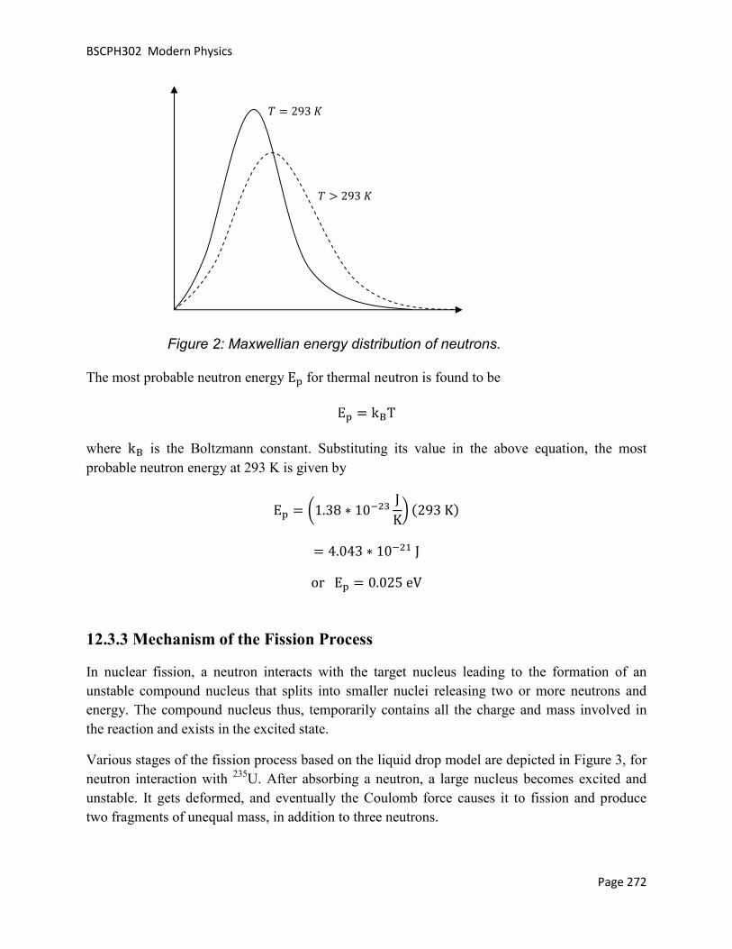
BSCPH302 Modern Physics
Page 272
The most probable neutron energy EQ for thermal neutron is found to be
EQ = kRT
where kR is the Boltzmann constant. Substituting its value in the above equation, the most probable neutron energy at 293 K is given by
EQ = a1.38 ∗ 10L D JKb �293 K�
= 4.043 ∗ 10L � J or EQ = 0.025 eV
12.3.3 Mechanism of the Fission Process
In nuclear fission, a neutron interacts with the target nucleus leading to the formation of an unstable compound nucleus that splits into smaller nuclei releasing two or more neutrons and energy. The compound nucleus thus, temporarily contains all the charge and mass involved in the reaction and exists in the excited state.
Various stages of the fission process based on the liquid drop model are depicted in Figure 3, for neutron interaction with 235U. After absorbing a neutron, a large nucleus becomes excited and unstable. It gets deformed, and eventually the Coulomb force causes it to fission and produce two fragments of unequal mass, in addition to three neutrons.
O = 293 ü
O > 293 ü
Figure 2: Maxwellian energy distribution of neutrons.

BSCPH302 Modern Physics
Page 273
12.3.4 Energy Released in Fission
When heavy nuclei undergo fission large amount of energy is released. It can be seen that the heaviest nuclei are less stable than the nuclei close to é ≅ 60, which suggests that the energy can be released if heavy nuclei split apart into more stable smaller nuclei. This process is called fission. For example, in the following thermal neutron induced fission reaction, the energy will be released and may be estimated from the binding energy per nucleon ÑÁ versus the mass number (A),
g�� + ã� DA → ã� D� ∗ → ÑåA���� + üiD�� + 3 g��
∆Ñ = ��ÑÁé�}!omV?n$ − ��ÑÁé�!�p?np&n$
= �8.4 ∗ 141 + 8.7 ∗ 92� − �7.6 ∗ 236����
= �1176 + 809� − 1786
= 199 ���
(b) Ellipsoidal (c) Narrowing
(d) Critical state (e) Separation
+ +
(a) Spherical
compound
nucleus n n
n
Figure 3: Schematics of a fission process.

BSCPH302 Modern Physics
12.3.5 Applications of Nuclear Fission
One of the most important applications of nuclear fission revolves around exploiting the massive energy that is generated in the fission reaction. The number of neutrons that are emitted in each fission reaction can initiate another fission reaction and ifoccurs and the neutron population will grow exponentially. Hence, nuclear fission chain reaction is a series of nuclear fissions each initiated by a neutron produced in a preceding fission. A schematic diagram of the nuclear fission chain reaction is shown in the figure below. In this example, a 235U nuclide absorbs a neutron and fissions to release three new neutrons. These three neutrons again initiate three new fissions which release nine new neutrons ready to initiatefission reactions and this chain continues to grow. On an average 2.5 neutrons are released by the fission of each 235U nucleus that absorbs a low
Figure 4: Schematic diagram of the nuclear fission chain reaction.
If the neutron population continues to grow in an uncontrolled manner and the fuel is more than the critical mass, the result is a fission bomb or an atomic bomb. This is one of the destructive applications of nuclear fission. We became aware of the ill effects of nuclear weapobombs were dropped on Hiroshima and Nagasaki in 1945 during World War 2. the mushroom cloud of the atomic bombing of the Japanese city of Nagasaki is shown in Figure 5.
If the neutrons that are released in each released in a controlled manner. Nuclear power plants produce heat through controlled nuclear fission chain reactions. A schematic diagram of a nuclear power plant using a pressurized watereactor is shown in Figure 6.
12.3.5 Applications of Nuclear Fission
One of the most important applications of nuclear fission revolves around exploiting the massive energy that is generated in the fission reaction. The number of neutrons that are emitted in each fission reaction can initiate another fission reaction and if left uncontrolled a chain reaction occurs and the neutron population will grow exponentially. Hence, nuclear fission chain reaction is a series of nuclear fissions each initiated by a neutron produced in a preceding fission. A
lear fission chain reaction is shown in the figure below. In this nuclide absorbs a neutron and fissions to release three new neutrons. These three
neutrons again initiate three new fissions which release nine new neutrons ready to initiatefission reactions and this chain continues to grow. On an average 2.5 neutrons are released by the
U nucleus that absorbs a low-energy neutron.
: Schematic diagram of the nuclear fission chain reaction.
ion continues to grow in an uncontrolled manner and the fuel is more than the critical mass, the result is a fission bomb or an atomic bomb. This is one of the destructive applications of nuclear fission. We became aware of the ill effects of nuclear weapobombs were dropped on Hiroshima and Nagasaki in 1945 during World War 2. the mushroom cloud of the atomic bombing of the Japanese city of Nagasaki is shown in Figure
If the neutrons that are released in each of the fission can be restricted, then the energy can be released in a controlled manner. Nuclear power plants produce heat through controlled nuclear fission chain reactions. A schematic diagram of a nuclear power plant using a pressurized wate
Page 274
One of the most important applications of nuclear fission revolves around exploiting the massive energy that is generated in the fission reaction. The number of neutrons that are emitted in each
left uncontrolled a chain reaction occurs and the neutron population will grow exponentially. Hence, nuclear fission chain reaction is a series of nuclear fissions each initiated by a neutron produced in a preceding fission. A
lear fission chain reaction is shown in the figure below. In this nuclide absorbs a neutron and fissions to release three new neutrons. These three
neutrons again initiate three new fissions which release nine new neutrons ready to initiate fission reactions and this chain continues to grow. On an average 2.5 neutrons are released by the
: Schematic diagram of the nuclear fission chain reaction.
ion continues to grow in an uncontrolled manner and the fuel is more than the critical mass, the result is a fission bomb or an atomic bomb. This is one of the destructive applications of nuclear fission. We became aware of the ill effects of nuclear weapons when twin bombs were dropped on Hiroshima and Nagasaki in 1945 during World War 2. A photograph of the mushroom cloud of the atomic bombing of the Japanese city of Nagasaki is shown in Figure
an be restricted, then the energy can be released in a controlled manner. Nuclear power plants produce heat through controlled nuclear fission chain reactions. A schematic diagram of a nuclear power plant using a pressurized water

BSCPH302 Modern Physics
Page 275
Figure 5: Photograph of the mushroom cloud of the atomic bombing of the Japanese city of Nagasaki on August 9, 1945.
Figure 6: Schematic diagram of a nuclear power plant using a pressurized water reactor.

BSCPH302 Modern Physics
Page 276
In order to turn nuclear fission into electrical energy, the controlled fission energy given off by the enriched uranium is allowed to heat water into steam. Enriched uranium is usually formed into pellets, which are then arranged into long rods, and the rods are arranged together into bundles. These bundles are submerged in water inside a pressure vessel. The water acts as a coolant. To prevent overheating, control rods made of a material that absorbs neutrons are inserted into the uranium bundle. Raising and lowering the control rods allows controlling the rate of the nuclear chain reaction. When the control rods are lifted out of the uranium bundle, fewer neutrons are absorbed and more heat is produced. When the control rods are lowered into the uranium bundle, more neutrons are absorbed and less heat is produced. The rods can also be lowered completely into the uranium bundle to shut down the reactor in the event of an accident or to replace or replenish the fuel. The heat that is produced in the nuclear reactor heats the water and turns it to steam. The steam drives a turbine, which spins an electric generator to produce power. This is one of the peaceful applications of nuclear fission. In America, more than 20% of the country’s electricity is produced by harnessing nuclear energy.
The advantages offered by nuclear energy are that no fossil fuels are burned and there are no combustion products like carbon dioxide or sulfur dioxide to pollute the air and water. The disadvantages include the cost (nuclear reactors are expensive to build and operate), limited supply of the fuel like 235U, accidents and safety hazards, and the disposal of nuclear waste. Radioactive wastes are generated in various stages of nuclear energy production. There are numerous waste streams having different physical and chemical properties and quantities of radionuclides. There are a number of dangers that surround nuclear waste disposal. The products of nuclear fission have long half lives, so they will continue to be radioactive and therefore hazardous for many years to come. One of the biggest concerns about the disposal of nuclear waste is the affect the hazardous materials could have on environment. Even though, more often than not the waste is well sealed inside huge steel and concrete structures, sometimes accidents can’t be avoided resulting in leaks. Nuclear waste can have drastically horrific effects on life, causing cancer, for instance, and even causing genetic problems for many generations of humans, animals and plants.
Apart from the nuclear reactors being used to generate power, there are research reactors as well. These non-power reactors serve primarily as a neutron source, for example, in neutron activation analysis (NAA) which is a qualitative and quantitative analytical technique for the determination of trace elements, in radioisotope production, in geological dating, in neutron transmutation doping (NTD) of silicon where high purity silicon is irradiated with thermal neutrons to convert some of the silicon to phosphorus, material irradiation to determine the effects of fast and thermal neutron irradiations on materials, in neutron radiography where an image on film that has been exposed to the secondary radiation is produced when neutrons from the reactor penetrate the specimen and interact with a neutron absorbing screen, and in material structure studies using research reactors in the low, intermediate and high power level range.

BSCPH302 Modern Physics
Page 277
Nuclear research reactors are also used in neutron capture therapy. When 10B absorbs a neutron it emits an α-particle that is highly ionizing and has a range in tissue about equal to the diameter of a cell. In boron neutron capture therapy the tumor is loaded with a borated compound and irradiated with neutrons. The tumor dose is much higher than that to the rest of the surrounding tissue, resulting in preferential killing of tumor cells. Research reactors are also used in instrument testing and calibration, and testing prototype fuel elements. India's first nuclear research reactor was APSARA built at Bhabha Atomic Research Centre (BARC) in 1956.
Example 1: In a nuclear reactor, 1 g of ã� DA (235.0439 u) fissions in 24 hours by a slow neutron (1.0087 u). Assuming that üiD�� (91.8973 u) and ÑåA���� (140.9139 u) are produced and no energy is lost, calculate the total energy released.
Solution:
The balanced equation for the nuclear fission reaction is:
g�� + ã� DA → üiD�� + ÑåA���� + 3 g��
The sum of the masses before reaction:
= 235.0439 + 1.0087 = 236.0526 �
The sum of the masses after reaction:
= 140.9139 + 91.8973 + 3�1.0087� = 235.8375 �
Therefore, the energy released in the reaction is
= ∆��931� ���
= �236.0526 − 235.8375 ���931� = 200 ���
Number of atoms in 1 g of ã� DA is 6.023 × 10 D/235. Therefore, the energy released in fission of 1 g of ã� DA is
= 6.023 × 10 D235 �200 ���� = 8.2 × 10��
Self Assessment Question (SAQ) 1: Fill in the blanks:
(a) The minimum amount of fissionable material required to sustain a chain reaction is called the ____________.

BSCPH302 Modern Physics
Page 278
(b) Very heavy elements tend to achieve stability by splitting into smaller nuclei, giving off energy in the process. This process is known as ___________. Conversely, very light nuclei can be made to merge to form heavier nuclei and giving off energy. This second process is called ____________.
(c) The fast neutrons can be slowed down by mixing the uranium fuel with a substance called a ____________.
(d) The ratio of the number of neutrons present at the beginning of a particular generation to the number present at the beginning of the next generation is called the __________________.
Self Assessment Question (SAQ) 2: Choose the correct option:
Which of the following is NOT used as a source of fuel for nuclear fission?
(a) Uranium-235
(b) Carbon-14
(c) Plutonium-239
(d) Thorium-231
Self Assessment Question (SAQ) 3: Choose the correct option:
If bombardment of uranium-235 with a thermal neutron generates tellurium-135 along with 3 neutrons, what is the other nuclide that is generated?
(a) strontium-99.
(b) zirconium-99.
(c) krypton-101.
(d) zirconium-98.
Self Assessment Question (SAQ) 4: Choose the correct option:
The difference between a nuclear reactor and an atomic bomb is that
(a) no chain reaction takes place in a nuclear reactor while in the atomic bomb there is a chain reaction

BSCPH302 Modern Physics
Page 279
(b) the chain reaction in a nuclear reactor is controlled
(c) the chain reaction in a nuclear reactor is not controlled
(d) no chain reaction takes place in atomic bomb while it takes place in a nuclear reactor
Self Assessment Question (SAQ) 5: Do all the thermal neutrons have the same energy?
12.4 NUCLEAR FUSION
15.4.1 Energy Released in Fusion
The lightest elements (like hydrogen and helium) have nuclei that are less stable than heavier one up to 56Fe. If two light nuclei fuse together to form a heavier nucleus, a significant energy could be released. This promising process, which is also the source of the sun’s energy, is called nuclear fusion.
It is exoergic only for nuclides of mass number below 60. The energy released in the fusion process may also be estimated from the binding energy per nucleon ÑÁ versus the mass number (A). For example, for the following fusion reaction,
ý� + ý� → ý� �
∆Ñ = ��ÑÁé�}!omV?n$ − ��ÑÁé�!�p?np&n$
= �7 ∗ 4� − �1.1 ∗ 2 + 1.1 ∗ 2�
= 23.6 ���
12.4.2 Reaction in the Sun and the Stars
Vast amounts of energy generated by the sun and stars cannot be accounted by chemical reactions and now it is established that these were because of the nuclear fusion reactions. At extremely high temperatures inside the core of the sun and the stars, gas will be in the form of completely ionized clouds known as plasma. There are two sets of reactions that occur inside the sun and the stars – Carbon cycle, also known as carbon-nitrogen-oxygen (CNO) cycle and proton-proton (p-p) chain. In the CNO cycle, carbon acts as a catalyst to facilitate the combination of four protons to form a helium nucleus. In the p-p chain, starting from proton-proton interaction followed by a series of intermediate reactions leads to the formation of the helium nucleus.

BSCPH302 Modern Physics
CNO cycle
In the sun and the stars, the main constituents are hydrogen and helium, but there are also relatively smaller amounts of other heavier nuclides present. In particular, there can be some presence of nuclides of carbon, nitrogen and oxygen. If these elements are present, they can also participate in the following sequential set of reactions:
In the CNO cycle, four protons fuse, using carbon, nitrogen and oxygen isotopes as a catalyst, to produce one alpha particle, two positrons and two electron neutrinos.regenerated. Combining all the above reacbe
4
The CNO cycle is represented as shown in the figure below:
Figure 7: Representation of the CNO cycle. The notation (a, b) indicates that the nucleus captures the particle ‘a’ and emits the particle ‘b’.
In the sun and the stars, the main constituents are hydrogen and helium, but there are also relatively smaller amounts of other heavier nuclides present. In particular, there can be some
resence of nuclides of carbon, nitrogen and oxygen. If these elements are present, they can also participate in the following sequential set of reactions:
¥�� + ý�� → çK�D + æ
çK�D → ¥��D + P� + �
¥��D + ý�� → çK�� + æ
çK�� + ý�� → �@�A + æ
�@�A → çK�A + P� + �
çK�A + ý�� → ¥�� + ý� �
In the CNO cycle, four protons fuse, using carbon, nitrogen and oxygen isotopes as a catalyst, to produce one alpha particle, two positrons and two electron neutrinos. In the final step, regenerated. Combining all the above reactions, the net reaction for the CNO cycle comes out to
4 1H � 4He + 2β+ +2ν + 26.7 MeV
The CNO cycle is represented as shown in the figure below:
Figure 7: Representation of the CNO cycle. The notation (a, b) indicates that the particle ‘a’ and emits the particle ‘b’.
Page 280
In the sun and the stars, the main constituents are hydrogen and helium, but there are also relatively smaller amounts of other heavier nuclides present. In particular, there can be some
resence of nuclides of carbon, nitrogen and oxygen. If these elements are present, they can also
In the CNO cycle, four protons fuse, using carbon, nitrogen and oxygen isotopes as a catalyst, to In the final step, 12C is
tions, the net reaction for the CNO cycle comes out to
Figure 7: Representation of the CNO cycle. The notation (a, b) indicates that the

BSCPH302 Modern Physics
p-p chain
The following sequential set of reactions take place in the p
On adding all the reactions, the net reaction is the same as that for the CNO cycle, i.e. the formation of 4He from four protons (
Since both p-p chains and CNO cycles can produce fusion energy by converting hydrogen to helium, they compete with each othe temperature increases, there is a sudden transition to dominance by the CNO cycle. Inside the sun with an interior temperature of around considerations suggest that the sun is producing about 98and only about 1% from the CNO cycle. Heavier stars, their interior temperatures being higher, favor the CNO cycle.
The figure below shows the energy production ratefunction of temperature (units of 10
Figure 8: Energy production rates for the ptemperature (units of 106 K).
The following sequential set of reactions take place in the p-p chain.
ý�� + ý�� → ý� + P� + �
ý�� + ý� → ý� D + æ
ý� D + ý� D → ý� � + ý�� + ý��
On adding all the reactions, the net reaction is the same as that for the CNO cycle, i.e. the He from four protons (1H).
p chains and CNO cycles can produce fusion energy by converting hydrogen to helium, they compete with each other. At low temperatures, the p-p chain predominates and as the temperature increases, there is a sudden transition to dominance by the CNO cycle. Inside the sun with an interior temperature of around 1.5 × 10K ü, the p-p chain predominates. Detailed
derations suggest that the sun is producing about 98-99% of its energy from the pand only about 1% from the CNO cycle. Heavier stars, their interior temperatures being higher,
The figure below shows the energy production rates for the p-p chain and the CNO cycle as a function of temperature (units of 106 K).
Figure 8: Energy production rates for the p-p chain and the CNO cycle as a function of
Page 281
On adding all the reactions, the net reaction is the same as that for the CNO cycle, i.e. the
p chains and CNO cycles can produce fusion energy by converting hydrogen to p chain predominates and as
the temperature increases, there is a sudden transition to dominance by the CNO cycle. Inside the p chain predominates. Detailed
99% of its energy from the p-p chain and only about 1% from the CNO cycle. Heavier stars, their interior temperatures being higher,
p chain and the CNO cycle as a
p chain and the CNO cycle as a function of

BSCPH302 Modern Physics
Page 282
12.4.3 Advantages of Nuclear Fusion over Nuclear Fission
Economically viable controlled nuclear fusion for power generation has not yet been achieved, even on a laboratory scale. However, thermonuclear fusion is the only energy technology that has been identified as a major energy source even before it has produced a single MeV of energy in a useful fashion. Many people believe that fusion is the answer to all of our future energy needs. Also, for a nuclear fusion reactor to work, the temperature and pressure would each have to be very high. These incredibly high temperatures and pressures are very expensive and extremely difficult to achieve. In the future, if we are able to achieve controlled thermonuclear fusion and build nuclear fusion reactor based power plants, there should be many advantages as compared to nuclear fusion reactor based power plants. Some of these advantages are listed below:
1- The energy released by fission is a million times more than that released in chemical reactions, but it is significantly lower than the energy released by nuclear fusion. The energy released per unit mass of the fuel by nuclear fusion is three to four times greater than the energy released by fission.
2- Another major advantage of fusion over fission is that unlike fission where highly penetrating radiations are liberated, which are extremely hazardous; the products of fusion reactions are not radioactive. Therefore, they are harmless and can be replaced easily.
3- If fusion is indeed harnessed as a source of controlled energy, we would have a seemingly infinite supply of fuel such as deuterium (2H), tritium (3H) etc. that can be obtained from the sea water, unlike fission where the fuels such as uranium-235 are in limited supply.
Example 2: Calculate the energy released from the following D-T fusion reaction,
D + T � n + 4He
Given : Mass of deuteron = 2.0149 u, mass of tritium nucleus = 3.01605 u, neutron mass = 1.008665 u, mass of 4He nuclide = 4.00260 u.
Solution:
The total mass of the reactants is = �W + �¢
= 2.0149 u + 3.01605 u = 5.03005 u
The total mass of the products is = �& + ���

BSCPH302 Modern Physics
Page 283
= 1.008665 u + 4.00260 u
= 5.011265 u
The mass difference is (the negative sign indicates that there will be release in energy, i.e. the reaction will be exoergic)
5.011265 u − 5.03005 u
= −0.018785 u
Since, I u = 931 MeV, the energy released in the reaction is
1 = ∆��
= Ö0.018785 uØ a931 ���� b
= 17.5 ���
Self Assessment Question (SAQ) 6: Choose the correct option:
It is proposed to use the nuclear fusion reaction: ý� + ý� → ý� � in a nuclear reactor of 200 MW rating and 25% efficiency. How many grams of deuterium will be needed per day? The masses of ý� and ý� � are 2.0141 and 4.0026 u, respectively.
(a) 120.83 g/day (b) 100.5 g/day (c) 52.44 g/day (d) 12.08 g/day
Self Assessment Question (SAQ) 7: Choose the correct option:
Why is the fusion reaction an unlikely source of energy on an industrial scale?
(a) The radioactive elements are too dangerous to handle.
(b) The temperatures that are needed are too high.
(c) It is illegal.
(d) None of these

BSCPH302 Modern Physics
Page 284
Self Assessment Question (SAQ) 8: Choose the correct option:
The processes that are responsible for generating massive amounts of energy inside a star are?
(a) Combustion of carbon
(b) Fusion reactions
(c) Fission reactions
(d) Chemical reactions
Self Assessment Question (SAQ) 9: Choose the correct option:
Which of the following potential fusion reactions will NOT result in the net release of energy?
(a) 6Li and 9Li
(b) 3Hand 6Li
(c) 4He and 3He
(d) 35Cl and 60Fe
Self Assessment Question (SAQ) 10: Choose the correct option:
Does the relation 1 = ∆�� apply to?
(a) fusion reactions only
(b) fission reactions only
(c) all the processes
(d) None of these
12.5 SUMMARY
In this unit, we studied the neutron-induced fission reaction that are initiated by slow or thermal neutrons. We understood how thermal neutrons follow Maxwell-Boltzmann distribution and hence do not possess a single value of energy. Their most probable energy at 293 K was found to

BSCPH302 Modern Physics
Page 285
be 0.025 eV. We then studied the mechanism of the fission process based on compound nucleus model and the liquid drop model.
We calculated the energy liberated in fission and fusion reactions using the binding energy curve and the mass defect, before discussing some of the applications of nuclear fission. We also discussed briefly some of the reactions that occur in the sun and the stars. In the last section, we listed some of the advantages that nuclear fusion enjoys over nuclear fission even though controlled nuclear fusion for power generation has not yet been achieved, even on a laboratory scale.
12.6 GLOSSARY
Atomic Nucleus - the nucleus of an atom is the central part of it. Neutrons and protons are bound together in the nucleus of an atom.
Chain Reaction – a self-sustaining nuclear reaction yielding energy or products that cause further reactions of the same kind.
CNO Cycle – the CNO cycle (for carbon–nitrogen–oxygen) is one of the two known sets of fusion reactions by which stars convert hydrogen to helium, the other being the proton–proton chain reaction. In the CNO cycle, four protons fuse, using carbon, nitrogen and oxygen isotopes as a catalyst, to produce one alpha particle, two positrons and two electron neutrinos. Unlike the latter, the CNO cycle is a catalytic cycle. Theoretical models suggest that the CNO cycle is the dominant source of energy in stars more massive than about 1.3 times the mass of the sun.
Control Rod – a neutron-absorbing material, such as cadmium, usually shaped into a rod and used to control nuclear fission.
Deuteron – a positively charged particle consisting of a proton and a neutron, equivalent to the nucleus of an atom of deuterium― symbol D.
Electron-Neutrino – is a subatomic elementary particle which has no net electric charge, and believed to have very small mass. An electron-neutrino is emitted along with a positron in positive beta decay, while an electron-antineutrino is emitted with an electron in negative beta decay.
Maxwell-Boltzmann distribution – in statistics, the Maxwell-Boltzmann distribution is a particular probability distribution named after James Clerk Maxwell and Ludwig Boltzmann. It was first defined and used in physics, for expressing the distribution of energy among the molecules of an idealized gas in thermal equilibrium.

BSCPH302 Modern Physics
Page 286
MeV (Mega Electron Volt) – energy equal to that acquired by a particle with one electronic charge in passing through a potential difference of one million volts.
Moderator – a substance (such as graphite or heavy water) used for slowing down the neutrons in a nuclear reactor.
Nuclear Fission – division of a heavy atomic nucleus into two fragments of roughly equal mass, accompanied by the release of a large amount of energy.
Nuclear Fusion – a nuclear reaction in which light nuclei combine to form more massive nuclei with the simultaneous release of energy.
Nuclear Reactor – any of several devices in which a chain reaction is initiated and controlled, with the resulting heat typically used for power generation and the neutrons and fission products used for military, experimental, and medical purposes.
p-p chain – the proton–proton chain reaction is one of the two (known) sets of fusion reactions by which stars convert hydrogen to helium. It dominates in stars the size of the sun or smaller.
Q value – Q value for a reaction is the amount of energy released by that reaction.
Radioactivity – the phenomenon, exhibited by and being a property of certain elements, of spontaneously emitting radiation resulting from changes in the nuclei of atoms of the element.
Radionuclide – a nuclide that exhibits radioactivity.
Tritium – a radioactive isotope of hydrogen that has one proton and two neutrons in its nucleus and that has three times the mass of ordinary hydrogen ― symbol T.
12.7 TERMINAL QUESTIONS
1. Explain the working of a nuclear fission reactor.
2. The basic energy-producing process in the sun is the fusion of hydrogen nuclei into helium nuclei. This can take place in different reaction sequences. Describe the two most common of such reaction sequences.
3. What is a nuclear reactor? How is it different from a nuclear power plant?
4. Why is fusion reaction preferred to fission reaction?
5. Describe what is meant by induced fission. How is it different from spontaneous fission?
6. What is meant by enriched uranium? Why is needed?

BSCPH302 Modern Physics
Page 287
7. What is a nuclear chain reaction? How can it be controlled?
8. Explain how in an atomic explosion, a large amount of energy is released.
9. What are the applications of nuclear research reactors?
10. Given that the mass of 1H atom is 1.007825 u and the mass of 4He atom is 4.002603 u, calculate the energy released in the fusion reaction when four protons combine to give a helium nuclide. 1 u of mass is equivalent to about 931 MeV of energy.
11. Choose the correct option:
A generic nuclear fission reaction is 235U + n � X + Y + 2n, where n is the symbol representing a neutron. Which of the following pairs can represent X and Y?
(a) 156Nd and 79Ge
(b) 121In and 113Ru
(c) 141Xe and 99Sr
(d) 139Cs and 95Rb
12. Choose the correct option:
If the earth’s sun were much more massive, its energy production mechanism
(a) will be dominated by the CNO cycle.
(b) will be dominated by the p-p chain.
(c) will have equal contributions from both p-p chain and the CNO cycle.
(d) will remain unaltered.
13. Write short notes on:
(i) proton-proton chain (ii) CNO cycle (iii) Mechanism of Nuclear Fission
12.8 ANSWERS
Selected Self Assessment Questions (SAQs):

BSCPH302 Modern Physics
Page 288
1. (a) Critical mass (b) Fission, fusion (c) moderator (d) multiplication factor
2. (b)
3. (d)
4. (b)
5. No. Thermal neutrons follow a Maxwellian energy distribution and have different energies. The most probable thermal neutron energy is found to be equal to �§O.
6. (a)
7. (b)
8. (b)
9. (d)
10. (c)
Selected Terminal Questions:
10. The given reaction is
4 1H � 4He + 2β+ +2ν
The mass of the neutrinos is negligibly small if at all, so they do not enter into the calculations of the disintegration energy. Therefore, the energy released in the above reaction is given by
1 = ∆��
= Ö4�1.007825 u� − 4.002603 uØ a931 ���� b
= 26.7 ���
11. (d)
12. (a)
12.9 REFERENCES
10. Nuclear Physics – Problem-based Approach including MATLAB, Hari M. Agrawal – PHI Learning, Delhi
11. Concepts of Modern Physics, Arthur Beiser – McGraw-Hill, New York

BSCPH302 Modern Physics
Page 289
12. Modern Physics, Stephen T. Thorthon – Cengage Learning India, New Delhi
12.10 SUGGESTED READINGS
10. Nuclear Physics – Problem-based Approach including MATLAB, Hari M. Agrawal – PHI Learning, Delhi
11. Concepts of Modern Physics, Arthur Beiser – McGraw-Hill, New York 12. Modern Physics, Stephen T. Thorthon – Cengage Learning India, New Delhi

BSCPH302 Modern Physics
Page 290
UNIT 13 RADIOACTIVITY
Structure
13.1 Introduction
13.2 Objectives
13.3 Radioactivity
13.3.1 Units of Radioactivity
13.4 α, β and γ Particles and Their Properties
15.4.1 Comparison of α, β and γ Particles
13.5 Radioactive Decay
13.6 Soddy Fajan’s Displacement Law
13.7 Law of Radioactive Disintegration
13.8 Half Life
13.9 The Mean Life
13.10 Activity of Radioactive Substance
13.11 Radioactive Dating
15.11.1 Carbon Dating
13.12 Geiger- Nuttal Law
13.13 Gamow’s Theory of α- decay
13.14 Theory of β- Decay
13.15 Summary
13.16 Glossary
13.17 References
13.18 Suggested Readings
13.19 Terminal Questions
13.20 Answers

BSCPH302 Modern Physics
Page 291
13.1 INTRODUCTION
In the previous unit, we have studied about elementary particles. In that unit, learners have studied the classifications of elementary particles, particles and anti-particles, conservation laws governing elementary particles, structure of hadrons, binding energy of Deuterium, energy production in stars, C-N cycle, p-p cycle etc. In this unit, we shall learn about radioactivity, alpha, beta and gamma particles, radioactive decay. We shall also study Soddy Fajan’s displacement law, law of radioactive disintegration, half life, mean life, carbon dating, Geiger-Nuttal law etc. We shall discuss Gamow’s theory of alpha decay and theory of beta decay in this unit.
13.2 OBJECTIVES
After studying this unit, you should be able to -
• understand radioactivity, radioactive decay, carbon dating • understand alpha and beta decay theory • understand Soddy Fajan’s displacement law • compute half life, mean life and decay constant
13.3 RADIOACTIVITY
In this section, we shall study the history and discovery of radioactivity. Radioactivity was discovered by Henri Becquerel in 1896. Becquerel observed that uranium and some of its salts emit spontaneously some invisible radiation which penetrates through opaque substances and affects the photographic plate. These rays are called ‘radioactive rays’ or ‘Becquerel rays’. The spontaneous emission of rays from a substance is called as ‘radioactivity’ and such substances are known as ‘radioactive substances’.
After the discovery of radioactivity, Madame Curie, Piere Curie, Rutherford etc., showed that the phenomenon was exhibited by heavy elements like uranium, polonium, radium, thorium etc. Radioactivity involves the spontaneous transmutation of one element into another. The phenomenon of spontaneous emission of highly penetrating radiations from heavy elements of atomic weights greater than about 206, occurring in nature, is called ‘natural radioactivity’. The atoms of radioactive elements emit radiations composed of three different types of rays- α, β and γ- rays. No radioactive substance emits both α- and β- particles simultaneously. Some substances emit α-particles and some other emits β- particles. γ- rays are emitted along with both α- and β- particles.
The modern technique of artificial transmutation of elements has been able to produce radioactivity in many other elements, much higher than those that occur in nature. Radioactivity induced in an element by bombarding it with α-particles, protons, neutrons and other particles or

BSCPH302 Modern Physics
Page 292
radiations is called artificial radioactivity. Therefore, there is a distinction between natural radioactivity i.e. of elements as found in nature and induced radioactivity, i.e. artificially produced.
13.3.1 Units of Radioactivity
Generally, we represent the activity of a radioactive substance in terms of Curie and its submultiples mill Curie (m Ci) and micro Curie (μ Ci). The curie is defined as the quantity of a radioactive substance which gives 3.70 × 10¹⁰ disintegrations/second.
Another unit of radioactivity is Rutherford (Rd). It is defined as the quantity of a radioactive substance which gives 106 disintegrations / second.
We can write-
1 Curie = 3.70 × 10¹⁰ disintegrations/second
1 Rutherford = 106 disintegrations / second
13.4 α, β AND γ- PARTICLES AND THEIR PROPERTIES
When an atom undergoes radioactivity, it emits particles like alpha, beta and gamma rays. Radioactivity basically occurs because the unstable atom tries to attain stability. Hence, when they are unstable, they eventually decay by emitting a particle transforming the nucleus into another nucleus, or into a lower energy state. This chain of decays continues till the nucleus attains the stage of stability.
Radioactive atom Energy
Radiation
Particle
Figure 1: Emission of particle from a radioactive atom

BSCPH302 Modern Physics
Page 293
There are basically three types of radiations that are emitted by radioactive particles. These three are called the alpha, beta and gamma rays. All these radiations are released from the nucleus of the atom. Though all three cause some ionization and have some penetration power, but their behavior differs from the others. We shall discuss the properties of alpha, beta and gamma rays in detail one by one:
Properties of α- particles
(i) Alpha rays or alpha particles are the positively charged particles. An α- particle has +3.2 ×10⁻¹⁹ coulomb positive charge which is just double the negative charge on the electron.
(ii) A highly energetic helium nucleus which contains two protons and two neutrons is called the alpha-particle. When both electrons are detached from the helium atom, then the helium nucleus is left which has two units positive charge. This is called α- particle. This particle is also called ‘doubly ionised helium atom’.
-2e 2He4 He++ Helium atom α-particle
(iii) The mass of an α- particle is 6.645×10⁻²⁷ Kg which is equal to the mass of the helium nucleus.
(iv) Α-particles are deflected in electric and magnetic fields and the direction of deflection indicates that these particles are positively charged.
(v) The velocity of α-particles is much less than the velocity of light. (vi) The range of these particles in air depends upon the radioactive substance and its value
for a given substance is constant. (vii) When α-particles pass through gases, they eject electrons from the atoms of the gases
i.e. the atoms are ionized. The ionizing power of these particles is 100 times that of β- particles and 10,000 times that of γ-rays i.e. α-particles have greatest ionizing power amongst α, β and γ-particles. Since these particles have great ionization power, so they can get into the body and can cause serious damage.
(viii) These particles are scattered while passing through thin foils of mica or gold. Most of the α- particles are scattered at small angles but some of them are scattered at larger angles also.
(ix) Alpha particles have the least penetration power. These particles can penetrate through matter. They cannot penetrate the skin but this does not mean that they are not dangerous.
(x) Α- particles affect photographic plates slightly. (xi) These particles may cause burns on human body. (xii) On being stopped, α-particles produce heating effect.

BSCPH302 Modern Physics
Page 294
(xiii) These particles produce fluorescence when allowed to fall on substances like barium-platinocyanide and zinc sulphide.
Properties of β- particles
(i) Beta particles are highly energetic electrons which are released from inside of a nucleus.
(ii) They are negatively charged and have a negligible mass. (iii) A β-particle has 1.6×10⁻¹⁹ coulomb of negative charge which is the charge of electron. (iv) These particles are deflected by electric and magnetic fields and the direction of
deflection shows that they are negatively charged. (v) β -particles are much lighter than the α-particles. (vi) Beta particles have a greater penetration power than the alpha particles and can easily
travel through the skin. (vii) Though beta particles have less ionization power than the alpha particles but still they are dangerous and so their contact with the body must be avoided. (viii) The velocity of β- particles vary from 1% to 99% of the velocity of light.
(ix) Since the velocity of β- particles is of the order of the velocity of light, their mass increases with increase in their velocity. If the rest mass of a β-particle be m0 and that in the state of velocity v be m, then according to Einstein’s theory of relativity, we have-
m = ����L�"�" …..(1)
where c is the velocity of light. Obviously, as the value of v approaches c, the value of m goes on increasing.
(x) The range of β-particle is not definite. (xi) β- particles affect photographic plate more than do the α- particles. (xii) These particles produce fluorescence in barium platinocyanide and zinc-sulphide.
Properties of γ- particles
(i) The waves from the high frequency end of the electromagnetic spectrum which do not have any mass are called the gamma rays.
(ii) These particles are electrically neutral i.e. they have no charge. (iii) γ- rays are not deflected by electric and magnetic field. (iv) γ- rays travel with the velocity of light i.e. 3×10⁸ m/sec. (v) They have greatest power of penetration and hence it is extremely difficult to stop them
from entering the body. (vi) They are the least ionizing. γ- rays ionize gases but their ionization power is very small
compared to that of α and β – particles.

BSCPH302 Modern Physics
Page 295
(vii) These rays carry huge amount of energy and can even travel through thin lead and thick concrete.
(viii) γ – rays produce fluorescence. (ix) γ- rays are absorbed by substances and give rise to the phenomenon of pair-production.
When a γ- ray photon strikes the nucleus of some atom, its energy is converted into an electron and a positron and its own existence is extinguished-
h υ → e- + e+
γ- photon electron positron
α- particle
β- particle
γ- particle
Palm Aluminum Lead
Figure 2: Comparison of penetrating power of three rays
13.4.1 Comparison of α, β and γ- Particles
The below table describes the characteristics of alpha, beta and gamma radiations and compares the masses and charges of the three rays as-
S. No. Property α- particle β- particle γ - rays 1. Nature Helium nucleus Fast moving electron Electromagnetic
waves or photons 2. Charge +3.2 ×10⁻¹⁹ coulomb -1.6 × 10⁻¹⁹ coulomb zero 3. Rest mass 6.6 × 10⁻²⁷ Kg 9.1 × 10⁻³¹ Kg zero 4. Velocity 1.4 × 10⁷ to 2.2 ×
107 m/sec 1% to 99% of the velocity of light
velocity of light (3 × 10⁸ m/sec)
5. Ionizing power 100 times that of β- particles
100 times that of γ - rays
Minimum

BSCPH302 Modern Physics
Page 296
6. Penetrating power
Minimum 100 times that of α- particles
100 times that of β- particles
7. Range ~10 cm in air, can be stopped by 1mm of Aluminum
Up to a few m in air, can be stopped by a thin layer of Aluminum
Several m in air, can be stopped by a thick layer of Lead
8. Natural sources By natural radioisotopes e.g. 92U236
By radioisotopes e.g. 29Co68
Excited nuclei formed as a result of Gamma decay
13.5 RADIOACTIVE DECAY
We have seen that in radioactivity the atoms of a radioactive element emit α – particles (or β – particles) and also γ – rays. As a consequence, both the mass number and the atomic number are changed. Thus, the original radioactive atoms decayed and an atom of some new elements is born. This phenomenon is known as ‘radioactive decay’ or ‘radioactive disintegration’. Thus, Radioactive decay (also known as nuclear decay, radioactivity or nuclear radiation) is the process by which an unstable atomic nucleus loses energy (in terms of mass in its rest frame) by emitting radiation, such as an alpha particle, beta particle with neutrino or only a neutrino in the case of electron capture, or a gamma ray or electron in the case of internal conversion. A material containing such unstable nuclei is considered radioactive. Certain highly excited short-lived nuclear states can decay through neutron emission, or more rarely, proton emission. We can represent this by the following equation as-
92U238 90Th234 + 2He4
Uranium Thorium α – particle
If the new atom is also radioactive then this too is decayed and a third new atom is obtained. For example, Thorium is also radioactive and by emitting a β – particle, it is converted into a Protactinium atom-
90Th 234 91 Pa 234 + -1 β 0 + W
Thorium Protactinium β – particle anti-neutrino
This process continues until a stable atom is obtained by radioactive decay. For example, Uranium atom, through a chain of radioactive decay, is finally converted into a Lead atom ( 82 Pb 206 ) which is the heaviest stable atom. All radioactive elements are finally converted into Lead. Radioactive decay is a nuclear process i.e. the radioactive rays are emitted from the nucleus of

BSCPH302 Modern Physics
Page 297
the atom. This process cannot be accelerated or slowed down by any physical or chemical process.
13.6 SODDY FAJAN’S DISPLACEMENT LAW
The law of radioactive displacements, also known as Fajans and Soddy law, in radiochemistry and nuclear physics, is a rule governing the transmutation of elements during radioactive decay. It is named after Frederick Soddy and Kazimierz Fajans, who independently arrived at it at about the same time in 1913.
When a radioactive disintegration occurs with the emission of α and β – particles, the original atom called the parent atom changes into something else, called the daughter. In 1913, Soddy and Fajan discovered a simple law known as the displacement law which governs these radioactive changes. It can be stated as follows-
(1) In all known radioactive transformations either an α or a β –particle is emitted by the atom.
(2) In alpha decay, an element is created with an atomic number less by 2 and a mass number less by four of that of the parent radioisotope, e.g.-
- α
92 U 238 90 Th 234
(3) In beta decay, the mass number remains unchanged while the atomic number becomes greater by 1 than that of the parent radioisotope, e.g.-
-β 90 Th 234 91 Pa 234
13.7 LAW OF RADIOACTIVE DISINTEGRATION
Rutherford and Soddy made an experimental study of the radioactive decay of various radioactive materials and found that the decay of all radioactive materials is governed by the same general law. According to the law of radioactive disintegration, “at any instant, the rate of decay of radioactive atoms is proportional to the number of atoms present at that instant”. Let us discuss this law.

BSCPH302 Modern Physics
Page 298
Let at any instant t, the number of atoms present = N
After time dt i.e. at instant (t+dt), the number of atoms = N- dN
The rate of decay of the atom = - dN/dt
According to Rutherford and Soddy law, we have-
− m#mn ∝ ç
or − m#mn = �ç
or m#mn = −�ç …..(2)
where λ is a constant and is known as ‘decay constant’. For a given element, the value of decay constant λ is constant. From equation (2), we get-
λ = − m#/mn#
i.e. decay constant or disintegration constant λ of a radioactive element is defined as the ratio of the amount of substance which disintegrates in a unit time to the amount of the substance present. Its unit is per year. From equation (2), we have-
m## = −� e� Integrating both sides, we get- loge N = - λ t + C …..(3) where C is a constant of integration. Initially, at t = 0, N = N0 , where N0 is the number of atoms in the beginning. Therefore, from equation (3), we have- loge N0 = - λ (0) + C or C = loge N0
Putting for C in equation (3), we have- loge N = - λ t + loge N0
loge ##� = - λ t
##� = e – λ t
or N = N0 e- λ t …..(4) Here N0 and N are the numbers of atoms in a radioactive substance at time t = 0 and after time t respectively. According to equation (4), it is clear that the decay of a radioactive substance is exponential i.e. the decay is rapid in the beginning and then its rate decreases continuously. It means that a radioactive substance will take infinite time in decaying completely. If t = 1/λ, then from equation (4), we get- N = N0 e-1 = N0 (1/e)

BSCPH302 Modern Physics
Page 299
In this way, in a radioactive material, after a time-interval equal to the reciprocal of decay constant, the number of undecayed atoms is (1/e) times the initial value.
13.8 HALF LIFE
The half life of a radioactive substance is defined as the time required for one-half of the radioactive substance to disintegrate. It is also known as half life period. The half life of a radioactive substance is constant but it is different for different substances. Its unit is year or day. It is denoted by ‘T’.
Let us discuss the relation between half life and decay constant.
We know-
N = N0 e- λ t
If the half life of a substance be T, then at time t = T, N = N0 /2
Therefore, N0/2 = N0 e-λ T
½ = e- λT
Or e λ T = 2
Or λ T = loge 2
Or T = � �7 V = �.��D�V …..(5)
The equation (5) represents the relationship between half life and decay constant. If the decay constant λ for a radioactive substance is known, then its half life can be calculated by this equation.
13.9 THE MEAN LIFE
Radioactive decay is a statistical process. A nucleus may take from zero to infinite time to decay. The average of the lives of all the nuclei is known as ‘average life’ i.e.
The mean life of a radioactive element = YV� oZ n4� Ó%0�$ oZ pÓÓ n4� pno�$¢onpÓ &V�~�! oZ pno�$
It is represented by τ. The mean life or average life of a radioactive material is equal to the reciprocal of the decay constant λ, i.e.
τ = �V …..(6)
But T = �.��D�V

BSCPH302 Modern Physics
Page 300
Therefore, T = 0.6931 τ …..(7)
The unit of mean life is year.
13.10 ACTIVITY OF RADIOACTIVE SUBSTANCE
The activity of a sample of any radioactive nuclide is the rate at which the nuclei of its constituent atoms decay.
If N is the number of nuclei present in the sample at a certain time, its activity R is given by-
R = - m#mn …..(8)
If N be the number of atoms of a radioactive substance left at some instant of time then the activity of the substance at that instant is-
R N
Let us consider that initially, at t = 0, the number of atoms in a substance = N0
After one half life, the number of atoms left in the substance will be N0/2 that is the number of atoms at time t = T is-
N = N0/2 = N0(1/2)1
After two half lives, the number of atoms left will be N0/4 that is at t = 2T, we shall have-
N = N0/4 = N0 (1/2)2
In general, the number of atoms left after n half lives is given as-
N = N0(1/2)n …..(9)
Where n = t/T
The SI unit of activity is named after Henri Becquerel.
1 Becquerel = 1Bq = 1 events/sec
1 MBq = 106 Bq
1 GBq = 109 Bq
Example 1: The half life of Radon is 3.8 days. Calculate how much radon will be left out of 1024 mg after 38 days?

BSCPH302 Modern Physics
Page 301
Solution: Given T = 3.8 days, t = 38 days, N0 = 1024 mg
Number of half lives n = t/T = 38/3.8 = 10
We know that N = N0(1/2)n
= 1024 × (1/2)10 = 1024/1024 = 1 mg
i.e. 1 mg radon will be left after 38 days.
Example 2: Prove that after two half lives the amount of a radioactive element is reduced to one-fourth part.
Solution: Given, n = 2
We know that N = N0(1/2)n = N0(1/2)2 = N0 (1/4) = N0/4
i.e. after two half lives the amount of a radioactive element is reduced to one-fourth part.
Example 3: A sample of radioactive substance has 106 radioactive nuclei. Its half life time is 20 seconds. How many nuclei will remain after 10 seconds?
Solution: Given N0 = 106 , T = 20 sec, t = 10 sec
Number of half lives n = t/T = 10/20 = ½
Using N = N0(1/2)n
N = 106 (1/2)1/2 = 106/√2 = 106/1.41 = 7× 10⁵ (Approximately)
Example 4: The half life of thorium –X is 3.64 days. After how many days will 0.1 of the mass of a sample of the substance remain undecayed?
Solution: Given T = 3.64 days, N/N0 =0.1 = 1/10 = 10-1
Using λ = 0.6931/T = 0.6931/3.64 = 0.1904 per day
We know N = N0 e- λ t
or N/N0 = e-λ t
or 10-1 = e-λ t
or eλ t = 10.
Taking loge on both sides, we get-
λ t = loge10 = 2.3026 × log10 10 = 2.3026×1 = 2.3026

BSCPH302 Modern Physics
Page 302
or t = 2.3026/λ = 2.3026/ 0.1904 = 12.1 days
Self Assessment Question (SAQ) 1: The half life of radium is 1600 years. After how many years 25% of a radium block remains undecayed?
Self Assessment Question (SAQ) 2: Find the half life of a radioactive material if its activity drops to 1/16th of its initial value in 30 years.
Self Assessment Question (SAQ) 3: The mean lives of a radioactive substance are 1620 years and 405 years for alpha emission and beta emission respectively. Find out the time during which 3/4th of a sample will decay if it is decaying both by alpha emission and beta emission simultaneously.
Self Assessment Question (SAQ) 4: Choose the correct option-
(i) The radioactive material does not emit-
(a) protons (b) gamma-rays (c) electrons (d) helium nuclei
(ii) Beta rays emitted by a radioactive material are-
(a) electromagnetic waves (b) electrons orbiting around the nucleus (c) charged particles emitted by the nucleus (d) neutral particles
(iii) The decay constant of a radioactive sample is λ. The half life and mean life of the sample are respectively given by-
(a) 1/λ and loge2/λ (b) loge2/λ and 1/λ (c) λ/loge2 and 1/λ (d) none of these
13.11 RADIOACTIVE DATING
In this section, we shall learn about radioactive dating. Half-lives are very useful for Geologists in estimating the age of mineral deposits, rocks and the earth. The age of the earth is estimated from the relative abundance of the two isotopes of uranium U238 and U235. The half periods of U238 and U235 are 4.5 × 10⁹ years and 7 × 10⁸ years respectively. Assume that at the beginning when the earth was formed the proportions of the two isotopes were equal. The present relative abundance of U238 and U235 in natural uranium is 99.3% to 0.7%.
Therefore, ##" = ��.D�.K = #���[\#���["\ = ��V"LV�n (Using N = N0 e- λ t)
Where, λ1 = �.��D��.A×�� and λ2 = �.��D�K×��J
Therefore, log� ;��.D�.K = = (λ2 – λ1) t

BSCPH302 Modern Physics
Page 303
or t = �V"LV log� ;��.D�.K =
= ��.H�_×�JL �.H�'.]×� log� ;��.D�.K = = 5.93× 10⁹ years .
This value agrees nearly with that given by astronomical evidence for the age of the universe.
13.11.1 Carbon Dating
The decay of radioactive elements is independent of the physical and chemical conditions imposed on them. Although the decay of an individual particle from a given nucleus is a random process, the gross decay of the many nuclei in a given sample provides a very convenient way of measuring times.
Cosmic rays coming from the outer space produce radioactive atoms by colliding with atoms present in the earth’s upper atmosphere. One of the radioactive atoms so produced is an isotope of carbon 6C14 which is formed by collision of neutron (0n1) present in cosmic rays and nitrogen atom (7N14) present in the atmosphere-
7N14 + 0n1 6C14 + 1H1 .
This carbon isotope is oxidized to carbon dioxide 14CO2 which is mixed with ordinary carbon dioxide 12CO2 of the atmosphere. The ratio of ordinary carbon 6C14 and radioactive carbon 6C14 in the atmospheric carbon dioxide is 1012 : 1.
In nature, all living trees and plants take carbon dioxide from the atmosphere. Therefore, in living plants also, the ratio of 6C12 and 6C14 is 1012 : 1. When a plant dies, it stops taking carbon dioxide from the atmosphere. Therefore, in the dead plant, the quantity of ordinary carbon 6C12 remains unchanged but the radioactive carbon 6C14 continues to decay according to the following equation-
6C14 7N14 + -1β0 + W .
The half life of this decay is 5568 years. The dead plant is ultimately converted into wood, coal, etc. If we measure the radioactive activity of some dead material like coal, wood, etc., then the age of that material can be determined.
Let us suppose that the activity of some dead material was R0 at the time of death and it has been reduced to R after t years. Then, according to the law of radioactive decay, we have-
R = R0 e-λ t .
Where λ is the decay constant of the radioactive carbon present in that material.
The above equation can be written as-

BSCPH302 Modern Physics
Page 304
eλ t = R0/R
λ t = loge(R0/R)
or t = (1/λ) loge(R0/R) = (2.3026/λ) log10(R0/R)
But T = �.��D�V or λ = �.��D�¢
Therefore, t = (2.3026 T/0.6931) log10 (R0/R)
= 3.3222 T log10 (R0/R) …..(10)
R0 is the activity of the plant at the time of its death. Obviously, it is the activity of some living plant. Hence, by measuring the activity R0 of living plant and the activity R of dead material and taking T = 5568 years, the age t of the material can be calculated. This method of determining the age is called ‘carbon dating’.
13.12 GEIGER- NUTTAL LAW
The Geiger–Nuttall law or Geiger–Nuttall rule relates the decay constant of a radioactive isotope with the energy of the alpha particles emitted. Roughly speaking, it states that short-lived isotopes emit more energetic alpha particles than long-lived ones. The relationship also shows that half-lives are exponentially dependent on decay energy, so that very large changes in half-life make comparatively small differences in decay energy, and thus alpha particle energy. In practice, this means that alpha particles from all alpha-emitting isotopes across many orders of magnitude of difference in half-life, all nevertheless have about the same decay energy.
Formulated in 1911 by Hans Geiger and John Mitchell Nuttall, in its modern form the Geiger–Nuttall law is –
log10 λ = - A1 �√M + A2 …..(11)
where λ is the decay constant (λ = ln2/half-life), Z the atomic number, E the total kinetic energy (of the alpha particle and the daughter nucleus), and A1 and A2 are constants. The law works best for nuclei with even atomic number and even atomic mass. The trend is still there for even-odd, odd-even, and odd-odd nuclei but not as pronounced.
The Geiger-Nuttal law can be expressed in terms of the range R of an α-particle. The distance through which an α-particle travels in a substance before coming to rest is called the range of the particle in that substance.
BSCPH302 Modern Physics
Page 305
The range R of an α-particle and the disintegration constant λ of the radioactive element that emits it are related as follows-
loge λ = A + B loge R …..(12)
This relation is called Geiger-Nuttal law. Here A and B are constants. This law is helpful in determining the decay constants of radioactive substances of very short or very long lives.
Example 5: The isotopes U238 and U235 occur in nature in the ratio 140:1. Assuming that at the time of earth’s formation they were present in equal ratio, make an estimation of the age of the earth. The half lives of U238 and U235 are 4.5 × 10⁹ years and 7.13 × 10⁸ years respectively. ( log10140 = 2.1461)
Solution: Let the number of their atoms be N1 and N2.
Using N = N0 e- λ t
##" = #���[\#���["\ = ��V"LV�n
Taking loge on both sides, we get-
loge (N1/N2) = (λ2 – λ1) t
or t = � �7ìì"V"LV =
� �7'�� �7 O ñ"L ñR = � �� ���� �� ; ¢"¢¢L¢"=
= .�����.D��� × [K.�D×��J\×[�.A×��\��.A×���L�K.�D×��J� = 6× 10⁹ years
Self Assessment Question (SAQ) 5: Write True or False-
(i) The age of the earth can be estimated by radioactive dating. (ii) The Geiger-Nuttal law was formulated in 1951. (iii) The Geiger-Nuttal law is helpful in determining the decay constants of radioactive
substances of very short or very long lives. (iv) The ratio of ordinary carbon 6C14 and radioactive carbon 6C14 in the atmospheric
carbon dioxide is 1012 : 10.
13.13 GAMOW’S THEORY OF α- DECAY
Classical physics fails to explain α-decay. Quantum mechanics provides a successful explanation of the problem of α-decay. The basic points of this theory are-

BSCPH302 Modern Physics
Page 306
1. An alpha (α) particle may exist as an entity within a heavy nucleus.
2. α-particle is in constant motion and bounces back and forth from the barrier walls. In each collision with wall, there is a definite probability that the particle will leak through the potential barrier. Let υ be the frequency with which the α-particle collides with the walls in order to escape from the nucleus and P the probability of transmission in each collision. Then the decay probability per unit time i.e. disintegration constant is given by-
λ = υ P …..(13)
Let us suppose that at any moment only one α-particle exists as such in a nucleus and that it moves back and forth along a nuclear diameter.
Then, υ = v/(2R) …..(14)
Where v is the α-particle velocity and R is the nuclear radius.
Since V ˃ T, [where V is the height of the potential barrier and T the kinetic energy of α-particle], according to classical physics P =0 but in quantum mechanics a moving particle is regarded as a wave and the result is a small but definite value for P. The probability P can be calculated quantum mechanically using WKB perturbation theory. According to this theory-
loge P = L �4/ �� y �2�:��M� − O;eM^^ …..(15)
where m is the mass of the α-particle.
V(x) = ��"����� is the electrostatic potential energy of an α-
particle at a distance x from the centre of a nucleus of charge Ze. Ze is the charge of the daughter nucleus ( i.e. nuclear charge – the α-particle charge of 2e).
The region from x = R to x = R1 is called the thickness of the barrier.
Putting the value of V(x) in equation (14), we have-
loge P = L �4/ �� y �2� ^ ��"����� − O_ eM^^ …..(16)
When x = R1 , T = V, therefore, T = ��"����^
Therefore, equation (16) takes the form as-
loge P = L �4/ �� ��2�O� y �^^� − 1_ eM^^

BSCPH302 Modern Physics
Page 307
or loge P = L �4/ �� ��2�O�Y� ���t L�� ^ − � ^ �;1 − ^=� …..(17)
The width of the potential barrier is very large compared with the nuclear radius i.e. R1 >> R. Therefore, cos-1 (R/R1)1/2 ≈ (π/2) – (R/R1)1/2 and (1- R/R1)1/2 ≈ 1
Hence, loge P = L �4/ �� ��2�O�Y� �ô� − � ^õ − � ^ �
= L �4/ �� ��2�O�Y� �ô� − 2� ^õ�
Substituting R1 = ��"����¢, we have-
loge P = ��; <"8= � ���� √Z√Y − �"; <"8=�� �� Z O�" …..(18)
or loge P = 2.97 √Z√Y - 3.95 Z T-1/2
where R is in Fermi and T is in MeV.
Since λ = υ P
Taking loge both sides, we get-
loge λ = loge υ + loge P
= loge (v/2R) + loge P
Therefore, loge λ = loge (v/2R) + 2.97 √Z√Y - 3.95 Z T-1/2 …..(19)
or log10 λ = log10 (v/2R) + 0.4343 × (2.97 √Z√Y - 3.95 Z T-1/2 )
or loge λ = loge (v/2R) + 1.29 Z1/2 R1/2 – 1.72 Z T-1/2 . …..(20)
This theory makes understandable the enormous variation in half life with disintegration energy. Nuclei emitting most energetic α-particles are short lived and those emitting least energetic α-particles are long lived. This correlation between the half life and the energy of the α-particle is predicted by equation (20).
13.14 THEORY OF β- DECAY
In 1934, Fermi developed a theory to explain the continuous β-ray spectrum. This theory is known as neutrino theory of β-decay. According to this theory, a β- particle and a neutrino are created in the nucleus and both are emitted simultaneously. The total energy of these two

BSCPH302 Modern Physics
Page 308
particles is a constant which is equal to the end point energy observed in the β-ray spectrum. This maximum energy is shared by the β-particle, the neutrino and also by the recoiling nucleus. The electron will carry the maximum energy when the energy of the neutrino is zero. In all other cases, electron will carry energy less than the maximum. The sum of the energies carried by the electron and the neutrino will always be the same. This energy may be shared by the two particles in any proportion. Hence, it explains the continuous β-ray spectrum.
When the nucleon shifts from the neutron quantum state to the proton quantum state, electron and antineutrino are emitted. This process is represented by-
n p + e- + W .
In ordinary β-decay, it is an antineutrino that is emitted.
Positron emission corresponds to the conversion of a nuclear proton into a neutron, a positron and a neutrino-
P n + e+ + υ .
Positron emission leads to a daughter nucleus of lower atomic number Z while leaving the mass number A unchanged. Thus negative and positive beta decays may be represented as-
ZXA Z+1XA + e- + W
ZXA Z-1XA + e+ + υ .
The electron, neutrino and product nucleus share among them the energy, angular momentum and linear momentum available from the nuclear transitions. Thus the neutrino theory of β- decay successfully explains the continuous energy spectrum of β-rays.
13.15 SUMMARY
In the present unit, we have study about radioactivity and the particles emitted during radioactivity i.e. α, β and γ-particles. We have discuss the comparison among the properties of these particles. We have study radioactive decay, decay constant, activity, half life, mean life etc. in this unit. We have learnt that the radioactive decay is the process by which an unstable atomic nucleus loses energy by emitting radiation, such as an alpha particle, beta particle with neutrino or only a neutrino in the case of electron capture, or a gamma ray or electron in the case of internal conversion. We have learnt about Soddy Fajan’s displacement law also. In the unit, we have study Geiger-Nuttal law, radioactive dating and carbon dating. We have discuss Gamow’s theory of α-decay and theory of β-decay. In the unit, we have included many solved examples and self assessment questions (SAQs) to check your progress.

BSCPH302 Modern Physics
Page 309
13.16 GLOSSARY
Induce – provoke, encourage
Decay – decompose, disintegration
Transmutation – change, transformation
Stability – constancy
Correlation- connection
13.17 REFERENCES
1. Modern Physics, R. Murugeshan, S. Chand and Company Ltd, New Delhi
2. Modern Physics, Sehgal, Chopra, Sehgal, Sultan Chand and Sons, New Delhi
3. IIT Physics, Gupta and Gupta, Jai Prakash Nath Publications, Meerut
13.18 SUGGESTED READINGS
1. Fundamentals of Physics; Halliday, Resnick and Walker; John Wiley and Sons (Asia) Ptd. Ltd, Singapore
2. Modern Physics, Beiser, Tata McGraw Hill
3. Elementary Text Book of Physics, J. P. Agarwal, Pragati Prakashan, Meerut
13.19 TERMINAL QUESTIONS
1. What do you mean by radioactivity? Explain.
2. Mention the properties of α, β and γ-particles.
3. Why all the radioactive elements ultimately are converted into lead?
4. Explain Soddy-Fajan’s displacement law.
5. Write notes on- (i) Decay constant (ii) Half life (iii) Mean life
6. Establish the law of radioactive disintegration.
7. What is activity of radioactive substance? Give its unit.
8. Give Geiger-Nuttal law? Give its importance.

BSCPH302 Modern Physics
Page 310
9. Explain the terms decay constant, half life and average life as applied to a radioactive substance. Find the relation between them.
10. What is meant by carbon dating? Explain.
11. Discuss radioactive dating. Give its significance.
12. Discuss Gamow’s theory of α-decay.
13. Explain and discuss the neutrino theory of beta decay.
13.20 ANSWERS
Self Assessment Questions:
1. Given, T = 1600 years, N = 25% of N0 = N0/4
We know that- N = N0(1/2)n
or N0/4 = N0 (1/2)n
or (1/2)2 = (1/2)n
or n = 2
Therefore, time of disintegration t = T × n = 1600×2 = 3200 years
2. Given, N = 1/16th of N0
or N/N0 = 1/16
We know- N = N0(1/2)n
or N/N0 = (1/2)n
or 1/16 = (1/2)n or (1/2)4 = (1/2)n
or n = 4 .
Half life of the substance T = t/n = 30 years/4 = 7.5 years
3. Given τ1= 1620 years, τ2 = 405 years , N = N0 – (3/4) N0 = N0/4
Therefore decay constants λ1 = � = 1/ 1620 per year, λ2 = �" = 1/405 per year .
Total decay constant λ = λ1 + λ2 = 1/1620 + 1/405 = 1/324 per year .
We know N = N0 e- λ t

BSCPH302 Modern Physics
Page 311
N0/4 = N0 e- λ t
or eλ t = 4
or λ t = loge4 or t = loge4/λ = 1.386× 324 = 449 years .
4. (i) – (a), (ii)- (c), (iii) – (b)
5. (i) True, (ii) False, (iii) True, (iv) False

BSCPH302 Modern Physics
Page 312
UNIT 14 ELEMENTARY PARTICLES
Structure
14.1 Introduction
14.2 Objectives
14.3 Fundamental Interactions
14.3.1 Strong Interaction
14.3.2 Electromagnetic Interaction
14.3.3 Weak Interaction
14.3.4 Gravitational Interaction
14.4 Constituent Particles of Matter
14.4.1 Leptons
14.4.2 Quarks
14.5 Classification of Particles
14.5.1 Baryon Number
14.4.2 Isospin
14.4.3 Strangeness and Strange Particles
14.4.4 Eightfold Way
14.6 Conservation Laws
14.6.1 Electric Charge
14.6.2 Baryon Number
14.6.3 Lepton Number
14.7 Deuteron
14.8 Energy Production in Stars
14.9 The Standard Model
14.10 Summary
14.11 Glossary
14.12 Terminal Questions
14.13 Answers
14.14 References
14.15 Suggested Readings

BSCPH302 Modern Physics
Page 313
14.1 INTRODUCTION
In this unit, we will be presenting the elementary particles. The desire to identify the smallest constituents of matter is indeed very old. Ancient Indian schools of philosophy proposed that space, water, fire, air and earth are the elementary constituents of nature. An almost similar view of the basic constituents of nature is also present in ancient Greek civilization who regarded water, fire, air and earth as four unchangeable elements of nature. The philosophers at that time also coined the word “atom.” The modern view of atom may be traced back to the discovery of the atomic model by Dalton in 1803 and later the Mendeleev’s periodic table of chemical elements. The modern genesis of the quest for the origin of the ultimate matter and kind of space-time we live in is over a hundred years old. With the discovery of the electron in 1897 and of the proton in 1900, the story of elementary particles beyond the physics of atoms started. The probing beam of α-particles from radioactive sources were limited in energy and to get a closer look at the nucleus, physicists needed a better source of higher energy particle beams. It was in 1930 when E. O. Lawrence built the first successful particle accelerator, that it was possible to make a detailed exploration of the inner structure of the atom. Since then, one accelerator followed another and with increasing sophistication and energy, it became the most powerful tool in the hands of particle physicists to explore and unravel the inner structure of the atom and to discover one elementary particle after another.
The Standard Model of particle physics is the theory that describes three of the four known fundamental forces – the electromagnetic, weak, and strong interactions (and not including the gravitational force) in the universe, and also classifies all known elementary particles. It evolved in stages throughout the latter half of the twentieth century through the work of many scientists around the world. The current formulation was finalized in the 1970s when the existence of quarks was experimentally confirmed. Since then, confirmation of the top quark (1995), the tau neutrino (2000), and the Higgs boson (2012) have added further credence to the Standard Model.
14.2 OBJECTIVES
After studying this unit, you should be able to
• describe what are the four fundamental interactions found in nature • describe what are the constituent particles of matter. • discuss the classification of elementary particles. • describe the conservation laws and symmetry governing elementary particles. • discuss the Standard model. • describe the deuteron problem.

BSCPH302 Modern Physics
14.3 FUNDAMENTAL INTERACTIONS
There are four fundamental interactionsFigure 14.1):
I. Gravitational interaction II. Electromagnetic interaction
III. Strong interaction IV. Weak interaction
Figure 14.1: Four fundamental interactions of nature.
The gravitation interaction, which is the weakest of all, is the force that holds the earth together, binds the sun and the planets into the solar system, and binds stars into galaxies. Between individual particles, however, it is extremely feeble that it can usually be ignored. The electromagnetic force acts between particles which carry an electronic charge. It holds the cloud of negatively charged electrons around the positively charged protons in the nu
14.3 FUNDAMENTAL INTERACTIONS
r fundamental interactions accounting for all the known forces in the universe (
Electromagnetic interaction
ure 14.1: Four fundamental interactions of nature.
The gravitation interaction, which is the weakest of all, is the force that holds the earth together, binds the sun and the planets into the solar system, and binds stars into galaxies. Between
particles, however, it is extremely feeble that it can usually be ignored. The electromagnetic force acts between particles which carry an electronic charge. It holds the cloud of negatively charged electrons around the positively charged protons in the nucleus, binds atoms
Page 314
for all the known forces in the universe (see
The gravitation interaction, which is the weakest of all, is the force that holds the earth together, binds the sun and the planets into the solar system, and binds stars into galaxies. Between
particles, however, it is extremely feeble that it can usually be ignored. The electromagnetic force acts between particles which carry an electronic charge. It holds the cloud
cleus, binds atoms

BSCPH302 Modern Physics
Page 315
together to form molecules and crystals. It is the significant interaction for all of chemistry and biology. The strong interaction glues the nucleons together; it binds neutrons and protons to form the nuclei of all the elements. The strongest force known in nature, it is also of quite short range. It is the dominating interaction in high-energy physics. The weak interaction causes the light elementary particles (the leptons: �, �, � and a) to interact with each other and with heavier particles. It is responsible for the radioactivity of a nucleus which can emit an �L when a neutron breaks up.
The interactions can be classified according to the value of a characteristic dimensionless constant related to the interaction strength.
14.3.1 Strong Interaction
Range of interaction is ~10L�A �. The interaction potential between two nucleons i distance apart has the form
�$�i� = �?i exp ;− iY= (14.1)
where Y = ?/��� is the Compton wavelength of pion. The exponential function indicates a short interaction length. The dimensionless constant 5$ gives the interaction strength.
5$ = �?
?� ≈ 1
14.3.2 Electromagnetic Interaction
The potential for two charged particles at distance i apart, has the form
���i� = � i (14.2)
The dimensionless constant characteristic of the interaction strength is the fine structure constant
5� = � 4|��?� = 1137
14.3.3 Weak Interaction
Its strength is represented by the Fermi coupling constant for the β-decay
d] = 1.4 ∗ 10L�� �i�. ��D
The potential of weak interaction has the form

BSCPH302 Modern Physics
Page 316
���i� = ��i exp a− iY�b (14.3)
where Y� = ?/2��� (as we did in the Yukawa theory). The dimensionless constant characteristic of the interaction strength is
5� = �4 ℏ� = d] �} �ℏD ≈ 10LA
14.3.4 Gravitational Interaction
The interaction potential between two protons has the form
�f�i� = d �} i (14.4)
The dimensionless constant characteristic of interaction strength is
d �} ?� ≈ 6 ∗ 10LD�
14.4 CONSTITUENT PARTICLES OF MATTER
Not long ago, protons, neutrons and electrons were regarded as the fundamental particles of nature when we learned in the 1900s through the experiments of Rutherford and others that atoms consist of mostly empty space with clouds of electrons surrounding a dense central nucleus constituting of the protons and the neutrons. The science of particle physics surged ahead with the arrival of particle accelerators that could accelerate protons and electrons to high energies, which are then made to collide with nuclei to produce many new particles. Based on these experiments and the theoretical studies, it became apparent that leptons and quarks are the basic building blocks of matter. i.e., they are now understood to be the “elementary particles.”
The developments of the past few years have revealed that quarks and leptons can themselves be grouped in families of four (quartet of quarks and leptons). Astonishingly, all the components of our normal everyday world can be explained in terms of just one such family – the first generation constituents, containing “up” and “down” quarks, the electron and the electron-type neutrino. At higher energies (that can be observed through some phenomena in outer space, or can be created artificially using particle accelerators) two further generations of quarks and leptons come into play. These quartets appear to be heavier copies of the normal quartet.
Table 14.1 illustrates the basic constituents of matter.

BSCPH302 Modern Physics
Page 317
In addition to these component particles are others (photons, bosons and gluons) which communicate the forces between the components.
Table 14.1: The basic constituents of matter
Quarks Leptons
In everyday matter
charge
Up 2/3 Electron
Down -1/3 Electron-type neutrino
In matter existing in higher
energy environments
Strange -1/3 Muon
Charm 2/3 Muon-type neutrino
Bottom -1/3 Tau
Top 2/3 Tau-type neutrino
14.4.1 Leptons
The electron was the first of the leptons3, actually, the first of the elementary particles observed by J. J. Thomson in 1897, for which an acceptable theory was developed by Paul A. M. Dirac in 1928. The intrinsic angular momentum (spin) of the electron was found to be ℏ/2 and therefore, constrained by the Pauli Exclusion Principle, a fact that has key implications for the creation of the periodic table of the elements. The existence of the antiparticles for the electrons was predicted by Dirac’s relativistic quantum theory of the electron. Carl D. Anderson, in 1932, during the study of the cosmic rays discovered the predicted positron, the first known antiparticle.
The lepton family has twelve members. The present standard model assumes that there are no more than three generations of having four members in each. The properties of the leptons are listed in Table 14.2. All the leptons are believed to have � = 1/2�. The neutrinos are uncharged; their masses are unknown but unlikely to exceed a few ��/� .
The muon, the member of the second family of leptons, was discovered in a cloud chamber by Anderson and Neddermayer in 1937. The muon decays through the following reaction
�± → �± + �¦ + ��g
where �¦ = 207��. Neutrinos associated with muon were seen in 1962. The reaction
�¦ + g → �L + d
3 Lepton in Greek means light particles.
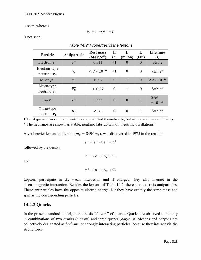
BSCPH302 Modern Physics
Page 318
is seen, whereas �¦ + g → �L + d is not seen.
Table 14.2: Properties of the leptons
Particle Antiparticle Rest mass (B�h/�/)
L (e)
L (muon)
L (tau)
Lifetimes (s)
Electron �L �� 0.511 +1 0 0 Stable
Electron-type neutrino i�
��g < 7 ∗ 10L� +1 0 0 Stable*
Muon jL �� 105.7 0 +1 0 2.2 ∗ 10L�
Muon-type neutrino ij
�jÁÁÁ < 0.27 0 +1 0 Stable*
Tau kL a� 1777 0 0 +1 2.96∗ 10L�D
Ϯ Tau-type neutrino ik
ikÁÁÁ < 31 0 0 +1 Stable*
Ϯ Tau-type neutrino and antineutrino are predicted theoretically, but yet to be observed directly. * The neutrinos are shown as stable; neutrino labs do talk of “neutrino oscillations.”
A yet heavier lepton, tau lepton (�` = 3490��), was discovered in 1975 in the reaction
�L + �� → aL + a� followed by the decays
aL → �L + ��g + �` and
a� → �� + �¦ + �g
Leptons participate in the weak interaction and if charged, they also interact in the electromagnetic interaction. Besides the leptons of Table 14.2, there also exist six antiparticles. These antiparticles have the opposite electric charge, but they have exactly the same mass and spin as the corresponding particles.
14.4.2 Quarks
In the present standard model, there are six “flavors” of quarks. Quarks are observed to be only in combinations of two quarks (mesons) and three quarks (baryons). Mesons and baryons are collectively designated as hadrons, or strongly interacting particles, because they interact via the strong force.

BSCPH302 Modern Physics
Page 319
The properties of six quarks are listed in Table 14.3. Besides the quarks, there also exist six anti quarks.
Table 14.3: Properties of six flavors of quarks
Quark Spin, parity
Charge Q/e
Baryon number
Isospin I
S C B T Mass* (l�h/�/)
Up (u) 1/2� +2/3 1/3 1/2 0 0 0 0 ~0.3
Down (d) 1/2� -1/3 1/3 1/2 0 0 0 0 ~0.3
Charm (c) 1/2� +2/3 1/3 0 0 +1 0 0 1.6
Strange (s)
1/2� -1/3 1/3 0 -1
0 0 0 0.5
Top (t) 1/2� +2/3 1/3 0 0 0 0 +1 174
Bottom (b)
1/2� -1/3 1/3 0 0 0 +1 0 4.5
Anti quarks mg, ng, �Á, *Á, G and og have opposite signs for charge, baryon number, S, C, B and T.
* The masses should not be taken too seriously, because the non-confinement of quarks implies that we can’t isolate them to measure their masses in a direct manner.
Each of these six flavors of quarks can have three different “colors” – red, green and blue. This color designation has absolutely nothing to do with the visual colors that we see. “Color” is the “charge” of the strong nuclear force, analogous to the electric charge for electromagnetism. The force between quarks is attractive only in the “colorless” combinations of three quarks (baryons) and quark-anti quark pairs (mesons). The interaction potential ��i� between two quarks inside a nucleon is given by
��i� = − åi + 3i (14.5)
where i is the distance between the quarks and a” and “b” are positive constants of suitable dimensions.
The up and down quarks are stable quarks. The other quarks, however, are unstable and decay. Their life-times and major decays are shown in the Table 14.4. Quarks get transformed by the exchange of W bosons and the rate and nature of the decay of hadrons by the weak interactions are determined by these transformations. Up and down quarks are the most common and least massive quarks, being the constituents of the protons and the neutrons and therefore, of most ordinary matter. In the PL-decay, a neutron transform to a proton via weak interaction

BSCPH302 Modern Physics
Page 320
g ��ee� → d �e��� + �L + ��g
is considered to be the result of a more fundamental quark process where a down quark becomes an up quark.
e → � + �L + ��g
Table 14.4: Life-times and major decay paths of unstable quarks
Quark Life-times (s) Major decays
Strange (s) 10L@ − 10L�� t → � + Mϯ
Charm (c) 10L� − 10L�D
� → ^t + Me + Mp Bottom (b)
10L� − 10L�D 3 → � + M
Top (t) ~10L A � → 3 + M
Ϯ “x” denotes other particles allowed by the appropriate conservation laws.
In 1947, during a study of cosmic ray interactions, a new particle Λ (lambda), which lived for longer time of 10L�� t than the expected 10L D t, was discovered. This behavior was dubbed as “strangeness.” The lambda is a baryon, which was proposed to be made up of u, d and s (strange quark). The strange quark has the strangeness value of �−1�, whereas the other two quarks (u and d) have þ = 0. The shorter life-time of 10L D t was expected because Λ (a baryon) participates in the strong interaction. The longer observed lifetime led to a new conservation law for such decays, i.e. “conservation of strangeness.” Particles decaying by the strong or the electromagnetic interactions preserve the strangeness quantum number.
The decay process for the Λ particle, however, must violate this rule, since there is no lighter particle containing a strange quark, to which it could decay. The following decay processes show that strangeness is not conserved.
q� ��et� → d���e� + |L ��Áe� þ = −1 ≠ 0 + 0
q� ��et� → g��ee� + |� T�Á� + ee√2 U þ = −1 ≠ 0 + 0
Strange quark gets transformed to another quark in the process. This can only occur by the weak interaction.
All the known hadrons could be specified by some combination of these three quarks and their anti quarks. But there was still a problem related to the discrepancy in the life-times of some of the known particles, and a fourth quark called the charmed quark (c) was proposed in 1970. In

BSCPH302 Modern Physics
Page 321
1974, a meson called the /s particle was discovered, which required the existence of the charmed quark and its antiquark. In 1977, an experimental group at Fermi lab found a new resonance at 9.4 d��/� . This new resonance was interpreted as a bottom-antibottom quark pair and called the upsilon meson.
Convincing evidence of top quark (t) was reported by Fermi lab’s Tevatron facility in April 1995, in the collision products of 0.9 TeV protons with equal energetic antiprotons in the d − d collider.
Till date, no one has ever seen an isolated quark. There are good reasons for the lack of direct observation. It seems that the color force doesn’t decrease with distance like the other observed forces. There is now a belief among the physicists that perhaps free quarks can’t be observed and that they can only exist within hadrons. Another way to visualize the quark confinement is called the “bag model.” Here, one can imagine the quarks as enclosed in an elastic bag, which allows the quarks to move freely around. But, when one tries to pull a quark out, the bag stretches and resists.
14.5 CLASSIFICATION OF PARTICLES
Table 14.5: Mass, mean-lifetimes and common decay modes of elementary particles
Particle Mass
(B�h/�/) τ(s)
Common decay
mode
Ha
dr
on
s M
es
on
s
Pions |L, |� 139 2 ∗ 10L@ μν |� 135 1.8 ∗ 10L�� γγ
Kaons
üL, ü� 494 1.2 ∗ 10L@ μν ü� 498 |±|�
Mixture of ü�, ü
ü� 0.89 ∗ 10L�� |�|L |�|� ü 5.18 ∗ 10L@
|�|�|� |�|L|�
Eta
meson
t 550 10L�@ æ + æ |� + |L + |� t¯ 958 10L � |� + |� + æ
Ba
ry
on
s Nucleons
d 938.2 > 10D@ Stable g 939.5 10D d�L�
Hyperons
ϯ
q 1,115 2.6 ∗ 10L�� d|L, g|� ∑� 1,189 0.8 ∗ 10L�� d|�, g|�

BSCPH302 Modern Physics
Page 322
∑� 1,192 10L � qæ ∑L 1,197 1.6 ∗ 10L�� g|L v� 1,314 3 ∗ 10L�� q|� vL 1,321 1.8 ∗ 10L�� q|L wL 1,675 1.3 ∗ 10L�� v| qüL
Photon γ 0 Stable
Leptons
aL 1,784 3.4 ∗ 10L�D �L� �L 105 2 ∗ 10L� ��� �L 0.51 Stable �� 0 Stable �¦ < 0.5 Stable �` < 164 Stable
ϯ q = �å�3eå, ∑ = tu��å, v = Mu, w = ����å
Table 14.5, gives the mass, mean-lifetimes (a) and common decay modes of the elementary particles excluding resonances and graviton. Their classification into hadrons, photon and leptons is also indicated. The subdivision of hadrons into baryons (nucleons and hyperons) and mesons (pions and kaons) is also shown.
Electron (�L), muon (�L), tau (aL), and the three neutrinos (�� , �¦, �`) constitute the class of leptons. A hadron stands for a strongly interacting particle distinguished from lepton which has only weak or electromagnetic interactions. Photon is the mass less carrier of the electromagnetic field. Mesons and photon are bosons (a particle of integral spin). Bosons obey the Bose-Einstein statistics; the wave function of two identical bosons is symmetric under particle exchange. The baryons and leptons are fermions (a particle with half integral spin – 1/2ℏ, 3/2ℏ, …). Fermions obey the Fermi-Dirac statistics; the wave function that describes two identical particles is anti-symmetric, i.e. changes sign under particle exchange.
Every particle has an associated antiparticle, which has exactly the same mass and lifetime but opposite values of the electric charge, magnetic moment, baryon number, lepton number, and the flavor. Thus, positron (��) is the antiparticle of electron, antiproton (dL) that of proton, antineutrino (��g ) that of neutrino, etc. Photon is the antiparticle of itself, so also |�. These antiparticles have not been shown explicitly in the table.
Properties and quark content of some baryons and mesons are given in the following tables.

BSCPH302 Modern Physics
Page 323
Table 14.6: Properties and quark content of some baryons (yz = )//�, ö = ))
Particle { {| } = ~ + ö Quark content
d 1/2 1/2 1 ��e g 1/2 -1/2 1 �ee q 0 0 0 �et ∑� 1 +1 0 ��t ∑� 1 0 0 �et ∑L 1 -1 0 eet v� 1/2 1/2 -1 �tt vL 1/2 -1/2 -1 ett
Table 14.7: Properties and quark content of some mesons
Particle { {| } = ~ Quark content
|L 1 -1 0 �Áe |� 1 1 0 �e
|� 1 0 0 ��Á − ee√2
üL 1/2 -1/2 -1 �Át ü� 1/2 +1/2 1 �t ü� 1/2 -1/2 1 et ü�ÁÁÁÁ 1/2 +1/2 -1 et
t 0 0 0 ��Á + ee − 2tt√6
t¯ 0 0 0 ��Á + ee + tt√3
14.5.1 Baryon Number
The baryons are the most abundant group of particles. For nucleons and hyperons Ñ = +1 for antibaryon Ñ = −1 for pions, kaons and other particles Ñ = 0. The baryon number is an additive quantum number and is conserved in all the three (i.e. strong, weak and electromagnetic) interactions.

BSCPH302 Modern Physics
Page 324
Example 1:
On the basis of the additive quark model, the total interaction cross-section is assumed to come from the sum of the cross-sections of various pairs. Assuming that ��ff� = ��ffÁ� prove the relation
��qd� = ��dd� + ��üLg� − ��|�d�
Solution
In the quark model from Tables 14.6 and 14.7, q = �et, d = ��e, g = �ee, üL = t�Á, |� = �e
��qd� = ���et����e�
= ���� + �� + �e + e� + e� + ee + t� + t� + te� = 9��ff�
Similarly,
��dd� = 9��ff�
��üLg� = 3��ff� + 3��ffÁ�
��|�d� = 3��ff� + 3��ffÁ�
Assuming ��ff� = ��ffÁ�, we get,
��qd� = ��dd� + ��üLg� − ��|�d�
14.5.2 Isospin
Isospin is a quantum number applicable to hadrons and is conserved in the strong interactions. The concept of isospin is rooted in the charge independence of the strong interaction, gg ≈ gd ≈dd. This doublet of neutrons and protons is said to have isospin 1/2 with the projection ¤� =+1/2 for the proton and ¤� = −1/2 for the neutron. The three pions (|�, |L, |�) compose a triplet, suggesting isospin ¤ = 1. The projections ¤� = +1 (for |�), 0 (for |�) and −1 (for |L). At the quark level, an isospin doublet (¤ = 1/2) is formed by the up and down quarks with the projection ¤� = +1/2 assigned to up quark and ¤� = −1/2 to down quark. The strange quark is in a class by itself and has isospin ¤ = 0.
¤ is the additive quantum number and Clebsch-Gordon coefficients are used for the addition of isospins. The charge multiplicity is given by 2¤ + 1. The antiparticle has the same ¤ as the particle but the opposite ¤�. Total isospin (¤) is conserved in the strong interaction but breaks down in the electromagnetic and the weak interactions. The third component ¤� of the system of

BSCPH302 Modern Physics
Page 325
hadrons is conserved in the strong and the EM interactions but is violated in the weak interactions (∆¤� = ±1/2).
For example, the process ∑� → d + t�
hasn’t been observed even though it conserves the charge, angular momentum and the baryon number. It is forbidden by the fact that it doesn’t conserve the isospin, ¤ = 1 ≠ 1/2 + 0, as required by the strong interaction.
Isospin is related to other quantum numbers for the particles by Gell-Mann formula, 1� = ¤� + þ + Ñ2 (14.6)
where þ is the strangeness and Ñ is the baryon number.
Baryons and mesons can be grouped into isospin multiplets as shown in Fig. 14.2.
Figure 14.2: Isospin multiplets: the nucleon doublet, the Λ singlet, the ∑ triplet, and the Ξ doublet

BSCPH302 Modern Physics
Page 326
14.5.3 Strangeness and Strange Particles
Heavy unstable particles such as the q and baryons, and the K mesons are produced at a rapid
rate in the high energy collisions but decay slowly, i.e. they have exceptionally long lifetimes. A typical production and decay sequence is
|L + d → ü� + q�
ü� → |� + |L, q� → |L + d
with lifetimes τ�~ 0.89 ∗ 10L�� s and τΛ~ 2.63 ∗ 10L�� s which is larger compared with the strong interaction time scale of 10L D s.
Table 14.8: I and S assignments
{/~ −� −/ −) � ) / � 0 wL q q w�ÁÁÁÁ d v� üL g ü� v�ÁÁÁ 1/2 vL ü� d ü�ÁÁÁÁ v�ÁÁÁÁ gÁ ∑� |� ∑LÁÁÁÁ 1 ∑� |� ∑�ÁÁÁÁ ∑L |L ∑�ÁÁÁÁ
They are named strange particles. A new quantum number S, strangeness is introduced to distinguish them from other particles. Gell-Mann formula (14.6) relates S with other quantum numbers. Strangeness S is an additive quantum number like the electric charge, and is thus, conserved in the production process. Table 14.8, summarizes the strangeness S for various hadron multiplets.
Strangeness S is conserved in the strong and the electromagnetic interactions, i.e. ∆þ = 0, but breaks down in the weak interactions, i.e. ∆þ = ±1.
14.5.4 Eightfold Way
In the preceding section, the particles could be grouped into isospin multiplets whose members are related by the isospin symmetry. This classification of the particles into isospin families helps bring some order into the confusing diversity of more than 300 known particles. The most successful classification scheme – “Eightfold Way” was proposed by M. Gell-Mann. On the

BSCPH302 Modern Physics
Page 327
basis of the eightfold way, the baryons and mesons are arranged into geometrical patterns, according to their charge and strangeness.
Figure 14.3: The baryon octet.
The eight lightest baryons form a hexagonal array, with two particles at the center as shown in Fig. 14.3. This group is known as the baryon octet. One can note that particles of like charge lie along the diagonal lines and horizontal lines associate particles of like strangeness þ. The eight lightest mesons fill a similar hexagonal pattern, forming the (pseudo-scalar) meson octet (Fig. 14.4).
Figure 14.4: The meson octet.

BSCPH302 Modern Physics
Page 328
14.6 CONSERVATION LAWS
In developing the standard model for the particles, certain types of interactions and decays are observed to be common while others to be forbidden. This observation has led to a number of conservation laws, which govern them. These conservation laws are beside the classical conservation laws such as the conservation of energy, charge etc. which are still applicable in the realm of particle interactions. There is an insightful connection between the conservation laws obeyed by a physical system and the symmetries. This connection is contained in the Noether’s theorem:
“To every symmetry, there corresponds a conservation law.”
All interactions obey the following conservation laws:
I. Conservation of the electric charge. II. Conservation of the four momenta (d�, dÒ, d� , ¸).
III. Conservation of the angular momentum. IV. Conservation of the baryon number. V. Conservation of the lepton number (the electron number, muon number and tau number
are all separately conserved.)
14.6.1 Electric Charge
The electric charge is one of the quantum numbers, which is exactly conserved. This conservation is associated with the mass less ness of the photon. It is due to the conservation of charge that the electron can’t decay, for instance, via �L → �� + �� + ��g
or, �L → �� + æ
14.6.2 Baryon Number
No known interaction or decay process in nature alters the net baryon number. The neutron along with all the heavier baryons decays directly to the proton or eventually forms proton, since the proton is the least massive baryon. This indicates that the proton can’t decay further without violating the conservation of baryon number, which means that if the conservation of baryon number holds exactly, the proton should be completely stable against any decay. One prediction of the grand unification of forces is that the proton can also decay. Until now, such a possibility hasn’t been experimentally verified.

BSCPH302 Modern Physics
Page 329
Conservation of baryon number forbids a decay of the type:
d + g → d + �� + �L
Ñ = 1 + 1 ≠ 1 + 0 + 0
Although, with adequate energy permits pair production in the reaction
d + g → d + g + d + d
Ñ = 1 + 1 = 1 + 1 + 1 − 1
14.6.3 Lepton Number
Each of the three sets of leptons (È� , Ȧ, È`) are to be conserved separately. For example, decay processes
|L → �L + �¦ÁÁÁ
�L → �L + ��g + �¦
were observed. Since, a well-defined muon energy is observed from the decay, the first reaction (decay of |L) is known to be a two-body decay. However, the decay of muon (�L) into electron produces a distribution of electron energies, indicating that it is at least a three-body decay. In order for both È� and Ȧ to be conserved, the other particles must be ��g and �¦.
Example 2:
Which of the following processes are forbidden by the law of conservation of lepton charge?
i. g → d + �L + �� ii. |� → �� + �L + ��
iii. |L → �L + �¦ iv. d + �L → g + �� v. �� → �� + �� + �¦ÁÁÁ
vi. üL → �L + �¦ÁÁÁ
Solution
i. The process can’t occur, since there are 2 more leptons (�L, ��) on the right hand side as compared to zero on the left.
ii. The process is forbidden because this corresponds to a change of lepton number by zero on the left and (-1) on the right.
iii. The process is forbidden because �L, �¦ are both leptons, which means ∆È = 2. iv. Allowed.

BSCPH302 Modern Physics
Page 330
v. Allowed. vi. Allowed.
14.7 DEUTERON
The only bound system of two nucleons found in nature is the deuteron, which consists of a neutron and a proton, such that
�m�2.013553 �� < Ö�}�1.007276 �� + �&�1.008665 ��Ø The other possibilities inconsistent with Pauli Exclusion Principle are di-neutron and di-proton, which don’t exist in nature.
The experiments have provided the following facts about deuteron:
Binding energy Ñm = −2.2244 ���. Total angular momentum and parity � = 1�. Root mean square radius (size) of deuteron ⟨im⟩ = 2.1 ��. Magnetic moment �m = 0.857393 g�. Electric quadrupole moment 1m = 0.00282 3. Deuteron doesn’t have any excited states.
The binding energy of the deuteron (¸) is found to be −2.2244 ��� and can be experimentally determined by various methods, like
Allowing slow neutrons to be captured by the protons in a material containing hydrogen and measuring the energy of γ-photon that is emitted in the reaction,
g + ý�� → ý� + æ
Another method uses the reverse reaction called photodisintegration, in which a γ-ray photon breaks down the deuteron.
ý� + æ → g + ý��
The detailed quantum mechanical analysis of deuteron (which is beyond the scope of this course) has given the following information about the two body nucleon forces:
1. Triplet interaction is just strong enough to bind deuteron while the singlet force is not enough strong to provide a bound singlet state. It means that the triplet interaction is strong enough than singlet interaction.
2. In deuteron, the two nucleons remain outside the range of the nuclear force for a considerable period of time, resulting in small binding energy of deuteron.

BSCPH302 Modern Physics
Page 331
3. To reproduce the quadrupole moment of deuteron, an admixture of S and D states is necessary. Thus, the ground state of the deuteron is {0.96(3þ�) + 0.04(3��)}.
4. The prolate shape of deuteron suggests that the force between the neutron and the proton in deuteron is attractive when they are one above the other, and less attractive or even repulsive when they are side-by-side as shown in Figure 14.5.
14.8 ENERGY PRODUCTION IN STARS
The first stage of the stellar nucleo-synthesis, which is still occurring in the stars like our sun, is the hydrogen burning. When the interior of a star heats up to about 10Kü, the first thermonuclear reactions begin to take place converting hydrogen into helium in a sequence of reactions known as the proton-proton chain. d + d → e + �� + �� + 0.42 ��� (14.7)
where most of the released energy is shared between the two leptons. The p-p reaction is a weak interaction process and has, therefore, a very small cross-section ~10L�K �� at these energies (�§O ~1 ���). This is the calculated cross-section value because measurement of this so tiny cross-section is rather not possible. The resultant reaction rate is 5 × 10L�@ reactions/s/proton.
N
S
N
S
N
S
N
S
(a) (c) (d) (b)
i
Figure 14.5: Illustration of the tensor force. (a) For two magnets kept as shown the force is repulsive, (b) the force is attractive, (c) for the deuteron, the force is repulsive when the spins are parallel and perpendicular to the line joining the nucleons, (d) when the parallel spins are also parallel to the line joining the

BSCPH302 Modern Physics
Page 332
In 1939, Bethe pointed out that in the proton-proton fusion process; deuterium is produced by the weak interaction in a quark transformation that converts one of the protons to a neutron. Simple fusion up to two protons via strong interaction is impossible because of the di-proton being unstable.
There is an improbable (0.4%) variant of the p-p reaction, called the pep reaction that also leads to the deuteron production. This rare reaction is a source of energetic neutrinos from the sun.
d + �L + d → e + �� + 1.42 ��� (14.8)
The next reaction in the sequence is
e + d → ý� D + æ + 5.49 ��� (10.9)
leading to the synthesis of ý� D . The rate of this strong interaction is ~10�� times greater than the weak p-p reaction. If weak but steady supply of deuterium is there, the above reaction can proceed indefinitely, until no more hydrogen is left. The ý� D that is formed in this way, can further react in three different ways to form ý� � .
In the first way, two ý� D nuclei react to form a ý� � nucleus along with a proton.
ý� D + ý� D → ý� � + d + 12.96 ��� (14.10)
The sequence of reactions given by (14.7) – (14.10) is known as the ppI chain. In a star consisting of pure hydrogen, ppI is the only option that is available. However, with the buildup of ý� � , or it being already present from the beginning, the ppII and ppIII processes become possible.
In the ppII and ppIII processes, ý� D available from the ppI chain, reacts with ý� � in the following sequence to ultimately yield ý� � ,
ý� D + ý� � → Ñ��K + æ (14.11)
The �DK thus formed undergoes subsequent EC decay,
�L + Ñ�DK → ÈuDK + �� + 0.86 ��� (14.12)
The resulting ÈuDK nuclide undergoes proton capture to form two ý� � nuclei,
ÈuDK + d → 2 ý� � + 17.35 ��� (14.13)
The sequence of reactions given by (14.11) – (14.13) is known as the ppII chain.
A small fraction of the �DK formed from the (14.11) reaction can undergo proton capture leading to

BSCPH302 Modern Physics
Page 333
Ñ�DK + d → ÑA@ + æ (14.14)
ÑA@ → 2 ý� � + �� + �� + 17.05 ��� (14.15)
The sequence of reactions represented by (14.11), (14.14) and (14.15) is known as the ppIII chain.
These three chains of nuclear reactions that constitute hydrogen burning and convert protons into ý� � . The rate-limiting step in all these reactions is the first reaction to create the deuterium, which is referred to as the “bottleneck.” In all the three processes, we see that the formation of one ý� � nucleus involves two β-transitions, since in each case we have in effect the reaction
4d → ý� � + 2�� + 2�� + 24.69 ��� (14.16)
This energy release of 24.69 ��� is, in fact, augmented by the energy (2 × 1.02 = 2.04 ���) coming from the annihilation of the two positrons with the star’s electrons. The ppI chain of reactions is responsible for ~91% of our sun’s energy. The ppII process accounts for ~7% and the ppIII chain provides ~0.015% of the sun’s energy.
14.9 THE STANDARD MODEL
The Standard Model describes matter and interactions due to electroweak and strong forces in a unified structure. Gravity is not included in the Standard model.
The two components of the standard model are electroweak theory, which describes interactions via the electromagnetic and weak forces, and quantum chromodynamics, the theory of the strong nuclear force. Both these theories describe the interactions between particles in terms of the exchange of intermediary “messenger” particles that have one unit of intrinsic angular momentum, or spin.
In addition to these force-carrying particles, the standard model encompasses two families of subatomic particles that build up matter and that have spins of one-half unit. These particles are the quarks and the leptons, and there are six varieties, or “flavors,” of each, related in pairs in three “generations” of increasing mass. Everyday matter is built from the members of the lightest generation: the “up” and “down” quarks that make up the protons and neutrons of atomic nuclei; the electron that orbits within atoms and participates in binding atoms together to make molecules and more complex structures; and the electron-neutrino that plays a role in radioactivity and so influences the stability of matter. Heavier types of quark and lepton have been discovered in studies of high-energy particle interactions, both at scientific laboratories with particle accelerators and in the natural reactions of high-energy cosmic-ray particles in the atmosphere.

BSCPH302 Modern Physics
Page 334
The standard model has proved a highly successful framework for predicting the interactions of quarks and leptons with great accuracy. Yet it has a number of weaknesses that lead physicists to search for a more complete theory of subatomic particles and their interactions. The present standard model, for example, cannot explain why there are three generations of quarks and leptons. It makes no predictions of the masses neither of the quarks and the leptons nor of the strengths of the various interactions. Physicists hope that, by probing the standard model in detail and making highly accurate measurements, they will discover some way in which the model begins to break down and thereby find a more complete theory. This may prove to be what is known as a grand unified theory, which uses a single theoretical structure to describe the strong, weak, and electromagnetic forces.
14.10 SUMMARY
In this unit, starting with the fundamental interaction in nature, we discussed the classification of elementary particles with the emphasis on hadrons, leptons, baryons and mesons. Various conservation laws have been discussed. With the introduction of the quark model, various hadrons structures have been discussed. Eightfold way of Gell-Mann has been covered next. Deuteron, which is a system of bound state of n, p is given in the next section. We have touched upon the energy production in stars in the next section before briefly discussing the Standard Model.
14.11 GLOSSARY
Baryon – a baryon is a type of composite subatomic particle which contains an odd number of valence quarks. Baryons belong to the hadrons family of particles, which are the quark-based particles. They are also classified as fermions, i.e., they have half-integer spin.
Deuterium – Deuterium is one of two stable isotopes of hydrogen. The nucleus of deuterium, called a deuteron, contains one proton and one neutron, whereas the far more common protium has no neutron in the nucleus. Deuterium has a natural abundance in Earth's oceans of about one atom in 6420 of hydrogen.
Eightfold Way – the eightfold way is an organizational scheme for a class of subatomic particles known as hadrons that led to the development of the quark model. American physicist Murray Gell-Mann and Israeli physicist Yuval Ne'eman both proposed the idea in 1961.
Electric Charge – it occurs in discrete natural units, equal to the charge of an electron or proton. It cannot be created or destroyed. Charge can be positive or negative; one positive charge can combine with one negative charge, and the result is a net zero charge. Two objects that have an excess of the same type of charge repel each other, while two objects with an excess of opposite

BSCPH302 Modern Physics
Page 335
charge attract each other. The SI unit of charge is the coulomb, which is represented by the symbol C.
Hadrons – Hadrons is a composite particle made of two or more quarks held together by the strong force in a similar way as molecules are held together by the electromagnetic force. Most of the mass of ordinary matter comes from two hadrons, the proton and the neutron.
Isospin – In nuclear physics and particle physics, isospin (I) is a quantum number related to the strong interaction. More specifically, isospin symmetry is a subset of the flavour symmetry seen more broadly in the interactions of baryons and mesons.
Lepton – a lepton is an elementary particle of half-integer spin that does not undergo strong interactions.
Mesons – mesons are hadronic subatomic particles composed of one quark and one anti quark, bound together by strong interactions.
MeV (Mega Electron Volt) – energy equal to that acquired by a particle with one electronic charge in passing through a potential difference of one million volts.
Nuclear Binding Energy – it is the energy required to separate an atomic nucleus into its constituent protons and neutrons. It is also the energy that would be released by combining individual protons and neutrons into a single nucleus.
Nuclear Fusion – is the union of light atomic nuclei to form heavier nuclei resulting in the release of enormous quantities of energy.
Nuclear Spin – it is a common practice to represent the total angular momentum of a nucleus by the symbol I or J and refer to it by the name "nuclear spin." Associated with each nuclear spin is a nuclear magnetic moment which produces magnetic interactions with its environment. The nuclear spins for individual protons and neutrons parallels the treatment of electron spin, with spin 1/2 and an associated magnetic moment. The magnetic moment is much smaller than that of the electron. For the combination neutrons and protons into nuclei, the situation is more complicated.
Nucleon – a constituent (proton or neutron) of an atomic nucleus.
Nuclide – a species of atom characterized by the constitution of its nucleus and hence by the number of protons and the number of neutrons.
Particle physics – Particle physics (also known as high energy physics) is a branch of physics that studies the nature of the particles that constitute matter and radiation.
p-p reaction – The proton–proton chain reaction is one of two known sets of nuclear fusion reactions by which stars convert hydrogen to helium.

BSCPH302 Modern Physics
Page 336
Quark – A quark is a type of elementary particle and a fundamental constituent of matter. Quarks combine to form composite particles called hadrons, the most stable of which are protons and neutrons, the components of atomic nuclei.
Standard Model – The Standard Model of particle physics is the theory describing three of the four known fundamental forces in the universe, as well as classifying all known elementary particles.
Strange Particles – unstable particles which are produced at a rapid rate in the high energy collisions, but decay slowly are known as strange particles.
14.12 TERMINAL QUESTIONS
1. Which of the following processes are allowed by the conservation laws?
i. g → d + æ ii. d → �� + æ
iii. d → |� + æ iv. g + d → |L + |�
2. Which of the following processes are forbidden by an absolute conservation law?
i. g → �� + üL ii. �L → �L + æ
iii. ü� → |� + �� + �L iv. aL → �L + æ v. g → gÁ
vi. aL → �L + |� + |L vii. ü� → �� + ��
viii. |� → �� + �� ix. d → �� + |� x. � → æ + æ
3. Describe the Standard Model. How many fundamental interactions are parts of the Standard Model?
4. Discuss how the energy is produced in stars.
5. Explain the different conservation laws and the symmetry governing the elementary particles.
6. Discuss the elementary ideas about quark structure of hadrons.

BSCPH302 Modern Physics
Page 337
7. Describe how on the basis of the eightfold way, the baryons and mesons are arranged into geometrical patterns, according to their charge and strangeness.
8. Enumerate the experimental facts about the deuteron and infer the properties of nuclear interaction.
9. Discuss the Eightfold way of classification of particles as proposed by Gell-Mann.
10. What are strange particles? Give two examples.
11. Explain the following terms:
(a) Hadrons
(b) Leptons
(c) Baryon Number
(d) Mesons
14.13 ANSWERS
Selected Terminal Questions:
1. Solution:
i. Doesn’t conserve electric charge. Forbidden. ii. Doesn’t conserve baryon number. Forbidden.
iii. Doesn’t conserve baryon number. Forbidden. iv. Allowed.
2. Solution:
i. Forbidden by conservation of baryon number and e lepton number. ii. Forbidden by conservation of μ lepton and e lepton numbers.
iii. Forbidden by conservation of e lepton and μ lepton numbers. iv. Forbidden by conservation of τ lepton and e lepton numbers. v. Forbidden by conservation of baryon number.
vi. Forbidden by conservation of τ lepton and e lepton numbers. vii. Forbidden by conservation of μ lepton and e lepton numbers.
viii. Forbidden by conservation of μ lepton and e lepton numbers. ix. Forbidden by conservation of baryon number and e lepton number. x. Forbidden by conservation of e lepton number.

BSCPH302 Modern Physics
Page 338
14.14 REFERENCES
13. Nuclear Physics – Problem-based Approach including MATLAB, Hari M. Agrawal – PHI Learning, Delhi
14. Concepts of Modern Physics, Arthur Beiser – McGraw-Hill, New York 15. Modern Physics, Stephen T. Thorthon – Cengage Learning India, New Delhi
14.15 SUGGESTED READINGS
13. Nuclear Physics – Problem-based Approach including MATLAB, Hari M. Agrawal – PHI Learning, Delhi
14. Concepts of Modern Physics, Arthur Beiser – McGraw-Hill, New York 15. Modern Physics, Stephen T. Thorthon – Cengage Learning India, New Delhi