body fluids and circulation - kopykitab€¦ · body fluids and circulation 1 ... extra cellular...
TRANSCRIPT
BODY FLUIDS AND CIRCULATION
1
It is the movement of body fluids inside the body of animals so as to transport
materials from the region of formation to the region of utilization or disposal. A
circulatory system is a complex of structures involved in the flow of body fluids of
an organism so as to accomplish transport of materials
Circulation of body fluids can be of the following types
1. Intracellular circulation
It occurs inside the individual cells through cyclosis or cytoplasmic streming.
Examples : Paramecium, Amoeba.
2. Extracellular circulation
In multi-cellular animals, the living cells are bathed in an intercellular or extra
cellular fluid which circulates in the body for transport of materials
Extra cellular circulation can be
a) Extra-organismic circulation: Outside water circulates in the body of an
organism
b) Intra-organismic circulation: It involves circulation of body fluid
i) Parenchymal circulation
In flatworms, fluid filled spaces present in parenchyma tissue between body
wall and internal organs are used in distribution of substances.
ii) Coelomic Circulation
Coelomic fluid is employed in transport of substances,. Pseudocelom is used
for this purpose in roundworms. Haemocoel does so in arthropods
iii) Blood vascular system
It contains blood and a pumping structure ( heart) for circulation of materials
inside the body. Lymphatic system accompanies blood vascular system.
FUNCTIONS OF CIRCULATORY SYSTEM
1) Transport of nutrients
2) Transport of waste products.
3) Transport of respiratory gases
4) Transport of metabolic intermediates like lactic acid from muscles to liver
5) Transport of hormones.
6) Regulation of pH by means of buffer.
7) Regulation of temperature.
8) Distribution of water.
9) Support or turgidity of certain organs like penis and nipples
10) Prevention of diseases by means of antibodies and antitoxin present in it
11) Disposal of cell wreckage.
12) Homeostatis or providing a stable internal environment for cells
BODY FLUIDS AND CIRCULATION
2
13) Determination of pigmentation in case of blood vascular system.
14) Plugging the area of injury.
15) As connective tissue
OPEN CIRCULATORY SYSTEM
Open circulation occurs in arthropods and mollusks.
The blood is not completely enclosed within vessels, the heart pumps blood
through arteries into large cavities or sinuses, where it mixes with interstitial
fluid and bathes the cells of the body.
Blood is a combination of blood and interstitial fluid called haemolymph, while
the spaces and lacumal are together called haemocoel.
The blood is slowly returned to the heart through small pores called ostia e.g.
arthropods ( cockroach)
Circulation is slower in an open system, because with some of the blood
pooled in sinuses, the heart cannot build up enough pressure to make blood
flow rapidly.
Open system cannot achieve the high rates of oxygen transport that active
animals require.
Animals with open systems are either small and sluggish or use open system
only for transport of food and wastes and use a different system for the
transport of gases.
Respiratory pigment, if present, is dissolved in the plasma, no red corpuscles
are present.
CLOSED CIRCULATORY SYSTEM
Closed circulatory system is a type of blood vascular system in which blood
remains confined and flows inside blood vessels only, never coming in direct
contact with body cells. It occurs in most annelids, cephalopods and
vertebrates. Annelids are the simplest animals to have closed circulatory
system.
Flow of blood is
Heart artery arteriole capillary venule vein heart
Circulatory system as discovered by William Harvey ( 1628), blood capillaries
by malpighi ( 1661) blood pressure by Halls (1732) and sphygmanometer by
Riva Rocci( 1896 )
BODY FLUIDS AND CIRCULATION
3
HEART
In prawn, heart is arterial as it pumps only oxygenated blood. Vertebrate heart
shows evolutionary development.
Sinus venosus is a distinct sac which is specialized to receive venous blood. It opens
into auricle.
Conus/ truncus arteriosus is another similar sac into which ventricle opens for
distribution of arterial blood.
In fishes, heart is two chambered with an auricle / atrium and a ventricle. Both
sinus venosa and conus arteriosus are present. There is a single circulation and
heart pumps only venous ( deoxygenated ) blood to gills form where it passes to
different body parts. Heart of fishes is therefore venous or branchial
Arteriovenous heart occurs in lungs fishes amphibians, reptiles, birds and mammals
because it receives both venous ( deoxygenated and arterial ( oxygenated) blood .
there is double circulation, pulmonary ( to and fro lungs) and systematic ( to and
fro other body parts)
In amphibians there are two auricles/ atria.
In amphibians there are two auricles/ atria, one ventricle, a sinus venosus and
conus/ truncus arteriosus. Mixing of oxygenated and deoxygenated bloods occur in
ventricle.
In reptiles, the heart has two atria and an incompletely divided ventricle. Sinus
venous is present but conus arterious has merged with ventricle and aorta.
In crocodiles, the ventricle is almost completely divided through mixing of bloods
does occur
Heart is completely four-chambered in mammals and birds with neither sinus
venous nor conus arteriosus. There are two atria and two ventriosus. There are two
atria and two ventricles. The left part of the heart is connected with oxygenated
blood ( scarlet red) and right part with deoxygenated blood ( purple red)
ARTERY
It is a blood vessel that carries blood away from the heart towards an organ.
Artery generally contains oxygenated blood ( deoxygenated in pulmonary artery).
The blood flows in an artery under alternate increased pressure and with jerks.
Arteries are deep seated with thick elastic wall and comparatively narrows lumen.
They become empty after death. Valves are absent. The wall is made up of three
regions tunica externa, tunica media or tunic adventitia is outer coat made of
loose connective tissue with abundant white ( collagen) and fewer yellow ( elastin
) fibres as well as longitudinal smooth or unstriped muscle fibres. There is a well
BODY FLUIDS AND CIRCULATION
4
developed external elastic lamina on the inner side. The middle coat or tunica
media is thick having unstriped circular muscles and elastic connective tissue.
The inner coat, tunica interna or tunica intima is also made of connective tissue. It
has a number of folds. The lumen is lined by an endothelium of elongated flat
thin squamous tissue. There is an elastic membrane of yellow fibers called
internal elastic membrane or lamina.
VEINS
It is a blood vessel that carries blood from an organ towards the heart. Vein generally
contains deoxygenated blood ( oxygenated in pulmonary veins). Flow of blood is
smooth, without jerks and under little pressure intervals, a vein contains semilunar
valves to maintain blood flow in one direction. Each semilunar valve has two cusps,
rarely three or one Venous flow of blood is maintained by milking action of
surrounding muscles, contraction of diaphragm and other body movements. Veins
are mostly superficial with thin wall and wide lumen. After death a vein retains
blood. Structurally, the wall of vein has the same three parts as in an artery, tunica
externa, tunica media and tunica interna. Tunica externa is the outer coat with loose
connective tissue, abundant white and fewer yellow fibres. It is well developed but
external elastic lamina is not much differentiated. Tunica media is comparatively
thinner in vein with a few smooth circular muscles. Tunica interna is similar to the
artery but with fewer folds, less developed internal elastic membrane and less
elongatated endothelial tissues. Semilunar valves are made of folds of endothelium
with some enclosed connective tissue.
CAPILLARY
It is a very fine blood vessel where the wall is made of a single layer of
endothelium of tasselated cells. Fine intercellular cleft occur between the
adjacent endothelial cells. Basement membrane lies on the outside. Blood
capillaries are formed by arterioles. They join to produce venules. The lumen of
blood capillary is so fine that red blood corpuscles can pass through it in a single
file. The WBC can come out of them through the process of diapedesis. Because
of their extremely thin walls, blood capillaries take part in exchange of materials
between blood and tissue fluid. In lungs, they pick up oxygen and give out CO2
through diffusion. All the blood capillaries are not functional all the time. Some
of them work only at the time of intense activity. Their working is controlled by
precapillary sphincters present in the area of their origin.
BODY FLUIDS AND CIRCULATION
5
ARTERIO-VENOUS ANASTOMOSIS
It is a direct vascular connection between an arteriole and venule bypassing capillary
supply. The connection occurs in certain exposed parts like finger tips, nose, pinnae,
eye lids, lips, tongue etc. It is meant for controlling blood supply and temperature of
the exposed parts.
VASCULAR PLEXUS
Anastomosis of blood vessels is like arteries in certain regions to provide extra blood
e.g. cutaneous plexus, papillary plexus, nasal plexus.
EFFIENCY OF CLOSED RESPIRATORY SYSTEM
i) It can regulate blood flow into an organ.
ii) Blood flows rapidly in blood vessels than in open spaces
iii) There is quick supply and removal of materials.
iv) Blood flows to all parts of the body with equal efficiency and speed.
BLOOD
It is complex mobile fluid connective tissue of reddish colour in which the fluid matrix
is not synthesized by the contained cells. An adult human has 5-5.5 litres of blood. pH
is 7.4. Blood consists of two parts, plasma and blood corpuscles ( formed elements)
PLASMA
It is slightly alkaline non-living intercellular substance which constitutes about 60%
part of the blood. It is a pale yellow but transparent and clear fluid.
It is composed of 91-92% of water, 7% proteins, 0.9% inorganic substances, 0.1%
glucose and traces of other constituents ( amino acids, fatty acid, fat drops,
cholesterol, anticoagulants, hormones, excretory products, vitamins etc.)
The main categories of protein are albumins, globulins and fibrinogen. Albumins
produce colloidal osmotic pressure. It also carry ca and some fatty acid α-globulin,
β-globulin carry fat soluble vitamins, cholesterol and ions other globulin are
prothrombin, thromoplastin and anti haemophiliac factors. Fibrinogen takes part in
blood coagulation by forming fibrin
Mineral salts like chlorides, bicarbonates, sulphates and phosphate of sodium,
potassium calcium, ironanad magnesium constitute about 0.9% of plasma. Buffer of
the blood is sodium bicarbonates
BODY FLUIDS AND CIRCULATION
6
FUNCTION OF BLOOD PLASMA
i) Transport
ii) Retention of fluid in blood.
iii) Maintenance of blood pH
iv) Body immunity
v) Prevention of blood loss
vi) Conducting heat to skin for dissipation
vii) Uniform distribution of heat all over body
BLOOD GLUCOSE
Usually blood glucose level is about 80-100 mg per 100 ml of blood 12 hours
after a normal meal
It blood glucose level exceeds 180 mg per 100 ml, it starts appearing in urine.
This condition is called glucosuria. If it is less it causes hypoglycemia and if it is
higher it causes hyperglycemia.
BLOOD CHOLESTEROL
Its normal amount is 80-180 mg in 100 ml of blood plasma. Increased blood
cholesterol may lead to its deposition in the internal wall of the blood vessels like
arteries and veins which causes high blood pressure and heart problem
FORMED ELEMENTS
Erythrocytes ( Rd blood corpuscles or RBCs)
A normal adult man and woman havae 5 to 4.5 million RBCs per cubic
millimeter of blood respectively.
Less amount of haemo globin leads to anemia which may be caused by loss of
blood or destruction of RBCs.
An abnormal rise in RBC count is called polycythemia. Decrease in the number
of RBC count is called erythrocytopenia which causes shortage in the blood
and tissues
They are biconcave, disc-shaped enucleate reddish coloured cells of 7-8μm in
diameter and 1-2μm thick. Red colour is due to the presence of haemoglobin
Haemoglobin is a conjugate protein which is made up of a protein called
globin and a non protein group heme (=haeme) hence the haemoglobin.
BODY FLUIDS AND CIRCULATION
7
Haemoglobin is oxygen carrying pigment. 100ml of blood of a normal man
contains 15g of haemoglobin and of normal woman an average of 13g of
haemoglobin
Erythropoiesis is the process by which red blood cells are produced. In human
adults, this usually occurs within the bone marrow.
The life of an RBC is about 120 days. The worn out RBCs are destroyed in the
spleen and liver
Their iron is returned to the red bone marrow for reuse in the synthesis of
fresh haemoglobin
Their pigment is degraded to yellowish pigment bilirubin which is excreted in
bile.
ERYTHROCYTE SEDIMENTATION RATE ( ESR)
If blood containing an anticoagulant (oxalate) is allowed to stand in a narrow vertical
tube the erythrocyte settle to the bottom half of the tube. The rate at which this
occurs is called the erythrocyte sedimentation rate. ESR is very useful in diagnosing
various diseases including tuberculosis. ESR in men is 0-5 mm/hr and in women it is
0-7mm/ hour.
Leucocytes ( White bold corpuscles or WBCs)
They are colourless, active and mobile nucleated blood corpuscles with a
number 7000±3500 /mm3. Leucytes are of two types granulocytes ( with
granules and polymorphic nucleus) and agranulocytes ( without granules and
monomorphic nucleus).
The life of granuloctes is normally 40 to 8 hours circulating in the blood and
another 4-5 days in the tissue
Monoctes have a short life span of 10-20 hours. The lymphocytes have life
span of few days or months or years
Granulocytes are of three types ( neutrophiles, basophiles, losinophiles) while
agranulocytes are of two types ( monocytes and lymphocytes)
Neutrophile
They have granules that stain with neutral dyes nucleus 2-7 lobed, nearly
circular, 62% of all leucoctes, phagocytic.
Esoinophile
Coarse granules that get stained with acidic dyes ( bright red with cosine), nucleus
bilobed, size 10-14μm, 2-3% of total leucocytes, number increases in asthama.
Basinophile
BODY FLUIDS AND CIRCULATION
8
Fewer coaese granyles stained with basic dye ( methylene blue ), nucleus S-
shaped and 3 lobed, 0.5% - 1%, allergic reactions by releasing histamine, also
heparin and serotonin.
Lymohocytes: Large nucleus with granule free pale blue cytoplasm, 30% of total
leucocytes, manufacture globins some of which function as antibodies in
immunological reactions. Lymphocytes have size of 7-10μm, nucleus stains more
deeply with basic dyes than surrounding cytoplasm. Large lymphocytes have 10-
14μm, nucleus stains more deeply with basic dyes than surrounding cytoplasm.
Large lymphocytes have 10-14 μm and more cytoplasm. On basis of site of
maturation, two kinds β-lymphocytes and T-lymphocytes
Monocytes
Largest leucocytes, 10-18 μm kidney shaped nucleus, 5-6% of total leucocytes
motile, phagocytic, scavengers, production of interleukin and pyrogen.
THROMBOCYTES ( BLOOD PLATELETS)
There are about 250,000 platelets in a cubic millimeters of blood. Increase and
decrease in the number of platilets is known as thrombocytosis and
thrombocytopenin respectively.
They are rounded or oval disc like bodies platelets are 2-3 μm in diameter.
They are colourless
Platelets are formed from the megakaryocytes( very large cells of the bone
marrow). Formation of thrombocytes is called thrombopoiesis.
Normal life span of blood platelets is about a week
When an injury is caused, the blood platelets release certain chemicals which
are called the platelet factors ( thrombo plastin). The platelet factors help in
the clotting of blood
BLOOD COAGULATION ( BLOOD CLOTTING)
When an injury is caused to a blood vessel, bleeding starts which is topped by a
process called blood clotting or blood coagulation
First step: At the site of an injury, the blood patelets disintegrate and release a
phospholipid called platelet factor-3 ( Thromboplastin). Injured tissues also
release a lipoprotein factor called hromoplastin. These two factors combine
with calcium ions Ca+2 and certain protein of the blood to form an enzyme
called pro-thrombinase.
BODY FLUIDS AND CIRCULATION
9
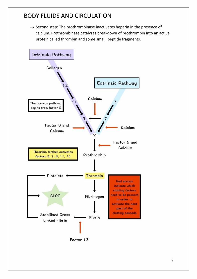
Second step: The prothrombinase inactivates heparin in the presence of
calcium. Prothrombinase catalyzes breakdown of prothrombin into an active
protein called thrombin and some small, peptide fragments.
BODY FLUIDS AND CIRCULATION
10
Third step: Thrombin acts as enzyme and first brings about depolymerization
of these monomers. Later thrombin stimulates repolymerization of these
monomers into long insoluble fibres – like polymers called fibrin. The thin,
long and solid fibres of fibrin from a dense network upon the wound and trap
blood corpuscles to form a clot. The clot seals the wound and stops bleeding.
Soon after the clot seals the wound and stops bleeding. Soon after the clot
starts contracting and a pale yellow fluid, the serum, starts oozing out. This
serum is blood plasma minus fibrinogen and blood corpuscles.
Vitamin K is essential for blood clotting as it is necessary for the synthesis of
prothrombin in the liver
List of Clotting Factors
Factor I
Name :Fibrinogen
Source :Liver
Pathway : Both extrinsic and intrinsic
Activator :Thrombin
Actions : When fibrinogen is converted into fibrin by thrombin, it forms long strands
that compose the mesh network for clot formation.
FactorII Name :Prothrombin Source :Liver Pathway : Both extrinsic and intrinsic Activator : Prothrombin activator Actions : Prothrombin is converted into thrombin which then activated fibrinogen into fibrin.
Factor III
Name : Thromboplastin / Tissue factor Source : Platelets (intrinsic) and damaged endothelium (cells) lining the blood vessel (extrinsic). Pathway : Both extrinsic and intrinsic Activator : Injury to blood vessel Action : Activates factor VII (VIIa).
BODY FLUIDS AND CIRCULATION
11
Factor IV
Name : Calcium Source : Bone and absorption from food in gastrointestinal tract Pathway : Both extrinsic and intrinsic Action : Works with many clotting factors for activation of the other clotting factors. These are called calcium-dependent steps.
Factor V
Name : Proaccerin / Labile factor / Ac-globulin (Ac-G) Source : Liver and platelets Pathway : Both extrinsic and intrinsic Activator : Thrombin Action : Works with Factor X to activate prothrombin (prothrombin activator).
Factor VII
Name : Proconvertin / Serum prothrombin conversion accelerator (SPCA) / stable factor Source : Liver Pathway : Extrinsic Activator : Factor III (tissue factor) Actions : Activates Factor X which works with other factors to convert prothrombin into thrombin.
Factor VIII
Name : Anti-hemoplytic factor / Antihemophilic factor (AHF) or globulin (AHG) / antihemophilic factor A Source : Endothelium lining blood vessel and platelets (plug) Pathway : Intrinsic Activator : Thrombin Actions : Works with Factor IX and calcium to activate Factor X. Deficiency : Hemophilia A
Factor IX
Name : Christmas factor / Plasma thromboplastin component (PTC) / Antihemophilic factor B Source : Liver Pathway : Intrinsic Activator : Factor XI and calcium
BODY FLUIDS AND CIRCULATION
12
Actions : Works with Factor VIII and calcium to activate Factor X. Deficiency : Hemophilia B
Factor X
Name : Stuart Prower factor / Stuart factor Source : Liver Pathway : Extrinsic and intrinsic Activator : Factor VII (extrinsic) / Factor IX + Factor VIII + calcium (intrinsic) Actions : Works with platelet phospholipids to convert prothrombin into thrombin. This reaction is made faster by activated Factor V.
Factor XI
Name : Plasma thromboplastin antecedent (PTA) / antihemophilic factor C Source : Liver Pathway : Intrinsic Activator : Factor XII + prekallikrein and kininogen Actions : Works with calcium to activate Factor IX. Deficiency : Hemophilia C
Factor XII
Name : Hageman factor Source : Liver Pathway : Intrinsic Activator : Contact with collagen in the torn wall of blood vessels Actions : Works with prekallikrein and kininogen to activate Factor XI. Also activates plasmin which degrades clots.
Factor XIII
Name : Fibrin stabilizing factor Source : Liver Activator : Thrombin and calcium Actions : Stabilizes the fibrin mesh network of a blood clot by helping fibrin strands to link to each other. Therefore it also helps to prevent fibrin breakdown (fibrinolysis).
Prekallikrein
Source : Liver Pathway : Intrinsic Actions : Works with kininogen and Factor XII to activate Factor XI.
BODY FLUIDS AND CIRCULATION
13
Kininogen
Source : Liver Pathway : Intrinsic Actions : Works with prekallikrein and Factor XII to activate Factor XI.
FUNCTIONS OF BLOOD
i) Transport o f food materials : Blood transports the digested food from the
alimentary canal to the different body cells
ii) Transport of respiratory gases : Oxygen is carried from the respiratory organs to
the tissues and carbon dioxide from the tissue to the respiratory organ by blood.
iii) Transport of hormones: Hormones are carried by blood from the endocrine
glands to the places of use
iv) Transport of excretory matter: Blood transport the excretory matter to the kidney
or other excretory organs.
v) Transport of heat: Blood allows the transfer of heat from the deeper tissue to
surface of the body where it can be lost.
vi) Defense against infection: Some white blood corpuscles are phagocytic in action,
however, certain blood corpuscles produce antitoxins to neutralize the toxins
released by the foreign germs.
vii) Temperature regulation : Blood maintains the body temperature to a constant
level after distributing heat within the body.
viii) Water balance: Blood maintains water to a constant level by bringing about
constant exchange of water between circulating blood and the tissue fluid
ix) Maintenance of pH: Blood helps to regulate the pH of the body.
x) Prevention of eccessive loss of blood: When any part of the body is injured, loss
of blood is prevented by the formation of a clot.
xi) Helps in healing: Blood maintains necessary supplies for the repair of damaged
tissue. Eosinophils and basophils help in the healing of wound.
xii) Maintenance of physiological co-operation: Blood maintains a physiological co-
oeration between parts of the body by circulating from one to other parts.
BLOOD GROUP
Karl Landsteiner reported first time ABO blood groups in human being ( 1900). AB
blood group was found out by de castellan and Steini ( 1902)
BODY FLUIDS AND CIRCULATION
14
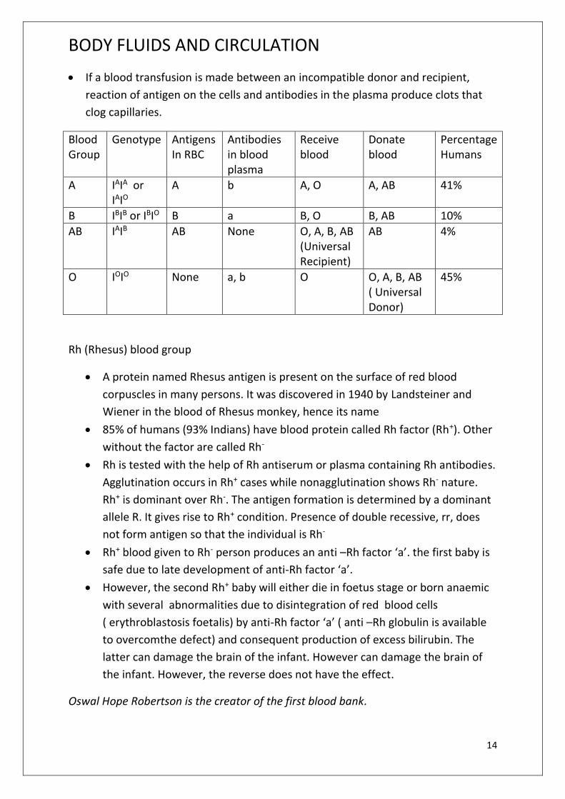
If a blood transfusion is made between an incompatible donor and recipient,
reaction of antigen on the cells and antibodies in the plasma produce clots that
clog capillaries.
Blood Group
Genotype Antigens In RBC
Antibodies in blood plasma
Receive blood
Donate blood
Percentage Humans
A IAIA or IAIO
A b A, O A, AB 41%
B IBIB or IBIO B a B, O B, AB 10%
AB IAIB AB None O, A, B, AB (Universal Recipient)
AB 4%
O IOIO None a, b O O, A, B, AB ( Universal Donor)
45%
Rh (Rhesus) blood group
A protein named Rhesus antigen is present on the surface of red blood
corpuscles in many persons. It was discovered in 1940 by Landsteiner and
Wiener in the blood of Rhesus monkey, hence its name
85% of humans (93% Indians) have blood protein called Rh factor (Rh+). Other
without the factor are called Rh-
Rh is tested with the help of Rh antiserum or plasma containing Rh antibodies.
Agglutination occurs in Rh+ cases while nonagglutination shows Rh- nature.
Rh+ is dominant over Rh-. The antigen formation is determined by a dominant
allele R. It gives rise to Rh+ condition. Presence of double recessive, rr, does
not form antigen so that the individual is Rh-
Rh+ blood given to Rh- person produces an anti –Rh factor ‘a’. the first baby is
safe due to late development of anti-Rh factor ‘a’.
However, the second Rh+ baby will either die in foetus stage or born anaemic
with several abnormalities due to disintegration of red blood cells
( erythroblastosis foetalis) by anti-Rh factor ‘a’ ( anti –Rh globulin is available
to overcomthe defect) and consequent production of excess bilirubin. The
latter can damage the brain of the infant. However can damage the brain of
the infant. However, the reverse does not have the effect.
Oswal Hope Robertson is the creator of the first blood bank.
AIMPT Study Material Of Biology Part 2
Publisher : Faculty Notes Author : Panel Of Experts
Type the URL : http://www.kopykitab.com/product/11137
Get this eBook
50%OFF