bone marrow cell recruitment mediated by inducible nitric ... · alfred l. nuttall,* ... irradiated...
TRANSCRIPT
Stem Cells, Tissue Engineering and Hematopoietic Elements
Bone Marrow Cell Recruitment Mediated byInducible Nitric Oxide Synthase/Stromal Cell-DerivedFactor-1� Signaling Repairs the AcousticallyDamaged Cochlear Blood-Labyrinth Barrier
Min Dai,* Yue Yang,* Irina Omelchenko,*Alfred L. Nuttall,*† Allan Kachelmeier,*Ruijuan Xiu,‡ and Xiaorui Shi*‡
From the Oregon Hearing Research Center,* Department of
Otolaryngology and Head and Neck Surgery, Oregon Health and
Science University, Portland, Oregon; the Kresge Hearing Research
Institute,† University of Michigan, Ann Arbor, Michigan; and The
Institute of Microcirculation,‡ Chinese Academy of Medical Sciences
and Peking Union Medical College, Beijing, China
Using a mouse model with noise-induced cochlearblood-labyrinth-barrier (CBLB) injury, we examinedthe effects of inducible nitric oxide synthase (iNOS)on the recruitment of bone marrow-derived cells(BMDCs) to the CBLB after acoustic injury. Lethallyirradiated C57BL/6J and B6.129P2-Nos2tm1Lau/J micewere transplanted with GFP�-BMDCs from C57Bl/6-Tg (UBC GFP) mice. Four weeks after transplanta-tion, we assessed the population of GFP�-BMDCs inthe CBLB. Only small numbers of GFP�-BMDCs werefound to infiltrate the area of the CBLB in the controlrecipient mice. However, robust GFP�-BMDC migra-tion occurred in the area of the CBLB within theinjured cochlea during the first week following acous-tic trauma, and further BMDC accumulation was seenby 2 weeks posttrauma. After 4 weeks, the BMDCswere integrated into vessels. Local iNOS from perivas-cular resident macrophages was found to be impor-tant for BMDC infiltration, since mice deficient iniNOS (Inos�/�) and mice with iNOS that had beeninhibited by 1400W displayed reduced BMDC infiltra-tion. Stromal cell-derived factor-1� (SDF-1�) and itschemokine receptor 4 (CXCR4) were required for theiNOS-triggered recruitment. BMDC recruitment wassignificantly reduced by the inhibition of SDF-1� ac-tivity. Inhibition of the iNOS/SDF-1� signaling path-way reduced vascular repair as observed by reducedvascular density. Our study revealed an intrinsic sig-naling pathway of iNOS that mediates SDF-1� to pro-
mote GFP�-BMDC infiltration/targeting in cochlearvascular repair. (Am J Pathol 2010, 177:3089–3099; DOI:10.2353/ajpath.2010.100340)
Ischemia causes considerable morbidity in various organsystems, and the pervasiveness of ischemic damagemakes repair of damaged vasculature an important ther-apeutic goal.1 Ischemia in the inner ear is closely relatedto several hearing disorders, including sudden sensori-neural hearing loss, presbyacusis, noise-induced hear-ing loss, tinnitus, and Meniere’s disease.2–5 Various clin-ical approaches to treatment of the ischemia have beentried, including use of vasoactive substances to improvecochlear blood flow; however, these generally have notbeen effective. A fundamental approach to determiningthe mechanisms of damage repair will enable develop-ment of more effective clinical treatments for vascular-related hearing disorders.
Acoustic trauma not only directly damages sensoryhair cells, but it also disrupts the blood flow and thecochlear blood-labyrinth-barrier (CBLB) in the stria vas-cularis,6 creating an ischemic and hypoxic environ-ment.3,6,7 Normal blood supply to the cochlea is criticalfor generating the ionic gradients and the endolymphaticpotential required for auditory transduction.6,8 Bettertreatment of noise-induced hearing loss requires an un-derstanding of parallel repair factors, one of which will bethe cellular repair mechanisms involved in restoration ofefficient cochlear blood flow after damage.
Increasing evidence highlights the importance of cir-culating bone marrow stem cells, which is home to sites
Supported by NIH NIDCD DC DC010844, NIH NIDCD DC 008888-02, NIHNIDCD DC 008888-02S1, NIH NIDCD DC R0100105, and NIH NIDCD DC005983 (p30).
Accepted for publication July 29, 2010.
Address reprint requests to Xiaorui Shi, M.D., Ph.D., Assistant Profes-sor, Oregon Hearing Research Center, Oregon Health and Science Uni-versity, 3181 SW Sam Jackson Park Rd, NRC04, Portland, OR 97239.E-mail: [email protected].
The American Journal of Pathology, Vol. 177, No. 6, December 2010
Copyright © American Society for Investigative Pathology
DOI: 10.2353/ajpath.2010.100340
3089
of ischemia and contribute to formation of new bloodvessels by differentiation.9–11 In the context of cancer, inhi-bition of bone marrow stem cell recruitment to tumors de-creases tumor angiogenesis,12 whereas the reverse, infu-sion of bone marrow-derived progenitor cells increasesangiogenesis13 and improves organ function. For example,transplantation of bone marrow cells into myocardium aug-ments cardiac function following myocardial infarct orchronic ischemia in rats and dogs.14,15 Transplantation ofbone marrow cells attenuates ischemic damage and im-proves functional recovery after brain injury.16
Coordination of an inflammatory response, entailing reg-ulation of chemokine release, is essential for bone marrowstem cell (BMSC)-associated neovascularization duringischemia and wound healing.17 For example, the release ofstromal cell-derived factor-1� (SDF-1�) from stressed tissueplays an important role in the mobilization of bone marrowcells to local ischemic sites on damaged vessels.18–20 Ad-ditionally, nitric oxide (NO), produced by endothelial nitricoxide synthase (eNOS), is shown to be a critical factor inbone marrow cell recruitment for vascular repair.21
Here, we report the finding that recruitment of bonemarrow cells to ischemic tissues in the noise traumatizedcochlea is signaled by a local inducible nitric oxide syn-thase (iNOS)-dependent SDF-1� pathway. Recruitmentof bone marrow derived cells (BMDCs) to the damagedstria vascularis results in repair of cochlear vessels.
Materials and Methods
Animals and Bone Marrow Cell Transplantation
Male C57BL/6J (Inos wild type [WT], aged 4 weeks; stocknumber: 000664), B6.129P2-Nos2tm1Lau/J (Inos�/�, aged4 weeks; stock number: 002609), and C57Bl/6-Tg mice(UBC-GFP, aged 4 to 6 weeks; stock number: 004353)were purchased from Jackson Laboratory (Bar Harbor,ME). C57Bl/6-Tg mice served as donor mice with InosWT, and Inos�/� mice of the same age served as recip-ients. The recipients were irradiated (at 9 Gy) with a�-emitting source and reconstituted with a single perior-bital sinus injection of 2 � 107 BMDCs in 200 �l ofmodified HBSS from donor transgenic mice. At 1 monthposttransplantation, the mice served as control, noise-exposed, and noise-exposed � drug treatment groups.The groups were sacrificed at different times after noiseexposure (immediately, 1 week, 2 weeks, and 4 weeks)for measurement. All procedures in this study were re-viewed and approved by the Institutional Animal Careand Use Committee at Oregon Health and ScienceUniversity.
Noise Exposure
Animals were placed in wire mesh cages and exposed tobroadband noise at 120 dB sound pressure level (SPL) ina sound exposure booth for 3 hours and for an additional3 hours the following day. The noise exposure regime,routinely used in our laboratory, produces permanentloss of cochlear sensitivity.6
Immunohistochemistry and FluorescenceMicroscopy
Normal and noise-exposed mice were sacrificed at day2, week 1, week 2, or week 4, subsequent to a 1-monthtransplantation recovery period. The cochleae were har-vested and fixed in 4% paraformaldehyde overnight at4°C, and then rinsed in 37°C PBS (pH 7.3) to remove anyresidual 4% paraformaldehyde. Immunohistochemistrywas performed as described before.6 Tissue sampleswere permeabilized in 0.5% Triton X-100 (Sigma, St.Louis, MO) for 1 hour, and then immunoblocked with asolution of 10% goat serum and 1% bovine albumin in0.02 mol/L PBS for an additional hour. The specimenswere incubated overnight at 4°C with the primary anti-bodies (listed in Table 1) diluted in PBS-bovine serumalbumin. After several washes in PBS, the samples wereincubated with secondary antibodies, Alexa Fluor 568-conjugated goat anti-rabbit (category number A21069;Invitrogen, Eugene, OR), Fluor 647-conjugated goat anti-rabbit (category number A-21244; Invitrogen), and AlexaFluor 568-conjugated goat anti-rat (category numberA-11077; Invitrogen) for 1 hour at room temperature. Thetissues were mounted in mounting medium (H-1000; Vec-tor Laboratories, Inc., Burlingame, CA) and visualizedunder an FV1000 Olympus laser-scanning confocal mi-croscope (Olympus, Tokyo, Japan). Controls were pre-pared by replacing primary antibodies with PBS.
Western Blot Analysis
To investigate the influence of noise exposure on theprotein level of iNOS expression, the cochlear lateral wallwas dissented from control and noise-exposed animals(cohorts of three mice). The collected lateral wall tissuewas homogenized in lysis buffer (RIPA Lysis buffer, Up-state, a Serologicals Company, Temecula, CA) with aprotease inhibitor cocktail (Protease Inhibitor cocktailsSet III, Calbiochem, Darmstadt, Germany) for 30 sec-onds. After centrifuging (4°C, 30 minutes, at 14,000 rpm),the supernatant was assayed for protein by using a DCprotein Assay kit (Bio-Rad, Hercules, CA). Samples wereheated to 100°C for 5 minutes with 2� SDS loading bufferand briefly cooled on ice. Fifty-microgram aliquots of totalprotein from each sample were run on 10% sodium do-decyl sulfate-polyacrylamide gels to detect iNOS (130kDa) and actin (43 kDa). Proteins were electrophoreti-cally transferred to polyvinylidene difluoride membranes(Millipore Corp., Bedford, MA), with the membranesblocked with nonfat milk and 0.1% Tween 20 in Tris-buffered saline for 1 hour at room temperature. The sam-ple was incubated with primary antibodies diluted 1:1000in skim milk overnight at 4°C for specific immunodetec-tion (rabbit polyclonal antibody to iNOS, � Diagnostic IntlInc., San Antonio, TX; mouse polyclonal antibody to actin,Millipore Corp.). After three washes with PBS, the mem-branes were incubated for another hour with horseradishperoxidase-conjugated goat anti-rabbit or goat anti-mouse IgG diluted 1:10,000 in PBS at room temperature.Antigens were revealed by using ECL Plus Western Blot-
3090 Dai et alAJP December 2010, Vol. 177, No. 6

ting Detection Reagents (GE Healthcare, Pittsburgh, PA).To quantify the changes of iNOS protein level, the banddensity was analyzed by using Image J software (V1.38X;NIH, West Chester, PA). The density of the bands of actinwas used to normalize the iNOS protein level.
Reverse Transcription-Polymerase ChainReaction
Total RNA from the cochlear lateral wall was separatelyextracted for each experimental group with a RNeasy kit(Qiagen, Valencia, CA) according to the manufacturer’ssuggestions. Each cohort of two mice was analyzed foriNOS mRNA. One microgram of total RNA was reverse-transcribed by using a RETROscript kit (Ambion, Austin,TX). Conserved regions spanning introns were selectedfor the primers of iNOS and glyceraldehyde-3-phosphatedehydrogenase. The primers used were as follows: iNOS(mouse Chr 11 NM_010927), forward, 5�-CTATCAGGAA-GAAATGCAGGAGAT-3�, reverse, 5�-GAGCACGCTGAG-TACCTCATT-3�, 145-bp product; SDF-1� (mouse Chr 6NM_013655), forward, 5�-CAAGAGGCTCAAGATGTGAG-AGGTG-3�, reverse, 5�-TGGCCTTGGCCTGTCACCAA-3�,258-bp; and glyceraldehyde-3-phosphate dehydrogenase(mouse Chr 6 NM_008084), forward, 5�-ATGTGTCCGTC-GTGGATCTGAC-3�, reverse, 5�-AGACAACCTGGTCCT-CAGTGTAG-3�, 132-bp product. The RT-PCR was cycledat 95°C for 2 minutes, up to 40 cycles at 95°C for 30seconds, 60°C for 45 seconds, 72°C for 30 seconds, and afinal 5-minute extension at 72°C. The products of RT-PCRwere visualized by agarose gel electrophoresis.
Quantitative Real-Time Polymerase ChainReaction
Total RNA from the cochlear lateral wall of differentgroups was separately extracted with RNeasy (Qiagen)
according to the manufacturer’s suggestions. Each co-hort of two mice was analyzed for mRNA levels of iNOSand SDF-1� with quantitative real-time PCR. One mi-crogram of total RNA was reverse-transcribed by usinga RETROscript kit (Ambion). The cDNA synthesizedfrom total RNA was diluted 10-fold with DNase-freewater, and each cDNA sample was independentlytested three times. Transcript quantities were assayedby TaqMan gene expression assay: iNOS (categorynumber Mm01309902_m1; AppliedBiosystems, Foster City,CA) and SDF-1� (category number Mm00457276_m1, Ap-plied Biosystems) were assayed in a model 7300 real-time PCR system (Applied Biosystems). Cycling con-ditions of the real-time PCR were 95°C for 20 seconds,40 cycles of 95°C for 1 second, and 60°C for 20seconds. Mouse glyceraldehyde-3-phosphate dehy-drogenase (category number 4352339E, Applied Bio-systems) expression was used as an endogenous con-trol. Quantitative PCR was performed according to theguidelines provided by Applied Biosystems. The com-parative cycle threshold (CT) method (��CT quantitation)was used to assess the difference between samples.Quantitative data analysis followed the suggestions of themanufacturer.
Enzyme-Linked Immunosorbent Assay
To compare the protein level of SDF-1 between differentgroups, whole cochlea was isolated from the animals(cohorts of three animals) and the lateral wall tissue care-fully dissected. All tissue samples were homogenized inlysis buffer (RIPA Lysis buffer, Upstate, a SerologicalsCompany) with a protease inhibitor cocktail (ProteaseInhibitor cocktails Set III, Calbiochem) for 30 seconds.After centrifuging (4°C, 30 minutes, at 14,000 rpm), thesupernatant was collected, and the protein assay wasperformed by using a DC protein Assay kit (Bio-Rad). Ten
Table 1. Primary Antibodies Employed
Primary antibodies Source Identification Dilution Type Specificity
NG2 Chemicon ab5320 1:200 (dilution with1% BSA-PBS)
Rabbit polyclonalantibody
Reacts with NG2 ofmouse, rat, and human
Desmin Epitomics 1466-1 1:400 (dilution with1% BSA-PBS)
Rabbitmonoclonalantibody
Reacts with desmin ofmouse, rat, and human
F4/80 Santa CruzBiotechnology
ab6640 1:100 (dilution with1% BSA-PBS)
Rat monoclonal(CI:A3-1) toF4/80
This antibody recognizesthe mouse F4/80antigen, a 160-kdglycoprotein expressedby macrophages.
Collagen type IV ResearchDiagnostics Inc.
7478 1:50 (dilution with1% BSA-PBS)
Rabbit polyclonalantibody
Reacts with mostmammalian type IVcollagen (dilution with1% BSA-PBS)
CD31 Abcam ab28364 1:100 (dilution with1% BSA-PBS)
Rabbit polyclonalantibody
Reacts with mouse
CXCR4 Abcam ab2074 1:100 (dilution with1% BSA-PBS)
Rabbit polyclonalantibody
Reacts with mouse
iNOS Alpha DiagnosticsIntl. Inc.
iNOS-A 0370A2 1:1000 (dilution with1% BSA-PBS)
Rabbit polyclonalantibody
Reacts with mouse
BSA, bovine serum albumin.
Bone Marrow Cell Recruitment in Cochlea 3091AJP December 2010, Vol. 177, No. 6
micrograms of protein per sample was used for enzyme-linked immunosorbent assay (ELISA) analysis. SDF-1protein concentration of different groups was assessedby using a Quantikine Mouse CXCL12/SDF-1 ELISA kit(R&D Systems, Burlington, ON) according to the manu-facturer’s recommendations.
GFP�-BMDC Count
GFP�-BMDCs in the stria vascularis of mouse cochleawere counted on a standard epifluorescence microscopewith a � 40 objective lens (cohorts of four mice). TheGFP�-BMDCs were counted in a region corresponding toan initial frequency response of 8–32 kHz, determined bythe frequency-map described by Wang et al.22 Noise-caused hearing loss occurs predominately between 8and 32 kHz with this sound protocol.6 The area studied inthis investigation (a length approximately 2 mm starting1.5 mm from the base) lies within this region. For plottingpurposes, the data were grouped.
Drug Treatment
The mice were pretreated with 1400W (10 mg/kg, i.p.;Enzo Life Sciences, Farmingdale, NY)23 to inhibit iNOSand with an antagonist of the SDF-1� receptor CXCR4,AMD3100 (7.5 mg/kg, i.p.; Sigma-Aldrich),24 to inhibitSDF-1� activity. Treatments were administered as single-dose injections 30 minutes before the animal receivednoise and were continued at one dose per day for the firstweek and one dose per week up to 4 weeks after noiseexposure. The side-effects of 1400W and AMD3100 onauditory function were evaluated. Auditory brain-stem re-sponse was used to measure hearing threshold. Neither1400W nor AMD3100 showed ototoxicity in the six miceof the two treated groups.
NO Measurement
The auditory bulla was dissected and rapidly opened in apetri dish filled with a physiological solution containing
125 mmol/L NaCl, 5.0 mmol/L KCl, 1.6 mmol/L CaCl2, 18mmol/L NaHCO3, 10 mmol/L glucose, and 10 mmol/LHEPES, pH 7.4. Small pieces of tissue from the basalmiddle turn of the cochlear lateral wall in noise exposedanimals, both with and without 1400W treatment, wereremoved and incubated in a physiological solution at37°C, pH 7.4, containing 10 �mol/L diaminofluorescein-2diacetate (category number 251505, Calbiochem) for de-tecting NO. The tissue was incubated with dye for 30minutes, subsequently washed in fresh physiological so-lution for 10 minutes, and assessed by confocal micros-copy. Quantitation of the NO indicator was performed onimages taken at the same gain and illumination powersetting (488 excitation, 520 nm emission filter for thediaminofluorescein diacetate). Fluorescence intensitywas analyzed by using Image J software (V1.38X; NIH)as described previously.4 In brief, images were acquiredwith a �40 objective lens. A total of 12 to 16 images wererecorded from four normal mice. Twelve to 14 imagesfrom four noise-exposed mice were recorded. A total of13 to 17 images were recorded from four 1400W-treatednoise-exposed mice. The area of the stria vascularis wasanalyzed for mean fluorescence intensity. Mean back-ground fluorescence, obtained for a small area locatedaway from the fluorescent tissue, was subtracted from thefluorescence intensity. Presented data are an average offour experimental and control animals.
Vascular Density Measurement
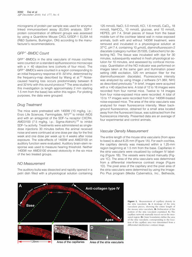
The entire length of the mouse stria vascularis (from apexto base) is about 6.25 mm (Figure 1A). For each cochlea,the capillary density was measured within a 1.25-mmregion beginning at 1.5 mm from the base. Capillaries inthe stria vascularis were visualized by collagen IV label-ing (Figure 1B). The vessels were traced manually (Fig-ure 1C). The area of the stria vascularis was determinedfrom a differential interference contrast image (Figure1D). The pixel area of the capillary and the pixel area ofthe stria vascularis were determined by using the Image-Pro Plus program (Media Cybernetics, Inc., Bethesda,
Figure 1. Measurement of capillary density inthe stria vascularis. A: A montage of the striavascularis pieces, showing the entire length ofthe mouse stria vascularis. B: Delineation of theportion of the stria vascularis examined. C: Thecapillary network manually traced out in the mea-sured region. D: Outer boundaries define the areaof the stria vascularis; corresponding to the loca-tion of the capillary area was determined from adifferential interference contrast image.
3092 Dai et alAJP December 2010, Vol. 177, No. 6
MD). Capillary density as a percentage of the strial tissue
was calculated asPixel area of capillaries
Pixel area of the stria vascularis� 100%.
Statistics and Analysis
Data are presented as means � SD and were evaluated byusing the Student’s t-test for two groups or by analysis ofvariance for comparisons of three or more groups. A 95%confidence level was considered statistically significant.
Results
Bone Marrow Cells Recruited to Noise-DamagedBlood-Labyrinth-Barrier
The cochlea of the mouse has 2.5 turns of lateral wall(Figure 2A). The normal mouse cochlear lateral wall con-tains two dense networks of capillaries: the capillaries ofthe stria vascularis (V/SV) and the capillaries of the spiralligament (V/SL; Figure 2B, modified from Mudry and
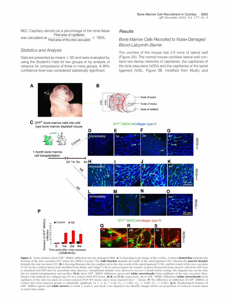
Figure 2. Noise initiates robust GFP�-BMDC infiltration into the damaged CBLB. A: A dissecting scope image of the cochlea. A drawn dotted line indicates thelocation of the stria vascularis (SV) where the CBLB is located. The wide bracket demarks the width of the spiral ligament (SL), whereas the narrow bracketdemarks the stria vascularis (SV). B: A drawing illustrates the two capillary networks, the vessels of the spiral ligament (V/SL), and the vessels of the stria vascularis(V/SV) in the cochlear lateral wall (modified from Mudry and Tange25). C: A cartoon depicts the transfer of green fluorescent bone marrow cells from GFP miceto irradiated non-GFP mice by periorbital sinus injection. Transplanted animals were allowed to recover 1 month before testing. The diagram sets out the timeline for animal transplantation and sacrifice. D–G: show GFP�-BMDC infiltration (green and white arrowheads) from capillaries of the stria vascularis (blue,labeled with antibody for collagen type IV) in a control iNOS WT mouse. H–K and L–O, respectively, show GFP�-BMDC infiltration (white arrowheads) fromcapillaries of the stria vascularis in a noise-exposed iNOS WT mouse and a noise-exposed Inos�/� mouse. P: The difference in infiltration of GFP�-BMDCs incontrol and noise-exposed groups is statistically significant (n � 4; P0 � 0.48; P1w 0.001; P2w 0.001; P4w � 0.001). Q–S: Morphological features ofGFP�-BMDCs (green and white arrows) at week 1, week 2, and week 4 are displayed (see Results). Images shown are projections of confocal z-stacks takenat tested time points.
Bone Marrow Cell Recruitment in Cochlea 3093AJP December 2010, Vol. 177, No. 6
Tange).25 The strial capillaries, shaped as polygonalloops, are highly specialized vascular epithelia that formthe CBLB in the stria vascularis. Sound trauma causes asignificant breech of the CBLB.6
In this study, we investigated whether BMDCs have anessential role in repair of damaged cochlear microves-sels in the stria vascularis. Lethally irradiated mice weretransplanted with GFP�-BMDCs from C57Bl/6-Tg (UBC-GFP) mice (Figure 2C). One month after transplantation,we assessed the population of GFP�-BMDCs in the re-gion of the stria vascularis in the reconstituted iNOS WTand Inos�/� mice under control (not exposed to loudsound) and acoustic trauma conditions. Irradiated andBMDC transplanted control mice showed normal hearingthresholds immediately after irradiation and 2 monthsafter irradiation (data not shown). No GFP�-BMDCs wereobserved to have migrated into the area of the striavascularis in the control mice at 1 month post transplan-tation. Only small numbers of GFP�-BMDCs had infil-trated into the area of CBLB 2 months after transplanta-tion in the reconstituted iNOS WT mice (Figure 2, D–G).Then, when animals were exposed to wide-band noise at120 dB for 3 hours per day for 2 consecutive days, robustBMDC migration was observed in the acoustically trau-matized iNOS WT animal cochlea (Figure 2, H–K). Imme-diately after noise exposure and flushing by cardiac per-fusion, large numbers of GFP�-BMDCs were observedadhered to vessel walls in the capillary network. Theselabeled cells had not yet transmigrated across the vesselwall at this time point. Remarkably, GFP�-BMDC infiltra-tion occurred in the first week post acoustic trauma.GFP�-BMDCs further accumulated at the second week(Figure 2, H–K). However, much less GFP�-BMDCs infil-tration was observed in the Inos�/� mice (Figure 2, L–O).The number of the infiltrated GFP�-BMDCs is shown inFigure 2P. There was a significant difference in GFP�-BMDC infiltration between the control and noise-exposediNOS WT mice.
Infiltrated BMDCs were previously identified as macro-phages.26 After taking residence in the stria vascularis,they underwent morphological changes (Figure 2, Q–S).At an early stage (approximately 1 week post noise ex-posure), infiltrated GFP�-BMDCs were frequently foundto be spherical or nodular shaped (possibly caught in theact of transmigration, Figure 2Q). Approximately 2 weekspost noise exposure, most of infiltrated GFP�-BMDCsdeveloped ramified processes, appeared dendriform inshape, and were irregularly distributed on the capillariesof the stria vascularis (Figure 2R). Approximately 4 weekspost noise exposure, the majority of infiltrated BMDCswere elongated and displayed a particular orientation—that is, their long processes were parallel to the vessels ofthe stria vascularis (Figure 2S).
BMDC Infiltration Mediated by NO from iNOS
eNOS was previously reported to be essential in therecruitment of peripheral circulating bone marrow cells tothe sites of ischemia.21 Our finding of noise-inducediNOS expression in the cochlear lateral wall,27 however,
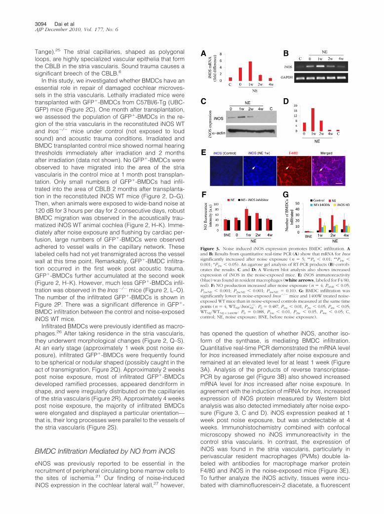
has opened the question of whether iNOS, another iso-form of the synthase, is mediating BMDC infiltration.Quantitative real-time PCR demonstrated the mRNA levelfor Inos increased immediately after noise exposure andremained at an elevated level for at least 1 week (Figure3A). Analysis of the products of reverse transcriptase-PCR by agarose gel (Figure 3B) also showed increasedmRNA level for Inos increased after noise exposure. Inagreement with the induction of mRNA for Inos, increasedexpression of iNOS protein measured by Western blotanalysis was also detected immediately after noise expo-sure (Figure 3, C and D). iNOS expression peaked at 1week post noise exposure, but was undetectable at 4weeks. Immunohistochemistry combined with confocalmicroscopy showed no iNOS immunoreactivity in thecontrol stria vascularis. In contrast, the expression ofiNOS was found in the stria vascularis, particularly inperivascular resident macrophages (PVMs) double la-beled with antibodies for macrophage marker proteinF4/80 and iNOS in the noise-exposed mice (Figure 3E).To further analyze the iNOS activity, tissues were incu-bated with diaminofluorescein-2 diacetate, a fluorescent
Figure 3. Noise induced iNOS expression promotes BMDC infiltration. Aand B: Results from quantitative real-time PCR (A) show that mRNA for Inossignificantly increased after noise exposure (n � 5; **P0 0.01; **P1w 0.001; *P2w 0.05). An agarose gel analysis of RT-PCR products (B) corrob-orates the results. C and D: A Western blot analysis also shows increasedexpression of iNOS in the noise-exposed mice. E: iNOS immunoreactivity(blue) was found in resident macrophages (white arrows, labeled for F4/80,red). F: NO production increased after noise exposure (n � 4; P0NE 0.05;P1wNE 0.001; P2wNE 0.001; P4wNE � 0.10). G: BMDC infiltration wassignificantly lower in noise-exposed Inos�/� mice and 1400W treated noise-exposed WT mice than in noise-exposed controls measured at the same timepoints (n � 4, WTNE/Inos
�/�NE : P0 � 0.487, P1w 0.01, P2w 0.05, P4w 0.05;
WTNE/WTNE�1400W: P0 � 0.088, P1w 0.01, P2w 0.05, P4w 0.05; C,control; NE, noise exposure; BNE, before noise exposure).
3094 Dai et alAJP December 2010, Vol. 177, No. 6
indicator for NO. Consistent with the results of increasedimmunoreactivity for iNOS, a significantly increased levelof NO production was found in the stria vascularis of thenoise-exposed mice (Figure 3F). Inhibition of iNOS activ-ity with the iNOS inhibitor 1400W markedly reduced NOproduction (Figure 3F). The number of infiltrated BMDCsin noise-exposed Inos�/� mice and in iNOS inhibitor1400W treated noise-exposed WT mice was significantlyless than in noise-exposed WT mice (Figure 3G). Thedata indicates that NO produced by iNOS is an importantdriving signal for the recruitment of bone marrow derivedcells.
BMDC Infiltration Linked to iNOS Up-Regulationof SDF-1�
SDF-1� and its receptor CXCR4 are known to regulatehoming and engraftment of BMDCs.28 To determinewhether iNOS/NO regulated BMDC recruitment involvesSDF-1�/CXCR4 signaling in the cochlea, SDF-1� expres-sion in control and noise-exposed WT mice, as well as inInos�/� mice and WT mice with iNOS inhibition, werecompared.
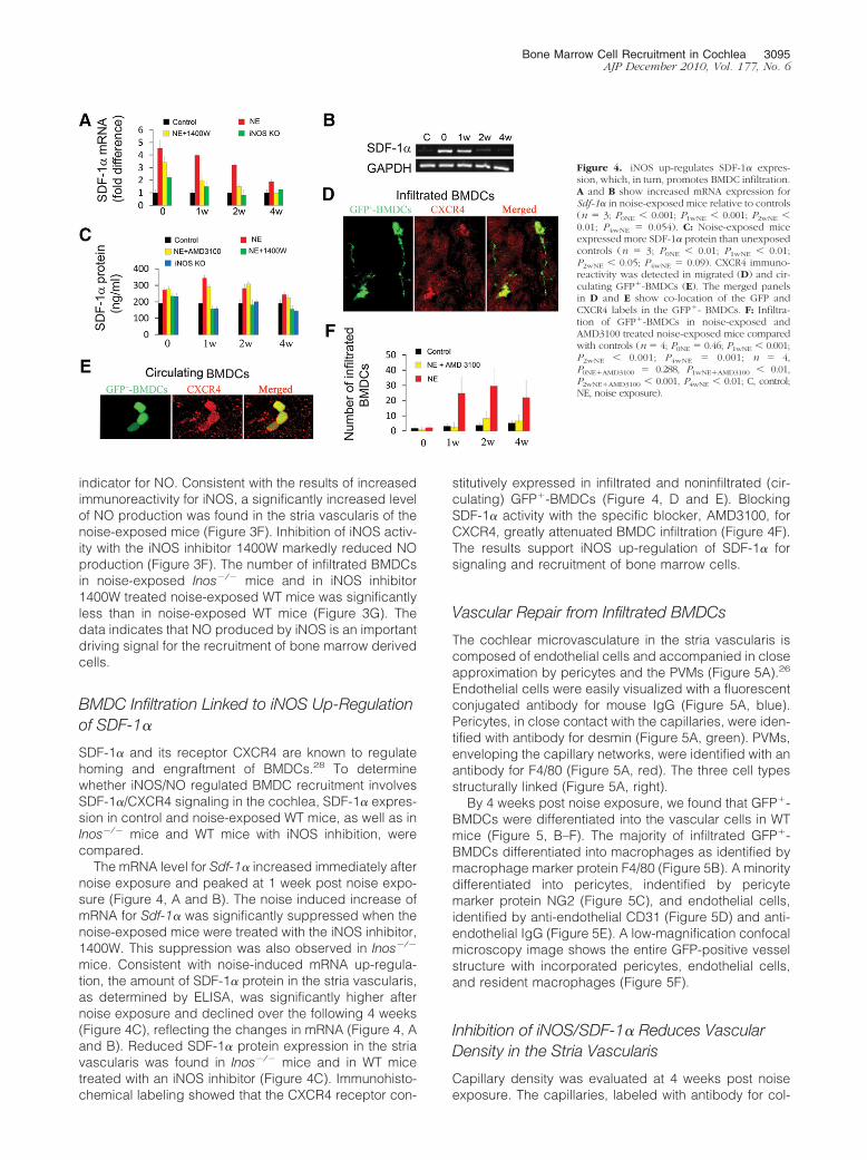
The mRNA level for Sdf-1� increased immediately afternoise exposure and peaked at 1 week post noise expo-sure (Figure 4, A and B). The noise induced increase ofmRNA for Sdf-1� was significantly suppressed when thenoise-exposed mice were treated with the iNOS inhibitor,1400W. This suppression was also observed in Inos�/�
mice. Consistent with noise-induced mRNA up-regula-tion, the amount of SDF-1� protein in the stria vascularis,as determined by ELISA, was significantly higher afternoise exposure and declined over the following 4 weeks(Figure 4C), reflecting the changes in mRNA (Figure 4, Aand B). Reduced SDF-1� protein expression in the striavascularis was found in Inos�/� mice and in WT micetreated with an iNOS inhibitor (Figure 4C). Immunohisto-chemical labeling showed that the CXCR4 receptor con-
stitutively expressed in infiltrated and noninfiltrated (cir-culating) GFP�-BMDCs (Figure 4, D and E). BlockingSDF-1� activity with the specific blocker, AMD3100, forCXCR4, greatly attenuated BMDC infiltration (Figure 4F).The results support iNOS up-regulation of SDF-1� forsignaling and recruitment of bone marrow cells.
Vascular Repair from Infiltrated BMDCs
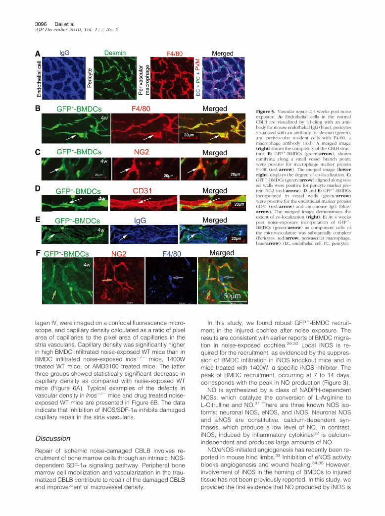
The cochlear microvasculature in the stria vascularis iscomposed of endothelial cells and accompanied in closeapproximation by pericytes and the PVMs (Figure 5A).26
Endothelial cells were easily visualized with a fluorescentconjugated antibody for mouse IgG (Figure 5A, blue).Pericytes, in close contact with the capillaries, were iden-tified with antibody for desmin (Figure 5A, green). PVMs,enveloping the capillary networks, were identified with anantibody for F4/80 (Figure 5A, red). The three cell typesstructurally linked (Figure 5A, right).
By 4 weeks post noise exposure, we found that GFP�-BMDCs were differentiated into the vascular cells in WTmice (Figure 5, B–F). The majority of infiltrated GFP�-BMDCs differentiated into macrophages as identified bymacrophage marker protein F4/80 (Figure 5B). A minoritydifferentiated into pericytes, indentified by pericytemarker protein NG2 (Figure 5C), and endothelial cells,identified by anti-endothelial CD31 (Figure 5D) and anti-endothelial IgG (Figure 5E). A low-magnification confocalmicroscopy image shows the entire GFP-positive vesselstructure with incorporated pericytes, endothelial cells,and resident macrophages (Figure 5F).
Inhibition of iNOS/SDF-1� Reduces VascularDensity in the Stria Vascularis
Capillary density was evaluated at 4 weeks post noiseexposure. The capillaries, labeled with antibody for col-
Figure 4. iNOS up-regulates SDF-1� expres-sion, which, in turn, promotes BMDC infiltration.A and B show increased mRNA expression forSdf-1� in noise-exposed mice relative to controls(n � 3; P0NE 0.001; P1wNE 0.001; P2wNE 0.01; P4wNE � 0.054). C: Noise-exposed miceexpressed more SDF-1� protein than unexposedcontrols (n � 3; P0NE 0.01; P1wNE 0.01;P2wNE 0.05; P4wNE � 0.09). CXCR4 immuno-reactivity was detected in migrated (D) and cir-culating GFP�-BMDCs (E). The merged panelsin D and E show co-location of the GFP andCXCR4 labels in the GFP�- BMDCs. F: Infiltra-tion of GFP�-BMDCs in noise-exposed andAMD3100 treated noise-exposed mice comparedwith controls (n � 4; P0NE � 0.46; P1wNE 0.001;P2wNE 0.001; P4wNE � 0.001; n � 4,P0NE�AMD3100 � 0.288, P1wNE�AMD3100 0.01,P2wNE�AMD3100 0.001, P4wNE 0.01; C, control;NE, noise exposure).
Bone Marrow Cell Recruitment in Cochlea 3095AJP December 2010, Vol. 177, No. 6
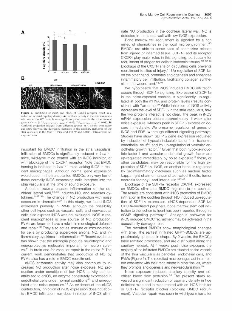
lagen IV, were imaged on a confocal fluorescence micro-scope, and capillary density calculated as a ratio of pixelarea of capillaries to the pixel area of capillaries in thestria vascularis. Capillary density was significantly higherin high BMDC infiltrated noise-exposed WT mice than inBMDC infiltrated noise-exposed Inos�/� mice, 1400Wtreated WT mice, or AMD3100 treated mice. The latterthree groups showed statistically significant decrease incapillary density as compared with noise-exposed WTmice (Figure 6A). Typical examples of the defects invascular density in Inos�/� mice and drug treated noise-exposed WT mice are presented in Figure 6B. The dataindicate that inhibition of iNOS/SDF-1� inhibits damagedcapillary repair in the stria vascularis.
Discussion
Repair of ischemic noise-damaged CBLB involves re-cruitment of bone marrow cells through an intrinsic iNOS-dependent SDF-1� signaling pathway. Peripheral bonemarrow cell mobilization and vascularization in the trau-matized CBLB contribute to repair of the damaged CBLBand improvement of microvessel density.
In this study, we found robust GFP�-BMDC recruit-ment in the injured cochlea after noise exposure. Theresults are consistent with earlier reports of BMDC migra-tion in noise-exposed cochlea.29,30 Local iNOS is re-quired for the recruitment, as evidenced by the suppres-sion of BMDC infiltration in iNOS knockout mice and inmice treated with 1400W, a specific iNOS inhibitor. Thepeak of BMDC recruitment, occurring at 7 to 14 days,corresponds with the peak in NO production (Figure 3).
NO is synthesized by a class of NADPH-dependentNOSs, which catalyze the conversion of L-Arginine toL-Citrulline and NO.31 There are three known NOS iso-forms: neuronal NOS, eNOS, and iNOS. Neuronal NOSand eNOS are constitutive, calcium-dependent syn-thases, which produce a low level of NO. In contrast,iNOS, induced by inflammatory cytokines32 is calcium-independent and produces large amounts of NO.
NO/eNOS initiated angiogenesis has recently been re-ported in mouse hind limbs.33 Inhibition of eNOS activityblocks angiogenesis and wound healing.34,35 However,involvement of iNOS in the homing of BMDCs to injuredtissue has not been previously reported. In this study, weprovided the first evidence that NO produced by iNOS is
Figure 5. Vascular repair at 4 weeks post noiseexposure. A: Endothelial cells in the normalCBLB are visualized by labeling with an anti-body for mouse endothelial IgG (blue), pericytesvisualized with an antibody for desmin (green),and perivascular resident cells with F4/80, amacrophage antibody (red). A merged image(right) shows the complexity of the CBLB struc-ture. B: GFP�-BMDCs (green/arrow), shownramifying along a small vessel branch point,were positive for macrophage marker proteinF4/80 (red/arrow). The merged image (lowerright) displays the degree of co-localization. C:GFP�-BMDCs (green/arrow) aligned along ves-sel walls were positive for pericyte marker pro-tein NG2 (red/arrow). D and E: GFP�-BMDCsincorporated in vessel walls (green/arrow)were positive for the endothelial marker proteinCD31 (red/arrow) and anti-mouse IgG (blue/arrow). The merged image demonstrates theextent of co-localization (right). F: At 4 weekspost noise-exposure incorporation of GFP�-BMDCs (green/arrow) as component cells ofthe microvasculature was substantially complete(Pericytes, red/arrow; perivascular macrophage,blue/arrow). (EC, endothelial cell; PC, pericyte).
3096 Dai et alAJP December 2010, Vol. 177, No. 6
important for BMDC infiltration in the stria vascularis.Infiltration of BMDCs is significantly reduced in Inos�/�
mice, wild-type mice treated with an iNOS inhibitor, orwith blockage of the CXCR4 receptor. Note that BMDChoming is inhibited in Inos�/� mice lacking iNOS in resi-dent macrophages. Although normal gene expressionwould occur in the transplanted BMDCs, only very few ofthese normally iNOS expressing cells integrate into thestria vascularis at the time of sound exposure.
Acoustic trauma causes inflammation of the co-chlear lateral wall,30,36 induces NO, and releases cy-tokines.6,37,38 The change in NO production with noiseexposure is dramatic.2,27 In this study, we found iNOSexpressed primarily in PVMs, although the possibilityother cell types such as vascular cells and nonvascularcells also express iNOS was not excluded. iNOS in res-ident macrophages is one source of NO production.PVMs are known to have a role in immunological defenseand repair.39 They also act as immune or immuno-effec-tor cells by producing superoxide anions, NO, and in-flammatory cytokines in inflammation.40 Recent evidencehas shown that the microglia produce neurotrophic andneuroprotective molecules important for neuron survi-val41 in brain and for vascular repair in the retina.42 Thecurrent work demonstrates that production of NO byPVMs also has a role in BMDC recruitment.
eNOS enzymatic activity may also contribute to in-creased NO production after noise exposure. NO pro-duction under conditions of low iNOS activity can beattributed to eNOS, an enzyme constitutely expressed inendothelial cells under normal conditions43 and unregu-lated after noise exposure.44 As evidence of the eNOScontribution, inhibition of iNOS expression does not abol-ish BMDC infiltration, nor does inhibition of iNOS elimi-
nate NO production in the cochlear lateral wall. NO isdetected in the lateral wall with low iNOS expression.
Bone marrow cell recruitment is signaled by a richmilieu of chemokines in the local microenvironment.45
BMDCs are able to sense sites of chemokine releasefrom injured or inflamed tissue. SDF-1� and its receptorCXCR4 play major roles in this signaling, particularly forrecruitment of progenitor cells to ischemic tissues.18,19,46
Blockage of the CXCR4 site on circulating cells preventsrecruitment to sites of injury.47 Up-regulation of SDF-1�,on the other hand, promotes angiogenesis and enhancesinflammatory cell infiltration, facilitating collagen synthe-sis in the wound bed.48,49
We hypothesize that iNOS induced BMDC infiltrationoccurs through SDF-1� signaling. Expression of SDF-1�in the noise-exposed cochlea is significantly up-regu-lated at both the mRNA and protein levels (results con-sistent with Tan et al).29 While inhibition of iNOS activitydecreases the level of SDF-1� in the stria vascularis, howthe two proteins interact is not clear. The peak in iNOSmRNA expression occurs approximately 1 week afternoise exposure, whereas peak in SDF-1� expression oc-curs immediately. We presume regulation of genes ofiNOS and SDF-1� through different signaling pathways.Studies have shown SDF-1� gene expression regulatedby induction of hypoxia-inducible factor-1 in ischemicendothelial cells50 and by up-regulation of vascular en-dothelial growth factor.51 Given that both hypoxia-induc-ible factor-1 and vascular endothelial growth factor areup-regulated immediately by noise exposure,6 these, orother candidates, may be responsible for the high ex-pression of SDF-1�. iNOS, on another hand, is regulatedby proinflammatory cytokines such as nuclear factorkappa-light-chain-enhancer of activated B cells, tumornecrosis factor-�, and interleukin.52,53
Blockage of the SDF-1� receptor CXCR4, expressedon BMDCs, eliminates BMDC migration to the cochlea.The results are consistent with iNOS promotion of BMDCinfiltration in the cochlea through NO induced up-regula-tion of SDF-1� expression. eNOS-dependent SDF-1�/CXCR4-mediated peripheral bone marrow stem cell infil-tration to the ischemic heart has been reported through acGMP signaling pathway.21 Analogous pathways foriNOS induced BMDC recruitment may be activated in theacoustically-damaged ear.
The recruited BMDCs show morphological changeswith time. The earliest infiltrated GFP�-BMDCs are ap-proximately spherical in shape. By 2 weeks, the BMDCshave ramified processes, and are distributed along thecapillary network. At 4 weeks post noise exposure, themajority of the infiltrated BMDCs are situated on the vesselsof the stria vascularis as pericytes, endothelial cells, andPVMs (Figure 5). The recruited macrophages act in a man-ner consistent with their recruitment in other tissues, wherethey promote angiogenesis and neovascularization.10
Noise exposure reduces capillary density and co-chlear blood flow perfusion.54 The present study re-vealed a significant reduction of capillary density in Inosdeficient mice and in mice treated with an iNOS inhibitoror SDF-1� receptor blocker (blocking BMDC recruit-ment). Vascular repair was seen in wild type mice after
Figure 6. Inhibition of iNOS and block of CXCR4 receptor result in areduction of strial capillary density. A: Capillary density in the stria vasculariswith respect to WT controls was significantly decreased in the experimentalgroups (n � 3; *PWTNE/WTNE�1400W 0.05; **PwtNE/Inos�/�NE 0.01). B:Confocal projection images from different groups at 4 weeks post noiseexposure showed the decreased densities of the capillary networks of thestria vascularis in the Inos�/� mice and 1400W and AMD3100 treated noise-exposed mice.
Bone Marrow Cell Recruitment in Cochlea 3097AJP December 2010, Vol. 177, No. 6

noise exposure, but little vascularization presented inInos�/� mice, wild type mice treated with an iNOS inhib-itor, or in mice in which the CXCR4 receptor was blocked.The results are consistent with angiogenesis from bonemarrow derived cells.
Tissue re-oxygenation and re-establishment of normalenergy supply are essential for functional recovery afternoise injury. Cochlear cells, especially sensory hair cells,are quite vulnerable to hypoxia. Metabolites that accu-mulate during increased sound activity must be quicklyremoved, and for this, the integrity of the capillary net-work is required. Repair and restoration of the cochlearcapillary network via bone marrow cell recruitment isessential for facilitating hearing recovery from noisedamage.
Conclusion
Postnoise injury repair in microvessels of the cochleainvolves NO, which is partly produced by the iNOS path-way. Up-regulation of iNOS expression in acoustically-traumatized cochlea promotes migration of BMDCs todamaged CBLB in the stria vascularis. iNOS associatedup-regulation of SDF-1� leads to BMDC migration andrepair of damaged vessels. Conversely, inhibition ofiNOS decreases resilience to noise insults. Given the roleof iNOS in vessel repair, modulation of NO productionmay provide a therapeutic target for treating acousticstroke.
Acknowledgment
We thank Dr. Teresa Wilson for her critical reading andediting of the article.
References
1. Angoulvant D, Fazel S, Li RK: Neovascularization derived from celltransplantation in ischemic myocardium. Mol Cell Biochem 2004,264:133–142
2. Shi X, Dai C, Nuttall AL: Altered expression of inducible nitric oxidesynthase (iNOS) in the cochlea. Hear Res 2003, 177:43–52
3. Nuttall AL: Sound-induced cochlear ischemia/hypoxia as a mecha-nism of hearing loss. Noise Health 1999, 2:17–32
4. Miller JM, Brown JN, Schacht J: 8-iso-prostaglandin F(2alpha), aproduct of noise exposure, reduces inner ear blood flow. AudiolNeurootol 2003, 8:207–221
5. Gratton MA, Schmiedt RA, Schulte BA: Age-related decreases inendocochlear potential are associated with vascular abnormalities inthe stria vascularis. Hear Res 1996, 102:181–190
6. Shi X: Cochlear pericyte responses to acoustic trauma and the in-volvement of hypoxia-inducible factor-1alpha and vascular endothe-lial growth factor. Am J Pathol 2009, 174:1692–1704
7. Lamm K, Arnold W: Successful treatment of noise-induced cochlearischemia, hypoxia, and hearing loss. Ann NY Acad Sci 1999,884:233–248
8. Nakashima T, Naganawa S, Sone M, Tominaga M, Hayashi H,Yamamoto H, Liu X, Nuttall AL: Disorders of cochlear blood flow.Brain Res Brain Res Rev 2003, 43:17–28
9. Takahashi T, Kalka C, Masuda H, Chen D, Silver M, Kearney M,Magner M, Isner JM, Asahara T: Ischemia- and cytokine-inducedmobilization of bone marrow-derived endothelial progenitor cells forneovascularization. Nat Med 1999, 5:434–438
10. Sanberg PR, Park DH, Kuzmin-Nichols N, Cruz E, Hossne NA, Jr.,Buffolo E, Willing AE: Monocyte transplantation for neural and cardio-vascular ischemia repair. J Cell Mol Med 2010, 14:553–563
11. Ferrara N, Kerbel RS: Angiogenesis as a therapeutic target. Nature2005, 438:967–974
12. Hamik A, Wang B, Jain MK: Transcriptional regulators of angiogen-esis. Arterioscler Thromb Vasc Biol 2006, 26:1936–1947
13. Halkos ME, Zhao ZQ, Kerendi F, Wang NP, Jiang R, Schmarkey LS,Martin BJ, Quyyumi AA, Few WL, Kin H, Guyton RA, Vinten-JohansenJ: Intravenous infusion of mesenchymal stem cells enhances regionalperfusion and improves ventricular function in a porcine model ofmyocardial infarction. Basic Res Cardiol 2008, 103:525–536
14. Tomita S, Li RK, Weisel RD, Mickle DA, Kim EJ, Sakai T, Jia ZQ:Autologous transplantation of bone marrow cells improves damagedheart function. Circulation 1999, 100:II247–II256
15. Silva GV, Litovsky S, Assad JA, Sousa AL, Martin BJ, Vela D, CoulterSC, Lin J, Ober J, Vaughn WK, Branco RV, Oliveira EM, He R, GengYJ, Willerson JT, Perin EC: Mesenchymal stem cells differentiate intoan endothelial phenotype, enhance vascular density, and improveheart function in a canine chronic ischemia model. Circulation 2005,111:150–156
16. Yalvac ME, Rizvanov AA, Kilic E, Sahin F, Mukhamedyarov MA,Islamov RR, Palotas A: Potential role of dental stem cells in the cellulartherapy of cerebral ischemia. Curr Pharm Des 2009, 15:3908–3916
17. Yu J, Fernandez-Hernando C, Suarez Y, Schleicher M, Hao Z, WrightPL, DiLorenzo A, Kyriakides TR, Sessa WC: Reticulon 4B (Nogo-B) isnecessary for macrophage infiltration and tissue repair. Proc NatlAcad Sci USA 2009, 106:17511–17516
18. Yin Y, Zhao X, Fang Y, Yu S, Zhao J, Song M, Huang L: SDF-1alphainvolved in mobilization and recruitment of endothelial progenitorcells after arterial injury in mice. Cardiovasc Pathol 2010, 19:218–227
19. Wright DE, Bowman EP, Wagers AJ, Butcher EC, Weissman IL:Hematopoietic stem cells are uniquely selective in their migratoryresponse to chemokines. J Exp Med 2002, 195:1145–1154
20. Hiasa K, Ishibashi M, Ohtani K, Inoue S, Zhao Q, Kitamoto S, Sata M,Ichiki T, Takeshita A, Egashira K: Gene transfer of stromal cell-derived factor-1alpha enhances ischemic vasculogenesis and angio-genesis via vascular endothelial growth factor/endothelial nitric oxidesynthase-related pathway: next-generation chemokine therapy fortherapeutic neovascularization. Circulation 2004, 109:2454–2461
21. Li N, Lu X, Zhao X, Xiang FL, Xenocostas A, Karmazyn M, Feng Q:Endothelial nitric oxide synthase promotes bone marrow stromal cellmigration to the ischemic myocardium via upregulation of stromalcell-derived factor-1alpha. Stem Cells 2009, 27:961–970
22. Wang Y, Hirose K, Liberman MC: Dynamics of noise-induced cellularinjury and repair in the mouse cochlea. J Assoc Res Otolaryngol2002, 3:248–268
23. Akita Y, Otani H, Matsuhisa S, Kyoi S, Enoki C, Hattori R, Imamura H,Kamihata H, Kimura Y, Iwasaka T: Exercise-induced activation ofcardiac sympathetic nerve triggers cardioprotection via redox-sensi-tive activation of eNOS and upregulation of iNOS. Am J Physiol HeartCirc Physiol 2007, 292:H2051–H2059
24. Young KC, Torres E, Hatzistergos KE, Hehre D, Suguihara C, HareJM: Inhibition of the SDF-1/CXCR4 axis attenuates neonatal hypoxia-induced pulmonary hypertension. Circ Res 2009, 104:1293–1301
25. Mudry A, Tange RA: The vascularization of the human cochlea: itshistorical background. Acta Otolaryngol Suppl 2009, 3–16
26. Shi X, Dai M, Yang Y, Kachelmeier A, Xiu R, Nuttall A: Bone MarrowCells to Resident Tissue Macrophages in Relation to INOS-DerivedNitric Oxide in the Blood-Labyrinth-Barrier. ARO Abstracts, 2010, 237
27. Shi X, Nuttall AL: Upregulated iNOS and oxidative damage to thecochlear stria vascularis due to noise stress. Brain Res 2003,967:1–10
28. Madlambayan GJ, Butler JM, Hosaka K, Jorgensen M, Fu D, GuthrieSM, Shenoy AK, Brank A, Russell KJ, Otero J, Siemann DW, Scott EW,Cogle CR: Bone marrow stem and progenitor cell contribution toneovasculogenesis is dependent on model system with SDF-1 as apermissive trigger. Blood 2009, 114:4310–4319
29. Tan BT, Lee MM, Ruan R: Bone-marrow-derived cells that home toacoustic deafened cochlea preserved their hematopoietic identity.J Comp Neurol 2008, 509:167–179
30. Hirose K, Discolo CM, Keasler JR, Ransohoff R: Mononuclear phago-cytes migrate into the murine cochlea after acoustic trauma. J CompNeurol 2005, 489:180–194
3098 Dai et alAJP December 2010, Vol. 177, No. 6
31. Moncada S, Palmer RM, Higgs EA: Biosynthesis of nitric oxide fromL-arginine: a pathway for the regulation of cell function and commu-nication. Biochem Pharmacol 1989, 38:1709–1715
32. Alderton WK, Cooper CE, Knowles RG: Nitric oxide synthases: struc-ture, function and inhibition. Biochem J 2001, 357:593–615
33. Schgoer W, Theurl M, Jeschke J, Beer AG, Albrecht K, Gander R,Rong S, Vasiljevic D, Egger M, Wolf AM, Frauscher S, Koller B,Tancevski I, Patsch JR, Schratzberger P, Piza-Katzer H, Ritsch A,Bahlmann FH, Fischer-Colbrie R, Wolf D, Kirchmair R: Gene therapywith the angiogenic cytokine secretoneurin induces therapeutic an-giogenesis by a nitric oxide-dependent mechanism. Circ Res 2009,105:994–1002
34. Lee PC, Salyapongse AN, Bragdon GA, Shears LL, 2nd, Watkins SC,Edington HD, Billiar TR: Impaired wound healing and angiogenesis ineNOS-deficient mice. Am J Physiol 1999, 277:H1600–H1608
35. Yamasaki K, Edington HD, McClosky C, Tzeng E, Lizonova A,Kovesdi I, Steed DL, Billiar TR: Reversal of impaired wound repair iniNOS-deficient mice by topical adenoviral-mediated iNOS genetransfer. J Clin Invest 1998, 101:967–971
36. Tornabene SV, Sato K, Pham L, Billings P, Keithley EM: Immune cellrecruitment following acoustic trauma. Hear Res 2006, 222:115–124
37. Ichimiya I, Yoshida K, Hirano T, Suzuki M, Mogi G: Significance ofspiral ligament fibrocytes with cochlear inflammation. Int J PediatrOtorhinolaryngol 2000, 56:45–51
38. Fujioka M, Kanzaki S, Okano HJ, Masuda M, Ogawa K, Okano H:Proinflammatory cytokines expression in noise-induced damaged co-chlea. J Neurosci Res 2006, 83:575–583
39. Hanisch UK, Kettenmann H: Microglia: active sensor and versatileeffector cells in the normal and pathologic brain. Nat Neurosci 2007,10:1387–1394
40. Block ML, Zecca L, Hong JS: Microglia-mediated neurotoxicity: un-covering the molecular mechanisms. Nat Rev Neurosci 2007,8:57–69
41. Kigerl KA, Gensel JC, Ankeny DP, Alexander JK, Donnelly DJ, PopovichPG: Identification of two distinct macrophage subsets with divergenteffects causing either neurotoxicity or regeneration in the injured mousespinal cord. J Neurosci 2009, 29:13435–13444
42. Ritter MR, Banin E, Moreno SK, Aguilar E, Dorrell MI, Friedlander M:Myeloid progenitors differentiate into microglia and promote vascular
repair in a model of ischemic retinopathy. J Clin Invest 2006, 116:3266–3276
43. Shi X, Ren T, Nuttall AL: Nitric oxide distribution and production in theguinea pig cochlea. Hear Res 2001, 153:23–31
44. Heinrich UR, Selivanova O, Feltens R, Brieger J, Mann W: Endothelialnitric oxide synthase upregulation in the guinea pig organ of Cortiafter acute noise trauma. Brain Res 2005, 1047:85–96
45. Zhang Y, Wittner M, Bouamar H, Jarrier P, Vainchenker W, LouacheF: Identification of CXCR4 as a new nitric oxide-regulated gene inhuman CD34� cells. Stem Cells 2007, 25:211–219
46. Cherla RP, Ganju RK: Stromal cell-derived factor 1 alpha-inducedchemotaxis in T cells is mediated by nitric oxide signaling pathways.J Immunol 2001, 166:3067–3074
47. Bonig H, Watts KL, Chang KH, Kiem HP, Papayannopoulou T: Con-current blockade of alpha4-integrin and CXCR4 in hematopoieticstem/progenitor cell mobilization. Stem Cells 2009, 27:836–837
48. Itoh T, Satou T, Ishida H, Nishida S, Tsubaki M, Hashimoto S, Ito H:The relationship between SDF-1alpha/CXCR4 and neural stem cellsappearing in damaged area after traumatic brain injury in rats. NeurolRes 2009, 31:90–102
49. Haider H, Jiang S, Idris NM, Ashraf M: IGF-1-overexpressing mesen-chymal stem cells accelerate bone marrow stem cell mobilization viaparacrine activation of SDF-1alpha/CXCR4 signaling to promote myo-cardial repair. Circ Res 2008, 103:1300–1308
50. Swinson DE, Jones JL, Cox G, Richardson D, Harris AL, O’Byrne KJ:Hypoxia-inducible factor-1 alpha in non small cell lung cancer: rela-tion to growth factor, protease and apoptosis pathways. Int J Cancer2004, 111:43–50
51. Hong X, Jiang F, Kalkanis SN, Zhang ZG, Zhang XP, DeCarvalho AC,Katakowski M, Bobbitt K, Mikkelsen T, Chopp M: SDF-1 and CXCR4are up-regulated by VEGF and contribute to glioma cell invasion.Cancer Lett 2006, 236:39–45
52. Saha RN, Pahan K: Regulation of inducible nitric oxide synthase genein glial cells. Antioxid Redox Signal 2006, 8:929–947
53. Saha RN, Pahan K: Signals for the induction of nitric oxide synthasein astrocytes. Neurochem Int 2006, 49:154–163
54. Vertes D, Axelsson A, Miller J, Liden G: Cochlear vascular andelectrophysiological effects in the guinea pig to 4 kHz pure tones ofdifferent durations and intensities. Acta Otolaryngol 1981, 92:15–24
Bone Marrow Cell Recruitment in Cochlea 3099AJP December 2010, Vol. 177, No. 6