bone metastases in patients with metastatic renal cell ... · bone metastases in patients with...
TRANSCRIPT
Santoni et al. Journal of Experimental & Clinical Cancer Research (2015) 34:10 DOI 10.1186/s13046-015-0122-0
RESEARCH Open Access
Bone metastases in patients with metastatic renalcell carcinoma: are they always associated withpoor prognosis?Matteo Santoni1, Alessandro Conti2, Giuseppe Procopio3, Camillo Porta4, Toni Ibrahim5, Sandro Barni6,Francesco Maria Guida7, Andrea Fontana8, Alfredo Berruti9, Rossana Berardi1, Francesco Massari10, Bruno Vincenzi7,Cinzia Ortega11, Davide Ottaviani12, Giacomo Carteni13, Gaetano Lanzetta14,15, Delia De Lisi7, Nicola Silvestris16,Maria Antonietta Satolli17, Elena Collovà18, Antonio Russo19, Giuseppe Badalamenti19, Stefano Luzi Fedeli20,Francesca Maria Tanca21, Vincenzo Adamo22, Evaristo Maiello23, Roberto Sabbatini24, Alessandra Felici25,Saverio Cinieri26, Rodolfo Montironi27, Sergio Bracarda28, Giuseppe Tonini7, Stefano Cascinu1 and Daniele Santini7*
Abstract
Purpose: Aim of this study was to investigate for the presence of existing prognostic factors in patients with bonemetastases (BMs) from RCC since bone represents an unfavorable site of metastasis for renal cell carcinoma (mRCC).
Materials and methods: Data of patients with BMs from RCC were retrospectively collected. Age, sex,ECOG-Performance Status (PS), MSKCC group, tumor histology, presence of concomitant metastases to othersites, time from nephrectomy to bone metastases (TTBM, classified into three groups: <1 year, between 1 and 5 yearsand >5 years) and time from BMs to skeletal-related event (SRE) were included in the Cox analysis to investigatetheir prognostic relevance.
Results: 470 patients were enrolled in this analysis. In 19 patients (4%),bone was the only metastatic site; 277 patientshad concomitant metastases in other sites. Median time to BMs was 16 months (range 0 − 44y) with Median OS of17 months. Number of metastatic sites (including bone, p = 0.01), concomitant metastases, high Fuhrman grade(p < 0.001) and non-clear cell histology (p = 0.013) were significantly associated with poor prognosis. Patients withTTBM >5 years had longer OS (22 months) compared to patients with TTBM <1 year (13 months) or between 1and 5 years (19 months) from nephrectomy (p < 0.001), no difference was found between these two last groups(p = 0.18). At multivariate analysis, ECOG-PS, MSKCC group and concomitant lung or lymph node metastases wereindependent predictors of OS in patients with BMs.
Conclusions: Our study suggest that age, ECOG-PS, histology, MSKCC score, TTBM and the presence of concomitantmetastases should be considered in order to optimize the management of RCC patients with BMs.
Keywords: Bone metastasis, Prognostic factors, Renal cell carcinoma, Time to distant metastasis
* Correspondence: [email protected] of Medical Oncology, Campus Bio-Medico University of Rome,Rome, ItalyFull list of author information is available at the end of the article
© 2015 Santoni et al.; licensee BioMed Central. This is an Open Access article distributed under the terms of the CreativeCommons Attribution License (http://creativecommons.org/licenses/by/4.0), which permits unrestricted use, distribution, andreproduction in any medium, provided the original work is properly credited. The Creative Commons Public DomainDedication waiver (http://creativecommons.org/publicdomain/zero/1.0/) applies to the data made available in this article,unless otherwise stated.
Santoni et al. Journal of Experimental & Clinical Cancer Research (2015) 34:10 Page 2 of 9
IntroductionRenal cell carcinoma (RCC) accounts for approximately5% of epithelial cancers worldwide with clear cell RCCrepresenting 85% of these tumors [1]. Almost one thirdof patients present with synchronous metastatic diseaseand another 20% experience recurrence or developmetastatic RCC (mRCC) after nephrectomy [2,3].The introduction of tyrosine kinase (TKIs) and mTOR
inhibitors has completely revolutionized the therapeuticscenario of mRCC, suddenly replacing immunotherapyas the standard of care for these patients. Seven agentshave been approved by the US Food and Drug Adminis-tration (FDA), starting from sorafenib in 2005, followedby sunitinib, bevacizumab plus interferon, everolimus,temsirolimus, pazopanib and axitinib. However, newclinical and molecular predictive and prognostic bio-markers are dramatically required in order to optimizethe use of novel effective agents for mRCC.Bone metastases (BMs) occurs in almost 35% of pa-
tients with advanced RCC [4]. Although the manage-ment of patients with BMs has been markedly improvedby the introduction of bone-directed targeted therapies,
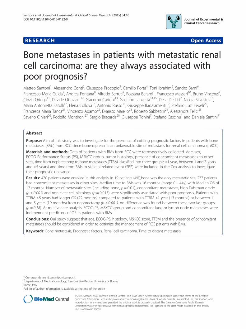
Figure 1 Bone metastases occurred at time of clear cell RCC diagnosi
their prognosis is still dismal, with a mean survival of12 months [4-6]. Skeletal involvement is commonly anaggressive, lytic process which causes substantial mor-bidity through skeletal related events (SREs), defined asa pathological fracture, surgical intervention, require-ment for palliative radiotherapy to bone, spinal cordcompression or hypercalcemia. Almost 70% of RCC pa-tients with BMs experience at least one SRE [7].The probability to develop BMs in RCC patients paral-
lels with increased survival related to the introduction ofbiological therapies (Figure 1). Several studies suggestthat the presence of BMs is associated with poor prog-nosis [8,9]. BMs are usually related to a more aggressivesubtype of disease as suggested by the higher percentageof patients with metastases or Fuhrman grade 4 at theinitial diagnosis, the shorter median time between neph-rectomy and diagnosis of metastatic disease and thegreater number of metastatic sites at the diagnosis [10].However, long survival in patients with BMs from RCCis not a rare event. This may be partially explained bydata on tumor biological heterogeneity [11,12], althoughother factors may affect the natural history of BMs in
s (A) and 12 years after radical nephrectomy (B).

Santoni et al. Journal of Experimental & Clinical Cancer Research (2015) 34:10 Page 3 of 9
RCC population. Aim of this study was to investigate forthe presence of existing prognostic factors in a largecohort of patients with BMs from RCC.
Patients and methodsData were extracted from the databases of 19 Italiancenters involved in the treatment of patients with mRCC.Inclusion criteria were: histologic diagnosis of RCC,clinical diagnosis of BMs, regularly conducted follow-upof the disease, minimum uneventful follow-up of 1 yearafter diagnosis of metastasis or alternatively knowndeath due to the disease. Pts were excluded from theanalysis if they had missing information about sites ofmetastasis or time to bone metastasis (TTBM).Patients characteristics and clinic-pathological variables
considered in this study were: gender, age, eastern co-operative oncology group performance status (ECOG-PS), tumor histology, clinical stage at time of surgery,time from surgery to diagnosis of BMs, MSKCC risk-group, type and number of sites of metastasis, timefrom BM to development of further metastatic sites andpresence/type of SREs. Since we gathered data fromRCC patients treated from 2001 onwards, we were notable to categorize all of them according to the novelIMDC (or Heng’s) prognostic classification.BMs were assessed with bone scan and confirmed by
contrast-enhanced CT, while other metastatic sites weredefined either with total body contrast-enhanced CT orMRI.Cancer-specific survival was computed both from any
site-metastasis and from BM to the event (death) andboth measurements were considered as outcomemeasures.Continuous covariates (age, TTBM) were grouped into
discrete ordinal categories. Age was divided in two or-dinal groups (<65 years and ≥65 years). Correlationbetween continuous variables was assessed by means ofPearson product-limit correlation coefficient [12]. Asoutcome variables, the overall survival (OS) from thediagnosis of BMs was analyzed.Patients were grouped according to TTBM: Group A
(<1 year), Group B (between 1 and 5 years) and GroupC (>5 years). The choice of these cut-offs was related tothe reported data on the prognostic effects of early(within 1 year) or late (>5 years) time to metastasis onthe outcome of RCC patients [13-16].Survival analysis was conducted via the Kaplan-Meier
method and Mantel-Haenszel log-rank test was employedto compare survival among groups. A Cox-regressionmodel was applied to the data with a univariate and multi-variate approach. The assumption of proportionality ofhazards was assessed using the Therneau and Grambschtest of the Schönefeld residuals [17]. Variables not fittingat univariate regression analysis were excluded for the
multivariate model. No-multicollinearity of the groupedco-variates was also checked. Significance level in the uni-variate model for inclusion in the multivariate final modelwas more liberally set at a 0.2 level, according to Hosmeret al. [17]. All other significance levels were set at a 0.05value. Statistical analysis was conducted with R –Softwareversion 3.0.1 (The R Company – Vienna –Austria).
ResultsWe retrospectively collected clinical data of 511 patientswith RCC BMs from 19 Italian Institutions followed be-tween January 2001 and April 2014. Of 470 patientswere available for this analysis (41 were excluded due tolack of data on BMs or follow-up). Median age was65 years (range 30 to 92 years). Three hundred andthirty-five patients (71%) were males; 398 (85%) hadclear cell RCC, whereas 72 (15%) presented with otherhistologies (5% papillary, 1% chromophobe, 9% other).The complete list of patient characteristics is shown inTable 1.Median number of bone metastases was 2. Median
number of SREs was 1 (range 0 to 6 events). The medianTTBM was 16 months (95% CI 0 – 64). Median time tofirst SRE was 2 months (95% CI 0 – 4) from BM diagno-sis. Median OS was 17 months (95% CI 14 to 19).Prognostic categories using MSKCC criteria were good
in 198 pts (42%), intermediate in 219 (47%) and poor in53 (11%). The median OS was 22 (95% CI 20 to 32), 18(95% CI 14 to 22) and 7 months (95% CI 6 to 10) inpatients with good, intermediate and poor prognosis,respectively (p < 0.001).A significantly higher mortality both from the diagno-
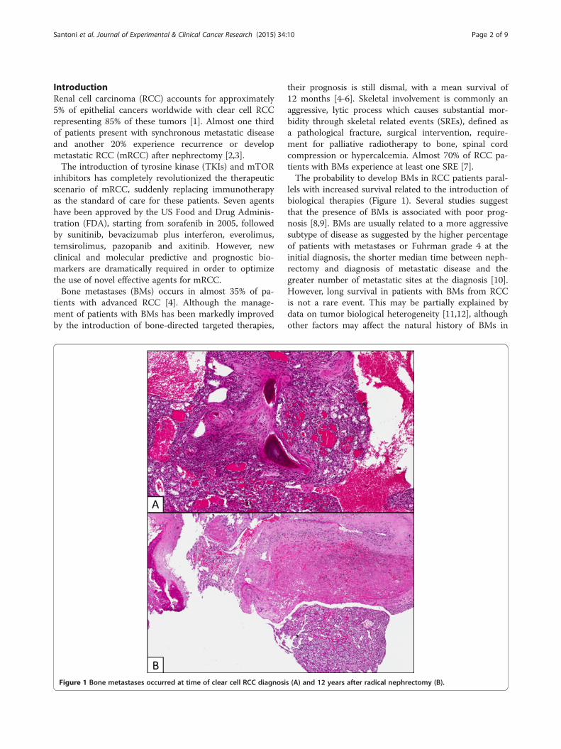
sis of mRCC and BMs was observed in patients develop-ing metastases < 65y compared to ≥ 65 (median OS:15 months (95% CI 13 to 20) vs 21 months (95% CI 18to 24), p = 0.038 (Figure 2A). No differences in OS wereobserved based on sex (p = 0.31).Clear-cell histology was correlated with a longer survival
as compared with other histologic types (18 months[95% CI 15 to 20] vs 12 months [95% CI 8 to 18],p = 0.014 Figure 2B), but this observation is somewayhampered by the small number of patients with non-clear cell histologies and the heterogeneous naturalhistory of the different histotypes. Also ECOG-PS at timeof diagnosis of metastatic disease was related to patients’survival at univariate analysis (log-rank p < 0.001; HR:1.22, 95% CI 1.05-1.40, p = 0.007).The number of BMs was associated neither with the
number of SREs (p = 0.096) nor with OS (HR: 1.17, 95% CI0.92-1.43, p = 0.21). No significant differences in termsof OS were found when comparing patients presentingwith visceral metastases as first metastatic sites withthose with BMs as first metastatic site (HR: 1.18, 95% CI0.89 to 1.54, p = 0.25).

Table 1 Patient demographics and disease characteristics
Patients, n (%) (N = 470)
Median age, y (range) 65 (30–92)
Sex
Male 335 (71)
Female 135 (29)
Tumor histology
Clear cell 398 (85)
Papillary 25 (5)
Chromophobe 5 (1)
Other 42 (9)
ECOG-Performance Status (PS) ≥2 60 (13)
Time to distant metastases (TTBM)
Group A (<1 year) 229 (49)
Group B (between 1 and 5 years) 107 (23)
Group C (>5 years) 134 (28)
MSKCC criteria
Good 198 (42)
Intermediate 219 (47)
Poor 53 (11)
Median number of bone metastases 2
Median number of SREs (range) 2 (1–6)
Sites of concomitant metastases
Lung 276 (59)
Lymph node 205 (44)
Liver 78 (17)
Brain 30 (6)
Adrenal gland 14 (3)
Treatment after onset of bone metastases 190 (41)
Sunitinib 61(12)
Sorefnib 4 (1)
Pazopanib 24 (5)
mTor inhibitors 191 (41)
Other treatments or no treatemnt
Santoni et al. Journal of Experimental & Clinical Cancer Research (2015) 34:10 Page 4 of 9
Furthermore, we analyzed the prognostic role of con-comitant visceral metastases in patients with BMs. Two-hundred seventy-six patients (59%) had concomitant lungmetastases, 205 (44%, with 28% retroperitoneal, 12% medi-astinal and 4% to other sites) had metastases to lymphnodes and 78 (17%) had liver metastases (Figure 3). Inter-estingly, the number and site of concomitant metastaseswas significantly associated with OS (p = 0.016) in ourpopulation. The presence of concomitant lung (p = 0.012),liver (p = 0.005), or lymph-node (p = 0.014) metastases wassignificantly correlated with OS at univariate analysis,while no correlation was shown for brain (p = 0.65) oradrenal metastases (p = 0.374) (Table 2).
At multivariate analysis, MSKCC risk group (p < 0.001),ECOG-PS (p < 0.001), lymph-node (p = 0.03) and lung(p = 0.009) metastases were independent prognosticfactors for OS (Table 2).Patients were grouped according to TTBM: Group
A (<1 year, 229 patients), Group B (between 1 and5 years, 107 patients) and Group C (>5 years, 134patients). The number of metastatic sites was statisti-cally different in the three groups, with a median of1 site in Group A, 2 In Group B and 2 in Group C(p = 0.001). TTBM was inversely correlated with MSKCCrisk group (testing for difference between groups withnon-homogeneous variances, Mann–Whitney U = 97,p < 0.001, Kendal tau = −0.40, p < 0.001). Indeed, pa-tients with longer TTBM presented more frequentlygood risk features.A significant difference was found for the distribution
of lung metastases (p < 0.001, Figure 3), with a higher in-cidence in Group B compared to the other groups. Nosignificant differences were found for the incidence oflymph-node (p = 0.20), liver (p = 0.24), adrenal, brain me-tastases (p = 0.15) or local recurrence (p = 0.75).As regard to OS, no significant difference was found
between Group A and B (13 months [95% CI 12 to 15]vs 19 months [95% CI 12 to 26], p = 0.36), while signifi-cant differences were found when comparing Group Awith Group C (13 months [95% CI 12 to 15] vs 22 months[95% CI 20 to 33] p < 0.001) and Group B with Group C(19 months [95% CI 12 to 26] vs 22 months [95% CI 20to 33] p < 0.001) (Figure 4).Finally, we identified three risk categories: the favor-
able risk group (Risk Score = 0–1 of previous confirmedthree prognostic factors, median OS = 21 months), theintermediate risk group (Risk Score = 2, median OS =12 months) and the poor risk group (Risk Score = 3,median OS = 6 months) (p < 0.001, HR 1.91, 95% CI:1.56-2.34, Figure 5).
DiscussionBMs are common in patients with mRCC, but the mech-anism by which this tumor preferentially metastasize tobone is poorly understood. The development of BMs is amultifactorial process that requires a series of interac-tions between invading tumor cells and the bone micro-environment [18-20] and is sustained by the release offactors, such as transforming growth factor-β (TGF-β),bone morphogenetic proteins (BMPs) and bone sialopro-tein (BSP), due to tumor-induced activity of osteoclasts[11,21,22]. Satcher and colleagues suggested a crucialrole for cadherin-11 in the homing/retention of RCCcells to bone and their subsequent proliferation. By usingan in vivo metastasis model of RCC, they revealed thatthe expression of cadherin-11 was enhanced in in BM-derived 786-O cells, and the knockdown of Cadherin-11
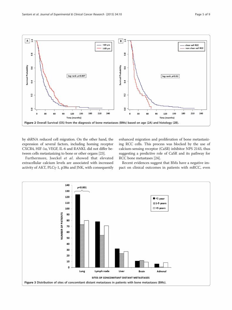
Figure 2 Overall Survival (OS) from the diagnosis of bone metastases (BMs) based on age (2A) and histology (2B).
Santoni et al. Journal of Experimental & Clinical Cancer Research (2015) 34:10 Page 5 of 9
by shRNA reduced cell migration. On the other hand, theexpression of several factors, including homing receptorCXCR4, HIF-1α, VEGF, IL-6 and RANKL did not differ be-tween cells metastasizing to bone or other organs [23].Furthermore, Joeckel et al. showed that elevated
extracellular calcium levels are associated with increasedactivity of AKT, PLCγ-1, p38α and JNK, with consequently
Figure 3 Distribution of sites of concomitant distant metastases in pa
enhanced migration and proliferation of bone metastasiz-ing RCC cells. This process was blocked by the use ofcalcium-sensing receptor (CaSR) inhibitor NPS 2143, thussuggesting a predictive role of CaSR and its pathway forRCC bone metastases [24].Recent evidences suggest that BMs have a negative im-
pact on clinical outcomes in patients with mRCC, even
tients with bone metastases (BMs).

Table 2 Univariate and multivariable analysis of predictors of OS from the diagnosis of bone metastases (BMs) inpatients with RCC
UVA* MVA**
HR 95% CI P HR 95% CI P
Age (<65 vs ≥65) 0.8 0.64-0.99 0.04 0.83 0.65-1.07 0.15
Gender (M vs F) 0.84 0.68-1.04 0.31
Histology (CC vs NCC) 1.45 1.08-1.95 0.013 1.23 0.83-1.81 0.31
MSKCC group 1.94 1.64-2.29 <0.001 1.82 1.50-2.20 <0.001
ECOG-PS 1.44 1.26-1.64 <0.001 1.40 1.19-1.66 <0.001
Time from diagnosis of primary disease (<1 y - 1-5y - >5 y) 0.72 0.64-0.81 <0.001 1.00 0.83-1.21 0.97
Number of bone metastases (single- vs multiple sites) 1.15 0.92-1.43 0.21
Number of other sites of metastasis 1.17 1.04-1.32 0.011 1.06 0.78-1.44 0.70
Other sites of metastasis
Local recurrence 1.05 0.76-1.46 0.78
Lung 1.19 0.96-1.48 0.012 1.41 1.09-1.84 0.009
Liver 1.43 1.11-1.84 0.005 1.2 0.87-1.66 0.26
Lymph-nodes 1.30 1.06-1.59 0.014 1.30 1.02-1.67 0.03
Adrenals 0.77 0.43-1.37 0.374
Brain 0.91 0.62-1.35 0.65
Other sites 0.60 0.37-0.97 0.04 0.59 0.32-1.09 0.09
HR and significance levels of significant variables are given as computed after removal of non-significant covariates. *UVA: Univariate analysis. **MVA:Multivariate analysis.cc = clear cell; ECOG-PS = Eastern Cooperative Oncology Group Performance Status; F = female; bold p value represents independent prognostic factor for OS.M =male; MSKCC =Memorial Sloan Kettering Cancer Center.
Figure 4 Overall survival (OS) from the diagnosis of bone metastases (BMs) based on the time of bone recurrence from nephrectomy (TTBM).
Santoni et al. Journal of Experimental & Clinical Cancer Research (2015) 34:10 Page 6 of 9
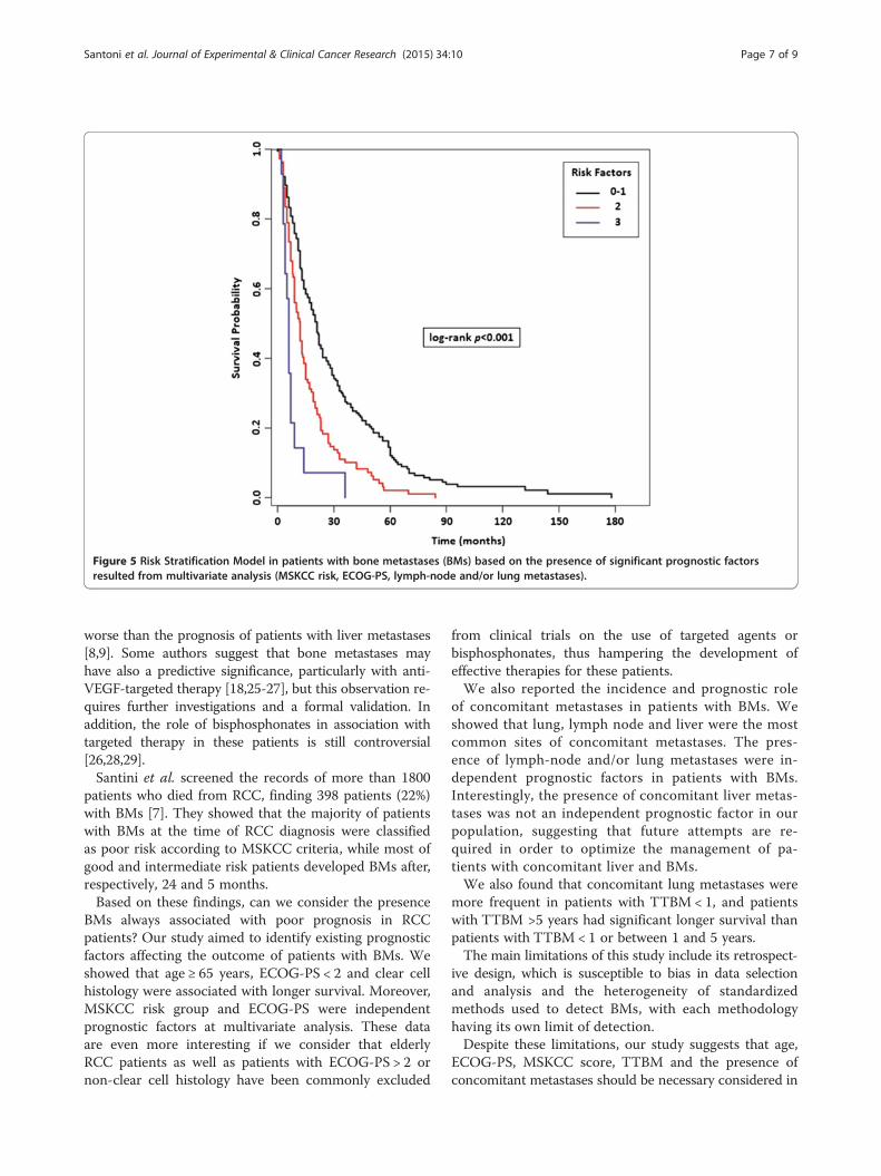
Figure 5 Risk Stratification Model in patients with bone metastases (BMs) based on the presence of significant prognostic factorsresulted from multivariate analysis (MSKCC risk, ECOG-PS, lymph-node and/or lung metastases).
Santoni et al. Journal of Experimental & Clinical Cancer Research (2015) 34:10 Page 7 of 9
worse than the prognosis of patients with liver metastases[8,9]. Some authors suggest that bone metastases mayhave also a predictive significance, particularly with anti-VEGF-targeted therapy [18,25-27], but this observation re-quires further investigations and a formal validation. Inaddition, the role of bisphosphonates in association withtargeted therapy in these patients is still controversial[26,28,29].Santini et al. screened the records of more than 1800
patients who died from RCC, finding 398 patients (22%)with BMs [7]. They showed that the majority of patientswith BMs at the time of RCC diagnosis were classifiedas poor risk according to MSKCC criteria, while most ofgood and intermediate risk patients developed BMs after,respectively, 24 and 5 months.Based on these findings, can we consider the presence
BMs always associated with poor prognosis in RCCpatients? Our study aimed to identify existing prognosticfactors affecting the outcome of patients with BMs. Weshowed that age ≥ 65 years, ECOG-PS < 2 and clear cellhistology were associated with longer survival. Moreover,MSKCC risk group and ECOG-PS were independentprognostic factors at multivariate analysis. These dataare even more interesting if we consider that elderlyRCC patients as well as patients with ECOG-PS > 2 ornon-clear cell histology have been commonly excluded
from clinical trials on the use of targeted agents orbisphosphonates, thus hampering the development ofeffective therapies for these patients.We also reported the incidence and prognostic role
of concomitant metastases in patients with BMs. Weshowed that lung, lymph node and liver were the mostcommon sites of concomitant metastases. The pres-ence of lymph-node and/or lung metastases were in-dependent prognostic factors in patients with BMs.Interestingly, the presence of concomitant liver metas-tases was not an independent prognostic factor in ourpopulation, suggesting that future attempts are re-quired in order to optimize the management of pa-tients with concomitant liver and BMs.We also found that concomitant lung metastases were
more frequent in patients with TTBM < 1, and patientswith TTBM >5 years had significant longer survival thanpatients with TTBM < 1 or between 1 and 5 years.The main limitations of this study include its retrospect-
ive design, which is susceptible to bias in data selectionand analysis and the heterogeneity of standardizedmethods used to detect BMs, with each methodologyhaving its own limit of detection.Despite these limitations, our study suggests that age,
ECOG-PS, MSKCC score, TTBM and the presence ofconcomitant metastases should be necessary considered in
Santoni et al. Journal of Experimental & Clinical Cancer Research (2015) 34:10 Page 8 of 9
order to optimize the prognosis of patients with RCC andBMs. The understanding of the molecular phenotypes ofBM-initiating cells in RCC could play a fundamental rolein developing therapeutic strategies for these patients.
Competing interestsThe authors declare that they have no competing interests.
Authors’ contributionsAll the authors cited have been involved in drafting the manuscript andrevising it critically for important intellectual content. All authors read andapproved the final manuscript.
Author details1Department of Medical Oncology, AOU Ospedali Riuniti, UniversitàPolitecnica delle, Marche, Ancona, Italy. 2Department of Clinical andSpecialist Sciences, Urology, Università Politecnica delle Marche, Ancona,Italy. 3Department of Medical Oncology, Fondazione IRCCS Istituto Nazionaledei Tumori, Milan, Italy. 4Division of Medical Oncology, I.R.C.C.S. San MatteoUniversity Hospital Foundation, Pavia, Italy. 5Osteoncology and Rare TumorsCenter, IRCCS Istituto Scientifico Romagnolo per lo Studio e la Cura deiTumori (IRST), Meldola, FC, Italy. 6Medical Oncology Department, AziendaOspedaliera Treviglio-Caravaggio, Treviglio, Italy. 7Department of MedicalOncology, Campus Bio-Medico University of Rome, Rome, Italy. 8Unit ofMedical Oncology 2, Istituto Toscano Tumori,Azienda-Ospedaliero-Universitaria Pisana, Pisa, Italy. 9Dipartimento diSpecialità Medico-Chirurgiche, Medical Oncology, Scienze Radiologiche eSanità Pubblica, Università degli Studi di Brescia, Azienda Ospedaliera SpedaliCivili, Brescia, Italy. 10Department of Medical Oncology, “G.B. Rossi” AcademicHospital, Azienda Ospedaliera Universitaria Integrata, University of Verona,Verona, Italy. 11Department of Medical Oncology, Institute for CancerResearch & Treatment (IRCC), Candiolo, Torino, Italy. 12Department of MedicalOncology, Presidio Sanitario Gradenigo, Turin, Italy. 13Department of MedicalOncology, Cardarelli Hospital, Naples, Italy. 14Department of NeurologicalSciences, Neuromed Institute, IRCSS, Pozzilli, IS, Italy. 15IstitutoNeurotraumatologico Italiano, Unità Funzionale di Oncologia, Grottaferrata,Italy. 16Medical Oncology Unit, National Cancer Research Centre “GiovanniPaolo II”, Bari, Italy. 17Department of Oncology, University of Turin, MedicalOncology 1, AOU Città della Salute e della Scienza, Turin, Italy. 18Division ofMedical Oncology, Hospital of Legnano, Milan, Italy. 19Department of Surgeryand Oncology, Section of Medical Oncology, University of Palermo, Palermo,Italy. 20Department of Medical Oncology, AOU Ospedali Riuniti, UniversitàPolitecnica delle Marche, Presidio San Salvatore, Pesaro, Italy. 21Departmentof Medical Oncology, University of Cagliari, Cagliari, Italy. 22Department ofHuman Pathology, Medical Oncology Unit AOOR Papardo-Piemonte,University of Messina, Messina, Italy. 23Oncology Unit, IRCCS Casa Sollievodella Sofferenza, San Giovanni Rotondo, FG, Italy. 24Dipartimento Integrato diOncologia ed Ematologia, Medical Oncology Division, Università degli Studidi Modena e Reggio Emilia, Modena, Italy. 25Department of MedicalOncology, Regina Elena National Cancer Institute, Rome, Italy. 26MedicalOncology Department & Breast Unit - Hospital of Brindisi and MedicalOncology Department - European Institute of Oncology, Milan, Italy.27Section of Pathological Anatomy, Polytechnic University of the MarcheRegion, School of Medicine, United Hospitals, Ancona, Italy. 28Department ofOncology, USL-8, Ospedale San Donato, Arezzo, Italy.
Received: 8 November 2014 Accepted: 3 January 2015
References1. Murai M, Oya M. Renal cell carcinoma: etiology, incidence and
epidemiology. Curr Opin Urol. 2004;14:229–33.2. Athar U, Gentile TC. Treatment options for metastatic renal cell carcinoma: a
review. Can J Urol. 2008;15:3954–66.3. Motzer RJ, Bander NH, Nanus DM. Renal-cell carcinoma. N Engl J Med.
1996;335:865–75.4. Woodward E, Jagdev S, McParland L, Clark K, Gregory W, Newsham A, et al.
Skeletal complications and survival in renal cancer patients with bonemetastases. Bone. 2011;48:160–6.
5. Sahi C, Knox JJ, Clemons M, Joshua AM, Broom R. Renal cellcarcinoma bone metastases: clinical advances. Ther Adv Med Oncol.2010;2:75–83.
6. Facchini G, Caraglia M, Santini D, Nasti G, Ottaiano A, Striano S, et al. Theclinical response on bone metastasis from breast and lung cancer duringtreatment with zoledronic acid is inversely correlated to skeletal relatedevents (SRE). J Exp Clin Cancer Res. 2007;26:307–12.
7. Santini D, Procopio G, Porta C, Ibrahim T, Barni S, Mazzara C, et al. Naturalhistory of malignant bone disease in renal cancer: final results of an Italianbone metastasis survey. PLoS One. 2013;8:e83026.
8. Beuselinck B, Oudard S, Rixe O, Wolter P, Blesius A, Ayllon J, et al. Negativeimpact of bone metastasis on outcome in clear-cell renal cell carcinomatreated with sunitinib. Ann Oncol. 2011;22:794–800.
9. Choueiri TK, Garcia JA, Elson P, Khasawneh M, Usman S, Golshayan AR, et al.Clinical factors associated with outcome in patients with metastatic clear-cellrenal cell carcinoma treated with vascular endothelial growth factor-targetedtherapy. Cancer. 2007;110:543–50.
10. Gerlinger M, Rowan AJ, Horswell S, Larkin J, Endesfelder D, Gronroos E, et al.Intratumor heterogeneity and branched evolution revealed by multiregionsequencing. N Engl J Med. 2012;366:883–92.
11. Santoni M, Santini D, Massari F, Conti A, Iacovelli R, Burattini L, et al.Heterogeneous drug target expression as possible basis for different clinicaland radiological response to the treatment of primary and metastatic renalcell carcinoma: suggestions from bench to bedside. Cancer Metastasis Rev.2014;33:321–31.
12. Newcombe RG. Interval estimation for the difference between independentproportions: comparison of eleven methods. Stat Med. 1998;17:873–90.
13. Motzer RJ, Bacik J, Schwartz LH, Reuter V, Russo P, Marion S, et al.Prognostic factors for survival in previously treated patients with metastaticrenal cell carcinoma. J Clin Oncol. 2004;22:454–63.
14. Heng DY, Xie W, Regan MM, Warren MA, Golshayan AR, Sahi C, et al.Prognostic factors for overall survival in patients with metastatic renal cellcarcinoma treated with vascular endothelial growth factor-targetedagents: results from a large, multicenter study. J Clin Oncol.2009;27:5794–9.
15. Kroeger N, Choueiri TK, Lee JL, Bjarnason GA, Knox JJ, MacKenzie MJ, et al.Survival outcome and treatment response of patients with late relapsefrom renal cell carcinoma in the era of targeted therapy. Eur Urol.2014;65:1086–92.
16. Santoni M, Conti A, Porta C, Procopio G, Sternberg CN, Basso U, et al.Sunitinib, Pazopanib or Sorafenib for the Treatment of Patients with LateRelapsing Metastatic Renal Cell Carcinoma. J Urol. 2015;193:41–7.doi:10.1016/j.juro.2014.07.011.
17. Hosmer WJ, Lemeshow S. Applied Survival Analysis: Regression Modeling ofTime to Event Data. New York: John Wiley; 1999.
18. Fizazi K, Yang J, Peleg S, Sikes CR, Kreimann EL, Daliani D, et al. Prostate cancercells-osteoblast interaction shifts expression of growth/survival-related genes inprostate cancer and reduces expression of osteoprotegerin in osteoblasts. ClinCancer Res. 2003;9:2587–97.
19. Shiirevnyamba A, Takahashi T, Shan H, Ogawa H, Yano S, Kanayama H, et al.Enhancement of osteoclastogenic activity in osteolytic prostate cancer cellsby physical contact with osteoblasts. Br J Cancer. 2011;104:505–13.
20. Chen Y, Liu H, Wu W, Li Y, Li J. Osteopontin genetic variants are associatedwith overall survival in advanced non-small-cell lung cancer patients andbone metastasis. J Exp Clin Cancer Res. 2013;32:45.
21. Chirgwin JM, Guise TA. Molecular mechanisms of tumor-bone interactionsin osteolytic metastases. Crit Rev Eukaryot Gene Expr. 2000;10:159–78.
22. Mundy GR. Metastasis to bone: causes, consequences and therapeuticopportunities. Nat Rev Cancer. 2002;2:584–93.
23. Satcher RL, Pan T, Cheng CJ, Lee YC, Lin SC, Yu G, et al. Cadherin-11 in renalcell carcinoma bone metastasis. PLoS One. 2014;9:e89880.
24. Joeckel E, Haber T, Prawitt D, Junker K, Hampel C, Thüroff JW, et al. Highcalcium concentration in bones promotes bone metastasis in renal cellcarcinomas expressing calcium-sensing receptor. Mol Cancer. 2014;13:42.
25. Yang L, You S, Kumar V, Zhang C, Cao Y. In vitro the behaviorsof metastasis with suppression of VEGF in human bone metastaticLNCaP-derivative C4-2B prostate cancer cell line. J Exp Clin Cancer Res.2012;31:40.
26. McKay RR, Lin X, Perkins JJ, Heng DY, Simantov R. Choueiri. Prognosticsignificance of bone metastases and bisphosphonate therapy in patientswith renal cell carcinoma Eur Urol. 2014;66:502–9.
Santoni et al. Journal of Experimental & Clinical Cancer Research (2015) 34:10 Page 9 of 9
27. Riechelmann RP, Chin S, Wang L, Tannock IF, Berthold DR, Moore MJ, et al.Sorafenib for metastatic renal cancer: the Princess Margaret experience.Am J Clin Oncol. 2008;31:182–7.
28. Lipton A, Colombo-Berra A, Bukowski RM, Rosen L, Zheng M, Urbanowitz G,et al. Skeletal complications in patients with bone metastases from renal cellcarcinoma and therapeutic benefits of zoledronic acid. Clin Cancer Res.2004;10:6397S–403.
29. Keizman D, Ish-Shalom M, Pili R, Hammers H, Eisenberger MA, Sinibaldi V,et al. Bisphosphonates combined with sunitinib may improve the responserate, progression free survival and overall survival of patients with bonemetastases from renal cell carcinoma. Eur J Cancer. 2012;48:1031–7.
Submit your next manuscript to BioMed Centraland take full advantage of:
• Convenient online submission
• Thorough peer review
• No space constraints or color figure charges
• Immediate publication on acceptance
• Inclusion in PubMed, CAS, Scopus and Google Scholar
• Research which is freely available for redistribution
Submit your manuscript at www.biomedcentral.com/submit