brain cancer treatment - surf · 0078936 brain cancer treatment picturing tomorrow’s treatment...
TRANSCRIPT

0078936
Brain cancer treatment
picturing tomorrow’s treatment using yesterday’s images
SURFsara Super D eventDecember 15, 2015
Dr Philip De Witt Hamer
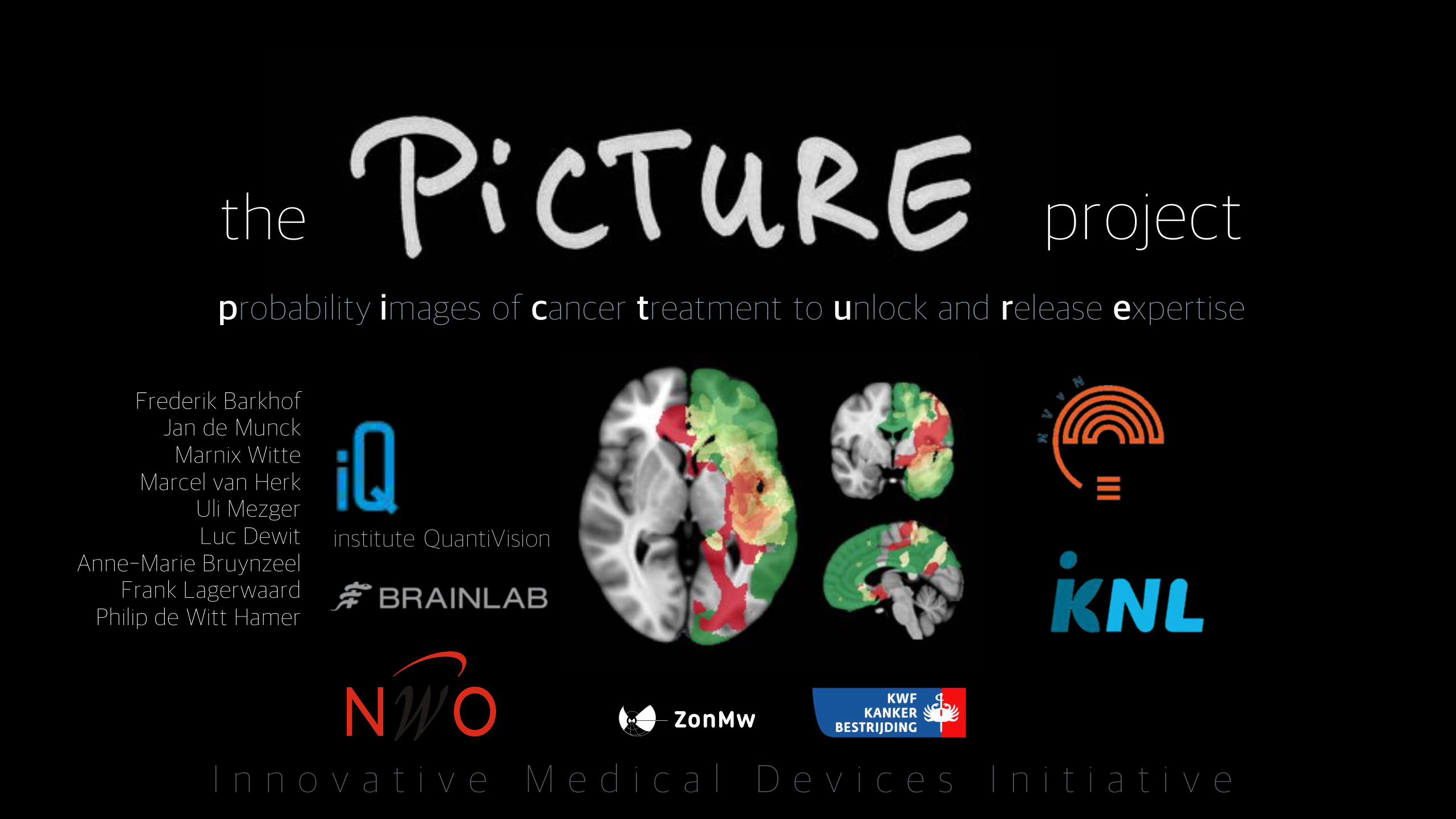
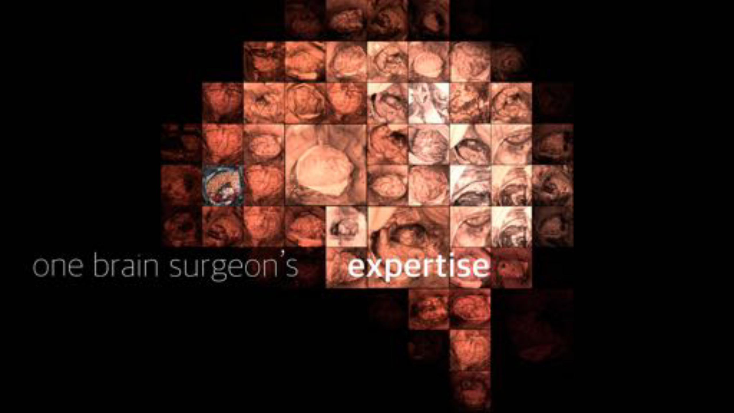
probability images of cancer treatment to unlock and release expertise
Frederik BarkhofJan de MunckMarnix Witte
Marcel van HerkUli MezgerLuc Dewit
Anne-Marie BruynzeelFrank Lagerwaard
Philip de Witt Hamer
institute QuantiVision
I n n o v a t i v e M e d i c a l D e v i c e s I n i t i a t i v e
the project
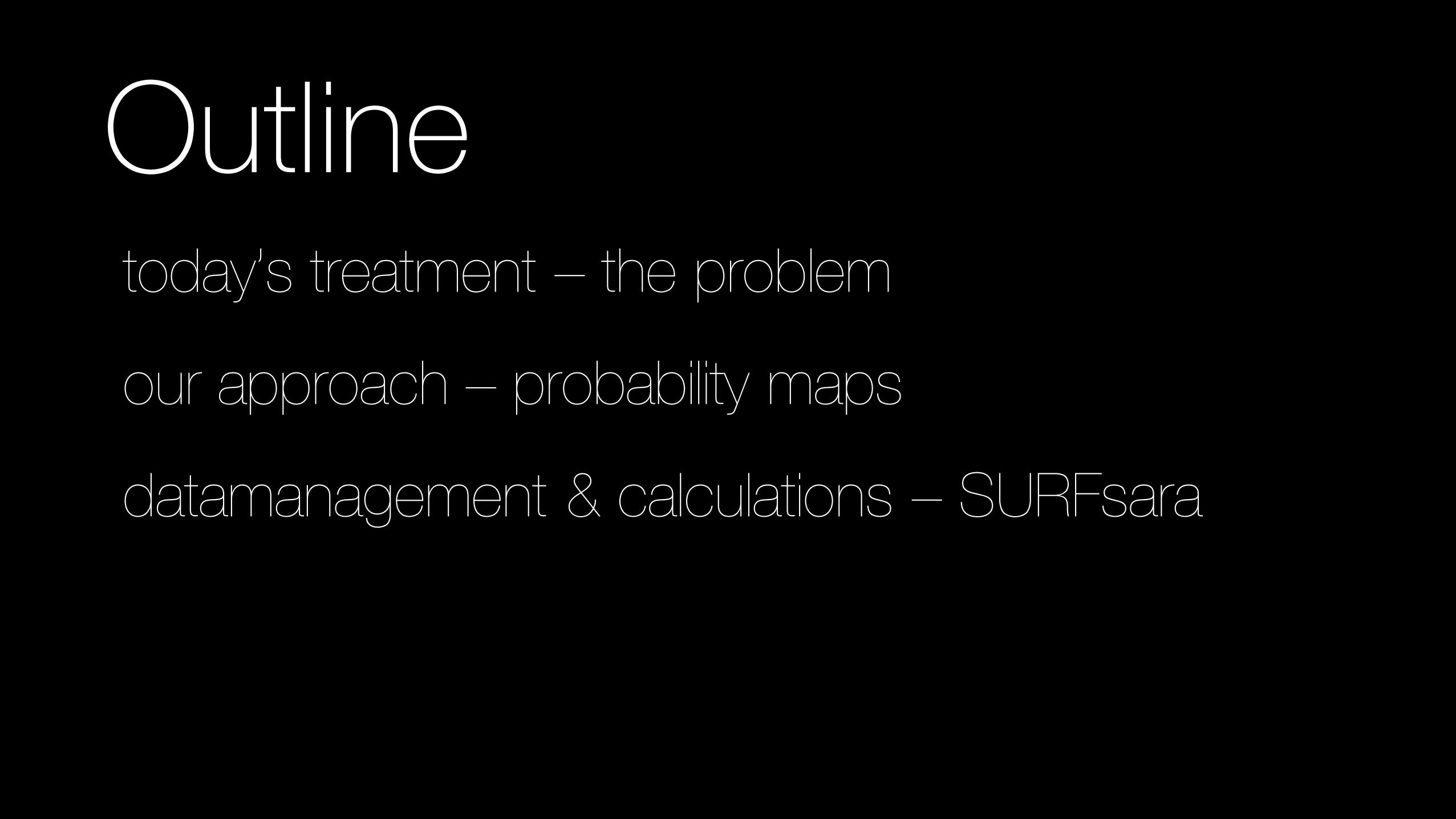
Outlinetoday’s treatment – the problem
our approach – probability maps
datamanagement & calculations – SURFsara
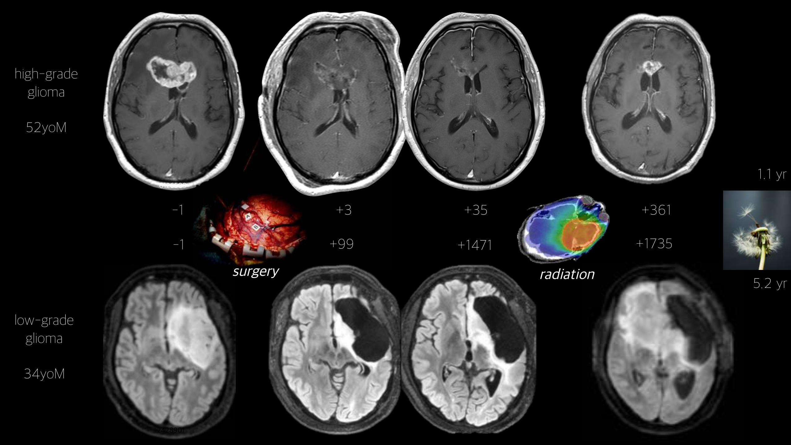
0126509
-1 +3 +35
-1 +99 +1471
2428901
+361
5.2 yr
high-gradeglioma
52yoM
low-gradeglioma
34yoM
1.1 yr
+1735
surgery radiation
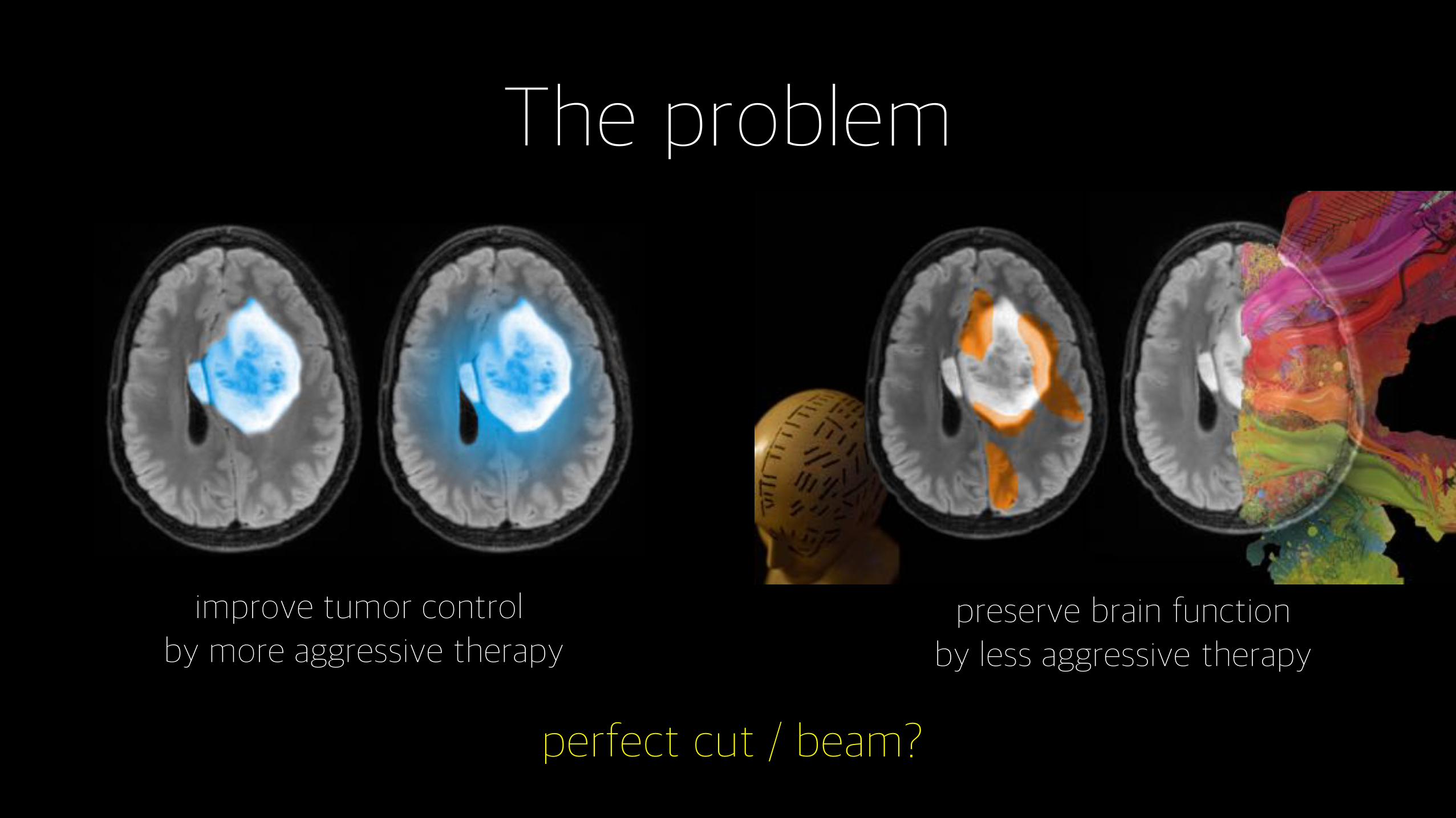
The problem
improve tumor control by more aggressive therapy
preserve brain functionby less aggressive therapy
perfect cut / beam?

balance
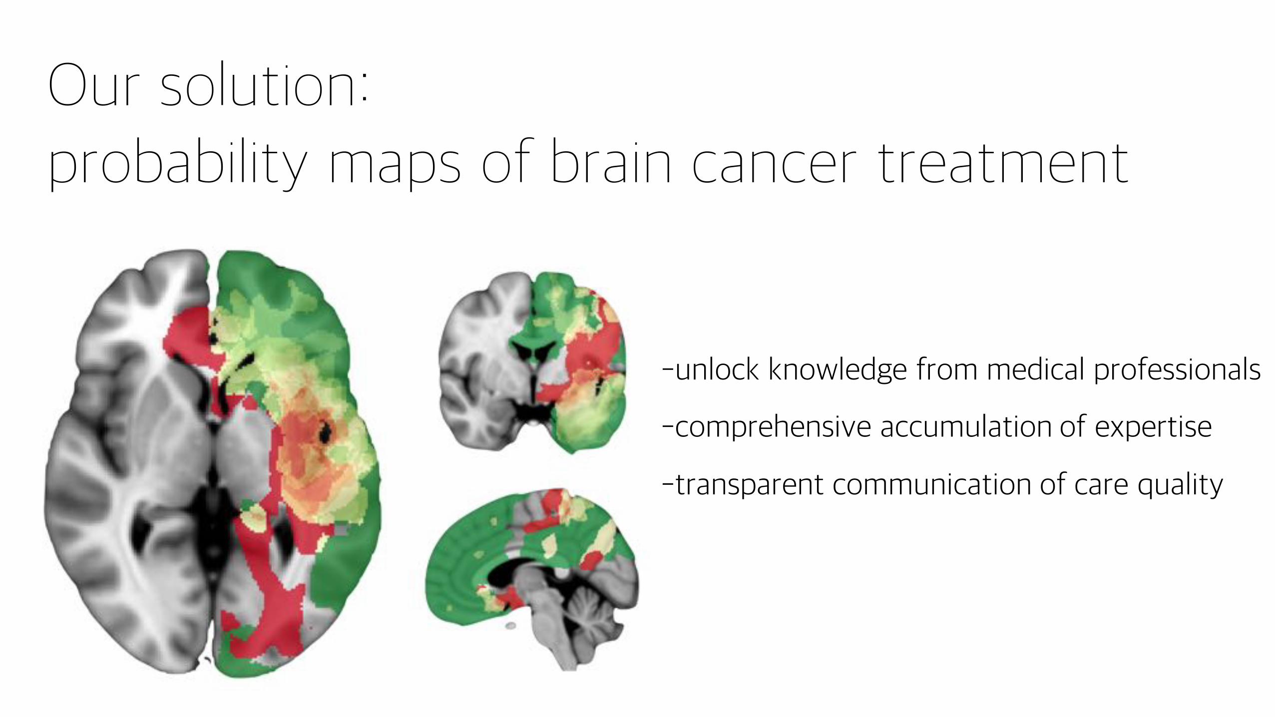
Our solution: probability maps of brain cancer treatment
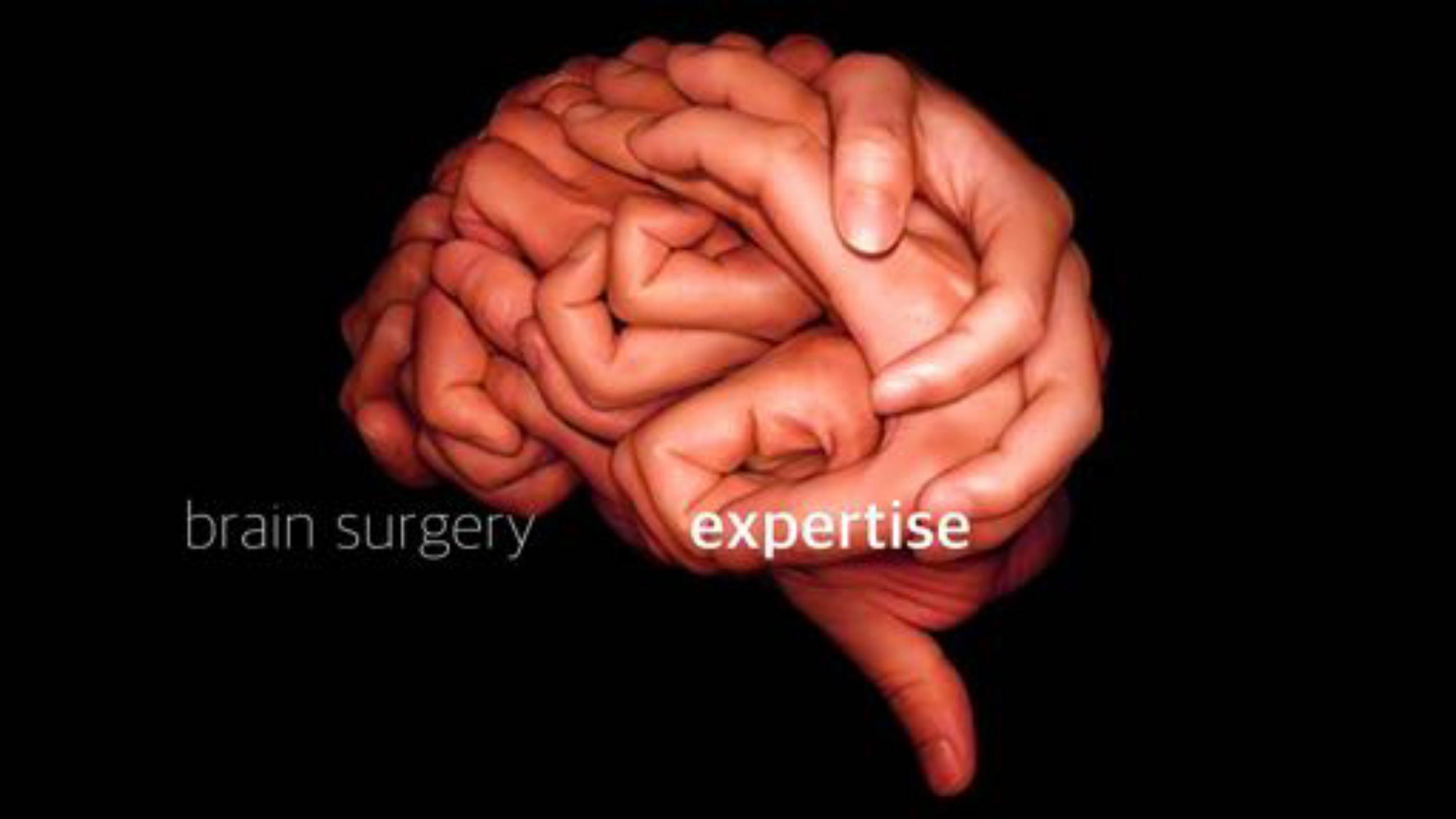
-unlock knowledge from medical professionals
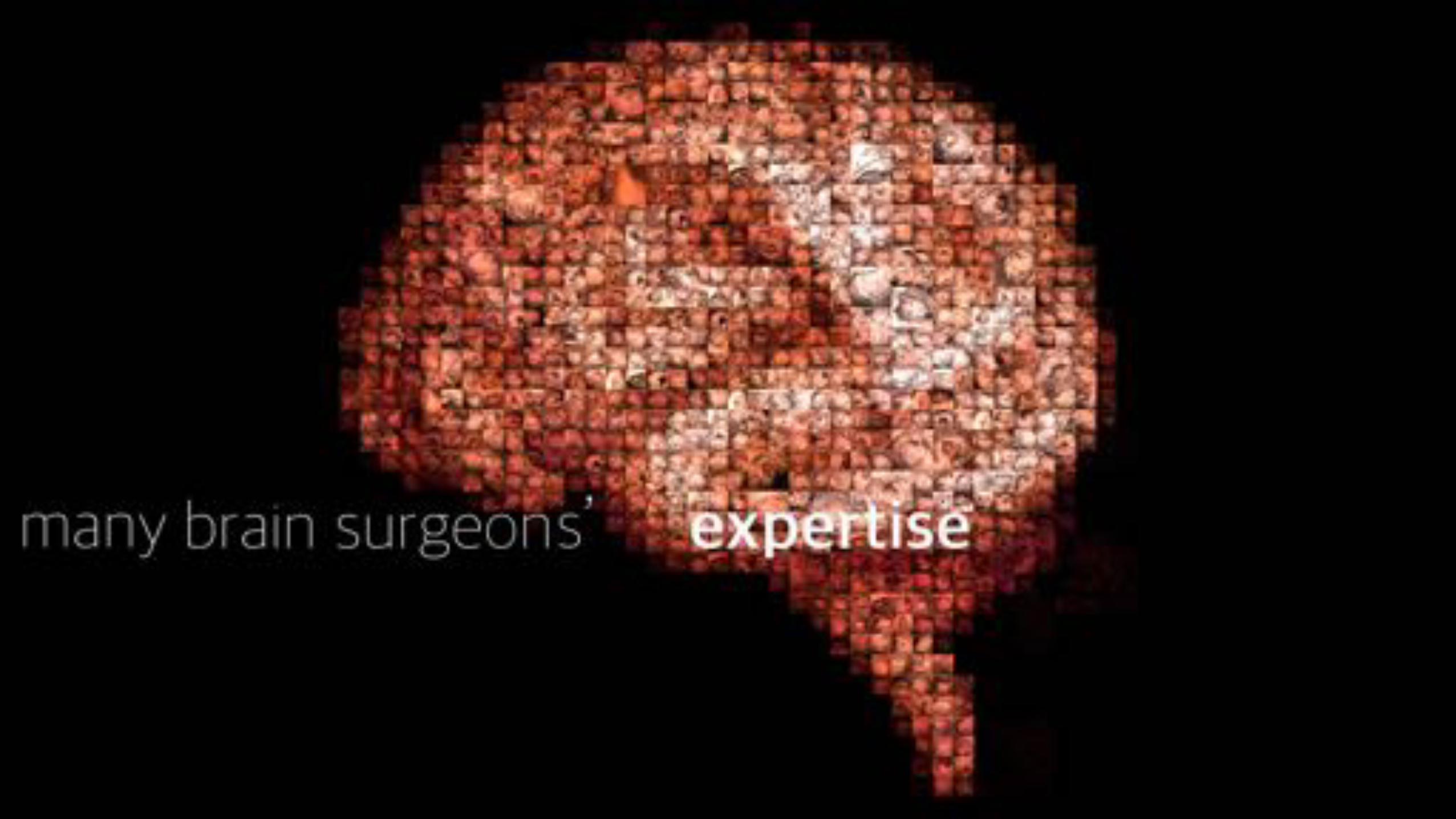
-comprehensive accumulation of expertise
-transparent communication of care quality
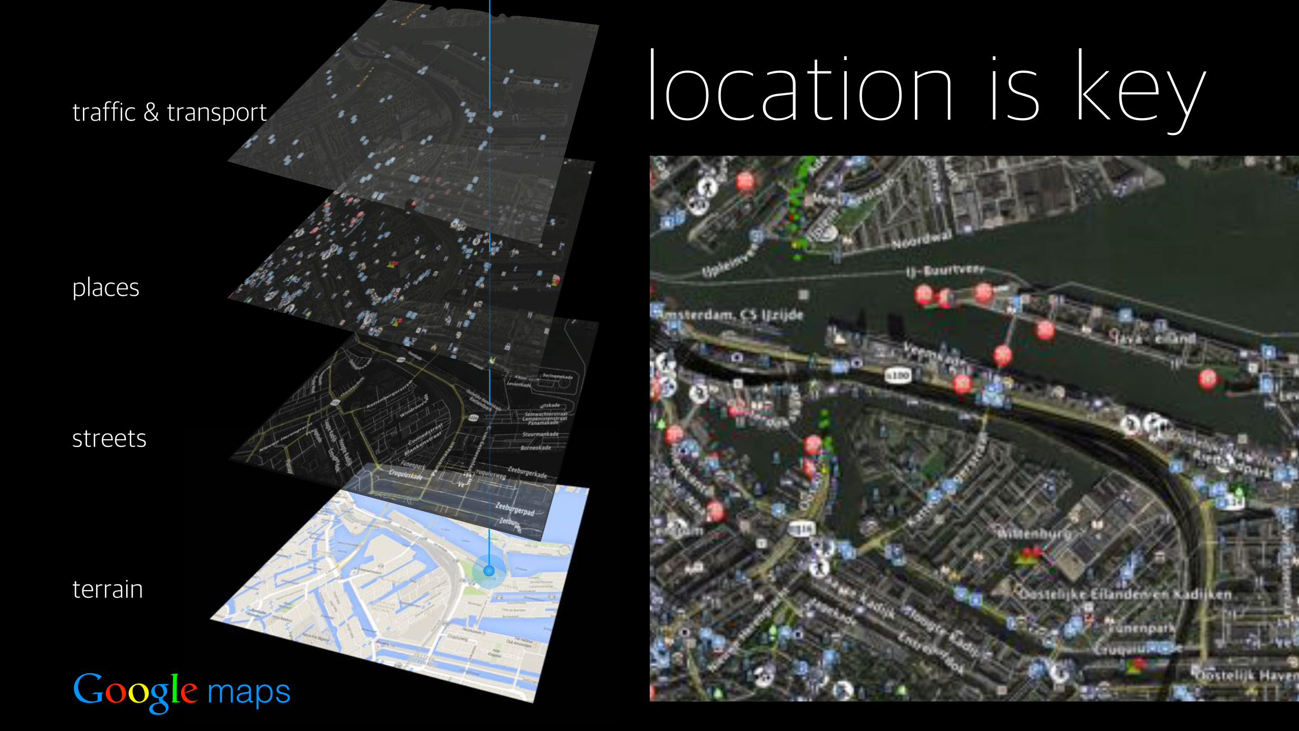
terrain
streets
places
location is keytraffic & transport
Google maps
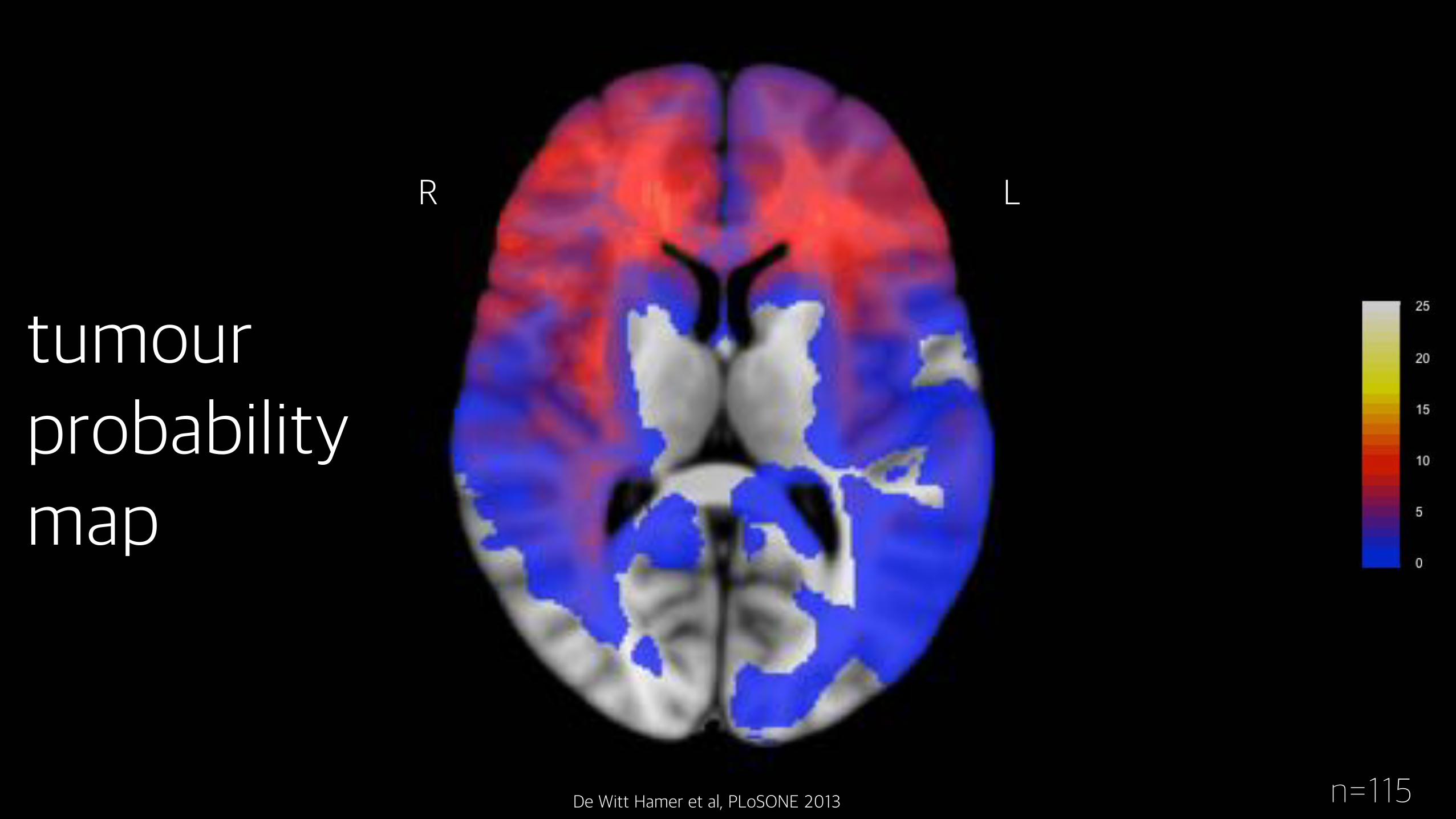
n=115De Witt Hamer et al, PLoSONE 2013
tumour probability map
LR
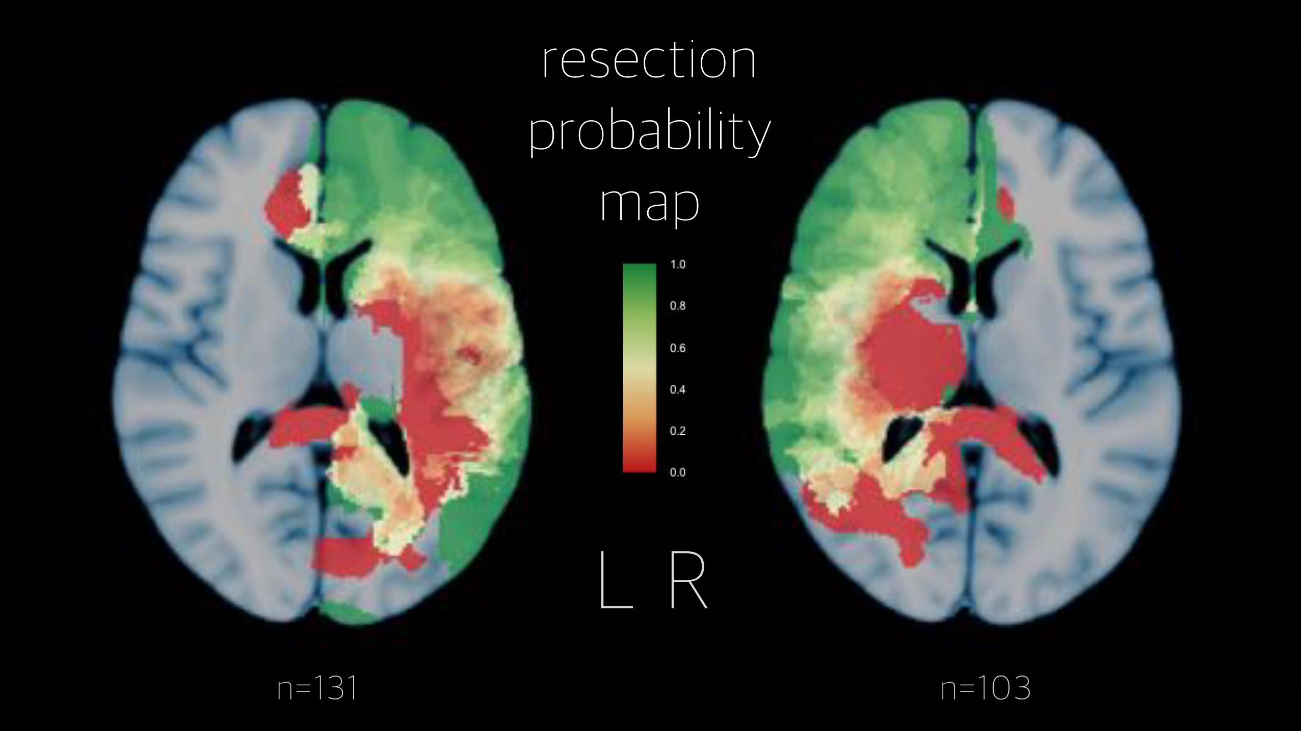
L Rn=131 n=103
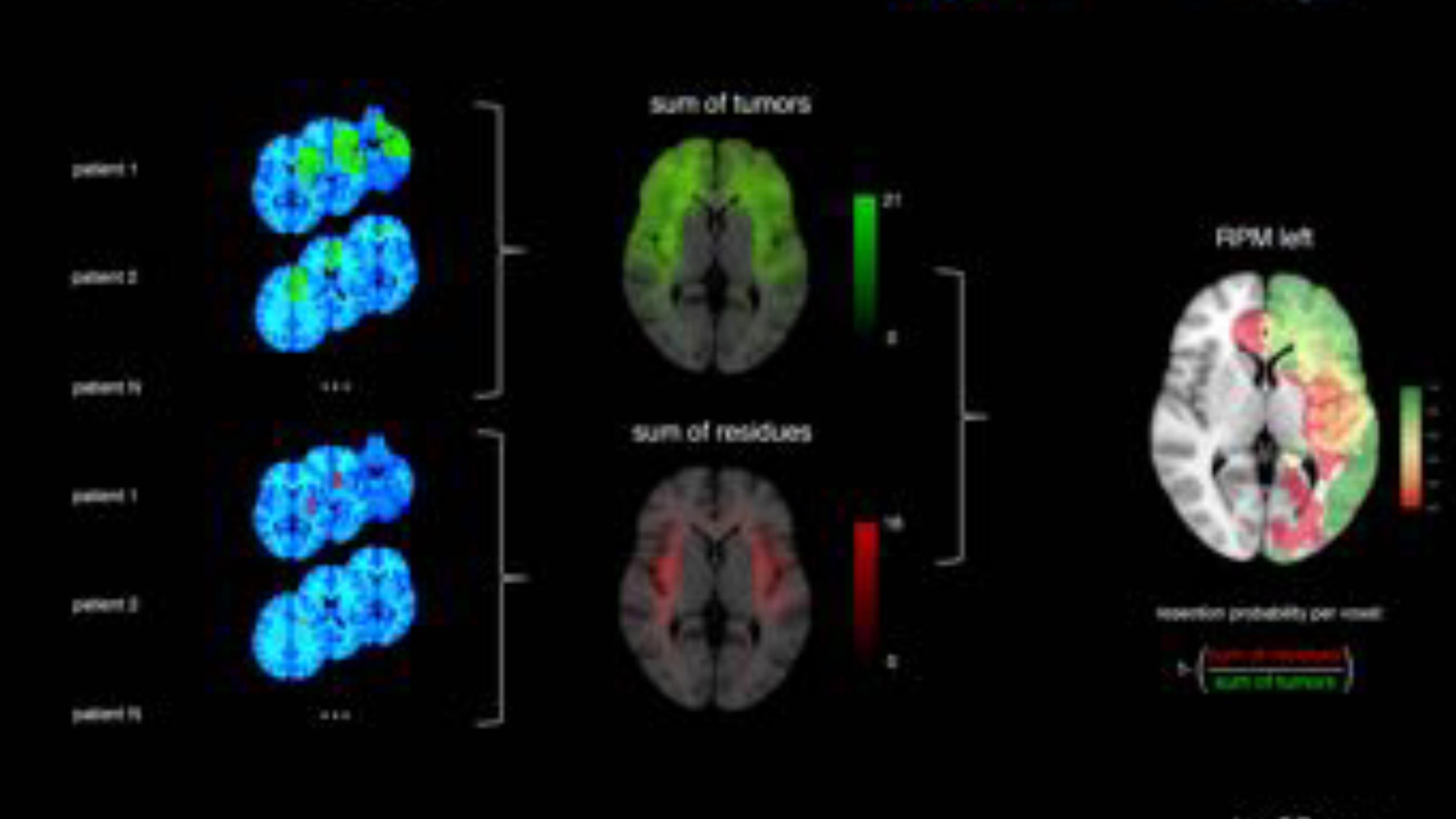
resectionprobability
map
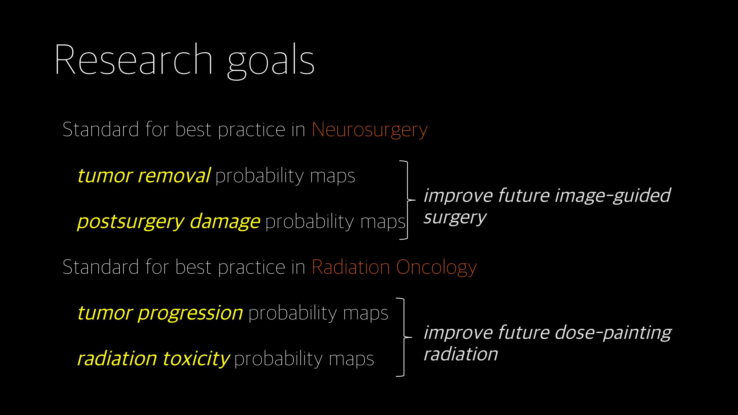
Research goals
Standard for best practice in Neurosurgery
tumor removal probability maps
postsurgery damage probability maps
Standard for best practice in Radiation Oncology
tumor progression probability maps
radiation toxicity probability maps
improve future image-guided surgery
improve future dose-painting radiation
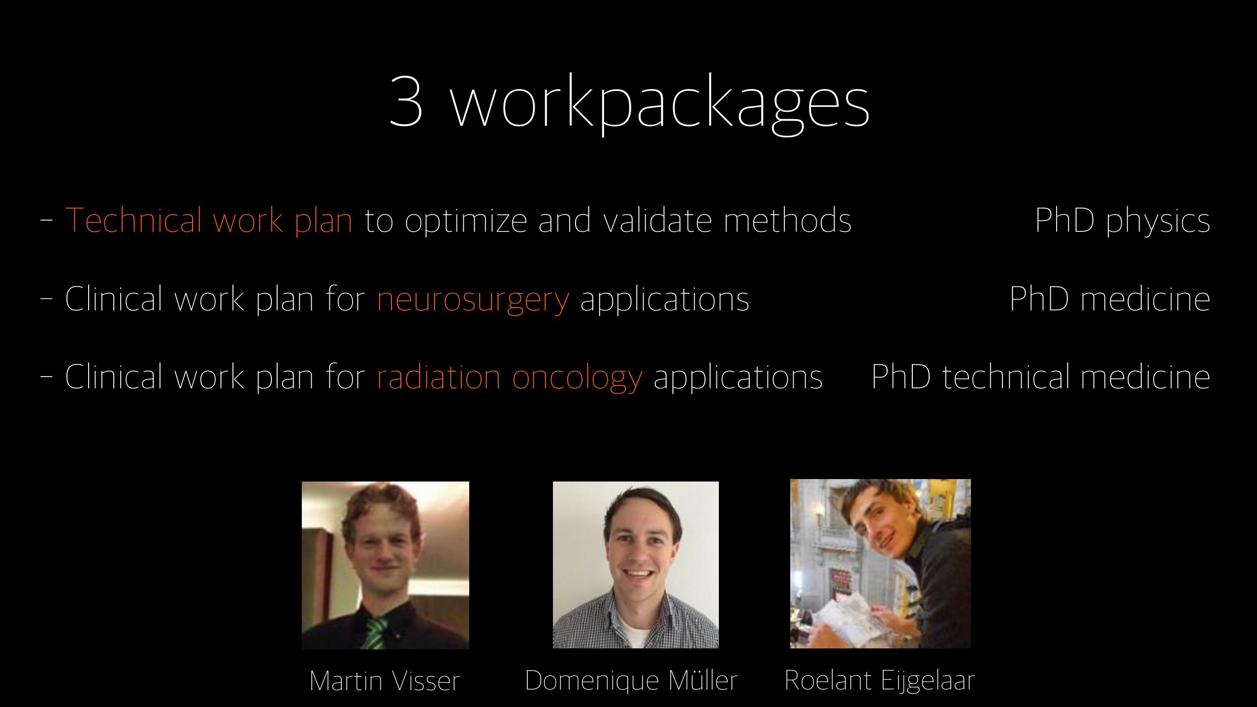
3 workpackages
- Technical work plan to optimize and validate methods PhD physics
- Clinical work plan for neurosurgery applications PhD medicine
- Clinical work plan for radiation oncology applications PhD technical medicine
Martin Visser Roelant EijgelaarDomenique M+ller
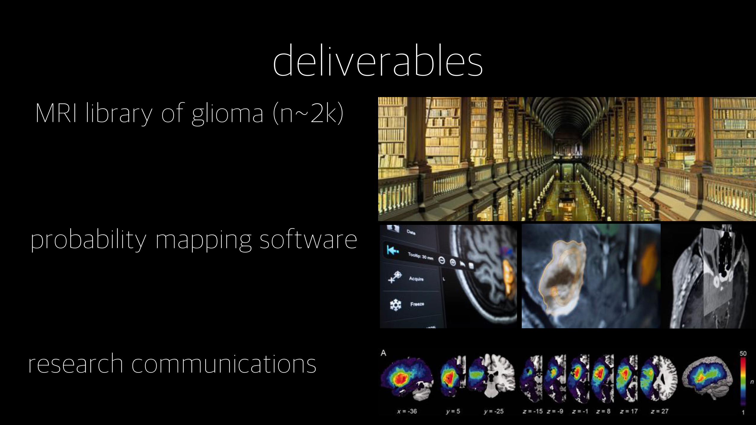
deliverablesMRI library of glioma (n~2k)
probability mapping software
research communications
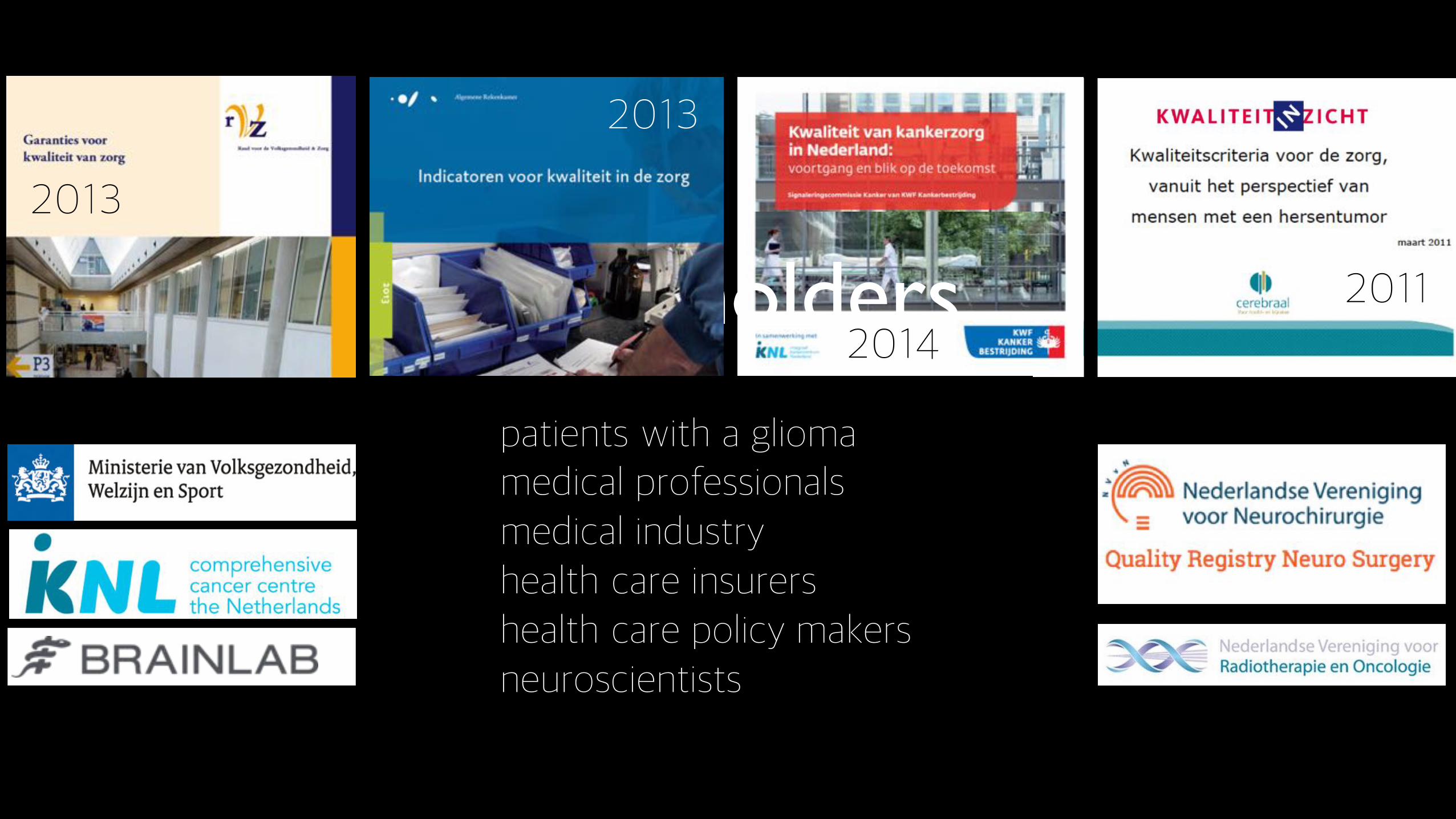
stakeholders2013
2013
20142011
patients with a gliomamedical professionalsmedical industryhealth care insurershealth care policy makersneuroscientists
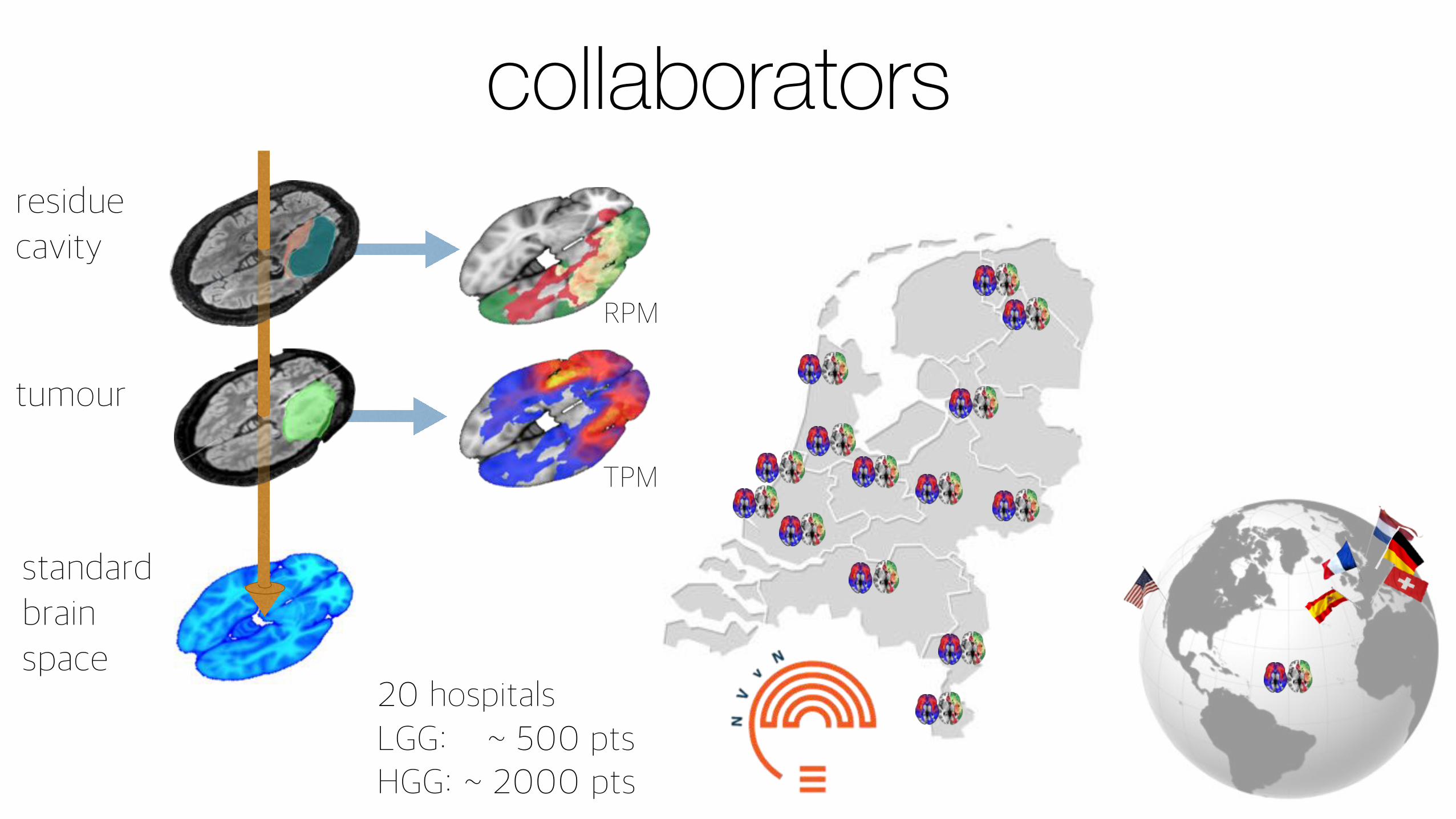
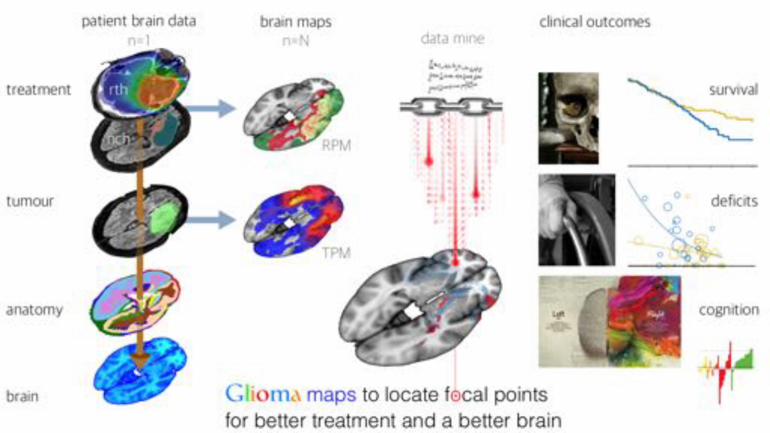
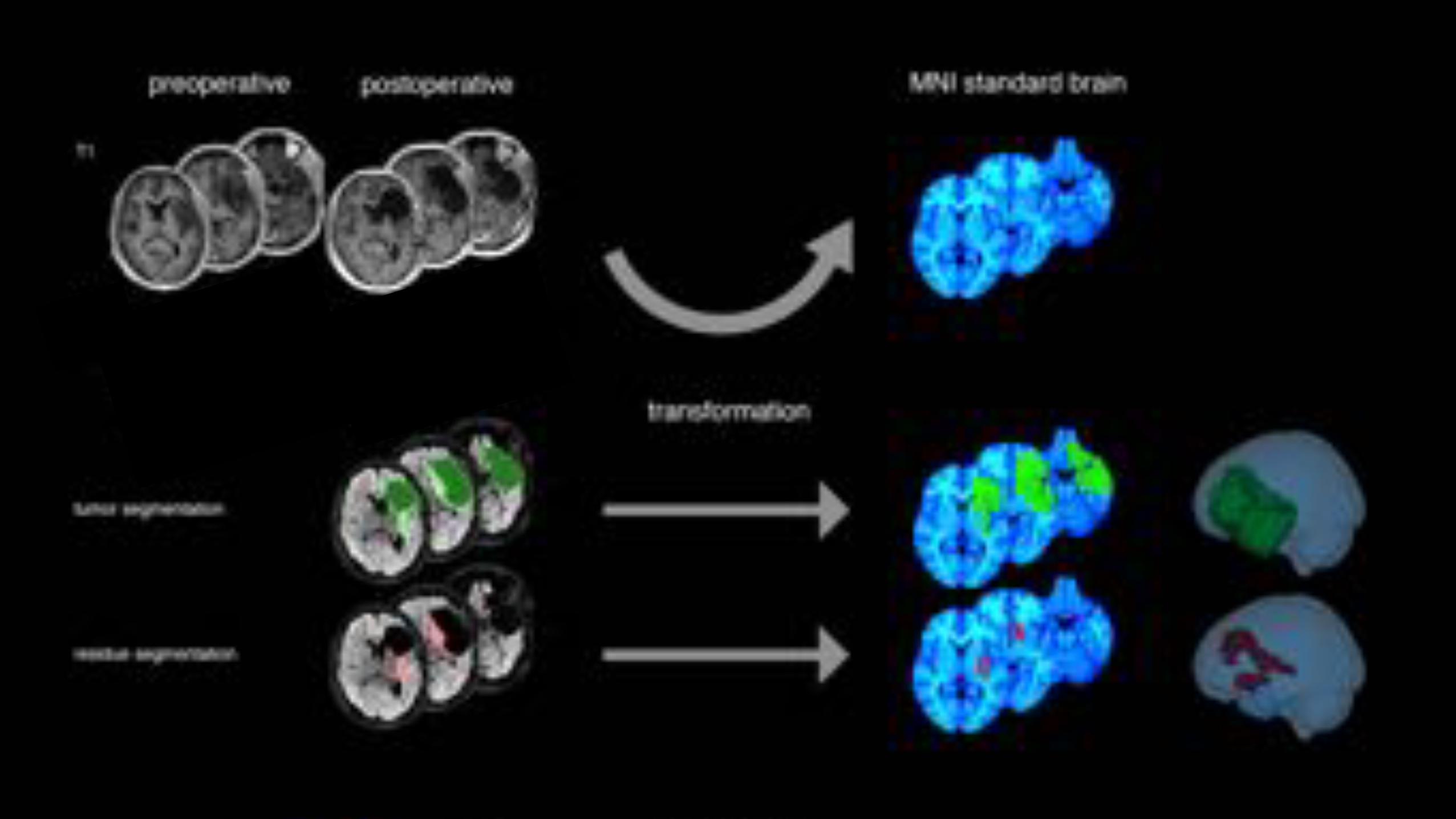
TPM
RPM
residuecavity
tumour
standardbrainspace
collaborators
20 hospitalsLGG: ~ 500 ptsHGG: ~ 2000 pts
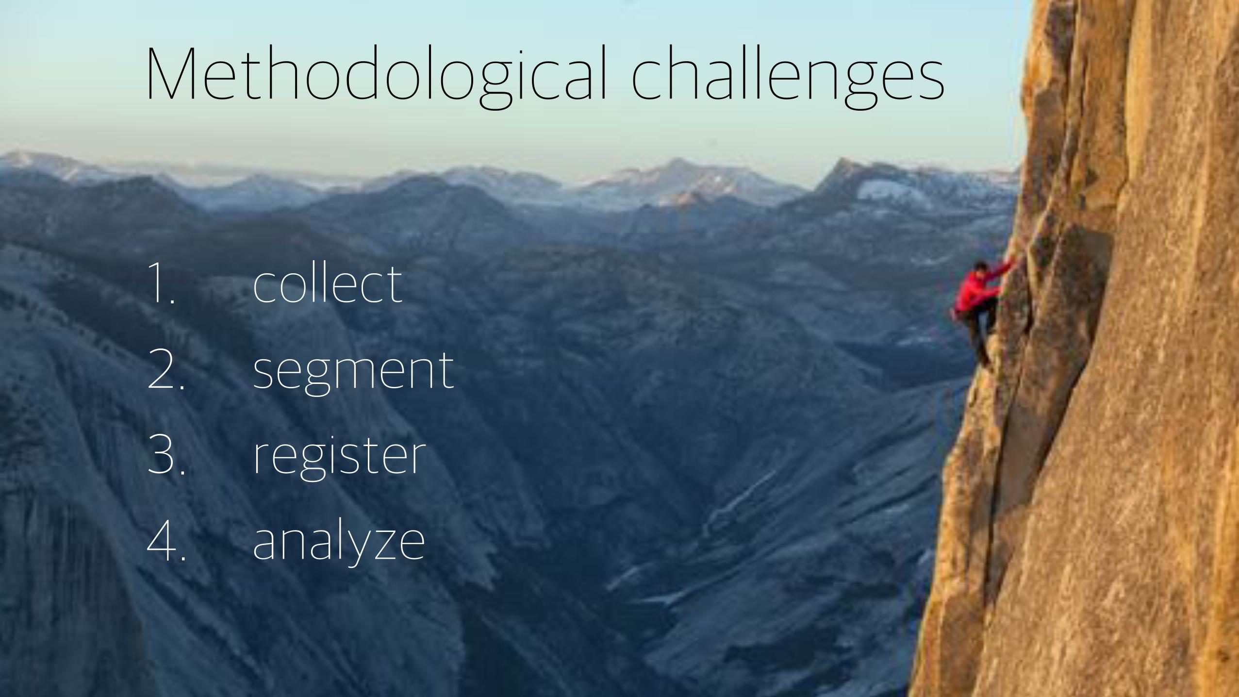
Methodological challenges
1. collect
2. segment
3. register
4. analyze
1-collect
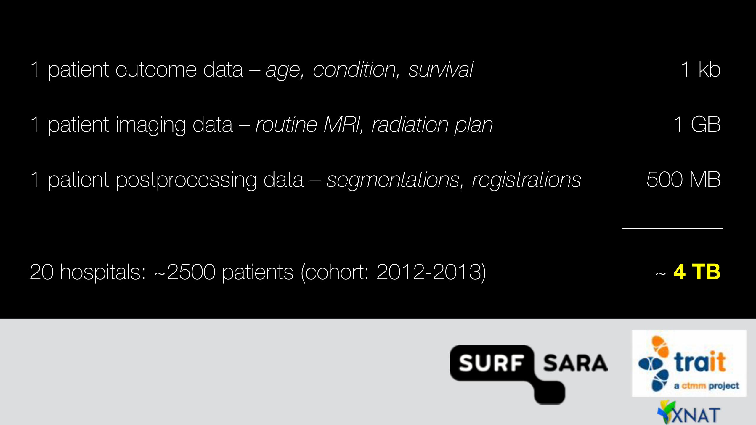
1 patient outcome data – age, condition, survival 1 kb
1 patient imaging data – routine MRI, radiation plan 1 GB
1 patient postprocessing data – segmentations, registrations 500 MB
20 hospitals: ~2500 patients (cohort: 2012-2013) ~ 4 TB
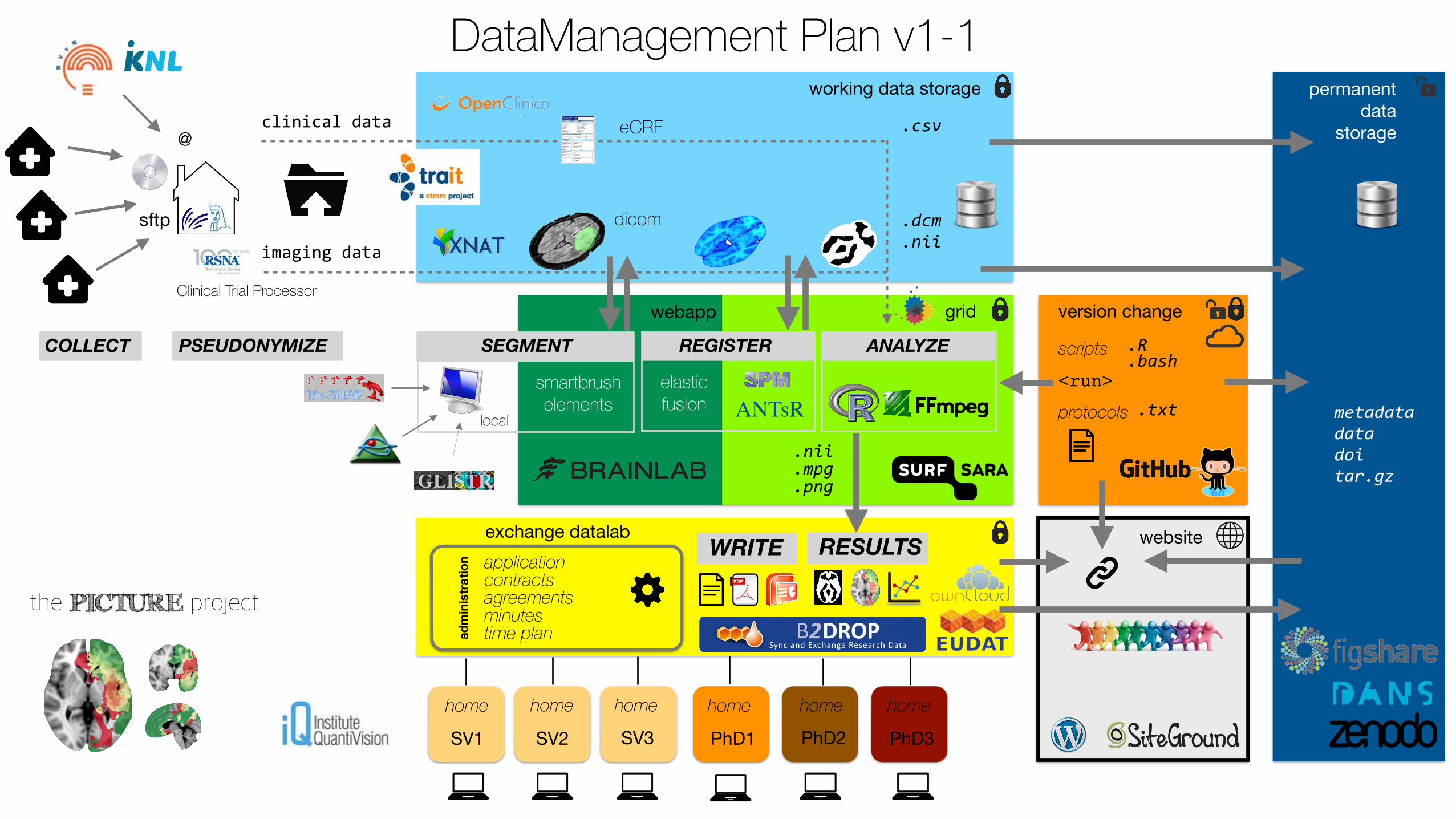
Clinical Trial Processor
PSEUDONYMIZECOLLECT
sftp
@
working data storage
eCRF
dicom .dcm%.nii
.csvclinical&data
permanent data
storage
imaging&data
application contracts agreements minutes time plan
SV1 SV2 SV3 PhD1 PhD2 PhD3
home home home home home home
smartbrush elements
elastic fusion
.R%
.bashscripts
exchange datalab
protocols .txt metadata%data%doi%tar.gz
<run>
version change
website
REGISTER
ANTsR
ANALYZESEGMENT
local
WRITE
administration
grid
.nii%
.mpg%
.png
RESULTS
webapp
DataManagement Plan v1-1
the PICTURE project
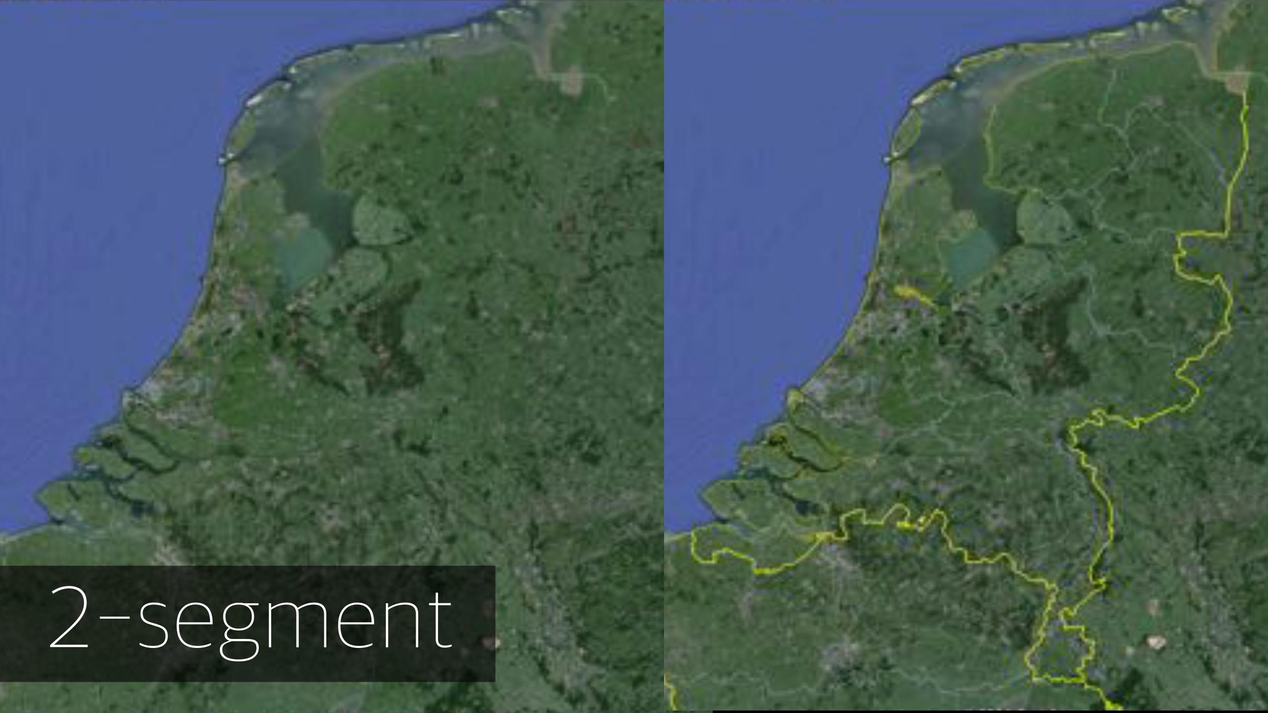
2-segment
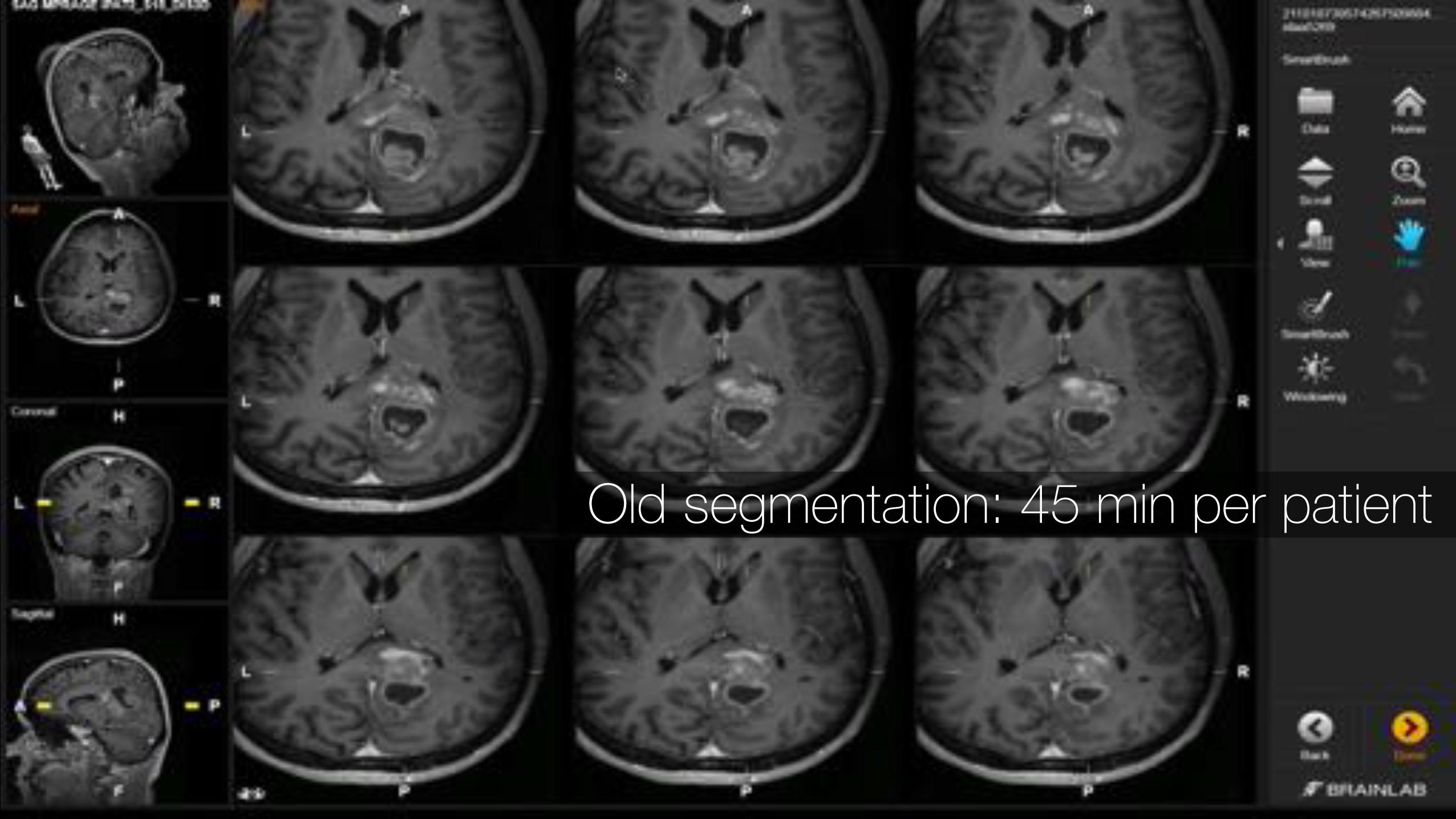
Old segmentation: 45 min per patient
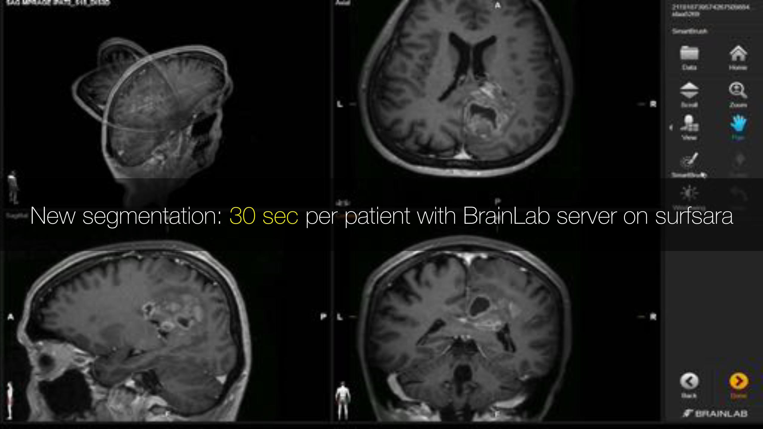
New segmentation: 30 sec per patient with BrainLab server on surfsara
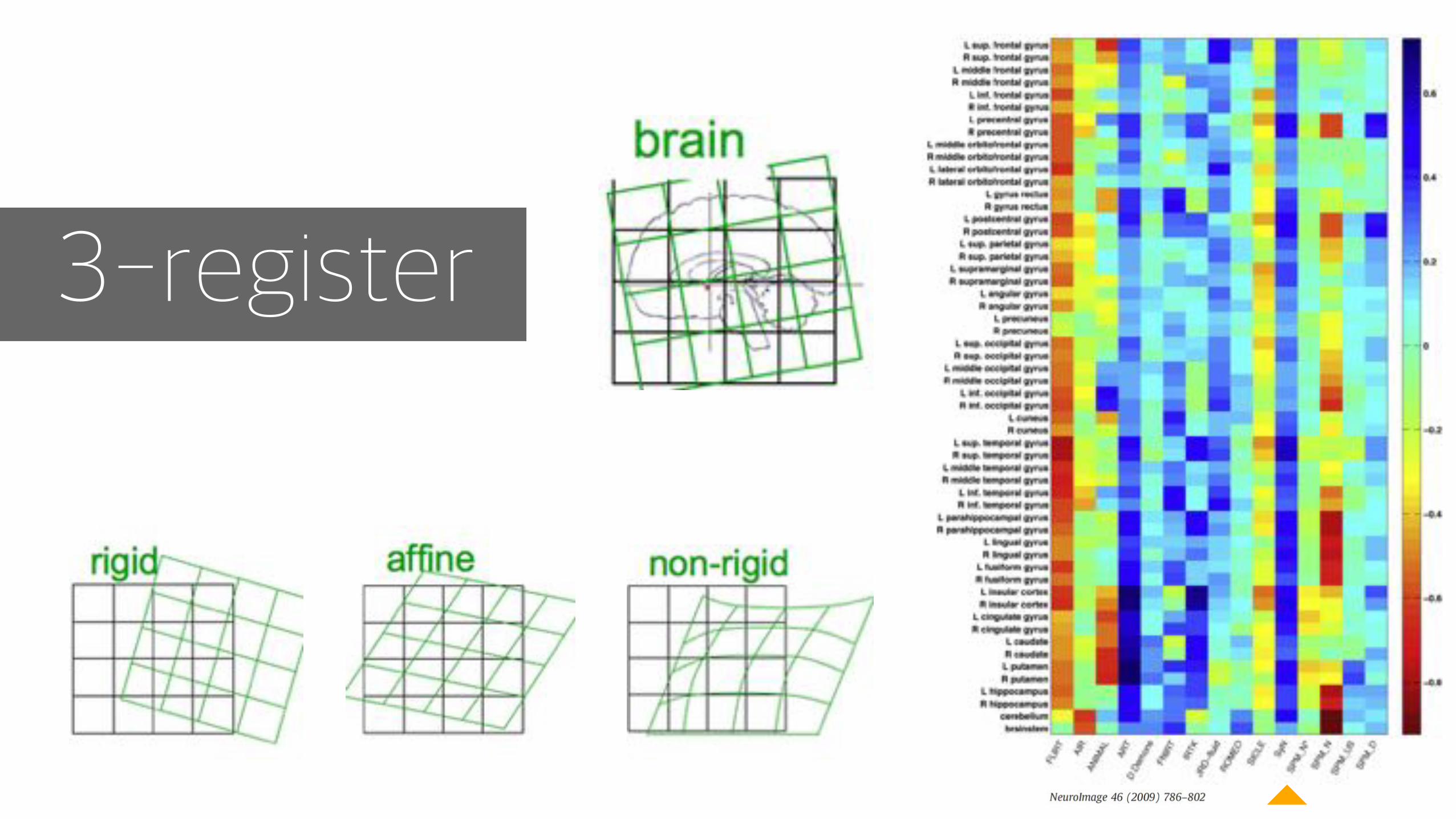
3-register
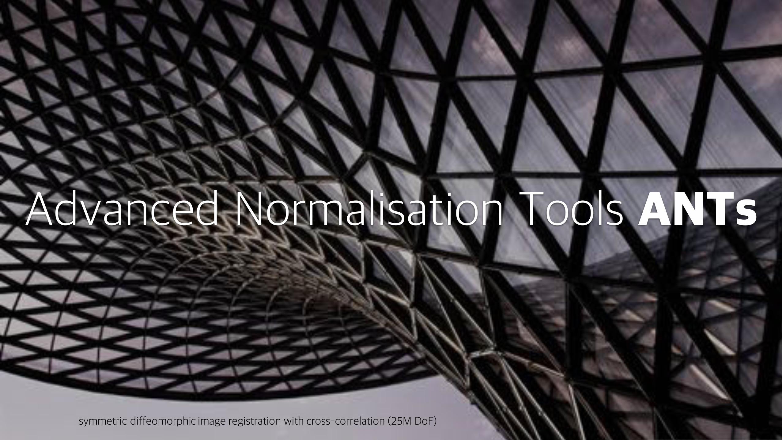
Advanced Normalisation Tools ANTs
symmetric diffeomorphic image registration with cross-correlation (25M DoF)
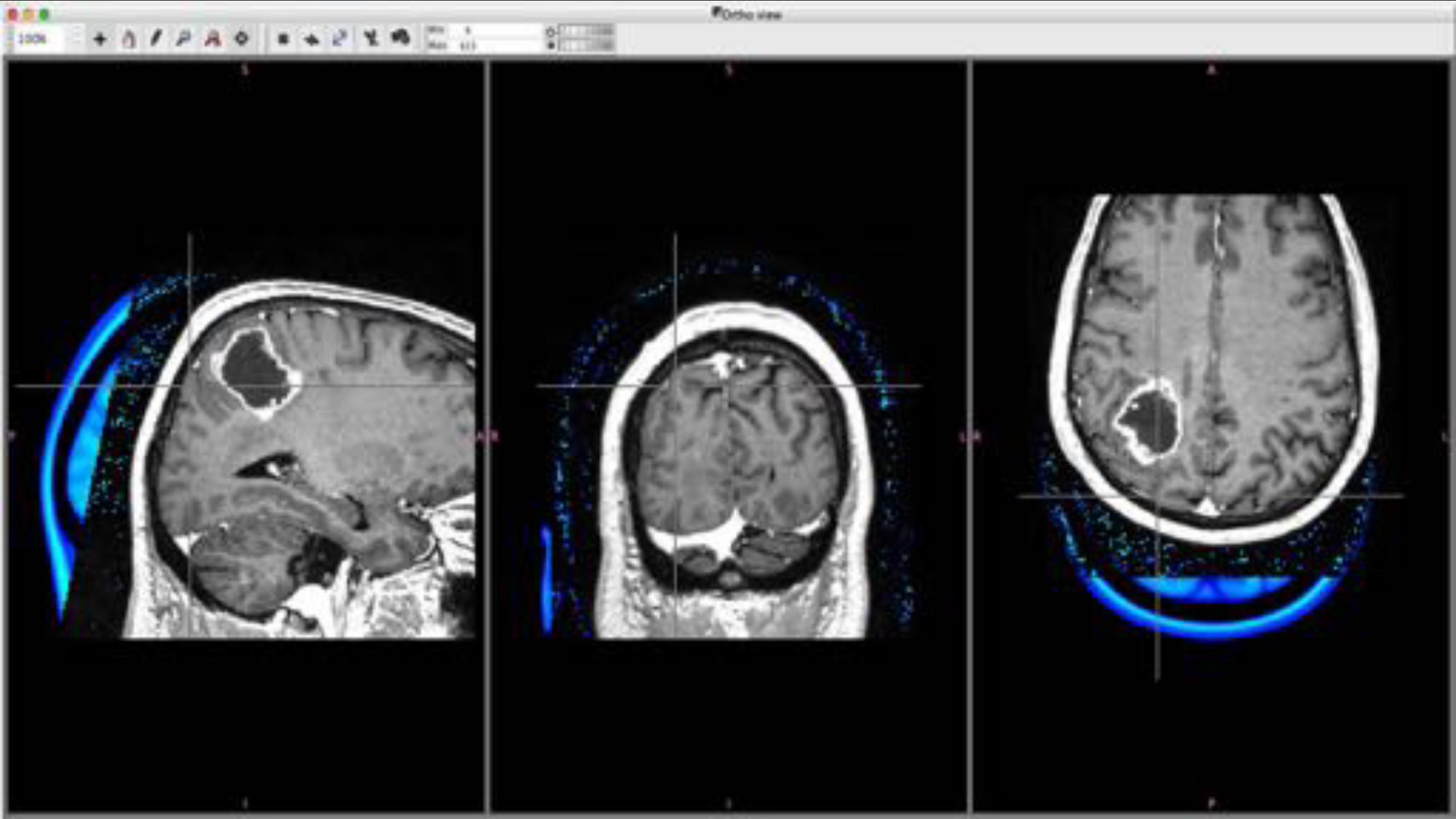
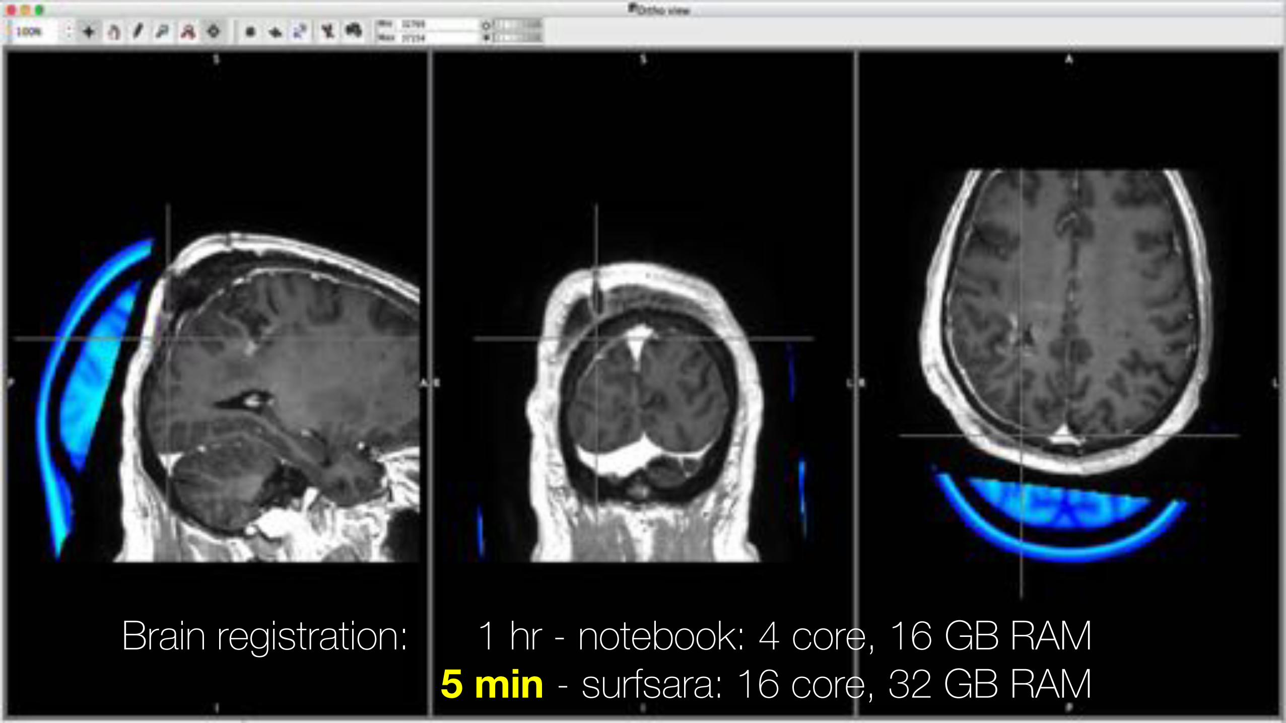
Brain registration: 1 hr - notebook: 4 core, 16 GB RAM5 min - surfsara: 16 core, 32 GB RAM
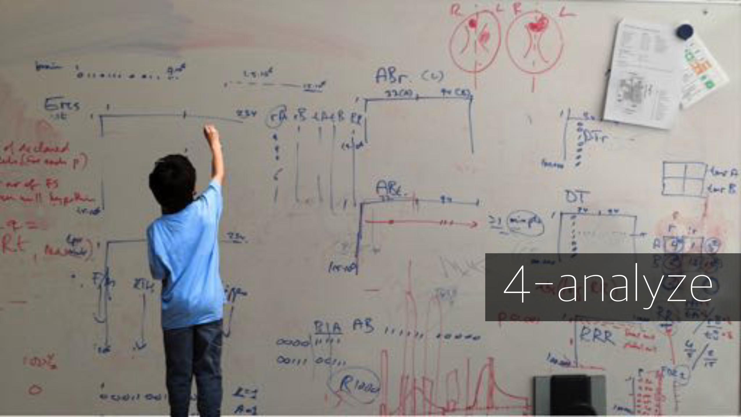
4-analyze
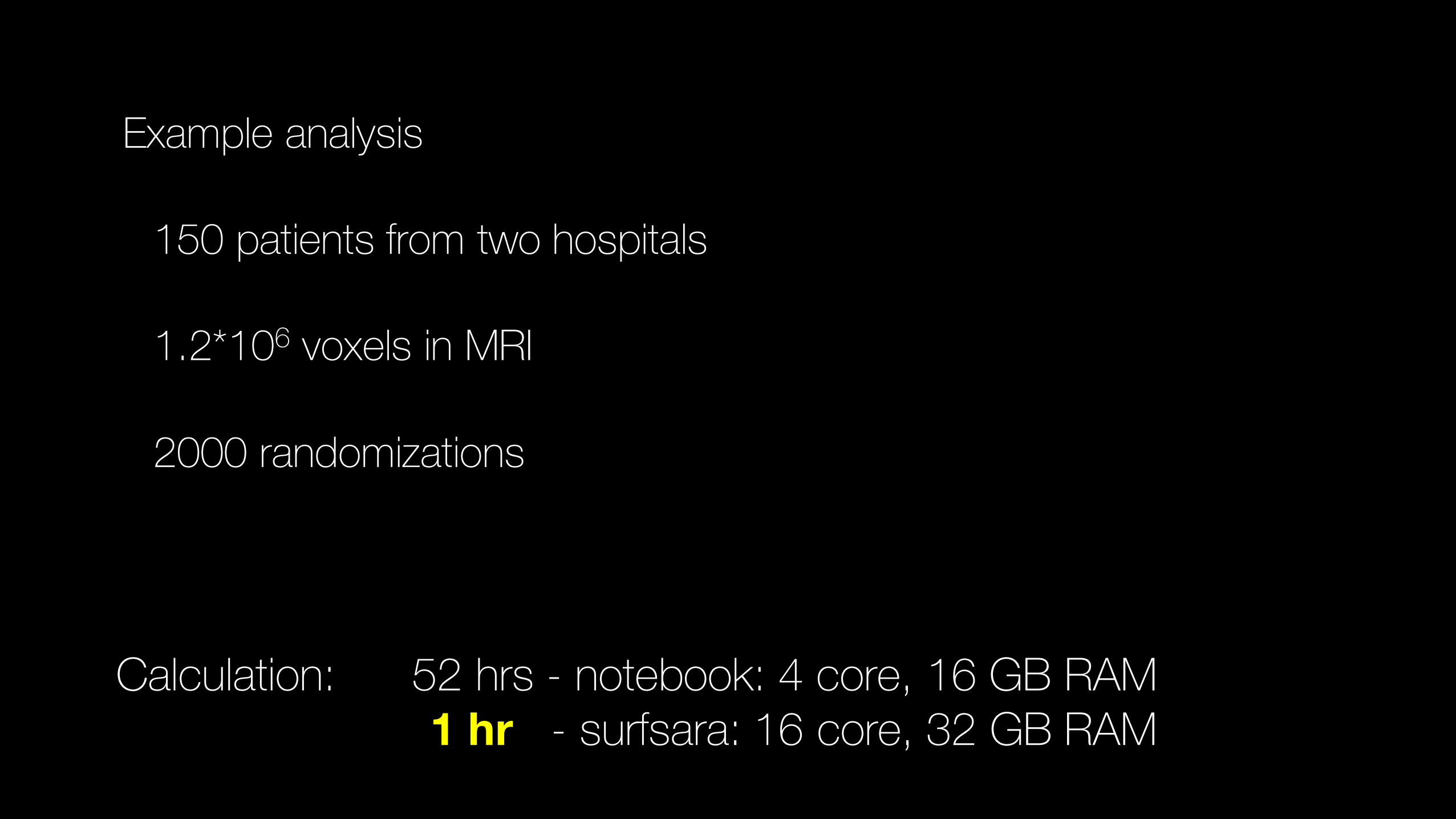
Example analysis
150 patients from two hospitals
1.2*106 voxels in MRI
2000 randomizations
Calculation: 52 hrs - notebook: 4 core, 16 GB RAM1 hr - surfsara: 16 core, 32 GB RAM
Initial resultstowards tomorrow’s treatment
E092
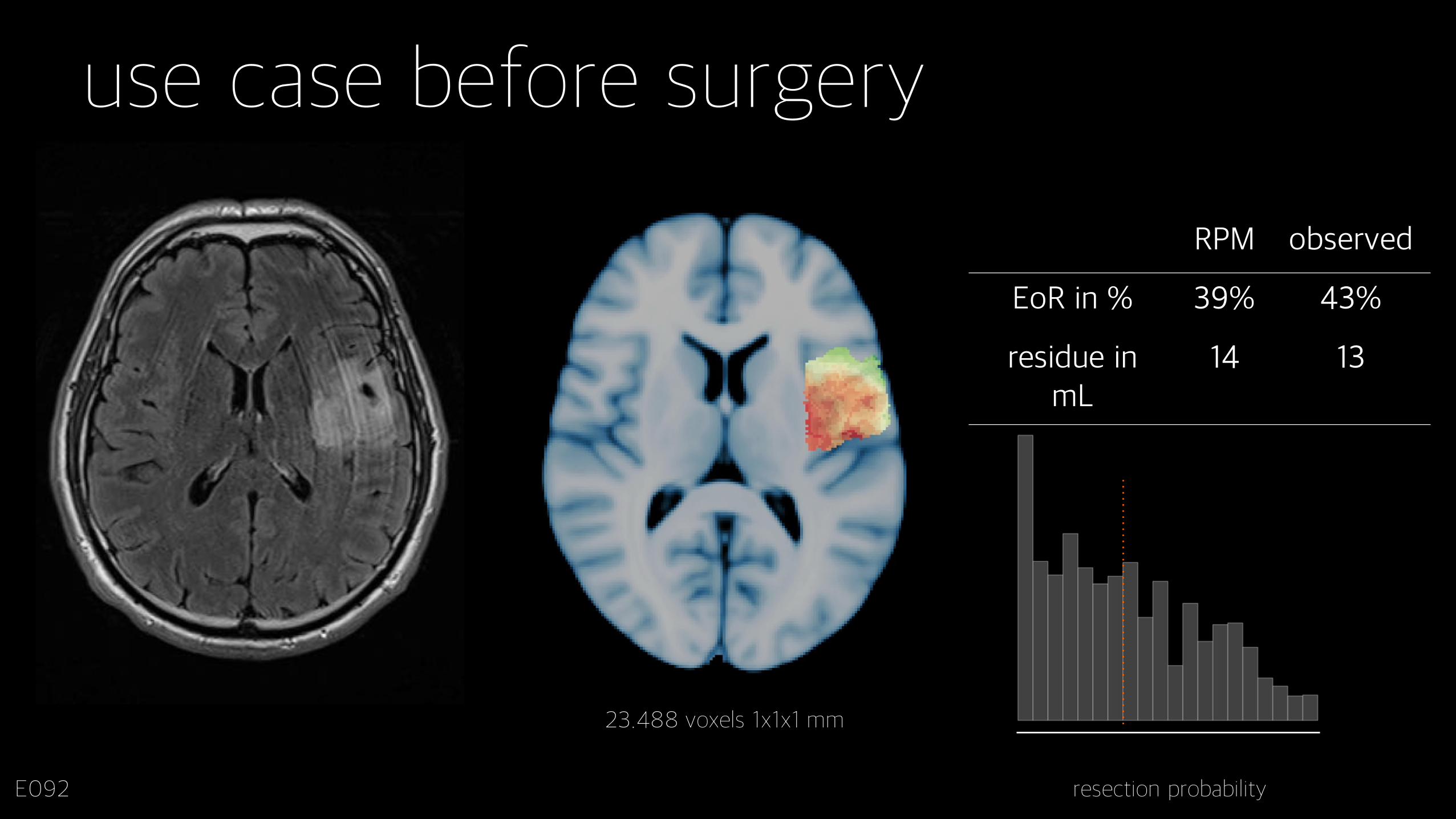
23.488 voxels 1x1x1 mm
resection probability
RPM observed
EoR in % 39% 43%
residue in mL
14 13
use case before surgery
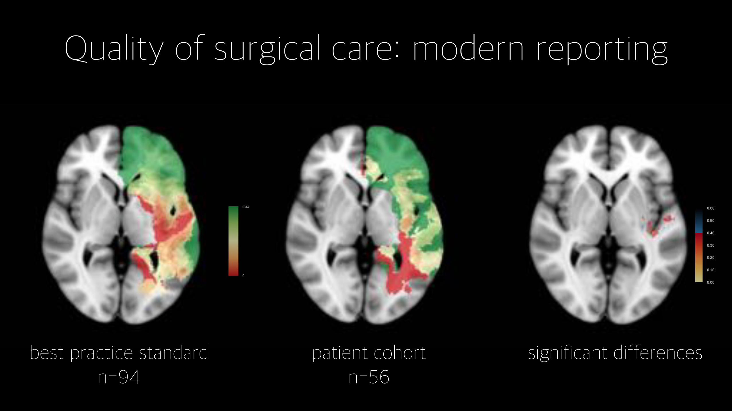
Quality of surgical care: modern reporting
best practice standardn=94
patient cohortn=56
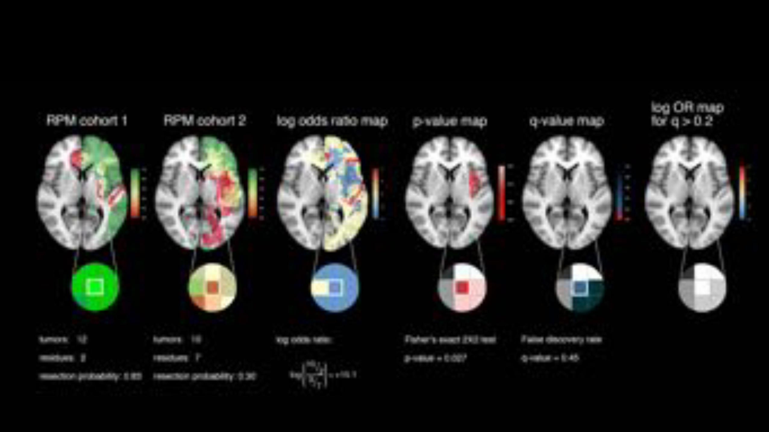
significant differences
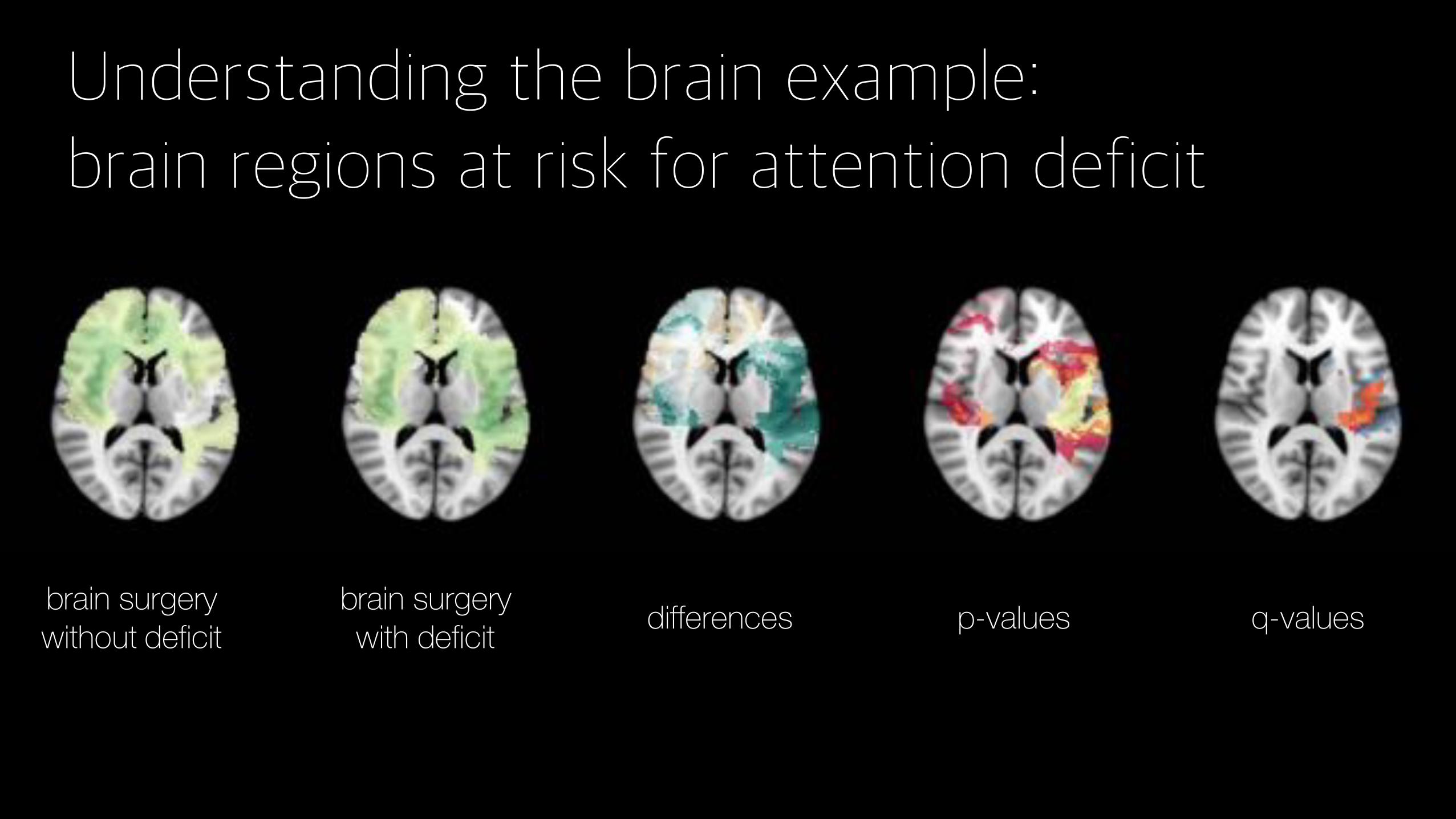
Understanding the brain example: brain regions at risk for attention deficit
brain surgerywithout deficit
brain surgery with deficit differences p-values q-values
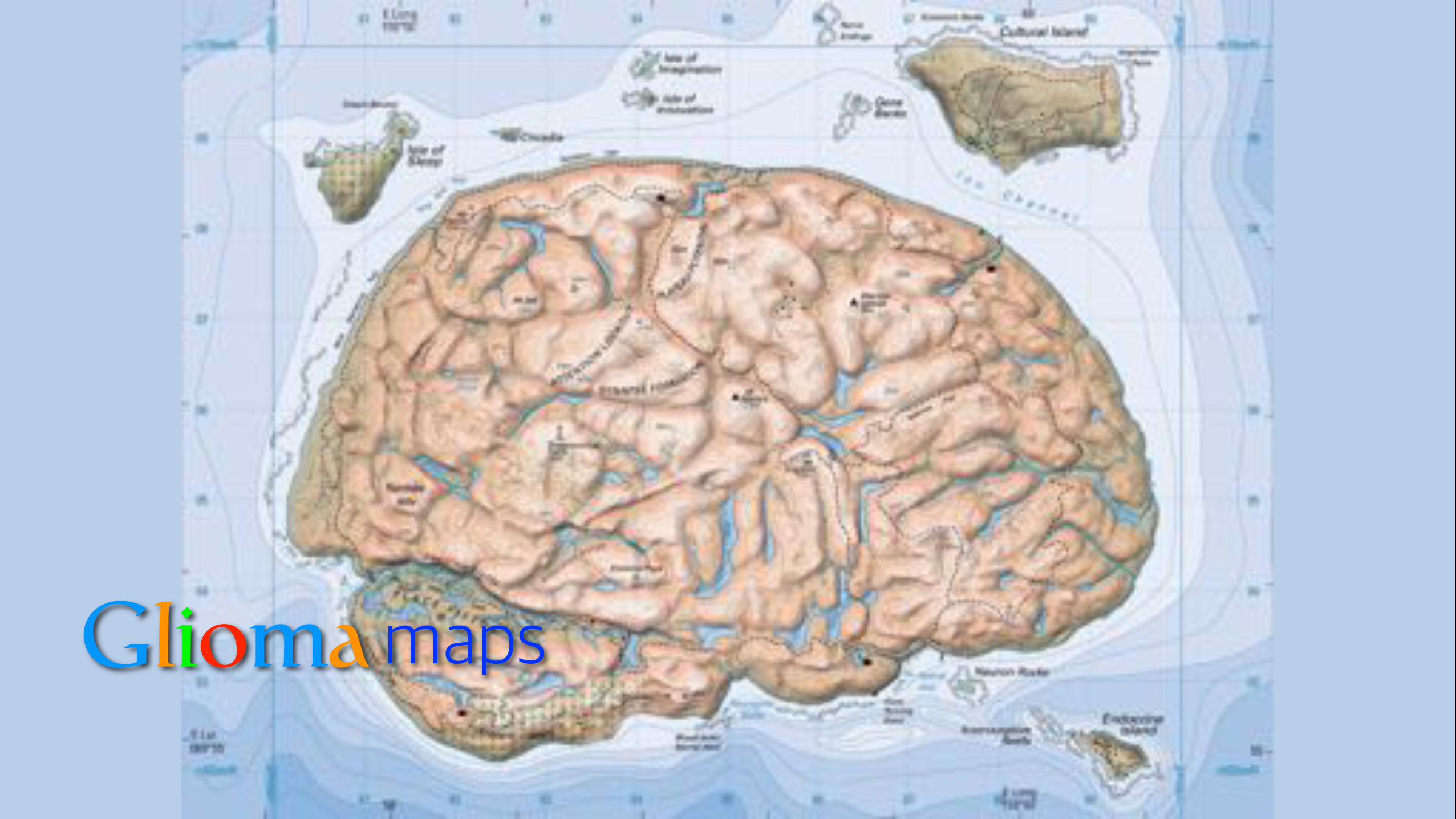
Gliomamaps
Thank youFrederik BarkhofJan de MunckMarnix WitteMarcel van HerkLuc DewitAnne-Marie BruynzeelFrank Lagerwaard
Roelant EijgelaarDomenique MullerMartin Visser
Uli MezgerBalint Varkuti
Linda Ackermans – MUMCHilko Ardon – St Elisabeth ZHSytske Boomstra – MSTWim Bouwknegt - SLZWimar van den Brink – Isala ZwolleClemens Dirven – Erasmus MCNiels van der Gaag – MCHBas Idema - MCAFred Kloet – MCHJan Koopmans - MZGMark ter Laan – UMCN/CWZPierre Robe – UMCUMarco Verstegen - LUMCMichiel Wagemakers – UMCG
Jan BotMathijs KattenbergLykle Voort
Stefan KleinRita Azevedo
Funding