brain-derived neurotrophic factor (bdnf) overexpression in the forebrain results in learning and...
TRANSCRIPT

Neurobiology of Disease 33 (2009) 358–368
Contents lists available at ScienceDirect
Neurobiology of Disease
j ourna l homepage: www.e lsev ie r.com/ locate /ynbd i
Brain-derived neurotrophic factor (BDNF) overexpression in the forebrain results inlearning and memory impairments
Carla Cunha a,b,1, Andrea Angelucci c, Angela D'Antoni a,d, Mate D. Dobrossy e,2, Stephen B. Dunnett e,Nicoletta Berardi c,f, Riccardo Brambilla a,⁎a San Raffaele Scientific Institute, Milan, Italyb Faculdade de Ciências da Universidade do Porto, Portugalc Institute of Neuroscience of CNR, Pisa, Italyd University of Milan, Italye School of Biosciences, Cardiff, UKf Department of Psychology, Florence University, Florence, Italy
⁎ Corresponding author. Fax: +39 0226434767.E-mail address: [email protected] (R. Brambi
1 Present address: Universita’ degli Studi di MilaBiotecnologie e Bioscienze, Milan, Italy.
2 Present address: Department of Stereotactic NeuFreiburg, Freiburg, Germany.
Available online on ScienceDirect (www.scienced
0969-9961/$ – see front matter © 2008 Elsevier Inc. Aldoi:10.1016/j.nbd.2008.11.004
a b s t r a c t
a r t i c l e i n f oArticle history:
In this study we analyzed th Received 5 March 2008Revised 7 October 2008Accepted 7 November 2008Available online 27 November 2008Keywords:BDNFSpatial learningPassive avoidanceShort-term memory
e effect on behavior of a chronic exposure to brain-derived neurotrophic factor(BDNF), by analysing a mouse line overexpressing BDNF under the αCaMKII promoter, which drives thetransgene expression exclusively to principal neurons of the forebrain. BDNF transgenic mice and their WTlittermates were examined with a battery of behavioral tests, in order to evaluate motor coordination,learning, short and long-term memory formation. Our results demonstrate that chronic BDNF overexpressionin the central nervous system (CNS) causes learning deficits and short-term memory impairments, both inspatial and instrumental learning tasks. This observation suggests that a widespread increase in BDNF inforebrain networks may result in adverse effects on learning and memory formation.
© 2008 Elsevier Inc. All rights reserved.
Introduction
Brain-derived neurotrophic factor (BDNF) is a member of theneurotrophin family of growth factors, which is widely expressedthroughout the mammalian brain, together with its high affinityreceptor TrkB (Lewin and Barde, 1996; Thoenen, 1995), and plays acrucial role in the development, maintenance and function of the CNS(Bonhoeffer, 1996; Huang and Reichardt, 2001; Huang and Reichardt,2003). In addition, in the past decade, accumulating data indicate thatneuronal activity regulates BDNF transcription, transport of BDNFmRNA and protein into dendrites and activity-dependent secretion ofBDNF that in turn modulate synaptogenesis, synaptic plasticity andmemory formation (Bekinschtein et al., 2007b; Lu et al., 2007; Poo,2001; Soule et al., 2006; Tyler et al., 2002).
A unique feature among the neurotrophin family is BDNF activity-dependent secretion, as a mixture of proBDNF andmature BDNF (Eganet al., 2003; Lu, 2003; Pang and Lu, 2004; Pang et al., 2004). Compellingevidence indicates that, by binding to its high affinity receptor TrkB,
lla).no-Bicocca, Dipartimento di
rosurgery, University Hospital
irect.com).
l rights reserved.
mature BDNF plays a critical role in both early and late forms of long-termpotentiation (E-LTP and L-LTP, respectively). On one side, Ca2+ andNMDA glutamate receptor dependent BDNF secretion, upon tetanicstimulation, facilitates maintenance of E-LTP (Aicardi et al., 2004;Balkowiec and Katz, 2002; Figurov et al., 1996; Gartner and Staiger,2002; Gottschalk et al., 1998; Hartmann et al., 2001; Lever et al., 2001;Rex et al., 2006; Yano et al., 2006). On the other, both genetic andpharmacological blockade of BDNF and TrkB signaling results in L-LTPimpairments, particularly upon theta burst stimulation or forskolinapplication (Korte et al., 1995; Korte et al., 1996; Pang et al., 2004;Patterson et al., 1996; Minichiello et al., 2002; Minichiello et al., 1999;Patterson et al., 2001). Moreover, at least in certain conditions, BNDFhas been shown to elicit by itself a form of synaptic plasticity, termedBDNF-LTP (Kang and Schuman, 1995; Kang and Schuman, 1996;Messaoudi et al., 2002; Ying et al., 2002). This is a form of late LTPinvolving brief, local application of BDNF which is transcription-dependent and requires Arc synthesis (Messaoudi et al., 2007).
From a behavioral perspective, several studies have shown thatBDNF and TrkB are implicated in learning and memory by directlyexamining their role in a variety of learning paradigms in rodents(Bekinschtein et al., 2007b; Bekinschtein et al., 2008; Lu et al., 2007;Yamada et al., 2002). BDNF expression was found to be increased inthe hippocampus of rats, following Morris Water Maze (MWM)(Kesslak et al., 1998), radial arm maze (Mizuno et al., 2000), passiveavoidance (Ma et al., 1998) or contextual fear conditioning (Hall et al.,

359C. Cunha et al. / Neurobiology of Disease 33 (2009) 358–368
2000; Rattiner et al., 2004b) and gene ablation of either BDNF or TrkBresulted in learning impairments (Linnarsson et al., 1997; Minichielloet al., 1999). Accordingly, a single intra-hippocampal BDNF adminis-tration in adult rats improves performance in the MWM test (Cirulliet al., 2004). Furthermore, pre-training infusions of either anti-BDNFantibodies or antisense BDNF oligonucleotides caused impairedspatial learning and memory in rats, as assessed in the MWM (Muet al., 1999), the radial arm maze (Mizuno et al., 2000), fearconditioning (Lee et al., 2004; Rattiner et al., 2004a; Rattiner et al.,2005) or passive avoidance (Alonso et al., 2005; Alonso et al., 2002;Tyler et al., 2002; Bekinschtein et al., 2007a; Ma et al., 1998).
A key aspect that has recently started to be investigated is therole of proBDNF in plasticity and memory formation. In humans, aval66met polymorphic substitution in the 5′ pro-region of thehuman BDNF protein results in hippocampal-dependent episodicmemory impairments that are linked to proBDNF deficits inlocalizing to secretory granules or synapses (Egan et al., 2003). Inaddition, strong evidence suggests that tissue-plasminogen activator(tPA), a protein whose secretion is also upregulated during L-LTP andlearning, through activation of plasmin, converts the proBDNFprecursor into mature BDNF and that this proteolytic step isnecessary for L-LTP expression (Pang et al., 2004). Together withthe observation that, at least in the hippocampus, a substantialfraction of the total BDNF secreted extracellularly is proBDNF(Farhadi et al., 2000; Mowla et al., 2001; Mowla et al., 1999) andthat its selective binding to the low affinity receptor p75NTR mayfacilitate neuronal apotosis (Lee et al., 2001; Teng et al., 2005) andpossibly other neuronal functions such as long-term depression(LTD), a contrasting bidirectional regulation of synaptic plasticity andmemory formation has been suggested for the mature and precursorforms of BDNF (Lu, 2003).
Unfortunately, very little work has so far been performed onanimal models in which BDNF levels have been increased chronicallyin the brain. Since the relevance of BDNF as potential therapeuticmolecule to treat a number of brain disorders, from Parkinson's andHuntington's diseases to depression and substance abuse, this aspectdeserves to be evaluated in detail (Chao et al., 2006). In the onlyavailable study, a widespread overexpression of BDNF in mice, underthe control of the ubiquitous β-actin promoter, resulted in learningdeficits in passive avoidance, increased neuronal excitability andsusceptibility to seizures (Croll et al., 1999). Such finding, highlyrelevant in our opinion for the future clinical use of BDNF but largelyneglected in the literature, has largely been confirmed by the presentwork. Herewe have analyzed amouse line, which overexpresses BDNFspecifically in the forebrain structures (Huang et al., 1999). We found,quite surprisingly, significant impairments in both short and long-term memory formation.
Materials and methods
Animals
All mice used in this work were maintained on a C57Bl/6 geneticbackground (more than 10 generations) from Charles River Labora-tories. Mice were kept 5 or 6 per cage in a SPF environment, with 50%relative humidity, a temperature of 21±1 °C and on a 12 h light-darkschedule, with food and water available ad libitum. Littermates wereweaned around day P21 and genotyped for the presence of the BDNFtransgene, using the forward primer 5′-TCAGTCAAGCCGGTTCTC-3′and the reverse primer 5′-AGTCCGCGTCCTTATGGT-3′. In this work,transgenic BDNF andWT control mice were littermates of 9–14 weeksof age and only naive males were used. For the behavioral analysis,mice were handled for 1 week before experiments and all experi-ments were performed in a low luminosity environment (20–25 lx)and carried out blind with respect to genotype. All animal procedureswere conducted according to EC guidelines (EC Council Directive 86/
609, 1986) and to the Italian legislation on animal experimentation(Decreto L.vo 116/92).
BDNF protein quantification by ELISA
Micewere sacrificed by cervical dislocation and decapitated. Brainswere quickly removed and microdissected by use of a stereotacticmicroscope. Samples from the selected regions were collected and theBDNF protein quantification was determined by use of the BDNF Emax
ImmunoAssay System (Promega), according to the manufacturer'sinstructions. Briefly, 96-well plates were incubated overnight at 4 °Cwith anti-BDNF monoclonal antibody, diluted 1:1000 in carbonatecoating buffer. Plates were blocked with 1X Block and Sample bufferfor 1 h at room temperature. Samples and standards (0–500 ρg/ml)were added to the plates and incubated for 6 h, followed by incubationwith a 1:500 anti-human BDNF polyclonal antibody, overnight at 4 °C.Anti-IgY horseradish peroxidase conjugate, diluted 1:200, was thenadded to each well and plates were incubated for 2 h at roomtemperature. Plates were developed by incubation 15 min at roomtemperature with 100 μl of TMB One Solution. The reaction wasstopped by adding 100 μl of 1N HCl and absorbance was measured at450 nmwithin 30 min. BDNF protein quantification for each structurewas performed in duplicate, and mean BDNF levels were reported inng/g of weight tissue. The results for the BDNF protein levels wereexpressed as mean±SEM, for both genotypes. Statistical analysisbetween genotypes for each structure analysed consisted on a two-tailstudent's t-Test.
SDS-PAGE and western blotting
WTand BDNF transgenic mice were decapitated and the brain wasrapidly removed and placed on ice. Hippocampus, striatum, cortexand cerebellum were dissected and frozen on liquid nitrogen. Tissueswere homogenized in a lysis buffer (150 mM NaCl, 20 mM Hepes,1 mM MgC, 1 mM EDTA) and western blotting was performed.Proteins (10 μg) were separated on 12% Tris-Glicine gel at a constantvoltage of 100 V and then transferred to Hybond-P PDVP membranes(Amersham Biosciences) at a constant voltage of 100 V for 1 h. Blotswere blocked in 5% non-fat milk in a Tris buffered saline solutioncontaining 1% Tween 20 (TBS-T) and this TBS-T solution was used forall subsequent washes. Primary and secondary antibodies werediluted in TBS-T containing 5% non-fat milk and were used at thefollowing concentrations: 1:5000 anti-BDNF (AP1779SP, Chemicon),1:1000 anti-GAPDH (Sigma) and used as previously described (Barnesand Thomas, 2008). HRP-conjugated secondary antibodies were usedat room temperature for 1 h in 5% milk in TBS plus 0.5% Tween 20followed by immunological detection with ECL Plus (AmershamBioscience). The immunoblots were exposed with Hyperfilm, scannedand analyzed with ImageJ software to measure the integrated opticaldensity of the bands.
Immunohistochemistry
WT and BDNF transgenic mice (five for each genotype) wereanaesthetized and perfused via intracardial infusion of ice-cold 4%PFA (dissolved in 0.1 M Na2HPO4/NaH2PO4 buffer, pH 7.4). Brainswere rapidly extracted, post-fixed overnight and transferred to 25%buffered sucrose for 24 h. Coronal sections were cut at 30 μmthickness on a freezing microtome and stored in a cryoprotectivesolution at −20 °C until they were processed for immunohisto-chemistry as previously described (Valjent et al., 2000). Free-floatingsections were incubated with a primary antibody against Phospho-p44/42 Map Kinase (1:200, Cell Signalling) overnight at 4 °C.Sections were then incubated with biotinylated goat anti-rabbit IgG(1:200, Vector Labs) for 2 h. Detection of the bound antibodies wascarried out using a standard peroxidase-based method (ABC-kit,
360 C. Cunha et al. / Neurobiology of Disease 33 (2009) 358–368
Vectastain, Vector Labs), followed by DAB staining. Quantification ofp-ERK positive neurons was performed for different regions of theHippocampus (DG, CA3, CA2, CA1) and Cortex (Cingulate Cx: Cin Cx;Prefrontal Cortex: Pf Cx) in 2 sections per mouse, bilaterally. Sampleareas were visualized under a 20× objective in a Leica DM IRBmicroscope by a blind investigator to condition and genotype andcounts per region were averaged across the sections using theImageJ software.
Accelerating Rota-Rod
The apparatus (Ugo Basile Instruments, Comerio, Italy) consists ona rotating cylinder, covered with textured rubber to provide grip. Micewalk forward on the rotating cylinder, at speeds increasing from 4 to40 rpm over a 6 min cut-off testing session. Latencies in falling off thecylinder were measured over 3 consecutive daily sessions, 3 trials persession (Biffi et al., 2004).
Locomotor activity
The apparatus (Ugo Basile Instruments, Comerio, Italy) consists ina 90×40×45 cm activity box containing UV photoelectric beamsencompassing the whole length of the cage, with a line of photocells2 cm above the floor to measure horizontal movements, and anotherline located 6 cm above the floor to measure vertical movements.Spontaneous locomotor activity and exploratory behavior wereassessed by individually placing the mice in the locomotor boxes for10 min: horizontal and vertical activity was measured as the totalnumber of beam disruptions during the monitoring period. A dailysessionwas performed, during 3 consecutive days (Mazzucchelli et al.,2002).
Passive avoidance
The apparatus (Panlab s.l., Barcelona, Spain) consists of twochambers: a white strongly illuminated chamber, separated by adoor from a black dark chamber, which has a floor grid that permitsthe passage of electric foot shocks (Pittenger et al., 2006). The taskbegan by placing the mouse in the illuminated compartment and thenopening the door between the chambers, after a period of exploration.When the mouse entered the dark compartment after a short latency,the door was closed and a single electric foot shock of 0.10 mA wasadministered, for 2.0 s. We performed the passive avoidance test intwo different conditions: in the first condition we used an apparatusin which the size of the white, illuminated compartment wasapproximately twice as large as that of the black, dark compartment,that mice were allowed to explore for 10.0 s before door opening. Inthe second condition we used a white compartment approximately ofthe same size than the black one, which mice were allowed to explorefor 3.0 s. After reintroducing the same mice in the same illuminatedchamber, after 1 h or 24 h, we determined the latency of time spent inthe illuminated side, as a measure of short- and long-term memory,respectively. For both cases, the acquired memory was recalled after10 days.
Eight-arm radial maze
Animals were food restricted for a week before the test andmaintained at 80% of their initial weight throughout the test. Theapparatus consisted in a plastic maze with 8 identical arms,numbered 1 to 8, radiating from a central platform. A series ofexternal cues were placed outside the maze for spatial orientation. Ateach daily trial, a food pellet reward was placed at the end of eacharm and the mouse was placed on the centre of the maze; the trialfinished when the mouse visited all the 8 baited arms and the overalllatency to complete the task was recorded. During each trial, two
parameters were scored: total number of errors and total number ofcorrect visits. An error corresponds to re-entering a previously visitedarm and the number of correct visits corresponds to the number ofarms visited before scoring an error. We performed one trial per dayduring 12 consecutive days and a recall trial after 10 days (Brambillaet al., 1997; Olton et al., 1978).
Morris water maze (MWM)
We performed the hidden platform version of the MWM test(Brambilla et al., 1997; Morris et al., 1982). A large water tank of120 cm of diameter was filled with white opaque water at 20 °C anddivided into four quadrants of equal area arbitrarily named Northeast,Southeast, Southwest and Northwest. The water tank was in a roomwith two- and three-dimensional visual cues in the walls. An escapeplatform of 11.4 cm of side was submerged 1 cm below the watersurface and placed in the center of the Southeast quadrant. Theplatform was maintained in this position for all the swim trialsthrough the test. Mice were trained to swim to the platform in 2 dailytrials, with a 30 min interval, during 10 consecutive days. Each trialwas initiated at one of four different starting positions at the outeredge of the pool and the swimming path of each mouse was videorecorded. Upon reaching the platform, each mouse was allowed torest for 20 s on it. The trial finished when the mouse found theplatform or when 60 s had elapsed. If the mouse did not reach theplatform within 60 s, it was guided to the platform and allowed torest for 20 s on it. After each trial each mouse was returned to itshome cage where it rested until the next trial. On the day 5 and onthe day 10, 30 min before the daily trials of hidden platform, eachmouse was tested in a probe trial in which the hidden platform wasremoved and the swimming path of each mouse was video recordedover 60 s. A recall probe trial was performed after 10 days. Differentparameters were scored as an average of the 2 daily trials and foreach probe trial, using Noldus EthoVision 3.0 platform, a videotracking system for automatic recording of movement. For each trialwe measured: latency to reach the platform, in sec, total distanceswam to the platform, in cm, and average swim speed, in cm/s. Foreach probe trial we measured the total amount of time spent in eachquadrant, in sec, and the total amount of time spent in the platformzone, in sec.
Statistical analysis
All results were expressed as mean±SEM and all statistical analysismade use of the Statistical Package for Social Sciences (SPSS 13.0software). For behavioral tests, removal of outliers was performed by aBlox plot analysis. Analysis of differences between groups consisted ona two-way analysis of variance (ANOVA) for repeated measures,considering both within- (time; time×genotype) and between-subjects (genotype) effects, followed by post-hoc comparisons(Scheffe test). Results were considered significant when pb0.05. ForBDNF level determination and body weight, a two-tailed t-Test wasused.
Results
BDNF has been repeatedly implicated in the regulation of synapticplasticity and cognitive functions (Bekinschtein et al., 2007b; Bram-ham andMessaoudi, 2005; Lu et al., 2007; Pang and Lu, 2004; Soule etal., 2006). In order to assess the effect of a sustained forebrain BDNFexpression in learning andmemory processing we took advantage of amouse line inwhich an accelerated postnatal rise of BDNFmRNA levelin the forebrain takes place (Huang et al., 1999). In this line, ectopicexpression of proBDNF, the unprocessed form of this neurotrophin, iscontrolled by the αCaMKII promoter, which drives the transgeneexpression specifically to forebrain structures, including the
361C. Cunha et al. / Neurobiology of Disease 33 (2009) 358–368
hippocampus, striatum, neocortex and amygdala (Mayford et al.,1996). Since expression in these forebrain structures may directlyaffect both behavioral performance and learning and memory wecarried out a battery of tests in order to compare WT and transgenicanimals at the locomotor and cognitive level.
Transgenic mice overexpress mature BDNF in most forebrain regions
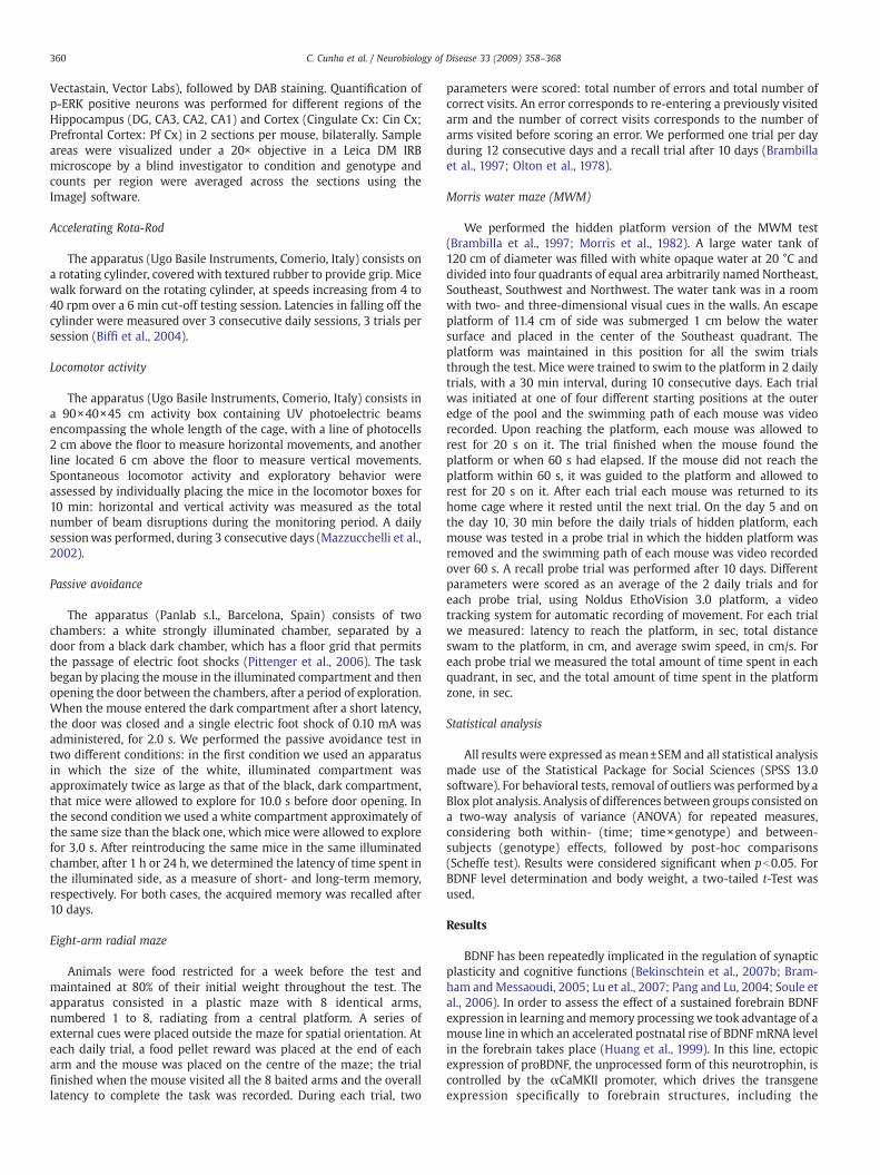
In order to evaluate the BDNF protein levels in the forebrain ofthese mice, we dissected the following structures: striatum, hippo-campus, frontal cortex, parietal cortex, occipital cortex and cerebellumand performed a BDNF specific quantitative ELISA. Cerebellum wasanalyzed as a control, since expression of αCaMKII driven BDNFtransgene should exclusively be restricted to the forebrain. As we cansee from Fig. 1A, transgenic mice showed higher BDNF immunor-eactivity in all forebrain structures analyzed, as compared to WTlittermates: we observed an increase of BDNF protein levels of 2.4 foldin the striatum (10.24±0.95 vs 4.18±0.32 ng/g wt tissue; pb0.01), 3.3fold in the hippocampus (54.36±10.68 vs 16.67±1.66 ng/g wt tissue;pb0.01), 2.8 fold in the frontal cortex (71.30±15.71 vs 19.53±1.54 ng/gwt tissue; pb0.05),1.8 fold in the parietal cortex (55.87±4.42 vs 31.00±2.44 ng/gwt tissue; pb0.01) and 2.0 fold in the occipital cortex (154.30±10.92 vs 78.67±12.46 ng/g wt tissue; pb0.01). However, BDNF proteinlevels in the cerebellumwere undistinguishable between transgenic andWT mice (29.12±3.38 vs 29.41±2.03 ng/g wt tissue). Altogether, theseresults are in agreementwith the reported data for BDNFmRNA levels inthe transgenic line and confirm that our transgenic mice do overexpressBDNFproteins (Huang et al.,1999). However, this assay does not allowusto distinguish between proBDNF and mature BDNF, that are equally
Fig. 1. Overexpression of BDNF in the transgenic mice. (A) Tissue levels of total BDNF proteparietal cortex, occipital cortex and cerebellum of WT (n=10) and BDNF (n=10) mice, as an(WT: n=3; BDNF: n=3) have been subjected towestern blot analysis for p32 proBDNF and noin the lower panels. Statistical analysis revealed no significant difference between genotype
recognized by the antibodyused in ELISA. Since thepotential contrastingeffects of proBDNF on mature BDNF, and considering the observedbehavioral phenotype (see below), we felt important to measure thelevel of theprecursor protein in the relevant brain structures for learningand memory. Western blot analysis on dissected brain areas, includingthe striatum, the hippocampus, the cortex and the cerebellum has beencarried out with specific antibodies recognizing p32 proBDNF (Fig. 1B)(Barnes and Thomas, 2008). After quantification of the data, no obviousstatistically significant changes were observed. Therefore, by comparingthe proBDNF data with the total BDNF levels observed in the transgenicanimals, we can confidently conclude that the large majority of BNDFoverexpress is in the mature form and that, at least in basal conditions,the level of proBDNF is tightly regulated in the brain, as recentlysuggested (Matsumoto et al., 2008).
Measurement of body weight
It has been previously reported that BDNF can function as ananorectic factor, contributing to food intake and body weightregulation, likely by regulating hypothalamic function (Lyons et al.,1999; Kernie et al., 2000; Fox and Byerly, 2004). We thereforeexpected that in our BDNF overexpressing mouse line, the averagebody weight would be significantly reduced as compared with WTmice. In order to test this possibility, we weighted a considerablenumber of WT (n=35) and BDNF (n=30) littermate males of9–14 weeks of age. As shown in Fig. 2A, the average bodyweight for WT mice is 27.47±0.76 g, while the value for BDNF mice is25.74±0.58 g. Mean comparison, by the student's t-Test, indicates onlya tendency, which was statistically non significant, for a reduction ofbody weight in the transgenics (t-Test, p=0.075). This result indicates
in, in ng of BDNF/g of wet tissue weight, in the striatum, hippocampus, frontal cortex,alyzed by ELISA. ⁎pb0.05, ⁎⁎pb0.01. (B) Protein extracts from the indicated brain areasrmalized for p37 GAPDH (upper panels). Quantification of data (mean±SEM) is indicateds for all structures analyzed.

Fig. 2. (A) Basal Body weight in BDNF overexpressing mice. The mean body weight for WT mice (n=35) is 27.47±0.76 g and the mean body weight for BDNF mice (n=30) is 25.74±0.58 g. No statistically significant difference was found. (B) Analysis of motor performance of BDNF transgenics and WT mice. Latencies in falling off the Rota-Rod apparatus, during3×3 daily sessions, for WT (n=25) and BDNF (n=23) mice. Statistical analysis shows a significant time effect (pb0.001, session 1 vs 9), but not a genotype or a genotype×timeinteraction. (C) Analysis of horizontal locomotor activity for WT (n=30) and BDNF (n=26) mice, measured by total number of photoelectric beam breaks in a 10 min daily session,during 3 consecutive days. Statistical analysis shows a significant time effect (pb0.001, day 1 vs 3), but not a genotype or a genotype×time interaction. (D) Analysis of verticallocomotor activity for WT (n=30) and BDNF (n=26) mice, measured by total number of photoelectric beam breaks in a 10 min daily session, during 3 consecutive days. Statisticalanalysis shows a significant time effect (pb0.001, day 1 vs 3), but not a genotype or a genotype×time interaction.
362 C. Cunha et al. / Neurobiology of Disease 33 (2009) 358–368
that a forebrain overexpression of BDNF may provide only a smallcontribution to an overall decreased body weight. Interestingly, it hasalso been reported that the conditional loss of BDNF in the forebrain,both in early or late adulthood, does not cause increase of bodyweight(Monteggia et al., 2004), as originally reported for global BDNF KOanimals, further suggesting that chronic changes in BDNF expressionrestricted to the forebrain may not be sufficient to cause thepreviously reported anorectic effect.
Motor performance of transgenic mice
In order to evaluate possible performance deficits in our BDNFtransgenic mice, we assayed motor coordination, balance and motorlearning in the Rota-Rod task, and spontaneous locomotor activity,exploratory behavior and habituation to a novel environment in theactivity boxes. Performance on the Rota-Rod test was measured bylatencies in falling off the cylinder, during 3 daily trials over 3consecutive days (Biffi et al., 2004). As shown in Fig. 2B, both genotypes(WT=25; BDNF=23)manifested a significant increase in the latency tofall, from trial 1 to trial 9 (F8,368=53.813; pb0.001), demonstrating thatboth groups significantly improved motor coordination over time.Altogether, BDNF mice showed normal motor coordination, balanceand motor learning performance since neither genotype effect(F1,46 = 0.001; p =0.993) nor a genotype × learning interaction(F8,368=1.077; p=0.379) was detected. Subsequently, horizontal andvertical activity was assayed for 3 consecutive days over a 10min daily
session (Mazzucchelli et al., 2002). As shown in the additional panels ofFig. 2, both genotypes (WT=30; BDNF=26) manifested similar per-formances for both horizontal (Fig. 2C) and vertical activity (Fig. 2D):no differences between genotypes for either horizontal (F1,54=1.368;p=0.247) or vertical activity (F1,54=0.932; p=0.339) nor genotype×time interaction for either horizontal (F2,108=1.723; p=0.183) orvertical activity (F2,108=0.109; p=0.897) were found. Altogether thesedata demonstrate that WT and BDNF mice bear no differences inspontaneous exploratory behavior. In addition, both genotypes showthe expected significant reduction in horizontal (F2,108=57.027;pb0.001) and vertical locomotion (F2,108=19.970; pb0.001), compar-ing day 1 to day 3, demonstrating that all experimental groupsproduced a normal habituation to the test environment.
Assessment of short and long-term memory in the passive avoidancetask
The strength of a memory trace has to be assessed through itsretrieval, although relatively little research has focused on under-standing the neurobiological processes underlying this function. Toassess memory formation and retrieval we used the passiveavoidance test, which consists in a single training trial (Brambillaet al., 1997; Mazzucchelli et al., 2002; Pittenger et al., 2006; Schutzand Izquierdo, 1979). We performed this test in two differentversions and with the test trials performed at different time points. AStepwise sequence of increasing footshocks from 0.02 mA to 0.50 mA

Fig. 3. Learning in passive avoidance reveals a deficit in short-termmemory in the BDNFoverexpressing mice. (A) Latency (in sec) to enter the dark compartment of the passiveavoidance apparatus after delivery of a 0.10 mA electric foot shock. WT (n=11) andBDNF (n=12) mice were assessed in untrained conditions (Initial), 24 h after training(24 h recall) and in a recall trial performed after 10 days (10 days recall). Whitecompartment was approximately double in size of the black compartment. (B) Latency(in sec) to enter the dark compartment of the passive avoidance apparatus after deliveryof a 0.10 mA electric foot shock. WT (n=22) and BDNF (n=18) mice were assessed inuntrained conditions (Initial), 24 h after training (24 h recall) and in a recall trialperformed after 10 days (10 days recall). White compartment was approximately of thesame size as the black compartment. (C) Latency (in sec) to enter the dark compartmentof the passive avoidance apparatus after delivery of a 0.10 mA electric foot shock. WT(n=18) and BDNF (n=11) mice were assessed in untrained conditions (Initial), 1 h aftertraining (1 h) and in a recall trial performed after 24 h (24 h recall) and 10 days (10 daysrecall). White compartment was approximately of the same size as the blackcompartment. ⁎pb0.05, ⁎⁎pb0.01.
363C. Cunha et al. / Neurobiology of Disease 33 (2009) 358–368
(0.02–0.05–0.10–0.20–0.50, 2.0 s shock duration) was used todetermine footshock sensitivity. When each mouse showed flinch-ing, jumping, or vocalization the sequence was terminated. In thesubsequent experiments, we used the shock intensity of 0.10 mA,which was the minimal value showing a clear fear response in bothexperimental groups (data not shown).
In the first experiment, WT (n=11) and BDNF (n=12) mice weresubjected to the passive avoidance test in which the size of theilluminated compartment was approximately twice as large as that ofthe dark compartment. The test trial was performed after 24 h inorder to measure long-term memory formation. The same group ofanimals was also subjected to a recall trial after 10 days of rest. Asshown in Fig. 3A, we found a significant learning effect (F2,42=29.751;pb0.001; initial latencies: WT=21.55±3.38 and BDNF=20.33±3.68;24 h recall latencies: WT=272.89±38.62 and BDNF=143.68±29.30;10 days recall latencies: WT=243.4±42.11 and BDNF=87.58±37.01),together with a clear genotype effect (F1,21=9.571; p=0.006) and agenotype× learning interaction (F2,42=5.272; p=0.009). Multiplecomparisons showed no differences between genotypes in the initiallatency to enter the dark compartment (p=0.810) but significantdifferences between genotypes both at 24 h (p=0.014) and at 10 days(p=0.011) were observed, This result suggests that BDNF transgenicmice may present impairments in the formation of long-termmemories, which cannot be recovered at least until 10 days fromlearning.
To confirmourfindings in a different learning context, in the secondexperiment, WT (n=22) and BDNF (n=18) mice were subjected to thetest, in which the white compartment has been reduced in size tomatch the black compartment. As before, the test trial was performedafter 24 h in order to evaluate long-term memory. The same group ofanimals was also subjected to a recall trial after 10 days. Resultsobtained are shown in Fig. 3B. In addition to a significant globallearning effect (F2,76=6.681; p=0.002), both a genotype effect(F1,38=7.950; p=0.008) and a learning×genotype interaction was alsosignificant (F2,76=4.214; p=0.018). Post hoc analysis showed nogenotype difference in the initial latency (p=0.133), but testing ateither 24 h (p=0.029) or at 10 days (p=0.011) revealed a clear memoryimpairment in the BDNF transgenics (initial latencies:WT=18.09±1.73and BDNF=11.87±1.07; 24 h latencies: WT=69.79±16.72 andBDNF=21.07 ± 3.02; 10 days latencies: WT=75.13 ± 21.10 andBDNF=11.29±1.76). In conclusion, two distinct protocols of passiveavoidance consistently showed that BDNF transgenic mice possiblymanifest impairments in long-term memory formation, which arepreserved up to 10 days from learning.
In order to determine whether this apparent deficit in long-termmemory reflects an inability to acquire the memory or an inability toconsolidate the trace, we performed a third experiment including atest trial after 1 h, to measure short-term memory. WT (n=18) andBDNF (n=11) mice were subjected to the passive avoidance test in atest environment with the white and black compartments of thesame size. The same group of animals also underwent two recalltrials at 24 h and 10 days. As shown in Fig. 3C, a significant learningeffect for both genotypes was observed (F3,81=8.700; pb0.001). Inaddition, both a significant genotype effect (F1,27=6.289; p=0.018)and a learning×genotype interaction were found (F3,81=4.487;p=0.006), confirming again a significant difference between BDNFoverexpressingmice and their littermateWTcontrols (initial latencies:WT=12.38±1.70 and BDNF=14.21±2.07; 1 h latencies: WT=80.86±22.47 and BDNF=32.60±8.76; 24 h latencies: WT=91.36±24.72 andBDNF=28.58±8.76). In this specific case though, the difference seemedto lie essentially in the 10d consolidation process. In fact, a multiplecomparison analysis indicated that no differences between genotypesexist in either the initial latency to enter the dark compartment(p=0.544), nor in the latency at 1 h (p=0.117) or 24 h (p=0.064), withonly a tendency toward significance for the latter. Instead, thedifference becomes highly significant after 10 days from training
(p=0.009). These results seem to indicate that the deficit in long-termmemory stabilization in the BDNF transgenics may be linked to apartial short-term deficit that is causing a reduction in the initial
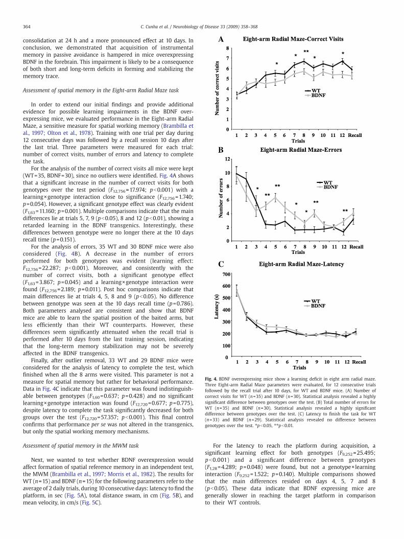
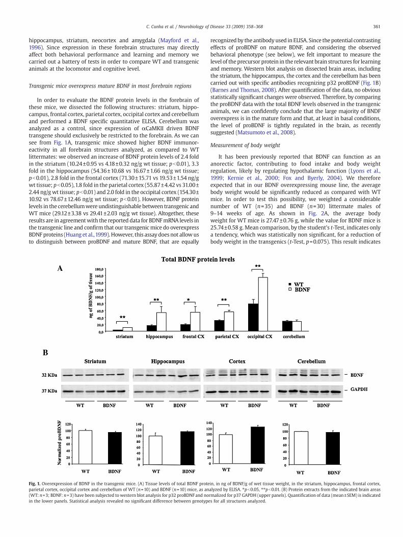
Fig. 4. BDNF overexpressing mice show a learning deficit in eight arm radial maze.Three Eight-arm Radial Maze parameters were evaluated, for 12 consecutive trialsfollowed by the recall trial after 10 days, for WT and BDNF mice. (A) Number ofcorrect visits for WT (n=35) and BDNF (n=30). Statistical analysis revealed a highlysignificant difference between genotypes over the test. (B) Total number of errors forWT (n=35) and BDNF (n=30). Statistical analysis revealed a highly significantdifference between genotypes over the test. (C) Latency to finish the task for WT(n=33) and BDNF (n=29). Statistical analysis revealed no difference betweengenotypes over the test. ⁎pb0.05, ⁎⁎pb0.01.
364 C. Cunha et al. / Neurobiology of Disease 33 (2009) 358–368
consolidation at 24 h and a more pronounced effect at 10 days. Inconclusion, we demonstrated that acquisition of instrumentalmemory in passive avoidance is hampered in mice overexpressingBDNF in the forebrain. This impairment is likely to be a consequenceof both short and long-term deficits in forming and stabilizing thememory trace.
Assessment of spatial memory in the Eight-arm Radial Maze task
In order to extend our initial findings and provide additionalevidence for possible learning impairments in the BDNF over-expressing mice, we evaluated performance in the Eight-arm RadialMaze, a sensitive measure for spatial working memory (Brambilla etal., 1997; Olton et al., 1978). Training with one trial per day during12 consecutive days was followed by a recall session 10 days afterthe last trial. Three parameters were measured for each trial:number of correct visits, number of errors and latency to completethe task.
For the analysis of the number of correct visits all mice were kept(WT=35, BDNF=30), since no outliers were identified. Fig. 4A showsthat a significant increase in the number of correct visits for bothgenotypes over the test period (F12,756=17.974; pb0.001) with alearning×genotype interaction close to significance (F12,756=1.740;p=0.054). However, a significant genotype effect was clearly evident(F1,63=11.160; p=0.001). Multiple comparisons indicate that the maindifferences lie at trials 5, 7, 9 (pb0.05), 8 and 12 (pb0.01), showing aretarded learning in the BDNF transgenics. Interestingly, thesedifferences between genotype were no longer there at the 10 daysrecall time (p=0.151).
For the analysis of errors, 35 WT and 30 BDNF mice were alsoconsidered (Fig. 4B). A decrease in the number of errorsperformed for both genotypes was evident (learning effect:F12,756=22.287; pb0.001). Moreover, and consistently with thenumber of correct visits, both a significant genotype effect(F1,63=3.867; p=0.045) and a learning×genotype interaction werefound (F12,756=2.189; p=0.011). Post hoc comparisons indicate thatmain differences lie at trials 4, 5, 8 and 9 (pb0.05). No differencebetween genotype was seen at the 10 days recall time (p=0.786).Both parameters analysed are consistent and show that BDNFmice are able to learn the spatial position of the baited arms, butless efficiently than their WT counterparts. However, thesedifferences seem significantly attenuated when the recall trial isperformed after 10 days from the last training session, indicatingthat the long-term memory stabilization may not be severelyaffected in the BDNF transgenics.
Finally, after outlier removal, 33 WT and 29 BDNF mice wereconsidered for the analysis of latency to complete the test, whichfinished when all the 8 arms were visited. This parameter is not ameasure for spatial memory but rather for behavioral performance.Data in Fig. 4C indicate that this parameter was found indistinguish-able between genotypes (F1,60=0.637; p=0.428) and no significantlearning×genotype interaction was found (F12,720=0.677; p=0.775),despite latency to complete the task significantly decreased for bothgroups over the test (F12,720=57.357; pb0.001). This final controlconfirms that performance per se was not altered in the transgenics,but only the spatial working memory mechanisms.
Assessment of spatial memory in the MWM task
Next, we wanted to test whether BDNF overexpression wouldaffect formation of spatial reference memory in an independent test,the MWM (Brambilla et al., 1997; Morris et al., 1982). The results forWT (n=15) and BDNF (n=15) for the following parameters refer to theaverage of 2 daily trials, during 10 consecutive days: latency to find theplatform, in sec (Fig. 5A), total distance swam, in cm (Fig. 5B), andmean velocity, in cm/s (Fig. 5C).
For the latency to reach the platform during acquisition, asignificant learning effect for both genotypes (F9,252=25.495;pb0.001) and a significant difference between genotypes(F1,28=4.289; p=0.048) were found, but not a genotype×learninginteraction (F9,252=1.522; p=0.140). Multiple comparisons showedthat the main differences resided on days 4, 5, 7 and 8(pb0.05). These data indicate that BDNF expressing mice aregenerally slower in reaching the target platform in comparisonto their WT controls.

Fig. 5. Learning retardation of BDNF overexpressing mice in the Morris Water Maze. MWM parameters were measured over the 10 daily sessions, for WT (n=15) and BDNF (n=15)mice. (A) Latency, in sec, to reach the platform. Statistical analysis revealed an overall significant difference between genotypes (pb0.05). (B) Distance swam, in cm. Statistical analysisrevealed an overall significant difference between genotypes (pb0.05). (C) Statistical analysis revealed no difference in the mean swim velocity between genotypes. ⁎pb0.05. D–F)MWM parameters were measured for the probe trials at day 5 and day 10, for WT (n=15) and BDNF (n=15) mice. (D) Total time, in sec, spent in the quadrants Northeast (NE),Northwest (NW), Southwest (SW) and in the target quadrant Southeast (SE), for the probe trial at day 5. Statistical analysis revealed no difference between genotypes. (E) Total time,in sec, spent in the quadrants Northeast (NE), Northwest (NW), Southwest (SW) and in the target quadrant Southeast (SE), for the probe trial at day 10. Statistical analysis revealed nodifference between genotypes. (F) Total time spent over the platform location, in sec. Statistical analysis revealed a significant difference between genotypes during the probe trial atday 5 (pb0.05). ⁎pb0.05.
365C. Cunha et al. / Neurobiology of Disease 33 (2009) 358–368
For the distance swam to the platform during acquisition, asignificant learning effect for both genotypes (F9,252 =23.007;p b0.001) and a significant difference between genotypes(F1,28=5.735; p=0.024) were detected, but not a learning×genotypeinteraction (F9,252=1.435; p=0.173). Multiple comparisons showedrevealed that the main differences were on days 5, 7 and 8 (pb0.05).Similarly to the previous parameter, these data indicate that BDNFmice had to swim a longer distance in order to reach the platform. Inconclusion, both parameters demonstrate that BDNF overexpressingmice are capable of improving the ability to reach the platform
position over the 10 daily sessions, since the both values for latencyand distance significantly decreased through the test, but not asefficiently as WT mice. BDNF mice showed a significantly higherlatency and higher distance swam to find the platform than WT mice,especially at sessions 7 and 8.
We also measured the mean swim velocity throughout the test, inorder to exclude differences in navigation speed, which could accountfor the differences observed in the previous parameters. Importantly,we observed a significant decrease in the mean swimvelocity throughthe test (F9,252=2.899; p=0.003), but neither difference between
366 C. Cunha et al. / Neurobiology of Disease 33 (2009) 358–368
genotypes (F1,28=0.240; p=0.628) nor a genotype×time interaction(F9,252=1.843; p=0.061).
Latency and distance to reach the platform are only crudemeasures of performance in the water maze and by no means anaccurate indicator for spatial memory deficits. Therefore, at day 5 andday 10, we performed a single probe trial in which the platform wasremoved, 30 min before the two daily trials of hidden platform. Totalduration, in sec, spent in the quadrants Northeast (NE), Northwest(NW), Southwest (SW) and in the target quadrant Southeast (SE), forWT (n=15) and BDNF (n=15) mice, is shown in Fig. 5D, for the probetrial at day 5, and in Fig. 5E, for the probe trial at day 10. In order tosimplify the statistical analysis of the data, we initially performed aone-way ANOVA with a Scheffe post hoc for multiple comparisons,considering the values for all four quadrants as independent(4 quadrants×2 genotypes×2 probe trials). We determined that the3 non-target quadrants were statistically indistinguishable for eachprobe trial and therefore they were pooled for subsequent analysis(data not shown). We then performed a three-way ANOVA, consider-ing 2 genotypes×2 quadrants×2 days (days 5 and 10), as a measure ofthe general tendency of the probe trials.
Learning effect was seen (F1,28=26.396, pb .0001) as well as thequadrant effect (F1,28=98.526, pb .0001) and the learning×quadrantinteraction (F1,28=13.453, pb .001). However, when the genotype wasconsidered, we found a non significant effect (F1,28=4.049; p=0.054)with only a tendency for BDNF mice to spend less time in the targetquadrant thanWTmice. This trendwas likely due to a small differenceduring the first probe trial at day 5. Indeed, comparing data at day 5only we found that both genotypes showed a significant differencebetween the time spent in the target quadrant versus non targetquadrants (F1,28=15.137; p=0.001) and a close to significant differencebetween genotypes (F1,28=3.359; p=0.078) (Fig. 5D). At the probe trialat day 10 instead, while both genotypes showed significant differencesbetween the time spent in the target/non target quadrants(F1,28 = 94.325; p b0.001), no difference between genotypes(F1,28=1.089; p=0.306) were found (Fig. 5E).
The previous analysis only showed a non significant trend for aretarded acquisition of the BDNF transgenics. In order to furtherevaluate the possibility that BDNF expressing mice might show subtlechanges in their ability to locate the platform, we analyzed the totaltime spent on the platform zone, for both genotypes and for bothprobe trials (Fig. 5F). This parameter is a more sensitive measure ofspatial memory, which detects mild impairments in the ability of miceto precisely locate the original platform location. A significant learningeffect from probe trial at day 5 to probe trial at day 10 was observed(F1,28=5.429; p=0.027), indicating that by day 10 both animal groupsincreased their precision in locating the platform position incomparison to day 5. Importantly, a significant genotype effect wasobserved (F1,28=7.159; p=0.012) and a post hoc comparison revealedthat the significant difference between genotypes reside on the probetrial at day 5 (p=0.014) but not at day 10 (p=0.427).
Altogether, these results show that BDNF transgenics are able tolearn the task but manifest mild spatial memory impairments incomparison to WT littermates. BDNF mice seem to have a significantretarded acquisition in the MWM test, even if in the probe trialsperformed this impairment was found minimal. Nevertheless, theresults presented here are consistent with those obtained in the Eight-arm Radial Maze test, by showing some forms of memory impairment.Importantly, since the Eight-arm Radial Maze results suggested adeficit in working memory, it is possible that the retarded spatiallearning in the MWM may be also linked to such alterations, ratherthan a true deficit in long-term spatial memory.
Discussion
In this study we demonstrated that a moderate overexpression ofBDNF in the forebrain results in modest but clear learning impair-
ments in both instrumental and spatial memory tasks. These resultsare somewhat surprising considering the well established role ofmature BDNF in promoting synaptic plasticity andmemory formation.However, these data are in agreement with early observations inwhich BDNF was mildly overexpressed (roughly 30%) in most bodytissues under the human β-actin promoter (Croll et al., 1999). In thattransgenic line, memory retention in passive avoidance was foundimpaired in younger animals (6–8 week old) but not in older ones (6–8 month old), suggesting only transitory deficits. Importantly, asignificant cognitive impairment was seen only in mutants carryingtwo copies of the transgene. Other non cognitive behavioralparameters were found to be normal. Although in our study we onlyused 2–3 month old mice, our comprehensive cognitive analysisclearly support the idea that a significant overexpression restricted tothe forebrain may be sufficient to cause similarly mild deficits inlearning. Importantly, our detailed analysis in passive avoidanceindicates a deficit in the acquisition of short-term memory, ratherthan a true deficit in memory consolidation. To support their findings,Croll et al., (1999) were able to measure several electrophysiologicalparameters in the dorsal hippocampus, the dendate gyrus and in theentorhinal cortex, three brain structures implicated in both spatialmemory and learning in the passive avoidance. Altogether, thoseresults suggest a general membrane hyperexcitability in comparisonto controls but no clear changes in either short-term or long-termplasticity, at least at the Schaffer collateral synapse. At present, wecannot provide direct evidence that synaptic transmission or plasticityis altered in our transgenic mice but certainly our behavioral data areconsistent with subtle changes at this level.
What may be the cellular basis for such detrimental effect ofBDNF overexpression on learning? In a recent publication it has beenshown that the same BDNF line we used manifests anxiety-likebehavior with a concomitant increase in spine density in thebasolateral amygdala. In contrast, upon chronic stress, such over-expression appears to prevent atrophy in the hippocampus a cellularphenotype likely linked to the antidepressant effect observed in theforced swim test (Govindarajan et al., 2006). Considering theserecent findings, the explanation of the observed learning deficits islikely to be a complex one, possibly involving also motivationalaspects of behavior. At the molecular level, one interesting possibilityis that in our transgenic animals the ratio between pro and matureBDNF may be in favor of the former, thus leading to an overallhyperactivation of the low affinity p75 receptors and a relativelylower engagement of the high affinity TrkB receptors. This possibilityis particularly exciting since an abnormal activation of p75 byproBDNF could lead to synaptic alterations that may be detrimentalto learning. Indeed, as recently shown, p75 receptor is involved inlong-term depression (LTD) in the hippocampus and proBDNF is ableto modulate LTD via p75 activation (Lu et al., 2005; Woo et al., 2005;Zagrebelsky et al., 2005; Rosch et al., 2005). Unfortunately, thispossibility seems to be quite unlikely based on our results. In fact, wefound, in most brain structures analyzed, very significant increases intotal BDNF levels (e.g. approximately 300% in the hippocampus)without significant changes in the proBDNF protein (only 15%increase in the same structure). Therefore, the most conservativeconclusion is that the large majority of BDNF produced by thetransgene is in the mature form.
The hypothesis we tend to favor is instead that an excess of matureBDNF may preferentially act on inhibitory interneurons, thus leadingto a general alteration of the inhibitory tone of the synaptic circuitryunderlying learning. Indeed, TrkB receptors have been found in anumber of interneurons in the forebrain and a number of functionshave been ascribed to BNDF in non pyramidal cells, including braindiseases such as schizophrenia and epilepsy. The increased excitabilityobserved by Croll et al (Croll et al., 1999) may suggest that a chronicBDNF expression could lead to a reduced inhibition by attenuatingIPSCs, as observed in a number of interneuron types upon acute BDNF

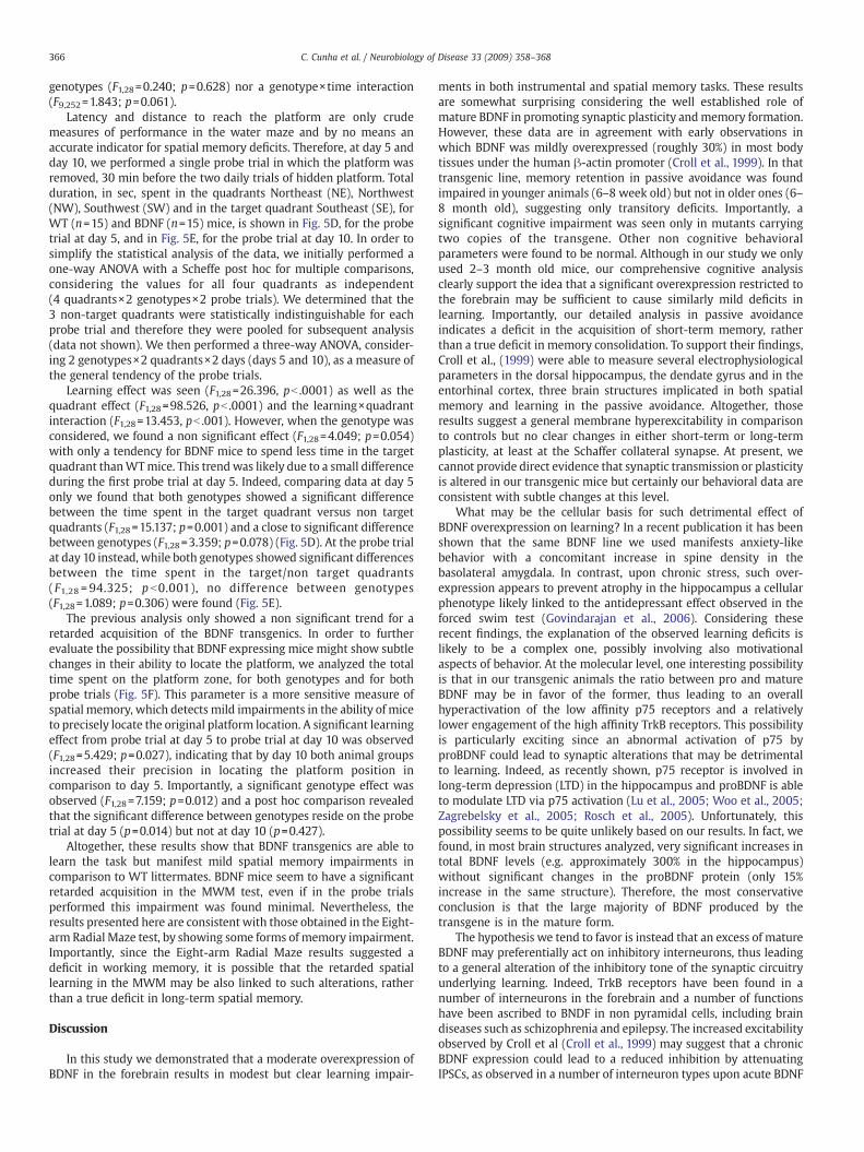
Fig. 6. Analysis of phospho-ERK induction in BDNF overexpressing mice. (A) WT (n=5) and BDNF (n=5) mice have been perfused and serial brain slices have been subjected to IHCanalysis with antibodies against the phosphorylated, activated form of ERK1/2 (P-ERK). Total number/mm2 of P-ERK positive puncta have been counted in the indicated structures(DG: dentate gyrus; CA3, CA2, CA1: dorsal hippocampus; Cig Cx: cingulated cortex; Pf Cx: prefrontal cortex). (B) Quantification of results (mean±SEM) is indicated. No significantdifference between genotypes was found.
367C. Cunha et al. / Neurobiology of Disease 33 (2009) 358–368
administration (for review see Woo and Lu, (2006)). In addition, as anon mutually exclusive possibility, an abnormal increase at specificexcitatory synapses that in turn inhibits the main hippocampalcircuitry cannot be in principle excluded. We have unsuccessfullyattempted to investigate this issue by monitoring TrkB dependentsignaling in the brain of the BDNF transgenics. In order to do that wemeasured, by immunohistochemical (IHC) techniques, the phosphory-lated levels of the fully activated forms of the Extracellular-signalRegulated Kinases (ERK1/2), as a main signaling pathway readoutdownstream to neurotrophin receptors. However, by quantitativelycomparing p-ERK levels in the wild-type and transgenic mice indifferent brain areas, we only observed minor and non significantchanges in p-ERK levels in the transgenics (Fig. 6) This result per sedoes not exclude that some changes occur in the cell signalingproperties in key brain areas for cognitive processing but actuallysuggests that the changesmay occur in specific subsets of cells, such asa fraction of interneurons part of the hippocampal circuitry, whichcannot be detected with IHC methods but rather require single cellelectrophysiological analysis, an approach which goes beyond thepurposes of the present work.
Altogether, with this study we believe to have demonstrated anunexpected complexity in the BDNF action in the CNS and wepropose that transgenic mice overexpressing this neurotrophin maybe useful animal models to study cognitive decline associated toeither neurodegenerative or psychiatric diseases. Indeed, the bestuse of our transgenics will be in combination with either genetic orpharmacological model of a disease for which BDNF administrationhas been proposed to be of therapeutic value, in order to studylong-term, chronic effects of this neurotrophin on the disease onsetand progression.
Acknowledgments
Authors would like to thank Susumu Tonegawa for reading themanuscript and having supplied the BDNF mouse strain and KerrieThomas for providing BDNF antibodies and for helpful suggestions.This research was supported by a grant (SFRH/BD/9625/2002) fromFundação para a Ciência e a Tecnologia, Portugal, through the
POCTI 2010 from the European Science Foundation (to CC), by theMariani Foundation for Neurological Diseases and by the ItalianMinistry of Research (to RB), and by the Medical Research CouncilUK (to MDD and SBD).
References
Aicardi, G., et al., 2004. Induction of long-term potentiation and depression is reflectedby corresponding changes in secretion of endogenous brain-derived neurotrophicfactor. Proc. Natl. Acad. Sci. U. S. A. 101, 15788–15792.
Alonso, M., et al., 2002. BDNF-triggered events in the rat hippocampus are required forboth short- and long-term memory formation. Hippocampus 12, 551–560.
Alonso, M., et al., 2005. Endogenous BDNF is required for long-term memory formationin the rat parietal cortex. Learn. Mem. 12, 504–510.
Balkowiec, A., Katz, D.M., 2002. Cellular mechanisms regulating activity-dependentrelease of native brain-derived neurotrophic factor from hippocampal neurons.J. Neurosci. 22, 10399–10407.
Barnes, P., Thomas, K.L., 2008. Proteolysis of proBDNF is a key regulator in the formationof memory. PLoS ONE 3, e3248.
Bekinschtein, P., et al., 2007a. Persistence of long-term memory storage requires a lateprotein synthesis- and BDNF- dependent phase in the hippocampus. Neuron 53,261–277.
Bekinschtein, P., et al., 2007b. BDNF and memory formation and storage. Neuroscientist14 (2), 147–156.
Bekinschtein, P., et al., 2008. BDNF is essential to promote persistence of long-termmemory storage. Proc. Natl. Acad. Sci. U. S. A. 105, 2711–2716.
Biffi, A., et al., 2004. Correction of metachromatic leukodystrophy in the mouse modelby transplantation of genetically modified hematopoietic stem cells. J. Clin. Invest.113, 1118–1129.
Bonhoeffer, T., 1996. Neurotrophins and activity-dependent development of theneocortex. Curr. Opin. Neurobiol. 6, 119–126.
Brambilla, R., et al., 1997. A role for the Ras signalling pathway in synaptic transmissionand long-term memory. Nature 390, 281–286.
Bramham, C.R., Messaoudi, E., 2005. BDNF function in adult synaptic plasticity: thesynaptic consolidation hypothesis. Prog. Neurobiol. 76, 99–125.
Chao, M.V., et al., 2006. Neurotrophin signalling in health and disease. Clin. Sci. (Lond.)110, 167–173.
Cirulli, F., et al., 2004. Intrahippocampal administration of BDNF in adult rats affectsshort-term behavioral plasticity in the Morris water maze and performance in theelevated plus-maze. Hippocampus 14, 802–807.
Croll, S.D., et al., 1999. Brain-derived neurotrophic factor transgenic mice exhibit passiveavoidance deficits, increased seizure severity and in vitro hyperexcitability in thehippocampus and entorhinal cortex. Neuroscience 93, 1491–1506.
Egan, M.F., et al., 2003. The BDNF val66met polymorphism affects activity-dependentsecretion of BDNF and human memory and hippocampal function. Cell 112, 257–269.
Farhadi, H.F., et al., 2000. Neurotrophin-3 sorts to the constitutive secretory pathway ofhippocampal neurons and is diverted to the regulated secretory pathway bycoexpression with brain-derived neurotrophic factor. J. Neurosci. 20, 4059–4068.

368 C. Cunha et al. / Neurobiology of Disease 33 (2009) 358–368
Figurov, A., et al., 1996. Regulation of synaptic responses to high-frequency stimulationand LTP by neurotrophins in the hippocampus. Nature 381, 706–709.
Fox, E.A., Byerly, M.S., 2004. A mechanism underlying mature-onset obesity: evidencefrom the hyperphagic phenotype of brain-derived neurotrophic factor mutants.Am. J. Physiol. Regul. Integr. Comp. Physiol. 286, R994–1004.
Gartner, A., Staiger, V., 2002. Neurotrophin secretion from hippocampal neurons evokedby long-term-potentiation-inducing electrical stimulation patterns. Proc. Natl.Acad. Sci. U. S. A. 99, 6386–6391.
Gottschalk, W., et al., 1998. Presynaptic modulation of synaptic transmission andplasticity by brain-derived neurotrophic factor in the developing hippocampus.J. Neurosci. 18, 6830–6839.
Govindarajan, A., et al., 2006. Transgenic brain-derived neurotrophic factor expressioncauses both anxiogenic and antidepressant effects. Proc. Natl. Acad. Sci. U. S. A. 103,13208–13213.
Hall, J., et al., 2000. Rapid and selective induction of BDNF expression in thehippocampus during contextual learning. Nat. Neurosci. 3, 533–535.
Hartmann, M., et al., 2001. Synaptic secretion of BDNF after high-frequency stimulationof glutamatergic synapses. Embo. J. 20, 5887–5897.
Huang, E.J., Reichardt, L.F., 2001. Neurotrophins: roles in neuronal development andfunction. Annu. Rev. Neurosci. 24, 677–736.
Huang, E.J., Reichardt, L.F., 2003. Trk receptors: roles in neuronal signal transduction.Annu. Rev. Biochem. 72, 609–642.
Huang, Z.J., et al., 1999. BDNF regulates the maturation of inhibition and the criticalperiod of plasticity in mouse visual cortex. Cell 98, 739–755.
Kang, H., Schuman, E.M., 1995. Long-lasting neurotrophin-induced enhancement ofsynaptic transmission in the adult hippocampus. Science 267, 1658–1662.
Kang, H., Schuman, E.M., 1996. A requirement for local protein synthesis inneurotrophin-induced hippocampal synaptic plasticity. Science 273, 1402–1406.
Kernie, S.G., et al., 2000. BDNF regulates eating behavior and locomotor activity in mice.Embo. J. 19, 1290–1300.
Kesslak, J.P., et al., 1998. Learning upregulates brain-derived neurotrophic factormessenger ribonucleic acid: a mechanism to facilitate encoding and circuitmaintenance? Behav. Neurosci. 112, 1012–1019.
Korte, M., et al., 1995. Hippocampal long-term potentiation is impaired in mice lackingbrain-derived neurotrophic factor. Proc. Natl. Acad. Sci. U.S.A 92, 8856–8860.
Korte, M., et al., 1996. Virus-mediated gene transfer into hippocampal CA1 regionrestores long-term potentiation in brain-derived neurotrophic factor mutant mice.Proc. Natl. Acad. Sci. U. S. A. 93, 12547–12552.
Lee, R., et al., 2001. Regulation of cell survival by secreted proneurotrophins. Science294, 1945–1948.
Lee, J.L., et al., 2004. Independent cellular processes for hippocampal memoryconsolidation and reconsolidation. Science 304, 839–843.
Lever, I.J., et al., 2001. Brain-derived neurotrophic factor is released in the dorsal horn bydistinctive patterns of afferent fiber stimulation. J. Neurosci. 21, 4469–4477.
Lewin, G.R., Barde, Y.A., 1996. Physiology of the neurotrophins. Annu. Rev. Neurosci. 19,289–317.
Linnarsson, S., et al., 1997. Learning deficit in BDNF mutant mice. Eur. J. Neurosci. 9,2581–2587.
Lu, B., 2003. Pro-region of neurotrophins: role in synaptic modulation. Neuron 39,735–738.
Lu, B., et al., 2005. The yin and yang of neurotrophin action. Nat. Rev. Neurosci. 6,603–614.
Lu, Y., et al., 2007. BDNF: a key regulator for protein synthesis-dependent LTP and long-term memory? Neurobiol. Learn. Mem.
Lyons, W.E., et al., 1999. Brain-derived neurotrophic factor-deficient mice developaggressiveness and hyperphagia in conjunction with brain serotonergic abnorm-alities. Proc. Natl. Acad. Sci. U. S. A. 96, 15239–15244.
Ma, Y.L., et al., 1998. Brain-derived neurotrophic factor antisense oligonucleotideimpairs memory retention and inhibits long-term potentiation in rats. Neu-roscience 82, 957–967.
Matsumoto, T., et al., 2008. Biosynthesis and processing of endogenous BDNF: CNSneurons store and secrete BDNF, not pro-BDNF. Nat. Neurosci. 11, 131–133.
Mayford, M., et al., 1996. Control of memory formation through regulated expression ofa CaMKII transgene. Science 274, 1678–1683.
Mazzucchelli, C., et al., 2002. Knockout of ERK1MAP kinase enhances synaptic plasticityin the striatum and facilitates striatal-mediated learning and memory. Neuron 34,807–820.
Messaoudi, E., et al., 2002. Brain-derived neurotrophic factor triggers transcription-dependent, late phase long-term potentiation in vivo. J. Neurosci. 22, 7453–7461.
Messaoudi, E., et al., 2007. Sustained Arc/Arg3.1 synthesis controls long-termpotentiation consolidation through regulation of local actin polymerization in thedentate gyrus in vivo. J. Neurosci. 27, 10445–10455.
Minichiello, L., et al., 1999. Essential role for TrkB receptors in hippocampus-mediatedlearning. Neuron 24, 401–414.
Minichiello, L., et al., 2002. Mechanism of TrkB-mediated hippocampal long-termpotentiation. Neuron 36, 121–137.
Mizuno, M., et al., 2000. Involvement of brain-derived neurotrophic factor in spatialmemory formation and maintenance in a radial arm maze test in rats. J. Neurosci.20, 7116–7121.
Monteggia, L.M., et al., 2004. Essential role of brain-derived neurotrophic factor in adulthippocampal function. Proc. Natl. Acad. Sci. U. S. A. 101, 10827–10832.
Morris, R.G., et al., 1982. Place navigation impaired in rats with hippocampal lesions.Nature 297, 681–683.
Mowla, S.J., et al., 1999. Differential sorting of nerve growth factor and brain-derivedneurotrophic factor in hippocampal neurons. J. Neurosci. 19, 2069–2080.
Mowla, S.J., et al., 2001. Biosynthesis and post-translational processing of the precursorto brain-derived neurotrophic factor. J. Biol. Chem. 276, 12660–12666.
Mu, J.S., et al., 1999. Deprivation of endogenous brain-derived neurotrophic factorresults in impairment of spatial learning and memory in adult rats. Brain. Res. 835,259–265.
Olton, D.S., et al., 1978. Hippocampal connections and spatial discrimination. Brain.Research. 139, 215–308.
Pang, P.T., Lu, B., 2004. Regulation of late-phase LTP and long-term memory in normaland aging hippocampus: role of secreted proteins tPA and BDNF. Ageing. Res. Rev. 3,407–430.
Pang, P.T., et al., 2004. Cleavage of proBDNF by tPA/plasmin is essential for long-termhippocampal plasticity. Science 306, 487–491.
Patterson, S.L., et al., 1996. Recombinant BDNF rescues deficits in basal synaptictransmission and hippocampal LTP in BDNF knockout mice. Neuron 16,1137–1145.
Patterson, S.L., et al., 2001. Some forms of cAMP-mediated long-lasting potentiation areassociated with release of BDNF and nuclear translocation of phospho-MAP kinase.Neuron 32, 123–140.
Pittenger, C., et al., 2006. Impaired bidirectional synaptic plasticity and proceduralmemory formation in striatum-specific cAMP response element-binding protein-deficient mice. J. Neurosci. 26, 2808–2813.
Poo, M.M., 2001. Neurotrophins as synaptic modulators. Nat. Rev. Neurosci. 2, 24–32.Rattiner, L.M., et al., 2004a. Brain-derived neurotrophic factor and tyrosine kinase
receptor B involvement in amygdala-dependent fear conditioning. J. Neurosci. 24,4796–4806.
Rattiner, L.M., et al., 2004b. Differential regulation of brain-derived neurotrophicfactor transcripts during the consolidation of fear learning. Learn. Mem. 11,727–731.
Rattiner, L.M., et al., 2005. Brain-derived neurotrophic factor in amygdala-dependentlearning. Neuroscientist 11, 323–333.
Rex, C.S., et al., 2006. Restoration of long-term potentiation in middle-agedhippocampus after induction of brain-derived neurotrophic factor. J. Neurophysiol.96, 677–685.
Rosch, H., et al., 2005. The neurotrophin receptor p75NTR modulates long-termdepression and regulates the expression of AMPA receptor subunits in thehippocampus. Proc. Natl. Acad. Sci. U. S. A. 102, 7362–7367.
Schutz, R.A., Izquierdo, I., 1979. Effect of brain lesions on rat shuttle behavior in fourdifferent tests. Physiol. Behavior 23, 97–105.
Soule, J., et al., 2006. Brain-derived neurotrophic factor and control of synapticconsolidation in the adult brain. Biochem. Soc. Trans. 34, 600–604.
Teng, H.K., et al., 2005. ProBDNF induces neuronal apoptosis via activation of a receptorcomplex of p75NTR and sortilin. J. Neurosci. 25, 5455–5463.
Thoenen, H., 1995. Neurotrophins and neuronal plasticity. Science 270, 593–598.Tyler, W.J., et al., 2002. From acquisition to consolidation: on the role of brain-derived
neurotrophic factor signaling in hippocampal-dependent learning. Learn. Mem. 9,224–237.
Valjent, E., et al., 2000. Involvement of the extracellular signal-regulated kinase cascadefor cocaine-rewarding properties. J. Neurosci. 20, 8701–8709.
Woo, N.H., Lu, B., 2006. Regulation of cortical interneurons by neurotrophins: fromdevelopment to cognitive disorders. Neuroscientist 12, 43–56.
Woo, N.H., et al., 2005. Activation of p75NTR by proBDNF facilitates hippocampal long-term depression. Nat. Neurosci. 8, 1069–1077.
Yamada, K., et al., 2002. Role for brain-derived neurotrophic factor in learning andmemory. Life Sci. 70, 735–744.
Yano, H., et al., 2006. BDNF-mediated neurotransmission relies upon a myosin VI motorcomplex. Nat. Neurosci. 9, 1009–1018.
Ying, S.W., et al., 2002. Brain-derived neurotrophic factor induces long-termpotentiation in intact adult hippocampus: requirement for ERK activationcoupled to CREB and upregulation of Arc synthesis. J. Neurosci. 22,1532–1540.
Zagrebelsky, M., et al., 2005. The p75 neurotrophin receptor negatively modulatesdendrite complexity and spine density in hippocampal neurons. J. Neurosci. 25,9989–9999.