brain expressed x linked 2 is pivotal for hyperactive mtor ... · cluding facial angiofibroma,...
TRANSCRIPT

mTOR up-regulation of BEX2
1
Brain expressed X-linked 2 is pivotal for hyperactive mTOR-mediated
tumorigenesis
Zhongdong Hu1, 2, Ying Wang3, Fuqiang Huang1, Rongrong Chen1, Chunjia Li1, Fang Wang1, June Goto4,
David J. Kwiatkowski5, Joanna Wdzieczak-Bakala6, Pengfei Tu2, Jianmiao Liu7, Xiaojun Zha8, 9, and
Hongbing Zhang1
1State Key Laboratory of Medical Molecular Biology, Department of Physiology, Institute of Basic
Medical Sciences & School of Basic Medicine, Graduate School of Peking Union Medical College,
Chinese Academy of Medical Sciences and Peking Union Medical College, Beijing 100005, China 2Modern Research Center for Traditional Chinese Medicine, Beijing University of Chinese Medicine,
Beijing 100029, China 3Department of Molecular Orthopaedics, Beijing Institute of Traumatology and Orthopaedics, Beijing
Jishuitan Hospital, Beijing 100035, China 4Division of Pediatric Neurosurgery, Cincinnati Children's Hospital Medical Center, Cincinnati, OH, USA
5Division of Translational Medicine, Department of Medicine, Brigham and Women’s Hospital, Harvard
Medical School, Boston, MA, USA 6Institut de Chimie des Substances Naturelles, CNRS UPR2301, 91198 Gif sur Yvette, France
7Sino-France Laboratory for Drug Screening, Key Laboratory of Molecular Biophysics of Ministry of
Education, College of Life Science and Technology, Huazhong University of Science and Technology,
Wuhan 430074, China 8Department of Biochemistry & Molecular Biology, School of Basic Medicine, Anhui Medical University,
Hefei 230032, China 9State Key Laboratory Incubation Base of Dermatology, Ministry of National Science and Technology,
Hefei 230032, China
*Running Title: mTOR up-regulation of BEX2
To whom correspondence should be addressed: Hongbing Zhang, Department of Physiology, Institute of
Basic Medical Sciences, Peking Union Medical College, 5 Dong Dan San Tiao, Beijing 100005, China.
Tel: 01186-10-65296495; Fax: 01186-10-65296491; E-mail: [email protected] or
[email protected]; Xiaojun Zha, Department of Biochemistry & Molecular Biology, School of
Basic Medicine, Anhui Medical University, 81 Meishan Road, Hefei 230032, China. E-mail:
Keywords: mTOR; STAT3; NF-κB; BEX2; VEGF; tumorigenesis
Background: mTOR signaling pathway is
frequently activated in cancer.
Results: Hyperactivation of mTOR stimulates
STAT3/NF-κB-BEX2-VEGF signaling cascade.
Conclusion: mTOR promotes tumorigenesis
through up-regulation of
STAT3/NF-κB-BEX2-VEGF signaling axis.
Significance: The components in this
mTOR-STAT3/NF-κB-BEX2-VEGF signaling
cascade are candidate targets for the treatment of
cancers associated with aberrant mTOR signaling.
ABSTRACT
Frequent alteration of upstream
proto-oncogenes and tumor suppressor genes
activates mechanistic target of rapamycin
(mTOR) and causes cancer. However, the
downstream effectors of mTOR remain largely
elusive. Here we report that brain expressed
X-linked 2 (BEX2) is a novel downstream
http://www.jbc.org/cgi/doi/10.1074/jbc.M115.665208The latest version is at JBC Papers in Press. Published on August 20, 2015 as Manuscript M115.665208
Copyright 2015 by The American Society for Biochemistry and Molecular Biology, Inc.
by guest on February 9, 2020http://w
ww
.jbc.org/D
ownloaded from

mTOR up-regulation of BEX2
2
effector of mTOR. Elevated BEX2 in Tsc2-/-
MEFs, Pten-/- MEFs, Tsc2-deficient rat uterine
leiomyoma cells, and brains of
neuronal-specific Tsc1 knockout mice were
abolished by mTOR inhibitor rapamycin.
Furthermore, BEX2 was also increased in the
liver of a hepatic-specific Pten knockout mouse
and the kidneys of Tsc2 heterozygous deletion
mice, and a patient with tuberous sclerosis
complex (TSC). mTOR up-regulation of BEX2
was mediated in parallel by both STAT3 and
NF-κB. BEX2 was involved in mTOR
up-regulation of VEGF production and
angiogenesis. Depletion of BEX2 blunted the
tumorigenesis of cells with activated mTOR.
Therefore, enhanced
STAT3/NF-κB-BEX2-VEGF signaling pathway
contributes to hyperactive mTOR-induced
tumorigenesis. BEX2 may be targeted for the
treatment of the cancers with aberrantly
activated mTOR signaling pathway.
INTRODUCTION
Largely due to mutations in either
proto-oncogenes or tumor suppressor genes, the
receptor tyrosine kinase-phosphoinositide 3-kinase
/PTEN-AKT-tuberous sclerosis complex
1/2-mechanstic target of rapamycin
(RTK-PI3K/PTEN-AKT-TSC1/2-mTOR)
signaling pathway is frequently altered in
cancer(1,2). PTEN is a tumor suppressor gene
which is mutated in multiple types of human
tumors(3). Loss of PTEN activates AKT(4). The
potentiated AKT then phosphorylates and inhibits
TSC2(5). TSC1/2 complex serves as a suppressor
of mTOR signaling. Inactivation of TSC1/2
complex leads to mTOR hyperactivation and
consequently causes tuberous sclerosis complex
(TSC), a tumor syndrome affecting multiple
organs(6). mTOR is a serine/threonine protein
kinase that promotes a subset of protein translation,
regulates metabolism and suppresses autophagy
through sensing the state of nutrition and energy,
and integrating the upstream input(2,7-10). By
associating with different proteins, mTOR
participates in the formation of two different
complexes: rapamycin-sensitive mTORC1
(composed of mTOR, Raptor, mLST8, DEPTOR,
and PRAS40) and rapamycin-resistant mTORC2
(composed of mTOR, Rictor, mLST8, DEPTOR,
and mSin1)(2,11,12).
Aberrant vascularization is a feature of TSC
lesions including facial angiofibroma, retinal
hamartomas, cardiac rhabdomyomsas, pulmonary
lymphangioleiomyomatosis (LAM), renal
angiomyolipoma (AML), and liver
hemangiomas(13). A variety of angiogenesis
activating factors have been elucidated, such as
bFGF, TGF-α, TGF-β, PDGF, and VEGF. The
VEGF serum levels in some cancer patients are
elevated(14) and VEGF is a therapeutic target for
anti-angiogenesis(15). Studies have revealed that
mTOR signaling pathway augments VEGF
expression(16-18). However, the underlying
mechanism of mTOR up-regulation of VEGF is
not completely understood.
Brain expressed X-linked 2 (BEX2), one of
the BEX family members, is abundantly expressed
in the central nervous system (19). Evidences have
shown that BEX2 is overexpressed in breast
cancer(20). BEX2 promotes the growth of breast
cancer cells and inhibits mitochondrial
apoptosis(21). Reduction of BEX2 expression
inhibits angiogenesis in vivo(22) and glioma cell
migration and invasion(23). Conversely, BEX2
has been found as a tumor suppressor in human
glioma, since overexpression of BEX2 in glioma
cells results in suppression of tumor growth in
vitro and in vivo(24). Hence, the precise function
as well as the regulatory mechanisms of BEX2 in
the development of malignant diseases are still not
clear.
In this study, we show that mTOR stimulated
BEX2 expression. Hyperactive mTOR promoted
carcinogenesis through up-regulation of
STAT3/NF-κB-BEX2-VEGF signaling cascade.
This study not only unveils an effector in the
mTOR signaling pathway, but also reveals a novel
mechanism for augmented VEGF expression
induced by hyperactive mTOR signaling. In
addition, our work provides candidate targets for
treatment of cancers associated with aberrant
mTOR signaling.
EXPERIMENTAL PROCEDURES
Cell culture, reagents, and Plasmids
by guest on February 9, 2020http://w
ww
.jbc.org/D
ownloaded from

mTOR up-regulation of BEX2
3
All mouse embryonic fibroblasts (MEFs)
were described previously(25). Retroviral
packaging PT67 cells were from Clontech
(Mountain View, CA, USA). SK-HEP-1 cells were
from Shanghai Institutes for Biological Sciences.
A549, HepG2 cells were from American Type
Culture Collection (Manassas, VA, USA).
Bel-7402 cells were from the Cell Institute of
Chinese Academy of Sciences. Tsc2-null uterine
leiomyoma ELT-3 cells have been reported
previously(26,27). All cells were cultured in
DMEM with 10 % FBS and 1%
penicillin/streptomysin in 5% CO2 at 37°C. BEX2 antibody was from Abcam (Cambridge, MA,
USA). Anti-phospho-S6 (Ser235/236) and anti-S6
antibodies have been described previously(28),
TSC2, β-actin, all HRP-labeled secondary
antibodies were from Santa Cruz Biotechnology
(Santa Cruz, CA, USA). STAT3, p-STAT3
(Tyr705), p65, p-p65 (Ser536), mTOR, Raptor,
Rictor, and PTEN antibodies were from Cell
Signaling Technology (Danvers, MA, USA).
VEGF antibody was from Millipore (Billerica,
MA, USA). Rapamycin, JSI-124, CAPE,
puromycin, and hygromycin B were acquired from
Sigma-Aldrich (St Louis, MO, USA).
Lipofectamine 2000 was from Invitrogen
(Carlsbad, CA, USA). FBS and DMEM were from
HyClone (Logan, UT, USA). pBabe-puro and
pBabe-STAT3C (control vector and the vector
expressing STAT3C, constitutively activated
STAT3, respectively) have been reported
previously(29). pLXIN-hygro and pLXIN-hTSC2
have been reported previously(30).
Quantitative real time PCR
Total RNA was extracted from cells or tissues
using Trizol reagent (Invitrogen). One microgram
RNA was reversely transcribed using the
PrimeScript RT Reagent Kit (TaKaRa, Shiga,
Japan). After 10-fold dilution, 4 μl of cDNA was
used as the template in a quantitative realtime PCR
reaction. Amplification was done for 40 cycles
using TransStart Green qPCR SuperMix
(TransGen Biotech, Beijing, China).
Oligonucleotide primers were synthesized to
detect BEX2 with β-actin as internal control. The
primer sequences are as follows:
Mouse BEX2 forward:
5’-GCGAGCGGGACAGATTGAC-3’, reverse:
5’-TCCATTTCTCCTGGGCCTATC-3’;
Mouse β-actin forward:
5’-AGAGGGAAATCGTGCGTGAC-3’ reverse:
5’-CAATAGTGATGACCTGGCCGT-3’.
Expression profiling analysis
RNA from WT and Tsc2−/− MEFs was
subjected to microarray on Affymetrix GeneChips
System with Affymetrix Mouse Genome 430 2.0
Array, and data was analyzed using Partek Express
software(31).
Immunoblotting analysis
Cells were washed with cold PBS and were
harvested on ice in lysis buffer [2% SDS, 100mM
DTT, 10mM Tris (pH 6.8), and 10% glycerol],
boiled for 10 min. Cell lysates were resolved by
SDS-PAGE. Subsequently, proteins were
transferred onto PVDF membrane (Millipore). The
membranes were blocked in TBST containing 3%
nonfat dry milk at room temperature and incubated
with the indicated antibodies diluted with TBST
containing 3% nonfat dry milk. After washing with
TBST, the membranes were incubated with
HRP-labeled secondary antibodies and then
detected by chemiluminescence.
Chromatin immunoprecipitation assay
Chromatin immunoprecipitation was
conducted to examine DNA-protein interactions
with an anti-STAT3 antibody and a SimpleChIP®
Enzymatic Chromatin IP Kit (Cell Signaling
Technology) according to the manufacturer’s
protocol. The released DNA was purified and then
used for analysis by PCR. The primer sequences
used for PCR are as follows: the putative STAT3
binding site region (PBR) of mouse BEX2 forward:
ATGCTCTAACAACTGGACTT; reverse:
CTCGACTCAATAGATTTACTC. A nonspecific
STAT3 binding region (NBR) of mouse BEX2
forward: GGTGCTGAATCTTTGAACA; reverse:
ATCCCTTTTGCTAGCATC.
RNA interference All the siRNA oligonucleotides were
purchased from GenePharma (Shanghai, China).
Cells seeded in 6-well plates were transfected with
siRNAs (200 nM) in Lipofectamine 2000. Cell
lysates were collected for immunoblotting analysis
48 hours later. The siRNA target sequences used
are as follows:
Negative control (NC):
5’-TTCTCCGAACGTGTCACGT-3’;
by guest on February 9, 2020http://w
ww
.jbc.org/D
ownloaded from

mTOR up-regulation of BEX2
4
TSC2 (human):
5’-CAATGAGTCACAGTCCTTTGA-3’;
mTOR (mouse):
5’-GAACTCGCTGATCCAGATG-3’;
Raptor (mouse):
5’-AAGGACAACGGTCACAAGTAC-3’;
Rictor (mouse):
5’-AAGCCCTACAGCCTTCATTTA-3’;
STAT3 (mouse):
5’-CTGGATAACTTCATTAGCA-3’;
STAT3 (human):
5’-AACATCTGCCTAGATCGGCTA-3’;
p65 (mouse):
5’-GGACCTATGAGACCTTCAA-3’;
p65 (human):
5’-GATGAGATCTTCCTACTGT-3’;
BEX2 (human):
5’-GCAGGAGAGTTTTACCTAT-3’;
BEX2 overexpression in WT MEFs
The coding sequence of BEX2 was inserted
into PLXIN-hyg vector between XhoI and ClaI
sites. PLXIN-hyg-BEX2 and PLXIN-hyg vector
were transfected into retroviral packaging PT67
cells using Lipofectamine 2000. After cell
selection with hygromycin B (100 μg/mL), cell
culture supernatants containing viruses were
harvested and filtered with 0.45-μm filter for
subsequent cell infection. Stably-expressing cell
lines were generated through selection with
hygromycin B (100 μg/mL).
BEX2 knockdown in Tsc2−/− and Pten−/− MEFs Mouse BEX2 target sequence was inserted
into pGPU6/Hygro shRNA expression vector
between BamHI and BbsI sites. The target
sequence of mouse BEX2 is
5’-GGAGACTACTACGTGCCTAGA-3’. This
construct and control vector were transfected into
Tsc2−/− or Pten−/− MEFs using Lipofectamine 2000.
Stably-expressing cell lines were generated
through selection with hygromycin B (100
μg/mL).
VEGF ELISA To determine the levels of VEGF secreted
from cells, cells (30,000~40,000 cells/well) were
seeded in 12-well plates in triplicates. The next
day, culture medium was replaced with 1ml of
fresh medium, and then cells were cultured in 5%
CO2 at 37°C for 48 hours. The number of cells was
counted by using Vi-CELL (Beckman Coulter,
Brea, CA, USA). Cell culture supernatants were
collected, and the secreted VEGF levels in the
supernatants were measured with a VEGF ELISA
kit (R&D Systems, Minneapolis, MN, USA). The
levels of VEGF secretion were normalized to the
number of cells(32).
Tumor engraftment onto chick chorio-allantoic
membrane
Fertilized chicken eggs were handled as
described(33). Briefly, on embryonic day 10, 5
000 000 cells with overexpressed BEX2 or WT
cells in 20 μL of culture medium were deposited
on chick chorio-allantoic membrane (CAM).
Digital pictures were taken under a
stereomicroscope (Nikon SMZ1500) at day 3, 5, 7
of tumor development.
Induction of subcutaneous tumors in nude mice Immunodeficient nude mice (BALB/c, 4~5
weeks old) were purchased from the institute of
Laboratory Animal Science, Chinese Academy of
Medical Sciences and Peking Union Medical
College. Eight male mice were in each cohort. The
subcutaneous tumor model was established as
previously described(25,31).
Human kidney tumor assessment
The kidney angioleiomyolipoma tissue and
adjacent normal tissue from a 16-year-old TSC
patient with a frameshift mutation in the TSC2
gene (g.10059delC, p.S132SfsX50) were extracted
with lysis buffer and then subjected to
immunoblotting(32). All the procedures were
performed under the permission of the Peking
Union Medical College Hospital Ethics Board.
Mouse kidney tumor assessment
The kidney cystadenoma tissues and
paratumor tissues from 4 mice with heterozygous
deletion of Tsc2(34) (Tsc2+/-, C57BL/6, and 17~18
months old) were extracted with lysis buffer and
then subjected to immunoblotting.
Mouse brain assessment
Wild-type (WT) mice, neuronal-specific Tsc1
knockout (Tsc1f/f Syn1Cre) mice, and the
neuronal-specific Tsc1 knockout mice treated with
rapamycin were sacrificed on postnatal day 21,
and brain tissues were extracted with lysis buffer
and then subjected to immunoblotting as described
previously(35). Total RNA was extracted from the
whole brain tissues of WT and neuronal-specific
Tsc1 knockout mice sacrificed on postnatal day 13
by guest on February 9, 2020http://w
ww
.jbc.org/D
ownloaded from

mTOR up-regulation of BEX2
5
for qRT-PCR analysis.
Mouse liver assessment
Mice with hepatocyte-specific deficiency of
Pten were generated by crossing Ptenf/f mice with
AlbCre transgenic mice(36). The liver tissue from
a WT mouse (Pten+/+ AlbCre, 9 months old) and a
liver-specific Pten-deficient mouse (Ptenf/f AlbCre,
9 months old) were extracted with lysis buffer and
then subjected to immunoblotting(37). All animal
protocols were approved by the Animal Center of
the Institute of Basic Medical Sciences, Chinese
Academy of Medical Sciences and Peking Union
Medical College, and were in accordance with the
regulation of Beijing Administration Office of
Laboratory Animal on the care of experimental
animals.
Statistical analysis
Mouse tumor development and survival data
were analyzed using the Kaplan-Meier log-rank
test, and the two-tailed Student’s t-test was used to
conduct the comparison between the groups. It
was of statistical significance when P<0.05.
RESULTS
Loss of PTEN or TSC2 potentiates BEX2
expression
To identify novel targets of TSC2, we
extracted mRNA from wild type (WT) and Tsc2-/-
mouse embryonic fibroblasts (MEF) and
conducted the gene expression profiling analysis
(Supplemental Table 1). We found that the
abundance of BEX2 was higher in Tsc2-/- MEFs
than in WT MEFs (Table 1). Quantitative real time
PCR revealed that the mRNA level of BEX2 in
Tsc2-/- MEFs was significantly higher than that of
WT MEFs (Fig. 1A). Furthermore, the protein
level of BEX2 in Tsc2-/- MEFs was also increased
(Fig. 1B). To examine whether the negative
regulation of TSC2 on BEX2 exists in human cells,
we knocked down TSC2 in human lung
adenocarcinoma cell line A549. Reduction of
TSC2 resulted in an increase of BEX2 expression
in A549 cells (Fig. 1C). Moreover, the ectopically
expressed human TSC2 in rat uterine leiomyoma
cell line ELT3 (Tsc2-deficient cells) inhibited
BEX2 expression (Fig. 1D). Similarly, BEX2
protein was much higher in Pten-/- MEFs than in
WT MEFs (Fig 1E). Furthermore, the protein level
of BEX2 was increased in the mouse liver tissue
with hepatocyte-specific Pten deletion (Fig. 1F).
BEX2 was much higher in renal tumor tissues than
in adjacent normal renal tissues from Tsc2+/− mice
(Fig. 1G). Moreover, the protein level of BEX2 in
renal tumor tissues from a 16 years old female
patient with TSC was up compared with adjacent
normal renal tissues (Fig. 1H).
mTOR positively regulates BEX2 expression
Since deletion of either TSC2 or PTEN leads
to mTOR activation(7) and BEX2 overexpression,
we explored whether there is a regulatory
relationship between mTOR and BEX2. We
treated WT and Tsc2-/- MEFs with mTOR inhibitor,
rapamycin. Rapamycin inhibited BEX2 expression
in WT and Tsc2-/- MEFs (Fig. 2A and B). Similar
change was presented in WT and Pten-/- MEFs
with rapmycin treatment (Fig. 2C). To test the in
vivo relevance of this finding, we examined the
expression of BEX2 in the brain tissues dissected
from wild-type mice (WT) and the
neuronal-specific Tsc1 knockout mice (Tsc1f/f
Syn1Cre). The level of BEX2 mRNA was elevated
in the brain tissues derived from the
neuronal-specific Tsc1 knockout mice over WT
mice (Fig. 2D). Moreover, the BEX2 protein level
was also increased in the neuronal-specific Tsc1
knockout mice, and the augmented BEX2 was
suppressed by rapamycin administration (Fig. 2E).
BEX2 expression declined after treatment with
rapamycin in human liver cancer cell lines
(HepG2, Bel-7402, SK-HEP-1), lung
adenocarcinoma cell line A549 and rat uterine
leiomyoma cell line ELT3 (Tsc2-deficient cells)
(Fig. 2F). To validate mTORC1 in the regulation
of BEX2, we knocked down mTOR, Raptor and
Rictor expression in Tsc2-/- MEFs, respectively.
Cells transfected with mTOR and Raptor siRNAs
had a remarkable decrease in the expression of
phospho-S6 and BEX2 (Fig. 2G). However there
was no dramatic change in both phospho-S6 and
BEX2 expression after knockdown of Rictor,
suggesting that BEX2 expression is controlled by
mTORC1 instead of mTORC2. Taken together,
these data show that mTORC1 is a positive
regulator of BEX2 expression.
Reduction of BEX2 inhibits the tumorigenic
capacity of cells with activated mTOR
by guest on February 9, 2020http://w
ww
.jbc.org/D
ownloaded from

mTOR up-regulation of BEX2
6
To investigate the role of BEX2 in the
tumorigenesis of cells with active mTOR, we
evaluated the tumorigenicity of Tsc2-/- MEFs
expressing shBEX2 or shV in a nude mouse model.
Depletion of BEX2 significantly attenuated tumor
initiation and progression of Tsc2-/- MEFs in nude
mice (Fig. 3A). Similarly, knockdown of BEX2
significantly compromised the tumorigenesis of
Pten-/- MEFs in nude mice (Fig. 3B).
Both STAT3 and NF-κB participate in mTOR
up-regulation of BEX2 expression
STAT3 is a transcription factor.
Phosphorylation of the tyrosine residue (Tyr705) is
required for activation of STAT3(38). Activation
of STAT3 is detectable in many types of
tumors(39). Since we have reported that mTOR
up-regulates STAT3(25,40), we investigated the
regulation of BEX2 by STAT3. Overexpression of
the constitutively activated STAT3 enhanced
BEX2 expression (Fig. 4A). To verify this finding,
we treated Tsc2-/- MEFs, Pten-/- MEFs, and
SK-HEP-1 cells with STAT3 inhibitor, JSI-124.
Inhibition of STAT3 dramatically reduced BEX2
expression in all cell lines examined (Fig. 4B).
Moreover, suppression of STAT3 with siRNA
impaired BEX2 expression in Tsc2-/- MEFs, Pten-/-
MEFs, and SK-HEP-1 cells (Fig. 4C). In addition,
qRT-PCR analysis showed that STAT3C
up-regulated BEX2 mRNA expression (Fig. 4D).
Moreover, the mRNA levels of BEX2 in Tsc2-/-
MEFs or Pten-/- MEFs were significantly
decreased in the presence of JSI-124 (Fig. 4E),
indicating that STAT3 regulates BEX2 at the
transcriptional level.
To explore whether STAT3 directly
transactivates BEX2 gene transcription, we
identified a putative STAT3 binding sequence
(-569/-561; TTCCAGGAA) within the promoter
of mouse BEX2 gene through the analysis of the
5’-flanking sequence of the BEX2 gene upstream
of the transcription start site (Fig. 4F, left). ChIP
analysis revealed that the binding of STAT3 to the
putative binding site in the promoter of BEX2
gene was increased in Tsc2-/- MEFs compared with
in WT cells. Moreover, the interaction between
STAT3 and the BEX2 promoter was decreased
after rapamycin treatment (Fig. 4F, right). Taken
together, STAT3 directly promotes the
transcription of BEX2 downstream of mTOR.
NF-κB is a family of transcription factors,
including RelA/p65, c-Rel, RelB, NF-κB1/p50 and
NF-κB2/p52(41), and is constitutively activated in
multiple types of tumors(42). NF-κB has been
reported locating in the downstream of mTOR(43).
To investigate whether BEX2 expression is
controlled by NF-κB, we transfected siRNA
targeting p65 into Tsc2-/- MEFs, Pten-/- MEFs, and
SK-HEP-1 cells to reduce NF-κB activity.
Knockdown of p65 suppressed BEX2 expression
in these cells (Fig. 4G). Moreover, inhibition of
NF-κB with NF-κB inhibitor, CAPE, resulted in a
remarkable reduction of BEX2 protein level in
Tsc2-/- MEFs, Pten-/- MEFs, and A549 cells (Fig.
4H). However, the mRNA levels of BEX2 in
Tsc2-/- MEFs or Pten-/- MEFs were not
significantly different in the absence or presence
of CAPE (Fig. 4I). Hence these results
demonstrate that NF-κB positively regulates
BEX2 protein level. Our previous report suggests
that STAT3 has no regulatory cross-talk with
NF-κB(25). In this study, inhibition of NF-κB or
STAT3 in Tsc2-/- MEFs were unable to affect the
activity of each other (Fig. 4J). Therefore, STAT3
and NF-κB stimulate BEX2 expression in parallel
downstream of mTOR.
BEX2 mediates mTOR augmentation of VEGF
expression Previous studies have shown that activation
of PI3K-AKT-mTOR signaling pathway increases
VEGF expression(16,17,44). Deficiency in either
Tsc2 or Pten indeed led to up-regulation of VEGF,
and inhibition of mTOR by rapamycin suppressed
the expression of VEGF (Fig. 5A and B). To
determine whether there was a potential
relationship between BEX2 and VEGF, we
examined the abundance of VEGF in cell lysates
and cell culture supernatants in Tsc2-/- MEFs
transfected with shRNA targeting BEX2.
Suppression of BEX2 expression reduced VEGF
expression (Fig. 5C) and secretion (Fig. 5D) in
Tsc2-null MEFs. In addition, BEX2
overexpression increased the expression of VEGF
in WT MEFs (Fig. 5E). To seek the relevance of
this finding in human cancer cells, we knocked
down BEX2 expression in human hepatocellular
by guest on February 9, 2020http://w
ww
.jbc.org/D
ownloaded from

mTOR up-regulation of BEX2
7
carcinoma SK-HEP-1 cells. Reduction of BEX2
suppressed VEGF expression in SK-HEP-1 cells
(Fig. 5F). Moreover, we examined the effect of
BEX2 on angiogenesis through chick
chorioallantoic membrane assay.
BEX2-overexpressing cells formed more blood
vessels, with severe bleeding, than the control
cells on day 7 (Fig. 5G). Thus, BEX2 mediates
mTOR up-regulation of VEGF production and
angiogenesis.
mTOR regulates STAT3/NF-κB-BEX2-VEGF
signaling in vitro and in vivo
It was reported that STAT3 and NF-κB
positively modulate VEGF expression(45,46).
Likewise, inhibition of STAT3 or NF-κB with
JSI-124 or CAPE decreased expression of VEGF
in SK-HEP-1, HepG2, and A549 cells,
respectively (Fig. 6A and B). We next investigated
whether this newly identified mTOR regulation of
STAT3/NF-κB-BEX2-VEGF signaling network
presents in vitro and in vivo. Inhibition of mTOR
by rapamycin downregulated p-S6, p-STAT3,
p-p65, BEX2, and VEGF in HepG2, SK-HEP-1,
ELT3, and A549 cells (Fig.6C). Moreover, there
are concurrent increases in the protein levels of
p-S6, p-STAT3, p-p65, BEX2, and VEGF in
kidney cystadenomas of four Tsc2+/− mice,
compared with that in paratumor tissues (Fig. 6D).
Taken together, these data indicate that the
mTOR-STAT3/NF-κB-BEX2-VEGF signaling
network exists in vitro and in vivo.
DISCUSSION
RTK-PI3K/PTEN-AKT-TSC1/2-mTOR
signaling pathway is frequently deregulated in
cancer, but the underlying mechanisms remain less
clear. In this study, we have elucidated that BEX2
is a novel downstream target of mTOR, and
activation of mTOR promotes tumorigenesis
through up-regulation of
STAT3/NF-κB-BEX2-VEGF signaling cassette.
As the significance of mTOR signaling in
physiology and diseases has been increasingly
appreciated, the molecular events downstream of
mTOR are under intensive investigation. In
addition, the underlying mechanism of mTOR
up-regulation of angiogenesis is not completely
understood. Evidences have shown that BEX2 is
overexpressed in breast cancer(20), indicating that
BEX2 may play a role in cancer development.
Reduction of BEX2 expression inhibits
angiogenesis in vivo(22). Our microarray analysis
revealed that the abundance of BEX2 was
~8.5-fold higher in Tsc2-/- MEFs than in WT MEFs
(Table 1). Therefore, BEX2 was chosen for
mechanistic study of tumorigenesis caused by
aberrant activation of mTOR.
By studying Tsc2-/- MEFs, Pten-/- MEFs, rat
Tsc2 mutant uterine leiomyoma cells, human
cancer cell lines, neuronal-specific Tsc1 knockout
mice, hepatic-specific Pten knockout mice,
heterozygous Tsc2 deletion mice, and TSC patient,
we have found that negative regulation of BEX2
expression by PTEN or TSC2 is mediated by
mTORC1 (Fig. 1 and 2). Since the abundance of
BEX2 is higher in TSC associated kidney
angiomyolipoma, the enhanced expression of
BEX2 may be involved in TSC development. We
speculate that BEX2 is a candidate target for the
treatment of TSC.
There is limited characterization on the
regulatory mechanisms upstream of BEX2. Our
data show that STAT3, a downstream effector of
mTOR, is a positive regulator of BEX2, and BEX2
is thus a novel effector of STAT3. Moreover,
STAT3 up-regulates BEX2 expression by directly
binding to the promoter of BEX2 gene (Fig. 4A-F).
In addition, NF-κB positively regulates BEX2
expression. However, NF-κB does not influence
the transcription of BEX2 gene (Fig. 4G-I) and
therefore NF-κB regulation of BEX2 is likely a
post-transcriptional event. Based on our previous
study (25) and the finding in this study (Fig. 4J),
there is no functional cross-talk between STAT3
and NFκB. We propose that STAT3 and NF-κB
positively modulates BEX2 expression
downstream of mTOR in parallel.
The role of BEX2 in tumorigenesis is
controversial. BEX2 is considered as a
proto-oncogene in breast cancer(21) and a tumor
suppressor in glioma(24). Our data demonstrate
that reduction of BEX2 inhibits tumorigenesis of
Tsc2-/- or Pten-/- MEFs (Fig. 3), supporting BEX2
as an mTOR-regulated proto-oncogene. Thus
BEX2 as a promising target may be harnessed for
the treatment of cancers associated with aberrant
mTOR signaling.
by guest on February 9, 2020http://w
ww
.jbc.org/D
ownloaded from
mTOR up-regulation of BEX2
8
It is well known that hyperactive mTOR
signaling leads to tumorigenesis with augmented
angiogenesis, and VEGF has been established as a
major stimulator of angiogenesis(18,47). Here we
have demonstrated that BEX2, as a novel
downstream target of mTOR, positively controls
VEGF expression, and BEX2 overexpression in
WT MEFs promotes angiogenesis in the chick
chorioallantoic membrane (Fig. 5). STAT3 and
NFκB augment BEX2 expression induced by
mTOR (Fig. 4). Moreover, STAT3 and NFκB are
positive upstream regulators of VEGF (Fig. 6A
and B). In addition, the
mTOR-STAT3/NF-κB-BEX2-VEGF signaling
cascade was shown to exist in human cancer cell
lines in vitro and renal tumors of Tsc2+/− mice in
vivo (Fig. 6C and D). Taken together, mTOR may
promote tumorigenesis through enhanced
STAT3/NF-κB-BEX2-VEGF signaling cassette
(Fig. 6E). As a target gene of c-Jun, BEX2
reciprocally regulates c-Jun in breast cancer(48).
mTOR signaling pathway augments VEGF
expression through up-regulation of
HIF-1α(18,49). c-Jun positively regulates VEGF
expression by stabilizing HIF-1α(50). Hence, we
postulate BEX2 enhances VEGF through
up-regulation of c-Jun-HIF-1α signaling cascade.
In summary, we have illustrated that BEX2
plays an important role in tumorigenesis caused by
aberrant activation of mTOR. Hyperactivation of
mTOR stimulates STAT3/NF-κB-BEX2-VEGF
signaling cascade. The components in the newly
established mTOR-STAT3/NF-κB-BEX2-VEGF
cascade are potential targets for the treatment of
cancer with aberrant
RTK-PI3K/PTEN-AKT-TSC1/2-mTOR signaling.
Acknowledgements: We thank Xinxin Chen and Yanling Jing for technical assistance and insightful
discussion. This work was supported by the National Basic Research Program of China 973 Program
Grant (2015CB553802), the Ministry of Science and Technology of China 863 Program Grant
(2012AA02A201), and the National Natural Science Foundation of China Grants (81101524, 81130085,
81372475, and 81403147).
Conflict of interest: The authors declare that they have no conflicts of interest with the contents of this
article.
Author contributions: ZH designed and carried out most of the experiments, analyzed data, and wrote
most of the paper. YW performed experiments, analyzed data, and revised the paper. FH performed
experiments and analyzed data. RC provided human kidney tumor tissue samples. CL provided ELT-3
cells stably expressing TSC2. FW performed the gene expression profiling analysis. JG and DJK provided
brain tissues of neuronal-specific Tsc1 knockout and wt control mice. JL and JWB performed chick
chorio-allantoic membrane assay for angiogenesis. PT supervised some of the study. HZ and XZ designed
experiments, analyzed data, and wrote the paper.
by guest on February 9, 2020http://w
ww
.jbc.org/D
ownloaded from

mTOR up-regulation of BEX2
9
REFERENCES 1. Manning, B. D., and Cantley, L. C. (2007) AKT/PKB signaling: navigating downstream. Cell 129,
1261-1274
2. Guertin, D. A., and Sabatini, D. M. (2007) Defining the role of mTOR in cancer. Cancer cell 12, 9-22
3. Eng, C. (2003) PTEN: one gene, many syndromes. Hum Mutat 22, 183-198
4. Di Cristofano, A., and Pandolfi, P. P. (2000) The multiple roles of PTEN in tumor suppression. Cell 100,
387-390
5. Inoki, K., Li, Y., Zhu, T., Wu, J., and Guan, K. L. (2002) TSC2 is phosphorylated and inhibited by Akt and
suppresses mTOR signalling. Nature cell biology 4, 648-657
6. Crino, P. B., Nathanson, K. L., and Henske, E. P. (2006) The tuberous sclerosis complex. N Engl J Med 355,
1345-1356
7. Hay, N., and Sonenberg, N. (2004) Upstream and downstream of mTOR. Genes & development 18,
1926-1945
8. Albert, V., and Hall, M. N. (2014) mTOR signaling in cellular and organismal energetics. Current opinion
in cell biology 33C, 55-66
9. Wang, S., Tsun, Z. Y., Wolfson, R. L., Shen, K., Wyant, G. A., Plovanich, M. E., Yuan, E. D., Jones, T. D.,
Chantranupong, L., Comb, W., Wang, T., Bar-Peled, L., Zoncu, R., Straub, C., Kim, C., Park, J., Sabatini, B.
L., and Sabatini, D. M. (2015) Metabolism. Lysosomal amino acid transporter SLC38A9 signals arginine
sufficiency to mTORC1. Science 347, 188-194
10. Jewell, J. L., Kim, Y. C., Russell, R. C., Yu, F. X., Park, H. W., Plouffe, S. W., Tagliabracci, V. S., and Guan,
K. L. (2015) Metabolism. Differential regulation of mTORC1 by leucine and glutamine. Science 347,
194-198
11. Kim, D. H., Sarbassov, D. D., Ali, S. M., King, J. E., Latek, R. R., Erdjument-Bromage, H., Tempst, P., and
Sabatini, D. M. (2002) mTOR interacts with raptor to form a nutrient-sensitive complex that signals to the
cell growth machinery. Cell 110, 163-175
12. Alessi, D. R., Pearce, L. R., and Garcia-Martinez, J. M. (2009) New insights into mTOR signaling:
mTORC2 and beyond. Sci Signal 2, pe27
13. Kwiatkowski, D. J., Whittemore, V. H., and Thiele, E. A. (2010) Tuberous sclerosis complex: genes,
clinical features and therapeutics, John Wiley & Sons
14. Kondo, S., Asano, M., Matsuo, K., Ohmori, I., and Suzuki, H. (1994) Vascular endothelial growth
factor/vascular permeability factor is detectable in the sera of tumor-bearing mice and cancer patients.
Biochim Biophys Acta 1221, 211-214
15. Ferrara, N., and Davis-Smyth, T. (1997) The biology of vascular endothelial growth factor. Endocr Rev 18,
4-25
16. El-Hashemite, N., Walker, V., Zhang, H., and Kwiatkowski, D. J. (2003) Loss of Tsc1 or Tsc2 induces
vascular endothelial growth factor production through mammalian target of rapamycin. Cancer research 63,
5173-5177
17. Brugarolas, J. B., Vazquez, F., Reddy, A., Sellers, W. R., and Kaelin, W. G., Jr. (2003) TSC2 regulates
VEGF through mTOR-dependent and -independent pathways. Cancer cell 4, 147-158
18. Karar, J., and Maity, A. (2011) PI3K/AKT/mTOR Pathway in Angiogenesis. Frontiers in molecular
neuroscience 4, 51
19. Alvarez, E., Zhou, W., Witta, S. E., and Freed, C. R. (2005) Characterization of the Bex gene family in
humans, mice, and rats. Gene 357, 18-28
20. Naderi, A., Teschendorff, A. E., Beigel, J., Cariati, M., Ellis, I. O., Brenton, J. D., and Caldas, C. (2007)
BEX2 is overexpressed in a subset of primary breast cancers and mediates nerve growth factor/nuclear
factor-kappaB inhibition of apoptosis in breast cancer cell lines. Cancer research 67, 6725-6736
21. Naderi, A., Liu, J., and Bennett, I. C. (2010) BEX2 regulates mitochondrial apoptosis and G1 cell cycle in
breast cancer. International journal of cancer. Journal international du cancer 126, 1596-1610
22. Le Mercier, M., Fortin, S., Mathieu, V., Roland, I., Spiegl-Kreinecker, S., Haibe-Kains, B., Bontempi, G.,
Decaestecker, C., Berger, W., Lefranc, F., and Kiss, R. (2009) Galectin 1 proangiogenic and promigratory
effects in the Hs683 oligodendroglioma model are partly mediated through the control of BEX2 expression.
Neoplasia 11, 485-496
23. Zhou, X., Xu, X., Meng, Q., Hu, J., Zhi, T., Shi, Q., and Yu, R. (2013) Bex2 is critical for migration and
by guest on February 9, 2020http://w
ww
.jbc.org/D
ownloaded from

mTOR up-regulation of BEX2
10
invasion in malignant glioma cells. Journal of molecular neuroscience : MN 50, 78-87
24. Foltz, G., Ryu, G. Y., Yoon, J. G., Nelson, T., Fahey, J., Frakes, A., Lee, H., Field, L., Zander, K., Sibenaller,
Z., Ryken, T. C., Vibhakar, R., Hood, L., and Madan, A. (2006) Genome-wide analysis of epigenetic
silencing identifies BEX1 and BEX2 as candidate tumor suppressor genes in malignant glioma. Cancer
research 66, 6665-6674
25. Ma, J., Meng, Y., Kwiatkowski, D. J., Chen, X., Peng, H., Sun, Q., Zha, X., Wang, F., Wang, Y., Jing, Y.,
Zhang, S., Chen, R., Wang, L., Wu, E., Cai, G., Malinowska-Kolodziej, I., Liao, Q., Liu, Y., Zhao, Y., Sun,
Q., Xu, K., Dai, J., Han, J., Wu, L., Zhao, R. C., Shen, H., and Zhang, H. (2010) Mammalian target of
rapamycin regulates murine and human cell differentiation through STAT3/p63/Jagged/Notch cascade. The
Journal of clinical investigation 120, 103-114
26. Kobayashi, T., Hirayama, Y., Kobayashi, E., Kubo, Y., and Hino, O. (1995) A germline insertion in the
tuberous sclerosis (Tsc2) gene gives rise to the Eker rat model of dominantly inherited cancer. Nature
genetics 9, 70-74
27. Zha, X., Hu, Z., Ji, S., Jin, F., Jiang, K., Li, C., Zhao, P., Tu, Z., Chen, X., Di, L., Zhou, H., and Zhang, H.
(2015) NFkappaB up-regulation of glucose transporter 3 is essential for hyperactive mammalian target of
rapamycin-induced aerobic glycolysis and tumor growth. Cancer letters 359, 97-106
28. Zhang, H., Bajraszewski, N., Wu, E., Wang, H., Moseman, A. P., Dabora, S. L., Griffin, J. D., and
Kwiatkowski, D. J. (2007) PDGFRs are critical for PI3K/Akt activation and negatively regulated by mTOR.
The Journal of clinical investigation 117, 730-738
29. Bromberg, J. F., Horvath, C. M., Besser, D., Lathem, W. W., and Darnell, J. E., Jr. (1998) Stat3 activation is
required for cellular transformation by v-src. Molecular and cellular biology 18, 2553-2558
30. Finlay, G. A., York, B., Karas, R. H., Fanburg, B. L., Zhang, H., Kwiatkowski, D. J., and Noonan, D. J.
(2004) Estrogen-induced smooth muscle cell growth is regulated by tuberin and associated with altered
activation of platelet-derived growth factor receptor-β and ERK-1/2. Journal of Biological Chemistry 279,
23114-23122
31. Sun, Q., Chen, X., Ma, J., Peng, H., Wang, F., Zha, X., Wang, Y., Jing, Y., Yang, H., Chen, R., Chang, L.,
Zhang, Y., Goto, J., Onda, H., Chen, T., Wang, M. R., Lu, Y., You, H., Kwiatkowski, D., and Zhang, H.
(2011) Mammalian target of rapamycin up-regulation of pyruvate kinase isoenzyme type M2 is critical for
aerobic glycolysis and tumor growth. Proceedings of the National Academy of Sciences of the United States
of America 108, 4129-4134
32. Peng, H., Liu, J., Sun, Q., Chen, R., Wang, Y., Duan, J., Li, C., Li, B., Jing, Y., Chen, X., Mao, Q., Xu, K. F.,
Walker, C. L., Li, J., Wang, J., and Zhang, H. (2013) mTORC1 enhancement of STIM1-mediated
store-operated Ca2+ entry constrains tuberous sclerosis complex-related tumor development. Oncogene 32,
4702-4711
33. Hagedorn, M., Javerzat, S., Gilges, D., Meyre, A., de Lafarge, B., Eichmann, A., and Bikfalvi, A. (2005)
Accessing key steps of human tumor progression in vivo by using an avian embryo model. Proceedings of
the National Academy of Sciences of the United States of America 102, 1643-1648
34. Onda, H., Lueck, A., Marks, P. W., Warren, H. B., and Kwiatkowski, D. J. (1999) Tsc2(+/-) mice develop
tumors in multiple sites that express gelsolin and are influenced by genetic background. The Journal of
clinical investigation 104, 687-695
35. Meikle, L., Talos, D. M., Onda, H., Pollizzi, K., Rotenberg, A., Sahin, M., Jensen, F. E., and Kwiatkowski,
D. J. (2007) A mouse model of tuberous sclerosis: neuronal loss of Tsc1 causes dysplastic and ectopic
neurons, reduced myelination, seizure activity, and limited survival. J Neurosci 27, 5546-5558
36. Horie, Y., Suzuki, A., Kataoka, E., Sasaki, T., Hamada, K., Sasaki, J., Mizuno, K., Hasegawa, G., Kishimoto,
H., Iizuka, M., Naito, M., Enomoto, K., Watanabe, S., Mak, T. W., and Nakano, T. (2004)
Hepatocyte-specific Pten deficiency results in steatohepatitis and hepatocellular carcinomas. The Journal of
clinical investigation 113, 1774-1783
37. Wang, Y., Hu, Z., Liu, Z., Chen, R., Peng, H., Guo, J., Chen, X., and Zhang, H. (2013) MTOR inhibition
attenuates DNA damage and apoptosis through autophagy-mediated suppression of CREB1. Autophagy 9,
2069-2086
38. Johnston, P. A., and Grandis, J. R. (2011) STAT3 signaling: anticancer strategies and challenges. Molecular
interventions 11, 18-26
39. Bromberg, J. F., Wrzeszczynska, M. H., Devgan, G., Zhao, Y., Pestell, R. G., Albanese, C., and Darnell, J. E.,
Jr. (1999) Stat3 as an oncogene. Cell 98, 295-303
by guest on February 9, 2020http://w
ww
.jbc.org/D
ownloaded from

mTOR up-regulation of BEX2
11
40. Zha, X., Wang, F., Wang, Y., He, S., Jing, Y., Wu, X., and Zhang, H. (2011) Lactate dehydrogenase B is
critical for hyperactive mTOR-mediated tumorigenesis. Cancer research 71, 13-18
41. Ghosh, S., and Karin, M. (2002) Missing pieces in the NF-kappaB puzzle. Cell 109 Suppl, S81-96
42. Karin, M., and Lin, A. (2002) NF-kappaB at the crossroads of life and death. Nature immunology 3,
221-227
43. Dan, H. C., Cooper, M. J., Cogswell, P. C., Duncan, J. A., Ting, J. P., and Baldwin, A. S. (2008)
Akt-dependent regulation of NF-{kappa}B is controlled by mTOR and Raptor in association with IKK.
Genes & development 22, 1490-1500
44. Fang, J., Ding, M., Yang, L., Liu, L. Z., and Jiang, B. H. (2007) PI3K/PTEN/AKT signaling regulates
prostate tumor angiogenesis. Cell Signal 19, 2487-2497
45. Xu, Q., Briggs, J., Park, S., Niu, G., Kortylewski, M., Zhang, S., Gritsko, T., Turkson, J., Kay, H., Semenza,
G. L., Cheng, J. Q., Jove, R., and Yu, H. (2005) Targeting Stat3 blocks both HIF-1 and VEGF expression
induced by multiple oncogenic growth signaling pathways. Oncogene 24, 5552-5560
46. Fujioka, S., Niu, J., Schmidt, C., Sclabas, G. M., Peng, B., Uwagawa, T., Li, Z., Evans, D. B., Abbruzzese, J.
L., and Chiao, P. J. (2004) NF-kappaB and AP-1 connection: mechanism of NF-kappaB-dependent
regulation of AP-1 activity. Molecular and cellular biology 24, 7806-7819
47. Carmeliet, P., and Jain, R. K. (2000) Angiogenesis in cancer and other diseases. Nature 407, 249-257
48. Naderi, A., Liu, J., and Hughes-Davies, L. (2010) BEX2 has a functional interplay with c-Jun/JNK and
p65/RelA in breast cancer. Molecular cancer 9, 111
49. Zhong, H., Chiles, K., Feldser, D., Laughner, E., Hanrahan, C., Georgescu, M. M., Simons, J. W., and
Semenza, G. L. (2000) Modulation of hypoxia-inducible factor 1alpha expression by the epidermal growth
factor/phosphatidylinositol 3-kinase/PTEN/AKT/FRAP pathway in human prostate cancer cells:
implications for tumor angiogenesis and therapeutics. Cancer research 60, 1541-1545
50. Yu, B., Miao, Z. H., Jiang, Y., Li, M. H., Yang, N., Li, T., and Ding, J. (2009) c-Jun protects
hypoxia-inducible factor-1alpha from degradation via its oxygen-dependent degradation domain in a
nontranscriptional manner. Cancer research 69, 7704-7712
Abbreviations: mTOR, mechanistic target of rapamycin; BEX2, brain expressed X-linked 2; TSC,
tuberous sclerosis complex; RTK, the receptor tyrosine kinase; PI3K, phosphoinositide 3-kinase; TSC1/2,
tuberous sclerosis complex 1/2
by guest on February 9, 2020http://w
ww
.jbc.org/D
ownloaded from

mTOR up-regulation of BEX2
12
FIGURE LEGENDS
Figure 1. Loss of PTEN or TSC2 potentiates BEX2 expression. Total RNA and cell lysates were extracted from WT, Tsc2−/− MEFs for qRT PCR (n=3, Data represent
mean ± SEM. *P < 0.05) (A) and immunoblotting (B). C, A549 cells were transfected with control or
TSC2 siRNAs for 24 h or 48 h and then subjected to immunoblotting. D, ELT3 cells were infected with
pLXIN-hygro or pLXIN-hTSC2 retroviruses and then subjected to immunoblotting. E, Cell lysates were
extracted from WT, Pten−/− MEFs for immunoblotting. F, Age- and genetic background-matched liver
tissues dissected from WT mouse (Pten+/+ AlbCre) and the mutant mouse (Ptenf/f AlbCre) were subjected
to immunoblotting. G, Kidney cystadenomas (T) and paratumor tissues (N) from four C57BL/6 Tsc2+/-
mice were immunoblotted. H, Kidney tumor tissue (T) and the adjacent normal tissue (N) from a TSC
patient were immunoblotted.
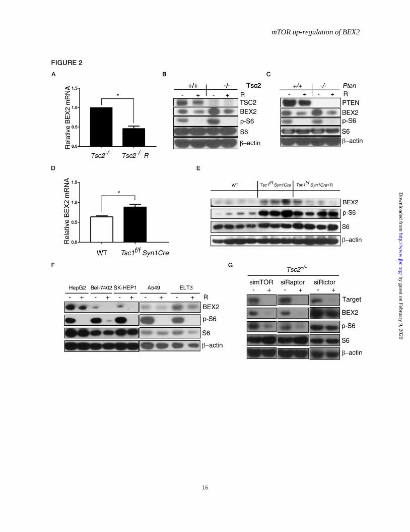
Figure 2. mTOR positively regulates BEX2 expression. A, Total RNA were extracted from Tsc2−/− MEFs treated with or without 10 nM rapamycin (R) for 24 h
for qRT PCR. n=3, Data represent mean ± SEM. *P < 0.05. B and C, Cell lysates were extracted from
WT, Tsc2−/− MEFs or Pten−/− MEFs treated with or without 10 nM rapamycin (R) for 24 h for
immunoblotting. D, Total RNA was extracted from brain tissues of wild-type mice (WT) and the
neuronal-specific Tsc1 knockout mice (Tsc1f/f Syn1Cre) for qRT PCR. n=3, Data represent mean ± SEM.
*P < 0.05. E, The brain tissues from WT mice, Tsc1f/f Syn1Cre mice and Tsc1f/f Syn1Cre mice treated with
rapamycin (R) (Tsc1f/f Syn1Cre +R) were immunoblotted. F, HepG2, Bel-7402, SK-HEP-1, A549, and
ELT3 cells were treated with or without 10 nM rapamycin (R) for 24 h and then subjected to
immunoblotting. G, Tsc2−/− MEFs were transfected with siRNA targeting mTOR, Raptor, or Rictor for
48 h and then subjected to immunoblotting.
Figure 3. Depletion of BEX2 reduces the tumorigenic capacity of cells with activated mTOR.
Tsc2−/− MEFs (A) or Pten−/− MEFs (B) were transfected with the plasmids (shBEX2 or scramble shRNA
(shV)) and then inoculated subcutaneously into nude mice (n=8). Left: immunoblotting. Middle: tumor
initiation. Right: survival of the mice. The Kaplan-Meier log-rank test, P < 0.05.
Figure 4. STAT3 and NF-κB up-regulate BEX2 downstream of mTOR.
A, WT MEFs transduced with the retroviruses for STAT3C in pBabe-puro or the control vector
pBabe-puro (V) were subjected to immunoblotting. B, Tsc2−/− MEFs, Pten−/− MEFs or SK-HEP-1 cells
were treated with or without JSI-124 (Tsc2−/− MEFs, Pten−/− MEFs: 0.5μM, SK-HEP-1: 0.1μM) for 24 h
and then subjected to immunoblotting. C, Tsc2−/− MEFs, Pten−/− MEFs or SK-HEP-1 cells were
transfected with STAT3 siRNAs for 48 h and then subjected to immunoblotting. Total RNA were
extracted from (D) WT MEFs transduced with the retroviruses for STAT3C in pBabe-puro or the control
vector pBabe-puro (V) and (E) Tsc2-/- MEFs or Pten-/- MEFs treated with or without 0.5μM JSI-124 for 24
h for qRT PCR. n=3, Data represent mean ± SEM. **P < 0.01, ***P < 0.001. F, left, Schematic
illustration of a putative STAT3 binding site within the promoter of mouse BEX2 gene. PBR, putative
STAT3 binding site region; NBR, nonspecific STAT3 binding region. F, right, WT and Tsc2−/− MEFs
treated with or without 20 nM rapamycin (R) for 24 h were subjected to ChIP assay with an anti-STAT3
antibody. Normal rabbit IgG antibody used as the negative control. Immunoprecipitated DNA was used
for PCR amplifications with primers surrounding PBR and NBR. G, Tsc2−/− MEFs, Pten−/− MEFs, or
SK-HEP-1 cells were transfected with control or p65 siRNAs for 48 h and then subjected to
immunoblotting. H, Tsc2−/− MEFs, Pten−/− MEFs, or A549 cells were treated with or without 10μM
CAPE for 24 h and then subjected to immunoblotting. I, Total RNA were extracted from Tsc2-/- MEFs or
Pten-/- MEFs treated with or without 10μM CAPE for 24 h for qRT PCR. n=3, Data represent mean ±
SEM. P > 0.05. J, Tsc2−/− MEFs were treated with or without 10μM CAPE or 0.5μM JSI-124 for 24 h and
by guest on February 9, 2020http://w
ww
.jbc.org/D
ownloaded from
mTOR up-regulation of BEX2
13
then subjected to immunoblotting.
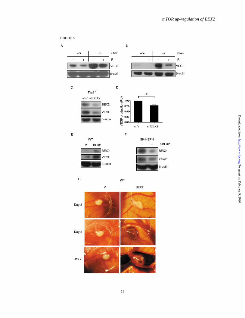
Figure 5. mTOR enhances VEGF expression through up-regulation of BEX2.
WT, Tsc2−/− MEFs (A), or Pten−/− MEFs (B) were treated with or without 10 nM rapamycin (R) for 24 h
and then subjected to immunoblotting. C, Cell lysates were extracted from Tsc2−/− MEFs stably
expressing the shRNA for BEX2 for immunoblotting, and (D) cell culture supernatants from these MEFs
were analyzed for VEGF. n=3, Data represent mean ± SEM. *P < 0.05. RU, relative unit. E, WT MEFs
transduced with the retroviruses for BEX2 in pLXIN-hyg or the control vector pLXIN-hyg (V) were
subjected to immunoblotting. F, SK-HEP-1 cells were transfected with control or BEX2 siRNAs for 48 h
and then subjected to immunoblotting. G, WT MEFs transduced with the retroviruses for BEX2
expression in pLXIN-hyg or the control vector pLXIN-hyg (V) were subjected to the chick
chorioallantoic membrane assay.
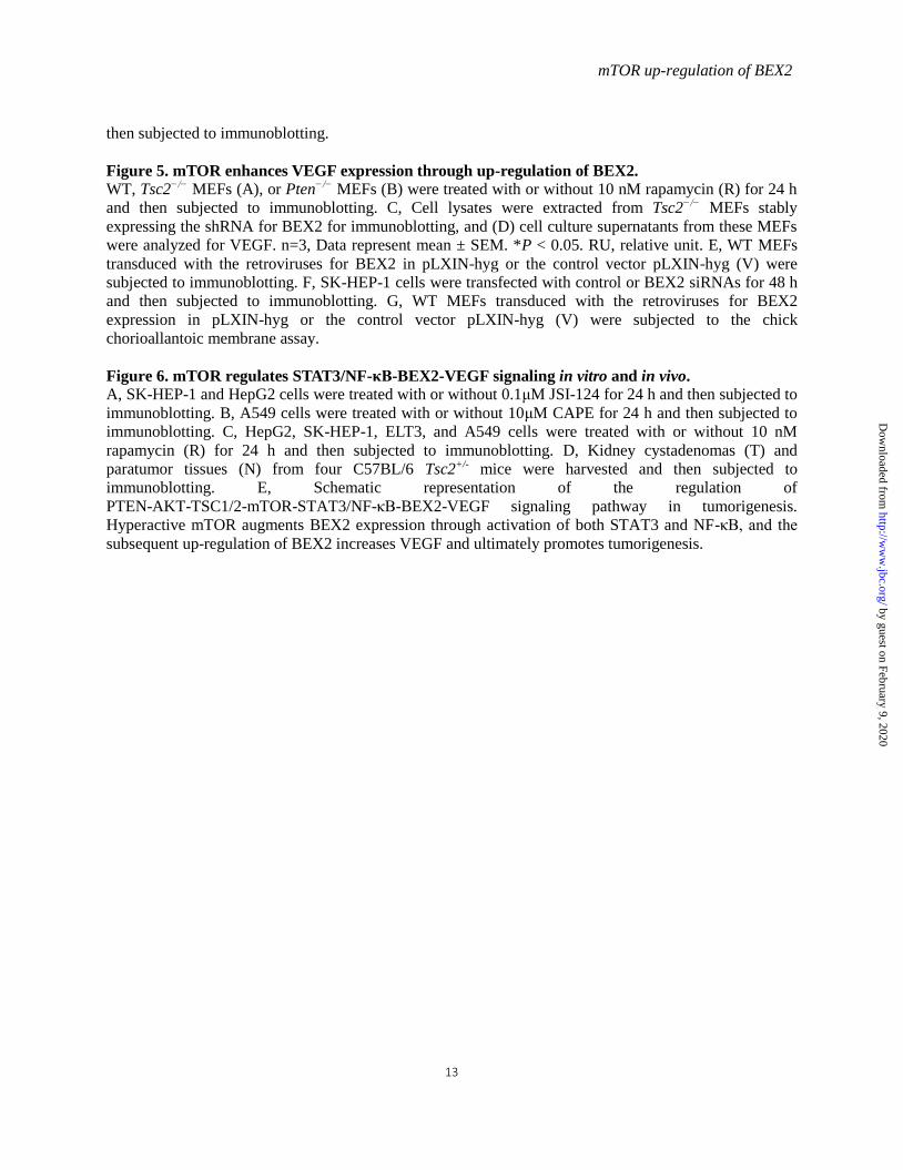
Figure 6. mTOR regulates STAT3/NF-κB-BEX2-VEGF signaling in vitro and in vivo.
A, SK-HEP-1 and HepG2 cells were treated with or without 0.1μM JSI-124 for 24 h and then subjected to
immunoblotting. B, A549 cells were treated with or without 10μM CAPE for 24 h and then subjected to
immunoblotting. C, HepG2, SK-HEP-1, ELT3, and A549 cells were treated with or without 10 nM
rapamycin (R) for 24 h and then subjected to immunoblotting. D, Kidney cystadenomas (T) and
paratumor tissues (N) from four C57BL/6 Tsc2+/- mice were harvested and then subjected to
immunoblotting. E, Schematic representation of the regulation of
PTEN-AKT-TSC1/2-mTOR-STAT3/NF-κB-BEX2-VEGF signaling pathway in tumorigenesis.
Hyperactive mTOR augments BEX2 expression through activation of both STAT3 and NF-κB, and the
subsequent up-regulation of BEX2 increases VEGF and ultimately promotes tumorigenesis.
by guest on February 9, 2020http://w
ww
.jbc.org/D
ownloaded from

mTOR up-regulation of BEX2
14
Tsc2-/-
vs. WT
Gene Fold change Description
Bex2 8.51992 Up
amRNA abundance of BEX2 in WT and Tsc2−/−
was measured using Affymetrix mouse genome
430 2.0 array.
Table 1. Increased BEX2 in Tsc2-/-
MEFsa
by guest on February 9, 2020http://w
ww
.jbc.org/D
ownloaded from

mTOR up-regulation of BEX2
15
by guest on February 9, 2020http://w
ww
.jbc.org/D
ownloaded from
mTOR up-regulation of BEX2
16
by guest on February 9, 2020http://w
ww
.jbc.org/D
ownloaded from

mTOR up-regulation of BEX2
17
by guest on February 9, 2020http://w
ww
.jbc.org/D
ownloaded from

mTOR up-regulation of BEX2
18
by guest on February 9, 2020http://w
ww
.jbc.org/D
ownloaded from
mTOR up-regulation of BEX2
19
by guest on February 9, 2020http://w
ww
.jbc.org/D
ownloaded from

mTOR up-regulation of BEX2
20
by guest on February 9, 2020http://w
ww
.jbc.org/D
ownloaded from

Xiaojun Zha and Hongbing ZhangJune Goto, David J. Kwiatkowski, Joanna Wdzieczak-Bakala, Pengfei Tu, Jianmiao Liu, Zhongdong Hu, Ying Wang, Fuqiang Huang, Rongrong Chen, Chunjia Li, Fang Wang,
TumorigenesisBrain Expressed X-Linked 2 Is Pivotal for Hyperactive mTOR-Mediated
published online August 20, 2015J. Biol. Chem.
10.1074/jbc.M115.665208Access the most updated version of this article at doi:
Alerts:
When a correction for this article is posted•
When this article is cited•
to choose from all of JBC's e-mail alertsClick here
Supplemental material:
http://www.jbc.org/content/suppl/2015/08/20/M115.665208.DC1
by guest on February 9, 2020http://w
ww
.jbc.org/D
ownloaded from