brain imaging: multi-modal computed tomography · pdf filebrain imaging: multi-modal computed...
TRANSCRIPT

Brain Imaging: Multi-Modal Computed
Tomography Workshop
MODULE 4: Brain Mapping for Translational Investigators
January 12, 2012
J. Pablo Villablanca, MD
Professor and Chief, Diagnostic Neuroradiology Medical Director of MRI
Director, Interventional Spine Service David Geffen School of Medicine [email protected]

General Strategy – Patient and managed care
driven- Ideal is to use a minimum studies to
perform:
• Detection
• Quantitation
• Characterization
• Treatment planning
• Non-invasive follow-up after intervention

Lecture Overview – Stroke
• Noncontrast CT
• CTA
• CTP
• CTV
• Dynamic imaging

CT Imaging: Goals of Emergent Evaluation of
Acute Stroke
• Establish vascular etiology of neurologic
deficit
• Rule-out stroke mimics
• Differentiate hemorrhagic from ischemic stroke
• Determine etiology or mechanism of the event
• Assess reversibility and eligibility for acute
therapies
• Provide early prognostic information
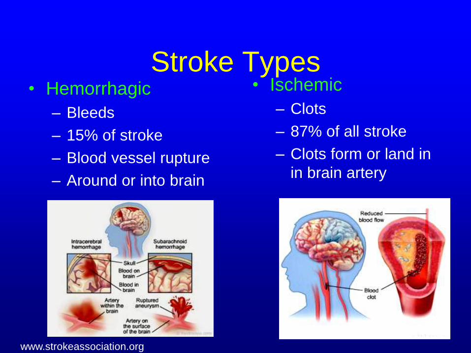
Stroke Types • Hemorrhagic
– Bleeds
– 15% of stroke
– Blood vessel rupture
– Around or into brain
• Ischemic
– Clots
– 87% of all stroke
– Clots form or land in
in brain artery
www.strokeassociation.org

Stroke Symptoms
• Right Brain
– Paralysis on the left side of the body
– Vision problems
– Personality change
– Memory loss
• Left Brain
– Paralysis on the right side of the body
– Speech/language problems
– Change in behavior
– Memory loss
• Brainstem
– Balance problems
– Coordination
– Breathing difficulty

Stroke Risk Factors • Age — Third leading cause of death in US. Stroke affects one person
every 45 seconds in the US. The probability of stroke doubles for each decade of life after age 55. Stroke is common among the elderly.
• Heredity (family history) and race — Your stroke risk is greater if a parent, grandparent, sister or brother has had a stroke. African Americans have a much higher risk of death from a stroke. This is partly because blacks have higher rates of high blood pressure, diabetes and obesity. Aneurysms predispose to hemorrhagic stroke if they rupture.
• Sex (gender) — Stroke is more common in men than in women. However, more than half of total stroke deaths occur in women, and more women than men die of stroke.
• Prior stroke, TIA or heart attack — The risk of stroke for someone with a prior stroke is much that of a person who has not. Transient ischemic attacks (TIAs) are "warning strokes" that produce stroke-like symptoms but no lasting damage. TIAs are strong predictors of future stroke. A person with one or more TIAs is almost 10 times more likely to have a stroke than someone of the same age and sex with no TIA. Recognizing and treating TIAs can reduce your risk of a major stroke. If you've had a heart attack, you're at higher risk of having a stroke, too. TIA should be considered a medical emergency and followed up immediately with a healthcare professional.

Ischemic Stroke: A blood clot in a brain artery
causes injury to a core of brain tissue, with
potential injury to additional surrounding brain if
blood flow is not re-established quickly
The Internet Stroke Center

During a stroke the cells of the brain stop sending
electrical signals and will die if blood flow is not restored quickly
The Internet
Stroke Center

Routine CT - Infarct Signs

Case #1
• 40 yo WM presents at 1 hour after
symptom onset with left hemiplegia and
neglect
• PMH: HTN, Paroxysmal Atrial
Fibrillation, Hypercholesterolemia
• Medications: Coumadin, Enalapril,
Zocor

Imaging Findings

Case Discussion – Late Infarct
Signs
• Finding – Hypodensity throughout MCA territory – findings
inconsistent with onset time
– Hyperdense MCA Sign
• Clinical Consequence – History retaken – onset time clarified – patient
actually last known well 5 hours prior to CT
– Large MCA territory completed infarct
– Therefore, IV tPA not given
– Anticipate poor functional recovery

Case #2
• 66 yo WM presents at 2 hours after
sudden onset of right hemiparesis and
global aphasia

Imaging Findings

Case Discussion- Early Infarct
Signs
• Finding: – Loss of gray-white differentiation
– Sulcal Effacement
– No hemorrhage
• Relevant Data: – In NINDS trials, early infarct signs not an
exclusion criterion; patients with early infarct signs benefited from therapy
• Clinical Consequence – Patient treated with thrombolytics with
improvement in NIHSS score from 14 to 3 at day 7
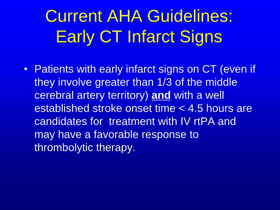
Current AHA Guidelines:
Early CT Infarct Signs
• Patients with early infarct signs on CT (even if
they involve greater than 1/3 of the middle
cerebral artery territory) and with a well
established stroke onset time < 4.5 hours are
candidates for treatment with IV rtPA and
may have a favorable response to
thrombolytic therapy.

AHA Guidelines:
Early CT Infarct Signs
• There are insufficient data to make a strong recommendation regarding the use of IV rt-PA treatment in the rare patient whose CT reveals extensive and clearly identifiable hypodensity yet show a well established stroke ictus onset time < 4.5 hours.
• While differences of opinion exist, some experts would recommend that thrombolytic therapy not be administered in these patients because a possibly unfavorable risk/benefit ratio.

Stroke Treatment
• Physical examination/conservative treatment
• Imaging study (CT or MRI) to determine if
hemorrhagic (bleed) or ischemic (clot) type
• Intravenous clot dissolving drug (tPA) if within
4.5 hours of stroke onset
• Intra-arterial clot dissolving drug if within 4.5-
6 hours of stroke onset
• Intra-arterial mechanical clot retrieval if within
6 hours of stroke onset
• Remember: Time is key!

Pre-
Retriever
Concentric
Retriever
Deployed
Post Retriever
Neurointerventional
Techniques

Multimodal CT - Ischemia,
Acute Stroke and Completed
Stroke: NECT, CTA and CT
Perfusion
NECT PCT CTA

Case #3
• 39 yo RH WM with history of
hyperlipidemia presents with recurrent
episodes of left hemiparesis while
therapeutic on warfarin therapy

Imaging Findings - CTA
Sensitivity and PPV of CTA for intracranial stenosis
(98% v. 70) and occlusion (100% v. 87%) is higher
than 3D TOF-MRA using DSA as gold standard*
*Bash, et al. AJNR 2004
DSA CTA CTA

MTT
Perfusion CT - Findings

CBV CBF
Perfusion CT - Findings

Quantitative CT Perfusion

Pre
Diamox
Post
Diamox
Note: Diamox (Acetazolamide) tests cerebrovascular reserve
Mean
Transit
Time
Map
(MTT)

CT Perfusion: Identification of
The Ischemic Penumbra
Tissue type CBF CBV MTT Tissue State
Normal tissue Normal
Viable, oligemic Penumbral
Viable,
ischemic Penumbral
Infarcted – bland Core
Infarcted,
reperfused Core
Wintermark, et.al, Ann Neurol. 2002;51:417-432
Infarct
Penumbra

Case Discussion- CTP:
Hypoperfusion and Impaired
Cerebrovascular Reserve • Finding
– CTA: 83% MCA stenosis
– Perfusion CT: resting perfusion deficit in MCA
territory worsened following diamox
– Indicates impaired cerebrovascular reserve
• Clinical Consequence
– Pt treated with florinef for presumed hemodynamic
events with resolution of symptoms
– Consider EC-IC bypass

Quantitative and qualitative analysis of the size and position of
the superficial temporal artery – 3D CTA and 2D MPR Images
3D-CTA 2D-CTA

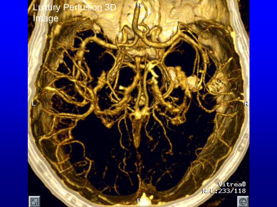
Luxury Perfusion 3D
Image

Loss of Autoregulatory Control in Reperfused
Infarct Zone - CT Hyperperfusion
Source
image
CBF
CBV MTT
CT and Perfusion CT
scan performed 24 hr
after thrombolysis for
left MCA occlusion
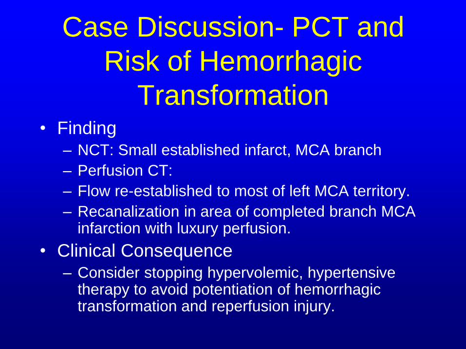
Case Discussion- PCT and
Risk of Hemorrhagic
Transformation • Finding
– NCT: Small established infarct, MCA branch
– Perfusion CT:
– Flow re-established to most of left MCA territory.
– Recanalization in area of completed branch MCA infarction with luxury perfusion.
• Clinical Consequence – Consider stopping hypervolemic, hypertensive
therapy to avoid potentiation of hemorrhagic transformation and reperfusion injury.

Computed Tomography (CT):
CT Perfusion – Whole Brain Blood Flow
Analysis
Courtesy Toshiba – Aquilion 1 5 mSv, 50 cc contrast, 20 second exposure

Detection Power of Perfusion CT
Author Territorial infarcts Non-territorial
infarcts
Sensitivity Specificity Sensitivity Specificity
Maruya,
2005
100% 100% 47.4% 91.3%
Mayer,
2000
93% -- 33% --

CT Monitoring of Stroke
Complications
Hemorrhagic
transformation Hemicraniectomy for
malignant cerebral edema

Comparative Evaluation of
Imaging Modalities Feature CTA DSA TOF-MRA US String sign
+ +/- - - Plaque
characterization + - - +/- Tandem stenosis
+ + + - Ulcerations
+ +/- - - Arterial
dissection + + +/- +/- Web stenosis
+/- + - + Metallic stents
+ + - -

CT Perfusion – Current
Limitations
Requires intact blood-brain barrier
Limited to 4-5 total slices (MR=7-15)
Motion sensitive
Ionizing radiation
Contrast allergy
Renal insufficiency

Carotid atherosclerosis and
Stroke
Up to 83% of all stroke, TIA or
amaurosis fugax believed to be due to
carotid bifurcation atheromatous
disease
Pharmacotherapy 1998;18:97s-93s.

Carotid Artery Source

Carotid Endarterectomy

NASCET: Endarterectomy
Protects from Stroke
Graph plots proportion without stroke

NASCET - Results
― CEA produces an absolute
reduction of 17% in stroke at 2
years when compared to ASA in
symptomatic patients with 70% or
greater ICA stenosis. Risk of no
treatment is 26%. Risk of CEA is
9%‖.1
1NASCET, N Engl J Med 1991;325:445-453
CTA and Cervical Stenosis-
Desirable Imaging Information
• High quality visualization
• Accurate quantitation
• Location relative to cervical column
• Eccentric stenosis
• Tandem stenosis
• Carotid and Vertebral artery stenosis
• Nonatherosclerotic stenosis

Helical CTA in Tandem stenosis

Non-atherosclerotic
causes of arterial
stenosis
Uncovertebral
Joint osteophyte

1/7/03 1/29/04

Recurrent
Atherosclerosis
Atherosclerosis
Sagittal 2D MPR
Axial 2D MPR

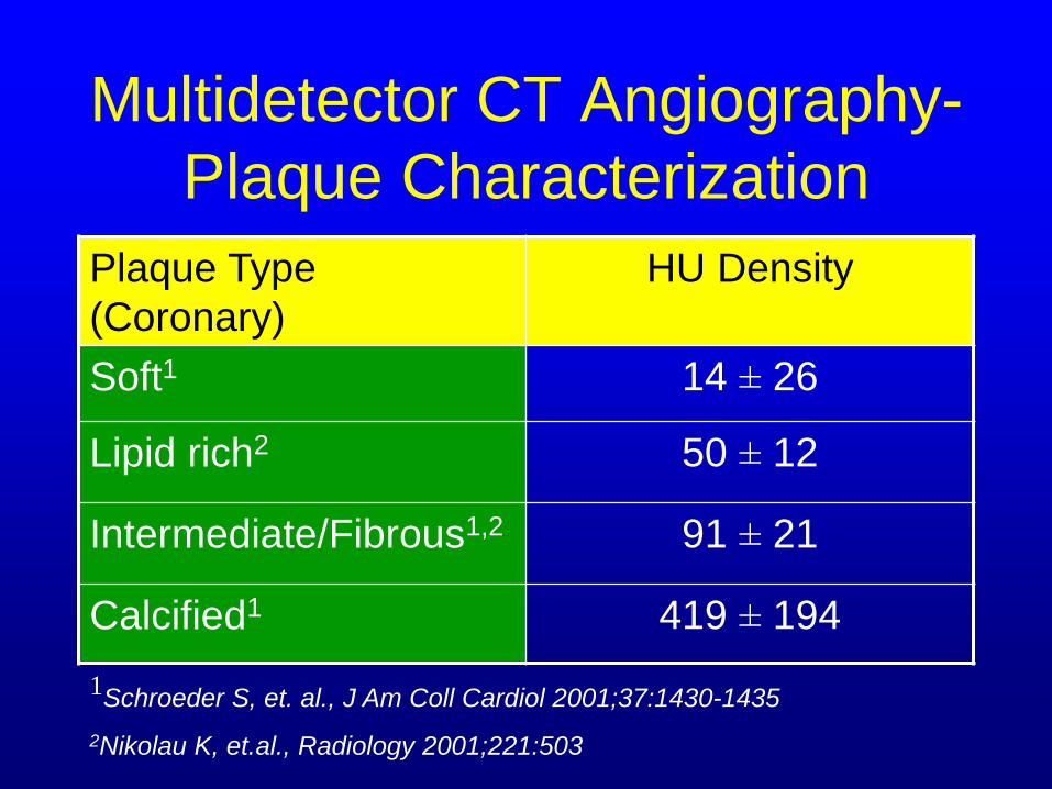
Multidetector CT Angiography-
Plaque Characterization
Plaque Type
(Coronary)
HU Density
Soft1 14 ± 26
Lipid rich2 50 ± 12
Intermediate/Fibrous1,2 91 ± 21
Calcified1 419 ± 194
1Schroeder S, et. al., J Am Coll Cardiol 2001;37:1430-1435
2Nikolau K, et.al., Radiology 2001;221:503

Multidetector CT Angiography-
Plaque Characterization
―Further studies will be required to
determine if MD-CTA of carotid
atherosclerosis with noncalcified
components can identify vulnerable
plaque, or patients at risk of developing
atheroembolic complications‖
Fayad ZA, et.al., Circulation. 2002;106:2026-2046

Multidetector Protocol T4-Vertex
• GE LightSpeed
• Pitch 3.75 HQ
• Collimation 1.25 mm
• Recon interval 0.625
• FOV 180 mm
• Martix 512 X 512
• Inj.Rate Min. 3-4 cc/sec
• Timing injection or SmartPrep
• 30-60 second exposure
• 120 kV, 300 mA
• Sensation 16
• Collimation 0.75 mm
• Recon interval 0.5
• mAs 350, kVp 120
• FOV 180 mm
• Matrix 512 X 512
• Inj. Rate 3-4 cc/sec.
• SmartPrep or hand inj
• <23 second exposure
• 6.8 mm/rotation feed
• H40f medium

CTA and Carotid Stenosis
• Accurate quantiation
• Anatomic
localization
• Luminal and non-
luminal information
• Tanden stenoses
• Longitudinal follow-
up
• Endoluminal
visualization
• 3D visualization
• Extended coverage
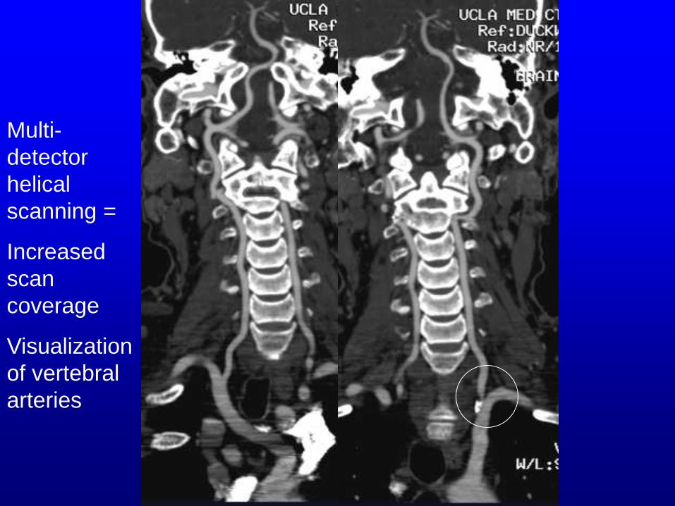
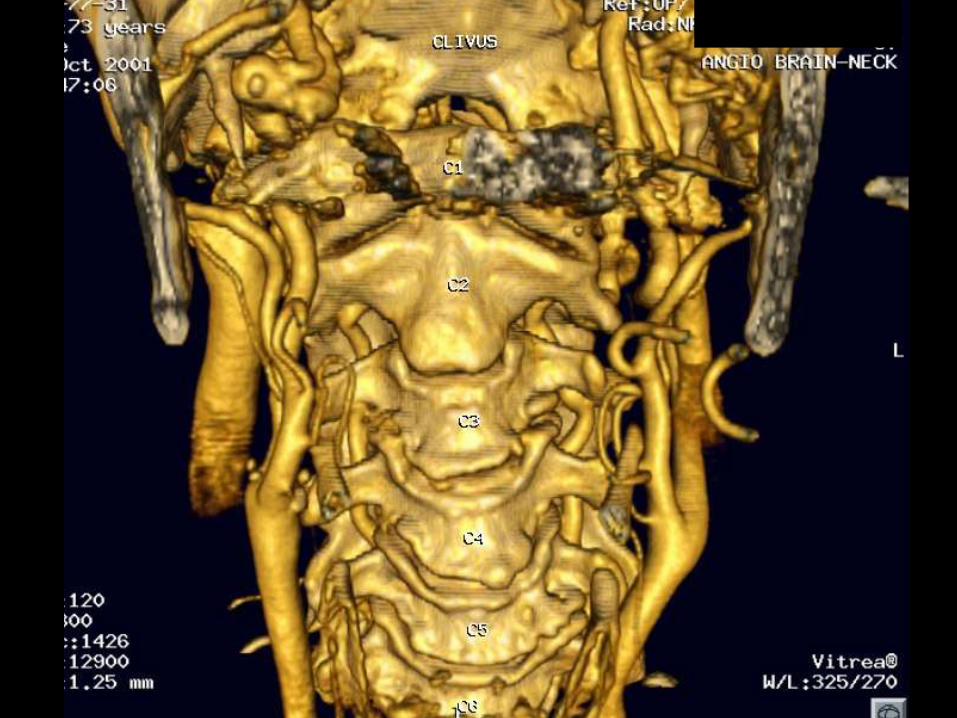
Multi-
detector
helical
scanning =
Increased
scan
coverage
Visualization
of vertebral
arteries

CTA axial or axial oblique source images correlate more
closely with DSA than do MIP or SSD images for all
degrees of stenosis (Stroke 2000;31:2168-2174)

Sagittal Coronal oblique
DSA may underestimates eccentric luminal stenosis


Volume Rendered Helical CTA of Neck:
Endoluminal Fly-Through Showing Calcific Left
Carotid Plaque
CTA and Carotid Stenosis
― CTA has been shown to have a pooled
sensitivity of 95% and a specificity of
98% for the detection of >70%, even if
only single slice techniques are used‖
Prokop M, et al. JBR-BTR. 2004;87:23-29.


3D and 2D MPR Helical CTA vs. DSA

Wall Thrombus
CTA in Acute Carotid Dissection with complete occlusion

Subacute/ chronic arterial dissection

CTA – Curved MPR CTA – 3D Volume rendered
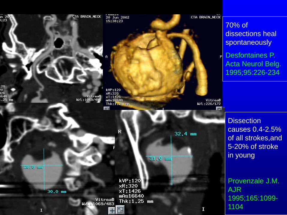
70% of
dissections heal
spontaneously
Desfontaines P.
Acta Neurol Belg.
1995;95:226-234
Dissection
causes 0.4-2.5%
of all strokes,and
5-20% of stroke
in young
Provenzale J.M.
AJR
1995;165:1099-
1104

Type I
Fibromuscular
Dysplasia – Left
Cervical Internal
Carotid Artery
Automated centerline
with background
included

Aneurysm Rupture

Intraoperative Aneurysm Rupture


3D-CTA Intraoperative Photo
Correlation Between CTA and Intraoperative Findings

3D CTA shows
Ca++ at neck

Surgical Planning with CTA – Giant Partially Thrombosed
Peripherally Calcified Fusiform Right MCA Aneurysm

Helical 3D CTA shows left PComA aneurysm, long
segment stenosis of left M1 segment.
Discordant findings

3D CTA with soft tissue window shows saccular
aneurysm with large thrombosed component
causing mass effect upon left M1
Value of soft tissue windows

37 year-old female: 1.2 mm laterally projecting
right SC-ICA aneurysm
2D-DSA 3D-CTA

Bone Subtracted CTV – Petrocavernous
ICA Segments
MIP CTA

Noninvasive Longitudinal
Follow-up: ACOM Aneurysm
4.9 mm AP X 2.9 mm TR X 3.2 mm CC
11/5/01 2/21/02 9/9/03
4.9 mm AP X 2.8 mm TR X 3.2 mm CC 7.0 mm AP X 2.8 mm TR X 3.2 mm CC

Time Resolved CT Angiogram
Left Carotico Ophthalmic Aneurysm

Helical CTA: Artifacts and
Limitations • Gross patient motion artifacts
• Beam hardening artifacts: amalgam, hyperconcentrated contrast
• Simultaneous arterial and venous imaging
• Reconstruction artifacts
• Contrast gradient artifacts
• Stent blooming artifacts
• Contrast allergy
• Low ejection fraction (heart failure)
• Overestimation of stenosis in thick calcific plaque
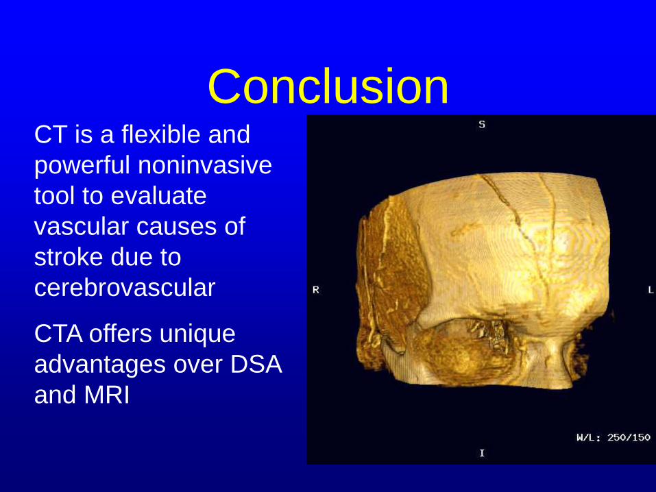
Conclusion CT is a flexible and
powerful noninvasive
tool to evaluate
vascular causes of
stroke due to
cerebrovascular
CTA offers unique
advantages over DSA
and MRI

Relationship Between Plaque
Rupture and Inflammation • No measurable FDG uptake in normal
carotids
• Autoradiography confirmed accumulation of deoxyglucose in macrophage rich areas of plaque
• Conclusion: Plaque rupture may be a consequence of inflammatory cell activation
Rudd JH, et.al. Circulation 2002;105:2708-2711

The Role of Shear Stress in
Atherogenesis - I • An essential feature of atherogenesis
• Fluid drag force on vessel wall is mechanotransduced into biochemical signals
• The endothelium controls local arterial responses by transduction of shear stress
• Physiological laminar shear stress is crucial for normal vascular functioning
• Therefore, laminar shear stress is atheroprotective by inhibiting vascular proliferation, thrombosis and inflammation

Carotid Shear Stress – Normal
Diastolic/Systolic Condition

Carotid Artery Pathology
• Carotid ―string-sign‖ – critical stenosis
• Bifurcation ulcer crater
• Plaque characterization and stroke risk
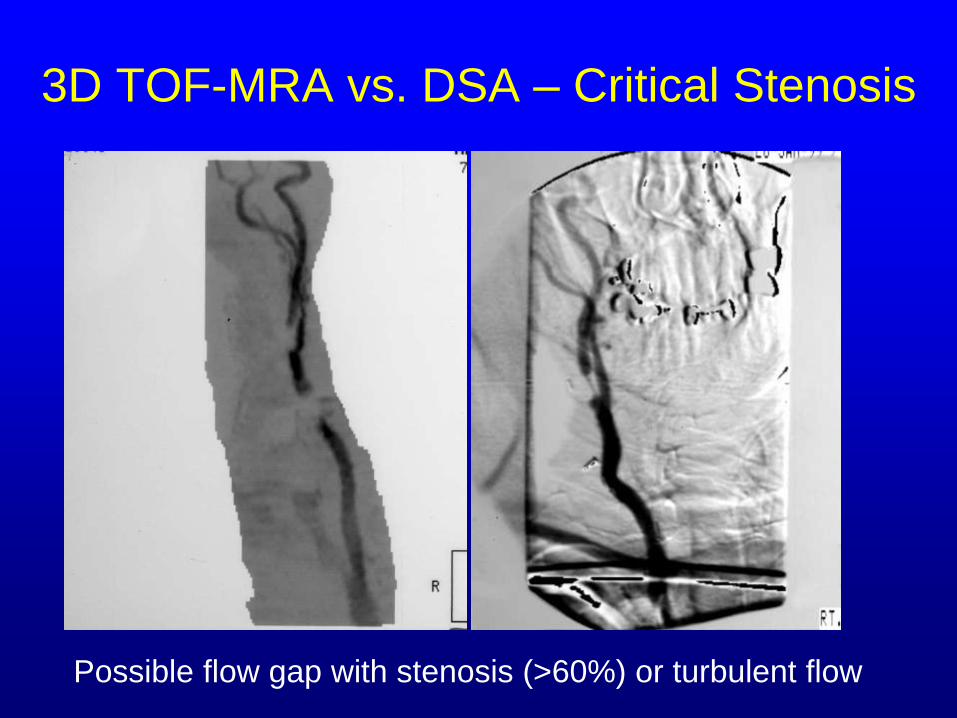
3D TOF-MRA vs. DSA – Critical Stenosis
Possible flow gap with stenosis (>60%) or turbulent flow


CE-MRA vs TOF-MRA
• Advantages – Shorter scan time
– Large coverage
– More accurate stenosis,
string sign and occlusion
– Contrast independent of
flow direction
– Less contamination from
short T1 materials
– Better SNR vs. TOF-MRA
– Less signal loss from
slow/turbulent flow
• Disadvantages – Longer prep time
– Lower spatial resolution vs. TOF-MRA
– Stents and metallic artifact
– T2* effects with bolus
– Maki effect (k-space ordering)
– May miss string sign
– Vessel diameter varies during contrast bolus cycle
– No calcifications
Willig DS, et al., 1996

3D TOF-MRA vs 1.5T CE-MRA
In-plane flow saturation effects
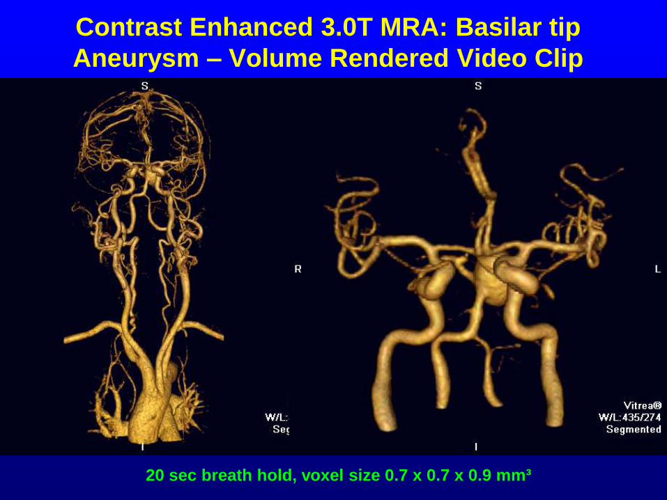
Contrast Enhanced 3.0T MRA: Basilar tip
Aneurysm – Volume Rendered Video Clip
20 sec breath hold, voxel size 0.7 x 0.7 x 0.9 mm³
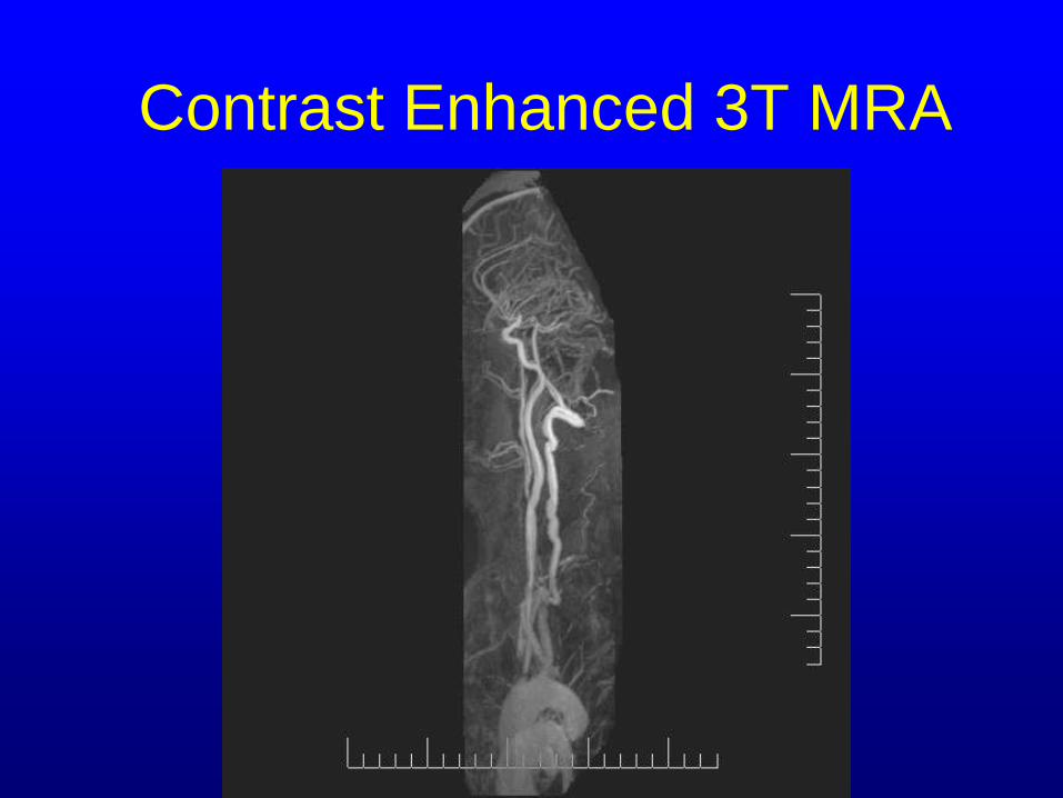
Contrast Enhanced 3T MRA

Phase Contrast MRA
Temp resolution 1.5s, In-plane
resolution: 1 x 1.3mm² , at 3.0T using
GRAPPA x3
Nael K et al. Invest Radiol. 2006 Feb;41(2):116-124.
Time-Resolved CE-MRA at 3.0T: Right Subclavian Steal
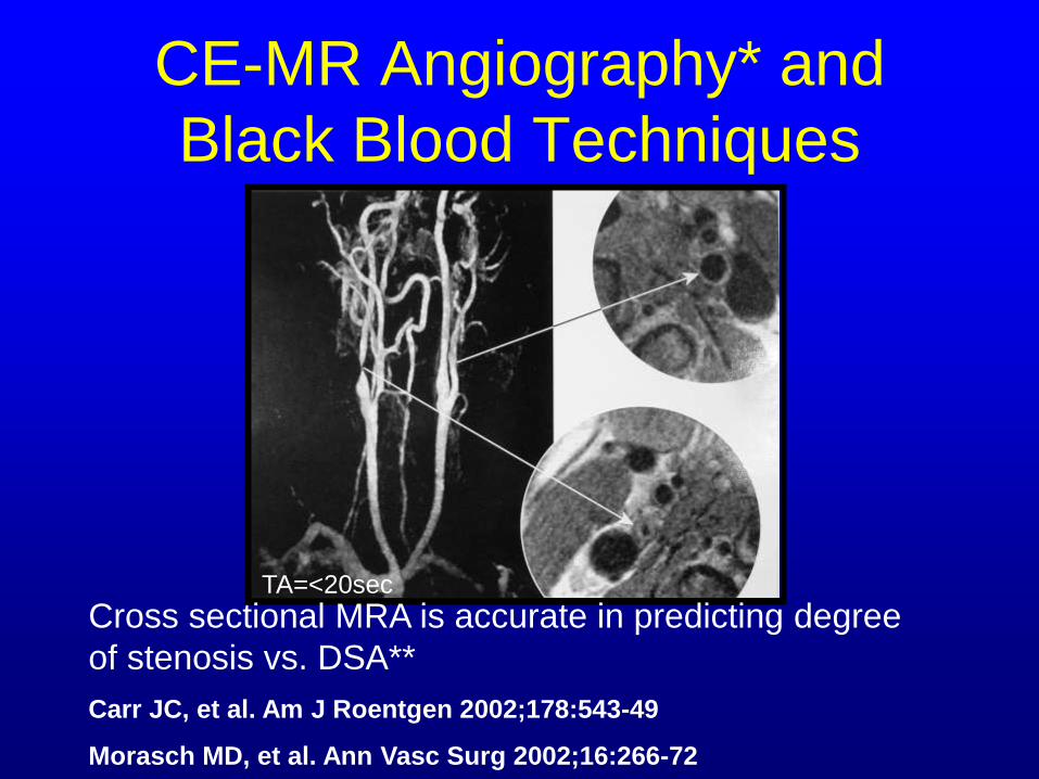
CE-MR Angiography* and
Black Blood Techniques
TA=<20sec
Cross sectional MRA is accurate in predicting degree
of stenosis vs. DSA**
Carr JC, et al. Am J Roentgen 2002;178:543-49
Morasch MD, et al. Ann Vasc Surg 2002;16:266-72
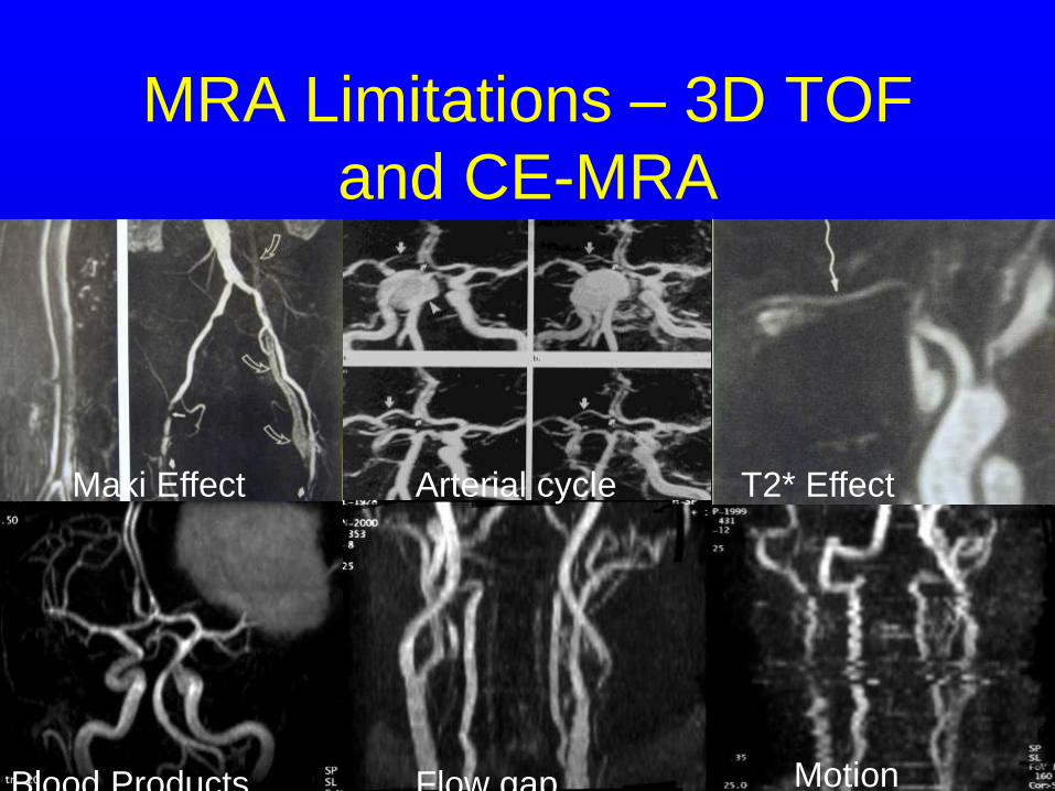
MRA Limitations – 3D TOF
and CE-MRA
Maki Effect T2* Effect
Blood Products Flow gap Motion
Arterial cycle

CE-MR Angiography – Major
Advantage over 2D-TOF
• Cross-sectional CE-MRA may be
accurate in predicting stenosis severity
vs. en-bloc endarterectomy specimen.1
1Morasch MD, Gurjala AN, Washington E, Chiou AC, Simonetty
OP, Finn JP, Yao JS. Ann Vasc Surg 2002;16:266-272.

Summary: Advantages of CTA
over TOF-and CE-MRA • Provides information about vessel lumen and
vessel wall in single study vs. contrast
enhanced MRA (CE-MRA) and TOF-MRA.
• No vascular signal artifacts arising from
slow/complex/turbulent/in-plane flow vs. TOF-
MRA and black blood MRA techniques.
• Higher spatial resolution
• Easier to acquire

Disadvantages of CTA vs.
TOF-MRA and CE-MRA
• Radiation
• Contrast allergy
• Less coverage vs. CE-MRA using single
detector systems
• Longer processing time
• Renal insufficiency

Carotid Ulceration
• Known thromboembolic source
• May be a marker of unstable plaque
• Presence and location
• Size
• Number
• Response to therapy

CT and MR Imaging Findings
CT MR- DWI
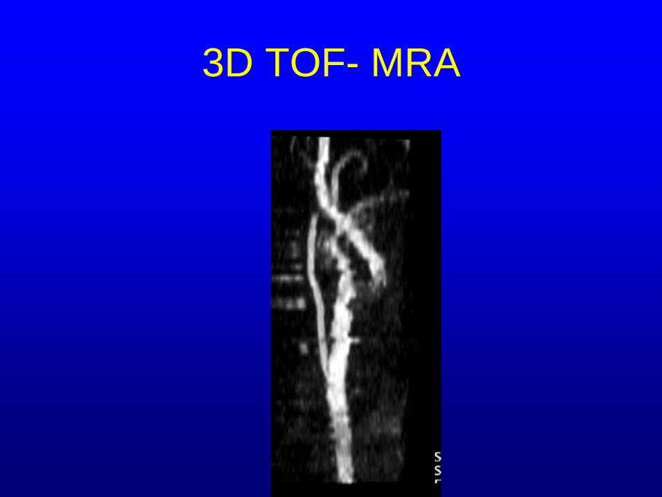
3D TOF- MRA

Discussion – Proximal Embolic Source
• Finding
– DWI-positive TIA
– DWI is positive in 56% of TIA patients
– Embolic appearing lesion
– CTA showed left carotid stenosis with ulcer crater
• Radiologic facts
– Sensitivity and specificity for the detection of >60%
stenosis is 87% and 90%, respectively and high for
ulcer crater detection1
1Cinat M. et.al., J Vasc Surg 1998;28:290-300

Carotid Dissection – Confirmation
and Aging of Injury

Imaging Findings – DWI hyperintense,
ADC hypointense, T2W and FLAIR
hyperintense = watershed infarct
DWI TOF-MRA

Carotid Dissection – Circumferential
Intramural Hematoma
Coronal Oblique 2D MPR Axial 2D MPR

Carotid Stenting – Intraluminal
Analysis

1/29/04 2/18/05
Pre-stent Post-stent

Recurrent Atherosclerosis is
generally eccentric and
irregular
Atherosclerosis
Sagittal 2D MPR
Axial 2D MPR
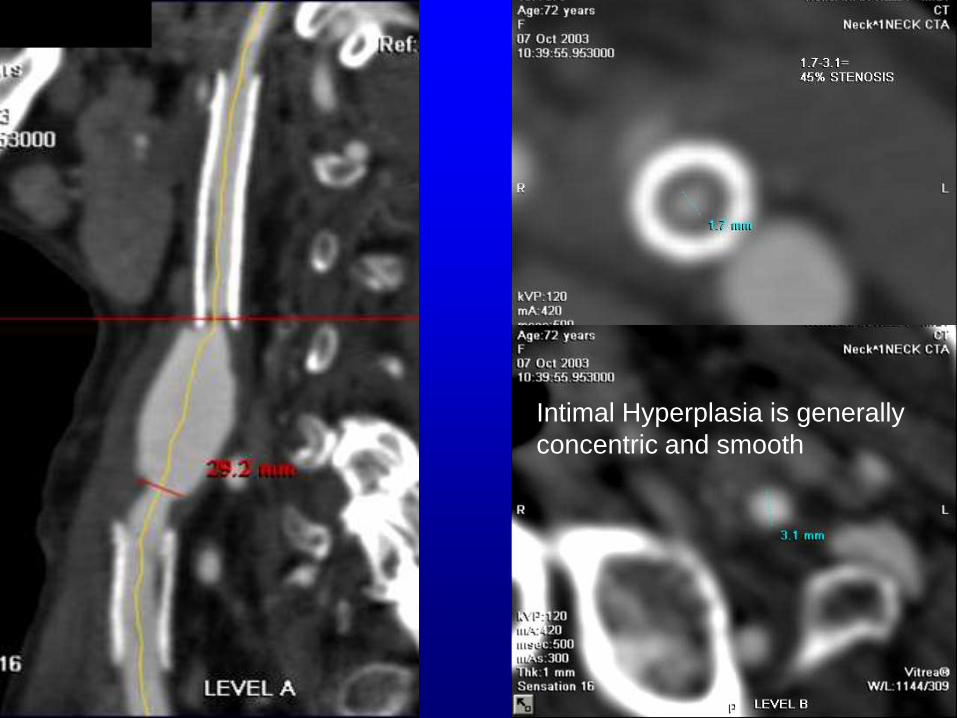
Intimal Hyperplasia is generally
concentric and smooth

Fibromuscular Dysplasia
• String of beads appearance
• Alternating strictures and dilatations
• ICA, 2nd most common site
• Type I, 80-85% -segmental beading
• Type II, 6-12% - long tubular
• Type III, 4-6% - one side of artery
• DSA and CTA unable to differentiate between intimal, medial and subadventitial types
• Ischemia 20%, TIA 30%, thromboembolic stroke 6%, dissection 10-20%
• 33% also have renal FMD
• 10% also involve vertebral artery

Type II
Fibromuscular
Dysplasia.
Note smooth
narrowing of long
segment of cervical
segment of the left
internal carotid
artery

Short Segment FMD – Type III
Coronal
2D curved
oblique
MPR
Sagittal
2D curved
oblique
MPR

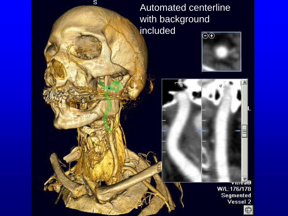
Rapid, Automated Post-
Processing of Carotid CTA’s

Centerline
segment with
isolated volume
rendered vessel
and curved oblique
reformatted image
in single display

Automated
segmentation of
smaller arteries
with segments
traveling close to
bone

Contrast
gradient
2D
Curved
MPR

Helical CTA
Artifacts and
Pitfalls:
Beam hardening
artifacts and
contrast entry
phenomena

CTA Advantages
• High spatial resolution: >MRA, < DSA
• Accurate measurements
• Comprehensive
• Minimally invasive
• Widely available
• Low cost
• Large coverage with multidetector systems

CTA Limitations
• Limited direct hemodynamic information
• Radiation – equivalent to CT of brain
performed with an without contrast
• Contrast reaction (1:30,000)
• Simultaneous arterial and venous imaging
• Renal insufficiency
• Gross motion in 3D images