brain research bulletin - uaeh
TRANSCRIPT
R
Er
CBa
Cb
c
d
Z
a
ARRAA
KESHCV
1
o
UVM
s(i(
h0
Brain Research Bulletin 131 (2017) 7–17
Contents lists available at ScienceDirect
Brain Research Bulletin
journa l homepage: www.e lsev ier .com/ locate /bra inresbul l
esearch report
lectroacupuncture improves gait locomotion, H-reflex and ventraloot potentials of spinal compression injured rats
arlos Escobar-Corona a, Sergio Torres-Castillo a, Erika Elizabeth Rodríguez-Torres b,ertha Segura-Alegría c, Ismael Jiménez-Estrada d, Salvador Quiroz-González a,∗
Department of Acupuncture and Rehabilitation, State University of Ecatepec Valley, Av. Central s/n, Esq. Leona Vicario, Col. Valle de Anáhuac, Secc. “A”,.P. 55210, Ecatepec Estado de Mexico, MexicoCenter for Research in Mathematics, Hidalgo State Autonomous University, MexicoFES Iztacala, UNAM, MexicoDepartment of Physiology, Biophysics and Neuroscience, Center for Research and Advanced Studies, Av. Instituto Politécnico Nacional 2508, Col. San Pedroacatenco, AP. 14-740, Mexico City, D.F. CP 07000, Mexico
r t i c l e i n f o
rticle history:eceived 25 November 2016eceived in revised form 7 February 2017ccepted 23 February 2017vailable online 6 March 2017
eywords:lectroacupuncturepinal cord injuryyper-excitabilityompressionentral root potentials
a b s t r a c t
This study explored the effect of electroacupuncture stimulation (EA) on alterations in the Hoffman reflex(H-reflex) response and gait locomotion provoked by spinal cord injury (SCI) in the rat. A compressionlesion of the spinal cord was evoked by insufflating a Fogarty balloon located in the epidural space atthe T8–9 spinal level of adult Wistar male rats (200–250 gr; n = 60). In different groups of SCI rats, EA(frequencies: 2, 50 and 100 Hz) was applied simultaneously to Huantiao (GB30), Yinmen (BL37), Jizhong(GV6) and Zhiyang (GV9) acupoints from the third post-injury day until the experimental session. At 1,2, 3 and 4 post-injury weeks, the BBB scores of the SCI group of rats treated with EA at 50 Hz showed agradual but greater enhancement of locomotor activity than the other groups of rats. Unrestrained gaitkinematic analysis of SCI rats treated with EA–50 Hz stimulation showed a significant improvement instride duration, length and speed (p < 0.05), whereas a discrete recovery of gait locomotion was observedin the other groups of animals. After four post-injury weeks, the H-reflex amplitude and H-reflex/Mwave amplitude ratio obtained in SCI rats had a noticeable enhancement (217%) compared to sham rats(n = 10). Meanwhile, SCI rats treated with EA at 50 Hz manifested a decreased facilitation of the H-reflexamplitude and H/M amplitude ratio (154%) and a reduced frequency-dependent amplitude depression of
the H-reflex (66%). In addition, 50 Hz-EA treatment induced a recovery of the presynaptic depression ofthe Gs-VRP evoked by PBSt conditioning stimulation in the SCI rat (63.2 ± 8.1%; n = 9). In concordance withthe latter, it could be suggested that 50 Hz-EA stimulation reduced the hyper-excitability of motoneuronsand provokes a partial improvement of the locomotive performance and H reflex responses by a possiblerecovery of presynaptic mechanisms in the spinal cord of experimentally injured rats.© 2017 Elsevier Inc. All rights reserved.
. Introduction
Spinal cord injury (SCI) is one of the most common impairmentsf the central nervous system that results in complete or partial
∗ Corresponding author at: Department of Acupuncture and Rehabilitation, Stateniversity of Ecatepec Valley, Avenida Central s/n, Esquina Leona Vicario, Coloniaalle de Anáhuac Sección “A”, Código Postal 55210, Ecatepec Estado de Mexico,exico.
E-mail addresses: kalitos [email protected] (C. Escobar-Corona),[email protected] (S. Torres-Castillo), [email protected]. Rodríguez-Torres), [email protected] (B. Segura-Alegría),[email protected] (I. Jiménez-Estrada), [email protected]. Quiroz-González).
ttp://dx.doi.org/10.1016/j.brainresbull.2017.02.008361-9230/© 2017 Elsevier Inc. All rights reserved.
loss of both sensory and motor functions due to mechanical spinaldamage, which often leads to permanent paralysis (Silverman et al.,2012). Traumatic and non-traumatic lesions represent the two pre-sentations of SCI. The first results from contusion, compression orstretching of the spinal cord, and the second is associated withvertebral spondylosis, tumor compression, vascular ischemia, andinflammatory spinal cord disorders (New et al., 2002). In bothhumans and animals, SCI induces physiological changes in themotor system which included hyperreflexia and abnormalities ofthe locomotor behavior (Silverman et al., 2012; Yablon and Stokic,
2004). Several researchers use the Hoffman reflex (H-reflex), toanalyze the hyperreflexia following SCI (Milanov, 1994; Yablon andStokic, 2004). The H-reflex is an electromyographic (EMG) responsethat results from activation of a synaptic pathway conformed by
8 Resea
tSa1ehdh2
s2e2ttm
Toonth(siarftt(aEaseaatiadustnt
2
2
ott(gacbo
C. Escobar-Corona et al. / Brain
he afferent-motoneuron-muscle circuit (Reyes et al., 2007). InCI, changes associated with hyperreflexia and spasticity included:lpha motor neuron hyperexcitability (Lin et al., 2007; Milanov,994), changes in the intrinsic properties of motoneurons (Bennettt al., 2001) and loss of presynaptic inhibition (Hultborn, 2006). Itas been shown that the measurement of H-reflex rate-sensitiveepression acquires a particular importance in the assessment ofyperreflexia following SCI (Thompson et al., 1992; Chen et al.,001).
In general, treatment for functional recovery of SCI includedurgery (Bregman et al., 2002), physical therapy (Silverman et al.,012), drugs (Attal et al., 2009), hormonal treatment (Calderónt al., 2015; Osuna et al., 2016), behavioral therapy (Norrbrink et al.,006), and supportive treatment (Huston et al., 2011). Becausehere is still a lack of effective treatment for spinal cord injuries,here has been an increased interest in alternative medical treat-
ents.Acupuncture is a therapeutic modality that emerged from
raditional Chinese Medicine. The World Health Organization rec-mmends the use of acupuncture for the treatment of a wide varietyf diseases (Zhang et al., 2014; Barnes et al., 2008). A relativelyovel form of acupuncture is the electrical stimulation of acupunc-ure points (APs), also known as electroacupuncture (EA), whichas been widely used in both clinical and experimental reportsVickers et al., 2012; Zhao, 2008). In previous studies, it has beenhown that application of EA (Dazhui (DU14), Mingmen (DU4), Sany-njiao (SP6), Huantiao (GB30), Zusanli (ST36) and Kunlun (BL60) as
treatment for SCI contributes to the recovery of several neu-ologic and functional alterations (Min et al., 2015). It has beenound that EA produces an improvement in the locomotor pat-ern (Peng et al., 2007), which is accompanied by reductions inhe process of glial scarring (Yang et al., 2005), oxidative stressPolitis and Korchinski, 1990), laminin expression (Zhu, 2002) andquaporin transport (AQP-4; Xie et al., 2006). It is thought thatA evokes its effects through the activation of peripheral sensoryfferents that in turn synaptically interact with sets of dorsal hornensory neurons in the spinal cord (Quiroz et al., 2014a,b). How-ver, EA effects on experimental animal models of SCI, particularlyt the motoneuron level, are scarcely studied. In this study, wenalyzed the effect of EA on locomotor behavior (evaluated withhe Open Field Test and gait kinematics analysis), H reflex facil-tation and H-reflex frequency-dependent depression evoked indult rats after a spinal cord compression injury. In addition, toisclose possible presynaptic mechanisms in the effect of EA stim-lation, we also analyzed the changes produced by conditioningtimulation of the posterior biceps and semitendinosus (PBSt) onhe amplitude of ventral root potentials (VRP) evoked by gastroc-emius nerve (Gs) stimulation, as a test for presynaptic inhibi-ion.
. Materials and methods
.1. Animals
Male Wistar rats (n = 60) weighing 200–250 g (8–10 weeks old)btained from our institution were used. All animals had free accesso food and water and were housed under identical environmen-al conditions of light and dark cycles (12:12 h) and temperature22–24 ◦C). All experiments were performed in accordance with theuidelines contained in NIH publications No. 80-23 (revised 1996)
nd the Mexican Official Norm (NOM-062-ZOO-1999) on the Prin-iples of Laboratory Animal Care. The study protocol was approvedy the institutional bioethics committee for the Care and Handlingf Laboratory Animals (Protocol 0267-05, CINVESTAV).rch Bulletin 131 (2017) 7–17
2.2. Surgical procedures and animals groups
Initially the rats were randomly assigned into five groups byusing a random number table: (1) sham control group (n = 10), (2)compression injury without EA treatment group (SCI-UT n = 12), (3)compression injury with EA treatment at 2 Hz (SCI-EA 2 Hz group;n = 11), (4) 50 Hz (SCI-EA 50 Hz group; n = 14) and (5) 100 Hz (SCI-EA100 Hz group; n = 13). The method used for producing a compres-sion injury was similar to that described previously (Lonjon et al.,2010). Animals were anesthetized with an intraperitoneal injec-tion of a mixed solution of ketamine (100 mg/kg) and xylazine(2 mg/kg). To provoke the compression injury in the rat spinal cord,a Fogarty catheter (2 French x 60 cm, size: 0.67 mm, Ethimed) wasinserted into the epidural space through a hole drilled in the poste-rior arch of the T-10 vertebra, and a groove was drilled on T-11 toguide the insertion of the catheter. The balloon was positioned atthe T8–9 level, inflated with sterilized water (10 �l) using a Hamil-ton syringe, and left in place for 5 min. The sham group of rats(n = 10) underwent the same protocol, except for balloon inflation.
After surgery, the back skin and muscles were sutured witha sterile thread and stainless wound clips, and the animals wereallowed to recover from surgery and anesthesia in a clean, heatedcage. All rats received an intramuscular injection of penicillin andprocaine (5,000,000 U/ml/day), and Furazolidone was spread ontothe lesion to prevent infection. The animals were housed in individ-ual cages and individually received intensive care, which includedmanual expression of urine and excrement until the day of theexperiment. Because nine rats died after SCI, and six died duringthe electrophysiological recording session, the final number of ani-mals included in each group was the following: SCI-UT (n = 10);SCI-EA 2 Hz group (n = 8), SCI-EA 50 Hz group (n = 9); and SCI-EA100 Hz group (n = 8).
2.3. Electroacupuncture treatment
EA was applied simultaneously at two pairs of acupoints. Onepair was situated at the Jizhong (GV6, anode) and Zhiyang (GV9cathode) acupoints and the other pair were Huantiao (GB30 anode)and Yinmen (BL37 cathode), bilaterally (Fig. 1). These acupointswere selected because they are needling points in acupuncturetreatments following SCI in humans (Heo et al., 2013a,b) and con-tributed to neuroplasticity in the spinal cord after injury (Min et al.,2015; Ding et al., 2011). The Huantiao (GB30) acupoint was locatedat the posterior upper border of the hip joint of the hindlimbs. TheYinmen (BL37) acupoint was situated where the long head of thebiceps femoris muscle and the semitendinosus muscle converged(upper to the middle part of the popliteal crease), whereas theJizhong (GV6) acupoint was located posterior to the midline, belowthe spinous process of the eleventh thoracic vertebra (maintainingthe animal in the prone position). The Zhiyang (GV9) acupoint wassituated on the posterior midline and in the depression below thespinous process of the seventh thoracic vertebra in the prone posi-tion. EA was applied daily (resting on weekends) by means of anelectroacupuncture stimulator (AWQ-104L, China), and it consistedof a sequence of biphasic pulse trains of 2.5 s in duration followedby periods of 2.5 s with no stimulation (Fig. 2). The biphasic pulsestimuli was chosen in this study to avoid the polarization of eachneedle because of electrolysis and break off in tissue (Pomeranz,1995). Another advantage of biphasic stimulus is that each pair ofneedles receives symmetrical current pulses which appears to bevery useful to neuromodulate the physiological state of the spinalcord in humans (Gerasimenko et al., 2015). The intensity of the EA
current pulses was adjusted to induce slight twitches in hind limbs(1 mA).Because it has been showed that EA stimulation induced neu-roprotective, remyelination, antioxidative and anti-inflammatory

C. Escobar-Corona et al. / Brain Research Bulletin 131 (2017) 7–17 9
FIGURE1
Fig. 1. Schematic diagram indicating the relative position of the selected acupoints Zhiyang (GV9), Jizhong (GV6) in the rat (A), and their corresponding acupoints in humans:Huantiao (GB30) (B), and Yinmen (BL37) (C);Diagram illustrating the operation process of electrode needles at the selected pairs of acupoints with their respective cathode(+) and anode (−) polarity. Right (R), Left (L).
FIGURE2
Fig. 2. Parameters of EA stimulation treatment applied to the three groups of SCI rats. The mode of stimulation was a discontinued biphasic pulse (0.37 ms) applied at 2 (A),50 (B) and 100 (C) Hz with a duration interval of 2.5 s per pulse over 30 min.

1 Resea
eidtiflp
2
BKmpt(t
2
gla((mw
2
epeadptpwcpmuaiLtahplssv
2
twaitu
0 C. Escobar-Corona et al. / Brain
ffects during the acute phase of SCI, starting from the third post-njury day (Mo et al., 2016; Huang et al., 2015; Liu et al., 2013) weecided to analyze the effect of EA treatment on H-reflex and ven-ral root potentials during a period of 4 weeks after the spinal cordnjury. In this way, EA was administered at 2 Hz, 50 Hz and 100 Hzrequencies as pointed out previously every other day (sessionasted 40 min) for 30 days (four weeks), starting from the secondost-injury day until the day prior to the experiment.
.4. Functional assessments
The locomotive activity was evaluated with theasso–Beattie–Bresnahan (BBB) open field test and with ainematic analysis of gait locomotion. All motor evaluations wereade between 9:30–10:30 a.m. and no details of the operative
rocedure or treatment protocol of animals were offered to thewo independent examiners who made the scoring of animals“double-blinded analysis”). Subsequently, the scores provided byhe two independent researchers were averaged
.4.1. Locomotor performance evaluation (BBB test)Animals were placed on one open-field arena 90 cm above
round where voluntary movements of the trunk, tail and hindimbs were recorded by a single commercial digital video camerand subsequently evaluated by the Basso, Beattie and BresnahanBBB) scale (Basso et al., 1995). BBB scale values varied from 0complete hindlimb paralysis) to 21 points (normal hindlimb loco-
otion). The experimental sessions were 3 days and 1, 2, 3 and 4eeks post-injury and lasted 5 min each.
.4.2. Kinematic analysis of gait locomotionUnrestrained gait locomotion of the different groups of rats was
valuated at 28 days post-injury (4 weeks) by a kinematic methodreviously used in other studies (Calderón et al., 2015; Osunat al., 2016). Briefly, gait locomotion of both the sham-operatednd experimental animals was recorded with a single commercialigital video camera located on one side of a transparent acrylicassageway. After shaving, the limbs, iliac crest, hip (e.g., greaterrochanter), knee, ankle (e.g., lateral malleolus), and fifth metatarsalhalangeal joints were marked with ink (Fig. 3A). The videotapesere digitized (30 frames/s), and their frames were individually
aptured as digital photographs using a commercial video editingrogram (Pinnacle Studio V.7, Pinnacle systems Inc.). Limb move-ents consisting of 4–6 step cycles were analyzed frame by frame
sing the Image J program (Scion Corporation, NIH), and the Xnd Y coordinates of each articulation joint were calculated andntroduced in a computer program (developed in our laboratory inabView environment) that calculated the angle and spatial loca-ion of each joint articulation. Line drawings constructed betweenrticulation joints illustrated the spatio-temporal sequences ofind limb movements during gait locomotion. Swing and stancehases were identified from each stride cycle, and the duration,
ength and speed of strides determined. The data obtained fromham-operated and experimental groups of animals were pooledeparately, and their corresponding average and standard erroralues were calculated.
.5. H reflex recording
At the recording day, the rats were anesthetized with an ini-ial dose of ketamine (100 mg/kg of weight, i.p.) supplementedith additional doses of 75 mg/kg i.p. as required to maintain an
dequate level of anesthesia, which was assessed routinely by ver-fying that the withdrawal reflex was absent after a strong pinch ofhe right hindlimb. The animal’s body temperature was monitoredsing a thermal probe located in the back muscles and connected
rch Bulletin 131 (2017) 7–17
to an automatic feedback control unit and heating blanket to main-tain the animal’s body temperature at 37 ◦C. All left hindlimb nerveswere dissected and sectioned with the exception of the distalbranch of the tibial nerve (first series of 10 experiments) or thelateral plantar nerve (second series of 6 experiments in which themedial plantar nerve was also sectioned) which were left intactwith their insertion to the plantar muscles (“closed loop condi-tion”). Single voltage pulses (0.05 ms duration) were delivered froma pulse generator every 8 s to avoid rate depression of the H reflex(Gozariu et al., 1998) and applied to the sciatic or proximal lateralplantar nerve through a pair of fine silver hook electrodes over onehour (n = 100 trials) and averaged. The stimulus strength was grad-ually adjusted from the minimal voltage strength needed to evokea barely discernible electroneurogram response (one times thresh-old, 1xT) in the distal lateral plantar nerve (1xT = 0.43 ± 0.19 V) toseveral multiples of the threshold (1–3 × T).
To reduce the possible mechanical artifacts derived from musclecontraction, electroneurogram (ENG) and electromyogram (EMG)recordings were performed with fine “floating” chloride silver elec-trodes located in the distal lateral plantar nerve and lateral plantarmuscles of the foot (between the 4th and 5th digit) (Reyes et al.,2007). The recording electrodes were connected to separate lownoise, high gain pre-amplifiers (band pass filters set at 0.3 Hz inthe low range and 10 KHz in the high range), which were subse-quently connected to a digital oscilloscope and to a digital computerthrough a data acquisition system (ATMIO 16E2, National Instru-ments; 12 bits resolution). EMG responses were digitized andstored by means of a specially designed computer program (at 100bins of resolution and a digitization rate of 3000 samples per sec-ond). Typically, EMG responses consisted of two components, theM wave and the H-reflex (Reyes et al., 2007). Subsequently, theHmax/Mmax amplitude ratio was calculated to estimate the rela-tive proportion of motor units recruited through the monosynapticreflex loop (Côté et al., 2014; Mazzocchio et al., 2001).
2.6. Rate-dependent depression of H-reflex
Low frequency-dependent depression of the H-reflex is definedas the gradual decrease in the H-reflex amplitude that occurs whena series of reflexes are elicited between 1 and 10 Hz. It has beenshown that measures of H-reflex frequency-sensitive depression isof particular importance in the assessment of hyper-reflexia fol-lowing SCI (Thompson et al., 1992; Chen et al., 2001). To inducethe frequency-dependent depression of the H-reflex, sequencesof stimulus pulses were applied to the exposed plantar nerve atdifferent frequencies (0.2, 1, 5, and 10 Hz), and alterations in thereflex excitability level of the plantar spinal motoneuron pool wereinferred from changes in the Hmax/Mmax amplitude ratio (Côtéet al., 2014).
2.7. Stimulation and ventral root recordings
The central ends of the posterior biceps and semitendinosus(PBSt) and gastrocnemius nerves (GS) were mounted on pairs of sil-ver hook electrodes connected to isolated-current-pulse generators(Isoflex D 4030) and square-current pulses (0.05 ms duration; 1 Hz)of graded intensity were applied to the nerves. The pulses weremonitored by recording the voltage drop across a resistor (1000 �)that was placed in the current return path to ground. The electri-cal threshold of the evoked spinal potentials (one times threshold,1 × T) was established as the minimum current intensity necessaryto evoke a barely discernible response in the ventral roots.
To record the VRP, a previously dissected L6 ventral rootledwas placed on two fine silver Ag-AgCl wire electrodes, one locatedvery close to, but not touching, the spinal cord and the other,distally on the severed end of the rootlet (Fig. 1A). Next, the record-
C. Escobar-Corona et al. / Brain Research Bulletin 131 (2017) 7–17 11
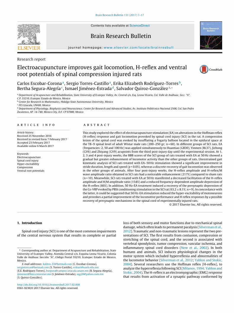
FIGURE3
F ; n = 1s quencn o-way
iawc
d(ssset2
2
fpTs(wata
2
aaacAs
2
wto0
ig. 3. BBB performance of the different groups of rats: sham-operated (black circlespinal cord) (squares SCI-UT; n = 10), and treated with electroacupuncture (EA) at fre
= 8). The baseline BBB score of sham-operated rats (control) was 21, * p < 0.05 (tw
ng electrodes were connected to low-noise, high-gain differentialmplifiers (Grass, model P511; band-pass filters set at 0.3–10 kHz),hich in turn, were attached to a digital oscilloscope and personal
omputer.To determine the time course of the effect produced by the con-
itioning stimulation on test responses, four conditioning stimuliat 300 Hz) were delivered to the PBSt nerve at different inter-timulus time intervals (between 10 and 200 ms) before the testtimulus applied to the GS nerve. The conditioning stimulationtarted 10 ms prior to the test stimulus to reduce the occlusiveffect that was provoked by the preceding stimuli on test poten-ials when short time intervals were used (Enríquez-Denton et al.,004; Rudomin and Schmidt., 1999).
.8. Histology
At the end of the recording session, animals were per-used intracardially with a mixed solution of Krebs saline andaraformaldehyde (4%). Subsequently, the compressed spinal T8-9 segments were dissected and placed in a solution of PBS + 30%ucrose. The segments were placed on an agar block and sliced50 �m thick) with a vibratome (Leica SM 2000R). Tissue slicesere stained with the crystal violet method for Nissl substance,
nd spinal cord sections showing the traumatic injury were pho-ographed with a digital camera to characterize the damaged spinalreas.
.9. Data analysis
All the data are expressed as the mean ± standard deviation,nd the motor performance (BBB test and kinematic gait analysis)nd frequency-dependent depression of the H-reflex values werenalyzed using a 2-way ANOVA followed by Bonferroni’s multipleomparison test. The significance level was established at p < 0.05.ll statistical analyses were performed using Graph-Pad Prism (ver-ion 5 San Diego Ca.) software.
.10. Ethics statements
All experimental procedures were performed in accordance
ith the guidelines of the Guide for the Care and Use of Labora-ory Animals (National Research Council, 2010, National Institutesf Health, Bethesda, MD, USA; Animal Welfare Assurance # A5036-1) and the Mexican Official Norm (NOM-062-ZOO-1999) and
0), untreated injured spinal cord (compression of the 8–9th segment of the thoracicies of 2 Hz (diamonds; n = 8), 50 Hz (inverted triangles; n = 9) and 100 Hz (triangles;
repeated measures ANOVA with Bonferroni’s post-test).
approved by the Institutional Bioethical Committee for Care andHandling of Laboratory Animals (Protocol 0267-05, CINVESTAV).
3. Results
3.1. Locomotor performance (BBB test)
At the third day after the spinal cord injury, all rats with spinallesions showed noticeable locomotor impairment in both the leftand right hind limbs, as determined using the BBB test (Fig. 3). At1, 2, 3 and 4 post-injury weeks, a gradual and progressive motorrecovery was observed in all groups of animals. SCI-UT rats had avery poor improvement in locomotor activity (Fig. 3), whereas SCIrats treated with EA stimulation at 2 and 50 Hz showed continuousand significant enhancement in their motor performance (p < 0.05,Fig. 3), which was characterized by the occurrence of movementsin the knee and ankle joints combined with slight movements ofthe hip joint during locomotor activity compared to SCI-UT rats(p < 0.05, n = 10). Moreover, SCI-EA 100 Hz rats exhibited a notice-able but not significant improvement in locomotor activity at thefourth week post-injury (Fig. 3).
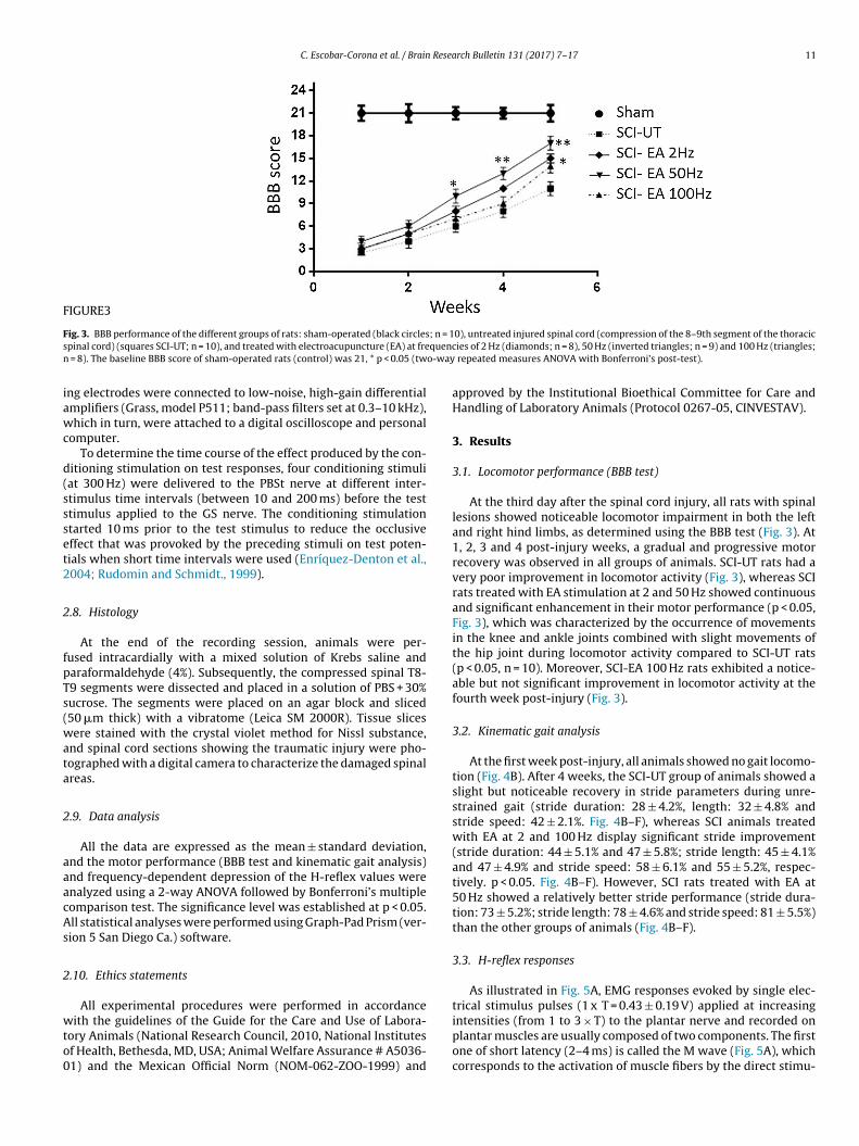
3.2. Kinematic gait analysis
At the first week post-injury, all animals showed no gait locomo-tion (Fig. 4B). After 4 weeks, the SCI-UT group of animals showed aslight but noticeable recovery in stride parameters during unre-strained gait (stride duration: 28 ± 4.2%, length: 32 ± 4.8% andstride speed: 42 ± 2.1%. Fig. 4B–F), whereas SCI animals treatedwith EA at 2 and 100 Hz display significant stride improvement(stride duration: 44 ± 5.1% and 47 ± 5.8%; stride length: 45 ± 4.1%and 47 ± 4.9% and stride speed: 58 ± 6.1% and 55 ± 5.2%, respec-tively. p < 0.05. Fig. 4B–F). However, SCI rats treated with EA at50 Hz showed a relatively better stride performance (stride dura-tion: 73 ± 5.2%; stride length: 78 ± 4.6% and stride speed: 81 ± 5.5%)than the other groups of animals (Fig. 4B–F).
3.3. H-reflex responses
As illustrated in Fig. 5A, EMG responses evoked by single elec-trical stimulus pulses (1 x T = 0.43 ± 0.19 V) applied at increasing
intensities (from 1 to 3 × T) to the plantar nerve and recorded onplantar muscles are usually composed of two components. The firstone of short latency (2–4 ms) is called the M wave (Fig. 5A), whichcorresponds to the activation of muscle fibers by the direct stimu-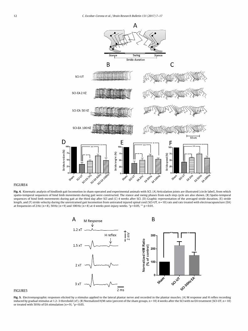
12 C. Escobar-Corona et al. / Brain Research Bulletin 131 (2017) 7–17
FIGURE4
Fig. 4. Kinematic analysis of hindlimb gait locomotion in sham-operated and experimental animals with SCI. (A) Articulation joints are illustrated (circle label), from whichspatio-temporal sequences of hind limb movements during gait were constructed. The stance and swing phases from each step cycle are also shown. (B) Spatio-temporalsequences of hind limb movements during gait at the third day after SCI and (C) 4 weeks after SCI. (D) Graphic representation of the averaged stride duration, (E) stridelength, and (F) stride velocity during the unrestrained gait locomotion from untreated injured spinal cord (SCI-UT, n = 10) rats and rats treated with electroacupuncture (EA)at frequencies of 2 Hz (n = 8), 50 Hz (n = 9) and 100 Hz (n = 8) at 4 weeks post-injury weeks. *p < 0.05, ** p < 0.01.
FIGURE5
Fig. 5. Electromyographic responses elicited by a stimulus applied to the lateral plantar nerve and recorded in the plantar muscles. (A) M response and H reflex recordinginduced by gradual stimulus at 1.2–3 threshold (xT). (B) Normalized H/M ratio (percent of the sham groups, n = 10) 4 weeks after the SCI with no EA treatment (SCI-UT, n = 10)or treated with 50 Hz of EA stimulation (n = 9). *p < 0.05.
C. Escobar-Corona et al. / Brain Research Bulletin 131 (2017) 7–17 13
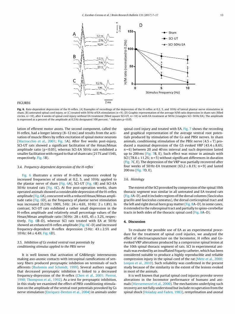
FIGURE6
Fig. 6. Rate-dependent depression of the H-reflex. (A) Examples of recordings of the depression of the H-reflex at 0.2, 5, and 10 Hz of lateral plantar nerve stimulation ins = 9).
c re SCIi ates p
lHv(Sasr
3
it5oatwcHHtsf1
3c
msatf1itn
ham, (B) untreated spinal cord injury, or (C) treated with 50 Hz of EA stimulation (nircles, n = 10), after 4 weeks of spinal cord injury without EA treatment (filled squas expressed as a percent of the amplitude at 0.2 Hz designated 100 percent; * indic
ation of efferent motor axons. The second component, called the-reflex, had a longer latency (8–12 ms) and results from the acti-ation of muscle fibers by reflex excitation of spinal motor neuronsMazzocchio et al., 2001; Fig. 5A). After five weeks post-injury,CI-UT rats showed a significant facilitation of the Hmax/Mmaxmplitude ratio (p < 0.05), whereas SCI-EA 50 Hz rats exhibited amaller facilitation with regard to that of sham rats (217% and 154%,espectively. Fig. 5B).
.4. Frequency-dependent depression of the H-reflex
Fig. 6 illustrates a series of H-reflex responses evoked byncreased frequencies of stimuli at 0.2, 5, and 10 Hz applied tohe plantar nerve of sham (Fig. 6A), SCI-UT (Fig. 6B) and SCI-EA0 Hz treated rats (Fig. 6C). At five post-operation weeks, shamperated animals showed a considerable depression of the H-reflexmplitude (Fig. 6A), concurrent with a reduced Hmax/Mmax ampli-ude ratio (Fig. 6D), as the frequency of plantar nerve stimulationas increased (0.2 Hz: 100%, 5 Hz: 24 ± 4.6%, 10 Hz: 2 ± 1.8%). In
ontrast, SCI-UT rats exhibited a rather small depression in the-reflex amplitude and relatively small percentage values of themax/Mmax amplitude ratio (50 Hz: 28 ± 4.6%, 45 ± 3.2%, respec-
ively. Fig. 6B–D), whereas SCI rats treated with EA at 50 Hzhowed an enhanced H-reflex amplitude (Fig. 6C–D) and increasedrequency-dependent H-reflex depression (5 Hz: 43 ± 2.5% and0 Hz: 64 ± 4.4%. Fig. 6D).
.5. Inhibition of Gs evoked ventral root potentials byonditioning stimulus applied to the PBSt nerve
It is well known that activation of GABAergic interneuronsaking axo-axonic contacts with intraspinal ramifications of sen-
ory fibers produced presynaptic inhibition on terminals of suchfferents (Rudomin and Schmidt, 1999). Several authors suggesthat decreased presynaptic inhibition is linked to a decreasedrequency-depression of the H-reflex (Chen et al., 2001; Pierrot,
990; Thompson et al., 1992). As a test for presynaptic inhibition,n this study we examined the effect of PBSt conditioning stimula-ion on the amplitude of the ventral root potentials provoked by Gserve stimulation (Enríquez-Denton et al., 2004) in animals under
(D) Graphic representation of the average H/M ratio depression in sham rats (filled-UT, n = 10) or with EA treatment at 50 Hz (triangles SCI–50 Hz EA). The amplitude
< 0.05.
spinal cord injury and treated with EA. Fig. 7 shows the recordingand graphical representation of the average ventral root poten-tials produced by stimulation of the Gs and PBSt nerves. In shamanimals, conditioning stimulation of the PBSt nerve (4.5 × T) pro-duced a maximal depression of the GS evoked VRP (43.4 ± 8.6%;n = 6) between 20 and 40 ms interval and such depression lastedup to 200 ms (Fig. 7B, E). Such effect was minor in animals withSCI (78.6 ± 11.2%; n = 5) without significant differences in duration(Fig. 7C, E). The depression of the VRP was partially recovered afterfour weeks of 50 Hz-EA treatment (63.2 ± 8.1%; n = 9) and lasted200 ms (Fig. 7D, E).
3.6. Histology
The extent of the SCI provoked by compression of the spinal 10ththoracic segment was similar in all untreated and EA treated rats(Fig. 8A–D), and it includes regions of the dorsal columns (fasciculusgracilis and fasciculus cuneatus), the dorsal corticospinal tract andthe left and right dorsal horn gray matter (Fig. 8A–D). In some cases,it extended to the rubrospinal tract and partially to spino-cerebellartracts in both sides of the thoracic spinal cord (Fig. 8A–D).
4. Discussion
To evaluate the possible use of EA as an experimental proce-dure for the treatment of spinal cord injuries, we analyzed theeffect of electroacupuncture on the locomotor, H reflex and Gs-evoked VRP alterations produced by a compressive spinal lesion atthe 10th spinal thoracic segment of rats. SCI in experimental ani-mals was evoked by an insufflated Fogarty catheter, which has beenconsidered suitable to produce a highly reproducible and reliablecompression injury in the spinal cord of the rat (Metz et al., 2000;Lonjon et al., 2010). Such reliability was confirmed in the presentstudy because of the similarity in the extent of the lesions evokedin most of the animals.
It is well known that partial spinal cord injuries provoke severe
alterations in the locomotor performance of humans and ani-mals (Hiersemenzel et al., 2000). The mechanisms underlying suchrecovery are not fully understood but include recuperation from thespinal shock (Holaday and Faden, 1983), remyelination and axonal
14 C. Escobar-Corona et al. / Brain Research Bulletin 131 (2017) 7–17
FIGURE7
Fig. 7. Amplitude depression of ventral root potentials (VRP) evoked by stimulus pulses applied to the gastrocnemius nerve (GS, 4.5 × T) by conditioning stimulation of theposterior biceps and semitendinosus nerve (PBSt, 4.5 × T). (A) Diagram of the experimental arrangement; (B) PBSt and Gs VRPs recordings evoked at different conditioning-test time intervals (20–180 ms) in sham animals; (C) Same recordings in rats with five weeks post-SCI without treatment; (D) VRP recordings obtained in rats with fivew t depri are SCa r trac
scwcsua(iawr
moayaiaSe
eeks post-SCI with 50 Hz of EA treatment. (E) Graph showing the average percenntervals in sham rats (filled circles, n = 10), SCI rats without EA treatment (filled squmplitude of the unconditioned VRP responses was considered as 100% (0 ms, uppe
prouting (Schwab and Batholdi, 1996). Several experimental andlinical therapies or combinations of them have been developedith the objective to alleviate the multiple complications asso-
iated with SCI, but most of the therapies are not completelyuccessful (Varma et al., 2013). In this study, we found that EA stim-lation at a frequency of 50 Hz that was applied simultaneouslyt Huantiao (GB30), Yinmen (BL37), Jizhong (GV6) and ZhiyangGV9) acupoints during 5 weeks post-injury provided significantmprovement in locomotor activity (according to the BBB testnd kinematic analysis of gait locomotion) and H-reflex responseshen compared with 2 Hz or 100 Hz EA stimulation applied to SCI
ats.The BBB test is a very useful tool for analyzing the free loco-
otive behavior of animals with spinal cord injury. One advantagef this test is that it does not need preoperative training for itspplication in animals (Metz et al., 2000). The kinematic gait anal-sis without restrictions is recommended to analyze the temporalnd spatial sequence of hind limb movements that occur during
ndividual strides of animals in terms of displacement, time, speednd acceleration (Metz et al., 2000). In our study, we found thatCI provokes alterations in length, duration and speed of stridesxecuted during the unrestrained gait locomotion of sham andession in amplitude of conditioned Gs-VRP, which was measured at different timeI-UT, n = 10) or with EA treatment at 50 Hz (filled triangles SCI–50 Hz EA n = 9). The
e in B). ** indicates p < 0.01.
experimental animals and that such kinematic changes are sub-stantially reverted by EA stimulation at 50 Hz. Taken together, thedata obtained from the BBB test and unrestrained gait kinemat-ics suggest that EA stimulation at 50 Hz provokes a substantialimprovement in the locomotor activity of SCI animals. However,the probable cellular or physiological mechanisms responsible forsuch motor recovery associated with EA stimulation remains to beexplored.
On the other hand, several studies have shown that after a spinalcord lesion occurs, there is a substantial facilitation of the spinalmonosynaptic reflex and H reflex (Lee et al., 2005; Thompson et al.,1992). The H reflex results from the activation of a di-synapticpathway composed of afferent fibers, motoneurons, and musclefibers (Sanes and Lichtman, 1999; Chen et al., 1998; Reyes et al.,2007). The Hmax/Mmax amplitude ratio estimates the fraction ofmotoneurons recruited via the H-reflex with respect to the activa-tion of the entire motor pool (inferred from the maximal M waveresponse). The ratio is facilitated after an SCI (Côté et al., 2014) and
provokes the occurrence of spasticity in animals (Charlotte et al.,2007). In our study, it was found that the H reflex amplitude andthe Hmax/Mmax ratio are substantially increased in SCI rats, andthe reflex facilitation was significantly reduced by EA stimulation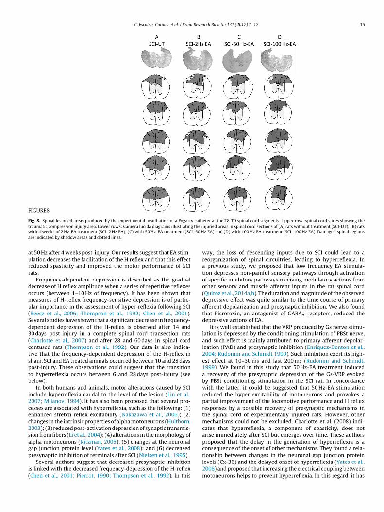
C. Escobar-Corona et al. / Brain Research Bulletin 131 (2017) 7–17 15
FIGURE8
Fig. 8. Spinal lesioned areas produced by the experimental insufflation of a Fogarty catheter at the T8-T9 spinal cord segments. Upper row: spinal cord slices showing thet g the iw I–50 Ha
aurr
domu(Sd3(ctsptb
i2cec2sagp
i(
raumatic compression injury area. Lower rows: Camera lucida diagrams illustratinith 4 weeks of 2 Hz-EA treatment (SCI–2 Hz EA); (C) with 50 Hz-EA treatment (SC
re indicated by shadow areas and dotted lines.
t 50 Hz after 4 weeks post-injury. Our results suggest that EA stim-lation decreases the facilitation of the H reflex and that this effecteduced spasticity and improved the motor performance of SCIats.
Frequency-dependent depression is described as the gradualecrease of H reflex amplitude when a series of repetitive reflexesccurs (between 1–10 Hz of frequency). It has been shown thateasures of H-reflex frequency-sensitive depression is of partic-
lar importance in the assessment of hyper-reflexia following SCIReese et al., 2006; Thompson et al., 1992; Chen et al., 2001).everal studies have shown that a significant decrease in frequency-ependent depression of the H-reflex is observed after 14 and0 days post-injury in a complete spinal cord transection ratsCharlotte et al., 2007) and after 28 and 60 days in spinal cordontused rats (Thompson et al., 1992). Our data is also indica-ive that the frequency-dependent depression of the H-reflex inham, SCI and EA treated animals occurred between 10 and 28 daysost-injury. These observations could suggest that the transitiono hyperreflexia occurs between 6 and 28 days post-injury (seeelow).
In both humans and animals, motor alterations caused by SCInclude hyperreflexia caudal to the level of the lesion (Lin et al.,007; Milanov, 1994). It has also been proposed that several pro-esses are associated with hyperreflexia, such as the following: (1)nhanced stretch reflex excitability (Nakazawa et al., 2006); (2)hanges in the intrinsic properties of alpha motoneurons (Hultborn,003); (3) reduced post-activation depression of synaptic transmis-ion from fibers (Li et al., 2004); (4) alterations in the morphology oflpha motoneurons (Kitzman, 2005); (5) changes at the neuronalap junction protein level (Yates et al., 2008); and (6) decreased
resynaptic inhibition of terminals after SCI (Nielsen et al., 1995).Several authors suggest that decreased presynaptic inhibitions linked with the decreased frequency-depression of the H-reflexChen et al., 2001; Pierrot, 1990; Thompson et al., 1992). In this
njuried areas in spinal cord sections of (A) rats without treatment (SCI-UT); (B) ratsz EA) and (D) with 100 Hz EA treatment (SCI–100 Hz EA). Damaged spinal regions
way, the loss of descending inputs due to SCI could lead to areorganization of spinal circuitries, leading to hyperreflexia. Ina previous study, we proposed that low frequency EA stimula-tion depresses non-painful sensory pathways through activationof specific inhibitory pathways receiving modulatory actions fromother sensory and muscle afferent inputs in the rat spinal cord(Quiroz et al., 2014a,b). The duration and magnitude of the observeddepressive effect was quite similar to the time course of primaryafferent depolarization and presynaptic inhibition. We also foundthat Picrotoxin, an antagonist of GABAA receptors, reduced thedepressive actions of EA.
It is well established that the VRP produced by Gs nerve stimu-lation is depressed by the conditioning stimulation of PBSt nerve,and such effect is mainly attributed to primary afferent depolar-ization (PAD) and presynaptic inhibition (Enríquez-Denton et al.,2004; Rudomin and Schmidt 1999). Such inhibition exert its high-est effect at 10–30 ms and last 200 ms (Rudomin and Schmidt,1999). We found in this study that 50 Hz-EA treatment induceda recovery of the presynaptic depression of the Gs-VRP evokedby PBSt conditioning stimulation in the SCI rat. In concordancewith the latter, it could be suggested that 50 Hz-EA stimulationreduced the hyper-excitability of motoneurons and provokes apartial improvement of the locomotive performance and H reflexresponses by a possible recovery of presynaptic mechanisms inthe spinal cord of experimentally injured rats. However, othermechanisms could not be excluded. Charlotte et al. (2008) indi-cates that hyperreflexia, a component of spasticity, does notarise immediately after SCI but emerges over time. These authorsproposed that the delay in the generation of hyperreflexia is aconsequence of the onset of other mechanisms. They found a rela-
tionship between changes in the neuronal gap junction proteinlevels (Cx-36) and the delayed onset of hyperreflexia (Yates et al.,2008) and proposed that increasing the electrical coupling betweenmotoneurons helps to prevent hyperreflexia. In this regard, it has
1 Resea
bst2Htcm2iil
rntppttoat
nraaaaticwraotanDti
eoEtmEeeoitm
wcttpvnq
6 C. Escobar-Corona et al. / Brain
een reported that EA stimulation modified the protein expres-ion of connexin 43 and improved the synaptic reorganization ofhe marginal zone of focal cerebral ischemia in rats (Luo et al.,011). In addition, Lee et al. (2007) observed an increase in the-reflex amplitude after 4 weeks post-injury which was posi-
ively correlated with an increased expression of 5-HT2R in theontused spinal cords of rats, and EA stimulation was able toodify the serotoninergic system in the spinal cord (Zhang et al.,
014). In this way, it could be proposed that EA stimulationnduces its effects on motoneuron hyper-excitability by modulat-ng multiple pre- and postsynaptic mechanisms at the spinal cordevels.
The significant effect of 50 Hz of EA on the motor functionecovery of SCI animals could offer several clinical paradigms foreurological recovery of SCI human patients. However, it is impor-ant to clarify that we cannot assume that acupoints, electricalarameters of EA such as intensity, amplitude of the stimulationulse (the resistance for current electricity is different in the humanhan in the rat), and that the model of SCI compression in rats areotally equivalent to those corresponding to humans. In this wayur results needs to be corroborated in further studies in humans,lone or in combination with other pharmacological or physicalherapeutic procedures.
In addition, there are several limitations in our study thateeds to be considered. One limitation is the lack of a group thateceived EA on non acupoints. It could be possible that the effectsscribed to a simple peripheral electrical stimulation or specificcupuncture stimulation on the acupoints located on the backnd hindlimb, are related to a combination of acupoint and non-cupoint actions. However, in a previous study we have showedhat EA on non-acupoint sites does not evoke significant changesn the SU nerve evoked cord dorsum potentials, suggesting a spe-ific acupoint effect of EA (Quiroz et al., 2014a,b). In such studye proposed that EA reduces the activation of dorsal horn neu-
ons provoked by low-threshold cutaneous afferent fibers by thectivation of specific sensory pathways in the spinal dorsal hornf the rat (Quiroz et al., 2014a,b). In this study we only analyzedhe effect of different frequencies of EA at the same model of SCInd with acupoints which previously has been reported to haveeuroprotective effect on SCI (Heo et al., 2013a,b; Min et al., 2015;ing et al., 2011). However, further studies are needed to analyze
he acupoint specificity of 50 Hz of EA stimulation for spinal cordnjury.
A second limitation is that our study do not contemplate theffect of muscle activation by EA stimulation on the improvementf locomotive performance of SCI rats. It could be proposed thatA stimulation provokes the electrical activation of muscles nearo acupoints and such action induce an improvement in the loco-
otive performance of SCI-rats. Previous research support thatA stimulation has the capacity to produce movement in den-rvated, paralyzed, or spastic muscles, but it is inherently lessfficient than human movement (Doucet et al., 2012). In this wayur study did not contemplate the analysis of muscle properties
n SCI rats to exclude the possible involvement of muscle activa-ion in the improved motor performance of SCI rats with EA treat-
ent.Another issue that has not been contemplated at our study
as the evaluation of a combined treatment of EA with physi-al therapy or pharmacological interventions that could maximizehe therapeutic effects of EA on SCI. Although biphasic pulse ishe most common mode of EA stimulation on basic and clinicractice other parameters as pulse width, electrode placement,
ariable frequency pulse, amplitude patterns and program durationeeds to be characterized. The proper characterization of an ade-uate treatment or combinations of treatments, in turn, will definerch Bulletin 131 (2017) 7–17
how quickly these advances can become commonplace in theclinic
5. Conclusions
The results obtained in this study demonstrated that EA stimula-tion applied at 50 Hz improves the motor performance and H-reflexamplitude by a possible recovery of presynaptic mechanisms in thespinal cord of experimentally injured rats. However, other mecha-nisms could not be excluded. The significant effect of 50 Hz of EAon the motor function recovery of SCI animals could offer severalclinical paradigms for neurological recovery of SCI human patients.
Acknowledgements
We would like to thank American Journal Experts for editing theEnglish version of the manuscript, José Carlos Guadarrama Olmosfor technical assistance, and Enrique Velazquez and Porfirio Reyesfor programming assistance. This work was partially supported byfellowships granted to E. Rodriguez-Torres, I. Jiménez-Estrada andS. Quiroz-González by the Mexican Sistema Nacional de investi-gadores (SNI) and partially supported by a PROMEP granted to E.Rodriguez-Torres and S. Quiroz-González.
References
Attal, N., Mazaltarine, G., Perrouin-Verbe, B., Albert, T., SOFMER French Society forPhysical Medicine and Rehabilitation, 2009. Chronic neuropathic painmanagement in spinal cord injury patients. What is the efficacy ofpharmacological treatments with a general mode of administration? (oral,transdermal, intravenous). Ann. Phys. Rehabil. Med. 52 (2), 124–141.
Barnes, P.M., Bloom, B., Nahin, R.L., 2008. Complementary and alternative medicineuse among adults and children. Natl. Health Stat. Rep. 10, 1–23.
Basso, D.M., Beatti, M.S., Bresnahan, J.C., 1995. A sensitive and reliable locomotorrating scale for open field testing in rats. J. Neurotrauma 12, 1–21.
Bennett, D.J., Li, Y., Harvey, P.J., Gorassini, M., 2001. Evidence for plateau potentialsin tail motoneurons of awake chronic spinal rats with spasticity. J.Neurophysiol. 86, 1972–1982.
Bregman, B.S., Coumans, J.V., Dai, H.N., 2002. Transplants and neurotrophic factorsincrease regeneration and recovery of function after spinal cord injury. Prog.Brain Res. 137, 257–273.
Côté, M.P., Gandhi, S., Zambrotta, M., Houlé, J.D., 2014. Exercise modulates chloridehomeostasis after spinal cord injury. J. Neurosci. 34, 8976–8987.
Calderón, V.D., Quintanar, S.A., Hernández, J.I., Jiménez, H.V., Ruiz, O.J., Jiménez, I.,Quintanar, J.L., 2015. Functional and structural recovery of the injured spinalcord in rats treated with gonadotropin-releasing hormone. Neurochem. Res.40, 455–462.
Charlotte, C., Yates, P.T., Charlesworth, A., Allen, S., Reese, N., 2007. The onset ofhyperreflexia in the rat following complete spinal cord transection. Exp.Neurol. 203, 502–511.
Chen, R., Corwell, B., Yaseen, Z., Hallett, M., Cohén, L.G., 1998. Mechanisms ofcortical reorganization in lower-limb amputees. J. Neurosci. 18, 3434–3450.
Chen, X.Y., Feng-Chen, K.C., Chen, L., Stark, D.M., Wolpaw, J.R., 2001. Short-termand medium-term effects of spinal cord tract transactions on soleus H-reflex infreely moving rats. J. Neurotrauma 18, 313–327.
Ding, Y., Yan, Q., Ruan, J.W., Zhang, Y.Q., Li, W.J., Zeng, X., Huang, S.F., Zhang, Y.J.,Wang, S., Dong, H., Zeng, Y.S., 2011. Bone marrow mesenchymal stem cells andelectroacupuncture down regulate the inhibitor molecules and promote theaxonal regeneration in the transected spinal cord of rats. Cell Transplant. 20,475–491.
Doucet, B.M., Lam, A., Griffin, L., 2012. Neuromuscular electrical stimulation forskeletal muscle function. Yale J. Biol. Med. 85, 201–215.
Enríquez-Denton, M., Manjarrez, E., Rudomin, P., 2004. Persistence of PAD andpresynaptic inhibition of muscle spindle afferents after peripheral nerve crush.Brain Res. 1027, 179–187.
Gerasimenko, Y., Gorodnichev, R., Moshonkina, T., Sayenko, D., Gad, P., ReggieEdgerton, V., 2015. Transcutaneous electrical spinal-cord stimulation inhumans. Ann. Phys. Rehabil. Med. 58, 225–231.
Gozariu, M., Roth, V., Keime, F., Le Bars, D., Willer, J.C., 1998. Anelectrophysiological investigation into the monosynaptic H-reflex in the rat.Brain Res. 782, 343–347.
Hiersemenzel, L.P., Curt, A., Dietz, V., 2000. From spinal shock to spasticity:
neuronal adaptations to a spinal cord injury. Neurology 54, 1574–1582.Holaday, J.W., Faden, A.I., 1983. Spinal shock and injury: experimental therapeuticapproaches. Adv. Shock Res. 10, 95–98.
Huang, S., Tang, C., Sun, S., Cao, W., et al., 2015. Protective effect ofelectroacupuncture on neural myelin sheaths is mediated via promotion of
Resea
H
H
H
H
H
K
L
L
L
L
L
L
L
M
M
M
M
M
N
N
N
N
electroacupuncture on persistent pain. Anesthesiology 120, 482–503.Zhao, Z.Q., 2008. Neural mechanism underlying acupuncture analgesia. Prog.
C. Escobar-Corona et al. / Brain
oligodendrocyte proliferation and inhibition of oligodendrocyte death aftercompressed spinal cord injury. Mol. Neurobiol. 52, 1870–1881.
ultborn, H., 2003. Changes in neuronal properties and spinal reflexes duringdevelopment of spasticity following spinal cord lesions and stroke: studies inanimal models and patients. J. Rehabil. Med. 41, 46–55.
ultborn, H., 2006. Spinal reflexes, mechanisms and concepts: from Eccles toLundberg and beyond. Prog. Neurobiol. 78, 215–232.
uston, T., Gassaway, J., Wilson, C., Gordons, S., Koval, J., Schwebe, A., 2011. The SCIRehab. project: treatment time spent in SCI rehabilitation: psychologytreatment time during inpatient spinal cord injury rehabilitation. J. Spinal CordMed. 34, 196–204.
eo, In, Byung, C.S., Young, D.K., Eui, H.H., Chang, W.H., Kwang, H.H., 2013a.Acupuncture for spinal cord injury and its complications: a systematic reviewand meta-analysis of randomized controlled trials. Evid. Based Complement.Altern. Med. 2013, 11–18.
eo, In, Byung, C.S., Young, D.K., Eui, H.H., Chang, W.H., Kwang, H.H., 2013b.Acupuncture for spinal cord injury and its complications: a systematic reviewand meta-analysis of randomized controlled trials. Evid. Based Complement.Altern. Medicine. 2013, 1–18.
itzman, P., 2005. Alteration in axial motoneuronal morphology in the spinal cordinjured spastic rat. Expert Neurol. 192, 100–108.
ee, J.K., Emch, G.S., Johnson, C.S., Wrathall, J.R., 2005. Effect of spinal cord injuryseverity on alterations of the H-reflex. Exp. Neurol. 196, 430–440.
ee, J.K., Johnson, C.S., Wrathal, J.R., 2007. Up-regulation of 5-HT2 receptors isinvolved in the increased H-reflex amplitude after contusive spinal cord injury.Exp. Neurol. 203, 502–511.
i, Y., Li, X., Harvey, P.J., Bennett, D.J., 2004. Effects of baclofen on spinal reflexesand persistent inward currents in motoneurons of chronic spinal rats withspasticity. J. Neurophysiol. 92, 2694–2703.
in, C.S., Macefield, V.G., Elam, M., Wallin, B.G., Engel, S., Kiernan, M.C., 2007.Axonal changes in spinal cord injured patients distal to the site of injury. Brain130, 985–994.
iu, F., Zou, Y., Liu, S., Liu, J., Wang, T., 2013. Electro-acupuncture treatmentimproves neurological function associated with downregulation of PDGF andinhibition of astrogliosis in rats with spinal cord transection. J. Mol. Neurosci.51, 629–635.
onjon, N., Kouyoumdjian, P., Prieto, M., Bauchet, L., Haton, H., Gaviria, M., Privat,A., Perrin, F.E., 2010. Early functional outcomes and historical analysis aftercord compression injury in rats. J. Neurosurg. Spine 12, 106–113.
uo, Y., Xu, N.G., Yi, W., Yu, T., Yang, Z.H., 2011. Study on the correlation betweensynaptic reconstruction and astrocyte after ischemia and the influence ofelectroacupuncture on rats. Chin. J. Integr. Med. 17, 750–757.
azzocchio, R., Scarfò, G.B., Mariottini, A., Muzii, V.F., Palma, L., 2001. Recruitmentcurve of the soleus H-reflex in chronic back pain and lumbosacralradiculopathy. BMC Musculoskeletal Disord. 2001, 2–4.
etz, G.A., Merkler, D., Dietz, V., Schwab, M.E., Fouad, K., 2000. Efficient testing ofmotor function in spinal cord injured rats. Brain Res. 883, 165–177.
ilanov, I., 1994. Examination of the segmental pathophysiological mechanisms ofspasticity. Electromyogr. Clin. Neurophysiol. 34, 73–79.
in, F.W., Shu, Q.Z., Jia, B.L., Ye, L., Qing, S.Z., Rui, G., 2015. Neuroprotective effectsof electroacupuncture on early- and late-stage spinal cord. Injury NeuralRegenr. Res. 10, 1628–1634.
o, Y.P., Yao, H.J., Lv, W., Song, L.Y., et al., 2016. Effects of electroacupuncture atgovernor vessel acupoints on Neurotrophin-3 in rats with experimental spinalcord injury. Neural Plast. 2016, 2371875.
akazawa, K., Kawashima, N., Akai, M., 2006. Enhanced stretch reflex excitabilityof the soleus muscle in persons with incomplete rather than complete chronicspinal cord injury. Arch. Phys. Med. Rehabil. 530, 71–75.
ew, P.W., Rawicki, H.B., Bailey, M.J., 2002. Nontraumatic spinal cord injury:demographic characteristics and complications. Arch. Phys. Med. Rehabil. 83,996–1001.
ielsen, J., Petersen, N., Crone, C., 1995. Changes in transmission across synapses of
Ia afferents in spastic patients. Brain 118, 995–1004.orrbrink, B.C., Kowalski, J., Lunderberg, T., 2006. A comprehensive painmanagement programme comprising educational, cognitive and behaviouralinterventions for neuropathic pain following spinal cord injury. J. Rehabil. Med.38, 172–180.
rch Bulletin 131 (2017) 7–17 17
Osuna, C.L.P., López, R.J.R., Mendizabal, R.E.G., De la Torre, V.B., Banuelos, P.J.,Jiménez, E.I., Duenas, J.S.H., 2016. Quantitative analysis of hindlimbslocomotion kinematics in spinalized rats treated with tamoxifen plus treadmillexercise. Neuroscience 333, 151–161.
Peng, B., Meng, X.F., Li, M., Luo, Y., Li, L.L., Zhang, J., 2007. Effects ofelectroacupuncture on the expression of epidermal growth factor receptor andglial fibrillary acidic protein after spinal cord injury in rats. Zhen Ci Yan Jiu 32,219–223.
Pierrot, D.P., 1990. Electrophysiological assessment of the spinal mechanismsunderlying spasticity. New Trends Adv. Tech. Clin. Neurophysiol. 41, 264–273.
Politis, M.J., Korchinski, M.A., 1990. Beneficial effects of acupuncture treatmentfollowing experimental spinal cord injury: a behavioral, morphological, andbiochemical study. Acupunct. Electrother. Res. 15, 37–49.
Pomeranz, B., 1995. Electroacupuncture and Transcutaneous Electrical NerveStimulation: From Basics of Acupuncture. In: Stux, G., Pomeranz, B. (Eds.).Springer-Verlag, Berlin-Heidelberg, pp. 258–268.
Quiroz, G.S., Segura, A.B., Jiménez, E.I., 2014a. Depressing effect ofelectroacupuncture on the spinal non-painful sensory input of the rat. Exp.Brain Res. 232, 2721–2729.
Quiroz, G.S., Segura, A.B., Guadarrama, O.J.C., Jiménez, E.I., 2014b. Cord dorsumpotentials evoked by electroacupuncture applied to the hind limbs of rats. J.Acupunct. Meridian Stud. 7, 25–32.
Reese, N.B., Skinner, R.D., Mitchell, D., Yates, C., Barnes, C.N., Kiser, T.S., Garcia, R.E.,2006. Restoration of frequency-dependent depression of the H-reflex bypassive exercise in spinal rats. Spinal Cord 44, 28–34.
Reyes, C., Segura, B., Reza, J.A., Pacheco, M.T., Lomelí, J., Guadarrama, J.C., Guevara,R., Jiménez, I., 2007. Absence of linear correlation between fluctuations in areaof simultaneous recorded monosynaptic responses and Hoffmann’s reflexes inthe rat. Neurosci. Lett. 411, 249–253.
Rudomin, P., Schmidt, R., 1999. Presynaptic inhibition in the vertebrate spinal cordrevisited. Exp. Brain Res. 129, 1–37.
Sanes, J.R., Lichtman, J.W., 1999. Development of vertebrate neuromuscularjunction. Annu. Rev. Neurosci. 22, 389–442.
Schwab, M.E., Batholdi, D., 1996. Degeneration and regeneration of axons in thelesioned spinal cord. Physiol. Rev. 76, 319–370.
Silverman, S.R., Schertz, L.A., Yuen, H.K., Lowman, J.D., Bickel, C.S., 2012. Systematicreview of the methodological quality and outcome measures utilized inexercise interventions for adults with spinal cord injury. Spinal Cord. 10,718–727.
Thompson, F.J., Reier, P.J., Lucas, C.C., Parme, R., 1992. Altered patterns of reflexexcitability subsequent to contusion injury of the rat spinal cord. J.Neurophysiol. 68, 1473–1486.
Varma, A.K., Das, A., Wallace, G., Barry, J., Vertegel, A.A., Ray, S.K., Banik, N.L., 2013.Spinal cord injury: a review of current therapy, future treatments, and basicscience frontiers. Neurochem. Res. 38, 895–905.
Vickers, A.J., Cronin, A.M., Maschino, A.C., Lewith, G., MacPherson, H., Foster, N.E.,2012. Acupuncture for chronic pain: individual patient data meta-analysis.Arch. Intern. Med. 172, 1444–1453.
Xie, J., Fang, J., Feng, X., Liu, Q., 2006. Effect of electroacupuncture at acupoints ofthe governor vessel on aquaporin-4 in rat with experimental spinal cordinjury. J. Tradit. Chin. Med. 26, 148–152.
Yablon, S.A., Stokic, D.S., 2004. Neurophysiologic evaluation of spastic hypertonia.Am J. Phys. Med. Rehabil. 83, 10–18.
Yang, C., Li, B., Liu, T.S., Zhao, D.M., Hu, F.A., 2005. Effect of electroacupuncture onproliferation of astrocytes after spinal cord injury. Zhongguo Zhen Jiu 25,569–572.
Yates, C.C., Charlesworth, A., Reese, N.B., Skinner, R.D., Garcia-Rill, E., 2008. Theeffects of passive exercise therapy initiated prior to or after the developmentof hyperreflexia following spinal transection. Exp. Neurol. 213 (2), 405–409.
Zhang, R., Lao, L., Ren, K., Berman, B.M., 2014. Mechanisms of acupuncture-
Neurobiol. 85, 355–375.Zhu, Z., 2002. Effects of electroacupuncture on laminin expression after spinal cord
injury in rats. Zhongguo Zhong Xi Yi Jie He Za Zhi 22, 525–527.