brain research, elsevier/north-holland biomedical press ...155.pdf · elsevier/north-holland...
TRANSCRIPT

Brain Research, 225 (1981) 155-170 155 Elsevier/North-Holland Biomedical Press
CYTOPLASMIC ALKALIZATION REDUCES CALCIUM B U F F E R I N G IN MOLLUSCAN CENTRAL NEURONS
ROBERT S. ZUCKER
Department Of Physiology-Anatomy, University of California, Berkeley, CA 94720 (U.S.A.)
(Accepted April 9th, 1981)
Key words: calcium buffering -- molluscan neuron - - pH
SUMMARY
The effect of raised cytoplasmic pH (pHi) on intracellular calcium concentration ([Ca2+]i) transients following calcium influx during membrane depolarization was studied in identified neurons in the abdominal ganglion of Aplysia californica. The pHi was monitored with pH-sensitive microelectrodes. Sea water containing 15 mM NH 4C1 at pH 7.7 elevated pHi about 0.35 pH units from the normal level of 7.17. These cells have an estimated buffering power of about 60 mM/pH unit. Calcium influx was elicited by depolarizing pulses under voltage clamp and [Ca2+]i transients were monitored with the photoprotein aequorin or the metallochromic dye arsenazo III. Aequorin photo-emissions increased by 21-131~o (mean, 4 8 ~ ) a n d arsenazo III absorbance changes accompanying depolarization increased by 9-33 ~ (mean, 20 ~ ) after 30 min in N H C , corresponding roughly to a 14 ~ increase in [Ca2+]i transients. Calcium-dependent potassium tail currents following a depolarizing pulse were somewhat slower and 4--91 ~ (mean, 38 ~ ) larger in NH4 ÷. The magnitude and time- and voltage-dependence of the membrane calcium conductance was studied using calcium tail currents following depolarizing pulses. The calcium current was unaffect- ed by NH4 +, so the enhanced [Ca2+]i transients must reflect reduced calcium buffering at high pHi. Either reduced cytoplasmic calcium binding or slowed active extrusion of calcium may be responsible for this effect.
INTRODUCTION
In a recent report 4a, Dr. S. Smith and I showed that when Aplysia central neurons filled with aequorin are bathed in a solution containing 100 mM or more tetraethylammonium (,TEA) ion, light emissions accompanying membrane depolariza- tions were increased. This indicates that depolarization evokes a larger intracellular
0006-8993/81/0000~000/$02.50 © Elsevier/North-Holland Biomedical Press

156
free calcium concentration ([CaZ+]i) transient in TEA than in its absence. In an effort to determine how TEA exerts this effect, I measured intracellular pH (pHi) with pH- sensitive microelectrodes and found that externally applied commercial preparations of TEA alkalized neural cytoplasm 44. The present study was therefore undertaken to disclose any effect of cytoplasmic alkalization on [Ca2+]i transients accompanying membrane depolarization.
Not much is known about the interaction between intracellular pH and calcium. Meech and Thomas z6 and Ahmed and Connor 3 found that injection of calcium or its entry through membrane channels could acidify neural cytoplasm. Rose and Rick 32 found a similar effect in salivary gland cells. Lea and Ashley za, Rose and Rick 32, Ahmed and Connor 3 and Rink et al. al found that acidification of muscle, salivary gland, molluscan neuron and Xenopus embryo cytoplasm with external CO2 or H ÷ injection often resulted in an increase in resting [Ca2+]i levels, while Mullins and Requena 29 found that cytoplasmic alkalization of squid axon by external NH a + reduced [Ca2+]i. In contrast, Baker and Honerj~tger 8 reported that alkalization of squid giant axon with external NHa ÷ sometimes increased [Ca2+]i, while acidification had the opposite effect. The only published study on the effect of pHi on [Ca2÷]i transients is that of Mullins and Requena 29, who found an increase in [Ca2+]i transients near the periphery, but a decrease in peak [CaZ+]t transients in the center of squid axons, when cytoplasm was alkalized by external NH4 +. The influence of pHi on [Ca2÷]i transients in Aplysia central neurons is the subject of this paper.
METHODS
Aplysia californica (200-400 g) were obtained from Pacific Biomarine (Venice, CA) and kept in a 60 gal Instant Ocean tank at 16 °C. All experiments were performed on identified neurons from the abdominal ganglion. Ganglia were usually desheathed without pronase, and small clumps of axotomized cells were pinned to a Sylgard- bottom dish in a temperature-controlled chamber. Intact ganglia were used for aequorin experiments only. Cells were usually bathed in a normal artificial sea water (NASW) consisting of (mM): 495 Na +, 10 K +, 10 Ca 2+, 50 Mg 2+, 620 CI- and 10 HEPES buffer pH 7.5. Solutions containing 0.24 mM dissolved NHz gas were made by adding 15 mM NH4CI to NASW, and adjusting the pH to 7.7. The ammonium concentration of such solutions, as measured with ammonium-specific electrodes, remains stable for months.
Procedures for voltage clamping neurons, and for filling cells with aequorin or arsenazo III and detecting signals from these calcium indicators have been described in detail elsewhere 35. In order to obtain simultaneous information about absorbance changes at several wavelengths, an air turbine spinning chopper wheel similar in design to that described by Brinley et al. lz and Gorman and Thomas 21 was constructed. Apparatus designed by Dr. Stephen J. Smith was used to de-multiplex the optical signal into its individual wavelength components. This device integrates each light signal during a pulse of light, corrects for dark current sampled between pulses, and measures the ratio of transmitted light to incident light as described earlier 85. An

157
output corresponding to each wavelength and a differential output for any selected wavelength pair are provided.
Intracellular pH measurements were made either with arsenazo III (see below) or with pH-sensitive recessed-tip microelectrodes fabricated as described by Thomas 87. The electrodes used in these experiments responded in 20 s with a slope of 57-58 mV/pH unit in the range pH 5-8. A signal linearly related to pH was obtained by recording differentially between the pH electrode and the voltage-sensing electrode.
In most experiments voltage, current, and pH or optical signals were recorded on magnetic tape and a chart recorder. In some experiments using arsenazo III, these signals were digitized, collected using a Processor Technology Sol 20 microcomputer, and stored on minifloppy discs and a chart recorder. In these experiments the microcomputer was also used to generate pulse sequences for the command potential of the voltage clamp. Signals stored on magnetic discs were later retrieved and displayed in analogue form on a storage oscilloscope. Finally, in experiments on rapid deactivation or 'tail' currents following voltage-clamp pulses, the currents following an equal number of pulses of equal and opposite magnitude were averaged using a Neurolog NL750 signal averager, with a time resolution of 256 bits for a 10 ms sweep duration. Individual tail currents following single pulses of either polarity were simultaneously displayed on an oscilloscope screen and photographed.
Calibration of arsenazo III
Arsenazo III was used to measure changes in [CaZ+]l at different pills. Arsenazo III signals corresponding to changes in pHi were also recorded. Arsenazo III is known to respond nearly linearly to changes in calcium ion concentration between 0.1 and 20 #M, and also to be sensitive to pH changes in the presence of excess Mg 2+ 13,1.8,21,35,86. We previously calibrated our sample of arsenazo III for calcium concentration changes under conditions of ionic strength, pH, and magnesium concentration appropriate for normal molluscan cytoplasm 85. It was now necessary to extend this calibration to include the effect of pH on the arsenazo III response to a given change in calcium concentration, and also to measure the response of arsenazo III to a change in pH in the absence of significant free calcium.
The published spectrum of arsenazo III absorbance changes accompanying changes in [Ca 2+] and pH in the presence of excess Mg 2+ 1.8,18,21,23 suggests that changes in these ions can best be discriminated by observing the wavelength pair 660 and 630 nm. For changes in [Ca2+], the absorbance change at 660 nm is about double the change at 630 nm, while the reverse is true for changes in pH. I have therefore measured the absorbance change at these wavelengths and at the isosbestic point for [Ca 2+] and pH changes (577 nm), in solutions resembling cytoplasm with different buffered calcium and pH levels. Arsenazo III was dissolved at 250/zM (the same as the final concentration after injection into neurons) in a solution of 0.4 M KC1, 50 mM HEPES to buffer pH, and 50 mM DPTA (1,3-diaminopropan-2-ol-tetraacetic acid, Sigma) to buffer [Ca 2+] to either nominally 0 or 10 #M, as described previously 35. These levels are comparable to the resting [Ca~+]i and the peak submembrane [Ca2+]i following a membrane depolarizationlS, a~. The pH of the mixture was adjusted with

158
NaOH to either 7.1 or 7.4. These levels are similar to the resting pHi and the pHi after alkalization by external NH4 + (see Results). Absorbance measurements of these solutions were made with 1 mm pathlength cuvetttes in a Cary 14 spectrophotometer. The results are presented in Table I.
Note that the absorbance change at 660 nm, divided by the isosbestic absorbance, corresponding to a [Ca 2+] increase of 10 #M, decreased from 0.139 to 0.114 when the pH was raised from 7.1 to 7.4. This result at first seemed paradoxical, because if H + competes with Ca 2+ for binding sites on arsenazo II114,27 one would expect to see a larger absorbance change, due to a larger apparent affinity for calcium binding at high pH 36. However, raising pH increases the absorbance at 660 nm in the absence of Ca z+, probably because the hydrogen ion is replaced by magnesium on an arsenazo lII binding site in high pH in excess Mg 2+ (cf. ref. 23). Therefore, the absorbance at 660 nm of the 'uncomplexed' arsenazo III species is closer to that of the Ca-arsenazo I lI complex at high pH, and consequently the change in absorbance on raising [Ca z+] is 18~ less. This result is consistent with other reports that the sensitivity of arsenazo III to changes in [Ca 2+] is diminished at alkaline pH 14, especially in excess magnesium 2z. These effects are even more pronounced at 630 nm. Consequently, the absorbance change for an increase in [Ca 2+] using the wavelength pair 660-630 nm is very slightly larger (about 6 ~ ) in going from pH 7.1 to pH 7.4.
Table I also shows the expected result that for changes in [Ca2+], the absorbance
change at 660 nm is 2-3 times that at 630 nm, while for changes in pH, the normalized absorbance change at 660 n m ( A ( A 6 6 0 / A 5 7 7 ) - - 0.070 for zero calcium) is about half the change at 630 nm (A(A6so/As77) = 0.123). That the same distinction in the spectral response of arsenazo III holds for calcium and pH changes in vivo is illustrated in Fig. 1.
This figure confirms that small cytoplasmic pH changes induced by exposure to NH4 + are not accompanied by measurable changes in [Ca2+]i, although small changes would go undetected. Moreover, the influx of calcium during depolarization is not accompanied by detectable changes in pHi, since the spectrum of arsenazo II1 absorbance changes matches that of a calcium concentration change. From the work of Ahmed and Connor ~, pHi would be expected to change by less than 0.01 units for the calcium influxes occurring in the present experiments.
TABLE I
Absorbance of arsenazo 111 solutions with different [Ca 2+] and pH
Abbreviations: An, absorbance at wavelength n; A(An/.4577), normalized absorbance change at n nm; A(A66o-63o/Asvv), normalized differential absorbance change at the wavelength pair 660 and 630 nm. Arsenazo III concentration: 250/~M. Path length: I mm.
[Ca 2+] A630 ZI A660 ZI A660-630 pH (l~M) A577 A630 .-4660 A A577 A577 A 5 7 ~
7.1 0 0.896 0.373 0.174 0.067 0.139 0.0722 7.1 10 0.900 0.435 0.300 7.4 0 0.907 0.490 0.240 0.037 0.114 0.0767 7.4 10 0.923 0.532 0.350

159
A ApH = -0.3 Arco2+'I i • I pM
6A$60-577 AS77
AA630-S77 AS77
0.01 I
-.~.~.~
2 min 50 mse¢
Fig. 1. Absorbance changes in a neuron filled with 0.29 mM arsenazo III, in response to changes in cytoplasmic pH and calcium concentration. A: reduction in pHi induced by washing out a solution of normal artificial sea water containing 15 mM NH 4C1 and buffered to pH 7.7. The absorbance change at 630 nm is about triple that at 660 nm. B: increase in [Ca2+]t during a 200 ms depolarizating pulse to +20 mV in normal sea water. The absorbance change at 660 nm is about 2.4 times that at 630 nm. Cell R15. Temperature 16 °C. Holding potential 4 0 mV in all experiments. The approximate changes in pH and [Ca2+]l shown in the headings are estimated from the peak normalized differential absorbance changes (AA66o-sao/A577) and the data of Table I, assuming near-zero calcium in A and pHi of 7.1 in B.
Table I also shows that arsenazo III is sensitive to a change in pH from 7.1 to 7.4 in zero-calcium. Such a change in pH is accompanied in vitro by a decrease in the normalized differential absorbance of the wavelength pair 660-630 nm (A(A~6o-6zo/ A577)) of 0.052. These data are inadequate to reveal the relation between ApH and AA. Thus, the pH-dependent arsenazo III absorbance change can only be referred to this particular pH change, and no extrapolation to other pH levels or changes is
warranted. The table does, however, provide calcium calibration factors appropriate for two pH levels, 7.1 and 7.4, and these values are used to convert absorbance changes to calcium concentration changes at these pH levels. Conversion at other pH levels by
interpolation or extrapolation would only be approximate.
RESULTS
Effect o f NH4 + on cytoplasmic p H Cytoplasmic pH was raised by exposing neurons to the permeant weak base
NHa. In a solution of artifidal sea water with 15 mM NH4CI buffered at pH 7.7, 0.24 mM of the ammonium will be in the form of NHa. Ammonia rapidly penetrates cell
membranes, and at equilibrium the cytoplasmic NHa concentration should also be 0.24 mM. The pH of the cytoplasm will change to a final value of pHi ~ from its initial level pHi ° by an amount ApHi depending on the buffering power, fl, of the cytoplasm. fl is defined as the concentration of the proton donor NHa + needed to change cytoplasmic pH by one unit 10. Substituting this definition into the Henderson-Hassel-

160
balch equation yields the following expression for a change in cytoplasmic pH:
[NH3] A p H i = pKa - - pHi ° -[- log10 - - (1)
pH
where pK a is the negative logarithm of the dissociation constant of NH4 ÷ in sea water and pKa = 9.51°. In four experiments, neurons were exposed to 15 mM NHaC1 at pH 7.7, and pHi was monitored with an intracellular hydrogen-selective micro- electrode or by recording absorbance changes at 630, 660 and 577 nm in cells filled with arsenazo III. pH-sensitive microelectrodes report the absolute value o f p H as well as its change, so that Eqn. 1 can be solved explicitly for ft. In seven cells, the average pHi ° in normal artificial sea water was 7.17 (range 7.0-7.3). In four of these cells treated with NH4C1, pHi rose slowly for 20-30 min to 0.35 units (range 0.2-0.5 units) above the initial level. On removing the ammonium, p h i recovered to near or even below its original level. Such an experiment is illustrated in Fig. 2. In four other experiments, the effect of NH4 + on arsenazo I I I absorbance was observed. In all cases, the absorbance at 630 nm increased more then the absorbance at 660 nm, and AAgro-fzo/A577 w a s about -43.050 (range --0.021 to --0.093). These results corres- pond to the observed in vitro changes in arsenazo I I I absorbance when pH is increased from 7.1 to 7.4, and so are consistent with such a change in pHi.
Using the values pHi = 7.1 and ApH = 0.35, a buffering power of fl ----- 86 m M / p H unit may be calculated from Eqn. 1. In other experiments, exposure to 5 CO2 sea water equilibrated with 29 mM HCO3- at pH 7.4 led to a drop in pHi of about 0.015 unit, measured with a hydrogen-sensitive microelectrode. This corresponds to an apparent buffering power of about 68 m M / p H unit. This is substantially higher than the values calculated for snail cytoplasm a7 and squid axoplasm 10. This difference in
buffering power may be more apparent than real, however. In snail and squid, NH4CI and CO2 treatment altered cytoplasmic pH more rapidly than in Aplysia, and the initial change was followed by a slow recovery due to the membrane permeability to NH4 + and to the action of active transport of protons or hydrogen buffers. Moreover,
7.S
- 73 I "1-
Z 2
7.1
70
f . . . .
NASW
Fig. 2. Effect of sea water containing 15 mM NHaCI buffered to pH 7.7 (15 mM NH4CIpH7.7 SW) on cytoplasmic pH. phi was monitored with a pH-sensitive microelectrode. After 25 min the cell was washed in normal artificial sea water (NASW). The neuron (cell L10 was voltage clamped at 40 inV. Temperature 15 °C.
161
NASW A
I G
B
Vm
Ia
nlA I o- _ j ' ~ . , _ , ~ . . ~
c
15 mM NH4CI pH Z7 SW NASW
/
1 sec
5 s e c
15-
iO-
-~ 5
0 ~ ,
0 20 40 60 Pulse potential (mY)
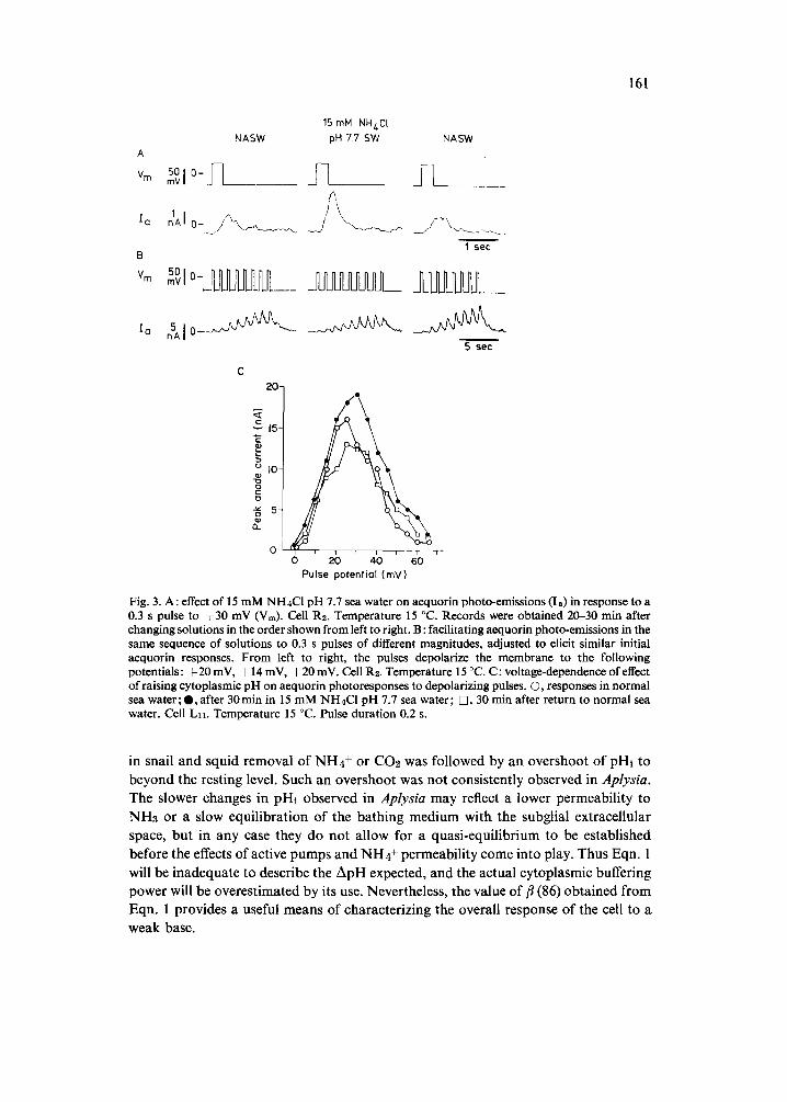
Fig. 3. A: effect of 15 mM NH4CI pH 7.7 sea water on aequorin photo-emissions (Is) in response to a 0.3 s pulse to ÷30 mV (Vm). Cell R2. Temperature 15 °C. Records were obtained 20-30 rain after changing solutions in the order shown from left to right. B: facilitating aequorin photo-emissions in the same sequence of solutions to 0.3 s pulses of different magnitudes, adjusted to elicit similar initial aequorin responses. From left to right, the pulses depolarize the membrane to the following potentials: + 20 mV, + 14 mV, + 20 mV. Cell R2. Temperature 15 °C. C: voltage-dependence of effect of raising cytoplasmic pH on aequorin photoresponses to depolarizing pulses. O, responses in normal sea water; O, after 30min in 15 mM NH4C1 pH 7.7 sea water; D, 30 rain after return to normal sea water. Cell Lll. Temperature 15 °C. Pulse duration 0.2 s.
in snail and squid removal o f N H 4 + or CO2 was fol lowed by an overshoot o f pHi to
beyond the resting level. Such an overshoot was not consistent ly observed in Aplysia. The slower changes in pHi observed in Aplysia may reflect a lower permeabi l i ty to
NH3 or a slow equi l ib ra t ion o f the ba th ing med ium with the subglial extracel lular
space, bu t in any case they do no t a l low for a quas i -equi l ibr ium to be es tabl ished
before the effects o f active pumps and N H 4 + permeabi l i ty come into play. Thus Eqn. 1
will be inadequa te to describe the A p H expected, and the actual cy toplasmic buffering
power will be overes t imated by its use. Nevertheless, the value o f fl (86) ob ta ined f rom
Eqn. 1 provides a useful means o f character iz ing the overal l response o f the cell to a
weak base.

162
A better estimate of cytoplasmic buffering power might be obtained by assuming that the late acidification often seen in NH4 + (Fig. 2) reflects recovery processes. These may be back-extrapolated to estimate the pHi change that would have occurred in the absence of such effects. This procedure results in an average ApHi estimate of 0.5 units, or a/3 of about 60 mM/pH unit.
Effect of cytoplasmic pH on aequorin signals The effect of cytoplasmic alkalization on intracellular calcium transients follow-
ing calcium influx through voltage-dependent membrane channels was assessed by 3 measures: the size of photo-emissions from neurons injected with aequorin, the size of absorbance changes from neurons filled with arsenazo III, and the size of calcium- dependent potassium currents. In six neurons injected with about 25 #M aequorin, a 30 min exposure to 15 mM NH4C1 pH 7.7 sea water increased calcium-evoked photo- emissions by 21-131 ~ (mean 48~). This enhancement of aequorin responses was reversed in 5 of these 6 experiments on washing out the ammonium. Recordings from a typical experiment illustrating this effect are reproduced in Fig. 3A.
The effect of NH4 ÷ on aequorin emissions is not dependent on the amplitude or duration of the depolarization used to trigger calcium influx (Fig. 3C). Thus changing cytoplasmic pH does not influence the voltage-dependence of calcium influx, but only the magnitude of the resulting calcium transient for any size depolarization. Note that the calcium-aequorin photo-reaction is virtually independent of pH in the range of pHi variation of these experiments2S, a3.
Another effect often observed is that the larger photo-emissions obtained in NH4 + facilitate slightly less to trains of pulses. This remained true even when the amplitude of the depolarizing pulse was reduced so that the first response was comparable to what it had been in normal artificial sea water. Fig. 3B illustrates such an experiment. In normal sea water, the sixth 0.3 s pulse to +20 mV at 1 Hz elicited a response that was 400~ greater than the first. In 15 mM NH4C1 pH 7.7 sea water, the first response to a pulse to +20 mV was now twice as large as before cytoplasmic alkalization, but the sixth response was only 350 ~ greater than the first. When the peak pulse potential was reduced to q- 14 mV to compensate the effect of pHi on the size of the first aequorin photo-emission, the sixth response was still only 350 ~ greater than the first. This small effect of NH4 + on aequorin-response facilitation, like the effect on the magnitude of aequorin photo-emissions, was reversible.
Effect of raising cytoplasmic pH on arsenazo III signals Aequorin is sensitive in a very non-linear fashion to calcium concentration
changes in the micromolar region, responding to the 2.5th power of [CaZ+]L Aequorin thus reports preferentially the changes in local high calcium concentration near the plasma membrane during a depolarization, and changes in aequorin signals might reflect only different distributions of the same amount of free [Ca2+]i3~. To confirm the apparent effect of pHi on free [CaZ+]i transients accompanying depolarizations, 4 neurons were filled with about 250 #M arsenazo III and exposed to 15 mM NH4C1 pH 7.7 sea water. When pHi had presumably risen an average of 0.35 units (see above),

163
t~
< 05 I
0
® 0.0 5 ~D ~D
< <3
0.008 -
f~ 0.006 - <~
0
O.O04- t 0 '.D
<3 0.002-
' ' ' z 3 ' - IO 80
Pulse potential (mV)
Fig. 4. Effect of cytoplasmic alkalization on arsenazo III absorbance changes in response to 0.2 s pulses to +20 mV. A: normalized differential absorbance changes in normal sea water (1), 30 min after changing to 15 mM NH4CI pH 7.7 sea water (2), and 30 min after a normal sea water rinse (3). B : dependence of peak absorbance changes on potential during 0.2 s depolarizating pulses in normal sea water (©), after 30 min in 15 mM NH4C1 pH 7.7 sea water (O), and 30 min after return to normal sea water ([i]). In A, cell Rls was injected with about 300 #M arsenazo III as estimated from the absorbance at 577 nm, and A A660-63o/A.577 of 0.005 corresponds roughly to an average [Ca2+]l change of 590 nM in normal sea water, and 560 nM in NH4CI sea water. In B, an unidentified 250 /~m diameter cell near R15 was filled with 250/~M arsenazo III, and AA68o-nao/Asvv of 0.005 corresponds to a A[Ca2+]l of about 690 mM in normal sea water and 650 nM in NH4C1 sea water. Temperature 16 ° C .
arsenazo I | I absorbance signals report ing a calcium concentra t ion change during a
0.2 s depolarizing pulse were increased on average by 2 0 ~ (range 9-339/0). In all
experiments, the absorbance change at 660 nm was 2-3 times that at 630 nm, indicating
that calcium influx changed [Ca2+]i with little change in pHi. This effect was
completely reversible on returning to normal sea water (Fig. 4A).
The effect o f elevated pH~ on [Ca2+]~ transients was independent o f the
ampl i tude o f the depolar iz ing pulse (Fig. 4B). The change in absorbance signals at the

164
wavelength pair 660-630 nm is greater than can be accounted for by the 6 ~ change in the sensitivity of arsenazo III to [CaZ÷]i transients due to the measured pHi change (see Methods). The results therefore suggest a real increase in free [Ca2+]i transients (about 14 ~ above normal on average) accompanying depolarization when cytoplasm is alkalized by NH4 ÷. This conclusion is further supported by the records of 660 nm absorbance changes. These often increased slightly in NH4 ÷. Such a change is in the opposite direction of the effect of pH on the calcium sensitivity of arsenazo III at this wavelength (see Table I).
Effect of raising cytoplasmic pH on calcium-dependent potassium current Molluscan neurons provide a natural indicator of submembrane calcium
concentration in the form of calcium-activated potassium current, IKtca) zS. Since the above chemical indicator results both suggest an increased [Ca2+]i transient accom- panying depolarization in elevated pHi, similar effects on Is(ca) are expected. The Ix(ca) component of potassium current can be most easily measured in isolation as a slow outward tail current several hundred msec following a depolarizing pulse 34,35, 38,39 at a holding potential positive to the reversial potential for lK(ca). Such tail currents are illustrated in Fig. 5. When measured 200 ms after the end of a pulse, this current increased an average of 38 ~ (range 4-91 ~ , 8 experiments) after 30 min in 15 mM NH4C1 pH 7.7 sea water. Furthermore, its decay rate was slowed an average of 29 ~ (range 4-65 ~). The effects were partially or completely reversible after a 30 min normal sea water rinse (mean recovery to within 14 ~ of the amplitude).
Interpretation of these observations of IK(Ca) is subject to the limitation that the reversal potential of 1KtCa) was not measured under the various experimental conditions. Increased potassium current with elevated pHi should accentuate external
0
Irn nA
i
0.2 sec
Fig. 5. Effect of raising cytoplasmic pH on calcium-activated potassium current, Ix(ca). 1,1 records lmca) deactivation tails following a 0.2 s pulse to ÷20 mV (Vm) in normal sea water (1), after 30 min in 15 mM NHaC1 pH 7.7 sea water (2), and after a 30 min normal sea water rinse (3). Cell Rt5. Temperature 16 °C.

165
~ f ~ . . . . . w / Im
w
Fig. 6. Calcium deactivation tail currents following depolarizing pulses. In the top row is membrane current (Ira) following single pulses of equal magnitude and opposite polarity from a hold potential of 4 0 inV. In the bottom row, 8 responses of each polarity are averaged. Pulse potentials and duration are --15 mV and --65 mV, 70 ms in A (d: 25 mV pulses), ÷ 15 mV and --95 mV, 70 ms in B (± 55 mV pulses), ÷45 mV and --125 mV, 70 ms in C (± 85 mV pulses), and ÷ 15 mV and --95 mV, 300 ms in D (± 55 mV pulses). Cell L3. Temperature 7 °C.
potassium accumulation and reduce outward potassium tail currents. The enhanced tail currents are thus in the opposite direction from the likely effect of changes in potassium equilibrium potential. The results suggest that elevated pHi increases the calcium-activated potassium permeability. This appears to reflect the effect of p h i on [Ca2--]i transients monitored with aequorin and arsenazo III, although a direct effect on the potassium channels cannot be excluded.
Eff~ect of raising cytoplasmic pH on calcium current The results so far point to an effect of cytoplasmic alkalization on the magnitude
of [Ca2+]i transients during membrane depolarization. These effects may be due to an influence of pHi on calcium influx through voltage-dependent calcium channels or an interaction between pHi and intracellular calcium buffering. To distinguish between these possibilities, membrane calcium currents were recorded under conditions of altered pHi. Calcium currents were isolated non-pharmacologically by measuring the rapidly deactivating inward calcium tail current following depolarizing pulses1,17, 35. These brief inward currents, occurring just after the repolarizing step which terminates a depolarizing pulse, reflect the current flowing through calcium channels that were opened by the depolarization. The current vanishes exponentially with a time-constant of 2 ms at 7 °C. (see Fig. 6), as the calcium channels close rapidly, or deactivate, at the hold potential.
The sodium current inactivates completely during a long depolarization 1, so sodium currents will not contribute to the tail current after such pulses. I f the potential following a depolarizing pulse is set to - -40 mV, there will be a small component of potassium deactivation current. But at 7 °C, this component decays with a time constant of about 35 ms 84 and so contributes little to the early changes in membrane current following a repolarizing step. The linear capacitance current comprises a mucb larger part of the early membrane current following a potential step. This component may be estimated from an equal and opposite potential step following a hyperpolari- zation, and may be subtracted from the current after a depolarizing step using an averaging computer. Fig. 6 shows how net inward calcium tail currents after
166
300-
250-
200-
15o-
o 1oo-
~- 50-
0 i
-15
A 150-
125 -
.5 = I 00 -
~= 7a-
~ ~°
~ 25-
O- r I I /
0 15 3O ns Pulse potential (mY)
B
~'¢ i i 1 i i "
3 I0 50 I00 500 I000 Pulse duration (msec)
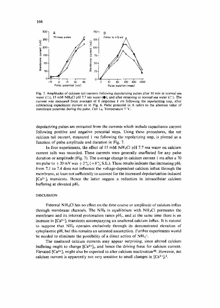
Fig. 7. Amplitudes of calcium tail currents following depolarizing pulses after 30 min in normal sea water (O), 15 mM NH4CI pH 7.7 sea water (O), and after returning to normal sea water ([]). The current was measured from averages of 8 responses 1 ms following the repolarizing step, after subtracting capacitance current as in Fig. 6. Pulse potential in A refers to the absolute value of membrane potential during the pulse. Cell Lz. Temperature 7 °C.
depolarizing pulses are extracted from the currents which include capacitance current following positive and negative potential steps. Using these procedures, the net calcium tail cmrent, measured 1 ms following the repolarizing step, is plotted as a function of pulse amplitude and duration in Fig. 7.
In four experiments, the effect of 15 mM NH4C1 pH 7.7 sea water on calcium current tails was recorded. These currents were generally unaffected for any pulse duration or amplitude (Fig. 7). The average change in calcium current 1 ms after a 70 ms pulse to +20 mV was + 2 ~ (-k8 ~ S.E.). These results indicate that increasing pHi from 7.1 to 7.4 does not influence the voltage-dependent calcium influx through the membrane, at least not sufficiently to account for the increased depolarization-induced [Ca2+]l transients. Hence the latter suggest a reduction in intracellular calcium buffering at elevated pHi.
D I S C U S S I O N
External NH4CI has no effect on the time course or amplitude of calcium influx through membrane channels. The NH3 in equilibrium with NH4CI permeates the membrane and its internal protonation raises pHi, and at the same time there is an increase in [Ca2÷]i transients accompanying an unaltered calcium influx. It is natural to suppose that NH3 operates exclusively through its demonstrated elevation of cytoplasmic pH, but this remains an untested assumption. Further experiments would be needed to eliminate the possibility of a direct action of NH4 ~-.
The unaltered calcium currents may appear surprising, since altered calcium buffering ought to change [Ca 2 ~]i, and hence the driving force for calcium current. Elevated [Ca2+]i might also be expected to alter calcium inactivation 40. However, net calcium current is apparently not very sensitive to small changes in [CaZ+]i4.

167
Mechanism of change in internal calcium buffering Elevated pHi could affect [Ca2+]i transients by several means. Calcium ions
entering a neuron are immediately partitioned into two compartments, one rapidly bound to sites in cytoplasm and a small fraction remaining free 7,35. Calcium is probably removed from cytoplasm by a surface membrane pump 80. There is also some ATP-dependent uptake of calcium into organelles, but this is so slow and so insensitive to physiological levels of [CaZ+]i as to be negligible in response to the small calcium influx accompanying a depolarizing pulse u. Thus, changes in uptake into organelles such as mitochondria are not likely to significantly affect brief [CaZ--]i transients. Moreover, elevating pHi should stimulate proton pumping by mitochondria, enhance calcium uptake, and so strengthen the mitochondrial contribution to calcium buffer- ing TM, not reduce it as observed in these experiments.
The two remaining possibilities are that increasing pHi could reduce either cytoplasmic binding of calcium or membrane extrusion systems for calcium removal. It is noteworthy that calcium binding to calmodulin isolated from pig brain does have a pH optimum, reported to be about 7.442. It is perhaps relevant that calcium pumping into sarcoplasmic reticulum is pH-dependent, with a pH optimum between 6.0 and 7.0 for calcium levels of 0.1-1.0 #M 19.
From the present results it is not possible to decide whether cytoplasmic alkalization primarily affects calcium binding or calcium extrusion. Reduction of either process would enhance aequorin and arsenazo II1 signals of [CaZ+]i transients following an unaltered calcium influx. However, the magnitude of [Ca2--]~ transients is much more sensitive to small changes in internal binding than to small changes in extrus- ion rate. Reduced calcium binding has the additional merit of speeding inward calcium diffusion from the submembrane space, because calcium diffusion in cytoplasm is retarded by binding 9. Reduced binding would reduce the level of residual calcium accumulating underneath the membrane following an influx, and so result in reduced aequorin response facilitation, which is caused by submembrane calcium accumula- tion and the non-linearity of aequorin's calcium sensitivity 35. On the other hand, a reduced rate of calcium extrusion has the merit of accounting for the slower calcium- dependent potassium tail currents observed at elevated phi. However, the decay of calcium-indicator responses should also be retarded by a slow pump, but this was not consistently observed. Neither mechanism alone can explain both the reduced aequorin response facilitation and the slowed calcium tail currents. Perhaps the most parsimonius explanation of the results, which can be advanced only very tentatively at this point, is to suppose that alkalization reduces cytoplasmic binding and also directly slows the kinetics of calcium-dependent potassium channels.
The results reported here may be compared to those of Mullins and Requena2L They found that NH4 ÷ lowered the resting level of [Ca2+]t, apparently by increasing cyanide-insensitive, hence non-mitochondrial, cytoplasmic calcium buffering in squid axon. Nevertheless, when aequorin photo-emissions from the core of the axon were screened by phenol red injection, the photo-emissions from the submembrane region accompanying electrical activity were increased on elevating pHi. Mullins and Requena interpreted this as a consequence of reduced submembrane mitochondrial
168
buffering at elevated pHi, due to the lowered resting [Ca2+]i level and the consequent de-priming of mitochondrial buffering of [Ca2+]i transients.
The aequorin signals accompanying brief depolarizations in Aplysia neurons decline with a time constant of about 0.5 s, compared to a decay time constant of 1-20 rain in squid axon following suprathreshold electrical stimulation at 120 s -1 for 1-3 min. Apparently the squid axons are heavily calcium-loaded by this stimulation and the slow decline of the aequorin response probably reflects removal of [Ca2+]i, while in Aplysia it reflects diffusion of [Ca2+]i away from the membrane 35. Thus the squid experiments were performed under conditions that would maximize the role of mitochondrial buffering, which turns on only sluggishly and at relatively high calcium levels, but then continues to work at lower [Ca2+]i. By lowering resting [Ca2+]~, mito- chondrial buffers are switched off, and may fail to activate quickly enough to regulate submembrane [CaZ+]i transients accompanying electrical activity, so these transients increase. In the present experiments on Aplysia neurons, smaller calcium loads and briefer [Ca2+]i transients should fail to activate even primed mitochondrial buffering to any significant extent, so the proposal of Mullins and Requena 29 to account for increased [Ca2+]i transients may not be appropriate for the present results. Never- theless, the similarity in effect in the two preparations, namely a reduced buffering of [Ca~+]i transients at high pHi, is remarkable.
Significance of changes in [CaZ+jl buffering Changes in cytoplasmic calcium binding or active extrusion can influence [Ca2+]i
transients accompanying neuronal activity. Such [Ca2+]i transients have been implica- ted in a variety of cellular processes, in particular neurosecretion 22, photoreceptor transduction 5 and adaptation 13,2°, and control of potassium permeability2L In the present experiments, alterations in calcium buffering were manifested as changes in calcium-dependent potassium current. Such currents appear to play a critical role in the regulation of neuronal activity, particularly in repetitively firing 41 and endoge- nously bursting s neurons. Thus changes in cytoplasmic pH might regulate a number of essential neural processes by modulating intracellular calcium concentration.
ACKNOWLEDGEMENTS
I wish to thank my colleague, Dr. Stephen J. Smith, for valuable discussion, for his design of the arsenazo III detection system and demultiplexer, for writing the computer programs for data acquisition, retrieval and analysis and voltage-clamp control, and for participating in the arsenazo III experiments. I am also grateful to Dr. Osamu Shimomura for his generous gift of aequorin, and to Dr. Sheldon Shen, Janet Alderton and Sheldon Lee for construction of the pH-sensitive microelectrodes and advice on their use. Supported by NIH grant NS 15114 and an Alfred P. Sloan Research Fellowship.

169
REFERENCES
1 Adams, D. J. and Gage, P. W., Characteristics of sodium and calcium conductance changes pro- duced by membrane depolarization in an Aplysia neurone, J. Physiol. (Lond.), 289 (1979) 143-161.
2 Allen,. D. G., Blinks, J. R. and Prendergast, F. G., Aequorin luminescence: relation of light emission to calcium concentration - a calcium-independent component, Science, 195 (1977) 996-998.
3 Ahmed, Z. and Connor, J. A., Intracellular pH changes induced by calcium influx during electrical activity in molluscan neurons, J. gen. Physiol., 75 (1980) 403-426.
4 Akaike, N., Lee, K. S. and Brown, A. M., The calcium current of Helix neuron, J. gen. Physiol., 71 (1978) 509-531.
5 Andresen, M. C., Brown, A. M. and Yasui, S., The role of diffusion in the photoresponse of an extraretinal photoreceptor of ,4plysia, J. Physiol. (Lond.), 287 (1979) 283-301.
6 Baker, P. F. and Honerj~iger, P., Influence of carbon dioxide on level of ionized calcium in squid axons. Nature (Lond.), 273 (1978) 160-161.
7 Baker, P. F. and Schlaepfer, W. W., Uptake and binding of calcium by axoplasm isolated from giant axons of Loligo and Myxicola, J. Physiol. (Lond.), 276 (1978) 103-125.
8 Blaustein, M. P. and Hodgkin, A. L., The effect of cyanide on the efflux of calcium from squid axons, J. PhysioL (Lond.), 200 (1969) 497-527.
9 Berridge, M. J. and Rapp, P. E., A comparative survey and the function, mechanism and control of cellular oscillators, J. exp. Btol., 81 (1879) 217-279.
10 Boron, W. F. and De Weer, P., Intracellular pH transients in squid axons caused by CO2, NHa, and metabolic inhibitors, J. gen. Physiol., 67 (1976) 91-112.
11 Brinley, F. J., Jr., Tiffert, T. and Scarpa, A., Mitochondria and other calcium buffers of squid axon studied in situ, J. gen. Physiol., 72 (1978) 101-127.
12 Brinley, F. J., Jr., Tiffert, T., Scarpa, A. and Mullins, L. J., lntracellular calcium buffering capacity in isolated squid axons, J. gen. Physiol., 70 (1977) 355-384.
13 Brown, J. E., Brown, P. K. and Pinto, L. H., Detection of light-induced changes of intracellular ionized calcium concentration in Limulus ventral photoreceptors using arsenazo Ill, J. Physiol. (Lond.), 267 (1977) 299-320.
14 Bud6,~insk~, B., Acidity of several chromotropic acid azo derivitives, Talanta, 16 (1969) 1277-1288. 15 Bygrave, F. L., Mitochondrial calcium transport, Curr. Tops. Bioenerg., 8 (1978) 259-318. 16 Carafoli, E. and Crompton, M., The regulation of intracellular calcium by mitochondria, Ann. N.
Y. Acad. Sci., 307 (1978) 269-284. 17 Connor, J. A., Time course separation of two inward currents in molluscan neurons, Brain
Research, 119 (1977) 487-492. 18 DiPolo, R., Requena, J., Brinley, F. J., Jr., Mullins, L. J., Scarpa, A. and Tiffert, T., Ionized
calcium concentrations in squid axons, J. gen. Physiol., 67 (1976) 433-467. 19 Fabiato, A. and Fabiato, F., Effects o fpH on the myofilaments and the sarcoplasmic reticulum of
skinned cells from cardiac and skeletal muscles, J. Physiol. (Lond.), 276 (1978) 233-255. 20 Fein, A. and Charlton, J. S., A quantitative comparison of the effects of intracellular calcium in-
jection and light adaptation on the photoresponse of Limulus ventral photoreceptors, J. gen. Physiol., 70 (1977) 591-600.
21 Gorman, A. L. F. and Thomas, M. V., Changes in the intracellular concentration of free calcium ions in a pace-maker neurone, measured with the metallochromic indicator dye arsenazo III, J. Physiol. (Lond.), 275 (1978) 357-376.
22 Katz, B., The Release of Neural Transmitter Substances, Charles C. Thomas, Springfield, IL, 1969. 23 Kendrick, N. C., Ratzlaff, R. W. and Blaustein, M. P., Arsenazo III as an indicator for ionized
calcium in physiological salt solutions: its use for determination of the CaATP dissociation constant, Analyt. Biochem., 83 (1977) 433-450.
24 Lea, T. J. and Ashley, C. C., Increase in free Ca ~+ in muscle after exposure to CO2, Nature (Lond.), 275 (1978) 236-238.
25 Meech, R. W., Calcium-dependent potassium activation in nervous tissues, Ann. Rev. Biophys. Bioeng., 7 (1978) 1-18.
26 Meech, R. W. and Thomas, R. C., The effect of calcium injection on the intracellular sodium and pH of snail neurones, J. PhysioL (Lond.), 265 (1977) 867-879.
27 Michaylova, V. and Ilkova, P., Photometric determination of micro amounts of calcium with arsenazo II1, Analyt. Chim. Acta, 53 (1971) 194-198.

170
28 Moisescu, D. G. and Ashley, C. C., The effect of physiologically occurring cations upon aequorin light emission. Determination of the binding constants, Biochim. Biophys. Acta, 460 (1977) 189-205.
29 Mullins, L. J. and Requena, J., Calcium measurement in the periphery of an axon, J. gen. Physiol., 74 (1979) 393-413.
30 Requena, J. and Mullins, L. J., Calcium movement in nerve fibres, Q. Rev. Biophys., 12 (1979) 371-460.
31 Rink, T. J., Tsien, R. Y. and Warner, A. E., Free calcium in Xenopus embryos measured with ion- selective microelectrodes, Nature (Lond.), 283 (1980) 658-660.
32 Rose, B. and Rick, R., Intracellular pH, intracellular free Ca, and junctional cell-cell coupling, J. Memb. Biol., 44 (1978) 377-415.
33 Shimomura, O., Johnson, F. H. and Saiga, Y., Extraction, purification and properties of aequorin, a bioluminescent protein from the luminous hydromedusan Aequorea, J. Cell comp. Physiol., 59 (1962) 223-239.
34 Smith, S. J., The Mechanism of Bursting Pacemaker Activity in Neurons o[ the Mollusc Tritonia diomedia, Ph D. Thesis, University of Washington, Seattle, WA.
35 Smith, S. J. and Zucker, R. S., Aequorin response facilitation and intracellular calcium accumula- tion in molluscan neurons, J. PhysioL (Lond.), 300 (1980) 167-196.
36 Thomas, M. V., Arsenazo III forms 2:1 complexes with Ca and 1:1 complexes with Mg under physiological conditions. Estimates of the apparent dissociation constants, Biophys. J., 25 (1979) 541-548.
37 Thomas, R. C., The effect of carbon dioxide on the intracellular pH and buffering power of snail neurones, J. Physiol. (Lond.), 255 (1976) 715-735.
38 Thompson, S. H., Membrane Currents Underlying Bursting in Molluscan Pacemaker Neurons, Ph. D. Thesis, University of Washington, Seattle, WA.
39 Thompson, S. H., Three pharmacologically distinct potassium channels in molluscan neurones, J. Physiol. (Lond.), 265 (1977) 465-488.
40 Tillotson, D., Inactivation of Ca conductance dependent on entry of Ca ions in molluscan neurons, Proc. nat. Acad. Sci. U.S.A., 76 (1979) 1497-1500.
41 Traub, R. D. and Llinas, R., The spatial distribution of ionic conductances in normal and axoto- mized motorneurons, Neuroscience, 2 (1977) 829-849.
42 Wolff, D. J. and Siegel, F. L., Purification of a calcium-binding phosphoprotein from pig brain, J. biol. Chem., 13 (1972) 4180-4185.
43 Zucker, R. S. and Smith, S. J., Effect of TEA on light emission from aequorin-injected Aplysia central neurons, Brain Research, 169 (1979) 91-102.
44 Zucker, R. S., Tetraethylammonium contains an impurity which alkalizes cytoplasm and reduces calcium buffering in neurons, Brain Research, 208 (1981) 473-478.