brain stem - univerzita karlovaanat.lf1.cuni.cz/souhrny/alekls0802.pdf · lamina quadrigemina =...
TRANSCRIPT
Brain stem
Veronika Němcová

Mes
Pons
Med
obl

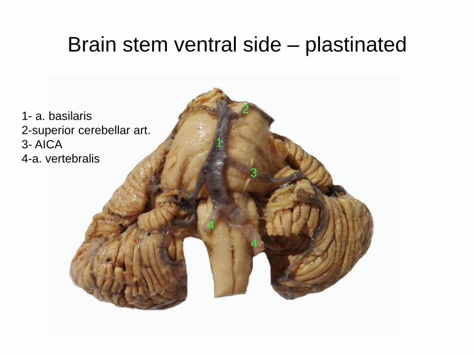
Brain stem ventral side – plastinated
1- a. basilaris
2-superior cerebellar art.
3- AICA
4-a. vertebralis
1
3
2
4
4
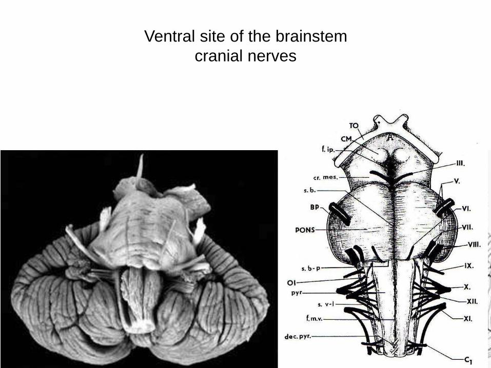
Ventral site of the brainstem
cranial nerves

Ventral site of the brainstem
cranial nerves
pedunculus
cerebri
oliva inferior
III.
IV.
V.VI.VII.VIII.IX.X.
XI.
XII.

Ventral site of the brainstem
cranial nerves and arteries
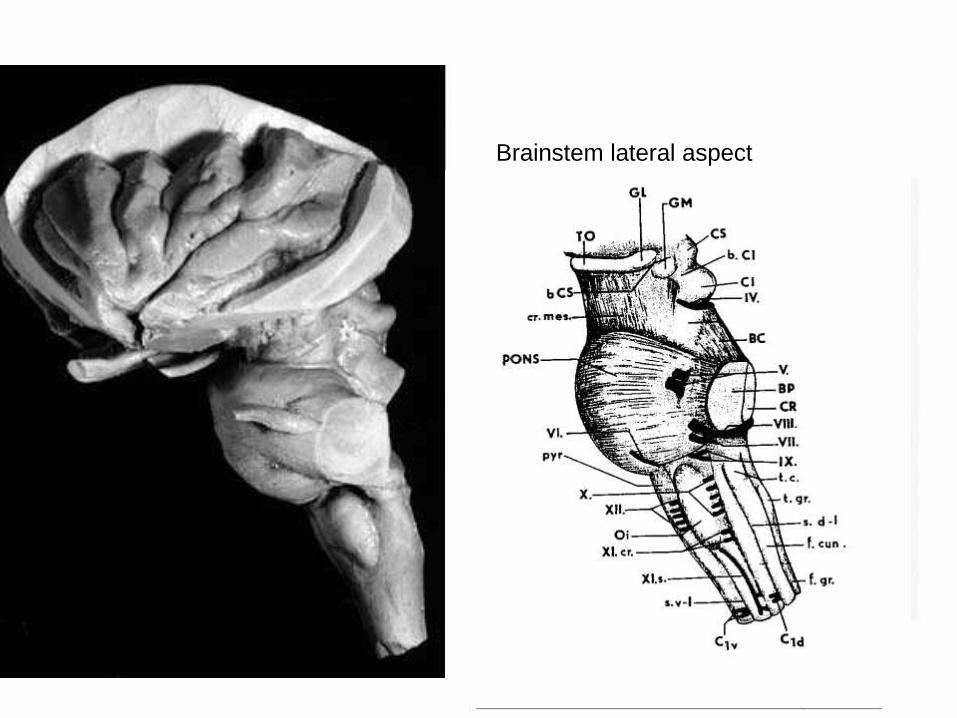
Brainstem lateral aspect

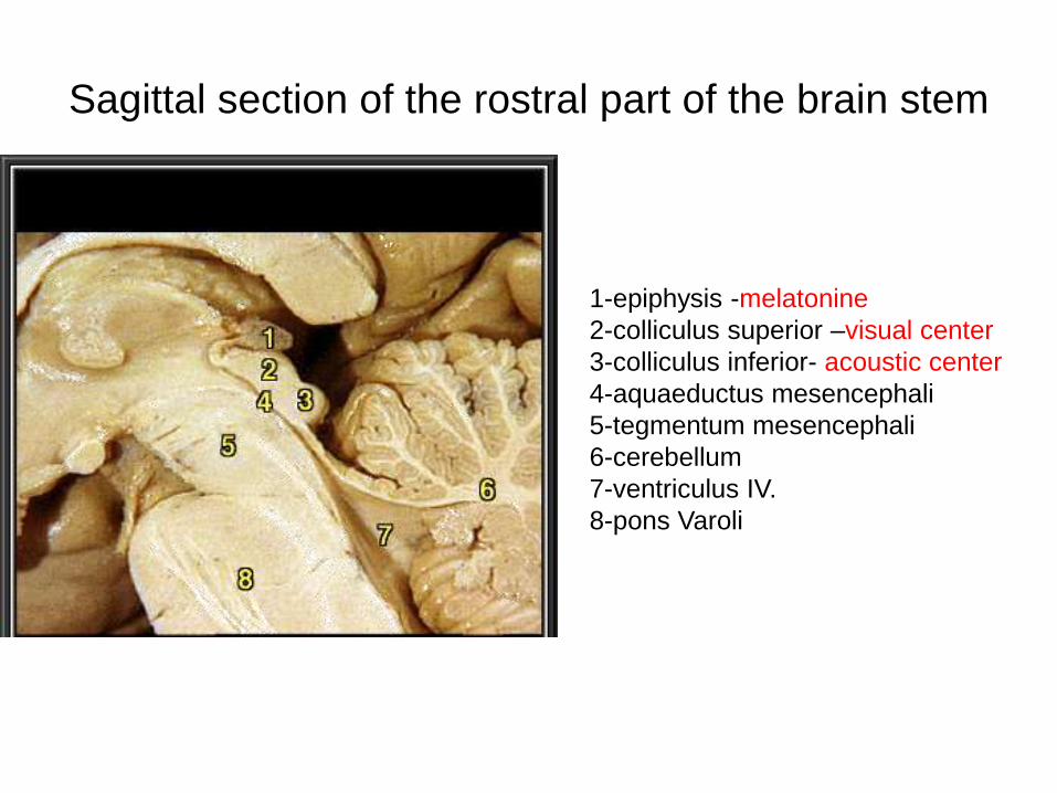
Sagittal section of the rostral part of the brain stem
1-epiphysis -melatonine
2-colliculus superior –visual center
3-colliculus inferior- acoustic center
4-aquaeductus mesencephali
5-tegmentum mesencephali
6-cerebellum
7-ventriculus IV.
8-pons Varoli

epi
III

Sagittal section of brainstem- impregnation
1-epiphysis -melatonin
2-colliculus superior -visual
3-colliculus inferior- acoustic
4-cerebellum
5-brachia conjunctiva
6-ventriculus IV.
7-pons Varoli – pars basilaris
8-tegmentum
9- pyramides
10- dorsal column nuclei
321
4
5
38
7
6
9
10

Rhomboid fossa – floor of the IV. ventricle

Brainstem – dorsal aspect
1
2
3
4
5
7
6
1- thalamus
2- tectum
3- brachia conjuntiva = PCS
4- brachia pontis = PCM
5- corpora restiformia = PCI
6- floor of the IV. ventricle
7- nervus vestibulocochlearis
8- epiphysis
8
Nervi vagi
trigonum nervi hypoglossi

Brainstem – dorsal aspectLamina quadrigemina = tectum mesencephali = colliculi sup et inf

Ncl. Edinger-Westphali
Ncl. salivatorius sup.
Ncl. salivatorius inf.
Ncl. dorsalis nervi vagi
Ncl ambiquus (IX,X,XI)
VII,IX,X:
Cranial nerves nuclei on the floor of the IV. ventricle
Somatomotor
Visceromotor
Viscerosensory
Somatosensory
Sensory
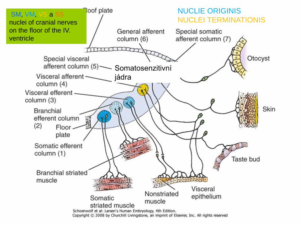
SM, VM, VS a SS
nuclei of cranial nerves
on the floor of the IV.
ventricle
Somatosenzitivní
jádra
NUCLIE ORIGINIS
NUCLEI TERMINATIONIS

Reticular formation
(Moruzzi and Magoun, 1949) brain stem grey matter without cranial nerve nuclei and nuclei of various
functional systems (e.g. acoustic, eye movement coordination, motor)
1) Medial system large cell nuclei, long connections
2) Lateral systém small cell nuclei , connections to the medial system
3) Rapheal systém, in the midline , serotonine
4) Precerebellar nuclei (ncl. olivaris inferior, ncl. tegmenti pontis Bechtěrevi)
5) Chemical systems: A1-10, B1-9, Ch1-6
glutamate, aspartate, GABA, peptids, NO
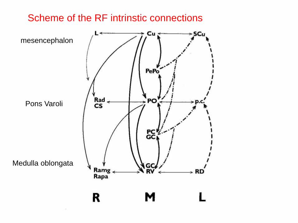
mesencephalon
Pons Varoli
Medulla oblongata
Scheme of the RF intrinstic connections
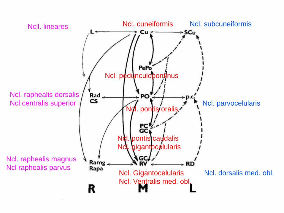
Ncl. raphealis dorsalis
Ncl centralis superior
Ncll. lineares
Ncl. raphealis magnus
Ncl raphealis parvus
Ncl. cuneiformis Ncl. subcuneiformis
Ncl. Gigantocelularis
Ncl. Ventralis med. obl.
Ncl. pedunculopontinus
Ncl. pontis oralis
Ncl. pontis caudalis
Ncl. gigantocelularis
Ncl. parvocelularis
Ncl. dorsalis med. obl.

RF connections
Spinal cord
brainstem
subcortical
cortex
cerebellum
Thalamus Pa intHypoth
Cornu ant Cornu post
RFmed
.RF lat Nuclei V.n.
Ncl. tr. solitarii
SM nuclei of cranial nerves
SN ret
RFraphe
VM nuclei of cranial nerves
tectumRFprecer
Habenula
Subthalamus
Septum verum Area 3,1,2,4,6
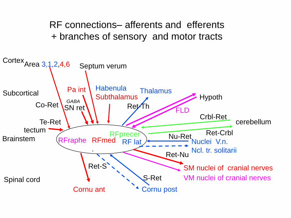
RF connections– afferents and efferents
+ branches of sensory and motor tracts
Spinal cord
Brainstem
Subcortical
Cortex
cerebellum
Thalamus Pa intHypoth
Cornu ant Cornu post
RFmed
.RF lat Nuclei V.n.
Ncl. tr. solitarii
SM nuclei of cranial nerves
SN retGABA
RFraphe
VM nuclei of cranial nerves
tectumRFprecer
Habenula
Subthalamus
Septum verum Area 3,1,2,4,6
Ret-S
Co-Ret
Te-Ret
S-Ret
Ret-Nu
Nu-RetRet-Crbl
Crbl-RetFLD
Ret-Th

Some functions of RF nuclei• Inspiration center – ncl. gigantocellularis ventral part
• Exspiration center – ncl. paragigantocellularis
dorsalis
• Pneumotaxic center – ncl. pontis oralis
• Vasomotor center – ncl. ventralis med. obl.,
• Heart rate center – dorsal part of ncl. ventralis med obl
and ncl. dorsalis med. obl.
• Ascendending activation system – mainly „chemical
systems“- ncl.laterodorsalis a pedunculopontinus, locus
coeruleus
• Inhibitory system – caudally, ventral part of ncl.
gigantocelularis, ncl. ventralis med. obl.

Micture center in rostral dorsolateral tegmentum
In bilateral lesion of this
center– cruel urine
retention
Barrington 1925
IV. komora
flm

Horizontal eye movements
VI.n.
III.n.
PPRFParamedin
pontinne RF
=
PONTINNE
GAZE
CENTRUM
Cortex (FEF)
Vestibular
nuclei
flm
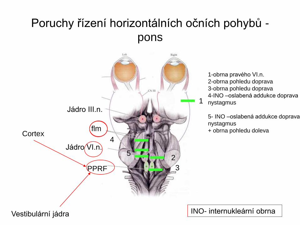
Poruchy řízení horizontálních očních pohybů -
pons
Jádro VI.n.
Jádro III.n.
Cortex
Vestibulární jádra
flm
1-obrna pravého VI.n.
2-obrna pohledu doprava
3-obrna pohledu doprava
4-INO –oslabená addukce doprava
nystagmus
5- INO –oslabená addukce doprava
nystagmus
+ obrna pohledu doleva
1
2
3
4
5
PPRF
INO- internukleární obrna

INTERNUCLEAR OFTHALMOPLEGIA
Paresis of right mlf – on the side of lesion is no adduction
http://pubs.rsna.org/doi/10.1148/rg.331125033?url_ver=Z39.88-
2003&rfr_id=ori:rid:crossref.org&rfr_dat=cr_pub%3dpubmed
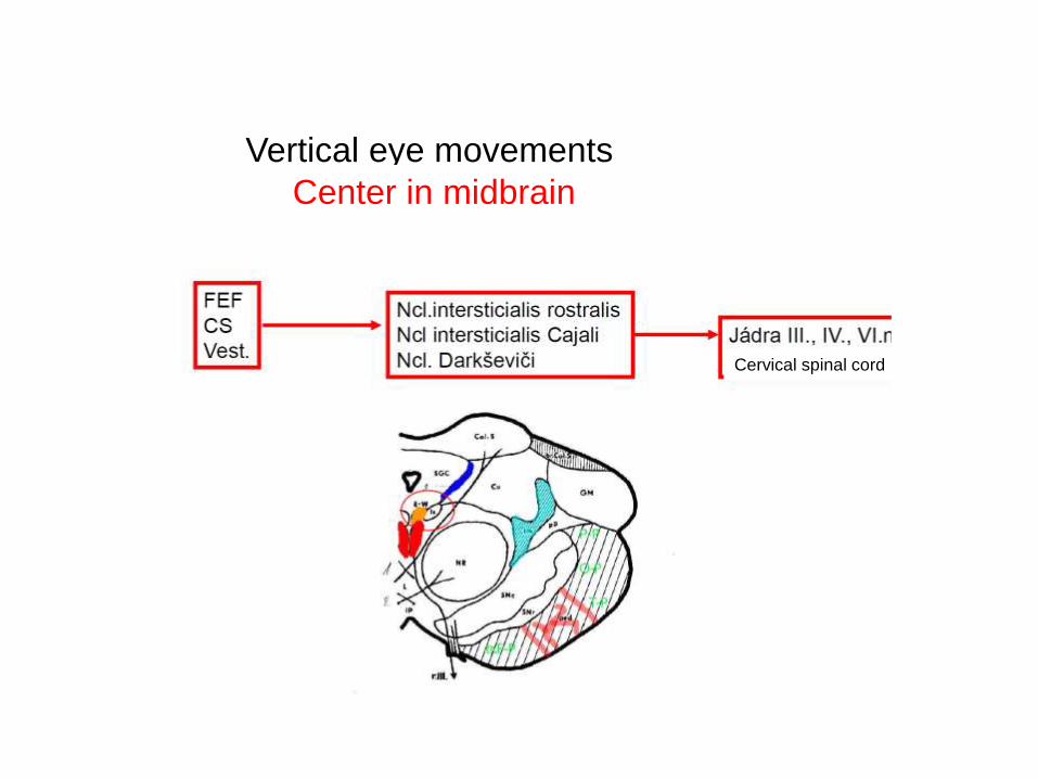
Vertical eye movements
Center in midbrain
Cervical spinal cord

Pupillary reflex center

Precerebellar nuclei
Ncl. Olivaris principalis
Ncl. Olivaris accessorius medialis
Ncl. Olivaris accessorius dorsalisNcll. Pontis
Ncl basalis
tegmenti
pontis
Bechterevi
ncl.
reticularis
lateralis

Cervae isole –section between colliculiFrédéric Bremer 1892–1982
Cat slept all time
Extreme miosis
Loss of olfactory and visual reflexes

Encephale isole
from the Bull Acad Roy Med Belg
1937; 4: 68–86).
Cat slept or was awake
It was possible to wake it up
Inner structures of brainstem
• Cranial nerves nuclei
• Reticular formation nuclei
• Specific nuclei
connected in the motor, sensory
or cerebellar circuits

White matter – visible fascicles
motor, sensory and cerebellar
1) crura cerebri – cortex – brainstem and spinal cord connections
2) pedunculi cerebellares brainstem – cerebellum connections
3) pyramides medullae oblongatae – cortico-spinal tract
4) fibrae arcuatae internae and lemniscus medialis, tr. spino-thalamicus –
thalamic connections
5) corpus trapezoideum a lemniscus lateralis – part of the acoustic tract
6) fasciculus logitudinalis medialis – coordination of eye movements – eye
movement nerve nuclei, vestibular nuclei and neck muscles nuclei connection
7) fasciculus longitudinalis dorsalis hypothalamus- CGM – VM nerve nuclei
connection
8) tr. spinalis n. trigemini, tr. mesencephalicus n. trigemini
tr. solitarius – fibres to sensory nerve nuclei
9) commissura posterior crossed fibers from Darksevic and Cajal nuclei
10) Fasciculus centralis tegmenti a) from the mesencephalon (red nucleus) to
the amiculum olivae inferioris a the RF nuclei b) from RF to thalamus and
subthalamus

Commissura
posterior
Lemniscus
medialis
Fasciculus
longitudinalis
medialis
Pedunculus
cerebri
Pyramidal tract

Macroscopic sections
MEDULLA SPINALISMEDULLA OBLONGATA

Brainstem sections – Nissl staining
MEDULLA SPINALIS
LOWER MEDULLA OBLONGATA
DORSAL COLUMN NUCLEI
DORSAL
COLUMN
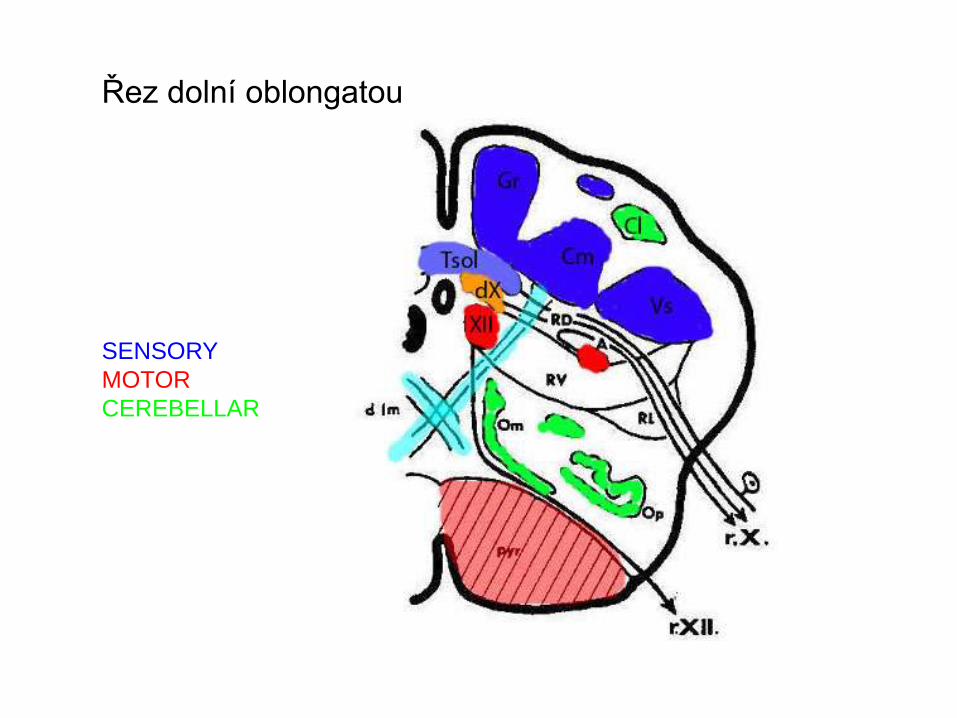
Řez dolní oblongatou
SENSORY
MOTOR
CEREBELLAR

Basal and alar plate development in the
brainstem
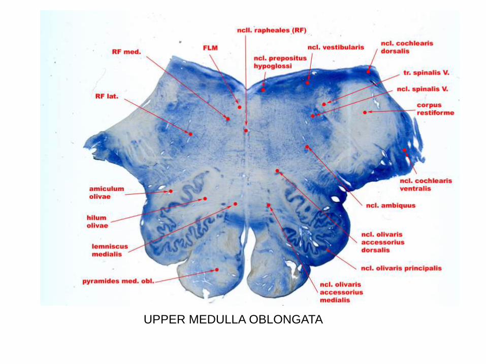
UPPER MEDULLA OBLONGATA

UPPER OBLONGATA SECTION
SENSORY
MOTOR
CEREBELLAR

LOWER PONS SECTION

UPPER PONS SECTION
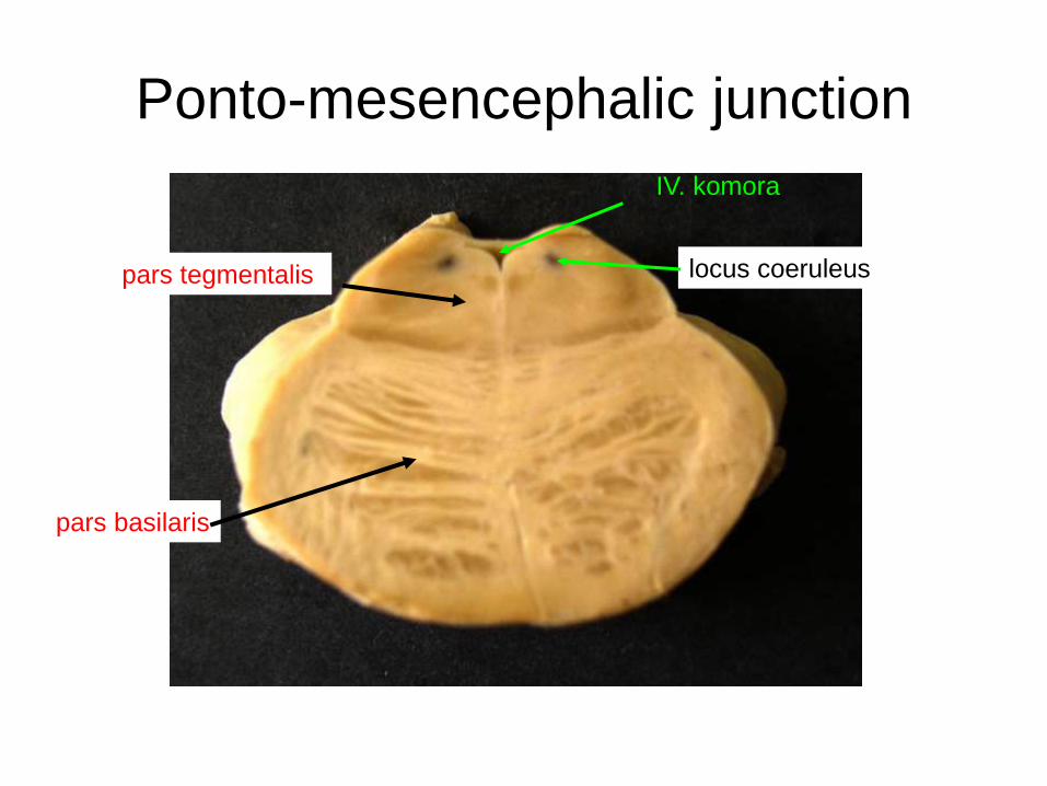
Ponto-mesencephalic junction
pars tegmentalis
pars basilaris
IV. komora
locus coeruleus
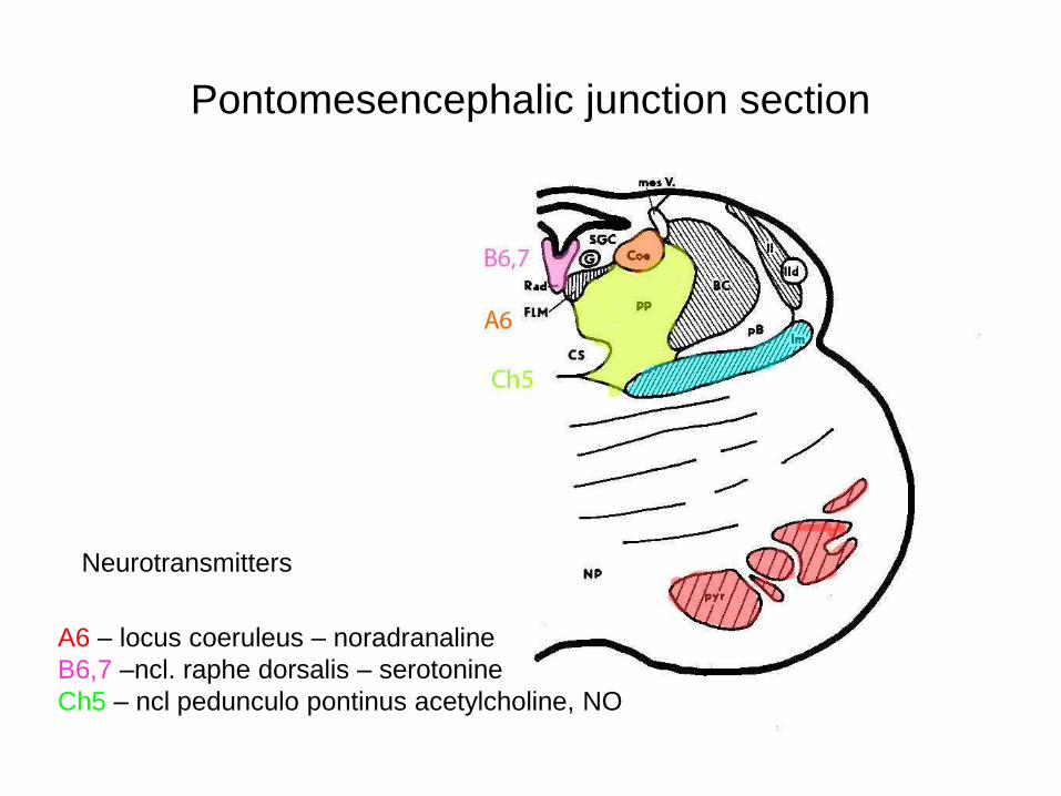
Pontomesencephalic junction section
A6 – locus coeruleus – noradranaline
B6,7 –ncl. raphe dorsalis – serotonine
Ch5 – ncl pedunculo pontinus acetylcholine, NO
Neurotransmitters

PONS VAROLI

serotonin
noradrenalin
acetylcholin
Pontomesencephalic junction section

Pontomesencephalic junction section - rat
tyrosin hydroxylase and NADPHdiaphorase labellinglocus coeruleus
Ch5
ncl pedunulopontinus
Ch 6
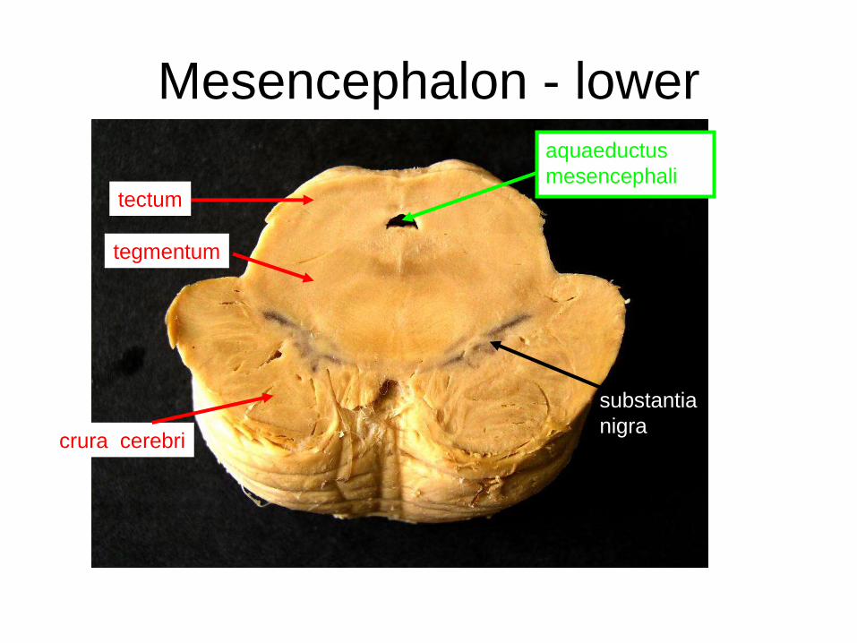
Mesencephalon - lower
tectum
crura cerebri
tegmentum
aquaeductus
mesencephali
substantia
nigra
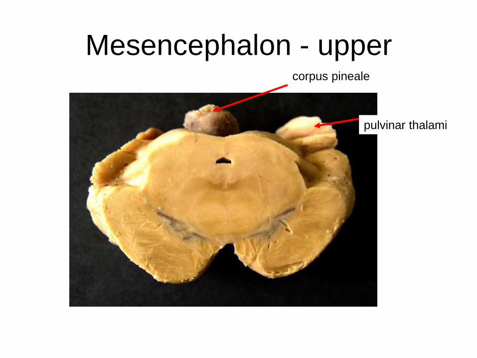
Mesencephalon - uppercorpus pineale
pulvinar thalami
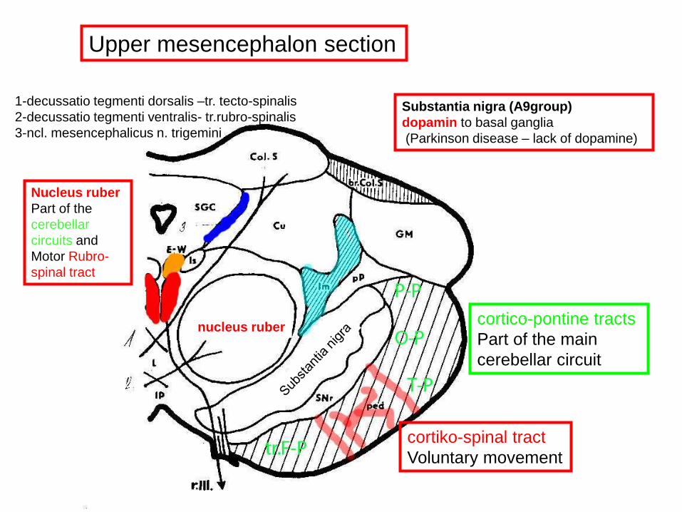
Upper mesencephalon section
cortico-pontine tracts
Part of the main
cerebellar circuit
cortiko-spinal tract
Voluntary movement
Nucleus ruber
Part of the
cerebellar
circuits and
Motor Rubro-
spinal tract
nucleus ruber
Substantia nigra (A9group)
dopamin to basal ganglia
(Parkinson disease – lack of dopamine)
1-decussatio tegmenti dorsalis –tr. tecto-spinalis
2-decussatio tegmenti ventralis- tr.rubro-spinalis
3-ncl. mesencephalicus n. trigemini

Colliculus superior connectionmesencephalic visual center (superficial layers)
and motor center (deep layers)
unconscious reaction to visual and acoustic stimuli – tecto-spinal tract
Tractus opticus
Cortex
Cortex
SNret + BG
Crbl
n.V.
MS
MS
RF
n.III., IV., VI.
Crbl
OI
Darkš. + Cajal

UPPER MESENCEPHALON
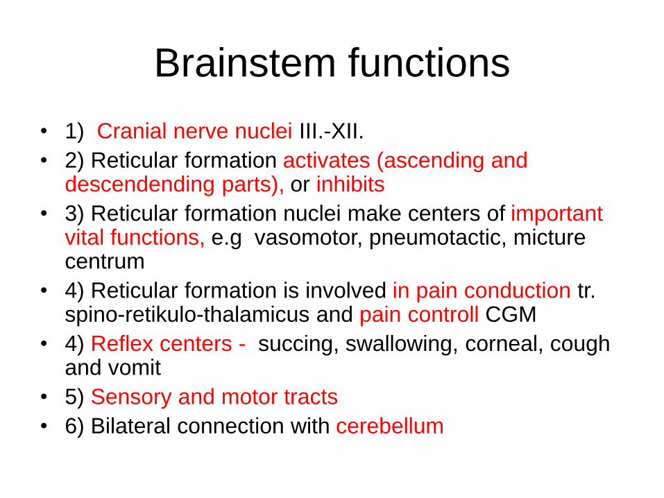
Brainstem functions
• 1) Cranial nerve nuclei III.-XII.
• 2) Reticular formation activates (ascending and descendending parts), or inhibits
• 3) Reticular formation nuclei make centers of important vital functions, e.g vasomotor, pneumotactic, micture centrum
• 4) Reticular formation is involved in pain conduction tr. spino-retikulo-thalamicus and pain controll CGM
• 4) Reflex centers - succing, swallowing, corneal, cough and vomit
• 5) Sensory and motor tracts
• 6) Bilateral connection with cerebellum

Hemiplegia alternans inferior Hemiplegia alternans media
Hemiplegia alternans superiorWallenberg´s sy of lateral medulla oblongate
Vascular lesions in the brain stem – motor and sensory defects

Ncl. et tr.
sp.n.V.
tr. S-Th
Ncl. amb
Wallenberg´s sy of lateral oblongate
(e.g occlusion of a. cerebellaris post.
inf.
Face:
Ipsilaterally
loss of pain
and
temperature
sensation
Body
Contralaterally
loss of pain and
temperature
sensation

Hemiplegia alternans
Syndrom of lateral medulla oblongataVertebral artery branches – a. cerebellaris inferior
posterior (PICA)
Wallenbergův sy
Sy medial medulla oblongataParamedian and circumferential
branches of a. spinalis anterior
Hemiplegia alternans inferior
(1) loss of pain and temperature sensation on the ipsilateral side of the face
(descending nucleus and tract of V) and the contralateral side of the body (spinothalamic/spinoreticular system);
(2) dysphagia and dysarthria (paralysis of ipsilateral pharyngeal and
laryngeal muscles resulting from damage to the ipsilateral nucleus ambiguus);
(3) ataxia of the limbs and falling to the ipsilateral side (inferior cerebellar peduncle and its afferent tracts);
(4) vertigo with nausea, vomiting, and nystagmus (vestibular nuclei); and
(5) ipsilateral Horner's syndrome, with ptosis, miosis, and anhidrosis (descending axons
from the hypothalamus to the T1–T2 intermediolateral cell column of the spinal cord).

Medial pontine syndrome
a. basilaris
Lateral pontine sya.cerebelaris inferior anterior
(AICA)
Hemiplegia alternans media

Weber´s syHemiplegia alternans superior
Contralateral limb palsy
+ ipsilateral III.n palsy
Benedikt´s syContralateral sensory loss
Ipsilateral III.n palsy
Tremor, loss of balance
Branches of a. cerebri post. and ramus comm. post.
Hemiplegia alternans superior
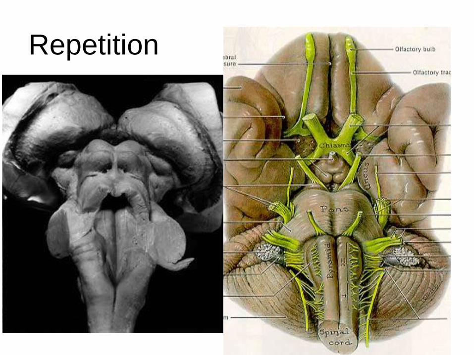
Repetition