brain structural plasticity in survivors of a major...
TRANSCRIPT

J Psychiatry Neurosci 2013;38(6) 381
Research Paper
Brain structural plasticity in survivors of a major earthquake
Su Lui, MD, PhD; Long Chen, MM; Li Yao, MM; Yuan Xiao, MM; Qi-Zhu Wu, PhD; Jun-Ran Zhang, PhD; Xiao-Qi Huang, PhD; Wei Zhang, MD, PhD; Yu-Qin Wang, PhD;
Hua-Fu Chen, PhD; Raymond C.K. Chan, PhD; John A. Sweeney, PhD; Qi-Yong Gong, MD, PhD
Lui, L. Chen, Yao, Xiao, Wu, J. Zhang, Huang, Gong — Department of Radiology, Huaxi MR Research Center (HMRRC), WestChina Hospital of Sichuan University, China; W. Zhang — Department of Psychiatry, the State Key Lab of Biotherapy, West ChinaHospital of Sichuan University, China; Wang, H.-F. Chen — School of Life Science and Technology, University of Electronic Scienceand Technology of China, China; Chan — Neuropsychology and Applied Cognitive Neuroscience Laboratory, Institute of Psych -ology, Chinese Academy of Sciences, China; Sweeney — University of Texas Southwestern Medical Center, Dallas, TX, USA
Introduction
Stress responses have been studied in animal models forsome time. Human studies demonstrate that stressful lifeevents have both short- and long-term physiologic effectsthat are mediated by several factors, including genetics, cog-nitive style and social support.1 However, effects of seriouspsychological stress on the human brain remain poorlyunder stood. Given the well-established chronic psychologicaldisability that many individuals experience after extreme lifestress, there is a need for a better understanding of the neuro-
biological effects of stress and an improved ability to identifyindividuals who most need early mental health interventions.Previous studies have reported altered brain structure in
patients with chronic posttraumatic stress disorder (PTSD),particularly involving the hippocampus, amygdala, anteriorcingulate cortex, subcallosal cingulate cortex and medialfrontal gyrus.2,3 However, unless response to acute stress isexamined, one cannot exclude the potential contributions ofincreased early life stress, secondary effects related to persis-tent PTSD or treatment effects on brain changes observed inclinical samples. Thus, studying people from the community
Correspondence to: Q.-Y. Gong, Professor of Radiology, Neurology and Psychiatry, Huaxi MR Research Center (HMRRC), Department ofRadiology, Center for Medical Imaging, West China Hospital of Sichuan University, No. 37 Guo Xue Xiang Chengdu, 610041 China; [email protected]
J Psychiatry Neurosci 2013;38(6):381-7.
Submitted Nov. 30, 2012; Revised Feb. 28, Mar. 7, 2013; Accepted Mar. 7, 2013.
DOI: 10.1503/jpn.120244
© 2013 Canadian Medical Association
Background: Stress responses have been studied extensively in animal models, but effects of major life stress on the human brain re-main poorly understood. The aim of this study was to determine whether survivors of a major earthquake, who were presumed to haveexperienced extreme emotional stress during the disaster, demonstrate differences in brain anatomy relative to individuals who have notexperienced such stressors. Methods: Healthy survivors living in an area devastated by a major earthquake and matched healthy con-trols underwent 3-dimentional high-resolution magnetic resonance imaging (MRI). Survivors were scanned 13–25 days after the earth-quake; controls had undergone MRI for other studies not long before the earthquake. We used optimized voxel-based morphometryanalysis to identify regional differences of grey matter volume between the survivors and controls. Results: We included 44 survivors(17 female, mean age 37 [standard deviation (SD) 10.6] yr) and 38 controls (14 female, mean age 35.3 [SD 11.2] yr) in our analysis.Compared with controls, the survivors showed significantly lower grey matter volume in the bilateral insula, hippocampus, left caudateand putamen, and greater grey matter volume in the bilateral orbitofrontal cortex and the parietal lobe (all p < 0.05, corrected for multiplecomparison). Limitations: Differences in the variance of survivor and control data could impact study findings. Conclusion: Acuteanatomic alterations could be observed in earthquake survivors in brain regions where functional alterations after stress have been de-scribed. Anatomic changes in the present study were observed earlier than previously reported and were seen in prefrontal–limbic, pari-etal and striatal brain systems. Together with the results of previous functional imaging studies, our observations suggest a complex pat-tern of human brain response to major life stress affecting brain systems that modulate and respond to heightened affective arousal.

shortly after serious psychological stress but before PTSD de-velops may help advance our understanding of more im -medi ate effects of stress on the human brain.Ethical concerns limit experimental studies of severe stress
response in humans; thus, studying people who survivednatural disasters has emerged as a strategy for investigatinghow people respond to major life stress. To date, few studieshave examined the association between life stress and brainstructure in adult participants without a history of psycho -pathology or brain disorder.3–6 Moreover, individuals re-cruited for previous studies had experienced different typesof stress-related events, including illness or injury, death of aclose friend or relative, unemployment and the ending of im-portant relationships, which might have variable effects onthe brain.6 Furthermore, the findings from these studies indi-cated that grey matter atrophy could only be observed3 months after stressful life events.6 A report that functionalbrain alterations have been observed in healthy survivors asearly as 25 days after a major earthquake7 raises the questionof whether acute neuroanatomical differences could also bedetected earlier than 3 months following major emotionalstress. To address this issue, we used optimized voxel-basedmorphometry (VBM) to compare the grey matter volume ofphysically healthy survivors of the Wenchuan earthquake tothat of healthy controls living in the same geographical re-gion who had undergone imaging before the earthquake.
Methods
Participants
We recruited healthy survivors of the Wenchuan earthquakefrom the local hospital, which serves as the medical centre forsouthwestern China. The distance between the hospital andthe epicentre of the earthquake is 46 miles. Survivors were re-cruited randomly from the hospital and from communitiesnear the hospital that were the most affected geographic re-gions, where peak seismic intensity ranged from 9 to 11 onthe Mercalli intensity scale. In these regions, thousands of in-dividuals were buried and died under collapsed buildings,and the community remained in fear of intense aftershocks.The survivors underwent scanning within 25 days after theearthquake. To be included in our analysis, survivors musthave physically experienced the earthquake, experienced nosevere personal medical injury and personally witnesseddeath or serious injury or the collapse of buildings. All sur-vivors were recruited by poster advertisement in which a freemagnetic resonance imaging (MRI) examination of the brainwas offered. Each survivor spent about 2 hours in our labora-tory for MRI examination and psychological analysis, and noobvious aftershock occurred during the study period. We recruited healthy controls matched for age, sex, height,
weight and years of education randomly from Chengdu, whichis located within 50 miles of the earthquake epicenter. Theywere recruited by poster advertisement, and they had under-gone MRI for other studies10,11 shortly before the earthquake.The following exclusion criteria applied to both groups: the
existence of any neurologic or psychiatric disorder, any re-
cent medications known to affect brain function, alcohol ordrug abuse, pregnancy or any systemic physical illness, suchas hepatitis or diabetes.An experienced psychiatrist (X.-Q.H.) ensured participants
met the inclusion criteria. After that, the researchers (L.C.,H.-F.C., S.L. or X.-Q.H.) presented and explained to all par -tici pants a standard consent form and the aim, detailed pro-cedures and information about the MRI scanner for thisstudy. All participants were told they could quit the study atany time for any reason.All participants completed the Structured Clinical Interview
for DSM-IV disorders (SCID)12 to rule out a current or pastpsychi atric disorder, especially anxiety disorders. Two psych -iatrists (X.-Q.H. and W.Z.) performed clinical assessments be-fore the MRI examination. Levels of anxiety and depressionwere evaluated using the Self-Rating Anxiety Scale (SAS)8 andthe Self-Rating Depression Scale (SDS).9 Brain MRI scans (i.e.,T1- and T2-weighted images) were inspected by an experiencedneuroradiologist (S.L.). The 2 groups were scanned on thesame research-dedicated 3 T MRI system. The ethics commit-tee of West China Hospital approved our study protocol, andall partcipants provided written informed consent.
Data acquisition
We acquired high-resolution T1-weighted images using a 3 TMRI system (EXCITE; General Electric) with a volumetric 3-dimentional spoiled gradient recall (SPGR) sequence (repe-tition time 8.5 ms, echo time 3.4 ms, flip angle 12°, slice thick-ness 1 mm) using an 8-channel phase array head coil. Weused a field of view of 240 × 240 mm2, with an acquisitionmatrix comprising 256 readings of 128 phase encoding steps,producing 156 contiguous coronal slices with slice thicknessof 1.0 mm. The final matrix of T1-weighted images was auto-matically interpolated in-plane to 512 × 512, which yields anin-plane resolution of 0.47 × 0.47 mm2. To assure the qualityof the data acquired at different times, a quality assurancescan was acquired, including a spin echo sequence to warmup the scanner and verify the signal-to-noise ratio of images,before each participant was examined.
Image analysis
Image preprocessing and statistical analyses were performedwith SPM8 (www.fil.ion.ucl.ac.uk/spm) using the VBM tool-box (VBM8). First, we constructed a whole brain template,and the individual native images of all participants were nor-malized to the whole brain template and segmented into greymatter, white matter and cerebrospinal fluid (CSF). We thengenerated a grey matter template from all the grey matter im-ages obtained in step 1. Third, the native images were seg-mented again into different tissue types and normalized tothe grey matter template. Next, the deformation parametersobtained from the preceding step were applied to the originalraw images of all participants to create optimally normalizedwhole brain images, which were recursively segmented intogrey matter, white matter and CSF. Next, reobtained greymatter images underwent Jacobian modulation (volume
Lui et al.
382 J Psychiatry Neurosci 2013;38(6)
Brain structural plasticity and stress response
J Psychiatry Neurosci 2013;38(6) 383
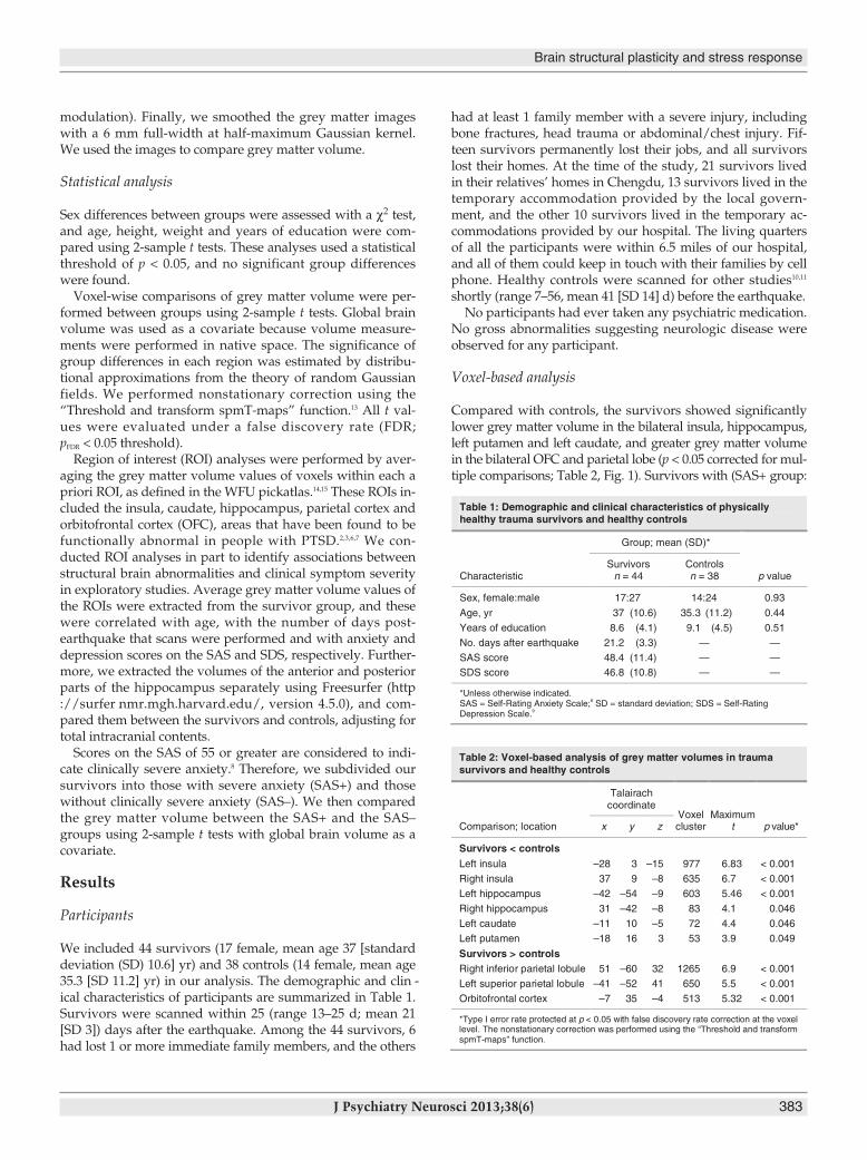
modulation). Finally, we smoothed the grey matter imageswith a 6 mm full-width at half-maximum Gaussian kernel.We used the images to compare grey matter volume.
Statistical analysis
Sex differences between groups were assessed with a χ2 test,and age, height, weight and years of education were com-pared using 2-sample t tests. These analyses used a statisticalthreshold of p < 0.05, and no significant group differenceswere found.Voxel-wise comparisons of grey matter volume were per-
formed between groups using 2-sample t tests. Global brainvolume was used as a covariate because volume measure-ments were performed in native space. The significance ofgroup differences in each region was estimated by distribu-tional approximations from the theory of random Gaussianfields. We performed nonstationary correction using the“Threshold and transform spmT-maps” function.13 All t val-ues were evaluated under a false discovery rate (FDR;pFDR < 0.05 threshold).Region of interest (ROI) analyses were performed by aver-
aging the grey matter volume values of voxels within each apriori ROI, as defined in the WFU pickatlas.14,15 These ROIs in-cluded the insula, caudate, hippocampus, parietal cortex andorbitofrontal cortex (OFC), areas that have been found to befunctionally abnormal in people with PTSD.2,3,6,7 We con-ducted ROI analyses in part to identify associations betweenstructural brain abnormalities and clinical symptom severityin exploratory studies. Average grey matter volume values ofthe ROIs were extracted from the survivor group, and thesewere correlated with age, with the number of days post-earthquake that scans were performed and with anxiety anddepression scores on the SAS and SDS, respectively. Further-more, we extracted the volumes of the anterior and posteriorparts of the hippocampus separately using Freesurfer (http://surfer .nmr.mgh.harvard.edu/, version 4.5.0), and com-pared them between the survivors and controls, adjusting fortotal intracranial contents.Scores on the SAS of 55 or greater are considered to indi-
cate clinically severe anxiety.8 Therefore, we subdivided oursurvivors into those with severe anxiety (SAS+) and thosewithout clinically severe anxiety (SAS–). We then comparedthe grey matter volume between the SAS+ and the SAS–groups using 2-sample t tests with global brain volume as acovariate.
Results
Participants
We included 44 survivors (17 female, mean age 37 [standarddeviation (SD) 10.6] yr) and 38 controls (14 female, mean age35.3 [SD 11.2] yr) in our analysis. The demographic and clin -ical characteristics of participants are summarized in Table 1.Survivors were scanned within 25 (range 13–25 d; mean 21[SD 3]) days after the earthquake. Among the 44 survivors, 6had lost 1 or more immediate family members, and the others
had at least 1 family member with a severe injury, includingbone fractures, head trauma or abdominal/chest injury. Fif-teen survivors permanently lost their jobs, and all survivorslost their homes. At the time of the study, 21 survivors livedin their relatives’ homes in Chengdu, 13 survivors lived in thetemporary accommodation provided by the local govern-ment, and the other 10 survivors lived in the temporary ac-commodations provided by our hospital. The living quartersof all the participants were within 6.5 miles of our hospital,and all of them could keep in touch with their families by cellphone. Healthy controls were scanned for other studies10,11
shortly (range 7–56, mean 41 [SD 14] d) before the earthquake.No participants had ever taken any psychiatric medication.
No gross abnormalities suggesting neurologic disease wereobserved for any participant.
Voxel-based analysis
Compared with controls, the survivors showed significantlylower grey matter volume in the bilateral insula, hippocampus,left putamen and left caudate, and greater grey matter volumein the bilateral OFC and parietal lobe (p < 0.05 corrected for mul-tiple comparisons; Table 2, Fig. 1). Survivors with (SAS+ group:
Table 1: Demographic and clinical characteristics of physically healthy trauma survivors and healthy controls
Characteristic
Group; mean (SD)*
p value Survivors
n = 44 Controls n = 38
Sex, female:male 17:27 14:24 0.93
73 ry ,egA (10.6) 35.3 (11.2) 0.44
Years of education 8.6 (4.1) 9.1 (4.5) 0.51
No. days after earthquake 21.2 (3.3) — —
SAS score 48.4 (11.4) — — SDS score 46.8 (10.8) — —
*Unless otherwise indicated. SAS = Self-Rating Anxiety Scale;8 SD = standard deviation; SDS = Self-Rating Depression Scale.9
Table 2: Voxel-based analysis of grey matter volumes in traumasurvivors and healthy controls
Comparison; location
Talairachcoordinate
Voxelcluster
Maximumt p value*x y z
Survivors < controlsLeft insula –28 3 –15 977 6.83 < 0.001
Right insula 37 9 –8 635 6.7 < 0.001
Left hippocampus –42 –54 –9 603 5.46 < 0.001
Right hippocampus 31 –42 –8 83 4.1 0.046
Left caudate –11 10 –5 72 4.4 0.046
Left putamen –18 16 3 53 3.9 0.049
Survivors > controlsRight inferior parietal lobule 51 –60 32 1265 6.9 < 0.001
Left superior parietal lobule –41 –52 41 650 5.5 < 0.001
Orbitofrontal cortex –7 35 –4 513 5.32 < 0.001
*Type I error rate protected at p < 0.05 with false discovery rate correction at the voxellevel. The nonstationary correction was performed using the “Threshold and transformspmT-maps” function.

15male, 9 female; mean age 37.9 [SD 10.1] yr, mean SAS score57 [SD 4.6]) and without (SAS– group: 12 male, 8 female; meanage 35.9 [SD 11.2] yr, mean SAS score 38 [SD 6.9]) clinically se-vere anxiety showed no significant differences in grey mattervolume (p > 0.05, corrected for multiple comparisons).
Region of interest analysis
As expected, ROI analysis yielded findings paralleling thosein the voxel-based analysis. Grey matter volume was lower inlimbic and subcortical regions, including the bilateral insula,hippocampus and caudate, and grey matter volume wasgreater in the bilateral OFC and parietal lobe (all p < 0.05;Fig. 2) in the survivor group than the control group. Further-more, the mean (and SD) volume of the anterior part of the bi-lateral hippocampus in the survivor group (left: 1.9 [0.4],right: 2.1 [0.3]) were lower than those of controls (left: 2.1 [0.3],right: 2.2 [0.5]; p < 0.05), whereas the volume of the posteriorpart of the bilateral hippocampus in the survivor group (left:1.6 [0.3], right: 1.5 [0.6]) did not differ from those of controls(left: 1.5 [0.4], right: 1.5 [0.5]; p > 0.05). Correlation analyses inthe survivor group did not reveal any significant associationsbetween the average grey matter volume values in brain re-gions in which abnormal volumes were detected and anxiety,depression, age or days from earthquake to the brain scan.
Discussion
Survivors of a severe earthquake who had no history of psychi -atric illness showed greater grey matter volume in areas of theassociation cortex and lower grey matter volume in the insula,hippocampus and ventral striatum than controls. These find-ings, evident within only 1 month after the earthquake, areconsistent with results of animal studies showing that new ex-perience can lead to both the formation and elimination ofsynapses in the cortex.16 In the context of major life stress, vol-ume reductions in the insula, ventral striatum and hippocam-pus may reflect disruptions to neuronal integrity or synapticpro cesses that are a result of high levels of sustained neural activity in affective brain systems.17–19 Such effects may parallelprevious findings in patients with a mood disorder showingincreased functional activity and lower grey matter volumewithin affective brain circuitry, which are believed to representneurotoxic effects of increased neural activity.20–23
Lui et al.
384 J Psychiatry Neurosci 2013;38(6)
Caudate
Insula
Hipp
Parietal
OFC
T0
0
–8 Decreased GMV
8 Increased GMV
Fig. 1: Voxel-based analysis showing regions with greater (red areas)and lower (blue areas) grey matter volume in survivors compared withcontrols (p < 0.05, corrected). Grey matter volumes (GMV) of the bilat-eral insula, hippocampus (Hipp), ventral striatum (caudate and puta-men), orbitofrontal cortex (OFC) and parietal lobe differed significantlybetween survivors and controls (all p < 0.05, corrected).
0.0
0.2
0.4
0.6
0.8
1.0
Gre
y m
atte
r vo
lum
e
Brain region of interestControlSurvivor
RightLeft
Insula Hippocampus Caudate OFC Parietal Insula Hippocampus Caudate OFC Parietal
Fig. 2: Region of interest analysis (mean and standard deviation) of the mean grey matter volume in the bi-lateral insula, hippocampus, ventral striatum (caudate), orbitofrontal cortex (OFC) and parietal lobe. The re-sults showed significantly different grey matter volume between survivors and controls in all of these regions(p < 0.05, corrected).

Brain structural plasticity and stress response
J Psychiatry Neurosci 2013;38(6) 385
Recent evidence supports a role of the OFC as an impor-tant control region for modulating affective responses in lim-bic structures, including the hippocampus, amygdala, ventralstriatum and insula.24 Thus, greater grey matter volume in theOFC may be a consequence of increased demands for top–down modulation to reduce sustained hyperactivation inlimbic systems. Greater grey matter volume in the parietallobe may reflect enhanced activity related to the hypervigi-lant state of many trauma survivors25 who actively scan theenvironment for perceived threat. In contrast to apparent ex-citotoxic effects of sustained high levels of neural activity inaffective brain circuitry, greater grey matter volume in theparietal cortex and OFC may result from elevated synapto -genesis, dendritic branching, increased dendritic spine den-sity17–19 or neurogenesis.26 Such effects have been seen in studiesof rats exposed to new experiences. Another contributing fac-tor might be an increased regional cerebral blood flow27 dueto increased neural activity in these regions.7
Though the mechanism for lower volume in the insula,ventral striatum and hippocampus is unclear, several possi-bilities may conrtribute to this change. First, acute stress isknown to elevate levels of corticotropin-releasing hormoneand activate the hypothalamic–pituitary–adrenal axis. Thatprocess can exert a profound effect on neurogenesis, leadingto both a rapid and prolonged decrease in the rate of cell pro-liferation and an increase in apoptotic cell death, especially inlimbic regions, such as the hippocampus.28 These processesmay contribute to volumetric differences in limbic circuitryobserved in our participants. In addition, heightened sus-tained excitatory drive from the OFC to local circuit inhibi -tory neurons29 in brain emotion systems might lead to habitu-ation or other alterations in local inhibitory circuitry thatcould indirectly enhance excitotoxic processes.28
In fact, the brain regions with altered grey matter volume insurvivors belongs to the prefrontal-limbic and striatal sys-tems, which have been recognized to be involved in affectiveprocessing30 and decision-making.31 Functional neuroimagingstudies have provided direct evidence that prefrontal-limbicand striatal systems play a critical role in anxiety disorders, in-cluding the recollection of traumatic memories and the pro-cessing of fear and pain,32,33 whereas the striatum and parietalregions are activated when making decisions under time pres-sure.31 Functional alterations in emotion circuitry have beendescribed previously in individuals who have experiencedmajor life stresses.7 Our observations in the present study in-dicate that not only functional or physiologic changes, butalso robust differences in gross brain anatomy, could be ob-served within 1 month after a traumatic event. While grossbrain anatomy has long been considered to be relatively fixedin adult life outside the context of neurologic disease, severallines of recent research show acute effects of learning34–36 andpsychiatric medications37 on brain anatomy. Furthermore, wealso noted that the anterior rather than the posterior part ofthe hippocampus showed lower grey matter volume in sur-vivors. This is consistent with the notion that the anterior partof the hippocampus was more involved in emotional process-ing while the posterior part was thought to be involved, forexample, in spatial navigation.38 The rapid anatomic changes
in the limbic cortex in a short time after major life stress, asshown in the present study, have not been previously re-ported in humans. The severe nature of the emotional stressexperienced by participants in the present study may havecontributed to our ability to detect anatomic changes earlierthan previously reported. The present findings contribute toour understanding of human stress response and providenovel evidence indicating a potential for robust and rapidneuroplasticity in emotional brain systems.Both animal models and patient studies demonstrate that
functional and anatomic changes in prefrontal-limbic andstriatal systems play a critical role in stress-related psychiatricdisorders.21,31,33,39,40 Our neuroanatomic findings may be of par-ticular clinical relevance for these conditions by showingstructural changes in brain areas where stress-related physio-logic changes have been previously reported. The presentfindings suggest that anatomic changes observed previouslyin patients with chronic stress-related disorders may in factoccur acutely, and perhaps early in the course of these disor-ders, rather than represent slowly progressive alterations orsecondary effects of chronic distress and disability. Althoughlonger term follow-up studies are necessary to document thefull time course of anatomic changes seen in stress-relatedpsychiatric disorders and after acute stressful events, ourdata indicate that substantial structural neuroplasticity inclinically relevant brain circuitry may manifest itself shortlyafter massive traumatic events. This observation providesfurther neurobiologic rationale for early mental health inter-ventions after major life stressors to reduce acute stress re-sponses and potentially minimize longer term morbidity.Although a resting-state fMRI study of earthquake sur-
vivors has previously shown functional changes within simi-lar brain regions where anatomic changes were observed inthe present study, those functional changes were related toanxiety ratings.7 In contrast, anatomic differences reportedhere were not related to anxiety or depression ratings. Thismay reflect a different or lagging manifestation of anatomicalterations that could result from physiologic and affectivechanges that are manifested earlier, which would suggest adifferent time course to their clinical relevance. Further, tothe extent that different levels of increased neural functionmay have a different magnitude and time course of effectacross brain regions and involve a complex interplay ofpathological and compensatory processes, there may be con-siderable variability across individuals in the association be-tween anatomic changes and immediate psychological dis-tress. It seems most plausible that physiologic changes maymore closely parallel emotional distress acutely, whereasanatomic alterations may be more related to cumulative con-sequences of persistent emotional distress and potentiallyother secondary effects. Longitudinal research is needed toevaluate these associations.
Limitations
It is important to consider that the use of peak volumetric dif-ferences in different brain regions as correlates for the SASand SDS scores may have led to an underestimation of their

association given intersubject variation in such measures.41
Differences in the variance of survivor and control data alsocould impact study findings. Thus, studies to replicate ourobservations are needed to confirm and validate our results,especially in individuals who experienced stressors of differ-ent types, duration and intensity. Furthermore, other factors,such as physical stress, nutritional deprivation and sleep dis-turbances, could have contributed to brain changes,42 thoughthe affected regions observed in the present study were morelikely to be related to emotion process.
Conclusion
To our knowledge, the present study demonstrated for thefirst time that individuals who experience severe emotionaltrauma show stress-induced neuroanatomic alterationsshortly (within 25 d) after massively traumatic psychologicalevents. This observation, together with previous reports offunctional brain alterations in trauma survivors, suggests thatsurvivors of severe emotional trauma may experience sub-stantial change with respect not only to function, but also tothe structural anatomy of prefrontal-limbic, parietal and stri-atal brain systems. From a public health perspective, the datahighlight the need to rapidly intervene to reduce stress levelsin people who have experienced major life stress so thatacute neurophysiological alterations do not lead to down-stream and potentially persistent alterations in psychologicaland brain function.
Acknowledgements: This study was supported by National NaturalScience Foundation (Grant Nos. 81222018,81030027, 81227002 and81220108013), the Programs for New Century Excellent Talents inUniversity (Grant No. NCET-10-0596) and National Key Technolo-gies R&D Program of China (Program No: 2012BAI01B03) and thevon Humboldt Foundation. Q.-Y. Gong acknowledges support fromhis American CMB Distinguished Professorship Award (Award No.F510000/G16916411), administered by the Institute of InternationalEducation. S. Lui is supported by the Distinguished Young Scholarsof Sichuan (Award No. 2011JQ0005).
Competing interests: J.A. Sweeney consults for Lilly, Takeda, Rocheand Pfizer. As above for Q.-Y. Gong and S. Lui. Otherwise, none declared.
Contributors: S. Lui, X.-Q. Huang and Q.-Y. Gong designed the arti-cle. S. Lui, L. Chen and W. Zhang acquired the data, which all au-thors but X.-Q. Huang analyzed. S. Lui, L. Chen, L. Yao, Y. Xiao, Q.-Z. Wu, X.-Q. Huang, W. Zhang, Y.-Q. Wang and J.A. Sweeneywrote the article. S. Lui, Q.-Z. Wu, J.-R. Zhang, S.-Q. Huang, H.-F. Chen, R.C.K. Chan, J.A. Sweeney and Q.-Y. Gong reviewed thearticle. All authors approved its publication.
References
1. Park CL. Making sense of the meaning literature: an integrative re-view of meaning making and its effects on adjustment to stressfullife events. Psychol Bull 2010;136:257-301.
2. McEwen BS. Physiology and neurobiology of stress and adapta-tion: central role of the brain. Physiol Rev 2007;87:873-904.
3. Cohen RA, Grieve S, Hoth KF, et al. Early life stress and morph -ometry of the adult anterior cingulate cortex and caudate nuclei.Biol Psychiatry 2006;59:975-82.
4. Ganzel BL, Kim P, Glover GH, et al. Resilience after 9/11: multi-modal neuroimaging evidence for stress-related change in thehealthy adult brain. Neuroimage 2008;40:788-95.
5. Gianaros PJ, Jennings JR, Sheu LK, et al. Prospective reports ofchronic life stress predict decreased grey matter volume in the hip-pocampus. Neuroimage 2007;35:795-803.
6. Papagni SA, Benetti S, Arulanantham S, et al. Effects of stressfullife events on human brain structure: a longitudinal voxel-basedmorphometry study. Stress 2011;14:227-32.
7. Lui S, Huang X, Chen L, et al. High-field MRI reveals an acute im-pact on brain function in survivors of the magnitude 8.0 earth-quake in China. Proc Natl Acad Sci U S A 2009;106:15412-7.
8. Zung WW. A rating instrument for anxiety disorders. Psychosomatics1971;12:371-9.
9. Zung WW. A Self-Rating Depression Scale. Arch Gen Psychiatry1965; 12:63-70.
10. Lui S, Deng W, Huang X, et al. Association of cerebral deficits withclinical symptoms in antipsychotic-naive first-episode schizophrenia:an optimized voxel-based morphometry and resting state functionalconnectivity study. Am J Psychiatry 2009;166:196-205.
11. Lui S, Parkes LM, Huang X, et al. Depressive disorders: focally al-tered cerebral perfusion measured with arterial spin-labeling MRimaging. Radiology 2009;251:476-84.
12. Spitzer RL, Williams JB, Gibbon M, et al. The Structured ClinicalInterview for DSM-III-R (SCID). I: history, rationale, and descrip-tion. Arch Gen Psychiatry 1992;49:624-9.
13. Hayasaka S, Nichols TE. Combining voxel intensity and cluster ex-tent with permutation test framework. Neuroimage 2004;23:54-63.
14. Lancaster JL, Woldorff MG, Parsons LM, et al. Automated Talairachatlas labels for functional brain mapping. Hum Brain Mapp 2000;10:120-31.
15. Maldjian JA, Laurienti PJ, Kraft RA, et al. An automated methodfor neuroanatomic and cytoarchitectonic atlas-based interrogationof fMRI data sets. Neuroimage 2003;19:1233-9.
16. Trachtenberg JT, Chen BE, Knott GW, et al. Long-term in vivoimaging of experience-dependent synaptic plasticity in adult cor-tex. Nature 2002;420:788-94.
17. Volkmar FR, Greenough WT. Rearing complexity affects branchingof dendrites in the visual cortex of the rat. Science 1972;176:1445-7.
18. Bock J, Gruss M, Becker S, et al. Experience-induced changes of den-dritic spine densities in the prefrontal and sensory cortex: correla-tion with developmental time windows. Cereb Cortex 2005;15:802-8.
19. Cerqueira JJ, Pego JM, Taipa R, et al. Morphological correlates ofcorticosteroid-induced changes in prefrontal cortex-dependent be-haviors. J Neurosci 2005;25:7792-800.
20. Canli T, Omura K, Haas BW, et al. Beyond affect: a role for geneticvariation of the serotonin transporter in neural activation during acognitive attention task. Proc Natl Acad Sci U S A 2005;102:12224-9.
21. Canli T, Qiu M, Omura K, et al. Neural correlates of epigenesis.Proc Natl Acad Sci U S A 2006;103:16033-8.
22. Lui S, Parkes LM, Huang X, et al. Depressive disorders: focally al-tered cerebral perfusion measured with arterial spin-labeling MRimaging. Radiology 2009;251:476-84.
23. Koolschijn PC, van Haren NE, Lensvelt-Mulders GJ, et al. Brain vol-ume abnormalities in major depressive disorder: a meta-analysis ofmagnetic resonance imaging studies. Hum Brain Mapp 2009;30: 3719-35.
Lui et al.
386 J Psychiatry Neurosci 2013;38(6)

Brain structural plasticity and stress response
J Psychiatry Neurosci 2013;38(6) 387
24. Davidson RJ, Putnam KM, Larson CL. Dysfunction in the neuralcircuitry of emotion regulation — a possible prelude to violence.Science 2000;289:591-4.
25. Bryant RA, Felmingham KL, Kemp AH, et al. Neural networks ofinformation processing in posttraumatic stress disorder: a functionalmagnetic resonance imaging study. Biol Psychiatry 2005;58:111-8.
26. Gross CG. Neurogenesis in the adult brain: death of a dogma. NatRev Neurosci 2000;1:67-73.
27. Matsuda H, Ohnishi T, Asada T, et al. Correction for partial-volumeeffects on brain perfusion SPECT in healthy men. J Nucl Med 2003;44: 1243-52.
28. de Kloet ER, Joels M, Holsboer F. Stress and the brain: from adap-tation to disease. Nat Rev Neurosci 2005;6:463-75.
29. Thayer JF, Lane RD. A model of neurovisceral integration in emo-tion regulation and dysregulation. J Affect Disord 2000;61:201-16.
30. Cardinal RN, Parkinson JA, Hall J, et al. Emotion and motivation:the role of the amygdala, ventral striatum, and prefrontal cortex.Neurosci Biobehav Rev 2002;26:321-52.
31. Forstmann BU, Dutilh G, Brown S, et al. Striatum and pre-SMA facili -tate decision-making under time pressure. Proc Natl Acad Sci U S A2008; 105:17538-42.
32. Lanius RA, Williamson PC, Densmore M, et al. The nature of trau-matic memories: a 4-T FMRI functional connectivity analysis. Am JPsychiatry 2004;161:36-44.
33. Bryant RA, Kemp AH, Felmingham KL, et al. Enhanced amygdalaand medial prefrontal activation during nonconscious processing
of fear in posttraumatic stress disorder: an fMRI study. Hum BrainMapp 2008;29:517-23.
34. Draganski B, Gaser C, Busch V, et al. Neuroplasticity: changes ingrey matter induced by training. Nature 2004;427:311-2.
35. Driemeyer J, Boyke J, Gaser C, et al. Changes in gray matter in-duced by learning — revisited. PLoS ONE 2008;3:e2669.
36. Scholz J, Klein MC, Behrens TE, et al. Training induces changes inwhite-matter architecture. Nat Neurosci 2009;12:1370-1.
37. Eack SM, Hogarty GE, Cho RY, et al. Neuroprotective effects ofcognitive enhancement therapy against gray matter loss in earlyschizophrenia: results from a 2-year randomized controlled trial.Arch Gen Psychiatry 2010;67:674-82.
38. Fanselow MS, Dong H-W. Are the dorsal and ventral hippocam-pus functionally distinct structures? Neuron 2010;65:7-19.
39. Simmons AN, Paulus MP, Thorp SR, et al. Functional activationand neural networks in women with posttraumatic stress disorderrelated to intimate partner violence. Biol Psychiatry 2008;64:681-90
40. Yamasue H, Kasai K, Iwanami A, et al. Voxel-based analysis ofMRI reveals anterior cingulate gray-matter volume reduction inposttraumatic stress disorder due to terrorism. Proc Natl Acad SciU S A 2003;100:9039-43.
41. Kriegeskorte N, Lindquist MA, Nichols TE, et al. Everything younever wanted to know about circular analysis, but were afraid toask. J Cereb Blood Flow Metab 2010;30:1551-7.
42. Sapolsky RM. Glucocorticoids and hippocampal atrophy in neuro -psychiatric disorders. Arch Gen Psychiatry 2000;57:925-35.
Correction
Are addictions diseases or choices?
There was an error in the affiliations listed for Marco Leyton in the July 2013 editorial (J Psychiatry Neurosci2013;38(4):219-21). Dr. Leyton is from the Department of Psychiatry, McGill University, Montréal, andthe Center for Studies in Behavioral Neurobiology, Department of Psychology, Concordia University,Montréal, Que., Canada.
We apologize for this error.