brainregion-specifictraffickingofthedopamine transporter · 12846 • j.neurosci.,september16,2015...
TRANSCRIPT

Cellular/Molecular
Brain Region-Specific Trafficking of the DopamineTransporter
Ethan R. Block,1 Jacob Nuttle,2 Judith Joyce Balcita-Pedicino,2 John Caltagarone,1 XSimon C. Watkins,1
Susan R. Sesack,2 and Alexander Sorkin1
1Department of Cell Biology, University of Pittsburgh School of Medicine, Pittsburgh, Pennsylvania 15261, and 2Department of Neuroscience, University ofPittsburgh Dietrich School of Arts and Sciences, Pittsburgh, Pennsylvania 15260
The dopamine (DA) transporter (DAT) controls dopaminergic neurotransmission by removing extracellular DA. Although DA reuptakeis proposed to be regulated by DAT traffic to and from the cell surface, the membrane trafficking system involved in the endocytic cyclingof DAT in the intact mammalian brain has not been characterized. Hence, we performed immunolabeling and quantitative analysis of thesubcellular and regional distribution of DAT using the transgenic knock-in mouse expressing hemagglutinin (HA) epitope-tagged DAT(HA-DAT) and by using a combination of electron microscopy and a novel method for immunofluorescence labeling of HA-DAT in acutesagittal brain slices. Both approaches demonstrated that, in midbrain somatodendritic regions, HA-DAT was present in the plasmamembrane, endoplasmic reticulum, and Golgi complex, with a small fraction in early and recycling endosomes and an even smallerfraction in late endosomes and lysosomes. In the striatum and in axonal tracts between the midbrain and striatum, HA-DAT was detectedpredominantly in the plasma membrane, and quantitative analysis revealed increased DAT density in striatal compared with midbrainplasma membranes. Endosomes were strikingly rare and lysosomes were absent in striatal axons, in which there was little intracellularHA-DAT. Acute administration of amphetamine in vivo (60 min) or to slices ex vivo (10 – 60 min) did not result in detectable changes inDAT distribution. Altogether, these data provide evidence for regional differences in DAT plasma membrane targeting and retention andsuggest a surprisingly low level of endocytic trafficking of DAT in the striatum along with limited DAT endocytic activity in somatoden-dritic areas.
Key words: dopamine; electron microscopy; endocytosis; transporter
IntroductionDopaminergic neurons control aspects of mood, cognition, andmovement and are involved in the pathogenesis and treatment of
a variety of disorders, including drug addiction, depression, andParkinson’s disease (Gainetdinov and Caron, 2003; Spiga et al.,2008; Wise, 2008; Willuhn et al., 2010). A primary regulator ofdopaminergic neurotransmission is the dopamine (DA) trans-
Received April 10, 2015; revised July 16, 2015; accepted Aug. 7, 2015.Author contributions: E.R.B., S.R.S., and A.S. designed research; E.R.B., J.N., J.J.B.-P., J.C., S.C.W., and A.S. per-
formed research; E.R.B., J.N., S.R.S., and A.S. analyzed data; E.R.B., S.R.S., and A.S. wrote the paper.This work was supported by National Institutes of Health/National Institute on Drug Abuse Grants DA014204
(A.S., E.R.B., J.C.) and F32DA034408 (E.R.B.). We are grateful to Dr. Mads Larsen, Callen Wallace, and Mara Sullivan(Center for Biological Imaging, University of Pittsburgh) for assistance with large-area confocal imaging, structuredillumination microscopy, and cryoelectron microscopy, respectively. We also thank Dr. R. Prekeris (University ofColorado) for the Syntaxin 13 antibody. The anti-LAMP1 1D4B antibody developed by August J. Thomas was ob-tained from the Developmental Studies Hybridoma Bank, created by the National Institute of Child Health and
Human Development of the National Institutes of Health and maintained at the University of Iowa (Department ofBiology).
The authors declare no competing financial interests.Correspondence should be addressed to either of the following: Susan R. Sesack, Department of Neuroscience,
University of Pittsburgh Dietrich School of Arts and Sciences, Pittsburgh, PA 15260, E-mail: [email protected]; orAlexander Sorkin, Department of Cell Biology, University of Pittsburgh School of Medicine, 3500 Terrace Street,Pittsburgh, PA 15261, E-mail: [email protected].
DOI:10.1523/JNEUROSCI.1391-15.2015Copyright © 2015 the authors 0270-6474/15/3512845-14$15.00/0
Significance Statement
The dopamine transporter (DAT) is the key regulator of the dopamine neurotransmission in the CNS. In the present study, wedeveloped a new approach for studying DAT localization and dynamics in intact neurons in acute sagittal brain slices from theknock-in mouse expressing epitope-tagged DAT. For the first time, the fluorescence imaging analysis of DAT was combined withthe immunogold labeling of DAT and quantitative electron microscopy. In contrast to numerous studies of DAT trafficking inheterologous expression systems and dissociated cultured neurons, studies in intact neurons revealed a surprisingly low amountof endocytic trafficking of DAT at steady state and after acute amphetamine treatment and suggested that non-vesicular transportcould be the main mechanism establishing DAT distribution within the dopaminergic neuron.
The Journal of Neuroscience, September 16, 2015 • 35(37):12845–12858 • 12845
porter (DAT; Giros et al., 1991; Jaber et al., 1997; Kristensen et al.,2011). The psychostimulants amphetamine, cocaine, and meth-ylphenidate elicit dramatic behavioral phenotypes in large part bytargeting DAT directly and increasing extracellular DA. Identify-ing mechanisms of DAT regulation not only contributes to ourbroader understanding of DA physiology but also suggests ther-apeutic options for treatment of DA pathologies, such as drugaddiction.
Somatodendritic regions of DA neurons are located in themidbrain substantia nigra (SN) and ventral tegmental area. Fromthere, DA neurons send bundles of axons rostrally to the dorsalstriatum and nucleus accumbens, as well as to the frontal cortex(Ciliax et al., 1999). Because DA reuptake requires the presence ofDAT in the plasma membrane (PM), proper subcellular traffick-ing and localization of the transporter is crucial for its function.Studies using primary neuronal cultures suggest that DAT passesthrough the biosynthetic pathway to the PM and undergoes con-stitutive endocytosis (Melikian 2004; Miranda et al., 2004;Sorkina et al., 2006; Eriksen et al., 2009; Zahniser and Sorkin,2009). However, there is disagreement on whether endocytosedDAT is preferentially targeted to lysosomes for degradation orrecycled back to the PM (Melikian, 2004; Chen et al., 2010; Erik-sen et al., 2010; Rao et al., 2011). Furthermore, PM delivery (re-cycling) and/or endocytosis can be enhanced by a variety ofstimuli, including cocaine, amphetamine, and the protein kinaseC (PKC) activator phorbol 12-myristate 13-acetate, findings thatare supported by biochemical studies with primary cultured neu-rons and ex vivo tissue preparations, such as synaptosomes orbrain slices (Huff et al., 1997; Vaughan et al., 1997; Melikian andBuckley, 1999; Hoover et al., 2007; Cremona et al., 2011; Gabrielet al., 2013). In contrast, PKC-mediated endocytosis of DAT hasnot been observed in several studies using primary neuronal cul-tures (Eriksen et al., 2009; Rao et al., 2012).
Little is known about the general organization of the endo-cytic trafficking system in the intact DA neuron. A number ofstudies using electron microscopy (EM) to gain valuable insightinto DAT distribution and function in the rat brain have revealedthat DAT is found at the PM in axons, although it may be ex-cluded from active zones (Nirenberg et al., 1996, 1997; Hersch etal., 1997; Ciliax et al., 1999). Although these original EM studiesprovided the first characterizations of DAT localization in theintact DA neuron, the immunohistochemical methods used inthese studies are sometimes associated with sparse labeling thatdoes not allow for quantitative analysis of DAT localization.Thus, a number of important questions have remained unan-swered. First, is DAT present in a higher concentration in striatalaxons, presynaptic terminals in particular, compared with den-drites and the soma? If so, the existence of a specific axon/synapsetargeting mechanism can be predicted. Second, is there a largefraction of DAT in endosomes and lysosomes? A large endolyso-somal pool of DAT would be indicative of substantial constitutiveendocytosis and other vesicular trafficking processes within theDA neuron.
For the present studies, we have examined DAT subcellularlocalization and regulation using a knock-in mouse that ex-presses a DAT with a hemagglutinin (HA) epitope inserted intothe second extracellular loop (HA-DAT; Rao et al., 2012). TheHA epitope is highly efficient for EM localization studies, permit-ting quantitative analysis and the identification of intracellularDAT pools. The HA epitope also provides a readily accessibleepitope to observe transporter dynamics by fluorescence micros-copy in acute brain slice preparations.
Materials and MethodsAntibodies and chemicals. Mouse monoclonal antibodies against the HAepitope HA11 (16B12) were from BioLegend (mms-101p); specificity ofHA11 for DA neurons in HA-DAT mice has been established previously(Rao et al., 2012). Rat monoclonal antibodies against the N terminus ofDAT were from Millipore (MAB369; characterized by Hersch et al., 1997;Garzon and Pickel, 2006), goat polyclonal antibodies against the C ter-minus of DAT (Salvatore et al., 2003) were from Santa Cruz Biotechnol-ogy, rat monoclonal anti-lysosome-associated membrane protein 1(LAMP1) antibodies were from the University of Iowa DevelopmentalStudies Hybridoma Bank (1D4B; deposited to the Developmental Stud-ies Hybridoma Bank by J. T. August, Johns Hopkins School of Medicine,Baltimore, MD; Chen et al., 1985), mouse monoclonal anti-early endo-somal antigen 1 (EEA.1) antibodies were from BD Biosciences (610457;Mu et al., 1995), rabbit anti-Syntaxin 13 (Stx13) antibodies were fromRytis Prekeris (University of Colorado, Denver, CO; Prekeris et al.,1999), and rabbit anti-tyrosine hydroxylase (TH) antibodies were fromEMD Millipore (AB152). Fluorophore-conjugated secondary antibodieswere from Jackson ImmunoResearch, and gold-conjugated secondaryantibodies were from Aurion (800.022). The specificity and optimal con-ditions of labeling by all antibodies were determined based on the max-imal difference between the immunofluorescence signal in the presenceof a primary antibody and the background fluorescence in the absence ofa primary antibody (replaced by corresponding nonspecific IgG). In ad-dition, specificity and conditions for specific labeling of dopaminergicneurons with HA11, TH, and DAT antibodies were established based onthe lack of labeling of nondopaminergic areas of the brain, cross-labelingof the same neurons by these antibodies, and detection of the same bandsby Western blotting (HA11 and DAT antibodies). The specificity of an-tibodies to endosomal markers and optimal labeling conditions wereadditionally determined based on the presence of strong specific signalsin cell bodies that exhibited the typical vesicular shape and perinuclearlocalization of endosomal compartments. All other reagents and supplieswere from Thermo Fisher Scientific unless noted otherwise.
Preparation of acute brain slices. All experimental procedures involvingthe use of laboratory mice were approved by the Institutional AnimalCare and Use Committee. Four- to 8-week-old HA-DAT mice of eithersex were killed by CO2 asphyxiation, followed by decapitation. Brainswere removed and submerged into an ice slush of oxygenated artificialCSF (ACSF; in mM: 124 NaCl, 4 KCl, 1.25 NaH2PO4, 25.7 NaHCO3, 1.2MgSO4, 2.45 CaCl2, 11 dextrose, and 0.15 ascorbic acid). After an initialsagittal cut along the midline, 0.8-mm-thick slices were made using mi-crotome blades and a stainless steel slicing block. Slices were allowed torecover in ACSF for 60 min at room temperature. For labeling of thecell-surface pool of HA-DAT (Figs. 1, 2), slices were incubated in ACSF atroom temperature with 1 �g/ml anti-HA antibodies for 1 h. For labelingof the cell-surface and endocytosed pool of HA-DAT (see Fig. 8), sliceswere incubated in ACSF at 37°C with 1 �g/ml anti-HA antibodies for 1 hin the absence or presence of 100 �M d-amphetamine hemisulfate. Afterremoving unbound HA antibodies, slices were fixed for 2 h in ACSF withfreshly prepared 4% paraformaldehyde (Electron Microscopy Sciences).
Immunohistochemical analysis of fixed brain slices. Paraformaldehyde-fixed acute slices (above) or slices cut as described above but allowed torecover for 20 min before paraformaldehyde fixation were subjected toadditional processing. Fixative was removed by washing in PBS, andslices were permeabilized by the addition of 0.3% Triton X-100 in PBS for30 min. Appropriate dilutions of primary antibodies were applied byovernight incubation at 4°C. DAPI and fluorophore-conjugated second-ary antibodies were applied for 1 h at room temperature before imaging.
Cell culture experiments. Human HEK293A cells were purchased fromInvitrogen. A previously described CFP–HA-DAT [N-terminally cyanfluorescent protein (CFP)-tagged DAT that contains an HA epitope inthe second extracellular loop; Vina-Vilaseca and Sorkin, 2010] was trans-fected at the time of seeding onto coverslips using Lipofectamine 2000(Invitrogen) according to the instructions of the manufacturer. Two daysafter transfection, cells were incubated with 1 �g/ml anti-HA antibodiesfor 30 min at 37°C in the absence or presence of 100 �M d-amphetamine.Subsequently, cells were washed, fixed for 30 min with 4% paraformal-
12846 • J. Neurosci., September 16, 2015 • 35(37):12845–12858 Block et al. • DAT Trafficking in Intact Brain

dehyde, permeabilized with 0.1% Triton X-100 for 5 min, and incubatedwith fluorophore-conjugated goat anti-mouse antibodies beforeimaging.
Confocal fluorescence microscopy and image analysis. Large-area imag-ing was performed by acquiring multiple images through a 2� objectiveusing a Nikon A1 scanning confocal microscope. The images were assem-bled in a single whole-slice montage image using Nikon Elementssoftware.
To obtain high resolution three-dimensional (3D) images of theDA neurons in the brain slices, a z stack of confocal images wasacquired 10 – 40 �m in from the cut face of the slice (to avoid debrisfrom the slicing procedure while maintaining good resolution). Theimaging system used was a spinning-disk confocal system based on aZeiss Axio Observer Z1 inverted fluorescence microscope [with 63�Plan Apo PH, 1.4 numerical aperture (NA)], equipped with acomputer-controlled Spherical Aberration Correction unit, Yok-ogawa CSU-X1, Vector photomanipulation module, PhotometricsEvolve 16-bit EMCCD camera, HQ2 cooled CCD camera, environ-mental chamber, and piezo stage controller and lasers (405, 445, 488,515, 561, and 640 nm; Intelligent Imaging Innovations), all controlledby SlideBook 6 software (Intelligent Imaging Innovations). Typically,up to 10 – 40 serial two-dimensional confocal images were recorded at300 – 400 nm intervals. Colocalization of the DAT with endocyticmarkers was determined by identifying clear overlapping structuresthat could be followed in multiple z planes.
To compare quantitatively the density of HA-DAT in different brainregions (Fig. 2C), the slices were incubated with HA11 antibody, fixed,and labeled with secondary antibody as described above. z stacks of 14confocal images were acquired at 400 nm intervals starting at 10 �mdistance from the slice surface. Voxels containing HA-DAT were selectedin each 3D image using automatic segmentation (Otsu algorithm), and amean fluorescence intensity was determined after background subtrac-
tion. For visual comparison of the fluorescence intensities of differentstructures in the DA neurons, high-resolution images are presented inthe pseudocolor mode (Fig. 2B). In this mode, the fluorescence intensityis displayed stretched between identical low and high renormalizationvalues, according to a temperature-based lookup table, with black (cold)indicating low values and red (hot) indicating high values. In these im-ages, data with fluorescence values greater than the high threshold of thesaturation channel are displayed at full saturation, whereas data valuesbelow the low threshold (background) are displayed with no saturation(i.e., black).
Structured illumination microscopy. Slices prepared and fixed as de-scribed above were imaged on an N-SIM (Nikon) super-resolution mi-croscopy system with lateral (x, y) resolution of �120 nm and axial (z)resolution of �240 nm, nearly double that of a conventional opticalmicroscope. Images were collected on an inverted Nikon ECLIPSE Ti-Eequipped with a 100� oil-immersion (1.49 NA) objective and AndorTechnology iXon DU897 EM CCD camera. The 3D-SIM image z stackswere processed and reconstructed in NIS Elements.
EM, immunocytochemistry, and image analysis. Pilot studies were per-formed on nine mice of either sex to determine the optimal fixationprotocol for maximizing HA-DAT immunolabeling without substan-tially compromising morphological integrity. All mice were anesthetizedwith sodium pentobarbital (60 mg/kg, i.p.) and then treated for 15 minwith 1 g/kg sodium diethyldithiocarbamate (intraperitoneally) to chelateany endogenous zinc that might produce spurious gold–silver labeling(Veznedaroglu and Milner 1992). Mice were then perfused transcardiallywith one of the following fixatives made in 0.1 M phosphate buffer, pH7.4: (1) 4% paraformaldehyde; (2) 4% paraformaldehyde together withglutaraldehyde ranging from 0.05 to 2%; or (3) 2% paraformaldehydewith 3.75% acrolein. The majority of data collected for the present studywas obtained from animals perfused with 4% paraformaldehyde and 1%glutaraldehyde as the optimal fixative.
Figure 1. Distribution of HA-DAT on the sagittal slice from mouse brain. Sagittal slices were incubated with HA11 for 1 h at room temperature, fixed, permeabilized, and labeled with TH andDAT-Nt antibodies, followed by secondary antibody specific to mouse, rat, or rabbit, and conjugated with Cy3 (HA, red), Alexa Fluor 488 (DAT-Nt, green), or Cy5 (TH, cyan), respectively. Multipleregions were imaged using a scanning confocal system and assembled in a single montage image. Insets represent high-magnification and high-contrast images of the regions of striatum (str),midbrain (mb), tracts (tract), and vasculature (vasc; anti-mouse host immunoreactivity) indicated by white rectangles. Scale bar, 2 mm.
Block et al. • DAT Trafficking in Intact Brain J. Neurosci., September 16, 2015 • 35(37):12845–12858 • 12847

Figure 2. Subcellular distribution of HA-DAT on sagittal slices. A, Slices were incubated with HA11 for 1 h at room temperature, fixed, permeabilized, and labeled with DAT-Nt antibodies, followedby secondary antibody specific to mouse or rat, and conjugated with Cy3 (HA, red) and Alexa Fluor 488 (DAT-Nt, green), respectively. Cell nuclei were stained with DAPI (blue). 3D images wereacquired using a spinning-disk confocal system through 488 nm, 561 nm, and DAPI channels. Individual optical sections are presented. Insets are high-contrast images of the regions indicated bywhite rectangles to illustrate labeling of the PM and ER in the midbrain soma and an overlap of HA and DAT-Nt labeling in striatum. B, Pseudocolor images of striatum, tracts, and midbrain acquiredat the same imaging settings and presented at the same intensity scale as described in Materials and Methods. The first section (10 �m from the edge of the slices) of the z stack is presented forstriatum and midbrain, whereas maximal projection image is presented for tracts (to better visualize tracts). C, Quantification of the mean intensities of the 3D images of HA11 fluorescence in thestriatum (Str), tracts (Tracts), and midbrain (Mb) was performed as described in Materials and Methods. The bar graphs represent mean � SD values from three independent experiments, each fiveto seven 3D images. ***p � 0.001, significant difference from striatum. Scale bars, 10 �m.
12848 • J. Neurosci., September 16, 2015 • 35(37):12845–12858 Block et al. • DAT Trafficking in Intact Brain
To examine the effect of amphetamine, four 6- to 8-week-old malemice (20 –25 g) were used, and d-amphetamine hemisulphate was dis-solved in 0.9% saline (0.3 mg/ml). Two mice each were injected intra-peritoneally with either amphetamine (3 mg/kg) or an equivalent volumeof saline (control). Forty-five minutes later, animals were anesthetized,treated with zinc chelator, and perfused with fixative as described above,with fixation occurring �1 h after amphetamine administration. Brainswere postfixed for 30 – 60 min, sectioned through the midbrain and stria-tum at 50 �m on a vibratome, and then treated for 30 min with 1%sodium borohydride in 0.1 M phosphate buffer to terminate fixation.After extensive rinsing, sections were then treated to a freeze–thaw pro-tocol in which they were incubated in cryoprotectant, frozen at �80°C,and then thawed in cryoprotectant. Tissue was then transferred to 0.1 M
Tris-buffered saline (TBS), pH 7.6, and blocked in 3% normal donkeyserum, 1% bovine serum albumin, and 0.04% Triton X-100 in TBS. Thesections were then incubated for �36 h at 4°C in monoclonal anti-HAantibody (HA11). After extensive rinsing in TBS, sections were thentransferred to 0.01 M PBS, pH 7.4, and blocked in blocking solution: 3%normal donkey serum, 0.8% bovine serum albumin, and 0.1% cold-water fish-skin gelatin (Aurion) in PBS. Tissue was then transferred toblocking solution containing 1:50 goat anti-mouse IgG conjugated with0.8 nm gold (Aurion). After overnight incubation in secondary antibody,sections were then rinsed in blocking solution, followed by PBS. Tissuewas then treated for 10 min with 2.5% glutaraldehyde in PBS and againrinsed. For silver enhancement, sections were transferred to 1:10 en-hanced conditioning solution (ECS; Aurion) before being placed inR-Gent SE-EM reagents (Aurion) for �2.5 h. After rinsing in 1:10 ECS,sections were then transferred to 0.1 M phosphate buffer.
Tissue preparation for EM included incubation for 30 min in 1%osmium tetroxide in phosphate buffer, followed by rinsing and thendehydration through increasing alcohol solutions and propylene oxide.Tissue was plasticized overnight using a 1:1 mixture of propylene oxideand epoxy resin (Embed-812; Electron Microscopy Sciences) and thenfor 2 h in straight epoxy resin. Sections were solidified in resin betweensheets of commercial plastic at 60°C for 72 h. Ultrathin sections (�60nm) through the striatum and SN were cut from the surface of this tissueand collected onto copper 400-mesh grids (open spacing, 3250 �m 2).Sections were then counterstained with uranyl acetate and lead citrate.
Tissue was examined on an FEI Morgagni transmission electron mi-croscope by randomly scanning and photographing representative im-ages at the tissue–plastic interface at which antibody penetration wasmaximal. Labeled profiles were included in the sample only if they con-
tained a minimum of three gold–silver particles, although the majority ofprofiles had considerably more than three particles. Within the striatum,HA-DAT-labeled axons, some of which formed synapses onto spines ordendrites, were selected for analysis, whereas in the SN, somata, andproximal and distal dendrites labeled for HA-DAT were selected, manyof which received synaptic input. Approximately 9000 –17,000 �m 2 tis-sue was examined per animal in the striatum, resulting in 382 micro-graphs. For the SN, �8000 –16,000 �m 2 was scanned per mouse, whichproduced 232 micrographs.
Digital electron micrographs of profiles in the sample were analyzedusing Neurolucida Morphometrics software (MBF Bioscience). The PMsof the selected profiles were traced to determine area and perimeter, anddiameter was assessed manually as the widest part of the short axis of eachprofile (Sesack et al., 1998). Two different markers were used to differ-entiate gold–silver particles within 20 nm of the PM versus all otherparticles that lay within the cytoplasm. The 20 nm criterion was based onthe estimated distance between a primary antibody and a gold tag on asecondary antibody (Paspalas and Goldman-Rakic, 2004). Organelleswhose membranes were within 20 nm of gold particles were also tracedindividually.
Statistical analysis. Statistical significance ( p value) was calculated us-ing paired Student’s t tests (GraphPad and Excel). Data normality of alldatasets was analyzed using the D’Agostino–Pearson test (GraphPadSoftware).
ResultsLocalization of HA-DAT in brain slicesTo examine DAT subcellular localization in intact living dopami-nergic neurons, we prepared acute sagittal brain slices from HA-DAT knock-in mice. Living slices cultured in ACSF wereincubated with monoclonal anti-HA antibodies (HA11) for 1 h atroom temperature, which allows labeling the cell-surface pool ofHA-DAT under conditions of minimal endocytosis (Sorkina etal., 2006). After fixation, HA antibodies were detected with Cy3-conjugated anti-mouse antibodies. To compare labeling of sur-face transporter with the total pool of transporters and with DAneurons, fixed slices were permeabilized and immunolabeledwith antibodies against the DAT N terminus (DAT-Nt) and TH.The sections were imaged at low magnification using a large-areaconfocal scanning microscope to assess labeling in the entire slice
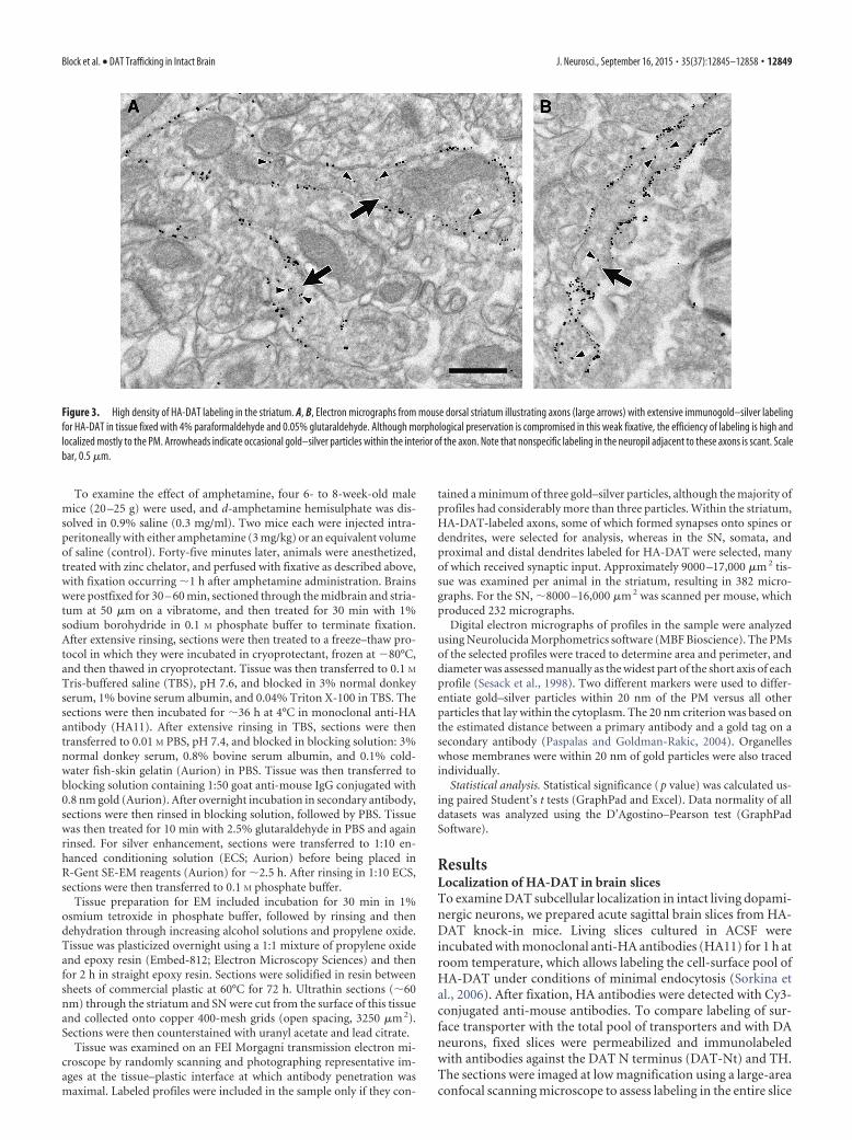
Figure 3. High density of HA-DAT labeling in the striatum. A, B, Electron micrographs from mouse dorsal striatum illustrating axons (large arrows) with extensive immunogold–silver labelingfor HA-DAT in tissue fixed with 4% paraformaldehyde and 0.05% glutaraldehyde. Although morphological preservation is compromised in this weak fixative, the efficiency of labeling is high andlocalized mostly to the PM. Arrowheads indicate occasional gold–silver particles within the interior of the axon. Note that nonspecific labeling in the neuropil adjacent to these axons is scant. Scalebar, 0.5 �m.
Block et al. • DAT Trafficking in Intact Brain J. Neurosci., September 16, 2015 • 35(37):12845–12858 • 12849

(Fig. 1). The labeling method directed against the HA epitopeadequately labeled all of the brain areas detected by anti-DATantibodies, and the labeling colocalized well with TH immuno-fluorescence in the midbrain, striatum, and neuronal tracts con-necting these two areas. Of note, non-obstructive host vascularelements were detected by anti-mouse antibodies, and TH wasalso detected in norepinephrine neurons not expressing DAT.Importantly, background fluorescence of HA11 labeling was lowin nondopaminergic areas of brain, indicating a high specificityof this labeling procedure. As expected, qualitatively, the majorityof HA and DAT immunoreactivity was observed in the striatum.
Detailed 3D images from various regions of sagittal slices (typ-ically 10 – 40 �m deep from the surface) prepared and labeled asin Figure 1 were acquired by spinning-disk confocal microscope(Fig. 2). In the midbrain (Fig. 2A), surface HA-DAT was foundon a network of processes (presumably, mostly dendrites andsoma-proximal axonal segments) and varicosities (typically�2–10 �m in diameter), as well as on cell bodies. Surface labelingon the cell bodies was comparatively weaker than on dendritic oraxonal structures as shown by pseudocolored intensity micro-graphs (Fig. 2B). The DAT-Nt antibody detected all DAT, includ-ing intracellular transporters (not labeled by HA11) in cell
bodies, as well as within proximal dendrites, in particular withinlarge varicosities. In the cell bodies, the pattern of labeling withDAT-Nt antibody resembled the tubuloreticular morphology ofthe endoplasmic reticulum (ER), with occasional punctate struc-tures (Fig. 2A, Midbrain insets). Interestingly, the DAT-Nt anti-body was less efficient at detecting HA-DAT in the PM than inintracellular membranes, possibly because of steric hindrance ofthe antibody recognition site in the N terminus of surfacetransporter.
HA antibody labeling revealed a relatively homogenous distri-bution of the surface transporter throughout the axonal tractsbetween the midbrain and striatum (Fig. 2A, Tracts) and a highlycomplex network of axonal processes and small varicosities (�1�m in diameter) in the striatum (Fig. 2A, Striatum). Labelingwith DAT-Nt antibodies detected the same neuronal structures,albeit with a relatively low intensity of labeling of axonal shafts. Instriatal varicosities, only few DAT-labeled structures were ob-served that were not also labeled with HA11, indicating a paucityof intracellular HA-DAT in this region. When the interior ofstriatal varicosities could be resolved, the two markers tended tocolocalize at the axonal surface (Fig. 2A, Striatum insets). Quan-titative analysis by pseudocolored intensity profiles (Fig. 2B) and
Figure 4. Distribution of HA-DAT in membrane compartments of the dorsal striatum. Electron micrographs from mouse dorsal striatum illustrating axons (large arrows) with immunogold–silverlabeling for HA-DAT. In A and B, particles are localized exclusively to the PM but not within the synaptic zones (medium arrows) formed onto unlabeled spines (us). One of the labeled axons in A isconnected to an unlabeled dendrite (ud) by a punctum adherens (clear arrow), which also exhibits no gold–silver labeling for HA-DAT. The labeled axon in B synapses onto a spine that receivesadditional synaptic input from an unlabeled axon terminal (ut). In C and D, both plasmalemmal and intracellular particles for HA-DAT are visible, the latter in association with vesicles and endosomes(small thin arrows) or with no apparent relationship to organelles (arrowheads). The axon in C synapses (medium arrow) onto an unlabeled dendrite (ud). Scale bar, 0.265 �m.
12850 • J. Neurosci., September 16, 2015 • 35(37):12845–12858 Block et al. • DAT Trafficking in Intact Brain

mean intensity calculations (Fig. 2C) revealed cell-surface HA-DAT in the midbrain to be �50% as dense as in the striatum andfurthermore that HA-DAT in the striatum is concentrated inaxonal varicosities as opposed to intervaricose segments. To-gether, the results in Figure 2 suggest that DAT in axons is mostlyat the cell surface; in cell bodies of the midbrain, DAT can befound in surface and intracellular membranes, especially the ER.Because the total amount of DAT is much greater in the striatum,most of the DAT in the brain is therefore at the cell surface.
Electron microscopic analysis of HA-DAT localization inmouse brainPrompted by the efficient labeling of HA-DAT in neurons ofprimary neuronal cultures (Rao et al., 2012) and brain slices(Figs. 1, 2), we performed EM labeling of HA-DAT in fixedmouse brain sections. Immunogold–silver labeling directedagainst HA11 yielded robust and efficient labeling in both thestriatum (Figs. 3, 4) and midbrain SN (Figs. 5, 6), especially inmildly fixed brains (Fig. 3).
Within the striatum, HA-DAT labeling was found predomi-nantly along the PM, in which it was notably excluded from
synaptic active zones (Figs. 3, 4; Table 1). Particles for HA-DATwere less frequently found along the intracellular membranes oforganelles resembling synaptic vesicles (SVs) or small endosomes(Fig. 4C,D; Table 1). In single planes of section, however, themajority of intracellular labeling had no evident organelle asso-ciation, an observation that does not preclude association withorganelles above or below the section plane.
For the SN, the distribution of HA-DAT immunoreactivitywas typically the reverse of the striatum, with the majority ofgold–silver particles being abundant along intracellular mem-branes, especially within somata, and with less frequent labelingat the plasmalemma (Figs. 5, 6; Table 2). Particles for HA-DATwere often observed along the nuclear membrane and mem-branes of the rough ER and Golgi complex (Fig. 5A–C). Labelingwas also evident on putative endosomes and multivesicular bod-ies (Figs. 5, 6). Clathrin-coated structures were devoid of HA-DAT labeling in both the SN and the striatum. By quantitativeanalysis of �600 images (Tables 1, 2), �85% of HA-DAT boundgold particles were associated with the PM in the dorsal striatum,whereas only �25% of gold particles were associated with theplasmalemma in dendrites of the SN (soma were only qualita-
Figure 5. Localization of HA-DAT within perikarya. Electron micrographs from mouse SN illustrating the distribution of immunogold–silver labeling for HA-DAT within perikarya shown at low (A,B) and high (C, D) magnification. Particles occur along the nuclear (n) membrane (small thin arrows) and within the Golgi apparatus (g), rough endoplasmic reticulum (rer), multivesicular bodies(mvb), and endosomes (e). Other particles are not localized to identifiable organelles in single sections (arrowheads). A few particles appear along the PM (medium-sized arrows). Scale bars: (in A)A, B, 0.5 �m; (in C) C, D, 0.2 �m.
Block et al. • DAT Trafficking in Intact Brain J. Neurosci., September 16, 2015 • 35(37):12845–12858 • 12851

tively assessed). These EM data are in strong agreement with theobservations from confocal immunofluorescence microscopythat the majority of DAT is in the PM, with an intracellular frac-tion that is greater in the midbrain, particularly in biosyntheticcompartments.
Endolysosomal compartments in dopaminergic neuronsBy confocal microscopy (Fig. 2) and EM (Figs. 3– 6), DAT waslocalized occasionally to intracellular membranes resembling en-dosomes. To identify compartments containing intracellularDAT, sagittal slices from HA-DAT mice were fixed, permeabil-ized, and colabeled for the DAT (using either DAT-Nt antibodyor HA11, depending on compatibility) and various endosomal
markers (Fig. 7). The vesicular SNARE protein Stx13 regulatesmembrane fusion of the early and recycling endosomes on whichit is found (Prekeris et al., 1998, 1999). Stx13 was detected inpunctate structures within DA neuronal cell bodies that colocal-ized occasionally with the DAT (0.82 DAT/Stx13 colocalizations/cell; Fig. 7A). EEA.1 is a Rab5 effector and the main component ofthe tethering complex in early and intermediate/sorting endo-somes. EEA.1 was found in punctate and tubular structures indopaminergic cell bodies in the midbrain (Fig. 7B). As withStx13, DAT exhibited occasional colocalization with EEA.1 in themidbrain cell bodies (0.87 DAT/EEA.1 colocalizations/cell).LAMP1 localizes mainly to late endosomes and lysosomes. Sim-ilarly to EEA.1, LAMP1 was found in punctate and tubular struc-
Figure 6. Localization of HA-DAT within dendrites. Electron micrographs from mouse SN illustrating immunogold–silver labeling for HA-DAT within dendrites. In A and B, the majority of particlesare not associated with identifiable structures in the cytoplasm (arrowheads) in single sections. In C and D, particles are associated with specific organelles, including endosomes (e) and amultivesicular body (mvb). Note the absence of labeling near clathrin-coated vesicles (c). Scale bar, 265 �m.
Table 1. Quantitative EM data: axons labeled for HA-DAT in the striatum
Labeled axon sizeHA-DAT gold particle density Organelle labeling
Labeledaxons
Diameter (�m)mean � SD
Perimeter (�m)mean � SD
Area (�m 2)mean � SD
Total (%)membraneparticles
Total (%)cytosolparticles
Particles perperimetermean � SD
Particles perareamean � SD
Total (%)organelleparticles
Perimeter (�m)mean � SD
AmphetamineAnimal 1 177 0.32 � 0.13 2.18 � 1.09 0.20 � 0.14 1997 (84%) 271 (11%) 5.5 � 3.3 83.2 � 57.3 102 (4%) 0.162 � 0.062Animal 2 228 0.30 � 0.13 2.02 � 1.26 0.18 � 0.15 3915 (89%) 330 (7%) 8.9 � 5.9 137.0 � 111.8 162 (4%) 0.300 � 0.234Combined 405 0.31 � 0.13 2.09 � 1.19 0.19 � 0.15 5912 (87%) 601 (9%) 7.4 � 5.2 113.5 � 95.8 264 (4%) 0.247 � 0.198
SalineAnimal 1 140 0.38 � 0.14 2.21 � 1.06 0.26 � 0.18 1420 (85%) 163 (10%) 4.9 � 3.0 59.1 � 42.7 95 (6%) 0.165 � 0.041Animal 2 156 0.37 � 0.16 2.07 � 1.16 0.23 � 0.20 2175 (84%) 294 (11%) 7.4 � 4.5 100.4 � 80.0 122 (5%) 0.150 � 0.027Combined 296 0.37 � 0.15 2.14 � 1.11 0.24 � 0.19 3595 (84%) 457 (11%) 6.2 � 4.1 80.9 � 68.2 217 (5%) 0.161 � 0.038
12852 • J. Neurosci., September 16, 2015 • 35(37):12845–12858 Block et al. • DAT Trafficking in Intact Brain

tures in dopaminergic cell bodies in the midbrain (Fig. 7C,D).Despite the abundance of LAMP1 compartments in DA cell bod-ies, DAT was rarely observed to colocalize with LAMP1 [0.17DAT/LAMP1 colocalizations/cell using DAT C-terminus (DAT-Ct) antibodies to detect DAT; 0.12 DAT/LAMP1 colocalizations/cell using HA11 to detect DAT]. Very few, if any, endosomes weredetected in midbrain dendritic varicosities.
In the striatum, Stx13-containing endosomes were abundantin nondopaminergic cells (Fig. 8A) but were rarely detected instriatal axons and varicosities (Fig. 8A detail). Because the smallsize of the observed structures and the resolution limits of theconfocal microscopy make it difficult to conclude whether Stx13colocalizes with the DAT in striatal axons, a super-resolution,structured illumination microscopy (SIM) was used. Lateral res-olution of SIM is �100 –120 nm, which is comparable with theminimal diameter of an endosome. Although confocal imagingdemonstrated overlapping DAT and Stx13 labeling, SIM imagingcould clearly separate the two labels, suggesting a lack of colocal-ization. EEA.1 (Fig. 8C) and LAMP1 (Fig. 8D) were detected nearthe nuclei of nondopaminergic cells in the striatum but were notdetected in striatal DA axons or in axonal tracts (Fig. 8E).
Together, these results (Figs. 2– 8) suggest that DAT is ex-pressed over the entirety of the neuronal PM (with the exceptionof active zones) and that the endolysosomal pool of DAT is small.This indicates that constitutive endocytosis of the transporter israre in axon tracts and varicosities and that transporter endocy-tosis is slow in the somatodendritic compartment. Furthermore,endocytic colocalizations that were observed more closely resem-ble a recycling route of internalized DAT as opposed to sorting tothe lysosomal degradation pathway. Strikingly, in DA axons, theendosomal trafficking system for DAT does not include early/sorting endosomes.
Acute administration of amphetamine does not affectsubcellular distribution of HA-DATGiven apparently slow rates of constitutive endocytosis, we nextexamined whether DAT localization could be altered by stimu-lating endocytosis with amphetamine, an abused and therapeuticpsychostimulant. Amphetamine increases monoamine neu-rotransmission by inducing reverse transport through reuptaketransporters (Sulzer et al., 2005; Robertson et al., 2009), and it hasalso been shown to induce DAT endocytosis in a variety of modelsystems (Saunders et al., 2000; Hong and Amara 2013). HA-DATmice were injected with 3 mg/kg d-amphetamine and used forexperiments after 1 h, conditions of acute exposure known toproduce marked behavioral changes (Chen et al., 2009; Rickhaget al., 2013). Quantitation after analysis by EM showed that thesubcellular distribution of HA-DAT in the striatum or SN was
not significantly altered from saline-injected littermate controlsafter a single treatment with amphetamine (Tables 1, 2; all pvalues �0.12, paired t test, two-tailed).
To examine the effect of amphetamine treatment on DATendocytosis in acute slice preparations ex vivo, we cultured theslices at 37°C for 10, 30, or 60 min with HA11 antibodies in thepresence or absence of amphetamine. After fixation and permea-bilization, endocytosed HA antibody could be observed by thecolocalization of anti-mouse secondary antibodies with immu-nolabeled Stx13. In agreement with the lack of significant endo-cytosis in naive animals (Figs. 2– 8), slices cultured in this mannerexhibited no localization of HA-DAT in Stx13 endosomes, andthe HA antibodies all appeared to remain at the PM (Fig. 9A,Vehicle). Treatment with 100 �M amphetamine did not alter thislocalization (Fig. 9A, AMPH). In control experiments in HEK293cells that stably express CFP–HA-DAT, treatment with 100 �M
amphetamine did induce a significant accumulation of DAT inendosomal compartments (Fig. 9B). Together, the results fromfluorescence and EM suggest that, in the intact brain, a singletreatment in vivo (60 min) or ex vivo (10 – 60 min) with amphet-amine does not redistribute the DAT significantly from the PM toendosomal compartments.
DiscussionDA neurons exhibit a distinct morphology with regionally segre-gated axonal and somatodendritic compartments, varicosities,and unusually dense axonal arborizations. Current understand-ing of how DAT traffics through this complex system to regulateextracellular DA concentration is incomplete. Although numer-ous important studies have investigated endocytosis and subcel-lular distribution of DAT in heterologous cells, neuronalcultures, striatal synaptosomes (for review, see Melikian, 2004;Zahniser and Sorkin, 2009; Kristensen et al., 2011), and striatalslices (Cremona et al., 2011; Gabriel et al., 2013), few studies haveexamined DAT trafficking in intact neurons in the brain. In thepresent work, we took advantage of the HA-DAT mouse anddeveloped a novel methodology to preserve entire DA neurons inacute sagittal slices for 3D fluorescence microscopy analysis ofDAT subcellular localization and trafficking. This approach wascombined with high-resolution quantitative EM analysis usingpreembedding immunogold–silver labeling of HA-DAT. HA an-tibodies labeled efficiently cell-surface and intracellular DAT,permitting quantitative analysis and localization to specific intra-cellular compartments. Comprehensive EM studies of DAT lo-calization in the rat brain have established the basic model ofDAT distribution over the entire DA neuron (Nirenberg et al.,1996, 1997; Hersch et al., 1997), but a number of questions re-mained unaddressed. Here, we have found that (1) DAT is pres-
Table 2. Quantitative EM data: dendrites labeled for HA-DAT in the SN
Labeled dendrite sizeHA-DAT gold particle density Organelle labeling
Labeleddendrites
Diameter (�m)mean � SD
Perimeter (�m)mean � SD
Area (�m 2)mean � SD
Total (%)membraneparticles
Total (%)cytosolparticles
Particles perperimetermean � SD
Particlesper areamean � SD
Total (%)organelleparticles
Perimeter(�m)mean � SD
AmphetamineAnimal 1 68 1.26 � 0.52 6.54 � 2.14 2.62 � 1.69 161 (20%) 533 (67%) 0.42 � 0.45 6.3 � 5.28 106 (13%) 0.508 � 0.348Animal 2 59 1.03 � 0.50 6.64 � 2.33 2.48 � 1.98 167 (15%) 720 (66%) 0.49 � 0.46 9.0 � 4.41 211 (19%) 0.534 � 0.414Combined 127 1.15 � 0.52 6.58 � 2.22 2.56 � 1.83 328 (17%) 1253 (66%) 0.45 � 0.45 7.5 � 5.06 317 (17%) 0.525 � 0.392
SalineAnimal 1 58 1.05 � 0.50 6.57 � 2.51 2.32 � 1.66 280 (27%) 567 (55%) 0.92 � 1.40 10.8 � 10.05 190 (18%) 0.442 � 0.349Animal 2 75 1.12 � 0.49 6.18 � 2.34 2.32 � 1.59 244 (24%) 640 (63%) 0.61 � 0.68 8.5 � 6.82 128 (13%) 0.590 � 0.477Combined 133 1.09 � 0.50 6.35 � 2.41 2.32 � 1.61 524 (26%) 1207 (59%) 0.75 � 1.06 9.5 � 8.43 318 (16%) 0.502 � 0.411
Block et al. • DAT Trafficking in Intact Brain J. Neurosci., September 16, 2015 • 35(37):12845–12858 • 12853

ent in higher concentrations in striatalaxons, presynaptic terminals in particu-lar, thus suggesting the existence of a spe-cific targeting mechanism, and (2) there isa relatively small endolysosomal pool ofDAT, which indicates the importance ofboth nonvesicular lateral membranemovement of the transporter, as well asPM retention mechanisms. Previousstudies have determined that DAT expres-sion levels, substrate uptake, and traffick-ing are unchanged by the insertion of theHA epitope and binding of HA antibodies(Sorkina et al., 2006; Eriksen et al., 2010;Rao et al., 2012), suggesting that the re-sults of these studies with HA-DAT applyto the wild-type transporter, as well.
High density of the PM DATthroughout the striatal axonal systemWhen living dopaminergic neurons were la-beled with HA antibodies under conditionsof negligible endocytosis to detect cell-surface DAT, the transporter was observedover the entirety of the neuron. Quantitativeanalysis of immunofluorescence signals re-vealed that cell-surface density of HA-DATwas twofold higher in striatal axons thanmidbrain. Moreover, by EM, the density ofgold particles marking HA-DAT at the cellsurface was �10-fold higher in striatal ax-ons than in SN dendrites. Although therewere differences in the relative efficiency ofsomatic PM labeling in EM versus immu-nofluorescence, the observed low density ofDAT in somatic PM agrees with previousEM observations using DAT antibodies inrat brain (Nirenberg et al., 1996, 1997) andlive-cell microscopy of primary cultures(Rao et al., 2012). The mechanisms under-lying this progressively increasing PM con-centration of DAT (soma � dendrites �axons) are unknown but may involve a de-creased endocytic rate or decreased mem-brane mobility of DAT (retention) in theareas in which DAT concentration is higher.Previous studies demonstrated that cell-surface retention of DAT is controlled byjuxtamembrane amino acid residues of theDAT-Nt (Sorkina et al., 2009).
Overall, our data suggest a high density ofDATalongtheentiresurfaceofaxons.Theab-sence of HA-DAT at axonal active zones ob-served by EM is unlikely attributable toinaccessibility of the HA epitope because sim-ilar observations were made using DAT anti-bodies (Hersch et al., 1997; Nirenberg et al.,1997). These observations are consistent withthe “volume transmission” model suggestedfor nigrostriatal projections whereby DAT regulates the range and mag-nitudeofextrasynapticDAsignaling(Craggetal., 2004;RiceandCragg,2008) to modulate multiple neurons over a large physical space (Ar-buthnott and Wickens, 2007; Rice et al., 2011). Although DAT is
certainly expressed over the entire DA neuron, we have found herethat, in the striatal axonal network, the mean fluorescenceintensity of surface HA-DAT was higher in varicosities than inter-varicose shafts. This increase in DAT density at putative presynaptic
Figure 7. Colocalization of DAT with endosomal markers in midbrain. Slices were fixed, permeabilized, and labeled withantibodies against HA and Stx13 (A), DAT-Nt and EEA.1 (B), DAT-Ct and LAMP1 (C), or HA and LAMP1 (D), followed by fluorophore-conjugated secondary antibodies. DAT is shown in green, and endosomal markers are shown in red. Maximum-intensityprojections of two to three consecutive confocal sections from the collected z stacks of the midbrain are presented. Insets showhigh-magnification images of areas indicated by white rectangles. Arrows show examples of colocalization. Inset in C shows anexample of localization of HA-DAT in a LAMP1-containing compartment. An example of partial overlap of DAT- andLAMP1-containing structures illustrating lack of true colocalization in insets (D) is indicated by circles. Scale bars, 10 �m.
12854 • J. Neurosci., September 16, 2015 • 35(37):12845–12858 Block et al. • DAT Trafficking in Intact Brain

areas suggests that DA reuptake, like release,is in fact spatially regulated.
Low levels of DAT endocytosis inaxonal and somatodendriticcompartmentsQuantitative EM showed that �84% ofgold particles were at the cell surface,whereas only 5% were associated clearlywith intracellular membranes in striatalaxons examined in single sections. Con-sistently, no endosomes containing HA-DAT together with markers of early,recycling, or late endosomes were foundin the striatum by immunofluorescence.Stx13-positive endosomes have been de-scribed in axons of other neurons(Prekeris et al., 1999) and cultured DAneurons (Rao et al., 2012). However, inthe present studies, Stx13-positive struc-tures were detected rarely in DA axons.Interestingly, when synaptic areas wereresolved by EM, intracellular HA-DATwas associated more frequently withstructures of the size and morphology ofSVs rather than with larger, endosome-like compartments. In contrast, previoussubcellular fractionation studies of ratstriatum did not identify DAT in the SVfraction, likely because the SV pool ofDAT is small (Rao et al., 2011). Our datapresent an alternative interpretation tostudies in which significant pools of intra-cellular transporters (�40%) have beendetected by surface biotinylation or stria-tal membrane fractionation (Johnson etal., 2005; Richards and Zahniser, 2009;German et al., 2012). Surface biotinyla-tion of synaptosomal preparations mayunderestimate the PM pool because oflimited biotinylation efficiency, and or-ganelle fractionation does not cleanly sep-arate early endosomes from the PM. Therelative paucity of intracellular axonalDAT detected in the present studies is alsoinconsistent with models describing rapiddelivery of a large intracellular pool oftransporters to the PM during stimulation(Johnson et al., 2005; Furman et al., 2009).
In SN dendrites analyzed by EM,only �25% of DAT was at the cell surface.Echoing previous studies (Nirenberg etal., 1996, 1997; Hersch et al., 1997), EMdetected a significant intracellular pool oftransporters in somatodendritic regionsin rough ER and Golgi, whereas only asmall pool of DAT was located in mem-brane compartments with endosome-likemorphology, including multivesicularbodies. This EM analysis, together withimmunofluorescence colocalization stud-ies that demonstrated a higher extent ofDAT localization in Stx13- and EEA.1-
Figure 8. Lack of early and late endosomes in tracts and striatal axons of DA neurons. Slices were fixed, permeabilized, andlabeled with antibodies against HA and Stx13 (A, B), DAT-Nt and EEA.1 (C, E), or HA and LAMP1 (D, E) followed by fluorophore-conjugated secondary antibodies. DAT is shown in green, and endosomal markers are shown in red. Images were acquired byspinning-disk confocal (A, C–E) or SIM (B) systems. Maximum-intensity projections of two to three consecutive confocal sectionsfrom the collected z stacks of the striatum (A, C, D) and tracts (E) are presented. Single optical section is presented in B. High-magnification images of areas indicated by white rectangles are shown on the right. In A, arrows show examples of overlap of Stx13and DAT fluorescence in axonal varicosities. The presence of a large number of Stx13 endosomes in nondopaminergic cells isindicated by circles. For B, SIM imaging of the striatum in slices processed as in A is shown; no overlap of DAT and Stx13 labeling wasobserved within the varicosity. In C and D, high-magnification images show abundance of EEA.1 and LAMP1 compartments,respectively, in nondopaminergic cells. Scale bars: A, C–E, 10 �m; B, 1 �m.
Block et al. • DAT Trafficking in Intact Brain J. Neurosci., September 16, 2015 • 35(37):12845–12858 • 12855

positive than in LAMP1-postive compartments in somata sug-gests that endocytosed DAT tends to traffic toward recyclingpathways as opposed to lysosomal degradation. Studies with pri-mary cultures of DA neurons have also observed transporter inearly and recycling compartments but have detected a significant
pool of DAT in late endosomes (Eriksen et al., 2010; Rao et al.,2011). These discrepancies suggest that trafficking and degrada-tion of DAT in cell culture and brain may be fundamentally dif-ferent and highlight the need for continued investigations intotrafficking mechanisms in intact neurons in the brain.
Figure 9. Amphetamine does not affect subcellular distribution of DAT. A, Acute slices were incubated with HA11 and either vehicle or 100 �M amphetamine (AMPH) for 10, 30, or 60min at 37°C. Slices were then fixed, permeabilized, and labeled with the Stx13 antibody, followed up by secondary antibodies labeled with Cy5 (HA-DAT, green) and Cy3 (Stx13, red). 3Dimage acquisition was performed through 561 nm and 640 nm channels. Individual optical sections are presented. Insets are high-magnification images of the regions indicated by whiterectangles showing parts of the soma. B, HEK293 cells expressing CFP–HA-DAT were incubated with HA11 and either vehicle or 100 �M amphetamine for 30 min at 37°C. Cells were fixedand labeled to detect HA-DAT as in A before imaging. Insets show high magnification of the region indicated by white rectangle to show examples of amphetamine-induced endosomes.Scale bars, 10 �m.
12856 • J. Neurosci., September 16, 2015 • 35(37):12845–12858 Block et al. • DAT Trafficking in Intact Brain

Acute administration of amphetamine does not increaseDAT endocytosisHigh-efficiency labeling of HA-DAT in the present EM studiespermitted a quantitative approach to investigate the effect ofacute in vivo amphetamine treatment on DAT distribution. Am-phetamine has been shown to induce DAT endocytosis or inhibitDA uptake in models that include cultured neurons (Hong andAmara, 2013), synaptosomes (Johnson et al., 2005; Richards andZahniser, 2009), and heterologous cells (Saunders et al., 2000).Amphetamine doses administered in vivo or to slices in thepresent studies correspond to doses used in single-treatment exp-eriments in which DAT endocytosis and other effects of amphet-amine were observed (Chen et al., 2009; Richards and Zahniser,2009; Rickhag et al., 2013). However, we did not detect anychanges in subcellular localization of DAT in striatal axons or SNdendrites by EM. Furthermore, treating acute slices with amphet-amine for 10 – 60 min did not stimulate endocytosis in any regionof DA neurons. Although methods used in the present study maynot be sensitive enough to detect rapid and small changes in DATendocytosis, our data are in agreement with findings that acuteamphetamine treatment does not stimulate substantial DAT en-docytosis in intact systems (German et al., 2012).
ConclusionsThe steady-state subcellular localization of DAT in the striatum isconsistent with the endocytosis rate being five to six times lowerthan the recycling rate, kinetics characteristic of a PM-retainedcargo, such as inactive growth factor receptors (Sorkin and Goh,2009). DAT is routed through conventional endocytic pathwaysin the somatodendritic region, but this trafficking activity is of asmall scale. Although dopaminergic neurons seem to possess thenecessary machinery for a normal itinerary of membrane trafficin the somatodendritic compartment, the lack of conventionalendosomes in axons is striking. We hypothesize that the majorityof DAT is not transported via vesicular trafficking mechanisms.Rather, in the intact brain, DAT is more likely transported be-tween midbrain and striatum by lateral diffusion in the PM. Thisis consistent with measurements of DAT half-life using radioac-tive tracers in intact rat brains, estimated to be �2– 6 d (Flecken-stein et al., 1996; Kimmel et al., 2000), which is orders ofmagnitude longer than the half-life of cell-surface DAT observedin heterologous expression systems (Boudanova et al., 2008). Thepresent study illustrates the potential of sagittal slice preparationsto define the kinetics and physiological mechanisms of DAT traf-ficking and turnover.
ReferencesArbuthnott GW, Wickens J (2007) Space, time and dopamine. Trends Neu-
rosci 30:62– 69. CrossRef MedlineBoudanova E, Navaroli DM, Melikian HE (2008) Amphetamine-induced
decreases in dopamine transporter surface expression are protein kinaseC-independent. Neuropharmacology 54:605– 612. CrossRef Medline
Chen JW, Pan W, D’Souza MP, August JT (1985) Lysosome-associatedmembrane proteins: characterization of LAMP-1 of macrophage P388and mouse embryo 3T3 cultured cells. Arch Biochem Biophys 239:574 –586. CrossRef Medline
Chen R, Furman CA, Zhang M, Kim MN, Gereau RW 4th, Leitges M, GnegyME (2009) Protein kinase Cbeta is a critical regulator of dopaminetransporter trafficking and regulates the behavioral response to amphet-amine in mice. J Pharmacol Exp Ther 328:912–920. CrossRef Medline
Chen R, Furman CA, Gnegy ME (2010) Dopamine transporter trafficking:rapid response on demand. Future Neurol 5:123. CrossRef Medline
Ciliax BJ, Drash GW, Staley JK, Haber S, Mobley CJ, Miller GW, Mufson EJ,Mash DC, Levey AI (1999) Immunocytochemical localization of the do-pamine transporter in human brain. J Comp Neurol 409:38 –56. CrossRefMedline
Cragg SJ, Baufreton J, Xue Y, Bolam JP, Bevan MD (2004) Synaptic release ofdopamine in the subthalamic nucleus. Eur J Neurosci 20:1788 –1802.CrossRef Medline
Cremona ML, Matthies HJ, Pau K, Bowton E, Speed N, Lute BJ, Anderson M,Sen N, Robertson SD, Vaughan RA, Rothman JE, Galli A, Javitch JA,Yamamoto A (2011) Flotillin-1 is essential for PKC-triggered endocyto-sis and membrane microdomain localization of DAT. Nat Neurosci 14:469 – 477. CrossRef Medline
Eriksen J, Rasmussen SG, Rasmussen TN, Vaegter CB, Cha JH, Zou MF,Newman AH, Gether U (2009) Visualization of dopamine transportertrafficking in live neurons by use of fluorescent cocaine analogs. J Neuro-sci 29:6794 – 6808. CrossRef Medline
Eriksen J, Bjørn-Yoshimoto WE, Jørgensen TN, Newman AH, Gether U(2010) Postendocytic sorting of constitutively internalized dopaminetransporter in cell lines and dopaminergic neurons. J Biol Chem 285:27289 –27301. CrossRef Medline
Fleckenstein AE, Pogun S, Carroll FI, Kuhar MJ (1996) Recovery of dopa-mine transporter binding and function after intrastriatal administrationof the irreversible inhibitor RTI-76 [3 beta-(3p-chlorophenyl) tropan-2beta-carboxylic acid p-isothiocyanatophenylethyl ester hydrochloride].J Pharmacol Exp Ther 279:200 –206. Medline
Furman CA, Chen R, Guptaroy B, Zhang M, Holz RW, Gnegy M (2009)Dopamine and amphetamine rapidly increase dopamine transporter traf-ficking to the surface: live-cell imaging using total internal reflection flu-orescence microscopy. J Neurosci 29:3328 –3336. CrossRef Medline
Gabriel LR, Wu S, Kearney P, Bellve KD, Standley C, Fogarty KE, Melikian HE(2013) Dopamine transporter endocytic trafficking in striatal dopami-nergic neurons: differential dependence on dynamin and the actin cyto-skeleton. J Neurosci 33:17836 –17846. CrossRef Medline
Gainetdinov RR, Caron MG (2003) Monoamine transporters: from genesto behavior. Annu Rev Pharmacol Toxicol 43:261–284. CrossRef Medline
Garzon M, Pickel VM (2006) Subcellular distribution of M2 muscarinicreceptors in relation to dopaminergic neurons of the rat ventral tegmentalarea. J Comp Neurol 498:821– 839. CrossRef Medline
German CL, Hanson GR, Fleckenstein AE (2012) Amphetamine and meth-amphetamine reduce striatal dopamine transporter function withoutconcurrent dopamine transporter relocalization. J Neurochem 123:288 –297. CrossRef Medline
Giros B, el Mestikawy S, Bertrand L, Caron MG (1991) Cloning and func-tional characterization of a cocaine-sensitive dopamine transporter. FEBSLett 295:149 –154. CrossRef Medline
Hersch SM, Yi H, Heilman CJ, Edwards RH, Levey AI (1997) Subcellular localiza-tion and molecular topology of the dopamine transporter in the striatum andsubstantia nigra. J Comp Neurol 388:211–227. CrossRef Medline
Hong WC, Amara SG (2013) Differential targeting of the dopamine trans-porter to recycling or degradative pathways during amphetamine- orPKC-regulated endocytosis in dopamine neurons. FASEB J 27:2995–3007. CrossRef Medline
Hoover BR, Everett CV, Sorkin A, Zahniser NR (2007) Rapid regulation ofdopamine transporters by tyrosine kinases in rat neuronal preparations.J Neurochem 101:1258 –1271. CrossRef Medline
Huff RA, Vaughan RA, Kuhar MJ, Uhl GR (1997) Phorbol esters increasedopamine transporter phosphorylation and decrease transport Vmax.J Neurochem 68:225–232. CrossRef Medline
Jaber M, Jones S, Giros B, Caron MG (1997) The dopamine transporter: acrucial component regulating dopamine transmission. Mov Disord 12:629 – 633. CrossRef Medline
Johnson LA, Furman CA, Zhang M, Guptaroy B, Gnegy ME (2005) Rapid deliveryof the dopamine transporter to the plasmalemmal membrane upon amphet-amine stimulation. Neuropharmacology 49:750–758. CrossRef Medline
Kimmel HL, Carroll FI, Kuhar MJ (2000) Dopamine transporter synthesisand degradation rate in rat striatum and nucleus accumbens usingRTI-76. Neuropharmacology 39:578 –585. CrossRef Medline
Kristensen AS, Andersen J, Jørgensen TN, Sørensen L, Eriksen J, Loland CJ,Strømgaard K, Gether U (2011) SLC6 neurotransmitter transporters:structure, function, and regulation. Pharmacol Rev 63:585– 640. CrossRefMedline
Melikian HE (2004) Neurotransmitter transporter trafficking: endocytosis,recycling, and regulation. Pharmacol Ther 104:17–27. CrossRef Medline
Melikian HE, Buckley KM (1999) Membrane trafficking regulates the activ-ity of the human dopamine transporter. J Neurosci 19:7699 –7710.Medline
Block et al. • DAT Trafficking in Intact Brain J. Neurosci., September 16, 2015 • 35(37):12845–12858 • 12857

Miranda M, Sorkina T, Grammatopoulos TN, Zawada WM, Sorkin A (2004)Multiple molecular determinants in the carboxyl terminus regulate dopa-mine transporter export from endoplasmic reticulum. J Biol Chem 279:30760 –30770. CrossRef Medline
Mu FT, Callaghan JM, Steele-Mortimer O, Stenmark H, Parton RG, Camp-bell PL, McCluskey J, Yeo JP, Tock EP, Toh BH (1995) EEA.1, an earlyendosome-associated protein. EEA.1 ia a conserved a-helical peripheralmembrane protein flanked by cysteine “fingers” and contains acalmodulin-binding IQ motif. J Biol Chem 270:13503–13511. CrossRefMedline
Nirenberg MJ, Vaughan RA, Uhl GR, Kuhar MJ, Pickel VM (1996) Thedopamine transporter is localized to dendritic and axonal plasma mem-branes of nigrostriatal dopaminergic neurons. J Neurosci 16:436 – 447.Medline
Nirenberg MJ, Chan J, Vaughan RA, Uhl GR, Kuhar MJ, Pickel VM (1997)Immunogold localization of the dopamine transporter: an ultrastructuralstudy of the rat ventral tegmental area. J Neurosci 17:5255–5262. Medline
Paspalas CD, Goldman-Rakic PS (2004) Microdomains for dopamine vol-ume neurotransmission in primate prefrontal cortex. J Neurosci 24:5292–5300. CrossRef Medline
Prekeris R, Klumperman J, Chen YA, Scheller RH (1998) Syntaxin 13 me-diates cycling of plasma membrane proteins via tubulovesicular recyclingendosomes. J Cell Biol 143:957–971. CrossRef Medline
Prekeris R, Foletti DL, Scheller RH (1999) Dynamics of tubulovesicular re-cycling endosomes in hippocampal neurons. J Neurosci 19:10324 –10337.Medline
Rao A, Simmons D, Sorkin A (2011) Differential subcellular distribution ofendosomal compartments and the dopamine transporter in dopaminer-gic neurons. Mol Cell Neurosci 46:148 –158. CrossRef Medline
Rao A, Richards TL, Simmons D, Zahniser NR, Sorkin A (2012) Epitope-tagged dopamine transporter knock-in mice reveal rapid endocytic traf-ficking and filopodia targeting of the transporter in dopaminergic axons.FASEB J 26:1921–1933. CrossRef Medline
Rice ME, Cragg SJ (2008) Dopamine spillover after quantal release: rethink-ing dopamine transmission in the nigrostriatal pathway. Brain Res Rev58:303–313. CrossRef Medline
Rice ME, Patel JC, Cragg SJ (2011) Dopamine release in the basal ganglia.Neuroscience 198:112–137. CrossRef Medline
Richards TL, Zahniser NR (2009) Rapid substrate-induced down-regulation in function and surface localization of dopamine transporters:rat dorsal striatum versus nucleus accumbens. J Neurochem 108:1575–1584. CrossRef Medline
Rickhag M, Hansen FH, Sørensen G, Strandfelt KN, Andresen B, Gotfryd K,Madsen KL, Vestergaard-Klewe I, Ammendrup-Johnsen I, Eriksen J,Newman AH, Fuchtbauer EM, Gomeza J, Woldbye DP, Wortwein G,Gether U (2013) A C-terminal PDZ domain-binding sequence is re-quired for striatal distribution of the dopamine transporter. Nat Com-mun 4:1580. CrossRef Medline
Robertson SD, Matthies HJ, Galli A (2009) A closer look at amphetamine-induced reverse transport and trafficking of the dopamine and norepi-nephrine transporters. Mol Neurobiol 39:73– 80. CrossRef Medline
Salvatore MF, Apparsundaram S, Gerhardt GA (2003) Decreased plasmamembrane expression of striatal dopamine transporter in aging. Neuro-biol Aging 24:1147–1154. CrossRef Medline
Saunders C, Ferrer JV, Shi L, Chen J, Merrill G, Lamb ME, Leeb-LundbergLM, Carvelli L, Javitch JA, Galli A (2000) Amphetamine-induced loss ofhuman dopamine transporter activity: an internalization-dependent andcocaine-sensitive mechanism. Proc Natl Acad Sci U S A 97:6850 – 6855.CrossRef Medline
Sesack SR, Hawrylak VA, Matus C, Guido MA, Levey AI (1998) Dopamineaxon varicosities in the prelimbic division of the rat prefrontal cortexexhibit sparse immunoreactivity for the dopamine transporter. J Neurosci18:2697–2708. Medline
Sorkin A, Goh LK (2009) Endocytosis and intracellular trafficking of ErbBs.Exp Cell Res 315:683– 696. CrossRef Medline
Sorkina T, Miranda M, Dionne KR, Hoover BR, Zahniser NR, Sorkin A(2006) RNA interference screen reveals an essential role of Nedd4-2 indopamine transporter ubiquitination and endocytosis. J Neurosci 26:8195– 8205. CrossRef Medline
Sorkina T, Richards TL, Rao A, Zahniser NR, Sorkin A (2009) Negativeregulation of dopamine transporter endocytosis by membrane-proximalN-terminal residues. J Neurosci 29:1361–1374. CrossRef Medline
Spiga S, Lintas A, Diana M (2008) Addiction and cognitive functions. AnnN Y Acad Sci 1139:299 –306. CrossRef Medline
Sulzer D, Sonders MS, Poulsen NW, Galli A (2005) Mechanisms of neu-rotransmitter release by amphetamines: a review. Prog Neurobiol 75:406 – 433. CrossRef Medline
VaughanRA,HuffRA,UhlGR,KuharMJ (1997) ProteinkinaseC-mediatedphos-phorylation and functional regulation of dopamine transporters in striatal syn-aptosomes. J Biol Chem 272:15541–15546. CrossRef Medline
Veznedaroglu E, Milner TA (1992) Elimination of artifactual labeling ofhippocampal mossy fibers seen following pre-embedding immunogold-silver technique by pretreatment with zinc chelator. Microsc Res Tech23:100 –101. CrossRef Medline
Vina-Vilaseca A, Sorkin A (2010) Lysine 63-linked polyubiquitination ofthe dopamine transporter requires WW3 and WW4 domains of Nedd4-2and UBE2D ubiquitin-conjugating enzymes. J Biol Chem 285:7645–7656.CrossRef Medline
Willuhn I, Wanat MJ, Clark JJ, Phillips PE (2010) Dopamine signaling in thenucleus accumbens of animals self-administering drugs of abuse. CurrTop Behav Neurosci 3:29 –71. CrossRef Medline
Wise RA (2008) Dopamine and reward: the anhedonia hypothesis 30 yearson. Neurotox Res 14:169 –183. CrossRef Medline
Zahniser NR, Sorkin A (2009) Trafficking of dopamine transporters in psy-chostimulant actions. Semin Cell Dev Biol 20:411– 417. CrossRef Medline
12858 • J. Neurosci., September 16, 2015 • 35(37):12845–12858 Block et al. • DAT Trafficking in Intact Brain