breast pathology helge stalsberg md university hospital of north norway
TRANSCRIPT

Breast Pathology
Helge Stalsberg MDUniversity Hospital of North Norway
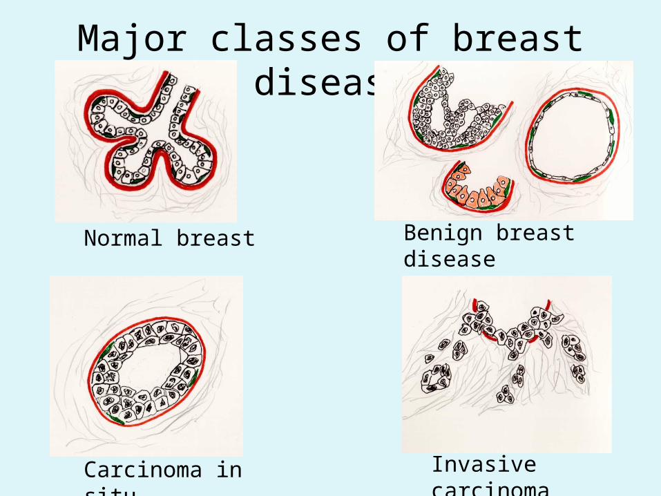
Major classes of breast disease
Normal breast Benign breast disease
Carcinoma in situ Invasive carcinoma
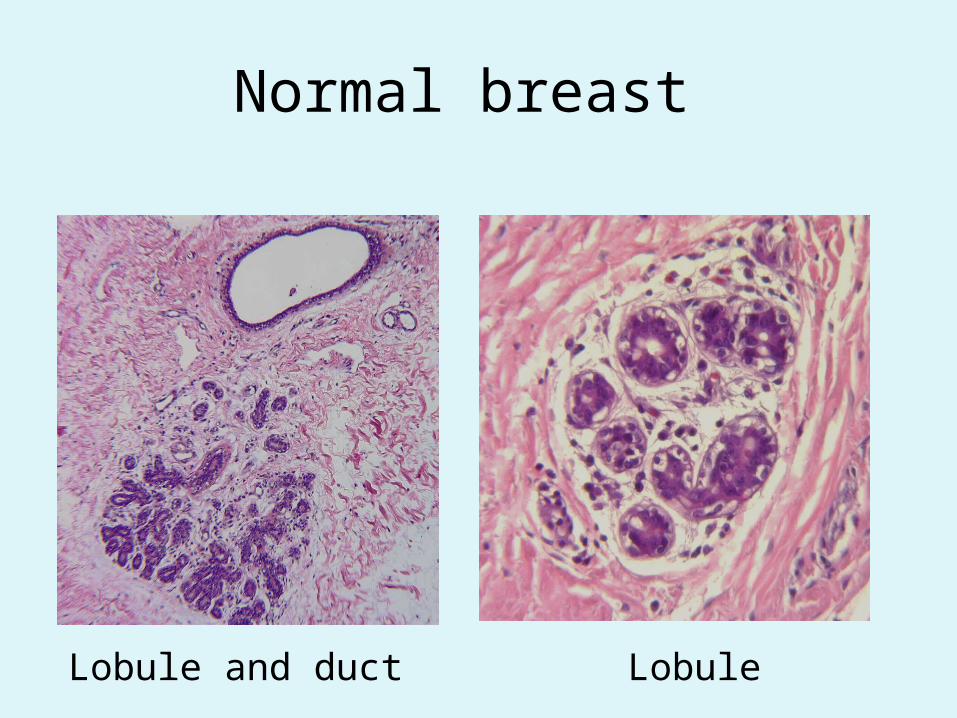
Normal breast
Lobule and duct Lobule

Benign Breast disease: Elements of fibrocystic disease
Cyst
Apo-crine meta-plasia
Ductal hyper-plasia
Sclero-sing adenosis

Benign breast disease:
Fibro-adenoma
Intraductal papilloma
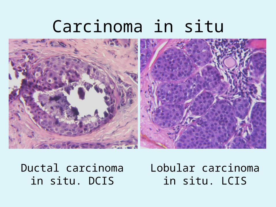
Carcinoma in situ
Ductal carcinoma in situ. DCIS
Lobular carcinoma in situ. LCIS

Ductal carcinoma in situ
Lobular carcinoma in situ
The distribution of carcinoma in situ
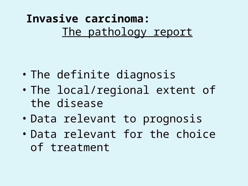
Invasive carcinoma: The pathology report
• The definite diagnosis• The local/regional extent of the disease• Data relevant to prognosis• Data relevant for the choice of treatment

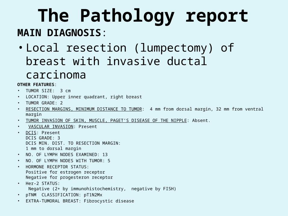
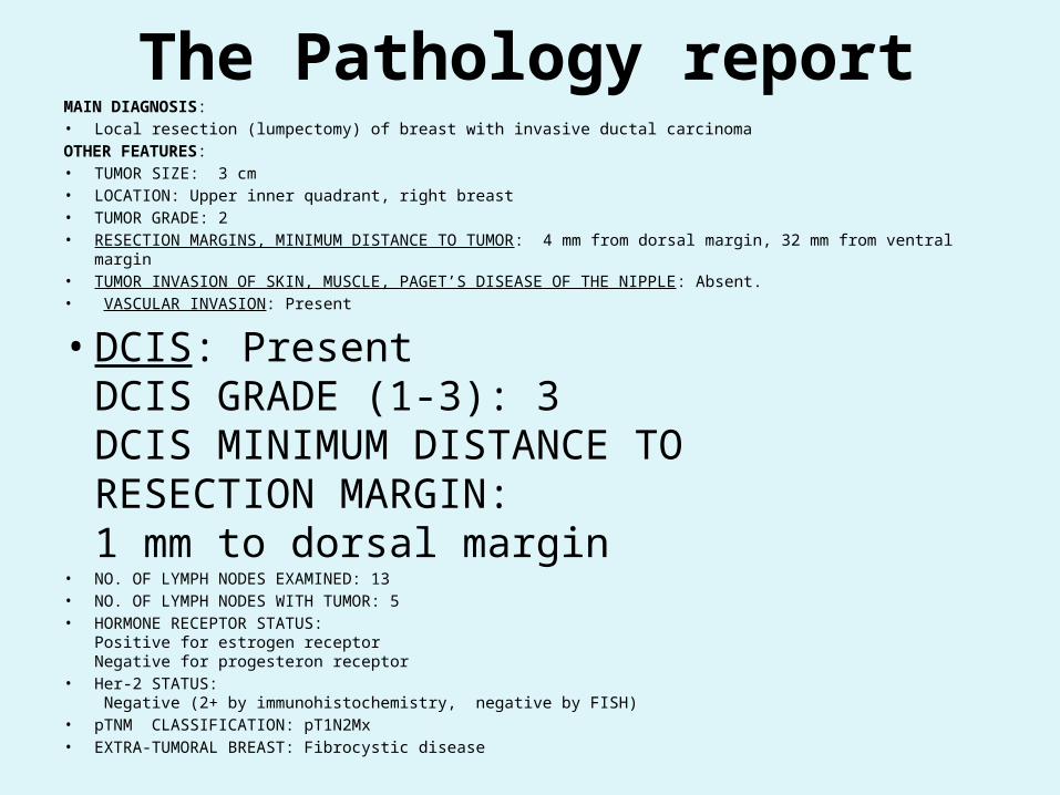
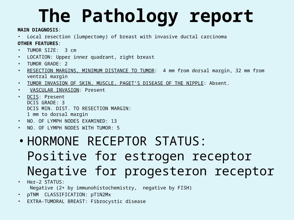
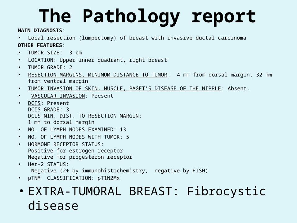
The Pathology report, Ca. resectionMAIN DIAGNOSIS: • Local resection (lumpectomy) of breast with invasive ductal carcinomaOTHER FEATURES:• TUMOR SIZE: 3 cm• LOCATION: Upper inner quadrant, right breast• TUMOR GRADE: 2• RESECTION MARGINS, MINIMUM DISTANCE TO TUMOR: 4 mm from dorsal margin, 32 mm from ventral
margin• TUMOR INVASION OF SKIN, MUSCLE, PAGET’S DISEASE OF THE NIPPLE: Absent.• VASCULAR INVASION: Present • DCIS: Present
DCIS GRADE: 3DCIS MIN. DIST. TO RESECTION MARGIN:
1 mm to dorsal margin• NO. OF LYMPH NODES EXAMINED: 13• NO. OF LYMPH NODES WITH TUMOR: 5• HORMONE RECEPTOR STATUS:
Positive for estrogen receptorNegative for progesteron receptor
• Her-2 STATUS: Negative (2+ by immunohistochemistry, negative by FISH)
• pTNM CLASSIFICATION: pT1N2Mx• EXTRA-TUMORAL BREAST: Fibrocystic disease
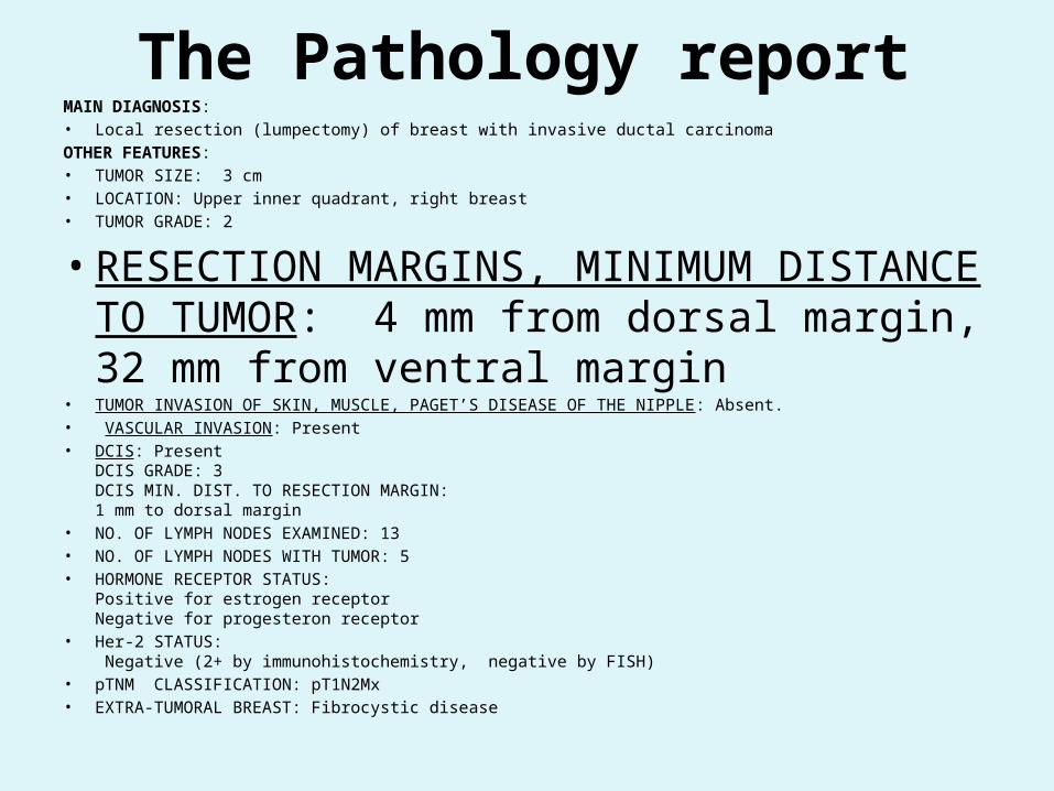
The Pathology reportMAIN DIAGNOSIS: • Local resection (lumpectomy) of breast with
invasive ductal carcinomaOTHER FEATURES:• TUMOR SIZE: 3 cm• LOCATION: Upper inner quadrant, right breast• TUMOR GRADE: 2• RESECTION MARGINS, MINIMUM DISTANCE TO TUMOR: 4 mm from dorsal margin, 32 mm from ventral margin• TUMOR INVASION OF SKIN, MUSCLE, PAGET’S DISEASE OF THE NIPPLE: Absent.• VASCULAR INVASION: Present • DCIS: Present
DCIS GRADE: 3DCIS MIN. DIST. TO RESECTION MARGIN:
1 mm to dorsal margin• NO. OF LYMPH NODES EXAMINED: 13• NO. OF LYMPH NODES WITH TUMOR: 5• HORMONE RECEPTOR STATUS:
Positive for estrogen receptorNegative for progesteron receptor
• Her-2 STATUS: Negative (2+ by immunohistochemistry, negative by FISH)
• pTNM CLASSIFICATION: pT1N2Mx• EXTRA-TUMORAL BREAST: Fibrocystic disease
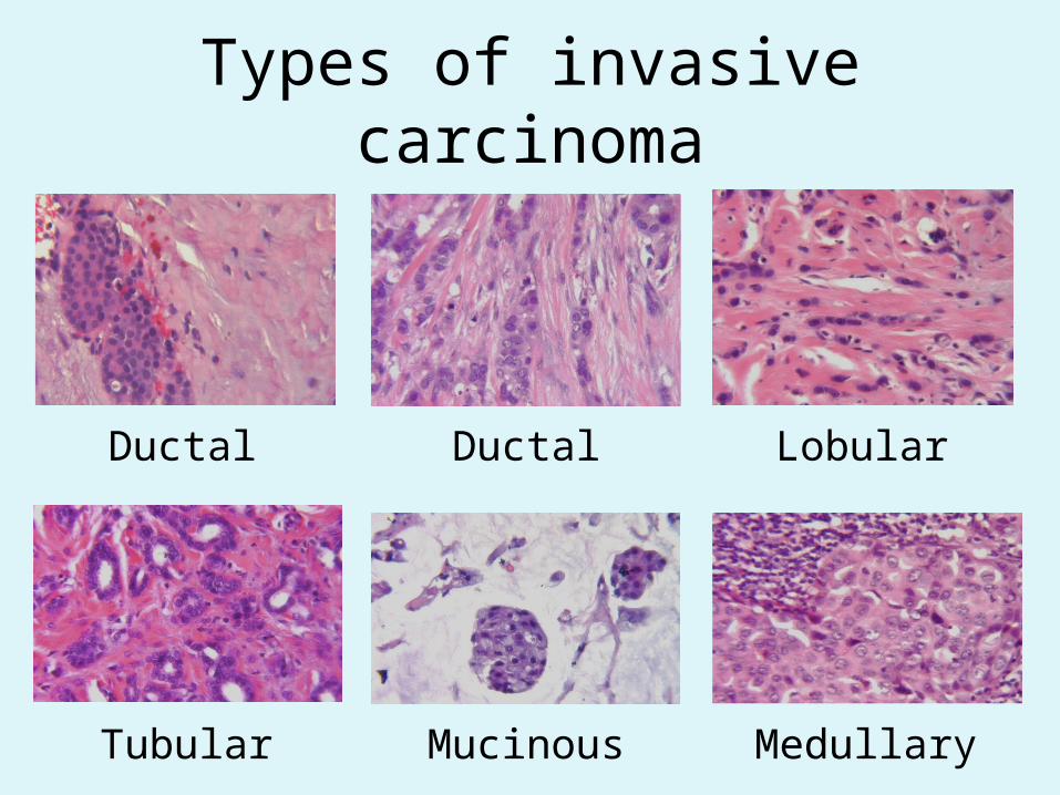
Types of invasive carcinoma
Ductal Ductal Lobular
Tubular Mucinous Medullary
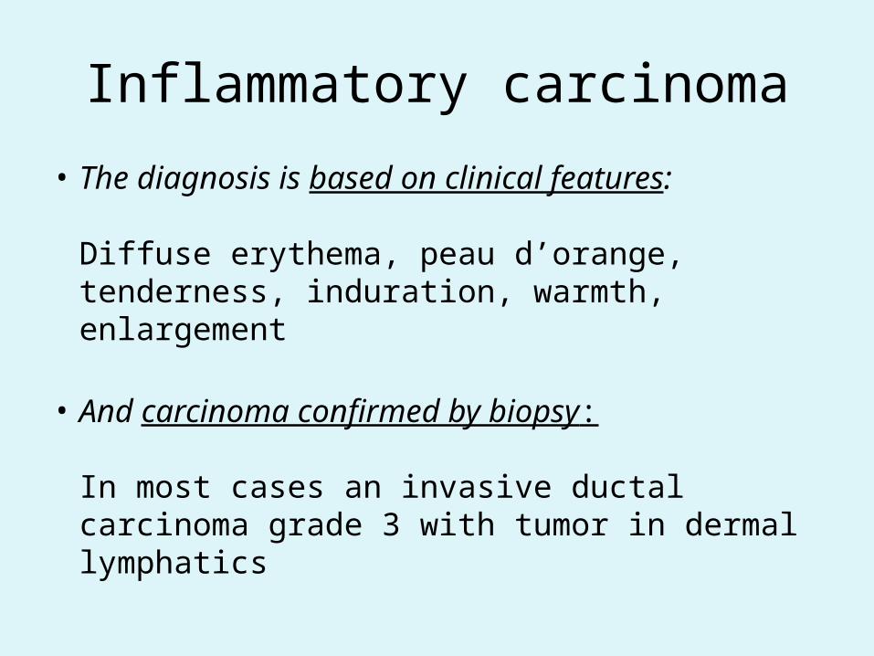
Inflammatory carcinoma
• The diagnosis is based on clinical features:
Diffuse erythema, peau d’orange, tenderness, induration, warmth, enlargement
• And carcinoma confirmed by biopsy:
In most cases an invasive ductal carcinoma grade 3 with tumor in dermal lymphatics

The Pathology reportMAIN DIAGNOSIS: • Local resection (lumpectomy) of breast with invasive ductal carcinomaOTHER FEATURES:• TUMOR SIZE: 3 cm• LOCATION: Upper inner quadrant, right breast
• TUMOR GRADE (1-3): 2• RESECTION MARGINS, MINIMUM DISTANCE TO TUMOR: 4 mm from dorsal margin, 32 mm from ventral
margin• TUMOR INVASION OF SKIN, MUSCLE, PAGET’S DISEASE OF THE NIPPLE: Absent.• VASCULAR INVASION: Present • DCIS: Present
DCIS GRADE: 3DCIS MIN. DIST. TO RESECTION MARGIN:
1 mm to dorsal margin• NO. OF LYMPH NODES EXAMINED: 13• NO. OF LYMPH NODES WITH TUMOR: 5• HORMONE RECEPTOR STATUS:
Positive for estrogen receptorNegative for progesteron receptor
• Her-2 STATUS: Negative (2+ by immunohistochemistry, negative by FISH)
• pTNM CLASSIFICATION: pT1N2Mx• EXTRA-TUMORAL BREAST: Fibrocystic disease

Tumor gradingFeature ScoreTubule formation 1-3Nuclear atypia 1-3Number of mitoses 1-3
Grade Sum of scoresGrade 1 3-5Grade 2 6-7Grade 3 8-9

Tumor grades
Grade 1 Grade 3
The Pathology reportMAIN DIAGNOSIS: • Local resection (lumpectomy) of breast with invasive ductal carcinomaOTHER FEATURES:• TUMOR SIZE: 3 cm• LOCATION: Upper inner quadrant, right breast• TUMOR GRADE: 2
• RESECTION MARGINS, MINIMUM DISTANCE TO TUMOR: 4 mm from dorsal margin, 32 mm from ventral margin
• TUMOR INVASION OF SKIN, MUSCLE, PAGET’S DISEASE OF THE NIPPLE: Absent.• VASCULAR INVASION: Present • DCIS: Present
DCIS GRADE: 3DCIS MIN. DIST. TO RESECTION MARGIN:
1 mm to dorsal margin• NO. OF LYMPH NODES EXAMINED: 13• NO. OF LYMPH NODES WITH TUMOR: 5• HORMONE RECEPTOR STATUS:
Positive for estrogen receptorNegative for progesteron receptor
• Her-2 STATUS: Negative (2+ by immunohistochemistry, negative by FISH)
• pTNM CLASSIFICATION: pT1N2Mx• EXTRA-TUMORAL BREAST: Fibrocystic disease
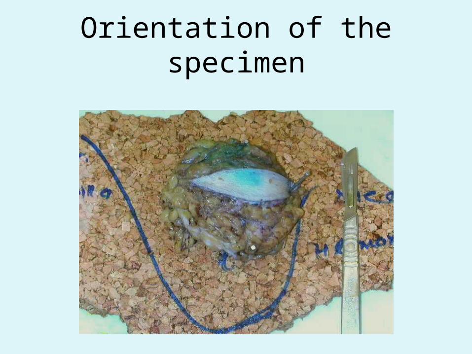
Orientation of the specimen

Inking of resection margins
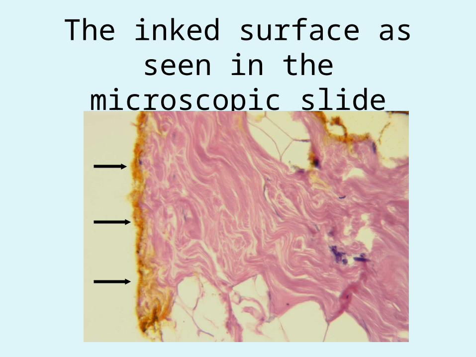
The inked surface as seen in the microscopic slide

Examination of resection margin
Section 2Section 1
Inked margin
Skin

The Pathology reportMAIN DIAGNOSIS: • Local resection (lumpectomy) of breast with invasive ductal carcinomaOTHER FEATURES:• TUMOR SIZE: 3 cm• LOCATION: Upper inner quadrant, right breast• TUMOR GRADE: 2• RESECTION MARGINS, MINIMUM DISTANCE TO TUMOR: 4 mm from dorsal margin, 32 mm from ventral
margin• TUMOR INVASION OF SKIN, MUSCLE, PAGET’S DISEASE OF THE NIPPLE: Absent.
• VASCULAR INVASION: Present
• DCIS: PresentDCIS GRADE: 3DCIS MIN. DIST. TO RESECTION MARGIN:
1 mm to dorsal margin• NO. OF LYMPH NODES EXAMINED: 13• NO. OF LYMPH NODES WITH TUMOR: 5• HORMONE RECEPTOR STATUS:
Positive for estrogen receptorNegative for progesteron receptor
• Her-2 STATUS: Negative (2+ by immunohistochemistry, negative by FISH)
• pTNM CLASSIFICATION: pT1N2Mx• EXTRA-TUMORAL BREAST: Fibrocystic disease

Tumor tissue
Lymph vessel
Vascular invasion
The Pathology reportMAIN DIAGNOSIS: • Local resection (lumpectomy) of breast with invasive ductal carcinomaOTHER FEATURES:• TUMOR SIZE: 3 cm• LOCATION: Upper inner quadrant, right breast• TUMOR GRADE: 2• RESECTION MARGINS, MINIMUM DISTANCE TO TUMOR: 4 mm from dorsal margin, 32 mm from ventral margin• TUMOR INVASION OF SKIN, MUSCLE, PAGET’S DISEASE OF THE NIPPLE: Absent.• VASCULAR INVASION: Present
• DCIS: PresentDCIS GRADE (1-3): 3DCIS MINIMUM DISTANCE TO
RESECTION MARGIN: 1 mm to dorsal margin
• NO. OF LYMPH NODES EXAMINED: 13• NO. OF LYMPH NODES WITH TUMOR: 5• HORMONE RECEPTOR STATUS:
Positive for estrogen receptorNegative for progesteron receptor
• Her-2 STATUS: Negative (2+ by immunohistochemistry, negative by FISH)
• pTNM CLASSIFICATION: pT1N2Mx• EXTRA-TUMORAL BREAST: Fibrocystic disease

The Pathology reportMAIN DIAGNOSIS: • Local resection (lumpectomy) of breast with invasive ductal carcinomaOTHER FEATURES:• TUMOR SIZE: 3 cm• LOCATION: Upper inner quadrant, right breast• TUMOR GRADE: 2• RESECTION MARGINS, MINIMUM DISTANCE TO TUMOR: 4 mm from dorsal margin, 32 mm from ventral margin• TUMOR INVASION OF SKIN, MUSCLE, PAGET’S DISEASE OF THE NIPPLE: Absent.• VASCULAR INVASION: Present • DCIS: Present
DCIS GRADE: 3DCIS MIN. DIST. TO RESECTION MARGIN: 1 mm to dorsal margin
• NO. OF LYMPH NODES EXAMINED: 13• NO. OF LYMPH NODES WITH TUMOR: 5• HORMONE RECEPTOR STATUS:
Positive for estrogen receptorNegative for progesteron receptor
• Her-2 STATUS: Negative (2+ by immunohistochemistry, negative by FISH)
• pTNM CLASSIFICATION: pT1N2Mx• EXTRA-TUMORAL BREAST: Fibrocystic disease
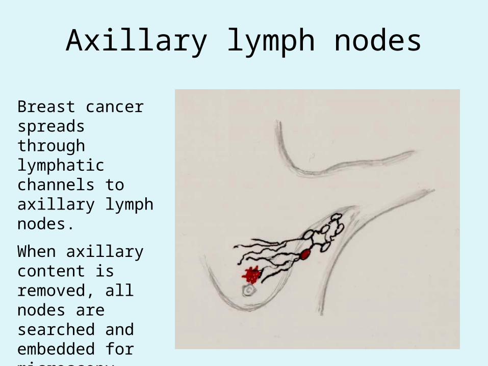
Axillary lymph nodes
Breast cancer spreads through lymphatic channels to axillary lymph nodes.
When axillary content is removed, all nodes are searched and embedded for microscopy

Micrometastasis
Micrometastasis spotted in otherwise negative sentinel node

Tumor deposits in lymph node
Size Designated pTNM class > 2 mm Metastasis pN1≤ 2 mm; > 0.2 mm Micrometastasis pN1(mi)≤ 0.2 mm Isolated tumor cells pN0 (i+)
The Pathology reportMAIN DIAGNOSIS: • Local resection (lumpectomy) of breast with invasive ductal carcinomaOTHER FEATURES:• TUMOR SIZE: 3 cm• LOCATION: Upper inner quadrant, right breast• TUMOR GRADE: 2• RESECTION MARGINS, MINIMUM DISTANCE TO TUMOR: 4 mm from dorsal margin, 32 mm from ventral margin• TUMOR INVASION OF SKIN, MUSCLE, PAGET’S DISEASE OF THE NIPPLE: Absent.• VASCULAR INVASION: Present • DCIS: Present
DCIS GRADE: 3DCIS MIN. DIST. TO RESECTION MARGIN:
1 mm to dorsal margin• NO. OF LYMPH NODES EXAMINED: 13• NO. OF LYMPH NODES WITH TUMOR: 5
• HORMONE RECEPTOR STATUS: Positive for estrogen receptorNegative for progesteron receptor
• Her-2 STATUS: Negative (2+ by immunohistochemistry, negative by FISH)
• pTNM CLASSIFICATION: pT1N2Mx• EXTRA-TUMORAL BREAST: Fibrocystic disease
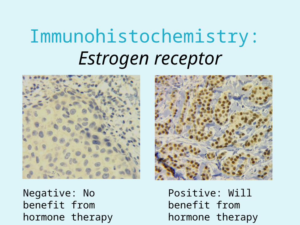
Immunohistochemistry: Estrogen receptor
Negative: No benefit from hormone therapy
Positive: Will benefit from hormone therapy
The Pathology reportMAIN DIAGNOSIS: • Local resection (lumpectomy) of breast with invasive ductal carcinomaOTHER FEATURES:• TUMOR SIZE: 3 cm• LOCATION: Upper inner quadrant, right breast• TUMOR GRADE: 2• RESECTION MARGINS, MINIMUM DISTANCE TO TUMOR: 4 mm from dorsal margin, 32 mm from ventral
margin• TUMOR INVASION OF SKIN, MUSCLE, PAGET’S DISEASE OF THE NIPPLE: Absent.• VASCULAR INVASION: Present • DCIS: Present
DCIS GRADE: 3DCIS MIN. DIST. TO RESECTION MARGIN:
1 mm to dorsal margin• NO. OF LYMPH NODES EXAMINED: 13• NO. OF LYMPH NODES WITH TUMOR: 5• HORMONE RECEPTOR STATUS:
Positive for estrogen receptorNegative for progesteron receptor
• Her-2 STATUS: Negative• pTNM CLASSIFICATION: pT1N2Mx• EXTRA-TUMORAL BREAST: Fibrocystic disease
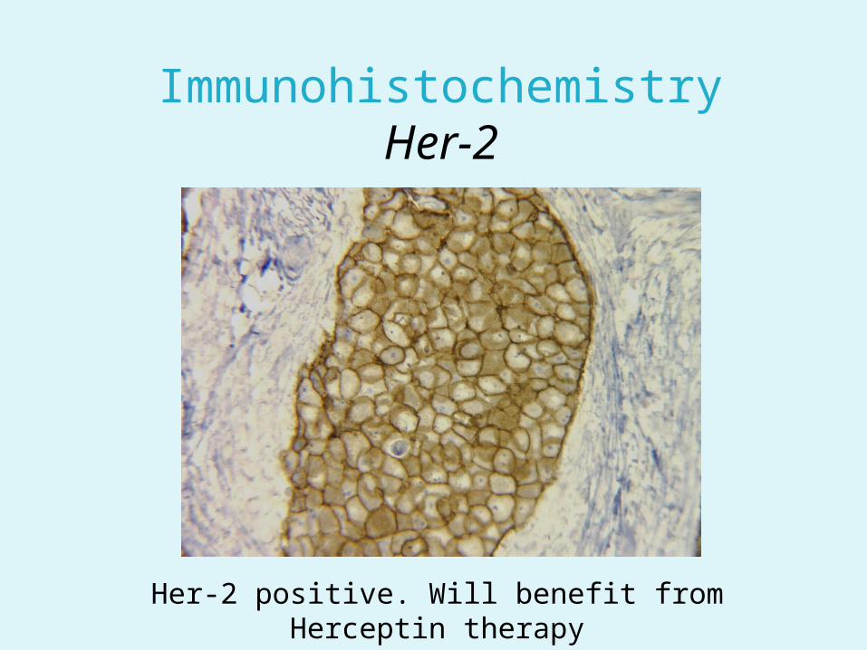
ImmunohistochemistryHer-2
Her-2 positive. Will benefit from Herceptin therapy

The Pathology reportMAIN DIAGNOSIS: • Local resection (lumpectomy) of breast with invasive ductal carcinomaOTHER FEATURES:• TUMOR SIZE: 3 cm• LOCATION: Upper inner quadrant, right breast• TUMOR GRADE: 2• RESECTION MARGINS, MINIMUM DISTANCE TO TUMOR: 4 mm from dorsal margin, 32 mm from ventral
margin• TUMOR INVASION OF SKIN, MUSCLE, PAGET’S DISEASE OF THE NIPPLE: Absent.• VASCULAR INVASION: Present • DCIS: Present
DCIS GRADE: 3DCIS MIN. DIST. TO RESECTION MARGIN:
1 mm to dorsal margin• NO. OF LYMPH NODES EXAMINED: 13• NO. OF LYMPH NODES WITH TUMOR: 5• HORMONE RECEPTOR STATUS:
Positive for estrogen receptorNegative for progesteron receptor
• Her-2 STATUS: Negative (2+ by immunohistochemistry, negative by FISH)
• pTNM CLASSIFICATION: pT1N2Mx• EXTRA-TUMORAL BREAST: Fibrocystic disease
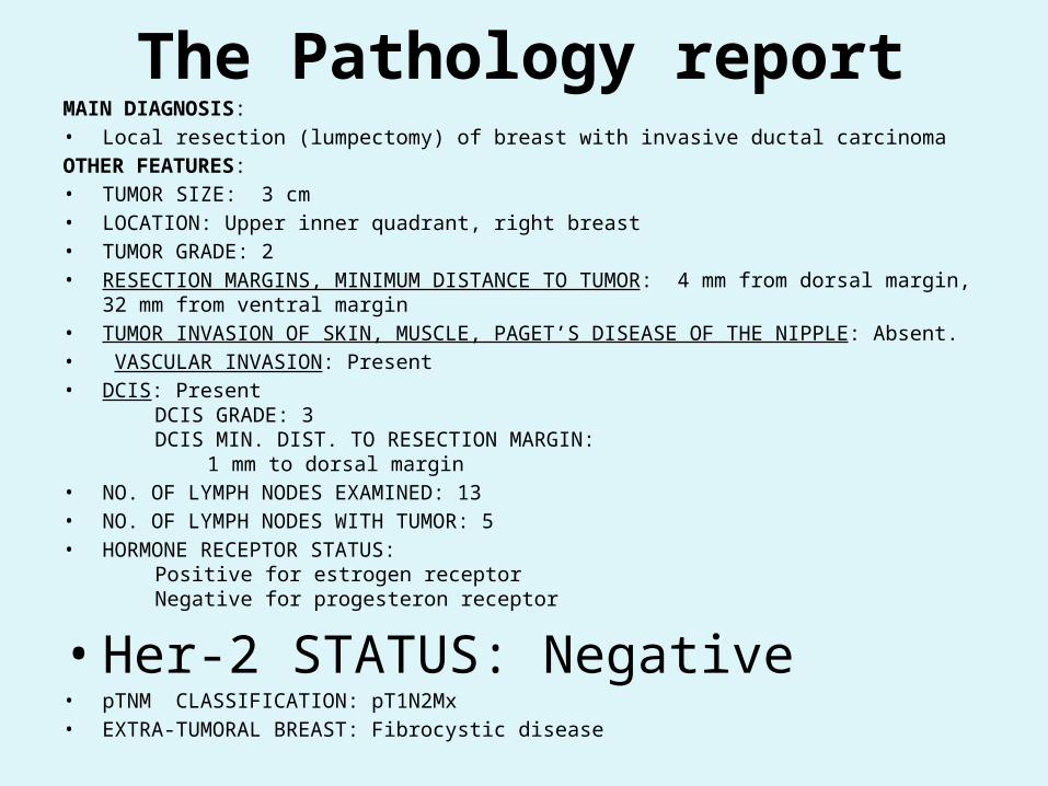
The Pathology reportMAIN DIAGNOSIS: • Local resection (lumpectomy) of breast with invasive ductal carcinomaOTHER FEATURES:• TUMOR SIZE: 3 cm• LOCATION: Upper inner quadrant, right breast• TUMOR GRADE: 2• RESECTION MARGINS, MINIMUM DISTANCE TO TUMOR: 4 mm from dorsal margin, 32 mm from ventral
margin• TUMOR INVASION OF SKIN, MUSCLE, PAGET’S DISEASE OF THE NIPPLE: Absent.• VASCULAR INVASION: Present • DCIS: Present
DCIS GRADE: 3DCIS MIN. DIST. TO RESECTION MARGIN:
1 mm to dorsal margin• NO. OF LYMPH NODES EXAMINED: 13• NO. OF LYMPH NODES WITH TUMOR: 5• HORMONE RECEPTOR STATUS:
Positive for estrogen receptorNegative for progesteron receptor
• Her-2 STATUS: Negative (2+ by immunohistochemistry, negative by FISH)
• pTNM CLASSIFICATION: pT1N2Mx
• EXTRA-TUMORAL BREAST: Fibrocystic disease


Is Breast pathology in Ghana similar to that in the industralized
countries ?
A comparison of non-inflammatory pathology diagnoses in Ghana (KATH)
and Norway (UNN)
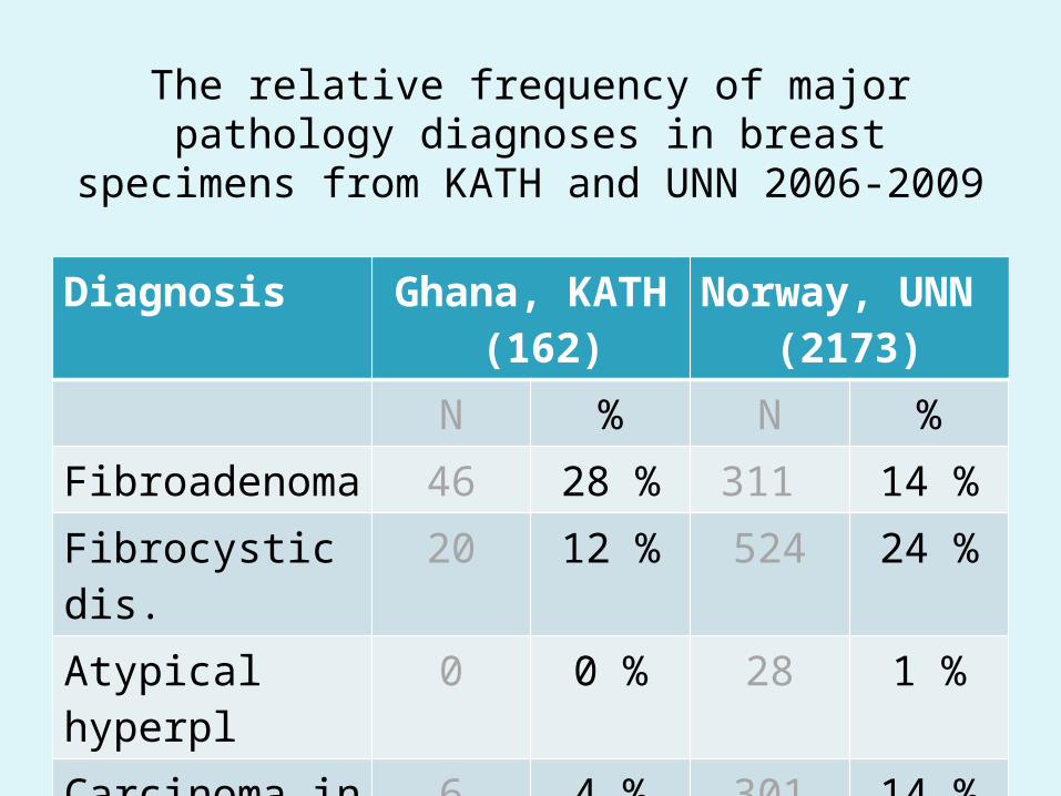
The relative frequency of major pathology diagnoses in breast specimens from KATH
and UNN 2006-2009Diagnosis Ghana, KATH
(162)Norway, UNN
(2173)
N % N %
Fibroadenoma 46 28 % 311 14 %
Fibrocystic dis. 20 12 % 524 24 %
Atypical hyperpl 0 0 % 28 1 %
Carcinoma in situ 6 4 % 301 14 %
Carcinoma 90 56 % 1009 46 %
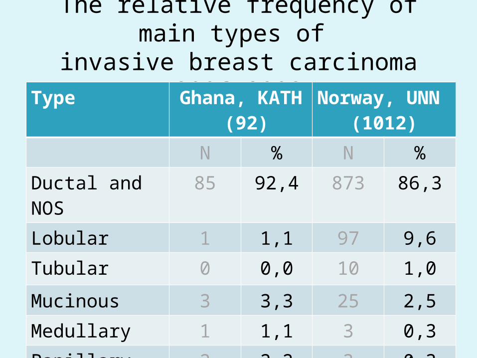
The relative frequency of main types of invasive breast carcinoma 2006-2009
Type Ghana, KATH (92)
Norway, UNN (1012)
N % N %
Ductal and NOS 85 92,4 873 86,3
Lobular 1 1,1 97 9,6
Tubular 0 0,0 10 1,0
Mucinous 3 3,3 25 2,5
Medullary 1 1,1 3 0,3
Papillary 2 2,2 3 0,3

Tumor grade inCore biopsies of Breast cancer
Preliminary data 2009Tumor grade Ghana
(25 cases)Norway
(93 cases)Grade 1 16% 33%Grade 2 28% 44%Grade 3 48% 16%
E. Adjei

Hormone receptors inCore biopsies of Breast cancer
Preliminary data 2009Hormone receptor
Ghana (25 cases)
Norway (93 cases)
Estrogen receptor
48% positive 89% positive
Progesteron receptor
12% positive 62% positive
E. Adjei

Her-2 overexpression inCore biopsies of Breast cancer
Preliminary data 2009
Ghana (25 cases)
Norway (93 cases)
32% positive 12% positive
E.Adjei