brit. toxicity of i, 8, 9-triacetoxyanthracene to cornea...
TRANSCRIPT
Brit. J. Ophthal. (I969) 53, 819
Toxicity of I, 8, 9-triacetoxyanthraceneto the cornea in rabbits
D. L. EASTY AND M. B. R. MATHALONE
From the Institute of Ophthalmology, University of London
I, 8, 9-Triacetoxyanthracene, marketed under the trade name Exolan, has been reportedto be a useful agent in the treatment of psoriasis (Hellier and Whitefield, I967). It wasintroduced to replace the drug Dithranol (anthracene-i, 8, 9-triol), which was previouslyemployed with some success, but had the disadvantage that it caused staining of linenand clothing, and was also associated with burning of the skin. Triacetoxyanthracenedoes not have these adverse reactions (Hellier and Whitefield, I967). Recently it wasnoted that triacetoxyanthracene caused corneal oedema when accidentally introducedinto the conjunctival sac of the human eye (Mathalone and Easty, I967). One of thepatients described in this report, using the preparation marketed as a cream suitable foruse on the scalp following a shampoo, inadvertently introduced some into both eyes. Theother patient, using the paste for a psoriatic eruption on the trunk, suffered a similarcorneal lesion, although he did not positively admit to introducing it into his eye. Follow-ing these clinical observations, the effect of triacetoxyanthracene on the rabbit cornea wasexamined in order to elucidate its site of action, and to demonstrate the pathogenesis ofthe lesion produced.
Method
Different quantities of Exolan were introduced into the conjunctival sac of one eye of each of sixrabbits. This treated eye was compared with the contralateral (control) eyc into which was instilledan equivalent concentration of the base suspended in water. The dose-levels of triacetoxyanthracenewere; 2 per cent. (2 rabbits), o 66 per cent. (i rabbit), o 2 per cent. (i rabbit), and o i per cent.(2 rabbits). One rabbit, into the eye of which 2 per cent. triacetoxyanthracene had been instilled,was assessed over a period of io weeks.The lesions produced were examined with the slit-lamp microscope and at the same time the
corneal thickness was measured using the pachometer developed by Maurice and Giardini (I95I).The permeability of the epithelium and endothelium was used as a test of cellular function employingthe fluorophotometric apparatus and technique as described by Maurice (1963).The epithelial permeability was measured by holding an aqueous solution of fluorescein (o.5 per
cent.) in contact with the cornea for a period of i minute, after which the eye was irrigated witho 9 per cent. saline. The fluorescein concentrations were measured 2 hours later in the stroma ofboth the treated and the control eyes and were compared. In the measurement of the endothelialpermeability, similar control studies were made as in the epithelial studies. Fluorescein concentra-tions were measured in the strorna and aqueous at 30-minute intervals over a period of 4 hours.The ratio of stromal to aqueous fluorescein was calculated on each occasion, and from these resultsa decay curve was constructed. All experiments were performed on eyes which had been treatedwith triacetoxyanthracene at least 24 hours previously.The histopathological changes were examined using haematoxylin and eosin, and the histo-
chemical characteristics were studied using Alcian blue, the periodic acid-Schiff reaction, and
Received for publication June 23, 1969Address for reprints: Moorfields Eye Hospital, City Road, London, E.C. i.
on 15 May 2018 by guest. P
rotected by copyright.http://bjo.bm
j.com/
Br J O
phthalmol: first published as 10.1136/bjo.53.12.819 on 1 D
ecember 1969. D
ownloaded from
D. L. Easty and M. B. R. Mathalone
colloidal iron staining, as suggested by Ashton ( 959). The endothelium was stained in flat prepara-tion using trypan blue, and examined for cellular damage (Stocker, King, Lucas, and Georgiade,I 967).
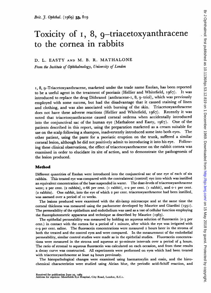
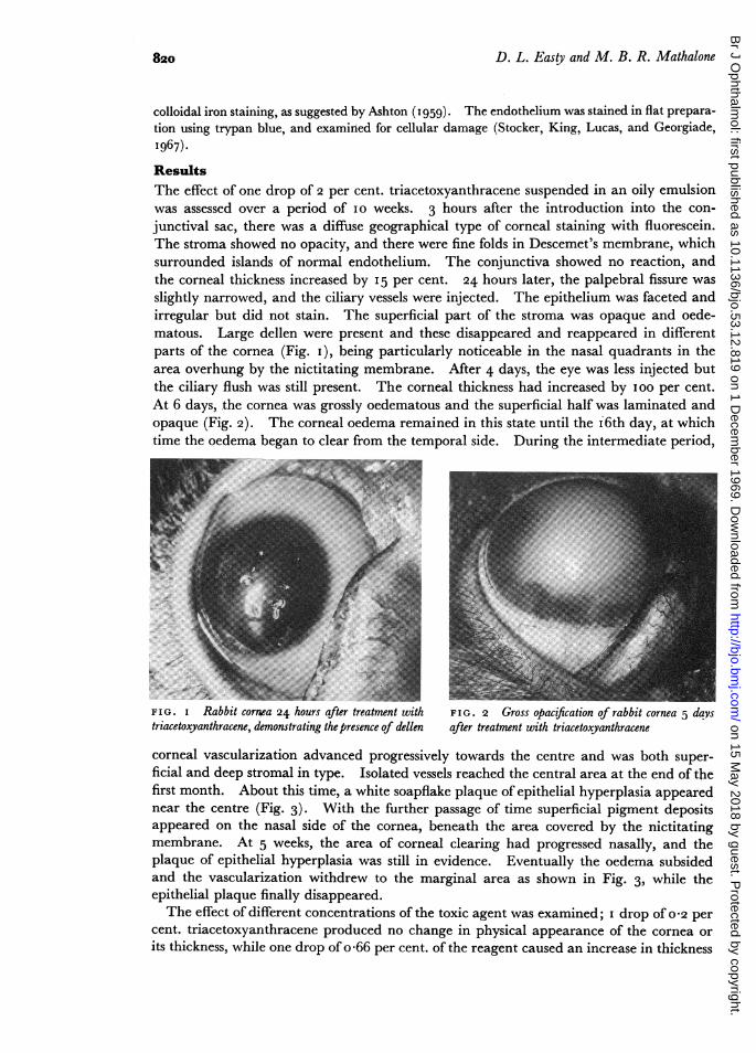
ResultsThe effect of one drop of 2 per cent. triacetoxyanthracene suspended in an oily emulsionwas assessed over a period of Io weeks. 3 hours after the introduction into the con-junctival sac, there was a diffuse geographical type of corneal staining with fluorescein.The stroma showed no opacity, and there were fine folds in Descemet's membrane, whichsurrounded islands of normal endothelium. The conjunctiva showed no reaction, andthe corneal thickness increased by I5 per cent. 24 hours later, the palpebral fissure wasslightly narrowed, and the ciliary vessels were injected. The epithelium was faceted andirregular but did not stain. The superficial part of the stroma was opaque and oede-matous. Large dellen were present and these disappeared and reappeared in differentparts of the cornea (Fig. i), being particularly noticeable in the nasal quadrants in thearea overhung by the nictitating membrane. After 4 days, the eye was less injected butthe ciliary flush was still present. The corneal thickness had increased by ioo per cent.At 6 days, the cornea was grossly oedematous and the superficial half was laminated andopaque (Fig. 2). The corneal oedema remained in this state until the i6th day, at whichtime the oedema began to clear from the temporal side. During the intermediate period,
*"'4'..E,B.,6z..,..s:..
FIG. I Rabbit cornea 24 hours after treatment with FIG. 2 Gross opacification of rabbit cornea 5 daystriacetoxyanthracene, demonstrating the presence of dellen after treatment with triacetoxyanthracene
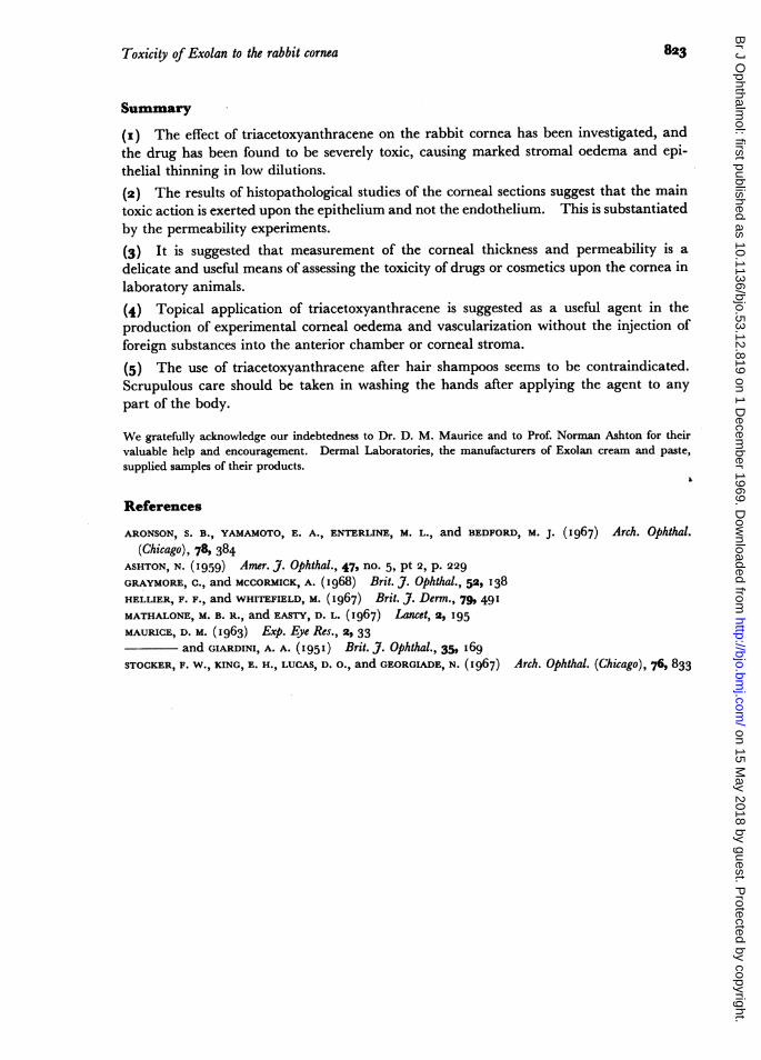
corneal vascularization advanced progressively towards the centre and was both super-ficial and deep stromal in type. Isolated vessels reached the central area at the end of thefirst month. About this time, a white soapflake plaque of epithelial hyperplasia appearednear the centre (Fig. 3). With the further passage of time superficial pigment depositsappeared on the nasal side of the cornea, beneath the area covered by the nictitatingmembrane. At 5 weeks, the area of corneal clearing had progressed nasally, and theplaque of epithelial hyperplasia was still in evidence. Eventually the oedema subsidedand the vascularization withdrew to the marginal area as shown in Fig. 3, while theepithelial plaque finally disappeared.The effect of different concentrations of the toxic agent was examined; I drop of 0 2 per
cent. triacetoxyanthracene produced no change in physical appearance of the cornea orits thickness, while one drop of o 66 per cent. of the reagent caused an increase in thickness
820
on 15 May 2018 by guest. P
rotected by copyright.http://bjo.bm
j.com/
Br J O
phthalmol: first published as 10.1136/bjo.53.12.819 on 1 D
ecember 1969. D
ownloaded from

Toxicity of Exolan to the rabbit cornea 82I
F I G . 3 Rabbit cornea at 5 weeks, showingepithelial soap-flake hyperplasia with peripheralvascularization. The stromal oedema has resolved.
of go per cent. in 5 days, with commensurate changes in the conjunctival and limbalvascularization. When a concentration of o-i per cent. triacetoxyanthracene was heldin the conjunctival basin for 4 minutes, the increase in thickness 2 days later was 8o percent. In this experiment there was minimal ciliary injection.
PERMEABILITY STUDIES
The measurement of the permeability of the epithelium in a rabbit which had been treatedwith o-i per cent. triacetoxyanthracene for 4 minutes, 2 days previously, indicated thatit had increased by a factor of 50. The ratio of fluorescein in the stroma and aqueouswas found to be 6: I in both the treated and the untreated eyes. The decay curve offluorescein loss from the stroma paralleled that constructed for the control eye. Thus thepermeability of the endothelium was unaffected. Other similar studies on two otherrabbits substantiated this finding, that the permeability of the epithelium was affectedmore than that of the endothelium.
PATHOLOGY
Sections of a cornea treated with a 2 per cent. solution of triacetoxyanthracene 6 dayspreviously were stained with haematoxylin and eosin and it was found that the epitheliumwas thinned down to one or two layers of cells (Fig. 4). There was a slight inflammatory
-~~~~~~~~l~~~* A# - $#
or
-.-4
* h p.uu.e .. ,qS..
...~~~~~~~~~ ~~~ ~~~~~~~~~~~~~~~~~~~~~~~~~~~~~...I.
F I G . 4 Rabbit cornea treated with triacetoxy-anthracene 6 days previously, demonstrating a normalendothelium below, and an attenuated laver ofepithelium above. x 70
on 15 May 2018 by guest. P
rotected by copyright.http://bjo.bm
j.com/
Br J O
phthalmol: first published as 10.1136/bjo.53.12.819 on 1 D
ecember 1969. D
ownloaded from

D. L. Easty and M. B. R. Mathalone
reaction at the limbus associated with a cellular exudate of polymorphs and round cells.Using differential staining techniques, there was no indication that either collagen ormucopolysaccharide had been preferentially affected. The stromal changes were thosethat might be expected in any type of corneal oedema. Using trypan blue in the studyof the endothelial viability, the damaged cornea did not stain differently from the normal.
Discussion
The toxic effect of triacetoxyanthracene on the cornea is now indisputable, though thisis not necessarily permanent, the lesion being one of stromal oedema that does notappear to result in scarring. The reaction can best be described as non-necrotizing, usingthe classification suggested by Aronson, Yamamoto, Enterline, and Bedford (i 967).It is of some importance that there is only a slight inflammatory reaction at the limbuswhen the toxin is used in dilute concentrations, and the lesion may present clinically as anisolated corneal opacity producing blurred vision with neither subjective nor objectivefindings suggestive of an inflammatory basis. This was the situation in one of the casesoriginally reported (Mathalone and Easty, I967). A recent report on the value of tri-acetoxyanthracene in psoriasis states that one of its side-effects is conjunctivitis (Hellierand Whitefield, I967). This is clearly understating the situation. The makers do stressthe importance of not introducing the drug into the eyes; however, in a product marketedas suitable for use following a shampoo, such an accident would seem inevitable onoccasions.Both the permeability studies and the histopathology show that damage to the epi-
thelium appears to be the main factor in the production of the oedema. Whether this isbecause the cell layers were reduced in number and the barrier function was consequentlyreduced, or whether it was the direct result of its toxic effect upon the metabolism of theremaining layers of cells in the epithelium cannot be said, though the former appears themore likely. At the same time, these factors are not mutually exclusive and it may be thatthey both help to produce stromal oedema.The endothelium was judged to be healthy on three criteria; normal permeability,
normal histopathology, and normal reaction to staining with trypan blue. Althoughthese three criteria suggested that the endothelium was normal, they could not categoricallyrule out a toxic effect on the inner cell layer.
Corneal vascularization is often induced for experimental purposes by injecting toxicagents into either the anterior chamber or the corneal stroma (Graymore and McCormick,I 968). Agents that produce corneal oedema by external application in low concentration,without inducing stromal damage ofa lasting nature in the form of scarring, are uncommon.Triacetoxyanthracene through its toxic effect may be of some value as a simple method ofinducing experimental corneal oedema and vascularization when required.
Manufacturers of drugs or cosmetics sometimes employ experimental methods basedupon a series of photographs showing varying levels of conjunctival injection and cornealoedema, but without the aid of a corneal microscope. It would appear that assessment bythe measurement of the corneal thickness and permeability might be a valuable aid in theinvestigation of these types of toxic effect. Corneal thickness seems to be a particularlysensitive method for determining early corneal changes as shown by the I5 per cent.increase found in an eye that had been treated with 2 per cent. triacetoxyanthracene only3 hours previously and which showed no other significant changes except some irregularstaining with fluorescein.
8g22
on 15 May 2018 by guest. P
rotected by copyright.http://bjo.bm
j.com/
Br J O
phthalmol: first published as 10.1136/bjo.53.12.819 on 1 D
ecember 1969. D
ownloaded from
Toxicity of Exolan to the rabbit cornea
Summary
(I) The effect of triacetoxyanthracene on the rabbit cornea has been investigated, andthe drug has been found to be severely toxic, causing marked stromal oedema and epi-thelial thinning in low dilutions.(2) The results of histopathological studies of the corneal sections suggest that the maintoxic action is exerted upon the epithelium and not the endothelium. This is substantiatedby the permeability experiments.
(3) It is suggested that measurement of the corneal thickness and permeability is adelicate and useful means of assessing the toxicity of drugs or cosmetics upon the cornea inlaboratory animals.(4) Topical application of triacetoxyanthracene is suggested as a useful agent in theproduction of experimental corneal oedema and vascularization without the injection offoreign substances into the anterior chamber or corneal stroma.
(5) The use of triacetoxyanthracene after hair shampoos seems to be contraindicated.Scrupulous care should be taken in washing the hands after applying the agent to anypart of the body.
We gratefully acknowledge our indebtedness to Dr. D. M. Maurice and to Prof. Norman Ashton for theirvaluable help and encouragement. Dermal Laboratories, the manufacturers of Exolan cream and paste,supplied samples of their products.
References
ARONSON, S. B., YAMAMOTO, E. A., ENTERLINE, M. L., and BEDFORD, M. j. (I967) Arch. Ophthal.
(Chicago), 78, 384ASHTON, N. (I959) Amer. J. Ophthal., 47, no. 5, pt 2, p. 229
GRAYMORE, C., and MCCORMICK, A. (I968) Brit. J. Ophthal., 52, 138HELLIER, F. F., and WHITEFIELD, M. (I967) Brit. J. Derm., 79, 491MATHALONE, M. B. R., and EASTY, D. L. (I967) Lancet, 2, 195
MAURICE, D. M. (I963) Exp. Eye Res., 2, 33and GIARDINI, A. A. (1951) Brit. J. Ophthal., 35, I69
STOCKER, F. W., KING, E. H., LUCAS, D. 0., and GEORGIADE, N. (I967) Arch. Ophthal. (Chicago), 76, 833
823
on 15 May 2018 by guest. P
rotected by copyright.http://bjo.bm
j.com/
Br J O
phthalmol: first published as 10.1136/bjo.53.12.819 on 1 D
ecember 1969. D
ownloaded from