bromo-organics. 2. irradiation-induced transformations of tetrabromocyclopentadienone dimer
TRANSCRIPT

J. Org. Chem. 1981,46, 1479-1481
Bromo-organics. 2. Irradiation-Induced Transformations of Tetrabromocyclopentadienone Dimer'
Benzion Fuchs,* Carmela Drucker, and Ramy Lidor
Department of Chemistry, Tel- Aviv University, Ramat Aviv, Tel-Aviv, Israel
Received August 6, 1980
The title compound (1) undergoes on photolysis decarbonylation to perbromoinden-1-one (2) and intramolecular [12 + 12] cycloaddition to the dissymetric perbromobishomocubanedione 3. The results from direct irradiation, sensitization and quenching experiments were taken to indicate that two excited states are involved.
In the framework of a comprehensive study on poly- bromo organic compounds, we have recently started an investigation of the dimer (1) of tetrabromocyclo- pentadienone (over 80% bromine content!) and have al- ready reported' its peculiar reactivity toward nucleophilic attack. We are now describing its photochemical behavior.
Direct and sensitized irradiations of 1 led to two prod- ucts in varying yields, dependent on the irradiation con- ditions: the known perbromoinden-1-one ( 2 ) ' ~ ~ and an unknown compound, the IR, UV, and mass spectra of which indicated it to be a saturated isomer of 1. The immediate proposition for the structure of this product was the dissymmetric octabromopentacyclo[5.3.0.02~5.03~g.04~8]- deca-6,lO-dione (3), following an internal [,2 + *2] pho- tocycloaddition in 1 and in analogy with its octachlor0~9~~ and oct&ydro4 derivatives which give 4 and 5, respectively. One should not, however, take this for granted since the dicyclopentadienone system may show great versatility.'-' Thus, various substituted cyclopentadienone dimers have led, on photolysis, not only to the dissymmetric, internal cycloaddition cage products but also to symmetric ones, be it of symmetry C2" (6) or Dut (7h5s6
Since the uniform perbromo substitution does not allow the mass spectrometric approach6 to be taken, we resorted to 13C NMR spectroscopy in order to prove unequivocally the structural assignment of 3. The rationale was simply that only 3 should exhibit five distinct signals, corre- sponding to ten pairwise equivalent carbon nuclei, as such a structure with C2 symmetry impliea. The general validity of this approach can be proven, if a t all needed, by ap- plying the same criterion to the octachloro analogue 4 of proven s t r ~ c t u r e . ~ . ~
The cage diketone 3 is difficult to purify-only by re- peated sublimation-due to its tendency to add water and form hydrates. In fact it was easier to isolate and char- acterize it as the dihydrate 8. As such, it could also be methylated by diazomethane to the corresponding tetra- methoxy derivative 9 whose 'H NMR spectrum exhibits two methyl signals (6 H each) by which, in fact, the cen- trosymmetric structure 7 could have been excluded-but not 6.3v6 Eventually, the 13C NMR spectra of both the octabromo and octachloro dihydrates (8 and 10, respec-
(1) Part I: B. Fuchs, R. Lidor, C. Kr6ger, and C. K. Liu, Nouu. J.
(2) R. G . Pews, Org. Prep. Proced. Znt., 3, 37 (1971). (3) G . W. Griffin and A. K. Price, J. Org. Chem., 29, 3192 (1964). (4) (a) E. Baggiolini, E. G . Herzog, S. Iwasaki, R. Schorta, and K.
Schaffner, Helv. Chim. Acta, 50,297 (1967); (b) U. Klinsmann, J. Gau- thier, K. Schaffner, M. Pastemak, and B. Fuchs, ibid., 55,2643 (1972).
( 5 ) (a) B. Fuchs, B. Pazhenchevsky, and M. Pasternak, Tetrahedron Lett., 3051 (1972); (b) B. Pazhenchevsky and B. Fuchs, ibid., 3047 (1972); (c) B. Fuchs, M. Pastemak, and B. Pazhenchevksy, Tetrahedron, in press.
(6) B. Fuchs and M. Pasternak, J. Am. Chem. Soc., 100,6225 (1978). (7) B. Fuche, Isr. J. Chem., 20, 203 (1980).
Chim., 4 , 361 (1980).
1 2
1479
3-X=Er 6 4-X:CI 5-X=H
7
8 - X=Er;R=H 9 - X=Er;R--Me
tively) were measured and the data are assembled in Table I, proving that they, as well as their parent diketones (3 and 4, respectively), are indeed C2 symmetry structures.
Returning to the photochemistry of the dimer (I), ita UV spectrum is depicted in Figure 1 and the irradiation results are assembled in Table 11. It should be mentioned a t the outset that the high-molecular-weight high-melting products and the sensitivity of the starting material (1) towards nucleophiles (e.g., H20, ROH)' render quantitative work very difficult and forced us to conduct our experi- menta a t high conversion, monitoring them by using low- precision product isolation techniques. Nevertheless, the results are reproducible and conclusive, following a set of carefully planned experiments, including variable-wave- length direct irradiations, as well as sensitization and quenching studies (Table 11). We rely on those to put forward a mechanism for the photochemical behavior of 1.
Discussion To begin with, the formation of perbromoinden-1-one
(2) is both interesting and somewhat surprising since it involves decarbonylation-debromination of 1 (be it in a concerted or stepwise process) induced by direct or sen- sitized irradiation, the latter process being, in fact, the more efficient one. To be sure, norbornen-7-one and its
10- X=CI ;R=H
0022-3263/81/1946-1479$01,25/0 0 1981 American Chemical Society
J. Org. Chem., Vol. 46, No. 7, 1981
15000
Fuchs, Drucker, and Lidor
other photodissociation processes of norbornen-7-0nes.~ Whether the accompanying debromination is taking place concertedly or as a discrete step is not known to date.
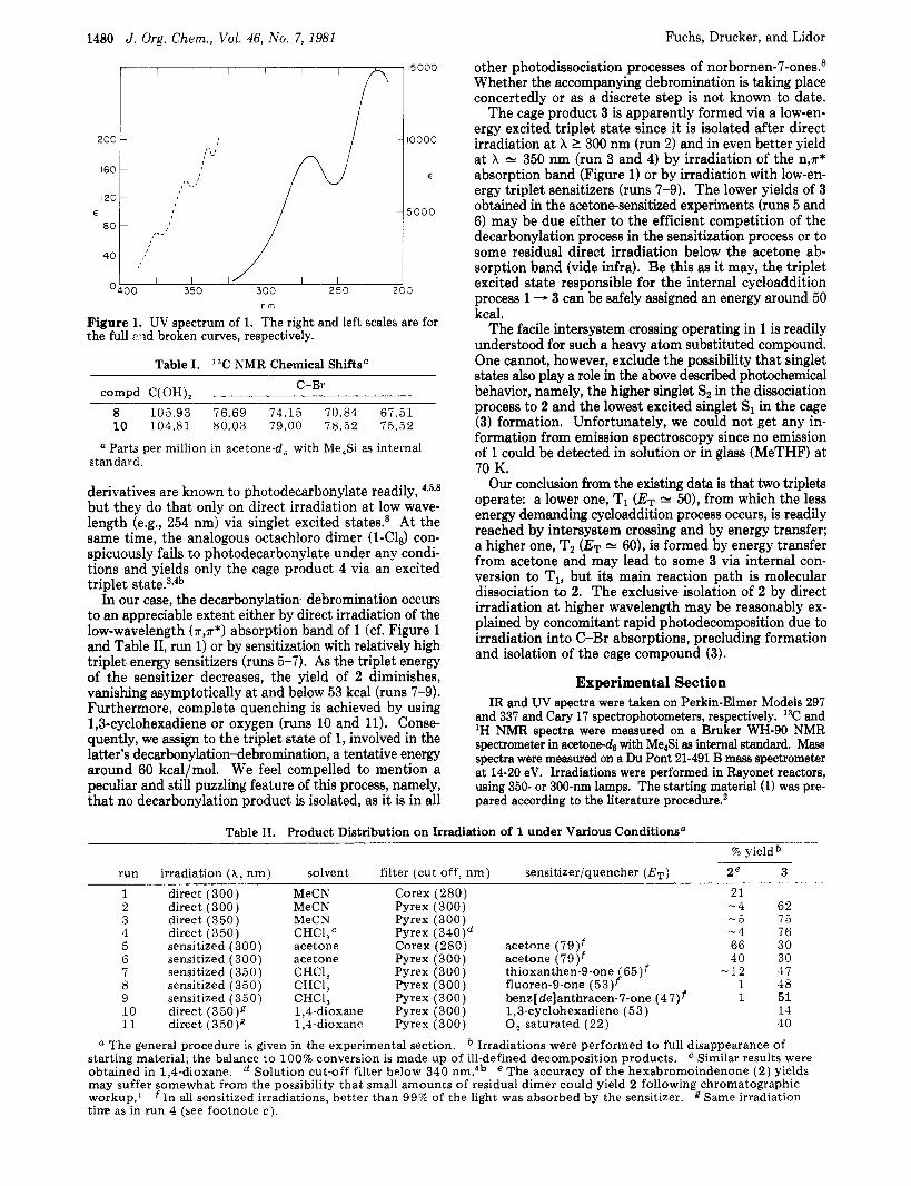
The cage product 3 is apparently formed via a low-en- ergy excited triplet state since it is isolated after direct irradiation at X I 300 nm (run 2) and in even better yield a t X N 350 nm (run 3 and 4) by irradiation of the n,r* absorption band (Figure 1) or by irradiation with low-en- ergy triplet sensitizers (runs 7-9). The lower yields of 3 obtained in the acetone-sensitized experiments (runs 5 and 6) may be due either to the efficient competition of the decarbonylation process in the sensitization process or to some residual direct irradiation below the acetone ab- sorption band (vide infra). Be this as it may, the triplet excited state responsible for the internal cycloaddition process 1 - 3 can be safely assigned an energy around 50 kcal.
The facile intersystem crossing operating in 1 is readily understood for such a heavy atom substituted compound. One cannot, however, exclude the possibility that singlet states also play a role in the above described photochemical behavior, namely, the higher singlet Sz in the dissociation process to 2 and the lowest excited singlet SI in the cage (3) formation. Unfortunately, we could not get any in- formation from emission spectroscopy since no emission of 1 could be detected in solution or in glass (MeTHF) at 70 K.
Our conclusion from the existing data is that two triplets operate: a lower one, T1 (ET. N 50), from which the less energy demanding cycloaddition process occurs, is readily reached by intersystem crossing and by energy transfer; a higher one, Tz (ET N 60), is formed by energy transfer from acetone and may lead to some 3 via internal con- version to T1, but ita main reaction path is molecular dissociation to 2. The exclusive isolation of 2 by direct irradiation a t higher wavelength may be reasonably ex- plained by concomitant rapid photodecomposition due to irradiation into C-Br absorptions, precluding formation and isolation of the cage compound (3).
Experimental Section IR and UV spectra were taken on Perkin-Elmer Models 297
and 337 and Cary 17 spectrophotometers, respectively. 13C and 'H NMR spectra were measured on a Bruker WH-90 NMR spectrometer in acetone-d6 with Me$i as intemal standard. Mass spectra were measured on a Du Pont 21-491 B mass spectrometer at 14-20 eV. Irradiations were performed in Rayonet reactors, using 350- or 300-nm lamps. The starting material (1) was pre- pared according to the literature procedure.*
1480
200
160
120
80 E
40
0
nm
Figure I. UV spectrum of 1. The right and left scales are for the full :?ad broken curves, respectively.
Table I. 13C NMR Chemical Shiftsa C-Br compd C(OH),
8 105.93 76.69 74.15 70.84 67.51 10 104.81 80.03 79.00 78.52 75.52
a Parts per million in acetone-d, with Me,Si as internal standard .
derivatives are known to photodecarbonylate readily, 43598
but they do that only on direct irradiation a t low wave- length (e.g., 254 nm) via singlet excited states.s At the same time, the analogous octachloro dimer (1-ClS) con- spicuously fails to photodecarbonylate under any condi- tions and yields only the cage product 4 via an excited triplet ~ t a t e . ~ ? ~ ~
In our case, the decarbonylation-debromination occurs to an appreciable extent either by direct irradiation of the low-wavelength (*,a*) absorption band of 1 (cf. Figure 1 and Table 11, run 1) or by sensitization with relatively high triplet energy sensitizers (runs 5-7). As the triplet energy of the sensitizer decreases, the yield of 2 diminishes, vanishing asymptotically at and below 53 kcal (runs 7-9). Furthermore, complete quenching is achieved by using 1,3-cyclohexadiene or oxygen (runs 10 and 11). Conse- quently, we assign to the triplet state of 1, involved in the latter's decarbonylation-debromination, a tentative energy around 60 kcal/mol. We feel compelled to mention a peculiar and still puzzling feature of this process, namely, that no decarbonylation product is isolated, as it is in all
Table 11. Product Distribution on Irradiation of 1 under Various Conditionsa 90 yield
run irradiation ( h , nm) solvent filter (cut off, nm) sensitizer/quencher ( E T ) 2e 3
1 2 3 4 5 6 7 8 9 10 11
direct (300) direct (300) direct (350) direct (350) sensitized (300) sensitized (300) sensitized (350) sensitized (350) sensitized (350) direct (350)R direct (350)g
MeCN MeCN MeCN CHC1,C acetone acetone CHCl, CHC1, CHC1, 1,4-dioxane 1,4-dioxane
Corex (280) Pyrex (300) Pyrex (300) Pyrex (340)d Corex (280) acetone (79)f Pyrex (300) acetone (79)f Pyrex (300) thioxanthen-9-one ( 65)f Pyrex (300) fluoren-9-one (53)f Pyrex (300) benz[de]anthracen-7-one (47)f Pyrex (300) 1,3-cyclohexadiene (53) Pyrex (300) 0, saturated ( 2 2 )
21 - 4 62 -5 15 - 4 76 66 30 40 30
- 1 2 37 1 48 1 51
11 40
a The general procedure is given in the experimental section. Irradiations were performed to full disappearance of starting material; the balance to 100% conversion is made up of ill-defined decomposition products. obtained in 1,4-dioxane. may suffer somewhat from the possibility that small amounts of residual dimer could yield 2 following chromatographic workup.' f In all sensitized irradiations, better than 99% of the light was absorbed by the sensitizer. g Same irradiation tine as in run 4 (see footnote c) .
Similar results were Solution cut-off filter below 340 nm.4b e The accuracy of the hexabromoindenone ( 2 ) yields

J. Org. Chem. 1981,46, 1481-1483 1481
Irradiation of Tetrabromocyclopentadienone Dimer (1) (See Table 11). A solution of 1 g of 1 in 450 mL of solvent was swept thoroughly with nitrogen, irradiated for 18 h, and monitored by IR and/or UV spectroscopy until the starting material was practically gone. The solvent was evaporated and the residue chromatographed on silica gel. The hexabromoindenone (21, mp 198 “C dec2 was eluted with petroleum ether while the cage (3 partly hydrated) emerged with CHC13/EtOAc (1:l). The data for the new compounds are given below. Octabromopentacyclo[5.3.O.Oz~6.O3~g.O4~*]deca-6,1O-dione (3):
purified by sublimation; IR (KBr) v, 1800 (CO) cm-’; mass spectrum, m/e 712 (M+ - Br), 684 (-CO), 604 (-Br).
Dihydrate of 3 (8): isolated from wet acetone and recrys- tallized from chloroform; IR (KBr) v, 3640-2800 (OH) cm-’; mass spectrum identical with that of 3; 13C NMR, see Table I.
(8) B. Fuchs and G. Scharf, Isr. J. Chem., 16, 335 (1977), and a dis- cussion therein on this subject.
Notes
Anal. Calcd for CloH40&.: C, 14.51; H, 0.49; Br, 77.26. Found: C, 14.33; H. 0.85; Br, 76.96.
Tetramethyl Diketal of 3 (9). This was obtained by treating 8, suspended in ether, with an ethereal solution of diazomethane. The mixture was stirred overnight and, after evaporation of the solvent in the hood, the residue was chromatographed on neutral alumina. The diketal (9) was eluted with CC14 and isolated in 1% yield: IR (KBr) Y- 2980, 2940, 2840 (CH) cm-’; mass spectrum, m/e 803 (M’ - Br); ‘H NMR (CDC13, Me4Si) 6 3.62 (6 H, s), 3.78 (6 H, s).
Acknowledgment. One of us (C.D.) acknowledges a Bat-Sheva de Rotschild teachers research grant for a sabbatical stay a t Tel-Aviv University. Mrs. Sarah Weinman and Mrs. Yardena Aboudi provided skillful technical assistance.
Registry No. 1, 31838-43-4; 2, 31838-44-5; 3, 76215-24-2; 8, 76215-25-3; 9, 76215-26-4; 10, 76252-05-6.
St ruc tu re Analysis by Carbon-13 Nuclear Magnetic Resonance Spectroscopy of Pandicine, a
Novel Bisindole Alkaloid from Pandacastrum saccharatum Pichon’
Christiane Kan-Fan, Georges Massiot,*2 Bhupesh C. Das,* and Pierre Potier
Znstitut de Chimie des Substances Naturelles, CNRS, 91190 Gif-sur- Yvette, France
Received August 28, 1980
From the leaves of Pandacastrum saccharatum Pichon (Apocynuceae) we have isolated a novel bisindole alkaloid, pandicine (I), possessing a hitherto unknown highly oxy- genated tabersonine skeleton linked a t its C(3) position to the C(18’) of a macroline3 moiety. We report the structure 1 of pandicine, established mainly from an analysis of its ‘H and 13C NMR spectra along with the consideration of its mass spectral fragmentation pattern. Although the chemistry of pandicine was little explored due to paucity of material, its facile oxidation to the i- minoquinone 2 was singularly helpful for the structural elucidation.
CH, k v
OCH3 COOCH,
1
(1) Part 21 in the series “Plantes Malgaches”. For Part 20, see N. Langlois, L. Diatta, and R. Z. Andriamialisoa, Phytochemistry, 18, 467 (1979).
(2) Present address: FaculW de Pharmacie, 51 rue Cognac-Jay, 51096 Reims Cedes, France.
and H. Schmid, Helu. Chim. Acta, 48, 689 (1965). (3) M. Hesse, H. Hiirzeler, C. W. Gemenden, B. S. Joshi, W. I. Taylor,
Pandicine was obtained as a brown amorphous solid which resisted all attempts toward crystallization. De- termination of its specific rotation was precluded due to the immediate development of a dark color (coloration may be due to traces of 2 and possibly other oxidation producb) whenever pandicine was dissolved in an organic solvent. The mass spectrum of pandicine (1) showed the molecular ion peak a t m / z 746.3698, corresponding to the formula C4HSON407 (calcd 746.3679). The UV spectrum of 1 showed maxima (EtOH) at 231,234,297,307, and 342 nm with a shoulder a t 250 nm. In acid medium the maxima were observed a t 228,265,297,304, and 342 nm while in alkaline medium there was a markedly visible batho- chromic shift above 350 nm with broadening of absorption but no appreciable change below 300 nm. Compound 2, which was obtained in quantitative yield by swirling a chloroform solution of 1 with activated MnOz, showed a very different UV spectrum, having maxima a t 233, 260, and 396 nm with a shoulder a t 288 nm. The IR spectrum of 1 showed complex C=O and C = C absorptions at 1680, 1640, and 1610 cm-’ and a broad NH/OH band a t 3400 cm-’. The latter disappeared in the IR spectrum of 2 while strong bands were observed a t 1690,1640, and 1580 cm-’.
The mass spectrum of pandicine (1) displayed frag- mentation peaks a t m/z 170 and 197 (base peak) typical
W i \ C H 3 I CH3 CH,
m/z 170 m/. 197
of the N(,)-CH, and Nb)-CH3 macroline skeleton3 Com- plementary peaks a t m/z 290 and 456 (M - 290) may be attributed to well-known retro-Diels-Alder opening of ring C followed by cleavage of the C(5)-C(6) bonda4
The complete structural elucidation of pandicine (1) followed from an analysis of its 13C NMR spectrum. Resonances due to the 44 carbons appeared as distinct
(4) H. Budzikiewicz, C. Djerassi, and D. H. Williams in “Structure Elucidation of Natural Products by Mass Spectrometry”, Vol. I, Hol- den-Day, Inc., San Francisco, CA, 1964.
0022-3263/81/1946-1481$01.25/0 0 1981 American Chemical Society