bronchiolitis - diagnosis and management
TRANSCRIPT
Version 6.1 July 20 – Review July 22 Author(s) Nurse Practice Group, Jamie Campbell Page 1 of 14
SETTING Bristol Royal Hospital for Children (BRHC)
FOR STAFF All medical and nursing staff
PATIENTS Patients diagnosed with bronchiolitis
_____________________________________________________________________________
BACKGROUND
Bronchiolitis is a seasonal respiratory viral illness affecting babies and children under two and is most prevalent in the first year of life, peaking between 3-6 months of age. The incidence is highest from October to March in the UK. Symptoms usually peak from days 3-5 of the illness, but can persist for 14 days or more.
DIAGNOSIS
Bronchiolitis is a clinical diagnosis. History and examination include -
Cough
Coryzal symptoms
Widespread inspiratory crackles and expiratory wheeze
Symptoms of respiratory distress including: head bobbing, nasal flaring, tracheal tug,subcostal, intercostal and sternal recession, grunting
Reduced oxygen saturation
Tachypnoea
Reduced feeding
Increased vomiting post feeds
Reduced urine output
Pyrexia, though may be afebrile
Consider the following alternative diagnoses Sepsis, especially if temperature ≥39°C
Underlying cardiac condition, particularly if: absent femoral pulses, murmur,hepatomegaly, sweating
Recurrent viral wheeze
If under 2 months, febrile and there is diagnostic uncertainty consider checking urine
Clinical Guideline
BRONCHIOLITIS – DIAGNOSIS & MANAGEMENT
Version 6 July 20 – Review July 22 Author(s) Nurse Practice Group, Jamie Campbell Page 2 of 14
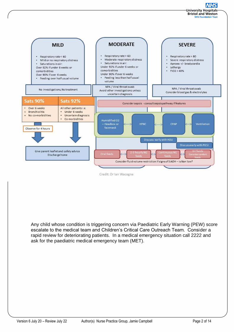
Any child whose condition is triggering concern via Paediatric Early Warning (PEW) score escalate to the medical team and Children’s Critical Care Outreach Team. Consider a rapid review for deteriorating patients. In a medical emergency situation call 2222 and ask for the paediatric medical emergency team (MET).
Version 6 July 20 – Review July 22 Author(s) Nurse Practice Group, Jamie Campbell Page 3 of 14
ADMISSION CRITERIA
Tachypnoea with moderate to severe increased work of breathing (refer to PEW scoring)
Hydration / nutrition support required
O2 Sats <90% in air for babies over 6 weeks with no co-morbidities, therefore requiring Oxygen. (If O2 Sats are 90-92% observe for a minimum 4 hours before discharge).
O2 Sats <92% in air for babies under 6 weeks or with co-morbidities, therefore requiring Oxygen, or if uncertain diagnosis
Apnoeas
Lower threshold for admission Age <6 weeks (corrected gestational age)
Premature (<35 weeks)
Low birth weight (<2.5kg)
Pre-existing lung disease
Congenital heart disease (CHD)
Neuromuscular weakness
Immunodeficiency
Trisomy 21
Psychosocial concerns or re-attender (require senior review) If not admitted, parents must be provided with key safety information to take away for reference. This should cover how to recognise developing “red flag” symptoms. (Parents guide to bronchiolitis).
INVESTIGATIONS These are not routinely needed for confirmation of diagnosis
If admitted, a nasopharyngeal aspirate (NPA) or viral throat swab should be sent for Polymerase Chain Reaction (PCR) to identify Respiratory Syncytial Virus (RSV) or other viruses to enable cohort nursing
Consider point of care / rapid testing as available
Consider Capillary blood gas (CBG) if requiring FiO2 ≥0.4, severe respiratory distress or exhaustion. CBG only gives information from the time it is performed, and should be interpreted in the context of the patient. A normal CBG is not indicative of severity
A chest X-ray should only be considered if there is diagnostic concern e.g. persistent pyrexia or FiO2 ≥0.5
Blood tests have a monitoring role if intravenous fluids are being used or if there is diagnostic uncertainty.
MANAGEMENT Treatment of bronchiolitis is primarily supportive in nature. Minimal handling is the cornerstone of care.
Respiratory
Maintain saturations ≥90% (92% if under 6 weeks or comorbidities)
Medical gases have a drying effect. Drying of mucous membranes can cause airway damage, increase work of breathing and can lead to thickened secretions which may
Version 6 July 20 – Review July 22 Author(s) Nurse Practice Group, Jamie Campbell Page 4 of 14
cause airway obstruction.
Oxygen should be humidified either via headbox or face mask (in an emergency or short term ≤1hour this can be via a non-rebreather mask)
Heat Humidified High Flow Nasal Cannula Therapy (HFNC) (Optiflow) is a well-tolerated form of respiratory support suitable for use on the wards or in a High Dependency Unit (HDU). It should be considered in patients with moderate and severe bronchiolitis as measured by a respiratory component of the PEW score of 3 or more.
HFNC therapy is not recommended in patients with respiratory acidosis and a pH <7.3, apnoea, air leak (pneumothorax or pneumomediastinum) or multi-organ failure. These patients need to be discussed with Paediatric Intensive Care Unit (PICU) for consideration of transfer for formal respiratory support.
Patients requiring FiO2 of 50% and / or have a respiratory component of the PEW score of 3 or more despite maximal HFNC therapy may benefit from Continuous Positive Airway Pressure (CPAP) . These patients should be managed on HDU.
Patients who require ventilation must be managed on PICU.
Nutrition and Hydration Routine suctioning of the nose and mouth is not indicated. In some patients suctioning
pre feeds may clear secretions to aid oral feeding.
When a patient is not tolerating adequate intake orally (<2/3 of feeds), nasogastric (NG) or orogastric (OG) feeds can be safe and effective, when inserted and used in accordance with trust policy
Feeds may be reduced to 100mls/kg or as medically directed, and given 2 – 3 hourly. Smaller feeds require less energy and effort to consume and digest. Larger volumes may affect respiratory effort by putting pressure on the diaphragm, affecting lung expansion
Strict fluid input / output to be documented on appropriate diet record or fluid chart and fluid balance recorded and escalate accordingly
Patients with worsening or severe respiratory distress should be considered for intravenous fluids. The minimum requirements are: o Plasma-Lyte with glucose please refer to Paediatric fluid administration guidelines o Infuse at 80% maintenance requirements o Check U&Es and glucose at onset of intravenous therapy and 24 hourly (risk of
hyponatraemia)
Consider comfort feeds (via NG) for those Nil by mouth (NBM). These must only be given in agreement with the medical and nursing teams
Fluid overload or dehydration will have significant implications on the patients clinical condition and must be addressed and treated early to prevent deterioration or development of associated complications
Consideration can be given to training of parents in NG feeding if suitable (see short-term home NG feeding in bronchiolitis guideline)
Handling & Posture Nurse infants prone (on their tummies) with minimal handling and the cot inclined at a 450
angle
Cluster patient cares together to minimise disturbances – i.e. nappy changes, observations & investigations
Consider environmental factors such as noise and harsh lighting to promote rest
Version 6 July 20 – Review July 22 Author(s) Nurse Practice Group, Jamie Campbell Page 5 of 14
Medication Consider regular analgesia if signs of discomfort
There is no role for bronchodilators, adrenaline, antibiotics, steroids, antivirals or physiotherapy in uncomplicated bronchiolitis.
Nebulised hypertonic saline is not recommended practice because of associated risks and the minimal benefit for our inpatient population.
MONITORING
At a minimum saturations and heart rate should be monitored continuously until the patient is in air and stable for 4 hours including a period of sleep.
Children requiring HFNC therapy should have continuous Electrocardiograph (ECG) and saturation monitoring.
Children requiring HFNC therapy may require High Dependency care. (See HFNC guideline)
Children with a history of apnoea, or at risk of apnoea should have continuous saturation and ECG monitoring.
Ensure the saturation probe is moved a minimum of 4 hourly, document this on the observation chart. Leaving the probe in the same position can cause pressure injuries or burns.
INFECTION CONTROL
Bronchiolitis viruses are highly contagious and barrier nursing procedures should be implemented.
DISCHARGE CRITERIA
Stable and improving
Stable continuously monitored oxygen saturations maintained ≥90% in air for a period of 4 hours including a period of sleep for babies over 6 weeks with no co-morbidities.
Stable continuously monitored oxygen saturations maintained ≥92% in air for a period of 4 hours including a period of sleep for babies under 6 weeks or those with co-morbidities or those considered for short-term home NG feeding.
Feeding at 2/3 normal feeds or more
Infants admitted with apnoea must have period of observation of at least 12 hours following last witnessed apnoea
Family confident in their ability to manage patient at home
When appropriate use the Bronchiolitis Criteria Led Discharge (CLD) form
DISCHARGE ADVICE
Avoid Passive Smoke
Symptoms may persist for 10-14 days
Re-infection may occur
Importance of handwashing
Increased risk of wheezing after bronchiolitis
Bronchiolitis parent advice leaflet including safety net advice
Version 6 July 20 – Review July 22 Author(s) Nurse Practice Group, Jamie Campbell Page 6 of 14
_____________________________________________________________________________
RELATED DOCUMENTS
My Child Has Been Admitted To Hospital With Bronchiolitis A Guide For Parents And Carers – Patient information leaflet
Parents guide to Bronchiolitis
High Humidity High Flow Nasal Cannula Oxygen therapy (AIRVO2 / Neonatal Optiflow) – Clinical Guideline
The Acutely Ill Child Parental Patient Involvement In Escalation Of Clinical Care – SOP (standard operating procedure)
Paediatric Medical Emergency Calls SOP
UHBristol Fluid (Paediatric) Clinical guideline
Clinical protocol for recording and acting upon physiological observations in paediatric in patient areas – on clinical guidelines pages under ‘R’
Enteral feeding policy
Short term home NG feeding in patients with bronchiolitis
Infection control
Oxygen Therapy Nursing Guidelines
Respiratory Failure Nursing Guidelines
NICE Guideline: Bronchiolitis in Children: diagnosis and management (NG9). 1st June 2015. https://www.nice.org.uk/guidance/ng9
AUTHORISING BODY
Paediatric Medical Governance
SAFETY If there are unusual or unexpected safety concerns (to staff or patient), emphasize them here
QUERIES Acute Medical team Bleep 2943 Children’s Critical Care Outreach Team Bleep 2968 Clinical Site Team Bleep 3217 HDU team 20921 PICU 28018
Version 6 July 20 – Review July 22 Author(s) Nurse Practice Group, Jamie Campbell Page 7 of 14
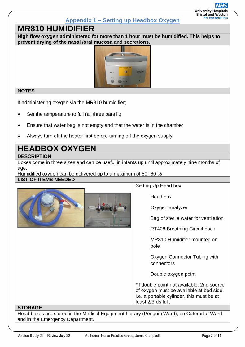
Appendix 1 – Setting up Headbox Oxygen
MR810 HUMIDIFIER
High flow oxygen administered for more than 1 hour must be humidified. This helps to prevent drying of the nasal /oral mucosa and secretions.
NOTES
If administering oxygen via the MR810 humidifier;
Set the temperature to full (all three bars lit)
Ensure that water bag is not empty and that the water is in the chamber
Always turn off the heater first before turning off the oxygen supply
HEADBOX OXYGEN
DESCRIPTION
Boxes come in three sizes and can be useful in infants up until approximately nine months of age. Humidified oxygen can be delivered up to a maximum of 50 -60 % LIST OF ITEMS NEEDED
Setting Up Head box
Head box
Oxygen analyzer
Bag of sterile water for ventilation
RT408 Breathing Circuit pack
MR810 Humidifier mounted on
pole
Oxygen Connector Tubing with
connectors
Double oxygen point
*if double point not available, 2nd source of oxygen must be available at bed side, i.e. a portable cylinder, this must be at least 2/3rds full.
STORAGE
Head boxes are stored in the Medical Equipment Library (Penguin Ward), on Caterpillar Ward and in the Emergency Department.
Version 6 July 20 – Review July 22 Author(s) Nurse Practice Group, Jamie Campbell Page 8 of 14
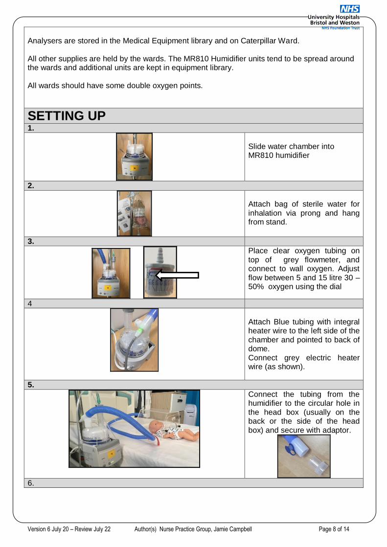
Analysers are stored in the Medical Equipment library and on Caterpillar Ward.
All other supplies are held by the wards. The MR810 Humidifier units tend to be spread around the wards and additional units are kept in equipment library. All wards should have some double oxygen points.
SETTING UP
1.
Slide water chamber into MR810 humidifier
2.
Attach bag of sterile water for inhalation via prong and hang from stand.
3.
Place clear oxygen tubing on top of grey flowmeter, and connect to wall oxygen. Adjust flow between 5 and 15 litre 30 – 50% oxygen using the dial
4
Attach Blue tubing with integral heater wire to the left side of the chamber and pointed to back of dome. Connect grey electric heater wire (as shown).
5.
Connect the tubing from the humidifier to the circular hole in the head box (usually on the back or the side of the head box) and secure with adaptor.
6.
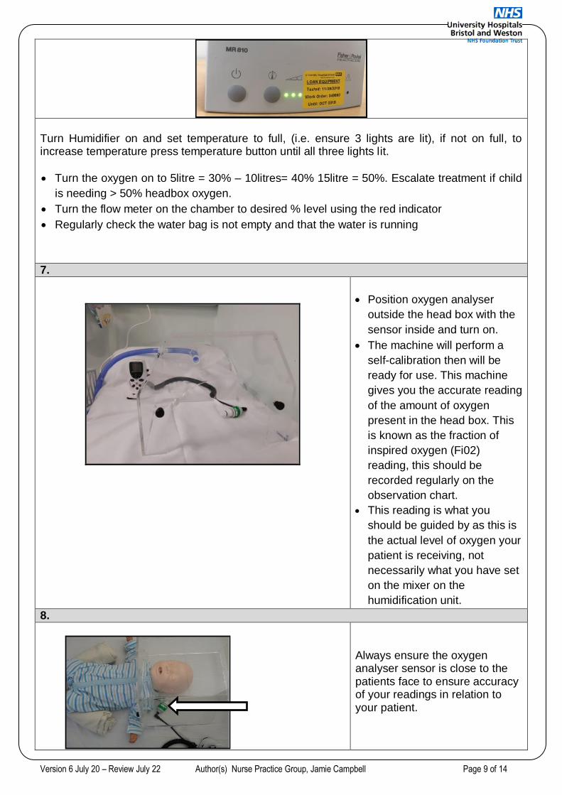
Version 6 July 20 – Review July 22 Author(s) Nurse Practice Group, Jamie Campbell Page 9 of 14
Turn Humidifier on and set temperature to full, (i.e. ensure 3 lights are lit), if not on full, to increase temperature press temperature button until all three lights lit.
Turn the oxygen on to 5litre = 30% – 10litres= 40% 15litre = 50%. Escalate treatment if child
is needing > 50% headbox oxygen.
Turn the flow meter on the chamber to desired % level using the red indicator
Regularly check the water bag is not empty and that the water is running
7.
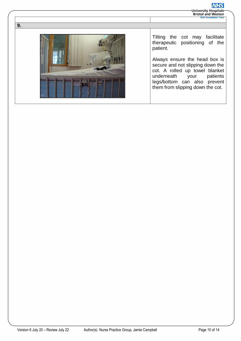
Position oxygen analyser
outside the head box with the
sensor inside and turn on.
The machine will perform a
self-calibration then will be
ready for use. This machine
gives you the accurate reading
of the amount of oxygen
present in the head box. This
is known as the fraction of
inspired oxygen (Fi02)
reading, this should be
recorded regularly on the
observation chart.
This reading is what you
should be guided by as this is
the actual level of oxygen your
patient is receiving, not
necessarily what you have set
on the mixer on the
humidification unit.
8.
Always ensure the oxygen analyser sensor is close to the patients face to ensure accuracy of your readings in relation to your patient.
Version 6 July 20 – Review July 22 Author(s) Nurse Practice Group, Jamie Campbell Page 10 of 14
9.
Tilting the cot may facilitate therapeutic positioning of the patient. Always ensure the head box is secure and not slipping down the cot. A rolled up towel blanket underneath your patients legs/bottom can also prevent them from slipping down the cot.
Version 4 Nov 16 – Review May 16 Author(s) Nurse Practice Group, Tom Hilliard Page 11 of 14
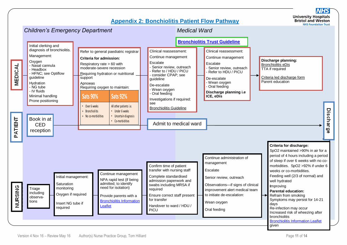
Appendix 2: Bonchiolitis Patient Flow Pathway
Children’s Emergency Department Medical Ward
Initial clerking and diagnosis of bronchiolitis.
Management:
Oxygen - Nasal cannula - Headbox - HFNC; see Optiflow guideline
Hydration - NG tube - IV fluids
Minimal handling
Prone positioning
Refer to general paediatric registrar
Criteria for admission:
Respiratory rate > 60 with moderate-severe recession
Requiring hydration or nutritional support
Apnoeas Requiring oxygen to maintain:
Sats 90% Sats 92%• Over 6 weeks+ Bronchiolitis+ No co-morbidities
All other patients i.e.• Under 6 weeks• Uncertain diagnosis• Co-morbidities
Clinical reassessment:
Continue management
Escalate
- Senior review, outreach - Refer to / HDU / PICU - consider CPAP; see guideline
De-escalate
- Wean oxygen - Oral feeding
Investigations if required: see
Bronchiolitis Guideline
Clinical reassessment:
Continue management
Escalate
- Senior review, outreach - Refer to HDU / PICU
De-escalate - Wean oxygen - Oral feeding
Discharge planning i.e ICE, eDis
Discharge planning: Bronchiolitis eDis TTA if required Criteria led discharge form Parent education
Admit to medical ward Book in at
CED reception
Triage including observa-tions
Initial management:
Saturation monitoring
Oxygen if required
Insert NG tube if required
Continue management
NPA rapid test (if being admitted; to identify need for isolation) Provide parents with a
Bronchiolitis Information
Leaflet
Confirm time of patient transfer with nursing staff
Complete standardised admission paperwork and swabs including MRSA if required
Ensure correct staff present for transfer
Handover to ward / HDU / PICU
Continue administration of
management
Escalate
Senior review, outreach
Observations—if signs of clinical
improvement alert medical team
to initiate de-escalation:
Wean oxygen
Oral feeding
Criteria for discharge:
SpO2 maintained >90% in air for a
period of 4 hours including a period
of sleep if over 6 weeks with no co-
morbidities. SpO2 >92% if under 6
weeks or co-morbidities.
Feeding well (2/3 of normal) and
well hydrated
Improving
Parental education: Refrain from smoking Symptoms may persist for 14-21 days Re-infection may occur Increased risk of wheezing after bronchiolitis Bronchiolitis Information Leaflet given
Bronchiolitis Trust Guideline
Vrs 7, 10/2020 Review 10/2021 Author (s): Jamie Campbell (General Paediatrics), Nurse Practice Group Page 12 of 14
Where to file in Evolve (tab): Inpatient Clinical Notes
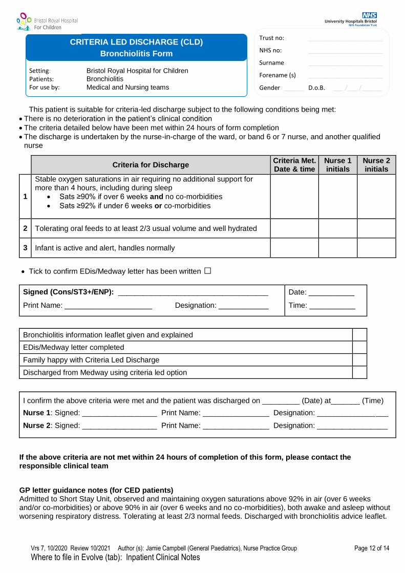
Trust no: ______________________
NHS no: ______________________
Surname ______________________
Forename (s) ______________________
Gender ______ D.o.B. ___ /___ /______
Setting: Bristol Royal Hospital for Children
Patients: Bronchiolitis
For use by: Medical and Nursing teams
CRITERIA LED DISCHARGE (CLD)
Bronchiolitis Form
This patient is suitable for criteria-led discharge subject to the following conditions being met:
There is no deterioration in the patient’s clinical condition
The criteria detailed below have been met within 24 hours of form completion
The discharge is undertaken by the nurse-in-charge of the ward, or band 6 or 7 nurse, and another qualified nurse
Criteria for Discharge Criteria Met. Date & time
Nurse 1 initials
Nurse 2 initials
1
Stable oxygen saturations in air requiring no additional support for more than 4 hours, including during sleep
Sats ≥90% if over 6 weeks and no co-morbidities
Sats ≥92% if under 6 weeks or co-morbidities
2 Tolerating oral feeds to at least 2/3 usual volume and well hydrated
3 Infant is active and alert, handles normally
Tick to confirm EDis/Medway letter has been written □
Signed (Cons/ST3+/ENP): ____________________________________
Print Name: _____________________ Designation: ____________
Date: ___________
Time: ___________
Bronchiolitis information leaflet given and explained
EDis/Medway letter completed
Family happy with Criteria Led Discharge
Discharged from Medway using criteria led option
I confirm the above criteria were met and the patient was discharged on _________ (Date) at_______ (Time)
Nurse 1: Signed: __________________ Print Name: ________________ Designation: _________________
Nurse 2: Signed: __________________ Print Name: ________________ Designation: _________________
If the above criteria are not met within 24 hours of completion of this form, please contact the responsible clinical team
GP letter guidance notes (for CED patients) Admitted to Short Stay Unit, observed and maintaining oxygen saturations above 92% in air (over 6 weeks and/or co-morbidities) or above 90% in air (over 6 weeks and no co-morbidities), both awake and asleep without worsening respiratory distress. Tolerating at least 2/3 normal feeds. Discharged with bronchiolitis advice leaflet.
Version 6 July 20 J Campbell. Nurse practice group Review July 22 Page 13 of 14
SETTING Bristol Royal Hospital for Children
FOR STAFF Nursing and medical staff
FOR PATIENTS Children and young people for whom the clinical team have agreed that criteria-led discharge is appropriate
Background/Introduction
The purpose of Criteria Led Discharge is to facilitate safe and timely discharge from Bristol Royal Hospital for Children (BRHC). Pre-requisite criteria for a patient being deemed ‘fit for discharge’ are agreed in advance of discharge. The decision to discharge the patient can then be safely undertaken by a delegated practitioner, avoiding delays associated with medical team review.
The responsibility for deciding that a patient has met the safe discharge criteria will be undertaken by the practitioners as below:
Inpatient wards: The Nurse in Charge of the Ward, or a band 6 or 7 nurse, in conjunction with a registered
nurse with a substantive contract within BRHC.
Children’s Emergency Department (CED)/The Observatory: A Band 6 or 7 nurse, in conjunction with a registered nurse with a substantive contract within BRHC.
Criteria Led Discharge Process
1. A patient is identified by an Emergency Nurse Practitioner (CED), ST3+ or Consultant as suitable for criteria-led discharge. Condition-specific criteria-led discharge forms can be found on the BRHC Patient Flow intranet pages.
2. The Clinician completes a criteria-led discharge form, which is stored in the patient’s nursing notes.
3. The Clinician informs the Nurse in Charge, who identifies the patient as criteria-led discharge on the electronic whiteboard.
4. The doctor or ENP completes the discharge summary and TTA, or Medway letter in the CED.
5. Once the discharge criteria have been met, the patient is reviewed by two designated practitioners to ensure all necessary actions have been taken to complete the criteria led discharge and sign the relevant section of the criteria-led discharge form.
6. The patient is discharged via usual processes, using the ‘Criteria Led’ option on Medway, and the Clinical Site Team is notified of bed availability.
7. If the relevant criteria are not met within 24 hours of the form being completed, the patient should be discussed with the responsible clinical team (or sooner if clinical concerns).
Developing criteria-led discharge proformas
New criteria-led discharge forms may be developed for other patient groups and should be circulated to the relevant clinical team and governance group for consultation before submission to the Nurse Practice Group.
The approved template should then be uploaded to the above Connect page by a member of the Women’s and Children’s Divisional Management team.
RELATED DOCUMENTS AUTHORISING BODY
A brief guide to developing criteria-led discharge, NHS Improvement. December 2017. https://improvement.nhs.uk/resources/guide-developing-criteria-led-discharge/
Nurse Practice Group Paediatric Medicine Governance group
SAFETY Any child who does not meet the criteria for criteria-led discharge should be discussed with the responsible clinical team.
QUERIES Contact the patient’s responsible clinical team.
Clinical Standard Operating Procedure (SOP)
CRITERIA LED DISCHARGE

Version 6 July 20 J Campbell. Nurse Practice Group Review July 22 Page 14 of 14