bu principles of humane use of laboratory animals
DESCRIPTION
Principles Of Humane use of Lab Animals.TRANSCRIPT

BUTLER UNIVERSITY
PRINCIPLES OF HUMANE CARE AND USE OF LABORATORY ANIMALS
REGULATIONS, GUIDELINES AND ACCREDITING BODIES 4 FEDERAL.................................................................................................................................................................... 4 STATE AND LOCAL ................................................................................................................................................. 8 INSTITUTIONAL POLICIES ..................................................................................................................................... 8 PROFESSIONAL ORGANIZATIONS ..................................................................................................................... 9
ETHICS 15 GENERAL ETHICAL PRINCIPLES ...................................................................................................................... 15 HISTORICAL PERSPECTIVES ............................................................................................................................. 15 CLASSIFICATION OF PROCEDURES ................................................................................................................ 17 CLASSIFICATION OF RESEARCH TECHNIQUES........................................................................................... 18 RISK-BENEFIT ANALYSIS.................................................................................................................................... 18 AESTHETICS VERSUS HUMANENESS ............................................................................................................. 19
ALTERNATIVES AND MODELS 20 GENERAL DISCUSSION OF THE THREE "R'S" ............................................................................................... 20 ANIMAL MODELS ................................................................................................................................................... 20 ALTERNATIVES TO ANIMAL MODELS ............................................................................................................. 22
SPECIES SUMMARIES 24 MOUSE ...................................................................................................................................................................... 24 RAT ............................................................................................................................................................................ 26 HAMSTER ................................................................................................................................................................. 28 GUINEA PIG ............................................................................................................................................................. 30 RABBIT...................................................................................................................................................................... 32 CAT ............................................................................................................................................................................ 34 DOG............................................................................................................................................................................ 36 PRIMATE (MACACA MULATTA) ................................................................................................................................ 38
BASIC NUTRITION 41 INTRODUCTION ...................................................................................................................................................... 41 FEEDING CONSIDERATIONS .............................................................................................................................. 41 WATER ...................................................................................................................................................................... 41 ENERGY.................................................................................................................................................................... 41 CARBOHYDRATES ................................................................................................................................................ 42 FATS .......................................................................................................................................................................... 42 PROTEINS ................................................................................................................................................................ 42 FIBER......................................................................................................................................................................... 43 VITAMINS.................................................................................................................................................................. 43 MINERALS ................................................................................................................................................................ 43 GOOD NUTRITION IS VITAL ................................................................................................................................. 44 FEED AND WATER REQUIREMENTS ................................................................................................................ 44
ANIMAL DISEASE: CONTROL AND RECOGNITION 45 VETERINARY CARE............................................................................................................................................... 45 ANIMAL QUALITY VS FACILITIES ...................................................................................................................... 45 PRINCIPLES OF DISEASE CONTROL ............................................................................................................... 45 RECOGNITION OF ANIMALS IN A DISEASED STATE ................................................................................... 46 DISEASES OF RATS AND MICE .......................................................................................................................... 47
ANESTHESIA AND ANALGESIA 49 INTRODUCTION ...................................................................................................................................................... 49 CONTROLLED SUBSTANCES ACT.................................................................................................................... 49 DEFINITIONS ........................................................................................................................................................... 50 GENERAL PRINCIPLES ........................................................................................................................................ 50
FACTORS AFFECTING CHOICE OF REGIMEN................................................................................................ 51 CLASSIFICATION OF AGENTS ........................................................................................................................... 51 SPECIES PECULIARITIES .................................................................................................................................... 57 TABLE OF POSTOPERATIVE PAIN .................................................................................................................... 58 TABLE OF DOSAGES ............................................................................................................................................ 58
PRE- AND POSTOPERATIVE CARE 62 REGULATIONS AND GUIDELINES ..................................................................................................................... 62 DEFINITIONS ........................................................................................................................................................... 66 PREOPERATIVE CARE ......................................................................................................................................... 67 OPERATIVE CARE ................................................................................................................................................. 68 FLUID THERAPY ..................................................................................................................................................... 68 MAINTAINING BODY TEMPERATURE............................................................................................................... 69 SURGICAL PROCEDURES ................................................................................................................................... 69 POSTOPERATIVE CARE....................................................................................................................................... 69 INVESTIGATOR RESPONSIBILITY..................................................................................................................... 70
EUTHANASIA 71 THE MEANING OF “EUTHANASIA”.................................................................................................................... 71 HUMANE CONSIDERATIONS .............................................................................................................................. 71 SELECTION OF A TECHNIQUE OF EUTHANASIA .......................................................................................... 71 ASSESSING PAIN AND DISTRESS..................................................................................................................... 72 ASSESSING UNCONSCIOUSNESS AND DEATH............................................................................................ 73 THE PRACTICAL REQUIREMENTS OF EUTHANASIA .................................................................................. 73 ACCEPTABLE AGENTS AND METHODS OF EUTHANASIA ........................................................................ 82 CONDITIONALLY ACCEPTABLE AGENTS AND METHODS OF EUTHANASIA....................................... 83 UNACCEPTABLE AGENTS AND METHODS OF EUTHANASIA................................................................... 83
3
REGULATIONS, GUIDELINES AND ACCREDITING BODIES FEDERAL
Animal Welfare Act The Animal Welfare Act of 1966 (P.L. 89-544), as amended by the Animal Welfare Act of 1970 (P.L. 91-579); 1976 Amendments to the Animal Welfare Act (P.L. 94-279); Subtitle F (Animal Welfare File Name: PL99198); and the Food and Agriculture Conservation and Trade Act of 1990 (P.L. 101-624), Section 2503, Protection of Pets (File Name: PL101624)-contains provisions to prevent the sale or use of animals that have been stolen; prohibit animal-fighting ventures; and ensure that animals used in research, for exhibition, or as pets receive humane care and treatment. The law provides for regulating the transport, purchase, sale, housing, care, handling, and treatment of such animals. Regulatory authority under the Animal Welfare Act is vested in the Secretary of the U.S. Department of Agriculture (USDA) and implemented by USDA's Animal and Plant Health Inspection Service (APHIS). Rules and regulations pertaining to implementation are published in the Code of Federal Regulations (CFR), Title 9 (Animals and Animal Products), Chapter1, Subchapter A (Animal Welfare), Parts 1, 2, and 3. The Improved Standards for Laboratory Animal Act, a part of the 1985 Farm Bill, was enacted in Public Law 99-198. The new law amends the Animal Welfare Act effective December, 1986, and requires the Secretary of Agriculture to promulgate new standards for the care, treatment, and use of laboratory animals, and to establish an information service at the National Agricultural Library. It also stipulates that the U.S. Department of Agriculture (USDA) shall inspect research facilities at least once a year, and that each facility must provide reports that verify compliance, train personnel involved with animal care, and establish at least one institutional animal committee to conduct semiannual reviews. Copies of Federal regulations can be obtained from USDA/APHIS/AC, 920 Main Campus Drive, Suite 200, Raleigh, NC 27606-5210, email: [email protected], Tel: (919) 716-5532, or contact your USDA Area Veterinarian-in-charge.
Public Health Service Regulations The Public Health Service (PHS) Policy on Humane Care and Use of Laboratory Animals was revised in 2002. In the policy statement, the PHS endorses the U.S. Government "Principles for the Utilization and Care of Vertebrate Animals Used in Testing, Research, and Education" (reprinted below), which were developed by the Interagency Research Animal Committee. The PHS policy implements and supplements these principles. The Health Research Extension Act of 1985, Public Law 99-158, revising and extending authorities of the National Institutes of Health (NIH) under the Public Health Service Act, requires the Director of NIH to establish guidelines for the care and use of laboratory animals and requires recipients of NIH funds to provide assurances of their compliance with these guidelines and to have institutional animal committees. In addition, the law provides that the Director of NIH must, by October 1986, develop a plan for research and training in valid alternatives to animal models, in methods to reduce the number of animals used, and in methods to minimize any pain and distress animals may experience. The NIH Office of Laboratory Animal Welfare (OLAW) has published the Public Health Service (PHS) Policy on Humane Care and Use of Laboratory Animals, revised as of August, 2002, incorporating the changes in the Public Health Service Act mandated by the Health Research Extension Act of 1985, Public Law 99-158. "All applications and proposals for award, which are either submitted to the PHS on or after November 1, 1986, or being conducted on or after July 1, 1987, must meet the requirements of the PHS Policy as amended. Institutions which currently have an approved or provisionally acceptable Animal Welfare Assurance on file with the OLAW must submit to OLAW by July 1, 1987, a document in the form of an
4

appendix or amendment which states the changes that the institution has made to conform to the amended Public Health Service Policy." As required by the new law, major revisions are:
1. The Policy will now apply to research that PHS conducts intramurally.
2. The Institutional Animal Care and Use Committee (IACUC) will be appointed by the chief executive officer of the institution.
3. 3. The institution's assurance must include a synopsis of the training or instructions made available
to scientists, animal technicians and other personnel involved in animal care, treatment or use.
4. The IACUC now must inspect and prepare reports on all of the institution's animal facilities (including satellite facilities) at least twice, instead of once, each year. The reports must be maintained by the institution and made available to OPRR upon request. AAALAC accredited facilities (Category 1) now must comply with this requirement since the law makes no distinction for them.
5. The IACUC, through the Institutional official, must submit written annual reports to OPRR updating
the institution's assurance. These reports now must include minority views filed by members of the committee. Information concerning the policy can be obtained from the Office of Laboratory Animal Welfare, National Institutes of Health, RKLI, Suite 1050, MSC 7982, 6705 Rockledge Drive, Bethesda, MD 20892-7982; [email protected]; Tel: (301) 496-7163.
Interagency Research Animal Committee
Principles for the Utilization and Care of Vertebrate Animals Used in Testing, Research, and Training
The development of knowledge necessary for the improvement of the health and well being of humans as well as other animals requires in vivo experimentation with a wide variety of animal species. Whenever U.S. Government agencies develop requirements for testing, research, or training procedures involving the use of vertebrate animals, the following principles shall be considered and whenever these agencies actually perform or sponsor such procedures, the responsible institutional official shall ensure that these principles are adhered to:
1. The transportation, care and use of animals should be in accordance with the Animal Welfare Act (7 U.S.C. 2131 et. seq.) and other applicable Federal laws, guidelines, and policies.
2. Procedures involving animals should be designed and performed with due consideration of their
relevance to human or animal health, the advancement of knowledge, or the good of society. 3. The animals selected for a procedure should be of an appropriate species and quality and the
minimum number required to obtain valid results. Methods such as mathematical models, computer simulation, and in vitro biological systems should be considered.
4. Proper use of animals, including the avoidance or minimization of discomfort, distress, and pain
when consistent with sound scientific practices, is imperative. Unless the contrary is established, investigators should consider that procedures that cause pain or distress in human beings may cause pain or distress in other animals.
5
5. Procedures with animals that may cause more than momentary or slight pain or distress should be performed with appropriate sedation, analgesia, or anesthesia. Surgical or other painful procedures should not be performed on unanesthetized animals paralyzed by chemical agents.
6. Animals that would otherwise suffer severe or chronic pain or distress that cannot be relieved
should be painlessly killed at the end of the procedure or, if appropriate, during the procedure. 7. The living conditions of animals should be appropriate for their species and contribute to their health
and comfort. Normally, the housing, feeding, and care of all animals used for biomedical purposes must be directed by a veterinarian or other scientist trained and experienced in the proper care, handling, and use of the species being maintained or studied. In any case, veterinary care shall be provided as indicated.
8. Investigators and other personnel shall be appropriately qualified and experienced for conducting
procedures on living animals. Adequate arrangements shall be made for their in-service training, including the proper and humane care and use of laboratory animals.
9. Where exceptions are required in relation to the provisions of these Principles, the decisions should
not rest with the investigators directly concerned but should be made, with due regard to Principle II, by an appropriate review group such as an institutional animal research committee. Such exceptions should not be made solely for the purposes of teaching or demonstration.
Guide for the Care and Use of Laboratory Animals
The Guide for the Care and Use of Laboratory Animals was originally prepared by the Institute for Laboratory Animal Resources of the National Research Council for NIH in 1963. The "Guide" has been revised several times since 1963 with the latest in 1996. These guidelines which provide information on the care and use of laboratory animals in research are not legally binding regulations, but a NIH grantee must make a commitment to follow these recommendations in order to be eligible for a NIH Grant/Contract. These recommendations cover the following subjects:
1. Institutional policies and responsibilities. The Guide discusses subjects that require policy attention: the role and function of the Institutional Animal Care and Use Committee, protocols for animal use, occupational health and safety, and personnel qualifications.
2. Animal environment, husbandry, and management. The Guide offers guidelines on how to design and run a management program, addressing environment, nutrition, sanitation, behavioral and social issues, genetics, and nomenclature.
3. Veterinary care. The Guide discusses animal procurement and transportation, disease and preventive medicine, surgery, pain recognition and relief, and euthanasia.
4. Physical plant. The Guide identifies design and construction issues, providing guidelines for animal-room doors, drainage, noise control, and surgery.
Copies of the "Guide" may be ordered from the Institute of Laboratory Animal Resources, National Research Council, 2101 Constitution Ave., NW, Washington, DC 20418; [email protected]; Bulk copies can be ordered from Superintendent of Documents, U.S. Government Printing Office, Washington, DC 20402; Tel: (202) 783-3238 GPO# 017-040-00498-2.
Endangered Species Act The Endangered Species Act of 1973 (P.L. 93-205; 87 Statute 884) became effective on December 28, 1973, supplanting the Endangered Species Conservation Act of 1969 (P.L. 91-135; 83 Statute 275). The new law seeks "to provide a means whereby the ecosystems upon which endangered species and threatened species depend may be conserved, to provide a program for the conservation of such
6
endangered species and threatened species, and to take such steps as may be appropriate to achieve the purposes of the treaties and conservation of wild flora and fauna worldwide." Regulatory authority under the Endangered Species Act is vested in the Secretary of the U.S. Department of the Interior (USDI) and implemented by the Fish and Wildlife Service. Implementing rules and regulations are published in the CFR, Title 50 (Wildlife and Fisheries), Chapter 1 (U.S. Fish and Wildlife Service, Department of Interior), Sub chapter B, Part 17 (Endangered and Threatened Wildlife and Plants). Copies of the regulations, including a list of species currently considered endangered or threatened; can be obtained by writing to the Office of Endangered Species, U.S. Department of Interior, Fish and Wildlife Service, Washington, DC 20240.
Good Laboratory Practice Regulations GLP compliance is intended to assure the quality and integrity of animal safety data in conducting any nonclinical laboratory study that supports or is intended to support applications for research or marketing permits regulated by the Food and Drug Administration.
Studies Included in GLP Regulations:
1. Safety studies in animals. 2. Mean lethal dose (LD 50). 3. Short and long term safety studies. 4. IN VITRO tests, if they have a bearing on product safety, e.g., short term mutagenicity). 5. Studies of safety of regulated products on target animals. 6. Acute toxicity studies on a final product formulation. 7. Studies of test article that are completed in fourteen (14) days or less.
Studies Not Included in GLP Regulations:
1. Exploratory safety studies. 2. Range-findings experiments. 3. Clinical studies. 4. Functionality studies. 5. Clinical tests performed solely in conjunction with product efficacy. 6. Chemical assays for quality control. 7. Stability tests on finished dosage forms and products. 8. Tests for conformance to pharmacopoeial standards. 9. Pharmacological and effectiveness studies. 10. Studies to develop new methodologies for toxicology experimentation. 11. Exploratory studies on viruses and cell biology. 12. Studies to develop methods of synthesis, analysis, mode of action, and formulation of test
articles. 13. Studies relating to stability, identity, strength, quality, and purity of test and/or control articles
that are covered by Good Manufacturing Practice (GMP) regulations. 14. Basic research on human and animal drugs. 15. Preliminary exploratory studies on human and animal drugs. 16. Studies done to determine the physical and chemical characteristics of the test article
independent of any test system. Published in Federal Register, Vol. 43, No. 247- Friday, December 22, 1978. Effective Date: June 20, 1979. Corrections Published in Federal Register, Vol. 44, No. 58-Friday, March 23, 1979. Amended in Federal Register, Vol. 45, No., 72-Friday, April 11, 1980. Effective Date: May 12, 1980.
7

STATE AND LOCAL On behalf of institutional members, the National Association for Biomedical Research (NABR) monitors legislation that could affect the use of animals in research. The Association maintains a computerized database and encourages its members to take advantage of this resource. Full copies of bills, summaries of bills, concise listings of current legislation and narrative summary on state activities are among the available products of this database. In addition, NABR has compiled State Laws Concerning the Use of Animals in Research, a useful reference tool. NABR, 818 Connecticut Avenue, N.W., Suite 200, Washington, D.C. 20006; Tel: 202-857-0540. INSTITUTIONAL POLICIES
Institutional Animal Care and Use Committee The Chief Executive Officer (CEO) of each research facility is required, by law, to appoint an Institutional Animal Care and Use Committee (IACUC). Those persons appointed to this Committee must be qualified through the experience and expertise of its members to oversee the institution's animal program, facilities and procedures. The Assurance must include the names, position titles and credentials of the IACUC chairperson and the members. The committee shall consist of not less than five members, and shall include at least:
1. One Doctor of Veterinary Medicine, with training or experience in laboratory animal science and medicine, who has direct or delegated program responsibility for activities involving animals at the institution.
2. One practicing scientist experienced in research involving animals.
3. One member whose primary concerns are in a nonscientific area (for example, ethicist, lawyer, member of the clergy).
4. One individual who is not affiliated with the institution in any way other than as a member of the
IACUC, and is not a member of the immediate family of a person who is affiliated with the institution.
5. An individual who meets the requirements of more than one of the categories may fulfill more than one requirement. However, no committee may consist of less than five members.
Functions of the Institutional Animal Care and Use Committee
As an agent of the institution the IACUC shall, with respect to PHS-supported activities:
1. Review at least twice annually the institution's program for humane care and use of animals.
2. Inspect at least twice annually all of the institution's animal facilities, including satellite facilities.
3. Prepare semiannual reports of the IACUC’s evaluations conducted and submit the reports to the Institutional official (IO).
4. Review concerns involving the care and use of animals at the institution.
5. Make recommendations to the institutional official regarding any aspect of the institution's animal
program, facilities or personnel training.
6. Review and approve, require modifications in (to secure approval) or withhold approval of those sections of PHS applications for proposals related to the care and use of animals.
8
7. Review and approve, require modifications in (to secure approval), or withhold approval of proposed
significant changes regarding the use of animals in ongoing activities.
8. Be authorized to suspend any activity involving animals in accordance with specifications set forth above.
PROFESSIONAL ORGANIZATIONS
AAALAC Accreditation The American Association for the Accreditation of Laboratory Animal Care (AAALAC) is nonprofit corporation formed in 1965 by leading U.S. scientific and educational organizations to promote high quality animal care and use through a voluntary accreditation program. Any institution maintaining, using, importing, or breeding laboratory animals for scientific purposes is eligible to apply for AAALAC accreditation. The animal care facilities of applicant institutions are visited and thoroughly evaluated by experts in laboratory animal science, who submit a detailed report to the Council on Accreditation. The council reviews applications and site visit reports, using the guidelines in the Guide for the Care and Use of Laboratory Animals (NIH Publication 85-23), to determine whether full accreditation should be granted. Accredited facilities are required to submit annual reports on the status of their animal facilities and site revisits are conducted at intervals of 3 years or less. The Council on Accreditation reviews the annual and site revisit reports to determine whether full accreditation should be continued. Fully accredited animal care facilities receive a certificate of accreditation and are included on a list of such facilities published in the association's Activities Report. Full accreditation by AAALAC is accepted by the Office of Laboratory Animal Welfare of the National Institutes of Health as strong evidence that the animal facilities are in compliance with PHS policy. For further information contact: AAALAC, 11300 Rockville Pike, Suite 1211, Rockville, MD 20852-3035; Tel: 301-231-5353; e-mail: [email protected]; URL: http://www.aaalac.org.
AALAS The American Association for Laboratory Animal Science (AALAS) is a nonprofit organization made up of individuals and institutions professionally concerned with the production, care and use of laboratory animals. It provides a means for collection and exchange of information on all phases of animal care and management. The association meets annually and publishes Laboratory Animal Science (a bimonthly journal), Contemporary Topics (a bimonthly journal), training manuals for laboratory animal technicians, and other documents. AALAS' Animal Technician Certification Board provides a means of developing uniform requirements for technician training by defining the qualifications, preparing, and approving examinations for training programs, and certifying successful candidates. American Association for Laboratory Animal Science, 70 Timber Creek, Suite 5; Cordova, TN 38018 (901) 745-8620; e-mail: [email protected]; URL: http://www.aalas.org.
ACLAM The American College of Laboratory Animal Medicine (ACLAM) is a specialty board recognized by the American Veterinary Medical Association (AVMA). It was founded in 1957 to encourage education, training, and research; to establish standards of training and experience for qualification; and to certify, by examination, qualified laboratory animal specialists as diplomats. To achieve these goals, the college seeks to interest veterinarians in furthering both training and qualifications in laboratory animal medicine.
9
The annual ACLAM Forum is a major continuing-education meeting. ACLAM also meets and sponsors programs in conjunction with the annual meetings of AVMA and AALAS. It emphasizes and sponsors continuing-education programs; cosponsors symposia; cosponsors approximately 30 auto tutorial programs on use, husbandry, and diseases of animals commonly used in research; and publishes texts, such as The Laboratory Rat and The Mouse in Biomedical Research. American College of Laboratory Animal Medicine (ACLAM), Dr. Charles W. McPherson, Executive Director, 200 Summerwinds Drive, Cary, NC 27511; Tel: (919) 859-5985. AHA The American Humane Association (AHA) is a professional, nonprofit organization of organizations and individuals concerned with the exploitation, abuse, and neglect of children and animals. AHA was founded in 1877 and was the first national organization to protect children and animals. AHA supports the 3 R's in biomedical research: refinement, reduction, and replacement where possible. AHA informs its members of issues in biomedical research through its magazine, Advocate, which is published quarterly. American Humane Association, 236 Massachusetts Avenue, NE, Suite 203, Washington, D.C. 20002; Tel: 202-543-7780.
ASLAP The American Society of Laboratory Animal Practitioners (ASLAP), founded in 1966, is open to any graduate of a veterinary college accredited or recognized by the American Veterinary Medical Association (AVMA) or Canadian Veterinary Medical Association (CVMA) who is engaged in laboratory animal practice and maintains membership in the AVMA, CVMA, or any other national veterinary medical association recognized by the AVMA. Its purpose is to disseminate ideas, experiences, and knowledge among veterinarians engaged in laboratory animal practice through education, training, and research at both pre- and postdoctoral levels. Two educational meetings are held annually, one each in conjunction with the annual meetings of the AVMA and American Association for Laboratory Animal Science. American Society of Laboratory Animal Practitioners (ASLAP), Dr. Bradford S. Goodwin, Jr., Secretary-Treasurer, University of Texas, Medical School-CLAMC, 6431 Fannin Street, Room 1132, Houston, TX 77030-1501; Tel: 713-792-5127.
AVMA The American Veterinary Medical Association (AVMA) is the major national organization of veterinarians. Its objective is to advance the science and art of veterinary medicine, including its relationship to public health and agriculture. The AVMA is the recognized accrediting agency for schools and colleges of veterinary medicine. It sponsors specialization in veterinary medicine through the formal recognition of specialty certifying organizations, including the American College of Laboratory Animal Medicine. The AVMA Committee on Animal Technician Activities and Training accredits 2-year programs in animal technology at institutions of higher learning throughout the United States. A list of accredited programs and a summary of individual state laws and regulations relative to veterinarians and animal technicians is available for the AVMA. American Veterinary Medical Association (AVMA). 1931 North Meacham Road, Suite100, Schaumburg, IL 60173-4360; Tel: 800-248-2862; URL: http:/Iwww.avma.org. AWIC
10
The Animal Welfare Information Center (AWIC), at the National Agricultural Library, was established by the 1985 amendments to the Animal Welfare Act. It provides information on employee training, improved methods of experimentation (including alternatives), and animal-care and animal-use topics through the production of bibliographies, workshops, resource guides, and The Animal Welfare Information Center Newsletter. AWIC services are geared toward those who must comply with the Animal Welfare Act, such as researchers, veterinarians, exhibitors, and dealers. Publications and additional information are available from AWIC. Animal Welfare Information Center (AWIC), National Agricultural Library, 5th floor, Beltsville, MD 20705-2351; Tel: 301-504-6212; e-mail: [email protected]; URL: http:Ilnetvet.wustl.edulawic.htm or http:II www.nalusda.gov. AWI AWI is a nonprofit educational organization dedicated to reducing the pain and fear inflicted on animals by humans. Since its founding in 1951, AWI has promoted humane treatment of laboratory animals, emphasizing the importance of socialization, exercise, and environmental enhancement. The institute supports the "3 R’s": replacement of experimental animals with alternatives, refinement to reduce animal pain and suffering, and reduction in the numbers of animals used. Educational material published by AWI includes the AWI Quarterly, Comfortable Quarters for Laboratory Animals, Beyond the Laboratory Door, and Animals and Their Legal Rights and is available free to scientific institutions and libraries and at cost to others. The institute welcomes correspondence and discussion with scientists, technicians, and IACUC members on improving the lives of laboratory animals. Animal Welfare Institute (AWI), P.O. Box 3650, Washington, DC 20007; Tel: 202-337-2332; e-mail: [email protected]. CAAT CAAT was founded in 1981 to develop alternatives to the use of whole animals for product development and safety testing. Although CAAT's mission focuses primarily on the development of alternatives for testing, the center also works with organizations seeking to implement the 3 R's in research and education. These organizations are throughout the world, primarily in North America, Europe, Australia, and Japan. CAAT is an academic research center based in the School of Hygiene and Public Health at Johns Hopkins University in Baltimore, whose programs encompass laboratory research, education/information, and validation of alternative methods. CAAT's primary outreach to scientific and lay audiences is its newsletter, which is published three times a year. A newsletter for middle-school students, CAATALYST, is published three times a year. Center for Alternatives to Animal Testing (CAAT), Johns Hopkins University, 111 Market Place, Suite 840, Baltimore, MD 21202-6709; Tel: 410-223-1693; e-mail: [email protected]; URL: http://infonet.welchjhu.edu/~caat.
FBR The Foundation for Biomedical Research was established in 1981 to take positive action to preserve the freedom of the scientific community to conduct biomedical research. FBR, a nonprofit educational organization provides the media and the public with accurate information about humane and responsible animal research. The Foundation has articulated the necessity for animal research in many forums and through a variety of media vehicles. More important, it has become the foremost resource in the nation for information on this critical subject, and has established a formal opposition to animal rights activists who formerly went unchallenged.
11
The Newsletter of the Foundation is published several times annually and provides information on FBR activities and educational materials, federal and state legislation, biomedical advances resulting from animal experimentation and animal rights/welfare activities. Foundation for Biomedical Research (FBR), 818 Connecticut Avenue, N.W., Suite 303, Washington, D.C. 20006; Tel: (202) 857-0654; email: [email protected]; URL: http://www.fiesta.com/fbr. HSUS The Humane Society of the United States (HSUS) is the nation's largest animal-protection organization. The society is active on a wide variety of humane issues, including those affecting wildlife, companion animals, and animals in laboratories and on farms. HSUS publishes a quarterly magazine (The HSUS News), a newsletter (The Animal Activist Alert), and a variety of reports, brochures, and other advocacy materials. The society works actively on issues involving the use of animals in research, safety testing, and education. This work is spearheaded by the HSUS Animal Research Issues Section, with the aid of a Scientific Advisory Council. The aims of this research are to promote the 3 R's of replacement, reduction, and refinement; strong regulations and their enforcement; openness and accountability among research institutions; and an end to egregious mistreatment of animals. HSUS pursues these aims through educational, legislative, legal, and investigative means. Staff are available to give presentations and write articles on these topics. The Humane Society of the United States (HSUS), 2100 L Street, NW, Washington, DC 20037; Tel: 202-452-1100; e-mail: HSUSLAB @ix.netcom.com).
ICLAS ICLAS is an international nongovernment scientific organization that was founded in 1961 under the auspices of UNESCO and several scientific unions. The aims of ICLAS are to promote and coordinate the development of laboratory animal science throughout the world, to promote international collaboration in laboratory animal science, to promote the definition and monitoring of quality laboratory animals, to collect and disseminate information on laboratory animal science, and to promote the humane use of animals in research, testing, and teaching through recognition of ethical principles and scientific responsibilities. ICLAS has programs addressing microbiologic and genetic monitoring and standardization, assisting developing countries in pursuing their objectives in improving the care and use of laboratory animals, and improving education and training in laboratory animal science. ICLAS accomplishes its goals through regional scientific meetings, an international scientific meeting held every 4 years, the dissemination of information, and expert consultation with those requesting assistance. ICLAS membership is composed of national members, scientific union members, scientific members, and associate members. The Governing Board is responsible for implementing the general policy of ICLAS and is elected by the General Assembly every 4 years. International Council for Laboratory Animal Science (ICLAS), Dr. Steven Pakes, Secretary General, Division of Comparative Medicine, University of Texas Southwestern Medical Center, 5323 Harry Hines Boulevard, Dallas, TX; Tel: 214-648-3340; e-mail: [email protected].
ILAR The Institute of Laboratory Animal Resources (ILAR) was founded in 1952 under the auspices of the National Research Council. A component of the Commission on Life Sciences, ILAR serves as a coordinating agency and a national and international resource for compiling and disseminating information on laboratory animals, promoting education, planning and conducting conferences and symposia, surveying existing and required facilities and resources, upgrading laboratory animal resources, and promoting high-quality, humane care of laboratory animals in the United States.
12
Institute of Laboratory Animal Resources, National Academy of Sciences, 2101 Constitution Ave, N.W. ; Washington, DC 20418; Tel: 202-334-2590; e-mail: [email protected]; ILAR Journal e-mail: ILARJ @nas.edu; URL: http:I/www2.nas.edulilarhome.
LAMA Laboratory Animal Management Association (LAMA) is a nonprofit educational organization. Membership includes individuals and institutions involved in laboratory animal management, medicine, and science. The mission of the association, founded in 1984, is to "enhance the quality of management and care of laboratory animals throughout the world." The objectives of LAMA include promoting the dissemination of ideas, experiences, and knowledge in the management of laboratory animals, encouraging continued education, acting as a spokesperson for the field of laboratory animal management, and assisting in the training of managers. The organization conducts a midyear forum on management issues and topics of interest to the general membership and an annual meeting in conjunction with the American Association of Laboratory Animals Science national meeting. LAMA Review is a quarterly journal on management issues published by the organization, and LAMA Lines is a bimonthly newsletter on topics of general interest to the membership. Laboratory Animal Management Association (LAMA), Mr. Paul Schwikert, Past-President, P.O. Box 1744, Silver Spring, MD 20915; phone: 313-577-1418.
NABR The National Association for Biomedical Research (NABR) is the only national, nonprofit organization dedicated solely to advocating sound public policy that recognizes the vital role of humane animal use in biomedical research, higher education and product safety testing. Founded in 1979, NABR provides the unified voice for the scientific community on legislative and regulatory matters affecting laboratory animal research. NABR's membership is comprised of over 300 public and private universities, medical and veterinary schools, teaching hospitals, voluntary health agencies, professional societies, pharmaceutical companies and other animal research-related firms. NABR supports the responsible use and humane care and treatment of laboratory animals in research, education and product safety testing. Further, the membership believes that only as many animals as necessary should be used; that any pain or distress animals may experience should be minimized; and that alternatives to the use of live animals should be developed and employed, wherever feasible. Still, the Association recognizes that now and in the foreseeable future it is not possible to completely replace the use of animals and that the study of whole, living organisms is an indispensable element of biomedical research and testing that benefits all animals. National Association for Biomedical Research (NABR), 818 Connecticut Avenue, N.W., Suite 303, Washington, D.C. 20006; Tel: (202) 857-0540; e-mail: [email protected]; URL: http://www.fiesta.comlnabr. OLAW The Office of Laboratory Animal Welfare (OLAW) fulfills responsibilities set forth in the Public Health Service (PHS) Act. These include developing and monitoring, as well as exercising compliance oversight relative to, the PHS Policy on Humane Care and Use of Laboratory Animals (Policy), which applies to animals involved in research conducted or supported by any component of PHS; establishing criteria for and negotiation of assurances of compliance with institutions engaged in PHS-conducted or PHS-supported research using animals; directing the development and implementation of educational and instructional programs with respect to the use of animals in research; and evaluating the effectiveness of PHS policies and programs for the humane care and use of laboratory animals.
13
Office for Protection from Research Risks (OPRR), National Institutes of Health, 6100 Executive Blvd., Suite 3B01, Rockville, MD 20892; Tel: 301-496-7163.
SCAW
SCAW is an independent organization supported by individuals and institutions involved in research with animals and concerned about maintaining the highest standards of humane care. SCAW publishes resource materials, organizes conferences, and supports a wide variety of educational activities. Scientists Center for Animal Welfare (SCAW), 7833 Walker Drive, Suite 340, Greenbelt, MD 20770; Tel: 301-345-3500.
REAC The missions of the Animal Care Program are to provide leadership in establishing acceptable standards of humane animal care and treatment and to monitor and achieve compliance through inspections and educational and cooperative efforts. Copies of the Animal Welfare Regulations and the Animal Welfare Act are available from REAC. United States Department of Agriculture, Animal and Plant Health Inspection Service, Regulatory Enforcement of Animal Care (REAC), 4700 River Road, Unit 84, Riverdale, MD 20737-1234; Tel: 301-734-4981; e-mail: sstith@ aphis.usda.gov.
14
ETHICS GENERAL ETHICAL PRINCIPLES The general ethical principles of humane use of animals in research were formulated by Marshall Hall in 1831. These principles are as useful in evaluating experimental procedures today as they were then. ""The first principle to be laid down for the prosecution of physiology is this: we should never have recourse to experiment, in cases in which observation can afford us the information required..." "As a second principle...it must be assumed that no experiment should be performed without a distinct and definite object, and without the persuasion, after the maturest consideration, that object will be attained by that experiment, in the form of a real and uncomplicated result..." "It must be admitted, as a third principle...that we should not needlessly repeat experiments which have already been performed by physiologists of reputation. If a doubt respecting their accuracy, or the accuracy of the deductions drawn from them, arise, it then, indeed, becomes highly important that they should be corrected or confirmed by repetition. This principle implies the necessity of a due knowledge of what has been done by preceding physiologists..." "...it must next be received as an axiom, or fourth principle, that a given experiment should be instituted with the least possible infliction of suffering..." "Lastly, it should be received as a fifth principle, that every physiological experiment should be performed under such circumstances as will secure a due observation and attestation of its results, and so obviate, as much as possible, the necessity for its repetition..." "In order to fully accomplish these objects, it would be desirable to form a society for physiological research. Each member should engage to assist the others. It should be competent to any member to propose a series of experiments, its modes, its objects. These should be first fully discussed--purged from all sources of complication, prejudice, or error--or rejected. If it be determined that such series of experiments be neither unnecessary nor useless...they should then be performed, repeated if necessary, and duly attested. Lastly, such experiments, with the deductions which may flow from them, may then be published with the inestimable advantage of authenticity." "Pursued in this manner, the science of physiology will be rescued from the charges of uncertainty and cruelty, and will be regarded by all men, at once as an important and essential branch of knowledge and scientific research."" HISTORICAL PERSPECTIVES
The History of the Use of Animals in Biomedical Research Aristotle (384-322 B C) is credited as the first person to study animals scientifically. He obtained a grant "to study the natures of animals", from Alexander the Great and studied anatomy and embryology, as well as zoology. Galen (129-199 AD) may have been the first to perform biomedical experiments on animals. William Harvey (1578-1657) proved the circulation of the blood by studying "the motion of the heart and blood in animals", a crucial landmark in medicine. John Hunter (1728-1793) is credited as the founder of experimental surgery and was a comparative morphologist, physiologist and zoologist. Claude Bernard (1813-1878), the great experimental physiologist wrote - "the observations of the appropriate animal species is the key to making observations relevant to the human situation under study;" and many other principles of experimentation in “An Introduction to the Study of Experimental Medicine” (1865). Louis Pasteur (1822-1895) studied anthrax, rabies, fowl cholera, and other diseases of animals and developed vaccines to prevent these diseases in animals and rabies in man. His work was severely criticized by antivivisectionists.
15
Enormous advances were made in medicine beginning at about 1870. The "greats" of medicine making their contribution through the study of animals include: Jenner, Darwin, Virchow, Lister, Koch, Ehrlich, Metchinikoff, Pavlov, Banting and Best, Cannon, Fleming, Chain and Florey and countless others. The first pathogenic bacterium, fungus, mycoplasma, protozoan, virus, prion were all discovered in animals. Our knowledge of infectious diseases, parasitology, physiology, inflammation, immunology, pharmacology, toxicology, embryology, oncology and many other medical subjects depended on experiments in animals. Another wave of medical advances began in the 1950's as a result of federally funded biomedical research. Testing of new drugs in animals gradually increased until 1959-60 when the thalidomide episode resulted in a tremendous increase in drug safety testing in animals. In the past 20 years there has been a reduction in the use of animals in biomedical research. The decrease is probably the result of many factors including decreased federal support for research, increased cost of animal research caused by inflation-recession and the imposition of many regulations controlling nearly every aspect of research using animals, increased use of non-animal testing procedures and increased public concern for use of animals in research. Despite the cost, there are probably few researchers and no directors of animal facilities who wish for the return of the "good old days" of diseased animals, poor housing, makeshift surgeries and poorly trained investigators and technicians. The need for the use of experimental animals continues. The conquest of the myriad of afflictions to which man is prone today and for the foreseeable future must continue to rely on the use of experimental animals.
Animal Protectionist/Rights Activist Movement The work of Darwin raised, in some minds, disturbing questions about experiments conducted by man on animals. In England, the Royal Society for the Prevention of Cruelty to Animals began to oppose most research on animals. This opposition was based on the premise that if Darwin was correct---that animals could feel pain similar to humans, then the infliction of pain was not desirable for any reason. The animal protectionist/rights activist movement is a very large, active, aggressive, well informed and wealthy (human and fiscal resources) movement. Based on 1982 information, this movement has over 400 organizations in this country with millions of members. Three major groups, the Friends of Animals (FOA), the Humane Society of the United States (HSUS) and the Fund for Animals (FFA) have a combined total membership of 446,000. The Massachusetts Society for the Prevention of Cruelty to Animals (MSPCA) has assets of about $42 million. The New England Anti-Vivisectionist Society (NEAVS) is also well endowed. These latter two organizations have been successful in obtaining very restrictive legislation regarding the acquisition and use of research animals in the state of Massachusetts. This movement, though large and powerful, has some deep divisions. These include difference in: Philosophy, e.g., animal welfare vs. animal rights and philosophical differences between those who seek an end to all animal research, those who seek to further restrict it by regulations and those who merely wish to be assured pets are never used as research subjects. Strategies, e.g., between those who advocate direct action such as personal attacks on researchers, destruction of research property, "liberating" research animals, and those who support public information and legislative initiatives. A major difference in the various groups is those that are oriented to animal welfare versus animal rights. Animal welfare groups, such as the humane societies, tend to be concerned with assuring proper care, treatment and shelter for animals as well as pet adoption and humane euthanasia. Animal rights groups are concerned with establishing the legal rights of the animals. These groups actively oppose nearly any use of animals in research, as well as the "exploitation" of animals for sport, food and fiber. They feel the use of animals in research is practicing a form of prejudice called "specieism". This term was coined in Peter Singer's book, “Animal Liberation, A New Ethic for Our Treatment of Animals". They hope to initiate a movement against "specieism" which will have the same impact as recent movements against sexism and racism.
16
The animal protectionists/rights activists movement has employed four major strategies.
1. To attack the validity of biomedical research as it is now conducted. They propose: that alternatives to the use of animals presently exist, or that alternatives should be created with federal funds; that many animal experiments are repeated needlessly and that much teaching and many experiments using animals are cruel and/or unnecessary.
2. A continuing effort to establish legal rights for animals.
3. A concerted effort to recruit scholars (philosophers), veterinarians, physicians and scientists into the
animal rights movement.
4. A major public education propaganda campaign. This movement with its goals and objectives can no longer be ignored by the research community. In order to counter this movement, the research community must face up to its responsibility to:
1. Inform and educate the public about the critical need for animals in research.
2. Reduce the numbers of animals used if possible, through careful selection of techniques and animal models.
3. Employ of non-animal techniques where appropriate.
4. Improve animal facilities where needed.
5. Reduce or eliminate experiments or procedures that may cause pain or distress, or insure relief of
any discomfort.
6. Insure that all animal users are fully informed and properly trained. The research community needs to be aware of the concerns of society and to adapt to the situation. Arrogance and complacency are not appropriate for the times. CLASSIFICATION OF PROCEDURES
Definitions Pain is awareness of acute or chronic discomfort occurring in varying degrees of severity resulting from injury, disease or emotional distress and evidenced by biological or behavioral changes or both. Acute Pain results from a traumatic, surgical or infectious event that is abrupt in onset, relatively short in duration (days to weeks), and generally alleviated by analgesics. Associated distress may be responsive to tranquilizers. Chronic Pain results from a long-standing physical disorder or emotional distress that is usually slow in onset, has a long duration, and is generally not totally alleviated by analgesics, but frequently responds to tranquilizers combined with environmental manipulation and behavioral conditioning. Distress is undesirable physical or mental stress resulting from pain, anxiety, or fear. Its acute form may be relieved by tranquilizers, whereas sustained distress requires environmental change and behavioral conditioning, and does not respond to drug therapy. A non-survival surgical procedure is one in which the animal never recovers from anesthesia. A survival surgical procedure is one in which the animal recovers from anesthesia even if only momentarily.
17
CLASSIFICATION OF RESEARCH TECHNIQUES
1. No Pain Or Negligible Pain
e.g., injections (1), tube feeding (1), dietary experiments (1), blood collection (2), breeding studies, behavioral studies without aversive conditioning, routine procedures from small animal veterinary practice.
2. Animals Painlessly Euthanized Or Anesthetized Animals That Are Not Permitted To Recover
e.g., blood pressure studies, organ or tissue harvesting, organ survival studies, perfusion studies.
3. Surgery Under Anesthetic From Which The Animal Recovers, The Surgery And/Or Procedure Being Of Such A Nature That There Will Be Minimal Postoperative Pain
e.g., biopsies, transfusion or vascular studies, cannulation, castration, minor surgical procedures.
4. As For (3) But With Considerable Postoperative Pain
e.g., major surgical procedures, burn studies, skin grafts, freezing injuries, fracture studies, trauma studies.
5. Experiments On Conscious Animals That Cause Pain, Or Experiments In Which The Animals
Are Expected To Become Seriously Ill And/Or Suffer Pain And/Or Distress
e.g., toxicity studies (LD50), radiation studies, tumor transplants (3), stress and shock studies, behavioral studies with aversive conditioning, end-point death studies (4), infectious disease studies, restraint/immobilization, pain studies.
a. These procedures may cause pathological states, e.g., injections of pathogens, feeding
toxic chemicals; and if so have to be classified differently.
b. Except intracardiac or periorbital blood collection.
c. IN VIVO studies of tumor growth and metastatic phenomena require careful experimental planning. A five gram tumor in a twenty five gram mouse will be a significant drain on the animal's resources. Tumor transplant site is also important since significant suffering can be avoided by careful selection of an appropriate site.
d. End-point death studies require special consideration and scientific justification. However,
in the face of distinct signs that such studies are causing irreversible pain and distress, alternate end-points should be sought to satisfy both the requirements of the study and the needs of the animal.
RISK-BENEFIT ANALYSIS The individual performing a procedure is responsible for the prevention of pain and distress to the animal. The level of pain and distress must be defined by the individual to properly alleviate the condition created by the procedure.
1. Will the procedure yield results beneficial to animal or human health and well being?
2. Has a literature search been performed to ensure that this procedure is not a replication of a well documented procedure?
18
3. What is the rational for involving the use of animals?
4. Is the specie and numbers of animals selected appropriate for the procedure?
5. Is the discomfort and injury to animals limited to that which is unavoidable in the conduct of this procedure?
6. Have the appropriate analgesic, anesthetic and tranquilizing drugs been used to minimize
discomfort and pain?
7. Has the method of euthanasia been considered?
8. Are the individuals performing the procedure properly trained? The assumption that if a procedure can be performed on humans without anesthesia, analgesia or tranquilization, then the same procedure can be performed on animals under the same conditions is false for the following reasons:
1. An animal cannot be informed of the consequences of a procedure, i.e., momentary pain, risk-benefit ratio.
2. An animal will not submit to certain procedures that cause discomfort without bodily movement
which may negate the benefit of the procedure, i.e. epidural injection. In general, procedures that cause minimal pain or discomfort defined by that experienced that experienced by humans and places the animal in minimal distress in performing the procedure are acceptable. However, if the animal is placed in a stressful situation no matter how minimal the pain; analgesia, anesthesia or tranquilization must be considered. AESTHETICS VERSUS HUMANENESS Because a procedure is not aesthetically appealing does not mean that is inhumane. However, the investigator should consider how others may view their procedure. The following examples demonstrate aesthetics versus humaneness. Decapitation, retrorbital bleeding, cardiac punctures and cervical dislocation appropriately performed by trained individuals may appear aesthetically displeasing but none the less are humane. The investigator may want to consider alternate methods of performing a particular procedure to avoid the confusion of aesthetics versus humaneness.
19
ALTERNATIVES AND MODELS Major portions of this section were contributed by Robert D. Gunnels, DVM, MS GENERAL DISCUSSION OF THE THREE "R's" The historical importance of animal models cannot be ignored. The use of animal models in research has contributed to the massive amount of medical knowledge on human diseases. However, even with the past contribution of animal models, concerted efforts need to be made by the research community to evaluate the use of animals in research. These efforts should be directed to a more prudent use of animals and the utilization of "alternative" techniques, which should lead to the three "R's" of Russell and Burch - Replacement, Reduction and Refinement and the fourth "R" of Bank (Responsibility). These "R's" are defined as replacement of animals with alternative techniques, reduction of the number of animals required for an experiment, and refinement of the experimental techniques in order to use fewer animals. The fourth R overlaps some of the refinement techniques. Bank states, "Responsibility toward research animals focuses new facility design and facility renovation toward accommodation of social interaction and behavioral interplay performing approved experimentation in a manner as distress free as possible, with analgesics or anesthetics used when necessary, of sufficient efficacy and dosage to ameliorate pain and distress." We also share a responsibility to educate the public and show them that we do care about the welfare of the animals. Even though alternative technique utilization is on the increase---until these techniques can duplicate all the complex, interacting, physiological factors of a living animal, the intact animal or human is required to discover the final answers to our biomedical research. ANIMAL MODELS The term animal model became increasingly important as a means by which disease processes occurring in humans could be investigated. Some specific strains and stocks of animals have biological and pathologic process bearing similarities to humans, and their study can lead to a better understanding of these mechanisms. The definition of a true animal model of a particular disease, is one in which the disease in the animal is reproducible and more important, is predictable. Animal Models are classified as follows:
1. Experimental Model - one in which the experimentally reproduced condition mimics a human disease (i.e., Leprosy in armadillos).
2. Negative Model - (Non-Model) – is an animal species in which a particular disease cannot be
produced. These are used to study why this animal is resistant to a particular disease, i.e., wood rat - immune to snake bite; opossum - resistant to rabies.
3. Spontaneous Model - is an animal species that has a disease which occurs naturally and "mimics"
a human disease at least in some way (i.e., Stumptailed macaques - baldness; Doberman Pinscher - VonWillebrands Disease - Factor A Hemophilia).
4. Orphan Model - is an animal disease that does not "mimic" a human disease. Even though the
animal disease pathogenesis is well understood, the similar human disease is not, therefore, the animal disease model may not be recognized as a true model.
In selecting an animal model, the investigator is required to establish his experimental objectives and determine that there is not an alternative technique to animal use. Once this is accomplished, the animal model should be selected based on the following considerations:
1. Species availability.
2. Facilities availability.
20
3. Husbandry and technical expertise availability.
4. Space and caging availability.
5. Special environmental requirements.
6. Genetic characteristics.
7. Nutritional requirements.
8. Microbial ecology of the animal.
9. Reproductive, anatomic, physiological, behavioral considerations.
10. Lifespan
11. Biohazard control.
12. Knowledge of comparative biology.
13. Literature survey. Animal models have several advantages and disadvantages which are listed below.
Advantages:
1. A good animal model is predictable and reproducible.
2. Adequate numbers of uniform animals available.
3. Small size.
4. Inexpensive (to raise, to buy, to maintain).
5. Genetic control possible.
6. Environment can be standardized and controlled.
7. Short lifespan.
8. Basic background information available.
9. Provides access to unlimited antemortem and postmortem samples - record keeping facilitated.
10. Readily available transmissible and transplantable tumor systems (for cancer research).
Disadvantages:
1. Not exact.
2. One must extrapolate.
3. Anatomic, physiologic, environmental, metabolic variations.
4. Results may be limited to standardized conditions of experiment.
21
5. Size of host and acute nature of disease often not well suited to clinical or applied studies.
6. Generally involves induced disease. ALTERNATIVES TO ANIMAL MODELS The most widely accepted definition of an alternative model is any technique which will reduce or eliminate the need for the use of animals in biomedical research, education or testing, as well as prevent needless suffering or pain by the animal. The six classes of alternative technique models are as follows.
Physio - Chemical Techniques The use of these techniques assists to identify human responses to chemicals and biological substances. These techniques separate complex substances and solutions into their basic elements through gas chromatography, which are then identified and measured via the use of mass spectrometry. This has been done in vitamin and drug research.
Computer or Mathematical Analysis This technique is of value when a biological effect can be represented by a known equation, computer or mathematical analysis. This technique can be applied as a substitute for animals but must be validated with animal studies. The computer can manipulate data but cannot create data. Until the basic data is understood by man on the complex physiological interactions of a living intact animal, the computer can only be used to "massage" the data obtained from animal studies. Microbiological Systems These test systems are used in toxicology and carcinogenesis (cancer producing) studies. Many of the tests measure the capability of chemicals to induce mutating changes in a cell's DNA, which is the genetic information center of the cell. The most frequently used test is the Ames Test. This system measures the ability of a chemical to cause a mutation in bacteria which is interpreted as the ability to induce cancer. This test has detected 80-90% of all carcinogenic chemicals that have been studied, when compared with testing results of the same compounds in animals. However, some chemicals that exhibit weak or negative reactions to this test are known to produce cancer in animals. These systems are used primarily as a screening system and must be validated with animal studies.
Tissue/Organ Culture Preparation These systems are used as a screening technique much the same as the Ames Test but must have animal studies conducted to validate the results.
Epidemiological Surveys This system uses existing data or previously exposed species data. These surveys are useful to limit the range of investigations regarding a chemical or other substance.
Plant Analysis Plant substitution has had limited success by demonstrating some effects of exposure to certain substances and relate the effect to humans. The use of in vitro (test tube) alternative techniques has several advantages and disadvantages which are listed below: Advantages:
22
1. Reduction of the number of animals used.
2. Ability to obtain results more quickly.
3. Reduction in the cost of the tests/experiments.
4. Flexibility to change conditions and variables of the experiment. Disadvantages:
1. Basic research requires the answers to an animal's metabolic responses in order to gain a fuller knowledge or understanding of the subject. With much of this unknown, the appropriate alternative cannot be selected.
2. Transplant studies involving substitution of an organ, tissue, or device, can not use alternative techniques, as no alternative has demonstrated the ability to accept or reject an implant.
3. Surgical techniques require animal models in which to develop and perfect new techniques before
use in humans.
4. Pathway studies to evaluate the body's metabolic response to chemicals and drugs require a living intact animal in which to test these responses.
5. Idiosyncratic responses of a substance which produce an allergic or an unpredicted response
cannot be tested in an alternative model. These effects do not fit any pattern or equation, which is the basis for alternative models.
6. Even though no animal model is a complete set of models for a process within a human being, the
intact animal does provide a better model of the complex interaction of the physiological process than does an alternative technique.
23
SPECIES SUMMARIES Major portions of this section were provided by Ralston Purina Company, Lab Chows Division. The care and feeding recommendations and other data presented are based upon current animal nutrition and practical management. The information on these charts is a compilation and is intended to serve as a guideline only. Specific data on individual species, strains and stocks of animals can be found in the current literature. MOUSE Mice were known to man almost 6,000 years before they were ever used in research. Since their 19th century introduction into the laboratory, mice rapidly have become the most utilized research animal. Approximately 600 or more different strains have been developed, many of them for specific kinds of research. Cancer research alone uses millions of mice each year. Mice are prolific, easy to breed and relatively inexpensive to house---qualities that recommend them to the researcher. Mice are especially useful in pharmacological experiments where they are used to screen chemical compounds for toxic effects. Strains of inbred mice are a special category of animals for research uses. Usually inbred mice have higher mortality and poorer growth rates than outbred mice. They are subject to cannibalism, uneven temperaments and birth defects. But these mice do serve important functions. Inbred strains of mice have been developed as models of human diseases (muscular dystrophy, anemia, obesity, diabetic, etc.). On arrival at the facility, new mouse shipments should be placed in quarantine and the shipping material should be disposed of. It's best, if possible, never to mix animals that came from different sources. Gentle handling of the mouse is important because it affects its disposition. Restraint, identification and technical manipulations of the mouse are described in detail in the Laboratory Manual for Basic Biomethodology of Laboratory Animals, Volume I, MTM Associates, 1985.
Species Variations
1. Genetic diversity presents the major biological variable.
2. "Barbering" is an expression of dominance.
3. Male mice fight and can cause severe injury to cage mates. 4. Teeth grow continuously, no deciduous dentition.
5. Esophagus contains no glands, extensive aglandular zone in the stomach.
6. Five pairs of mammary glands that are restricted to the thoracic and inguinal zones, very extensive,
encroach on subcutaneous tissues of the flank and pectoral regions.
7. Sexual dimorphism in the salivary glands and glomerular capsules in kidney cortex.
8. X-zone in adrenals of young females.
9. Left lung, one lobe; right lung, four lobes.
10. Frequent, wide distribution mononuclear cells in mesentery, liver, kidneys.
11. Kidneys unipyramidal.
24
12. Male spleen 50% larger than female; accessory splenic tissue found in pancreas and mesenteric fat lobules.
13. Extramedullary hematopoiesis.
14. Not a true endotherm, newborn is ectothermic, temperature control not fully developed until day 20.
15. Large surface area per gram of body weight.
16. Coprophagous.
17. Chloroform Toxicity - Only a few PPM of chloroform vapors have a toxic effect on sexually mature
male mice.
18. Can not vomit.
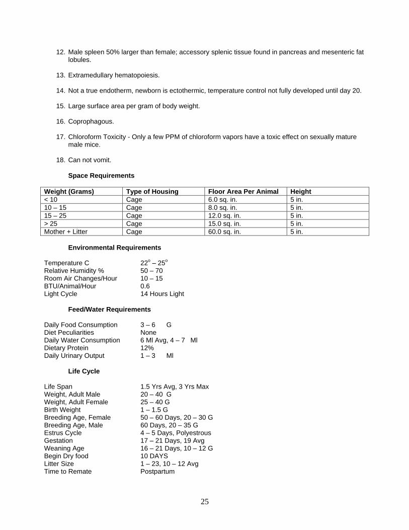
Space Requirements Weight (Grams) Type of Housing Floor Area Per Animal Height < 10 Cage 6.0 sq. in. 5 in. 10 – 15 Cage 8.0 sq. in. 5 in. 15 – 25 Cage 12.0 sq. in. 5 in. > 25 Cage 15.0 sq. in. 5 in. Mother + Litter Cage 60.0 sq. in. 5 in.
Environmental Requirements Temperature C 22o – 25o Relative Humidity % 50 – 70 Room Air Changes/Hour 10 – 15 BTU/Animal/Hour 0.6 Light Cycle 14 Hours Light
Feed/Water Requirements Daily Food Consumption 3 – 6 G Diet Peculiarities None Daily Water Consumption 6 Ml Avg, 4 – 7 Ml Dietary Protein 12% Daily Urinary Output 1 – 3 Ml Life Cycle Life Span 1.5 Yrs Avg, 3 Yrs Max Weight, Adult Male 20 – 40 G Weight, Adult Female 25 – 40 G Birth Weight 1 – 1.5 G Breeding Age, Female 50 – 60 Days, 20 – 30 G Breeding Age, Male 60 Days, 20 – 35 G Estrus Cycle 4 – 5 Days, Polyestrous Gestation 17 – 21 Days, 19 Avg Weaning Age 16 – 21 Days, 10 – 12 G Begin Dry food 10 DAYS Litter Size 1 – 23, 10 – 12 Avg Time to Remate Postpartum
25
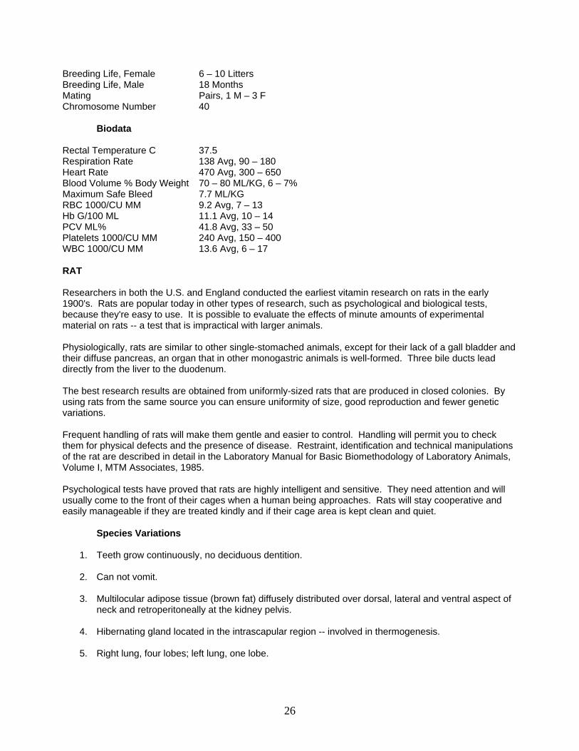
Breeding Life, Female 6 – 10 Litters Breeding Life, Male 18 Months Mating Pairs, 1 M – 3 F Chromosome Number 40 Biodata Rectal Temperature C 37.5 Respiration Rate 138 Avg, 90 – 180 Heart Rate 470 Avg, 300 – 650 Blood Volume % Body Weight 70 – 80 ML/KG, 6 – 7% Maximum Safe Bleed 7.7 ML/KG RBC 1000/CU MM 9.2 Avg, 7 – 13 Hb G/100 ML 11.1 Avg, 10 – 14 PCV ML% 41.8 Avg, 33 – 50 Platelets 1000/CU MM 240 Avg, 150 – 400 WBC 1000/CU MM 13.6 Avg, 6 – 17 RAT Researchers in both the U.S. and England conducted the earliest vitamin research on rats in the early 1900's. Rats are popular today in other types of research, such as psychological and biological tests, because they're easy to use. It is possible to evaluate the effects of minute amounts of experimental material on rats -- a test that is impractical with larger animals. Physiologically, rats are similar to other single-stomached animals, except for their lack of a gall bladder and their diffuse pancreas, an organ that in other monogastric animals is well-formed. Three bile ducts lead directly from the liver to the duodenum. The best research results are obtained from uniformly-sized rats that are produced in closed colonies. By using rats from the same source you can ensure uniformity of size, good reproduction and fewer genetic variations. Frequent handling of rats will make them gentle and easier to control. Handling will permit you to check them for physical defects and the presence of disease. Restraint, identification and technical manipulations of the rat are described in detail in the Laboratory Manual for Basic Biomethodology of Laboratory Animals, Volume I, MTM Associates, 1985. Psychological tests have proved that rats are highly intelligent and sensitive. They need attention and will usually come to the front of their cages when a human being approaches. Rats will stay cooperative and easily manageable if they are treated kindly and if their cage area is kept clean and quiet.
Species Variations
1. Teeth grow continuously, no deciduous dentition.
2. Can not vomit.
3. Multilocular adipose tissue (brown fat) diffusely distributed over dorsal, lateral and ventral aspect of neck and retroperitoneally at the kidney pelvis.
4. Hibernating gland located in the intrascapular region -- involved in thermogenesis.
5. Right lung, four lobes; left lung, one lobe.
26
6. One third of stomach is aglandular (forestomach); glandular stomach has no cardiac glands, is rich in histamine-producing gastric mast cells, pyloric glands restricted to antrum.
7. Does not have a gall bladder, large cecum.
8. Diffuse pancreas.
9. Kidney, unipyramidal, superficial nephrons.
10. Greater number of accessory sex glands than other rodents, os penis.
11. Coprophagous.
Space Requirements
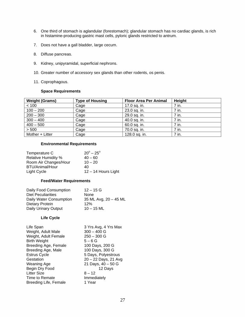
Weight (Grams) Type of Housing Floor Area Per Animal Height < 100 Cage 17.0 sq. in. 7 in. 100 – 200 Cage 23.0 sq. in. 7 in. 200 – 300 Cage 29.0 sq. in. 7 in. 300 – 400 Cage 40.0 sq. in. 7 in. 400 – 500 Cage 60.0 sq. in. 7 in. > 500 Cage 70.0 sq. in. 7 in. Mother + Litter Cage 128.0 sq. in. 7 in.
Environmental Requirements Temperature C 20o – 25o
Relative Humidity % 40 – 60 Room Air Changes/Hour 10 – 20 BTU/Animal/Hour 40 Light Cycle 12 – 14 Hours Light
Feed/Water Requirements Daily Food Consumption 12 – 15 G Diet Peculiarities None Daily Water Consumption 35 ML Avg, 20 – 45 ML Dietary Protein 12% Daily Urinary Output 10 – 15 ML
Life Cycle Life Span 3 Yrs Avg, 4 Yrs Max Weight, Adult Male 300 – 400 G Weight, Adult Female 250 – 300 G Birth Weight 5 – 6 G Breeding Age, Female 100 Days, 200 G Breeding Age, Male 100 Days, 300 G Estrus Cycle 5 Days, Polyestrous Gestation 20 – 22 Days, 21 Avg Weaning Age 21 Days, 40 – 50 G Begin Dry Food 12 Days Litter Size 8 – 12 Time to Remate Immediately Breeding Life, Female 1 Year
27
Breeding Life, Male 1 Year Mating Pairs, 1 M, 3 – 4 F Chromosome Number 42
Biodata Rectal Temperature C 37.5 Respiration Rate 92 Avg, 80 – 150 Heart Rate 350 Avg, 260 – 450 Blood Volume % Body Weight 50 – 65 ML/KG, 6 – 7% Maximum Safe Bleed 5.5 ML/KG RBC 1000/CU MM 8.5 Avg, 6 – 10 Hb G/100 ML 14.2 Avg, 11 – 17 PCV ML% 45.9 Avg, 40 – 50 Platelets 1000/CU MM 330 Avg, 150 – 460 WBC 1000/CU MM 9.8 Avg, 5 – 13 HAMSTER The Golden hamster, sometimes known as the Syrian Golden, is the most popular in U.S. laboratories and is used also in many other countries. The European, or black-bellied hamster, is used almost exclusively on that continent. The third laboratory type, the Chinese hamster, is used less frequently in the U.S. but often in Asia. The Golden hamster's color ranges all the way from albino to dark brown, with a light gray belly. The European hamster has a black belly and brown back. The Chinese strain is sometimes called a striped hamster because of its striped sides. If they are treated kindly and given a comfortable cage, hamsters are docile animals. During the day they often sleep, but at night they are very active and require room in which to exercise. They enjoy playing on exercise wheels. The Golden hamster with 44 chromosomes, has twice as many as either of the other two. Its friendly, gentle disposition and better adaptation to captivity are two of the reasons it is preferred by breeders and researchers. The hamster is prolific and easy to handle, making it useful and manageable in a research environment. Anatomically, the hamster is more like the rat than any other member of the rodent family. Its large cheek pouches and short stubby tail clearly distinguish it from other lab rodents. The pouches are valuable to the researcher because part of their circulatory system is very near the surface, allowing an excellent study of circulation. The laboratory uses of the hamster are similar to those of other rodents. But they are used less often because their reactions to tests are not yet well enough known. The hamster has been shown to have a more specific reaction to certain experiments, for example, those involving the growth of cancerous tumors. Besides cancer research, hamsters are used in diabetes and radiation research, and in vascular physiology studies.
Species Variations
1. Solitary animals.
2. Short gestation period, 15-19 days.
3. Susceptible to dental caries and periodontal disease.
4. Certain strains of Chinese hamster susceptible to diabetes mellitus.
28
5. Chinese hamster, 22 chromosomes; Syrian hamster, 44 chromosomes.
6. Can hibernate for short time periods when temperature is 5 +/- 2 degrees C.
7. Well developed check pouches, eversible; privileged immunologically.
8. Aglandular, forestomach-like diverticulum anterior to the cardiac region of the stomach provides
pregastric fermentation similar to ruminants.
9. Left lung, one lobe; right lung three lobes.
10. Kidney, unipyramidal; urine pH 8, rich in crystalluria.
11. Hamsters like their food on the cage floor.
12. Very radioresistant.
13. Susceptible to many induced infections.
14. Model for pancreatic and renal tumors.
15. Sebaceous glands (territorial marking) located in the costovertebral area, more prominent in male than female.
16. Coprophagous.
Space Requirements
Weight (Grams) Type of Housing Floor Area Per Animal Height < 60 Cage 10 sq. in. 6 in. 60 – 80 Cage 13.0 sq. in. 6 in. 80 – 100 Cage 16.0 sq. in. 6 in. > 100 Cage 19.0 sq. in. 6 in. Mother + Litter Cage 121.0 sq. in. 6 in.
Environmental Requirements Temperature C 21o – 24o Relative Humidity % 45 – 65 Room Air Changes/Hour 10 – 15 BTU/Animal/Hour 2.5 Light Cycle 12 Hours Light
Feed/Water Requirements Daily Food Consumption 7 – 15 G Diet Peculiarities Food On Floor Of Cage Daily Water Consumption 8 – 12 ML Dietary Protein 16% Daily Urinary Output 6 – 12 ML
Life Cycle Life Span 1 Yr Avg, 2 Yrs Max
29
Weight, Adult Male 85 – 110 G Weight, Adult Female 95 – 120 G Birth Weight 2 G Breeding Age, Female 2 Months, 95 – 120 G Breeding Age, Male 2 Months, 85 – 110 G Estrus Cycle 3 – 4 Days, Polyestrous Gestation 15 – 19 Days, 16 Avg Weaning Age 21 Days, 35 G Begin Dry Food 7 – 9 Days Litter Size 4 – 12, 6 – 8 Avg Time to Remate 4 Days Breeding Life, Female 1 Year Breeding Life, Male 1 Year Mating Pairs, 1 M, 3 – 4 F Chromosome Number 44
Biodata Rectal Temperature C 39 Respiration Rate 77 Avg, 40 – 120 Heart Rate 332 Avg, 286 – 400 Blood Volume % Body Weight 65 – 80 ML/KG, 6 – 9% Maximum Safe Bleed 5.5 ML/KG RBC 1000/CU MM 7.2 Avg, 4 – 10 Hb G/100 ML 16.4 Avg, 13 – 19 PCV ML% 50.8 Avg, 39 – 59 Platelets 1000/CU MM 386 Avg, 300 – 570 WBC 1000/CU MM 8.1 Avg, 5 – 11 GUINEA PIG Guinea pigs are highly valuable research animals because of the variety of experiments in which they can be used. Their skin so resembles the skin of human beings that guinea pigs are often used in dermatologic research. Long use of the guinea pig in bacteriological and serological research has resulted in a sizeable accumulation of baseline data on blood values, cell counts, elementary analysis and physical constants. Guinea pigs have been used frequently in nutritional research, immunology experiments and otology. Guinea pigs became known to research scientists after sailors brought them to England from Peru as pets. The Dunkin-Hartley strain, developed by the English, is the most widely used in research. It's almost always an albino with short, smooth hair. A second guinea pig type, the Abyssinian, has short rough hair that grows in rosettes. The present day Peruvian type of guinea pig, with long hair, is seldom used in research. Of all the commonly used lab animals, guinea pigs are one of the most nervous and high strung species. The guinea pig needs to be approached quietly and confidently. It will seldom scratch when picked up, provided it feels well-supported. If the guinea pig does become alarmed, however, the whole colony can detect that fright and become apprehensive. Restraint, identification and technical manipulations of the guinea pig are described in detail in the Laboratory Manual for Basic Biomethodology of Laboratory Animals, Volume I, MTM Associates, 1985. Good sanitation is extremely important in the guinea pig colony. Guinea pigs are very susceptible to natural and induced infections. Cages need to be cleaned often to keep the animals as free of disease. Like monkeys and human beings, guinea pigs need daily doses of vitamin C. Without it, scurvy can develop within seven to ten days. Normally a daily intake of 10 mg. of ascorbic acid will be adequate to keep guinea pigs free of scurvy.
30
Species Variations
1. Hystricomorph (porcupine-like) suborder of rodents.
2. Vitamin C required.
3. "Barbering".
4. Long gestation, 63 days average.
5. Pregnancy toxemia.
6. Constantly erupting hysodont teeth.
7. Subject to gastric ulcers.
8. Fur consists entirely of guard hairs.
9. May respond unfavorably to antibiotics or stress.
10. Smooth muscle contractions of the bronchial tree due to histamine may be severe or fatal.
11. Perivascular lymphoid nodules in the lungs.
12. Right lung, four lobes; left lung, three lobes.
13. Large pancreas.
14. Cervically located thymus, easy to remove surgically. 15. Cellular membrane closes over vaginal orifice, except at estrus and parturition.
16. Two nipples and mammary glands located in inguinal region.
17. Mature male, large vesicular glands.
18. Relatively resistant to steroids.
19. Coprophagous.
Space Requirements
Weight (Grams) Type of Housing Floor Area Per Animal Height <350 Cage 60.0 sq. in. 7 in. >350 Cage 101.0 sq. in. 7 in. Mother + Litter Cage Single Cage 7 in.
Environmental Requirements Temperature C 16o – 20o Relative Humidity % 50 – 60 Room Air Changes/Hour 10 – 15 BTU/Animal/Hour 5 – 6 Light Cycle 12 – 15 Light
31
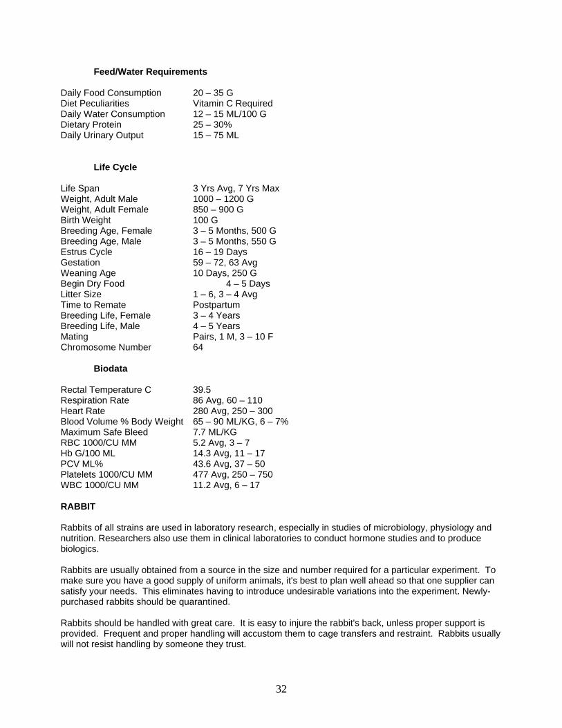
Feed/Water Requirements Daily Food Consumption 20 – 35 G Diet Peculiarities Vitamin C Required Daily Water Consumption 12 – 15 ML/100 G Dietary Protein 25 – 30% Daily Urinary Output 15 – 75 ML
Life Cycle Life Span 3 Yrs Avg, 7 Yrs Max Weight, Adult Male 1000 – 1200 G Weight, Adult Female 850 – 900 G Birth Weight 100 G Breeding Age, Female 3 – 5 Months, 500 G Breeding Age, Male 3 – 5 Months, 550 G Estrus Cycle 16 – 19 Days Gestation 59 – 72, 63 Avg Weaning Age 10 Days, 250 G Begin Dry Food 4 – 5 Days Litter Size 1 – 6, 3 – 4 Avg Time to Remate Postpartum Breeding Life, Female 3 – 4 Years Breeding Life, Male 4 – 5 Years Mating Pairs, 1 M, 3 – 10 F Chromosome Number 64
Biodata Rectal Temperature C 39.5 Respiration Rate 86 Avg, 60 – 110 Heart Rate 280 Avg, 250 – 300 Blood Volume % Body Weight 65 – 90 ML/KG, 6 – 7% Maximum Safe Bleed 7.7 ML/KG RBC 1000/CU MM 5.2 Avg, 3 – 7 Hb G/100 ML 14.3 Avg, 11 – 17 PCV ML% 43.6 Avg, 37 – 50 Platelets 1000/CU MM 477 Avg, 250 – 750 WBC 1000/CU MM 11.2 Avg, 6 – 17 RABBIT Rabbits of all strains are used in laboratory research, especially in studies of microbiology, physiology and nutrition. Researchers also use them in clinical laboratories to conduct hormone studies and to produce biologics. Rabbits are usually obtained from a source in the size and number required for a particular experiment. To make sure you have a good supply of uniform animals, it's best to plan well ahead so that one supplier can satisfy your needs. This eliminates having to introduce undesirable variations into the experiment. Newly-purchased rabbits should be quarantined. Rabbits should be handled with great care. It is easy to injure the rabbit's back, unless proper support is provided. Frequent and proper handling will accustom them to cage transfers and restraint. Rabbits usually will not resist handling by someone they trust.
32
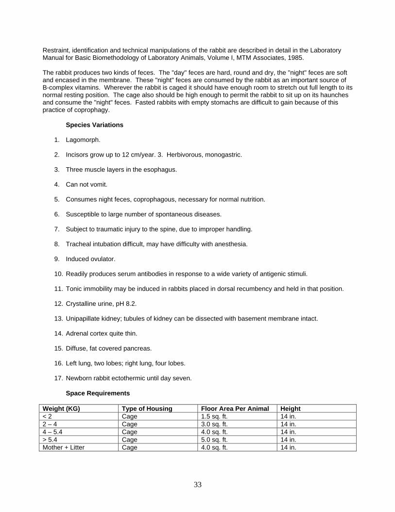
Restraint, identification and technical manipulations of the rabbit are described in detail in the Laboratory Manual for Basic Biomethodology of Laboratory Animals, Volume I, MTM Associates, 1985. The rabbit produces two kinds of feces. The "day" feces are hard, round and dry, the "night" feces are soft and encased in the membrane. These "night" feces are consumed by the rabbit as an important source of B-complex vitamins. Wherever the rabbit is caged it should have enough room to stretch out full length to its normal resting position. The cage also should be high enough to permit the rabbit to sit up on its haunches and consume the "night" feces. Fasted rabbits with empty stomachs are difficult to gain because of this practice of coprophagy.
Species Variations
1. Lagomorph.
2. Incisors grow up to 12 cm/year. 3. Herbivorous, monogastric.
3. Three muscle layers in the esophagus.
4. Can not vomit.
5. Consumes night feces, coprophagous, necessary for normal nutrition.
6. Susceptible to large number of spontaneous diseases.
7. Subject to traumatic injury to the spine, due to improper handling.
8. Tracheal intubation difficult, may have difficulty with anesthesia.
9. Induced ovulator.
10. Readily produces serum antibodies in response to a wide variety of antigenic stimuli.
11. Tonic immobility may be induced in rabbits placed in dorsal recumbency and held in that position.
12. Crystalline urine, pH 8.2.
13. Unipapillate kidney; tubules of kidney can be dissected with basement membrane intact.
14. Adrenal cortex quite thin.
15. Diffuse, fat covered pancreas.
16. Left lung, two lobes; right lung, four lobes.
17. Newborn rabbit ectothermic until day seven.
Space Requirements Weight (KG) Type of Housing Floor Area Per Animal Height < 2 Cage 1.5 sq. ft. 14 in. 2 – 4 Cage 3.0 sq. ft. 14 in. 4 – 5.4 Cage 4.0 sq. ft. 14 in. > 5.4 Cage 5.0 sq. ft. 14 in. Mother + Litter Cage 4.0 sq. ft. 14 in.
33
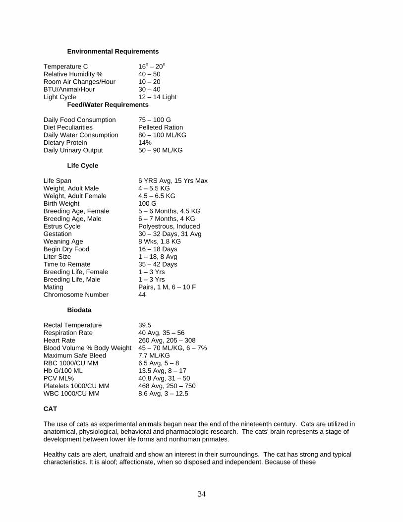
Environmental Requirements
Temperature C 16o – 20o Relative Humidity % 40 – 50 Room Air Changes/Hour 10 – 20 BTU/Animal/Hour 30 – 40 Light Cycle 12 – 14 Light
Feed/Water Requirements Daily Food Consumption 75 – 100 G Diet Peculiarities Pelleted Ration Daily Water Consumption 80 – 100 ML/KG Dietary Protein 14% Daily Urinary Output 50 – 90 ML/KG
Life Cycle Life Span 6 YRS Avg, 15 Yrs Max Weight, Adult Male 4 – 5.5 KG Weight, Adult Female 4.5 – 6.5 KG Birth Weight 100 G Breeding Age, Female 5 – 6 Months, 4.5 KG Breeding Age, Male 6 – 7 Months, 4 KG Estrus Cycle Polyestrous, Induced Gestation 30 – 32 Days, 31 Avg Weaning Age 8 Wks, 1.8 KG Begin Dry Food 16 – 18 Days Liter Size 1 – 18, 8 Avg Time to Remate 35 – 42 Days Breeding Life, Female 1 – 3 Yrs Breeding Life, Male 1 – 3 Yrs Mating Pairs, 1 M, 6 – 10 F Chromosome Number 44
Biodata Rectal Temperature 39.5 Respiration Rate 40 Avg, 35 – 56 Heart Rate 260 Avg, 205 – 308 Blood Volume % Body Weight 45 – 70 ML/KG, 6 – 7% Maximum Safe Bleed 7.7 ML/KG RBC 1000/CU MM 6.5 Avg, 5 – 8 Hb G/100 ML 13.5 Avg, 8 – 17 PCV ML% 40.8 Avg, 31 – 50 Platelets 1000/CU MM 468 Avg, 250 – 750 WBC 1000/CU MM 8.6 Avg, 3 – 12.5 CAT The use of cats as experimental animals began near the end of the nineteenth century. Cats are utilized in anatomical, physiological, behavioral and pharmacologic research. The cats' brain represents a stage of development between lower life forms and nonhuman primates. Healthy cats are alert, unafraid and show an interest in their surroundings. The cat has strong and typical characteristics. It is aloof; affectionate, when so disposed and independent. Because of these
34
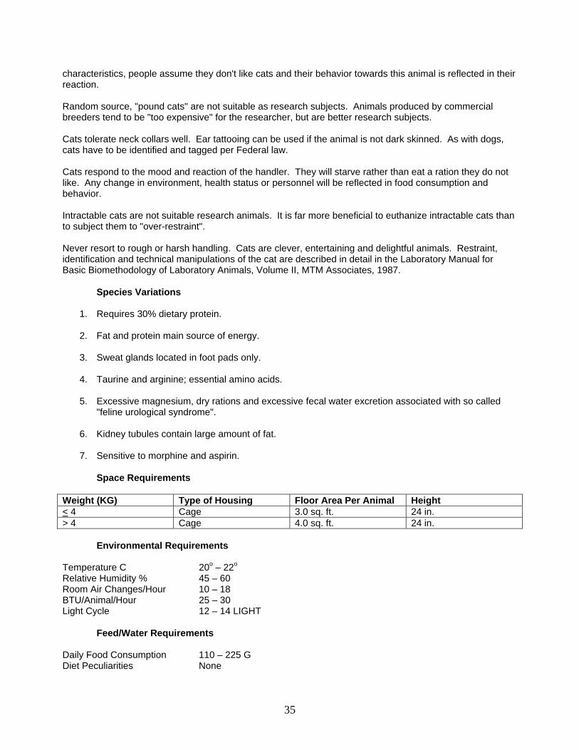
characteristics, people assume they don't like cats and their behavior towards this animal is reflected in their reaction. Random source, "pound cats" are not suitable as research subjects. Animals produced by commercial breeders tend to be "too expensive" for the researcher, but are better research subjects. Cats tolerate neck collars well. Ear tattooing can be used if the animal is not dark skinned. As with dogs, cats have to be identified and tagged per Federal law. Cats respond to the mood and reaction of the handler. They will starve rather than eat a ration they do not like. Any change in environment, health status or personnel will be reflected in food consumption and behavior. Intractable cats are not suitable research animals. It is far more beneficial to euthanize intractable cats than to subject them to "over-restraint". Never resort to rough or harsh handling. Cats are clever, entertaining and delightful animals. Restraint, identification and technical manipulations of the cat are described in detail in the Laboratory Manual for Basic Biomethodology of Laboratory Animals, Volume II, MTM Associates, 1987.
Species Variations
1. Requires 30% dietary protein.
2. Fat and protein main source of energy.
3. Sweat glands located in foot pads only.
4. Taurine and arginine; essential amino acids.
5. Excessive magnesium, dry rations and excessive fecal water excretion associated with so called "feline urological syndrome".
6. Kidney tubules contain large amount of fat.
7. Sensitive to morphine and aspirin.
Space Requirements
Weight (KG) Type of Housing Floor Area Per Animal Height < 4 Cage 3.0 sq. ft. 24 in. > 4 Cage 4.0 sq. ft. 24 in.
Environmental Requirements Temperature C 20o – 22o Relative Humidity % 45 – 60 Room Air Changes/Hour 10 – 18 BTU/Animal/Hour 25 – 30 Light Cycle 12 – 14 LIGHT
Feed/Water Requirements Daily Food Consumption 110 – 225 G Diet Peculiarities None
35
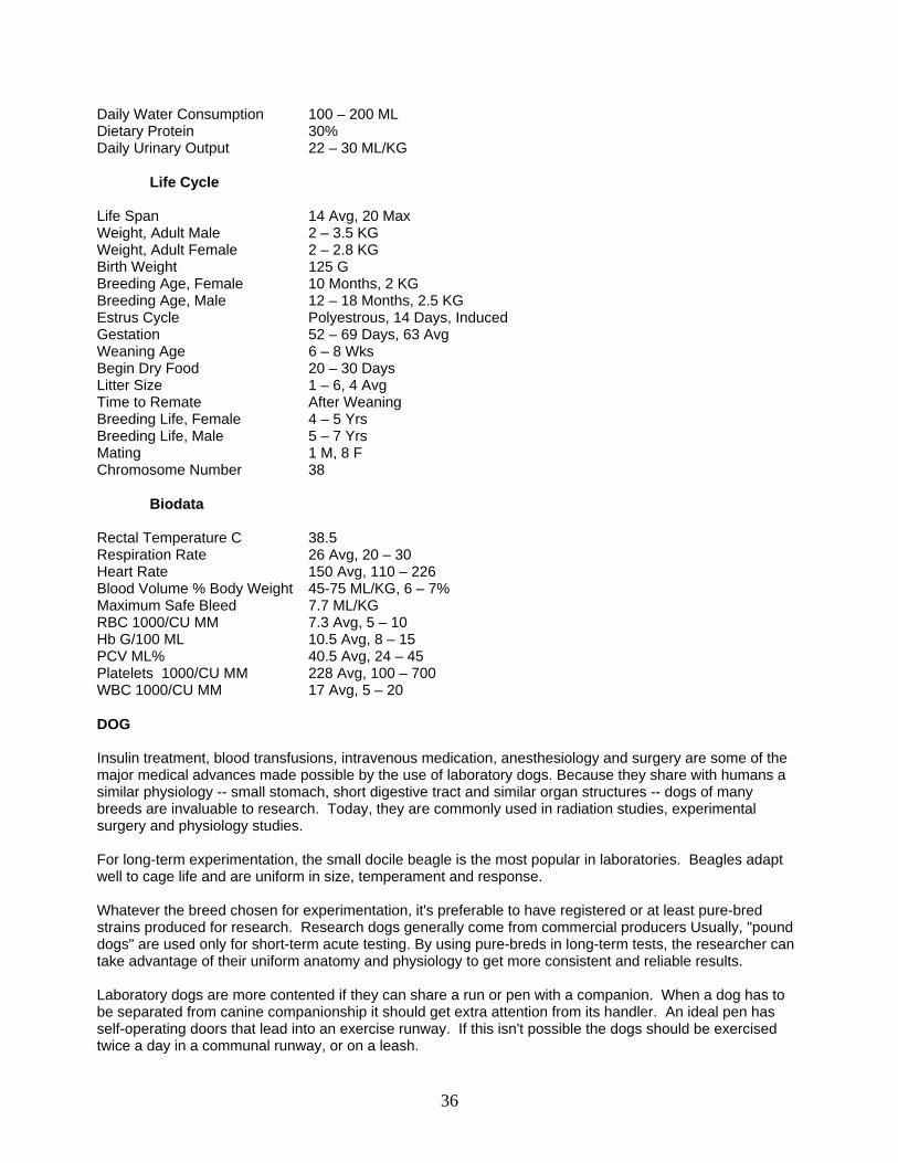
Daily Water Consumption 100 – 200 ML Dietary Protein 30% Daily Urinary Output 22 – 30 ML/KG
Life Cycle Life Span 14 Avg, 20 Max Weight, Adult Male 2 – 3.5 KG Weight, Adult Female 2 – 2.8 KG Birth Weight 125 G Breeding Age, Female 10 Months, 2 KG Breeding Age, Male 12 – 18 Months, 2.5 KG Estrus Cycle Polyestrous, 14 Days, Induced Gestation 52 – 69 Days, 63 Avg Weaning Age 6 – 8 Wks Begin Dry Food 20 – 30 Days Litter Size 1 – 6, 4 Avg Time to Remate After Weaning Breeding Life, Female 4 – 5 Yrs Breeding Life, Male 5 – 7 Yrs Mating 1 M, 8 F Chromosome Number 38
Biodata Rectal Temperature C 38.5 Respiration Rate 26 Avg, 20 – 30 Heart Rate 150 Avg, 110 – 226 Blood Volume % Body Weight 45-75 ML/KG, 6 – 7% Maximum Safe Bleed 7.7 ML/KG RBC 1000/CU MM 7.3 Avg, 5 – 10 Hb G/100 ML 10.5 Avg, 8 – 15 PCV ML% 40.5 Avg, 24 – 45 Platelets 1000/CU MM 228 Avg, 100 – 700 WBC 1000/CU MM 17 Avg, 5 – 20 DOG Insulin treatment, blood transfusions, intravenous medication, anesthesiology and surgery are some of the major medical advances made possible by the use of laboratory dogs. Because they share with humans a similar physiology -- small stomach, short digestive tract and similar organ structures -- dogs of many breeds are invaluable to research. Today, they are commonly used in radiation studies, experimental surgery and physiology studies. For long-term experimentation, the small docile beagle is the most popular in laboratories. Beagles adapt well to cage life and are uniform in size, temperament and response. Whatever the breed chosen for experimentation, it's preferable to have registered or at least pure-bred strains produced for research. Research dogs generally come from commercial producers Usually, "pound dogs" are used only for short-term acute testing. By using pure-breds in long-term tests, the researcher can take advantage of their uniform anatomy and physiology to get more consistent and reliable results. Laboratory dogs are more contented if they can share a run or pen with a companion. When a dog has to be separated from canine companionship it should get extra attention from its handler. An ideal pen has self-operating doors that lead into an exercise runway. If this isn't possible the dogs should be exercised twice a day in a communal runway, or on a leash.
36
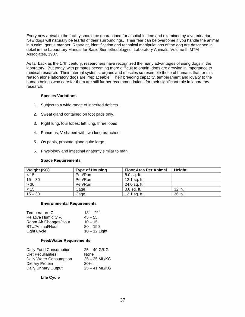
Every new arrival to the facility should be quarantined for a suitable time and examined by a veterinarian. New dogs will naturally be fearful of their surroundings. Their fear can be overcome if you handle the animal in a calm, gentle manner. Restraint, identification and technical manipulations of the dog are described in detail in the Laboratory Manual for Basic Biomethodology of Laboratory Animals, Volume II, MTM Associates, 1987. As far back as the 17th century, researchers have recognized the many advantages of using dogs in the laboratory. But today, with primates becoming more difficult to obtain, dogs are growing in importance to medical research. Their internal systems, organs and muscles so resemble those of humans that for this reason alone laboratory dogs are irreplaceable. Their breeding capacity, temperament and loyalty to the human beings who care for them are still further recommendations for their significant role in laboratory research.
Species Variations
1. Subject to a wide range of inherited defects.
2. Sweat gland contained on foot pads only.
3. Right lung, four lobes; left lung, three lobes
4. Pancreas, V-shaped with two long branches
5. Os penis, prostate gland quite large.
6. Physiology and intestinal anatomy similar to man.
Space Requirements Weight (KG) Type of Housing Floor Area Per Animal Height < 15 Pen/Run 8.0 sq. ft. 15 – 30 Pen/Run 12.1 sq. ft. > 30 Pen/Run 24.0 sq. ft. < 15 Cage 8.0 sq. ft. 32 in. 15 – 30 Cage 12.1 sq. ft. 36 in.
Environmental Requirements Temperature C 18o – 21o Relative Humidity % 45 – 55 Room Air Changes/Hour 10 – 15 BTU/Animal/Hour 80 – 150 Light Cycle 10 – 12 Light
Feed/Water Requirements Daily Food Consumption 25 – 40 G/KG Diet Peculiarities None Daily Water Consumption 25 – 35 ML/KG Dietary Protein 20% Daily Urinary Output 25 – 41 ML/KG
Life Cycle
37
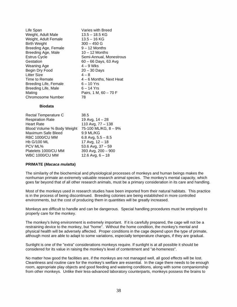
Life Span Varies with Breed Weight, Adult Male 13.5 – 18.5 KG Weight, Adult Female 13.5 – 16 KG Birth Weight 300 – 450 G Breeding Age, Female 9 – 12 Months Breeding Age, Male 10 – 12 Months Estrus Cycle Semi-Annual, Monestrous Gestation 60 – 66 Days, 63 Avg Weaning Age 4 – 9 Wks Begin Dry Food 20 – 30 Days Litter Size 4 – 8 Time to Remate 4 – 6 Months, Next Heat Breeding Life, Female 6 – 10 Yrs Breeding Life, Male 6 – 14 Yrs Mating Pairs, 1 M, 60 – 70 F Chromosome Number 78
Biodata Rectal Temperature C 38.5 Respiration Rate 19 Avg, 14 – 28 Heart Rate 110 Avg, 77 – 138 Blood Volume % Body Weight 75-100 ML/KG, 8 – 9% Maximum Safe Bleed 9.9 ML/KG RBC 1000/CU MM 6.8 Avg, 5.5 – 8.5 Hb G/100 ML 17 Avg, 12 – 18 PCV ML% 53.6 Avg, 37 – 59 Platelets 1000/CU MM 393 Avg, 200 – 900 WBC 1000/CU MM 12.6 Avg, 6 – 18 PRIMATE (Macaca mulatta) The similarity of the biochemical and physiological processes of monkeys and human beings makes the nonhuman primate an extremely valuable research animal species. The monkey's mental capacity, which goes far beyond that of all other research animals, must be a primary consideration in its care and handling. Most of the monkeys used in research studies have been imported from their natural habitats. This practice is in the process of being discontinued. Breeding colonies are being established in more controlled environments, but the cost of producing them in quantities will be greatly increased. Monkeys are difficult to handle and can be dangerous. Special handling procedures must be employed to properly care for the monkey. The monkey's living environment is extremely important. If it is carefully prepared, the cage will not be a restraining device to the monkey, but "home". Without the home condition, the monkey's mental and physical health will be adversely affected. Proper conditions in the cage depend upon the type of primate, although most are able to adapt to some variations, especially temperature changes, if they are gradual. Sunlight is one of the "extra" considerations monkeys require. If sunlight is at all possible it should be considered for its value in raising the monkey's level of contentment and "at-homeness". No matter how good the facilities are, if the monkeys are not managed well, all good effects will be lost. Cleanliness and routine care for the monkey's welfare are essential. In the cage there needs to be enough room, appropriate play objects and good feeding and watering conditions, along with some companionship from other monkeys. Unlike their less-advanced laboratory counterparts, monkeys possess the brains to
38
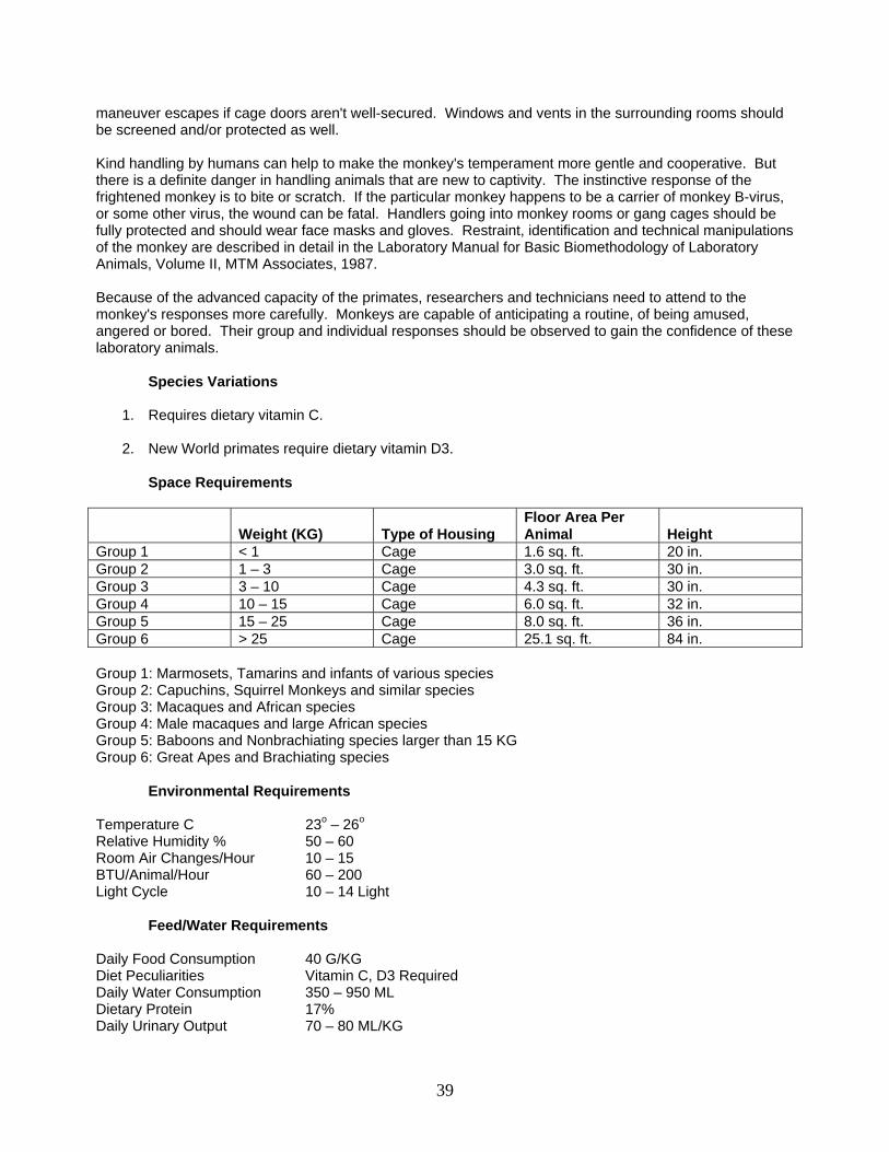
maneuver escapes if cage doors aren't well-secured. Windows and vents in the surrounding rooms should be screened and/or protected as well. Kind handling by humans can help to make the monkey's temperament more gentle and cooperative. But there is a definite danger in handling animals that are new to captivity. The instinctive response of the frightened monkey is to bite or scratch. If the particular monkey happens to be a carrier of monkey B-virus, or some other virus, the wound can be fatal. Handlers going into monkey rooms or gang cages should be fully protected and should wear face masks and gloves. Restraint, identification and technical manipulations of the monkey are described in detail in the Laboratory Manual for Basic Biomethodology of Laboratory Animals, Volume II, MTM Associates, 1987. Because of the advanced capacity of the primates, researchers and technicians need to attend to the monkey's responses more carefully. Monkeys are capable of anticipating a routine, of being amused, angered or bored. Their group and individual responses should be observed to gain the confidence of these laboratory animals.
Species Variations
1. Requires dietary vitamin C.
2. New World primates require dietary vitamin D3.
Space Requirements
Weight (KG) Type of Housing
Floor Area Per Animal
Height
Group 1 < 1 Cage 1.6 sq. ft. 20 in. Group 2 1 – 3 Cage 3.0 sq. ft. 30 in. Group 3 3 – 10 Cage 4.3 sq. ft. 30 in. Group 4 10 – 15 Cage 6.0 sq. ft. 32 in. Group 5 15 – 25 Cage 8.0 sq. ft. 36 in. Group 6 > 25 Cage 25.1 sq. ft. 84 in. Group 1: Marmosets, Tamarins and infants of various species Group 2: Capuchins, Squirrel Monkeys and similar species Group 3: Macaques and African species Group 4: Male macaques and large African species Group 5: Baboons and Nonbrachiating species larger than 15 KG Group 6: Great Apes and Brachiating species
Environmental Requirements Temperature C 23o – 26o Relative Humidity % 50 – 60 Room Air Changes/Hour 10 – 15 BTU/Animal/Hour 60 – 200 Light Cycle 10 – 14 Light
Feed/Water Requirements Daily Food Consumption 40 G/KG Diet Peculiarities Vitamin C, D3 Required Daily Water Consumption 350 – 950 ML Dietary Protein 17% Daily Urinary Output 70 – 80 ML/KG
39

Life Cycle
Life Span 16 Yrs Avg, 30 Max Weight, Adult Male 12 KG Weight, Adult Female 10 KG Birth Weight 500 – 700 G Breeding Age, Female 4 – 5 Yrs Breeding Age, Male 3 – 6 Yrs Estrus cycle 27 – 32 Days Gestation 168 Days Avg Weaning Age 3-6 Months Begin Dry Food 20 – 30 Days Litter Size 1 Time to Remate After Weaning Breeding Life, Female 12 – 15 Yrs Breeding Life, Male 12 – 15 Yrs Mating Pairs, 1 M, 10 F Chromosome Number 42
Biodata Rectal Temperature C 39o
Respiration Rate 40 Avg, 30 – 54 Heart Rate 220 Avg, 165 – 243 Blood Volume % Body Weight 55 – 75 ML/KG, 6 – 7% Maximum Safe Bleed 6.6 ML/KG RBC 1000/CU MM 5 Avg, 3.6 – 7 Hb G/100 ML 12.9 Avg, 8.8 – 16.5 PCV ML% 37 Avg, 35 – 43 Platelets 1000/CU MM 359 Avg, 109 – 597 WBC 1000/CU MM 8 Avg, 2.5 – 27
40
BASIC NUTRITION INTRODUCTION
Major portions of this section were provided by Ralston Purina Company, Lab Chows Division. Laboratory animal nutrition is far too complex to deal with comprehensively. Instead, this section offers a basic overview of the role proper nutrition has in the maintenance of the biological processes. More is known about the nutrition of laboratory animals than human nutrition. Most if not all laboratory animals are adapted to the omnivorous state, consume a totally balanced diet and "eat" better than most humans. In practice, most laboratory animals are fed ad libitum - without restraint; available on a free choice basis. The animal will regulate its food intake to meet its requirements for energy. The care and feeding recommendations and other data presented are based upon current animal nutrition and practical management. The information on these charts is a compilation and is intended to serve as a guideline only. Specific data on individual species, strains and stocks of animals can be found in the current literature. FEEDING CONSIDERATIONS Type of Diet - each species has slightly different requirements. Demand - Most animals fed ad lib, but there may be special dietary studies. Balanced Diet - diets are standardized and balanced. Diet Supplements - usually not needed. WATER Regular amounts of clean, pure water are an absolute must if life is to be sustained. Water makes up more than one-half the total composition of the mature animal's body and is involved in almost every metabolic process. Blood, the medium by which nutrients are carried to various parts of the body, is approximately 80% water. Water serves as an efficient solvent in cells, where the major biochemical processes of digestion, assimilation, metabolism and respiration occur. Water is necessary for the regulation of body temperature. And, finally, water is valuable in the elimination of waste from the body. Without proper water intake, tissues dehydrate and digestion, along with other metabolic processes, is severely altered. The body's cells, which derive their oxygen from the blood stream, are starved as the blood becomes concentrated. If only one-tenth of the body's water is lost, death will result. ENERGY The most common sources of energy for laboratory animals are fats and carbohydrates. Energy also is derived, to a lesser extent, from protein. The unit of energy is expressed in terms of heat units (calories). A "small calorie" is defined as the amount of heat required to raise 1 gram of water 1 degree C. The "large calorie" or "KCal" is the unit of energy commonly used in expressing the energy content of foods. There are four ways of specifying energy: Gross energy is the energy of complete combustion measured as heat when a material is burned to its oxidation products; digestible energy is equal to the gross energy minus the energy remaining in the fecal matter; metabolizable energy equals the gross energy minus the energy lost in the fecal material and that lost via the urine and combustible gases; and net energy is the energy remaining for production uses after deducting from the gross energy those energies lost in the feces,
41
urine, combustible gases and body heat losses. When nourishment is withheld from the body, energy is derived from the carbohydrate, glycogen, in the liver; fat stores and protein in the body tissues. The largest function of a food is to supply energy for body processes and to form nonnitrogenous, organic matter of tissues and secretions. Without adequate energy other important organic nutrients are not used for the normal needs, such as tissue maintenance, growth processes, reproduction and lactation as well as for work and heat. Without sufficient energy, the young animal's body cannot grow. Because of the additional energy requirements of the young animal and the breeding female, they obviously need more energy producing nutrients than other animals. CARBOHYDRATES Carbohydrates normally serve as the most important source of bodily energy. The utilizable ones are found mostly as sugars and starches. In animal feed, corn is an often used ingredient because it is rich in carbohydrates. Crude fiber, or roughage, also is considered as a source of carbohydrates. But its precise usefulness to an animal depends upon the particular food the fiber comes from and how well the animal is able to digest the fiber. Ruminant animals have microorganisms to aid in fiber digestion; monogastric animals are less efficient in digesting fiber but need fiber for other purposes. FATS Fats are a concentrated form of energy. Per unit of weight, they contain 2-1/4 times as much energy as the equivalent weight of either carbohydrates or proteins. They also supply a source of essential fatty acids, the absence of which causes metabolic disorders. Normally 1 to 2% fat will supply the necessary amount of essential fatty acids. The remainder is used as an energy source. Since fat is a concentrated source of energy is used as a convenient ingredient to increase the dietary energy. Fat tends to make feeds and some food more palatable to animals and people. When high fat diets are used, other nutrients need to be properly adjusted to ensure that, as the animal is satisfied with smaller amounts of feed, it is still getting the proper balance of essential nutrients. Antioxidants are now used to help prevent rancidity. If rancidity is permitted in fats, there is a more rapid destruction of some fat soluble vitamins, usually a decreased palatability and frequently the appearance of unpleasant odors. PROTEINS The protein, or amino acids, an animal takes in provides the nitrogen required for the body's growth, tissue maintenance, reproduction and lactation. When a young animal gets too little protein, its growth will be limited and its energy intake will be thwarted. Protein-calorie malnutrition can result. Protein intake requirements vary with the kind of animal and performance expected of it. Adult animals have a lower protein requirement than do young ones. Approximately 10 amino acids are considered essential for the growing animal. An essential amino acid can be defined as one which cannot be synthesized at a sufficiently rapid rate to permit optimum growth of the young animal. Sources of protein vary in their nutrient value, depending upon the content and availability of the essential amino acids. Vegetable seed proteins contain more essential amino acids than cereal grains or by-products. Among them, soybean meal is perhaps the best source of amino acids and is also a major source of protein for non-ruminants. Animal proteins derived from fish meal, meat and bone meal, and dried milk products, tend to be excellent sources of the essential amino acids. If processed properly these sources have a high level of amino acids
42
and are very useful as supplementary proteins to complement the amino acid balance of cereal grain proteins. FIBER Fiber has received a great deal of research attention among animal scientists because of its importance to the ruminant. In the ruminant, it represents the plant cell wall which utilized as an energy source by the rumen micoflora, and is extensively degraded. In the monogastric animal, fiber represents the insoluble matter of plant cell walls which is indigestible by animal enzymes, but can be partially degraded by gastrointestinal microflora. Fiber is actually very complex. It is a combination of at least four major components which are distinctly different in chemical composition. These four major components are cellulose, hemicellulose, lignin and pectin. Because of the complexity of defining and measuring fiber, any procedure for its measurement must strike a compromise between a complete, fractionated measurement of all the various species, and a simplified system involving grouping in different compounds, (cellulose, hemicellulose, lignin and pectin). To say that a ration has "X" amount of fiber makes no sense without some understanding of the ingredients used and/or a knowledge of the amounts of each of the major fiber components that are present. The constituents of fiber affect the gastrointestinal tract differently, ultimately affecting the nutrition of the animal. Some fibers have a high water-holding capacity, which affects the speed at which the diet passes through the intestinal tract (transit time). Other fiber constituents have extensive cation-exchange capacity, which can tie up dietary minerals. Current research indicates that various fibers may have these physiological effects:
1. A decrease in the absorption of minerals; 2. The binding of bile acids which are integral to cholesterol homeostasis and fat absorption; 3. A change in the potency of intestinal toxins and carcinogens; 4. The production of volatile fatty acids which are used for energy or for inhibiting pathogens; 5. Changes in transit time; 6. Alterations in gastrointestinal bacteria.
The changes produced by fiber in these and other body functions have been implicated in color cancer, diverticular disease, diabetes, atherosclerosis, coronary artery disease and hemorrhoids. VITAMINS Chemical compounds known as vitamins are necessary, in small amounts, for maintenance, growth, reproduction and lactation. As components of certain enzymes, vitamins are essential to maintaining life processes. Vitamins A, D, E and K are fat soluble vitamins. They can be stored in the liver or in other organs to provide the needs of the animal. A reasonable daily intake is recommended, however. The water soluble vitamins: the B-complex group and vitamin C can be stored only in very limited quantities and therefore need to be a regular part of the daily diet. Monkeys and guinea pigs are two species of animals, besides human beings, that need regular doses of vitamin C. MINERALS As many as 20 different minerals may be required in some degree for optimum bodily functioning, some in relatively large amounts and others only as trace elements. The mineral element required in largest amount is calcium. The 2% of calcium found in the body is present primarily in the bones and teeth. Phosphorus, closely associated with calcium in bone development, makes up about 1% of the body's composition.
43
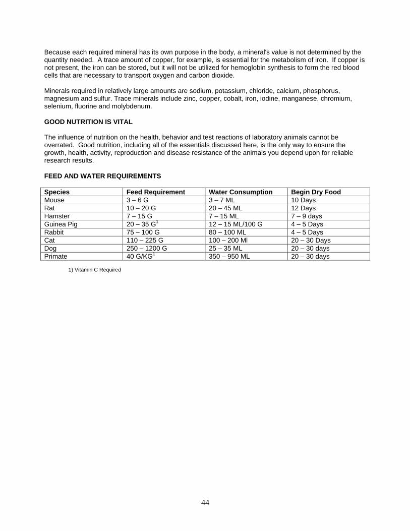
Because each required mineral has its own purpose in the body, a mineral's value is not determined by the quantity needed. A trace amount of copper, for example, is essential for the metabolism of iron. If copper is not present, the iron can be stored, but it will not be utilized for hemoglobin synthesis to form the red blood cells that are necessary to transport oxygen and carbon dioxide. Minerals required in relatively large amounts are sodium, potassium, chloride, calcium, phosphorus, magnesium and sulfur. Trace minerals include zinc, copper, cobalt, iron, iodine, manganese, chromium, selenium, fluorine and molybdenum. GOOD NUTRITION IS VITAL The influence of nutrition on the health, behavior and test reactions of laboratory animals cannot be overrated. Good nutrition, including all of the essentials discussed here, is the only way to ensure the growth, health, activity, reproduction and disease resistance of the animals you depend upon for reliable research results. FEED AND WATER REQUIREMENTS Species Feed Requirement Water Consumption Begin Dry Food Mouse 3 – 6 G 3 – 7 ML 10 Days Rat 10 – 20 G 20 – 45 ML 12 Days Hamster 7 – 15 G 7 – 15 ML 7 – 9 days Guinea Pig 20 – 35 G1 12 – 15 ML/100 G 4 – 5 Days Rabbit 75 – 100 G 80 – 100 ML 4 – 5 Days Cat 110 – 225 G 100 – 200 Ml 20 – 30 Days Dog 250 – 1200 G 25 – 35 ML 20 – 30 days Primate 40 G/KG1 350 – 950 ML 20 – 30 days
1) Vitamin C Required
44
ANIMAL DISEASE: CONTROL AND RECOGNITION VETERINARY CARE Adequate veterinary care for disease control, recognition and prevention consists of:
• Observing all animals daily to assess their health and welfare. • Using appropriate methods to prevent, control, diagnosis, and treat diseases and injuries.
ANIMAL QUALITY VS FACILITIES Select animals on the basis of what your needs are for the research project. The quality of the animal needs to be matched to the ability of the animal facility to house the animals and provide appropriate care. Do not expect to house "clean" animals in the same facility as "dirty" animals without special equipment and care. PRINCIPLES OF DISEASE CONTROL
Transmission Diseases are transmitted by the following routes:
• Vector - a living carrier that transfers an infective agent from one host to another. • Fomite - an inanimate object that is not in itself harmful, but is able to harbor pathogenic
microorganisms and thus may serve as an agent of transmission of an infection (i.e., gloves, needles, equipment etc.).
• Genes - congenital abnormalities or mutation passed to the offspring via DNA. • Zoonoses are infections that are transmitted between animals and man. Animals must be
considered as potential sources of zoonoses but the risk involved will vary depending on the class and species of animal. In general, the more closely a species is related to man phylogenetically, the greater risk of zoonoses.
Control
Evaluate the quality of animal, the type of research and the ability of the animal facility to maintain the animal. Utilize isolation and quarantine. Keep new animals away from the established animals until health status is evaluated. Isolate diseased animals away from the established animals. Maintain animal quality by:
• Species isolation • Source isolation • Protective isolation
Maintain the barrier between the animal and personnel, i.e., gloves, mask and protective clothing. Maintain the barrier between animal and animal, i.e., change or disinfect gloves when handling animals from different cages, disinfect instruments and equipment used to manipulate different animals. Maintain barrier between animals and equipment, i.e., disinfect cleaning utensils such as mops, brooms, sponges; sanitize caging, do not transfer food, water, caging or equipment between rooms.
45
Establish animal health and genetic monitoring programs matched to the quality of animal and the needs of the research project. If you must breed animals, isolate the breeding colony away from other animals. RECOGNITION OF ANIMALS IN A DISEASED STATE Observe your animals on a day to day basis and maintain an awareness of the following conditions that may indicate overt or unapparent disease:
General Physical Condition
Debilitated Dehydrated Emaciated Listless Comatose Dyspnea Alopecia Circling/Head tilt Coughing/Sneezing Discharges from body orifices Scratching Rough hair coat Growth retardation Tumor(s) Abscess Diarrhea Constipation Blood in feces Worms in feces Blood and /or exudates on cage surfaces Evidence of vomiting Worms in vomitus Blood in vomitus
Evidence of Injury (nonspecific) Limping Paralysis Ataxia Dilatation of pupils Convulsions Limb fracture Postoperative complications Hemorrhage Wounds or contusions
Abnormal Physiological Findings
Lack of urine Excessive urine Lack of feces Evidence of anorexia Low water consumption Excessive water consumption
46
Observation of any of the above conditions should be followed up by a diagnosis, prognosis and treatment regimen after consulting the institutional veterinarian. Animals may be killed humanely for disease control or for the relief of pain or distress. DISEASES OF RATS AND MICE The following section is condensed from "Manual of Microbiological Monitoring of Laboratory Animals”, NIH Publication No. 86-2498.
Introduction An increasing awareness and demand for laboratory animals of high quality and definition is evident today. Researchers are striving to reduce variability by using a "defined" animal, and government regulations require that the laboratory animals not be infected with microbial agents which would interfere with drug testing and cancer studies. Though research workers historically have been concerned about the quality of laboratory animals, there was little hope for upgrading or expecting better quality until the late 1950's or early 1960's when cesarean derivation and production of mice and rats under barrier conditions became a reality. The literature of the first half of this century contains many accounts of spontaneous or naturally occurring diseases of laboratory animals which were studied and recorded because of interference with the intended research. During the past two decades we have seen an increased use of cesarean-derived, barrier-maintained mice and rats to study the naturally occurring disease conditions of conventionally raised rats and mice. The utilization of mice and rats free of pathogens (except for vertically transmitted agents such as tumor viruses) is rapidly becoming commonplace. This does not mean that once free of pathogens, a production colony remains so indefinitely. Numerous so-called "breaks" have been recorded. Furthermore, the maintenance of pathogen-free mice and rats during the conduct of research requires the use of special isolation facilities and procedures designed to prevent the introduction and spread of unwanted microbial agents in the animals. To a great extent, the state of the art has been such that producers have been able to provide cleaner mice and rats than some researchers were prepared to use properly. The pendulum swings both ways, however. The increasing visibility assigned to the defined animal and the increasing awareness of the researcher regarding the effect of infectious agents, including some not normally thought of as pathogens, have catalyzed the need to develop the means to monitor the animals more precisely. Thus, there is need for further research on the naturally occurring diseases, including the development of more sensitive and specific diagnostic techniques. Many people with diverse objectives have an interest in defining the laboratory animal, of which microbiologic monitoring is only a part. To assist our communication and understanding, the definition of a few commonly used terms is appropriate.
Quality Control This is a broad and inclusive term which embraces all measures taken to define, maintain, and monitor those factors that might result in animal variability. It includes, but is not limited to, environment (e.g., temperature, humidity, lighting, air filtration, noise), husbandry (e.g., caging, cleaning, feeding, watering), nutrition, toxic substance detection, genetic profile, and health assessment. It is the sum of the activity which permits a statement regarding the definition of the animal.
Health Assessment This is also a broad term which includes all efforts to evaluate or appraise the state of health (or freedom from disease) of animals. It is an integral part of a good quality control program. Health assessment of a single animal or group of animals might include information regarding reproductive performance, morbidity and mortality data, gross and histologic pathology, detection of endo- and ectoparasites, isolation of actual
47

or potential pathogenic microbial agents (bacteria, rickettsia, fungi, or viruses), and various serologic procedures to detect previous exposure to specific or serologically related agents. It is the total activity which describes the health of an animal (or group of animals) at one point in time.
Health Assurance Though this term implies desirable elements of confidence and security, it is a poor term for health assessment because it suggests a guarantee, a term from which biologists tend to shy away.
Microbiologic Monitoring The term monitoring implies to check or test for quality, to keep track of, to regulate or control. Microbiologic monitoring of laboratory animals is the process of repeated testing for evidence of infection with specific agents at specified time intervals. It might be considered a process for testing whether the system (e.g., cesarean derivation, environmental control, husbandry) is functioning properly. Such testing should permit a statement regarding the expected quality of animals coming from a highly regulated environment such as a closed strict barrier-maintained colony or isolator-maintained animals. From the foregoing discussion of terms it should be clear that microbiologic monitoring is but one aspect of health assessment, which is but one aspect of the quality control program designed to produce optimally defined animals for the researcher.
48
ANESTHESIA AND ANALGESIA INTRODUCTION As stated in the Animal Welfare Act and the "Guide", anesthetic, analgesic, and sedative agents should be used on animals utilized in biomedical research for humane and scientific reasons. These agents are used to control pain and distress unless the use of these agents would interfere with the objectives of the study. If these agents are not used, the procedures must be directly supervised by the responsible investigator in accordance with all regulations and guidelines governing these situations. Investigators requiring the non-relief of "necessary pain or distress" in their protocol procedures are required to provide well documented evidence that relief of this pain will interfere with the research results. The use of these agents by investigators/technicians should be reviewed and approved after consulting with the institution's veterinarian. CONTROLLED SUBSTANCES ACT Regulates via the Drug Enforcement Agency (DEA) the manufacture, distribution and use of potentially abusive drugs. Schedule I
• The drug or other substance has a high potential for abuse. • The drug or other substance has no currently accepted medical use in treatment in the United
States. • There is a lack of accepted safety for use of the drug or other substance under medical supervision.
Schedule II
• The drug or other substance has a high potential for abuse. • The drug or other substance has a currently accepted medical use in treatment in the United States
or a currently accepted medical use with severe restrictions. • Abuse of the drug or other substances may lead to severe psychological or physical dependence.
Schedule III
• The drug or other substance has a potential for abuse less than the drugs or other substances in schedules I and II.
• The drug or other substance has a currently accepted medical use in treatment in the United States. • Abuse of the drug or other substance may lead to moderate or low physical dependence or high
psychological dependence. Schedule IV
• The drug or other substance has a low potential for abuse relative to the drugs or other substances in schedule III.
• The drug or other substance has a currently accepted medical use in treatment in the United States. • Abuse of the drug or other substance may lead to limited physical dependence or psychological
dependence relative to the drugs or other substances in schedule III. Schedule V • The drug or other substance has a low potential for abuse relative to the drugs or other substances
in schedule IV. • The drug or other substance has a currently accepted medical use in treatment in the United States. • Abuse of the drug or other substance may lead to limited physical dependence or psychological
dependence relative to the drugs or other substances in schedule IV.
49
To insure that drug supplies are adequately protected drugs should be stored and inventoried in the following manner:
1. Maintain a daily inventory on Controlled Dangerous Substances. 2. Keep a record of all C.D.S. used on animals. 3. Keep a record of any drugs that are wasted or disposed. 4. Make sure all drugs are locked in a secure cabinet or safe when unattended. 5. Do not leave drugs or drug paraphernalia lying around on desk tops, lab tables, etc. 6. Keep unauthorized people out of the lab areas.
The Law addresses itself to Controlled Dangerous Substances (C.D.S.), however, all drugs whether C.D.S. or not, should be locked in a secure place. Even though drugs are not in the controlled category they still have high street value to the drug user. DEFINITIONS Analgesia refers to relief from pain. Tranquilization is a state of behavioral change in which the animal is relaxed and unconcerned by its surroundings. In this state the animal is often indifferent to minor pain. Sedation is a mild degree of CNS depression in which the animal is awake but calm. Narcosis in man is defined as a drug produced state of deep sleep accompanied by analgesia. The narcotized animal is seldom asleep but is sedated and oblivious to moderate pain. Hypnosis is a condition of artificially induced sleep, or a trance resembling sleep, resulting from moderate CNS depression. Local anesthesia is the loss of sensation in a limited area of the body. Regional anesthesia is insensibility in a larger but limited area of the body. Basal anesthesia is a light level of general anesthesia usually produced by pre-anesthetic agents. It serves as a basis for deeper anesthesia on administration of other agents. General anesthesia is complete unconsciousness. Surgical anesthesia is unconsciousness accompanied by muscular relaxation to such a degree that surgery can be performed painlessly and without struggling on the part of the animal. GENERAL PRINCIPLES
1. If possible, evaluate the anesthetic technique via a literature search and in a limited trial before depending on it in experimental procedures.
2. Evaluate the health of the animal before the procedure.
3. Drug selection should provide the minimal level of CNS depression necessary to provide anesthesia
or analgesia.
4. Consider the effect of anesthetic drugs and techniques on the experimental results and the interaction with other drugs or agents being used.
5. Provide basic equipment and supplies to ensure pulmonary ventilation and to respond to emergencies.
50
6. Monitor and maintain body temperature.
7. Provide adequate post operative care until animal is fully recovered.
8. Observe all safety precautions and avoid environmental contamination.
FACTORS AFFECTING CHOICE OF REGIMEN
Individual animal - species, age, etc. Surgical procedure - duration and site Postoperative fate of animal Available equipment Inherent disease Personal knowledge or preference CLASSIFICATION OF AGENTS
Anticholinergics
Block parasympathetic impulses to cardiopulmonary system, glands and smooth muscle and reduce salivary secretion are frequently used in combination with other sedatives and analgesics as pre-medication to general anesthesia (i.e. atropine sulfate, glycopyrrolate).
Tranquilizers They may be used to calm the animal by reducing fear and apprehension which facilitates restraint without marked sedation. These animals can be readily aroused by painful stimulation because tranquilizers do not produce analgesia. Reduce the amount of anesthetic required for induction and maintenance of general anesthesia which decreases the undesirable side-effects of the anesthetic agent. Reduce involuntary reflex responses that may occur unless deep levels of anesthesia are maintained throughout a surgical procedure. Provide greater skeletal muscle relaxation when the chemical nature of the anesthetic does not produce enough. This effect is seen in rabbits and cats with the administration of a combination of acetylpromazine and ketamine hydrochloride. This activity is not uniformly as strong, however, as the skeletal muscle relaxation that results from the administration of neuromuscular blocking agents like succinylcholine.
Phenothiazine Derivatives Make animals more tractable; cause hypotension secondary to peripheral vasodilation mediated by a-adrenergic blockade; minimally reduce respiratory rate; may lower seizure threshold; cause CNS effects by depressing the brainstem and connections to the cerebral cortex; are metabolized in the liver. NOTE: These agents augment hypothermia through their hypotensive effects so they should be used with caution in small animals or very old animals. These compounds act through dopaminergic receptor blockade and cause blockade of peripheral a-adrenergic receptors leading to profound hypotension.
51
• Acetylpromazine maleate has antiemetic, hypotensive, and hypothermic properties; it is often used in combination with ketamine hydrochloride to increases muscle relaxation; it has been observed to precipitate seizures in gerbils and other individual seizure prone animals.
• Chlorpromazine hydrochloride potentiates barbiturate anesthetics; IM injections in rabbits
associated with severe myositis at the site of injection; produces teratogenic effects in rats and mice.
Butyrophenones
Cause animals to act indifferently to their surroundings and decreases their motor activity; cause hypotension, but less than the phenothiazines, and slightly increase the respiratory rate. Effects of the group are similar to the phenothiazines but more potent. Used in combination with narcotics to produce neuroleptanalgesic combinations. These compounds act through dopaminergic receptor blockade and to a lesser extent cause some a-adrenergic blockade. (i.e. droperidol, azaperone, haloperidol, fluanisone)
Rauwolfia Alkaloids
None of these drugs are commonly used in laboratory animal medicine. (i.e. reserpine and metoserpate)
Sedatives They may be used to depress the CNS and produce drowsiness which also serves to reduce the amount of anesthetic agent that is needed for induction and maintenance of general anesthesia. There is wide species variation in the reaction to these drugs. Some sedatives, such as xylazine, also possess analgesic properties.
Barbiturates
High doses will produce general anesthesia whereas lower doses cause an excitement stage. This stage can be alleviated by the administration of an additional anesthetic. Sodium pentothal and sodium thiamylal, ultrashort acting barbiturates, can be used at lower doses as a sedative and pre-medication before anesthesia but the dose must be carefully controlled to avoid the excitement stage. (i.e. sodium pentothal, pentobarbital sodium, thiamylal sodium)
Benzodiazepines These drugs cause CNS depression through agonist activity at g-amino butyric acid (GABA) a-type receptors. They have mild cardiovascular depressant effects at low doses; have little effect on respiration. They may have significant species variations in that they cause minimal sedation in most animals but marked in rabbits and rodents. Since they cause minimal cardiopulmonary depression, they are excellent for old animals and those that are metabolically compromised for some other reason. Benzodiazepines bind very well to most plastics; therefore, do not store in plastic syringes. Diazepam (Valium) - Schedule IV drug with anticonvulsant properties; acts on thalamus and hypothalamus with no peripheral blocking actions; metabolized in liver. Good skeletal muscle relaxation. Diazepam does not mix efficiently with most other drugs in the same syringe but it mixes efficiently with ketamine hydrochloride. Midazolam (Versed) is a water soluble benzodiazepine. (i.e. diazepam, midazolam)
Chloral Derivatives Chloral hydrate is a reliable sedative hypnotic but it has poor analgesic properties, even at anesthetic doses. Consequently, it has frequently been used in combination with some other drug such as magnesium sulfate when general anesthesia was the desired endpoint. Intraperitoneal administration of chloral hydrate to rats is associated with paralytic ileus. a-Chloroalose is a hypnotic frequently used in neuroscience experiments.
52
These drugs have no analgesic activity but can be combined with local or regional anesthesia (novocaine, marcaine, bupivicaine), to produce sufficient tranquilization and anesthesia to allow skin incisions and minor surgical procedures. (i.e. chloral hydrate)
Thiazine Derivatives (Alpha-2-adrenergic agonists) These drugs produce dose-related CNS depression; cause reflex bradycardia (vagal mediated) via increased sympathetic tone, decreased cardiac output, and increases central venous pressure. They are potent sedatives and hypnotics with excellent analgesic activity. This class of drugs causes cardiovascular and respiratory depression. Bradycardia (slow heart rate) is associated with the reflex increase in parasympathetic tone and 2nd degree heart block may occur. Other electrical cardiac abnormalities have been associated with the overall increase in systemic adrenalin levels due to a2 stimulated adrenergic release from the presynaptic sympathetic in the adrenal glands. This can be prevented by administration of anti-cholinergics. The onset of activity is rapid metabolism in swine. These drugs cross the placental barrier, may cause vomiting due to increased vagal tone, and they alter GI transit time and GI sphincter tone. (i.e. xylazine, metdetomidine)
• Xylazine (Rompun) acts as a central nervous system depressant which induces muscle relaxation by inhibiting the transmission of impulses in the central nervous system. This is a non-narcotic sedative and analgesic muscle relaxant agent. Wide safety margin; occasionally causes emesis via direct central stimulation; potentiates barbiturate anesthesia; do not use on animals in last month of pregnancy as may precipitate early parturition.
Analgesics Narcotic agents and narcotic antagonists all must be handled in adherence to the Controlled Substances Act. Narcotic Agents (Opiates and Opioids) Narcotics produce their major effects on the central nervous system (CNS) and gastrointestinal system. These agents produce hypnotic and analgesic effects with resultant depression of the cardiovascular and thermoregulatory systems; attach to opiate sites in the CNS and block neurotransmitters, elevating the pain threshold. Opiates are associated with systemic histamine release from mast cells. Effects of narcotics include analgesia, respiratory depression, decreased gastrointestinal motility, nausea, vomiting (mediated centrally), and alterations of the endocrine and autonomic nervous systems. Narcotics decrease the amount of other agents needed for general anesthesia by 1/3 to 1/2; most are metabolized by the liver and excreted in the bile and urine.
• Morphine may produce atropine sensitive bradycardia; also causes adverse GI side effects; duration of action of 4-6 hours.
• Meperidine (Demerol) is preferred over morphine because of fewer side effects; 1/10 as potent as
morphine; duration of action of 2-3 hours.
• Fentanyl is a potent short acting narcotic used in Innovar – Vet; 100 times more potent than morphine; effects last 30-60 minutes.
• Etorphine hydrochloride (M 99) is a synthetic derivative of the opium alkaloids; approximately
1000 times more potent than morphine; used for immobilization of zoo animals and wild game.
• Sufentanil (Sufental) is 200 to 250 times more potent than morphine.
53
Narcotic Antagonists Narcotic antagonists (e.g., naloxone, nalorphine) can prevent or promptly reverse the analgesic, gastrointestinal, depressant, and convulsant effects of opioids by displacing another compound at the receptor site, will not reverse the sedative or depressant effects of other drugs. The narcotic antagonists are Schedule III drugs.
Mixed Agonists/Antagonists Cause blockade of some opiate receptor subtypes while stimulating others (nalbuphine, buprenorphine). These drugs can provide analgesia without euphoria (so are schedule IV) and produce minimal respiratory depression.
• Buprenorphine, a derivative of the morphine alkaloid thebaine, has both strong analgesic and marked narcotic antagonist activities. The drug is not effective orally (except sublingually) but when administered parenterally is at least as effective as other strong analgesics and lasts longer (6-12 hours). This is also a schedule V (low abuse potential) controlled substance and could be useful for relief of moderate to severe pain.
Non-Narcotic Analgesics
• Xylazine (Rompun) and Medetomidine are thiazine derivatives which causes sedation, muscle relaxation, and analgesia; wide margin of safety; may cause emesis via direct central stimulation or by indirect increase in parasympathetic activity; potentiates barbiturate anesthesia; may precipitate early parturition if given to animals in last month of pregnancy; yohimbine partially reverses the effects of xylazine.
• Non-Steroidal Anti-inflammatory Drugs (NSAIDs) have analgesic, antipyretic, and anti-inflammatory effects; aspirin (salicylate) is the best known; most effective for the relief of muscular pain and has minimal effect for the relief of visceral pain. This class of compounds includes acetaminophen, ibuprofen, carprofen, naprosin, etc.
• Pentazocine (Talwin) is 1/2 as potent as morphine; duration of action of action averages 2 hours; produces little or no sedation; minimal effect on the cardiovascular and respiratory systems.
Neuroleptanalgesics
Produce a state of sedation and analgesia produced by the combination of a tranquilizer (neuroleptic) and narcotic. The animal remains conscious and responds to certain stimuli but minor surgery can be done. Maximum analgesia persists for 30-40 minutes, after which there may be a reaction to cutaneous stimulation even though generalized sedation and some analgesia are still evident.
• Innovar-Vet - narcotic analgesic fentanyl (0.4 mg/ml) plus tranquilizer droperidol (20 mg/ml); good analgesia and muscle relaxation; produces mild, atropine sensitive bradycardia; contraindicated for IM use in guinea pigs due to severe necrotic myositis at injection site. Narcotic antagonists (naloxone, nalorphine) can reverse the effects of the fentanyl only.
General Anesthetics
Dissociative Anesthetic Agents
Dissociative Anesthetics (Ketamine, Tiletamine) produce a state of chemical restraint and anesthesia characterized by a form of muscle rigidity and an apparent dissociation of the mind from the external environment. Reflexes remain intact; tracheal intubation possible; analgesic properties questionable; produce excessive salivation that is controllable with atropine. These agents cause blockade of N-methyl D
54
aspartate (NMDA) receptors and have been demonstrated to be neuroprotective for some neurons during ischemia. These agents are do not have strong analgesic properties so their use must be limited to minor surgery or other procedures that are not likely to cause deep levels of pain. They can also be used in conjunction with other agents to provide greater analgesia. Larger doses may produce convulsions. Many adverse effects can be minimized by the addition of tranquilizers, like the benzodiazepines.
• Ketamine hydrochloride (i.e. Vetalar, Ketaset) is the preferred dissociative anesthetic as has a wide margin of safety, shorter duration and recovery time, and fewer adverse side effects; FDA approved for use in cats and nonhuman primates only.
Injectable Anesthetics (Barbiturates)
Once these agents are administered their effects cannot be reversed because the drug must be metabolized or counteracted upon by the action of another drug before the anesthetic action is terminated. The use of many injectable anesthetic agents as controlled substances, i.e. barbiturates, must be documented in compliance with the Controlled Substances Act. Many of these agents will produce sedation associated with severe respiratory depression and general anesthesia as larger doses are administered. At high doses these agents are utilized for euthanasia. Prolonged recovery from barbiturate anesthesia is associated with glucose, epinephrine, chloramphenicol, and hypothermia. Softwood beddings which induce hepatic microsomal enzymes in rodents, decrease barbiturate sleeping time. Some barbiturates are caustic substances when injected into living tissue and so must be give intravenous or diluted and given intraperitoneal. Avoid subcutaneous or intra-muscular injections with these drugs. Intra-peritoneal injections are acceptable only with pentobarbital sodium (Nembutal). All barbiturates are poor analgesics unless administered to unconsciousness.
• Pentobarbital sodium (i.e. Thiopental, Nembutal) is long acting; small safety margin, intravenous or intraperitoneal injection.
• Thiamylal sodium (Surital) is short acting (15 to 30 minutes). Intravenous or intraperitoneal
injection only. Avoid repeated doses due to cumulative effects. Calculate dose on lean body weight only.
Other Agents
• Urethane (i.e. ethyl carbamate) can be given IV (1 gm/kg) or IP (1-2 gm/kg); anesthesia lasts
several hours; hepatotoxic and carcinogenic.
• Alpha chloralose has actions similar to chloral hydrate; usually used in non-survival physiological experiments - does not affect baroreceptor or chemoreceptor activities; causes increased spinal reflex activities; difficult to dissolve in aqueous medium but should never be boiled.
• Chloral hydrate is a hypnotic; Schedule IV drug; given orally, IV or IP; narrow margin of safety;
weak analgesic properties.
Inhalant Anesthetic Agents
55
The duration of effect of these agents can be terminated quickly because expiration of the anesthetic gas begins immediately upon termination of the administration of the agent. Inhalant anesthetics have a rapid onset and high degree of controllability. The reversal process can be hastened significantly by the administration of oxygen after the anesthetic gas is stopped. Oxygen therapy is recommended after inhalation anesthesia as the anesthetic agent floods lung alveolar spaces when the source is removed and may cause transient dilutional hypoxia in the immediate post-anesthetic period.
• Isoflurane is non-flammable (in anesthetic concentrations), non-irritating, non-toxic, and relatively insoluble in blood. This low solubility results in rapid induction and recovery and permits the level of anesthesia to be altered quickly and precisely. Maintains cardiac output better than other volatile agents. Respiratory depression. Rapid recovery may cause possible emergence delirium. Less bio-transformation than other inhalation agents. Pungent smell may cause breath-holding during mask or chamber induction. Good muscle relaxation and analgesia. As with halothane, most applications require use of a precision vaporizer. Due to their very similar vapor pressures, isoflurane may be used in a halothane vaporizer which has been properly cleaned and recalibrated.
• Halothane (Flurane) highly volatile, relatively insoluble; requires use of vaporizer for precise concentrations; potent cardiovascular depressant; gives fair muscle relaxation and analgesia; nonexplosive vapors.
• Methoxyflurane (Metafane) is highly soluble but low volatility; muscle relaxation and analgesia
good; cardiovascular and respiratory depressant; inhalation anesthetic of choice for rodents; has a prolonged onset in larger species and prolonged recovery; nonexplosive.
• Nitrous oxide is a potent analgesic, useful in conjunction with other agents; very insoluble in blood
and tissues resulting in rapid induction and recovery; nonirritating; nonexplosive.
• Diethyl ether is highly volatile and soluble; provides good analgesia and muscle relaxation; vapors irritate respiratory mucosa; VERY DANGEROUS as is flammable and explosive.
Muscle Relaxants
Neuromuscular blocking agents
These agents inhibit the transmission of nerve impulses of the neuromuscular junction resulting in skeletal muscle paralysis and profound muscular relaxation. These agents produce motor paralysis only, and do not produce either sedation or analgesia. These agents should not be used as anesthetics or analgesic agents alone.
Depolarizing neuromuscular blocking agents These agents interact with and depolarize the receptor areas, causing a lack of responsiveness to acetylcholine. These agents can not be reversed. (i.e. succinylcholine).
Competitive neuromuscular blocking agents These agents combine with the receptors and render them inaccessible to acetylcholine. These agents can be reversed. (i.e. d. tubocuraine, pancuronium).
Local Anesthetics Local anesthetics may be used by themselves to block the nerve supply to a limited area for the performance of usually rather minor or rapid procedures. Local anesthesia is also frequently used as an
56
adjunct to various sedative and hypnotic agents in somewhat more prolonged and invasive procedures, as in a caesarian section. Local anesthetic agents may be used for the regional infiltration of a surgical site, field blocking, nerve blocks, and for epidural and spinal anesthesia. Expert (veterinary) assistance should be sought in the initial use of the last three procedures. SPECIES PECULIARITIES
Mouse
Use of chloroform as an anesthetic can cause renal tubular calcification and/or necrosis, particularly in male mice; DBA/2 strain most susceptible.
Rat Methoxyflurane use contraindicated for F 344 rats as it produces diabetes - insipidus-like syndrome in this strain. Bell jar open drop method of using halothane for anesthesia not advised due to high and rapid mortality in animals subjected to a saturated atmosphere where halothane concentrations can reach 30%; halothane can be safely used in rats providing gas is delivered through a vaporizer at concentrations below 5%. Male rats, in general, require higher doses of barbiturates than females.
Guinea Pig Large cecum can act as reservoir for anesthetics. Fast 12 hours prior to anesthesia to prevent vomiting. Ether and halothane considered high risk due to initial breath holding when animals first exposed to irritating gas vapors. Innovar -Vet given IM can cause severe tissue necrosis. Must give atropine prior to exposure to ether to prevent laryngeal spasm and salivation. Repeated exposure to halothane can cause hepatotoxicity. Methoxyflurane safest inhalant anesthetic to use.
Rabbit Large cecum can act as anesthetic reservoir. Give IV injections via marginal ear veins. Unique hypnotism or immobilization reflex. Ether and halothane considered high risks as animals initially hold their breath when exposed to these gases. Combination of 35 mg ketamine with 5 mg xylazine/kg given IM safe and effective; duration of anesthesia 20-75 minutes.
Cat Morphine contraindicated.
Primate
57
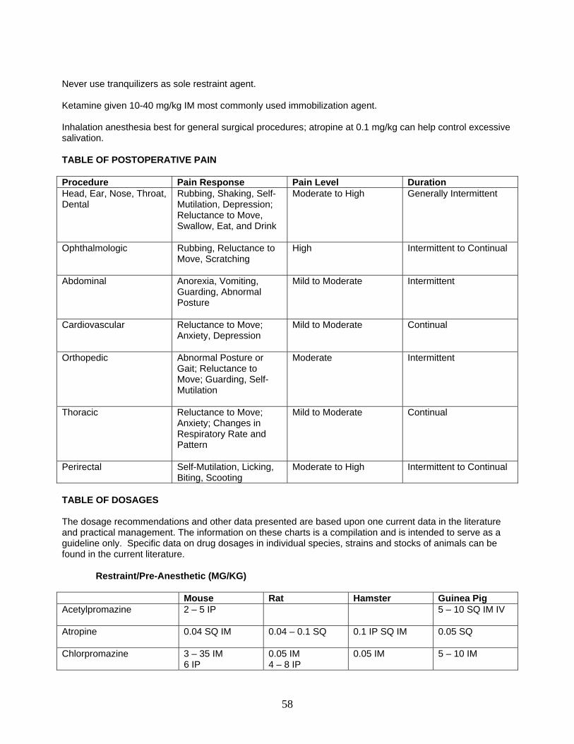
Never use tranquilizers as sole restraint agent. Ketamine given 10-40 mg/kg IM most commonly used immobilization agent. Inhalation anesthesia best for general surgical procedures; atropine at 0.1 mg/kg can help control excessive salivation. TABLE OF POSTOPERATIVE PAIN Procedure Pain Response Pain Level Duration Head, Ear, Nose, Throat, Dental
Rubbing, Shaking, Self-Mutilation, Depression; Reluctance to Move, Swallow, Eat, and Drink
Moderate to High Generally Intermittent
Ophthalmologic Rubbing, Reluctance to Move, Scratching
High Intermittent to Continual
Abdominal Anorexia, Vomiting, Guarding, Abnormal Posture
Mild to Moderate Intermittent
Cardiovascular Reluctance to Move; Anxiety, Depression
Mild to Moderate Continual
Orthopedic Abnormal Posture or Gait; Reluctance to Move; Guarding, Self-Mutilation
Moderate Intermittent
Thoracic Reluctance to Move; Anxiety; Changes in Respiratory Rate and Pattern
Mild to Moderate Continual
Perirectal Self-Mutilation, Licking, Biting, Scooting
Moderate to High Intermittent to Continual
TABLE OF DOSAGES The dosage recommendations and other data presented are based upon one current data in the literature and practical management. The information on these charts is a compilation and is intended to serve as a guideline only. Specific data on drug dosages in individual species, strains and stocks of animals can be found in the current literature.
Restraint/Pre-Anesthetic (MG/KG) Mouse Rat Hamster Guinea Pig Acetylpromazine 2 – 5 IP 5 – 10 SQ IM IV
Atropine 0.04 SQ IM
0.04 – 0.1 SQ 0.1 IP SQ IM 0.05 SQ
Chlorpromazine 3 – 35 IM 6 IP
0.05 IM 4 – 8 IP
0.05 IM 5 – 10 IM
58
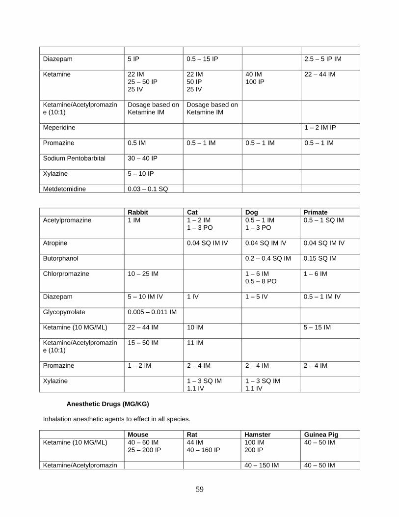
Diazepam 5 IP
0.5 – 15 IP 2.5 – 5 IP IM
Ketamine 22 IM 25 – 50 IP 25 IV
22 IM 50 IP 25 IV
40 IM 100 IP
22 – 44 IM
Ketamine/Acetylpromazine (10:1)
Dosage based on Ketamine IM
Dosage based on Ketamine IM
Meperidine 1 – 2 IM IP
Promazine 0.5 IM
0.5 – 1 IM 0.5 – 1 IM 0.5 – 1 IM
Sodium Pentobarbital
30 – 40 IP
Xylazine
5 – 10 IP
Metdetomidine 0.03 – 0.1 SQ Rabbit Cat Dog Primate Acetylpromazine 1 IM
1 – 2 IM 1 – 3 PO
0.5 – 1 IM 1 – 3 PO
0.5 – 1 SQ IM
Atropine 0.04 SQ IM IV
0.04 SQ IM IV 0.04 SQ IM IV
Butorphanol 0.2 – 0.4 SQ IM
0.15 SQ IM
Chlorpromazine 10 – 25 IM
1 – 6 IM 0.5 – 8 PO
1 – 6 IM
Diazepam 5 – 10 IM IV
1 IV 1 – 5 IV 0.5 – 1 IM IV
Glycopyrrolate 0.005 – 0.011 IM
Ketamine (10 MG/ML) 22 – 44 IM 10 IM
5 – 15 IM
Ketamine/Acetylpromazine (10:1)
15 – 50 IM 11 IM
Promazine 1 – 2 IM
2 – 4 IM 2 – 4 IM 2 – 4 IM
Xylazine 1 – 3 SQ IM 1.1 IV
1 – 3 SQ IM 1.1 IV
Anesthetic Drugs (MG/KG)
Inhalation anesthetic agents to effect in all species. Mouse Rat Hamster Guinea Pig Ketamine (10 MG/ML) 40 – 60 IM
25 – 200 IP
44 IM 40 – 160 IP
100 IM 200 IP
40 – 50 IM
Ketamine/Acetylpromazin 40 – 150 IM 40 – 50 IM
59
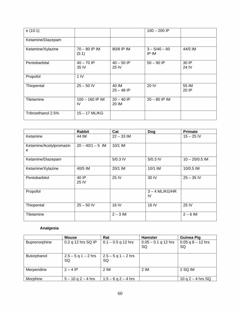
e (10:1)
100 – 200 IP
Ketamine/Diazepam
Ketamine/Xylazine 70 – 80 IP IM (5:1)
80/8 IP IM 3 – 5/40 – 60 IP IM
44/5 IM
Pentobarbital 40 – 70 IP 35 IV
40 – 50 IP 25 IV
50 – 90 IP 30 IP 24 IV
Propofol
1 IV
Thiopental 25 – 50 IV 40 IM 25 – 48 IP
20 IV
55 IM 20 IP
Tiletamine 100 – 160 IP IM IV
20 – 40 IP 20 IM
20 – 80 IP IM
Tribroethanol 2.5%
15 – 17 ML/KG
Rabbit Cat Dog Primate Ketamine 44 IM 22 – 33 IM
15 – 25 IV
Ketamine/Acetylpromazine
20 – 40/1 – 5 IM 10/1 IM
Ketamine/Diazepam 5/0.3 IV
5/0.3 IV 10 – 20/0.5 IM
Ketamine/Xylazine 40/5 IM 20/1 IM
10/1 IM 10/0.5 IM
Pentobarbitol 40 IP 25 IV
25 IV 30 IV 25 – 35 IV
Propofol 3 – 4 ML/KG/HR IV
Thiopental 25 – 50 IV 16 IV
16 IV 25 IV
Tiletamine 2 – 3 IM
2 – 6 IM
Analgesia Mouse Rat Hamster Guinea Pig Buprenorphine 0.2 q 12 hrs SQ IP 0.1 – 0.5 q 12 hrs 0.05 – 0.1 q 12 hrs
SQ 0.05 q 8 – 12 hrs SQ
Butorphanol 2.5 – 5 q 1 – 2 hrs SQ
2.5 – 5 q 1 – 2 hrs SQ
Merperidine 2 – 4 IP
2 IM 2 IM 2 SQ IM
Morphine 5 – 10 q 2 – 4 hrs 1.5 – 6 q 2 – 4 hrs 10 q 2 – 4 hrs SQ
60
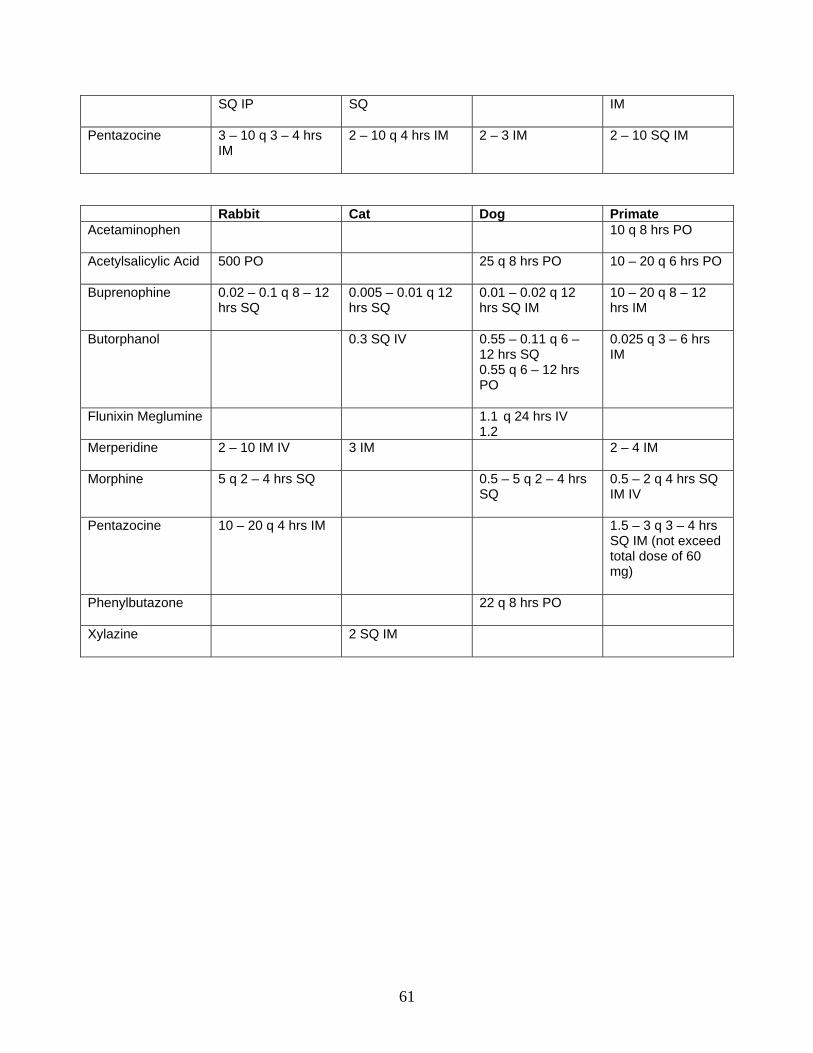
SQ IP
SQ IM
Pentazocine 3 – 10 q 3 – 4 hrs IM
2 – 10 q 4 hrs IM 2 – 3 IM 2 – 10 SQ IM
Rabbit Cat Dog Primate Acetaminophen
10 q 8 hrs PO
Acetylsalicylic Acid 500 PO
25 q 8 hrs PO 10 – 20 q 6 hrs PO
Buprenophine 0.02 – 0.1 q 8 – 12 hrs SQ
0.005 – 0.01 q 12 hrs SQ
0.01 – 0.02 q 12 hrs SQ IM
10 – 20 q 8 – 12 hrs IM
Butorphanol 0.3 SQ IV 0.55 – 0.11 q 6 – 12 hrs SQ 0.55 q 6 – 12 hrs PO
0.025 q 3 – 6 hrs IM
Flunixin Meglumine 1.1 q 24 hrs IV 1.2
Merperidine 2 – 10 IM IV
3 IM 2 – 4 IM
Morphine 5 q 2 – 4 hrs SQ
0.5 – 5 q 2 – 4 hrs SQ
0.5 – 2 q 4 hrs SQ IM IV
Pentazocine 10 – 20 q 4 hrs IM
1.5 – 3 q 3 – 4 hrs SQ IM (not exceed total dose of 60 mg)
Phenylbutazone 22 q 8 hrs PO
Xylazine 2 SQ IM
61
PRE- AND POSTOPERATIVE CARE Inappropriate or inadequately performed preoperative care, operative technique and postoperative care results in distress to the animal and may be considered "unnecessary pain". Individuals performing procedures (minor or major; survival or non-survival) should have adequate knowledge of anatomy, physiology and pharmacology in regard to the species they are utilizing in their research project. All persons performing procedures should have demonstrated ability via training and experience. Good technique, adequate facilities and support equipment; and proper pre and postoperative care are an essential part of an animal care and use program. Veterinary consultation and medical care must be available at all times. REGULATIONS AND GUIDELINES
USDA Regulations
Section 2.31 The IACUC shall determine that the proposed activities or significant changes in ongoing activities meet the following requirements:
(i) Procedures involving animals will avoid or minimize discomfort, distress, and pain to the animals; (ii) The principal investigator has considered alternatives to procedures that may cause more than momentary or slight pain or distress to the animals, and has provided a written narrative description of the methods and sources, e. g., the Animal Welfare Information Center, used to determine that alternatives were not available; (iii) The principal investigator has provided written assurance that the activities do not unnecessarily duplicate previous experiments; (iv) Procedures that may cause more than momentary or slight pain or distress to the animals will:
(A) Be performed with appropriate sedatives, analgesics or anesthetics, unless withholding such agents is justified for scientific reasons, in writing, by the principal investigator and will continue for only the necessary period of time; (B) Involve, in their planning, consultation with the attending veterinarian or his or her designee; (C) Not include the use of paralytics without anesthesia;
(v) Animals that would otherwise experience severe or chronic pain or distress that cannot be relieved will be painlessly euthanized at the end of the procedure or, if appropriate, during the procedure; (vi) The animals' living conditions will be appropriate for their species in accordance with part 3 of this subchapter, and contribute to their health and comfort. The housing, feeding, and nonmedical care of the animals will be directed by the attending veterinarian or other scientist trained and experienced in the proper care, handling, and use of the species being maintained or studied;
(vii) Medical care for animals will be available and provided as necessary by a qualified veterinarian; (viii) Personnel conducting procedures on the species being maintained or studied will be appropriately qualified and trained in those procedures;
62
(ix) Activities that involve surgery include appropriate provision for pre-operative and post-operative care of the animals in accordance with established veterinary medical and nursing practices. All survival surgery will be performed using aseptic procedures, including surgical gloves, masks, sterile instruments, and aseptic techniques. Major operative procedures on non-rodents will be conducted only in facilities intended for that purpose which shall be operated and maintained under aseptic conditions. Non-major operative procedures and all surgery on rodents do not require a dedicated facility, but must be performed using aseptic procedures. Operative procedures conducted at field sites need not be performed in dedicated facilities, but must be performed using aseptic procedures; (x) No animal will be used in more than one major operative procedure from which it is allowed to recover, unless:
(A) Justified for scientific reasons by the principal investigator, in writing; (B) Required as routine veterinary procedure or to protect the health or well-being of the animal as determined by the attending veterinarian; or (C) In other special circumstances as determined by the Administrator on an individual basis. Written requests and supporting data should be sent to the Animal and Plant Health Inspection Service, Animal Care, 4700 River Road, Unit 84, Riverdale, Maryland 20737-1234;
(xi) Methods of euthanasia used must be in accordance with the definition of the term set forth in 9 CFR part 1, Sec. 1.1 of this subchapter, unless a deviation is justified for scientific reasons, in writing, by the investigator.
Health Research Extension Act of 1985 Public Law 99-158, November 20, 1985: Animals in
Research Sec. 495. (a) The Secretary, acting through the Director of NIH, shall establish guidelines for the following:
(1) The proper care of animals to be used in biomedical and behavioral research.
(2) The proper treatment of animals while being used in such research. Guidelines under this paragraph shall require -
(A) the appropriate use of tranquilizers, analgesics, anesthetics, paralytics, and euthanasia for animals in such research; and (B) appropriate pre-surgical and post-surgical veterinary medical and nursing care for animals in such research.
Guidelines to meet the above requirements are contained in the Guide for the Care and Use of Laboratory Animals, NIH Publication 85-23.
AAALAC As per the Guide for the Care and Use of Laboratory Animals, NIH Publication 85-23: Surgery
Appropriate attention to presurgical planning, personnel training, aseptic and surgical technique, animal well-being, and animal physiologic status during all phases of a protocol will enhance the outcome of surgery (see Appendix A, "Anesthesia, Pain, and Surgery"). The individual impact of those factors will vary according to the complexity of procedures involved and the species of animal used. A team approach to a surgical project often increases the likelihood of a successful outcome by providing input from persons with different expertise (Brown and Schofield 1994; Brown and others 1993).
A continuing and thorough assessment of surgical outcomes should be performed to ensure that appropriate procedures are followed and timely corrective changes instituted. Modification of standard
63
techniques might be desirable or even required (for instance, in rodent or field surgery), but it should not compromise the well-being of the animals. In the event of modification, assessment of outcomes should be even more intense and might have to incorporate criteria other than obvious clinical morbidity and mortality.
Presurgical planning should include input from all members of the surgical team, including the surgeon, anesthetist, veterinarian, surgical technicians, animal-care staff, and investigator. The surgical plan should identify personnel, their roles and training needs, and equipment and supplies required for the procedures planned (Cunliffe-Beamer 1993); the location and nature of the facilities in which the procedures will be conducted; and preoperative animal-health assessment and postoperative care (Brown and Schofield 1994). If a nonsterile part of an animal, such as the gastrointestinal tract, is to be surgically exposed or if a procedure is likely to cause immunosuppression, preoperative antibiotics might be appropriate (Klement and others 1987). However, the use of antibiotics should never be considered as a replacement for aseptic procedures.
It is important that persons have had appropriate training to ensure that good surgical technique is practiced, that is, asepsis, gentle tissue handling, minimal dissection of tissue, appropriate use of instruments, effective hemostasis, and correct use of suture materials and patterns (Chaffee 1974; Wingfield 1979). People performing and assisting in surgical procedures in a research setting often have a wide range of educational backgrounds and might require various levels and kinds of training before they participate in surgical procedures on animals. For example, persons trained in human surgery might need training in inter species variations in anatomy, physiology, and the effects of anesthetic and analgesic drugs, or in postoperative requirements. Training guidelines for research surgery commensurate with a person's background are available (ASR 1989) to assist institutions in developing appropriate training programs. The PHS Policy and the AWRs place responsibility with the IACUC for determining that personnel performing surgical procedures are appropriately qualified and trained in the procedures to be performed.
In general, surgical procedures are categorized as major or minor and in the laboratory setting can be further divided into survival and nonsurvival. Major survival surgery penetrates and exposes a body cavity or produces substantial impairment of physical or physiologic functions (such as laparotomy, thoracotomy, craniotomy, joint replacement, and limb amputation). Minor survival surgery does not expose a body cavity and causes little or no physical impairment (such as wound suturing; peripheral-vessel cannulation; such routine farm-animal procedures as castration, dehorning, and repair of prolapses; and most procedures routinely done on an "outpatient" basis in veterinary clinical practice).
Minor procedures are often performed under less-stringent conditions than major procedures but still require aseptic technique and instruments and appropriate anesthesia. Although laparoscopic procedures are often performed on an "outpatient" basis, appropriate aseptic technique is necessary if a body cavity is penetrated.
In nonsurvival surgery, an animal is euthanized before recovery from anesthesia. It might not be necessary to follow all the techniques outlined in this section if nonsurvival surgery is performed; however, at a minimum, the surgical site should be clipped, the surgeon should wear gloves, and the instruments and surrounding area should be clean (Slattum and others 1991).
Emergency situations sometimes require immediate surgical correction under less than ideal conditions. For example, if an animal maintained outdoors needs surgical attention, movement to a surgical facility might pose an unacceptable risk to the animal or be impractical. Such situations often require more-intensive aftercare and might pose a greater risk of postoperative complications. The appropriate course of action requires veterinary medical judgment.
Aseptic technique is used to reduce microbial contamination to the lowest possible practical level (Cunliffe-Beamer 1993). No procedure, piece of equipment, or germicide alone can achieve that objective (Schonholtz 1976). Aseptic technique requires the input and cooperation of everyone who enters the operating suite (Belkin 1992; McWilliams 1976). The contribution and importance of each practice varies with the procedure. Aseptic technique includes preparation of the patient, such as hair removal and disinfection of the operative site (Hofmann 1979); preparation of the surgeon, such as the provision of decontaminated surgical attire, surgical scrub, and sterile surgical gloves (Chamberlain and Houang 1984; Pereira and others 1990; Schonholtz 1976); sterilization of instruments, supplies, and implanted materials (Kagan 1992b); and the use of operative techniques to reduce the likelihood of infection (Ayliffe 1991; Kagan 1 992a; Ritter and Marmion 1987; Schofield 1994; Whyte 1988).
Specific sterilization methods should be selected on the basis of physical characteristics of materials to be sterilized (Schofield 1994). Autoclaving and gas sterilization are common effective methods. Sterilization indicators should be used to identify materials that have undergone proper sterilization (Berg
64
1993). Liquid chemical sterilants should be used with adequate contact times, and instruments should be rinsed with sterile water or saline before use. Alcohol is neither a sterilant nor a high-level disinfectant (Rutala 1990).
In general, unless an exception is specifically justified as an essential component of the research protocol and approved by the IACUC, nonrodent aseptic surgery should be conducted only in facilities intended for that purpose. Most bacteria are carried on airborne particles or fomites, so surgical facilities should be maintained and operated in a manner that ensures cleanliness and minimizes unnecessary traffic (AORN 1982; Bartley 1993). In some circumstances, it might be necessary to use an operating room for other purposes. In such cases, it is imperative that the room be returned to an appropriate level of cleanliness before its use for major survival surgery.
Careful surgical monitoring and timely attention to problems increase the likelihood of a successful surgical outcome. Monitoring includes checking of anesthetic depth and physiologic function and assessment of clinical signs and conditions. Maintenance of normal body temperature minimizes cardiovascular and respiratory disturbances caused by anesthetic agents (Dardai and Heavner 1987) and is of particular importance.
The species of animal influences the components and intensity of the surgical program. The relative susceptibility of rodents to surgical infection has been debated; available data suggest that subclinical infections can cause adverse physiologic and behavioral responses (Beamer 1972; Bradfield and others 1992; Cunliffe-Beamer 1990; Waynforth 1980, 1987) that can affect both surgical success and research results. Some characteristics of common laboratory-rodent surgery-such as smaller incision sites, fewer personnel in the surgical team, manipulation of multiple animals at one sitting, and briefer procedures-as opposed to surgery in larger species, can make modifications in standard aseptic techniques necessary or desirable (Brown 1994; Cunliffe-Beamer 1993). Useful suggestions for dealing with some of the unique challenges of rodent surgery have been published (Cunliffe-Beamer 1983, 1993).
Generally, farm animals maintained for biomedical research should undergo surgery with procedures and in facilities compatible with the guidelines set forth in this section. However, some minor and emergency procedures that are commonly performed in clinical veterinary practice and in commercial agricultural settings may be conducted under less-stringent conditions than experimental surgical procedures in a biomedical-research setting. Even when conducted in an agricultural setting, these procedures require the use of appropriate aseptic technique, sedatives, analgesics, anesthetics, and conditions commensurate with the risk to the animal's health and well-being. But they might not require the intensive surgical settings, facilities, and procedures outlined here.
Presurgical planning should specify the requirements of postsurgical monitoring, care, and record-keeping, including the personnel who will perform these duties. The investigator and veterinarian share responsibility for ensuring that postsurgical care is appropriate. An important component of postsurgical care is observation of the animal and intervention as required during recovery from anesthesia and surgery. The intensity of monitoring necessary will vary with the species and the procedure and might be greater during the immediate anesthetic recovery period than later in postoperative recovery. During the anesthetic-recovery period, the animal should be in a clean, dry area where it can be observed often by trained personnel. Particular attention should be given to thermoregulation, cardiovascular and respiratory function, and postoperative pain or discomfort during recovery from anesthesia. Additional care might be warranted, including administration of parenteral fluids for maintenance of water and electrolyte balance (FBR 1987), analgesics, and other drugs; care for surgical incisions; and maintenance of appropriate medical records.
After anesthetic recovery, monitoring is often less intense but should include attention to basic biologic functions of intake and elimination and behavioral signs of postoperative pain, monitoring for postsurgical infections, monitoring of the surgical incision, bandaging as appropriate, and timely removal of skin sutures, clips, or staples (UFAW 1989). Facilities for Aseptic Surgery As per the Guide for the Care and Use of Laboratory Animals, NIH Publication 85-23:
The design of a surgical facility should accommodate the species to be operated on and the complexity of the procedures to be performed (Hessler 1991; see also Appendix A, "Design and Construction of Animal Facilities"). For most rodent surgery, a facility may be small and simple, such as a dedicated space in a laboratory appropriately managed to minimize contamination from other activities in the
65
room during surgery. The facility often becomes larger and more complex as the number of animals, the size of animals, or the complexity of procedures increases, for instance, large-volume rodent procedures, the need for special restraint devices, hydraulic operating tables, and floor drains for farm-animal surgery, and procedures that require large surgical teams and support equipment and thus large space. The relationship of surgical facilities to diagnostic laboratories, radiology facilities, animal housing, staff offices, and so on should be considered in the overall context of the complexity of the surgical program. Surgical facilities should be sufficiently separate from other areas to minimize unnecessary traffic and decrease the potential for contamination (Humphreys 1993). Centralized facilities provide important advantages in cost savings in equipment, conservation of space and personnel resources, reduced transit of animals, and enhanced professional oversight of facilities and procedures.
For most surgical programs, functional components of aseptic surgery include surgical support, animal preparation, surgeon's scrub, operating room, and postoperative recovery. The areas that support those functions should be designed to minimize traffic flow and separate the related, nonsurgical activities from the surgical procedure in the operating room. The separation is best achieved by physical barriers (AORN 1982) but might also be achieved by distance between areas or by the timing of appropriate cleaning and disinfection between activities. The number of personnel and their level of activity have been shown to be directly related to the level of bacterial contamination and the incidence of postoperative wound infection (Fitzgerald 1979). Traffic in the operating room itself can be reduced by the installation of an observation window, a communication system (such as an intercom system), and judicious location of doors.
Control of contamination and ease of cleaning should be key considerations in the design of a surgical facility. The interior surfaces should be constructed of materials that are monolithic and impervious to moisture. Ventilation systems supplying filtered air at positive pressure can reduce the risk of postoperative infection (Ayscue 1986; Bartley 1993; Bourdillon 1946; Schonholtz 1976). Careful location of air supply and exhaust ducts and appropriate room-ventilation rates are also recommended to minimize contamination (Ayliffe 1991; Bartley 1993; Holton and Ridgway 1993; Humphreys 1993). To facilitate cleaning, the operating rooms should have as little fixed equipment as possible (Schonholtz 1976; UFAW 1989). Other features of the operating room to consider include surgical lights to provide adequate illumination (Ayscue 1986), sufficient electric outlets for support equipment, and gas-scavenging capability.
The surgical-support area should be designed for washing and sterilizing instruments and for storing instruments and supplies. Autoclaves are commonly placed in this area. It is often desirable to have a large sink in the animal-preparation area to facilitate cleaning of the animal and the operative site. A dressing area should be provided for personnel to change into surgical attire; a multipurpose locker room can serve this function. There should be a scrub area for surgeons, equipped with foot, knee, or electric-eye surgical sinks (Knecht and others 1981). To minimize the potential for contamination of the surgical site by aerosols generated during scrubbing, the scrub area is usually outside the operating room.
A postoperative-recovery area should provide the physical environment to support the needs of the animal during the period of anesthetic and immediate postsurgical recovery and should be so placed as to allow adequate observation of the animal during this period. The electric and mechanical requirements of monitoring and support equipment should be considered. The type of caging and support equipment will depend on the species and types of procedures but should be designed to be easily cleaned and to support physiologic functions, such as thermoregulation and respiration. Depending on the circumstances, a postoperative recovery area for farm animals might be modified or nonexistent in some field situations, but precautions should be taken to minimize risk of injury to recovering animals. DEFINITIONS Major operative procedure means any surgical intervention that penetrates and exposes a body cavity or any procedure which produces permanent impairment of physical or psychological functions. Minor operative procedure means any surgical intervention that does not expose a body cavity and causes little or no physical impairment. Non-survival surgical procedure is one in which the animal is euthanized before recovery from anesthesia.
66
Painful procedure as applied to any animal means any procedure that would reasonably be expected to cause more than slight or momentary pain or distress in a human being to which that procedure was applied, that is, pain in excess of that caused by injections or other minor procedures. Survival surgical procedure is one in which the animal recovers from anesthesia even if only momentarily. PREOPERATIVE CARE
1. Physical Exam
• Check temperature, pulse and respiratory system. • Check heart. • Check mucus membranes. • Check face for discharges. • Check anal area for signs of diarrhea. • Check locomotion.
OPTIONAL - depends on procedure
• Complete blood count; WBC, RBC, HCT, Hbg. • Kidney function. • Liver function. • Other tests or evaluation as required.
2. Diet
Animals should be maintained on a nutritionally balanced diet.
Water can be given ad lib right up to the time of the procedure.
Withhold food:
• Small rodents • Guinea pigs - 6 hours; 12 if barbiturates are used. • Rabbits - 6-8 hours. • Dogs and cats - 8-12 hours. • Primates - 18 hours. • Ruminants - 48 hours
3. Preanesthetic Drugs
• Facilitates restraint • Minimizes side effect of anesthesia • Reduces reflex responses • Provides analgesia • Adjunct to regional anesthesia and reduces the dosage of anesthetic agents
4. Antibiotics
Antibiotics are given as a prophylactic treatment for anticipated infection:
• 12 hours prior to the procedure and for 2-4 days following the procedure • Any time the procedure lasts more than an hour • If asepsis is broken
67
Consider dosage and species sensitivity Give the antibiotic that is appropriate for the procedure Antibiotics can potentiate anesthesia Consideration should be given as to the effect of preanesthetic drugs, anesthetic agents and antibiotics on the experiment. Consider and have available analgesics, life support drugs and equipment; and agents for euthanasia. OPERATIVE CARE Prepare surgical area - instruments and equipment Anesthetize animal in prep room - monitor animal at all times and be prepared for emergencies (life support equipment and drugs) Remove hair around surgical site in prep room. Move animal to surgical room. Prepare surgical site utilizing standard technique. Observe aseptic technique at all times:
• For surgeon and assistants. • When draping surgical site. • During surgery and when using experimental equipment.
All instruments that enter the sterile field and come in contact with the animal or a skin opening should be sterilized. When performing surgery, the surgeon should wear sterile gloves, gown, and mask. Animals that vomit readily require endotracheal intubation. FLUID THERAPY It is good practice to have an intravenous catheter inserted and fluids administered during surgery. There are situations when fluid administration is required. An intravenous catheter provides an emergency route for administering life support drugs. Fluids should be administered:
• When the procedure lasts over an hour. • When large volume of blood loss is expected. • During any high risk surgery.
Fluids are administered:
To replace loss due to:
• Hemorrhage • Dehydration –
o No water intake
68
o Blood loss o Exposed tissue o Loss via respiration
• Tissue edema • Vasodilation • Maintain kidney function • Prevent shock • Maintain tissue perfusion
MAINTAINING BODY TEMPERATURE Excessive heat loss may occur during surgery. Heating pads or blankets should be used but monitored carefully to prevent burns. When an animal’s body temperature drops, a given dosage of anesthetic produces a greater effect. SURGICAL PROCEDURES These surgical factors promote wound healing:
• Gentle tissue handling • Asepsis • Hemostasis • Proper skin closure • Wound irrigation • Correct suture pattern and technique
Electrocautery should be used with care. It can cause severe tissue necrosis, which retards healing. Exposed tissue should be kept moist with sterile physiological fluids. Throughout surgery, the animal should be monitored for pulse, respiration, and tissue color and perfusion. Plan ahead to reduce surgical time as much as possible. POSTOPERATIVE CARE When the animal is aware of its surroundings and in an upright position it is considered recovered from anesthesia, however, this does not constitute adequate postoperative care.
• Keep the animal warm with supplemental heating. • Observe animal for signs of shock and be prepared for emergency care and euthanasia if
necessary. • Remove intravenous catheters if animal is recovering without complications.
When animal is fully recovered small amounts of water and food may be given. Be prepared to water and feed by hand to compensate for postoperative disability.
• Give antibiotics as necessary. • Give analgesics if required.
Surgical site, including indwelling catheters should be cared for:
• Check sutures. • Check for overt bleeding.
69
• Check for adequate blood perfusion to the surgical site. • Minor incision may require topical antibiotics without steroids. • Major incision may require topical antibiotics and bandaging. • May have to protect incision to prevent self-mutilation. • Check animal daily and change bandages as needed.
Sutures should be removed in 7-14 days (7 days - healthy animal, no complications; 14 days - with steroids, major surgery, infection or complications). INVESTIGATOR RESPONSIBILITY
1. The investigator is responsible for compliance with all regulations and guidelines.
2. The individual performing the procedure is responsible for the animal(s) care and use. 3. The following must be provided:
• Consultation with the institutional veterinarian. • Review and approval of protocol for the procedure by the IACUC. • Training of all individuals using animals. • Proper use of analgesics, tranquilizers and anesthetics. • Proper preoperative, operative and postoperative care. • Provisions for emergency care and euthanasia if necessary.
4. Maintain adequate records.
70
EUTHANASIA THE MEANING OF “EUTHANASIA”
Dictionary Definition
The word "euthanasia" is derived from the Greek eu-, well + thanatos-, death. An easy and painless death.
United States Department of Agriculture For the purposes of the Animal Welfare Act, “euthanasia” means the humane destruction of an animal accomplished by a method that produces rapid unconsciousness and subsequent death without evidence of pain or distress, or a method that utilizes anesthesia produced by an agent that causes painless loss of consciousness and subsequent death
Guide for the Care and Use of Laboratory Animals For the purposes of the Guide for the Care and Use of Laboratory Animals, “euthanasia" is the procedure of killing animals rapidly and painlessly. It should be carried out by trained personnel using acceptable techniques in accordance with institutional policies and applicable laws. The method should not interfere with postmortem evaluation. HUMANE CONSIDERATIONS
Primary Criterion for Evaluating a Technique
A method of euthanasia must cause loss of consciousness and have an initial depressive action on the central nervous system which will ensure insensitivity to pain without fear or anxiety. Death does not necessarily follow loss of consciousness or CNS depression.
Aesthetics versus Humaneness Some methods of euthanasia, such as decapitation or cervical dislocation, are not aesthetic to the observer but none the less are humane. A technique of euthanasia is considered humane if it causes CNS depression and insensitivity to pain. Pain recognition in an animal and man is dependent on impulses from pain receptors reaching the thalamus and cerebral cortex. If these structures are not functioning, pain will not be felt. Therefore, a technique of euthanasia is considered humane if this occurs. In the unconscious animal, stimuli that invoke pain will elicit reflex responses manifested by motor movement but this does not indicate cerebral pain reception. SELECTION OF A TECHNIQUE OF EUTHANASIA
Killing an Animal at the End of an Experiment Many animal experiments require data that is collected during a postmortem examination. Animals may be painlessly killed in order to harvest tissues or organs for in-vitro experiments. In the majority of experiments, animals are painlessly killed because they are surplus to the requirements of the experiment. Endangered species or valuable animals are not generally killed but are recycled to other projects.
Killing an Animal on Humane Grounds
71
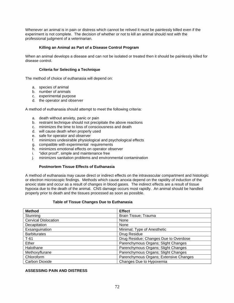
Whenever an animal is in pain or distress which cannot be relived it must be painlessly killed even if the experiment is not complete. The decision of whether or not to kill an animal should rest with the professional judgment of a veterinarian.
Killing an Animal as Part of a Disease Control Program
When an animal develops a disease and can not be isolated or treated then it should be painlessly killed for disease control.
Criteria for Selecting a Technique The method of choice of euthanasia will depend on:
a. species of animal b. number of animals c. experimental purpose d. the operator and observer
A method of euthanasia should attempt to meet the following criteria:
a. death without anxiety, panic or pain b. restraint technique should not precipitate the above reactions c. minimizes the time to loss of consciousness and death d. will cause death when properly used e. safe for operator and observer f. minimizes undesirable physiological and psychological effects g. compatible with experimental requirements h. minimizes emotional effects on operator observer i. "idiot proof", simple and maintenance free j. minimizes sanitation problems and environmental contamination
Postmortem Tissue Effects of Euthanasia
A method of euthanasia may cause direct or indirect effects on the intravascular compartment and histologic or electron microscopic findings. Methods which cause anoxia depend on the rapidity of induction of the anoxic state and occur as a result of changes in blood gases. The indirect effects are a result of tissue hypoxia due to the death of the animal. CNS damage occurs most rapidly. An animal should be handled properly prior to death and the tissues processed as soon as possible.
Table of Tissue Changes Due to Euthanasia Method Effect Stunning Brain Tissue; Trauma Cervical Dislocation None Decapitation None Exsanguination Minimal; Type of Anesthetic Barbiturates Drug Residue T-61 Drug Residue; Changes Due to Overdose Ether Parenchymous Organs; Slight Changes Halothane Parenchymous Organs; Slight Changes Methoxyflurane Parenchymous Organs; Slight Changes Chloroform Parenchymous Organs; Extensive Changes Carbon Dioxide Changes Due to Hypoxemia ASSESSING PAIN AND DISTRESS
72
Abnormal Behavioral and Physiological Responses
In the conscious animal the following demonstrates abnormal reaction to fear, pain and distress:
a. distress vocalization b. struggling and attempts to escape c. defensive aggression d. freezing, tremors e. dilated pupils f. salivation g. evacuation of anal glands, urination and defecation h. tachycardia i. panting
A method of euthanasia may cause an animal to pass through stage two of anesthesia in which the following may occur:
a. excitement and uninhibited activity b. exaggerated response to painful stimuli
ASSESSING UNCONSCIOUSNESS AND DEATH
Determining the Stage of Unconsciousness and Death
The level of consciousness and an animal's ability to feel pain may be measured by testing the palpebral, corneal or "blinking" reflex. This reflex is absent in the cat and when curariform drugs or dissociative anesthetics are used in other species. The signs of death in an animal are indicated by:
a. cessation of respiration b. absence of a heartbeat or pulse - verify before disposal c. loss of reflex responses d. a flat or isoelectric electroencephalogram e. total flaccidity of the muscles which is followed in minutes or hours by rigor mortis
THE PRACTICAL REQUIREMENTS OF EUTHANASIA
Techniques of the Most Commonly Used Methods This section is adapted from the 2000 Report of the AVMA Panel on Euthanasia. The authors’ recommendations may differ from those of the AVMA Panel.
Physical The physical methods of euthanasia are technically difficult to perform and may be aesthetically displeasing. These techniques when properly performed by trained personnel are humane.
Cervical Dislocation Cervical dislocation is a technique of separating the skull and brain from the spinal cord by applying pressure posterior to the base of the skull and spinal cord. It is utilized to kill mice and immature rats.
73
"The thumb and index finger are placed on either side of the neck at the base of the skull or a rod is pressed at the base of the skull. With the other hand the base of the tail or hind limbs are quickly pulled causing separation of the cervical vertebrae from the skull."
Advantages:
a. produces rapid unconsciousness and death b. tissues and blood not contaminated by chemicals
Disadvantages:
a. aesthetically displeasing b. required mastering technical skills to ensure loss of consciousness is rapidly induced c. limited to mice immature rats and rabbits
Recommendations
Acceptable only for mice, rats weighing less than 200 g, and rabbits weighing less than 1 kg. Animals must be sedated or anesthetized prior to cervical dislocation. Institutional Animal Care and Use Committee must determine that personnel who perform cervical dislocation techniques have been properly trained.
Decapitation with Guillotine Decapitation is the method of rapidly and completely severing the head from the body using a specifically designed apparatus (guillotine).
Advantages:
a. guillotines are commercially available b. tissues and blood are not contaminated by chemicals c. appears to induce rapid loss of consciousness
Disadvantages:
a. aesthetically displeasing b. some studies indicate that the animal may not lose consciousness for 13-14 seconds after
decapitation. c. personnel performing this techniques should take adequate precautions to prevent personal
injury
Recommendations
This technique is conditionally acceptable if performed correctly, required by the experimental design and approved the Institutional Animal Care and Use Committee. The equipment used should be maintained and in good working order and serviced on a regular basis to endure sharpness of blades. The use of plastic cones to restrain animals appears to reduce distress from handling, minimizes the chance of injury to personnel, and improves positioning of the animal in the guillotine. Personnel must be properly trained to perform decapitation. Penetrating Captive Bolt Captive bolt guns are powered by gunpowder or compressed air and must provide sufficient energy to penetrate the skull of the species on which they are being used. Adequate restraint is important to ensure proper placement of the captive bolt. A cerebral hemisphere and brainstem must be sufficiently disrupted by the projectile to induce sudden loss of consciousness and subsequent death.
74
Advantages:
a. effective method of euthanasia for use in slaughterhouses, in research facilities, and on the farm when use of drugs is inappropriate.
Disadvantages
a. aesthetically displeasing b. death may not occur if equipment is not maintained and used properly
Recommendations
Use of the penetrating captive bolt is an acceptable and practical method of euthanasia for horses, ruminants, and swine. It is conditionally acceptable for other appropriate species. Electrocution Electrocution induces death by cardiac fibrillation, which causes cerebral hypoxia. However, animals do not lose consciousness for 10 to 30 seconds or more after onset of cardiac fibrillation. It is imperative that animals be unconscious before being electrocuted. Advantages:
a. humane if the animal is first rendered unconscious b. it does not chemically contaminate tissues c. it is economical
Disadvantages
a. may be hazardous to personnel b. when conventional single-animal probes are used, it may not be a useful method for mass
euthanasia because so much time is required per animal c. is not a useful method for dangerous, intractable animals d. aesthetically objectionable because of violent extension and stiffening of the limbs, head,
and neck e. may not result in death in small animals (< 5 kg) because ventricular fibrillation and
circulatory collapse do not always persist after cessation of current flow
Recommendations Euthanasia by electrocution requires special skills and equipment to ensure passage of sufficient current through the brain to induce cardiac fibrillation. Although the method is conditionally acceptable if the aforementioned requirements are met, its disadvantages far outweigh its advantages in most applications. Techniques that apply electric current from head to tail, head to foot, or head to moistened metal plates on which the animal is standing are unacceptable.
Microwave Irradiation Heating by microwave irradiation is used primarily by neurobiologists to fix brain metabolites in vivo while maintaining the anatomic integrity of the brain. Microwave instruments have been specifically designed for use in euthanasia of laboratory mice and rats. Advantages:
a. loss of consciousness is achieved in less than 100 ms, and death in less than 1 second
75
b. this is the most effective method to fix brain tissue in vivo for subsequent assay of enzymatically labile chemicals
Disadvantages:
a. instruments are expensive b. only animals the size of mice and rats can be euthanized with commercial instruments that
are currently available
Recommendations Microwave irradiation is a humane method for euthanizing small laboratory rodents if instruments that induce rapid loss of consciousness are used. Only instruments that are designed for this use and have appropriate power and microwave distribution can be used. Microwave ovens designed for domestic and institutional kitchens are absolutely unacceptable for euthanasia. Chemical Agents The use of injectable euthanasia agents is the most rapid and reliable method of performing euthanasia. It is the most desirable method when it can be performed without causing fear or distress in the animal. When the restraint necessary for giving an animal an intravenous injection would impart added distress to the animal or pose undue risk to the operator, sedation, anesthesia, or an acceptable alternate route of administration should be employed. Aggressive, fearful, wild, or feral animals should be sedated or given a nonparalytic immobilizing agent prior to intravenous administration of the euthanasia agent. When intravenous administration is considered impractical or impossible, intraperitoneal administration of a nonirritating euthanasia agent is acceptable, provided the drug does not contain neuromuscular blocking agents. Intracardiac injection is acceptable only when performed on heavily sedated, anesthetized, or comatose animals. It is not considered acceptable in awake animals, owing to the difficulty and unpredictability of performing the injection accurately. Intramuscular, subcutaneous, intrathoracic, intrapulmonary, intrahepatic, intrarenal, intrasplenic, intrathecal, and other nonvascular injections are not acceptable methods of administering injectable euthanasia agents. When injectable euthanasia agents are administered into the peritoneal cavity, animals may be slow to pass through stages I and II of anesthesia. Accordingly, they should be placed in small cages in a quiet area to minimize excitement and trauma.
Barbiturates Barbiturates depress the central nervous system in descending order, beginning with the cerebral cortex. Within seconds of intravenous administration, unconsciousness is induced and it progresses to deep anesthesia. Apnea occurs due to depression of the respiratory center, and cardiac arrest quickly follows. Several barbiturates are acceptable, but pentobarbital sodium most commonly is used for euthanasia.
Advantages:
a. speed of action b. barbiturates induce euthanasia smoothly, with minimal discomfort to the animal c. aesthetically pleasing when administered by trained personnel d. may be less expensive than other agents
Disadvantages:
a. must be administered intravenously by trained personnel b. animal(s) must be restrained c. controlled substance (United States Drug Enforcement Agency)
76
d. terminal gasp may occur e. these drugs tend to persist in the carcass and may cause sedation or even death of animals
that consume the body
Recommendations
Intravenous injection of a barbituric acid derivative is the preferred method for euthanasia of dogs, cats, other small animals, and horses. Intraperitoneal injection may be used in situations when an intravenous injection would be distressful or even dangerous. Intracardiac injection must only be used if the animal is heavily sedated, unconscious, or anesthetized. Potassium Chloride in Conjunction with Prior General Anesthesia Although unacceptable and condemned when used in unanesthetized animals, the use of a supersaturated solution of potassium chloride injected intravenously or intracardially in an animal under general anesthesia is an acceptable method to produce cardiac arrest and death. Advantages:
a. potassium chloride is not a controlled substance. It is easily acquired, transported, and mixed in the field.
b. potassium chloride, when used with appropriate methods to render an animal unconscious, results in a carcass that is potentially less toxic for scavengers and predators in cases where carcass disposal is impossible or impractical
Disadvantages:
a. rippling of muscle tissue and clonic spasms may occur on or shortly after injection
Recommendations
It is of utmost importance that personnel performing this technique are trained and knowledgeable in anesthetic techniques, and are competent in assessing anesthetic depth appropriate for administration of potassium chloride intravenously. Administration of potassium chloride intravenously requires animals to be in a surgical plane of anesthesia characterized by loss of consciousness, loss of reflex muscle response, and loss of response to noxious stimuli. Saturated potassium chloride solutions are effective in causing cardiac arrest following rapid intracardiac or intravenous injection. Residual tissue concentrations of general anesthetics after anesthetic induction have not been documented. Whereas no scavenger toxicoses have been reported with potassium chloride in combination with a general anesthetic, proper carcass disposal should always be attempted to prevent possible toxicosis by consumption of a carcass contaminated with general anesthetics.
Inhalant Anesthetics Inhalant anesthetics (e.g., ether, halothane, methoxyflurane, isoflurane, sevoflurane, desflurane, and enflurane) have been used to euthanize many species. Halothane induces anesthesia rapidly and is the most effective inhalant anesthetic for euthanasia. Enflurane is less soluble in blood than halothane, but, because of its lower vapor pressure and lower potency, induction rates may be similar to those for halothane. At deep anesthetic planes, animals may seizure. It is an effective agent for euthanasia, but the associated seizure activity may be disturbing to personnel. Isoflurane is less soluble than halothane, and it should induce anesthesia more rapidly. However, it has a slightly pungent odor and animals often hold their breath, delaying onset of loss of consciousness. Isoflurane also may require more drug to kill an animal, compared with halothane. Although isoflurane is acceptable as a euthanasia agent, halothane is preferred. Sevoflurane is less soluble than halothane and does not have an objectionable odor. It is less potent than isoflurane or halothane and has a lower vapor pressure. Anesthetic concentrations can be achieved and
77
maintained rapidly. Desflurane is currently the least soluble potent inhalant anesthetic, but the vapor is quite pungent, which may slow induction. This drug is so volatile that it could displace oxygen (O2) and induce hypoxemia during induction if supplemental O2 is not provided. Methoxyflurane is highly soluble, and slow anesthetic induction with its use may be accompanied by agitation. It is a conditionally acceptable agent for euthanasia in rodents. Ether has high solubility in blood and induces anesthesia slowly. It is irritating to the eyes and nose, poses serious risks associated with its flammability and explosiveness, and has been used to create a model for stress. With inhalant anesthetics, the animal can be placed in a closed receptacle containing cotton or gauze soaked with an appropriate amount of the anesthetic, or the anesthetic can be introduced from a vaporizer. The latter method may be associated with a longer induction time. Vapors are inhaled until respiration ceases and death ensues. Because the liquid state of most inhalant anesthetics is irritating, animals should be exposed only to vapors. Also, sufficient air or O2 must be provided during the induction period to prevent hypoxemia. In the case of small rodents placed in a large container, there will be sufficient O2 in the chamber to prevent hypoxemia. Larger species placed in small containers may need supplemental air or O2. Nitrous oxide (N2O) may be used with other inhalants to speed the onset of anesthesia, but alone it does not induce anesthesia in animals, even at 100% concentration. When used by itself, N2O produces hypoxemia before respiratory or cardiac arrest. As a result, animals may become distressed prior to loss of consciousness. Occupational exposure to inhalant anesthetics constitutes a human health hazard. Spontaneous abortion and congenital abnormalities have been associated with exposure of women to trace amounts of inhalation anesthetic agents during early stages of pregnancy. Regarding human exposure to inhalant anesthetics, the concentrations of halothane, enflurane, and isoflurane should be less than 2 ppm, and less than 25 ppm for nitrous oxide. There are no controlled studies proving that such concentrations of anesthetics are safe, but these concentrations were established because they were found to be attainable under hospital conditions. Effective procedures must be used to protect personnel from anesthetic vapors. Advantages:
a. inhalant anesthetics are particularly valuable for euthanasia of smaller animals (< 7 kg) or for animals in which venipuncture may be difficult
b. halothane, enflurane, isoflurane, sevoflurane, desflurane, methoxyflurane, and N2O are nonflammable and nonexplosive under ordinary environmental conditions
Disadvantages:
a. animals may struggle and become anxious during induction of anesthesia because
anesthetic vapors may be irritating and can induce excitement b. ether is flammable and explosive. Explosions have occurred when animals, euthanized with
ether, were placed in an ordinary (not explosion proof) refrigerator or freezer and when bagged animals were placed in an incinerator
c. Induction with methoxyflurane is unacceptably slow in some species. (4) Nitrous oxide will support combustion
d. personnel and animals can be injured by exposure to these agents. (6) There is a potential for human abuse of some of these drugs, especially N2O
Recommendations
In order of preference, halothane, enflurane, isoflurane, sevoflurane, methoxyflurane, and desflurane, with or without nitrous oxide, are acceptable for euthanasia of small animals (< 7 kg). Ether should only be used in carefully controlled situations in compliance with state and federal occupational health and safety regulations. It is conditionally acceptable. Nitrous oxide should not be used alone, pending further scientific studies on its suitability for animal euthanasia. Although acceptable, these agents are generally not used in larger animals because of their cost and difficulty of administration.
78
Carbon Dioxide Room air contains 0.04% carbon dioxide. Pure carbon dioxide is heavier than air and nearly odorless. Inhalation of carbon dioxide in concentrations of 7.5% increases the pain threshold, and higher concentrations of carbon dioxide have a rapid anesthetic effect. CO2 concentrations for euthanasia of neonates should be especially high. A CO2 concentration of 60% to 70% with a 5-minute exposure time appears to be optimal.
Advantages:
a. carbon dioxide will produce a narcosis with rapid anesthesia and death. b. carbon dioxide is supplied in pressurized cylinders c. inexpensive, nonflammable, nonexplosive d. normally not hazardous to personnel or other animals e. does not distort cellular architecture
Disadvantages:
a. chamber must be completely filled and precharged to prevent animals from climbing to
avoid exposure b. immature animals require more exposure time to produce unconsciousness and death c. euthanasia by exposure to CO2 may take longer than euthanasia by other means d. induction of loss of consciousness at lower concentrations (< 80%) may produce pulmonary
and upper respiratory tract lesions e. high concentrations of CO2 may be distressful to some animals
Recommendations
Carbon dioxide is acceptable for euthanasia in appropriate species. Compressed CO2 gas in cylinders is the only recommended source of carbon dioxide because the inflow to the chamber can be regulated precisely. Carbon dioxide generated by other methods such as from dry ice, fire extinguishers, or chemical means (e.g., antacids) is unacceptable. Species should be separated and chambers should not be overcrowded. With an animal in the chamber, an optimal flow rate should displace at least 20% of the chamber volume per minute. Loss of consciousness may be induced more rapidly by exposing animals to a CO2 concentration of 70% or more by prefilling the chamber for species in which this has not been shown to cause distress. Gas flow should be maintained for at least 1 minute after apparent clinical death. It is important to verify that an animal is dead before removing it from the chamber. If an animal is not dead, CO2 narcosis must be followed with another method of euthanasia. Adding O2 to the CO2 may or may not preclude signs of distress. Additional O2 will, however, prolong time to death and may complicate determination of consciousness. There appears to be no advantage to combining O2 with carbon dioxide for euthanasia.
Nitrogen, Argon Nitrogen (N2) and argon (Ar) are colorless, odorless gases that are inert, nonflammable, and nonexplosive. Nitrogen comprises 78% of atmospheric air, whereas Ar comprises less than 1%. Euthanasia is induced by placing the animal in a closed container that has been prefilled with N2 or Ar or into which the gas is then rapidly introduced. Nitrogen/Ar displaces O2, thus inducing death by hypoxemia. Advantages:
a. nitrogen and Ar are readily available as compressed gases. (2) Hazards to personnel are minimal.
79
Disadvantages:
a. loss of consciousness is preceded by hypoxemia and ventilatory stimulation, which may be distressing to the animal
b. reestablishing a low concentration of O2 (i.e., 6% or greater) in the chamber before death will allow immediate recovery
Recommendations
Nitrogen and Ar can be distressful to some species (e.g., rats). Therefore, this technique is conditionally acceptable only if O2 concentrations < 2% are achieved rapidly and animals are heavily sedated or anesthetized. With heavy sedation or anesthesia, it should be recognized that death may be delayed. Although N2 and Ar are effective, other methods of euthanasia are preferable. Carbon Monoxide Carbon monoxide (CO) is a colorless, odorless gas that is nonflammable and nonexplosive unless concentrations exceed 10%. It combines with hemoglobin to form carboxyhemoglobin and blocks uptake of O2 by erythrocytes, leading to fatal hypoxemia. In the past, mass euthanasia has been accomplished by use of 3 methods for generating CO: (1) chemical interaction of sodium formate and sulfuric acid, (2) exhaust fumes from idling gasoline internal combustion engines, and (3) commercially compressed CO in cylinders. The first 2 techniques are associated with problems such as production of other gases, achieving inadequate concentrations of carbon monoxide, inadequate cooling of the gas, and maintenance of equipment. Therefore, the only acceptable source is compressed CO in cylinders. Advantages:
a. carbon monoxide induces loss of consciousness without pain and with minimal discernible discomfort
b. hypoxemia induced by CO is insidious, so that the animal appears to be unaware c. Death occurs rapidly if concentrations of 4 to 6% are used
Disadvantages:
a. safeguards must be taken to prevent exposure of personnel b. any electrical equipment exposed to CO (e.g., lights and fans) must be explosion proof
Recommendations Carbon monoxide used for individual animal or mass euthanasia is acceptable for dogs, cats, and other small mammals, provided that commercially compressed CO is used and the following precautions are taken: (1) personnel using CO must be instructed thoroughly in its use and must understand its hazards and limitations; (2) the CO chamber must be of the highest quality construction and should allow for separation of individual animals; (3) the CO source and chamber must be located in a well-ventilated environment, preferably out of doors; (4) the chamber must be well lit and have view ports that allow personnel direct observation of animals; (5) the CO flow rate should be adequate to rapidly achieve a uniform CO concentration of at least 6% after animals are placed in the chamber, although some species (e.g., neonatal pigs) are less likely to become agitated with a gradual rise in CO concentration;98 and (6) if the chamber is inside a room, CO monitors must be placed in the room to warn personnel of hazardous concentrations. It is essential that CO use be in compliance with state and federal occupational health and safety regulations.
Adjunctive Methods
80
Stunning and pithing, when properly done, induce loss of consciousness but do not ensure death. Therefore, these methods must be used only in conjunction with other procedures, such as pharmacologic agents, exsanguination, or decapitation to euthanize the animal.
Exsanguination
Exsanguination can be used to ensure death subsequent to stunning, or in otherwise unconscious animals. Because anxiety is associated with extreme hypovolemia, exsanguination must not be used as a sole means of euthanasia. Animals may be exsanguinated to obtain blood products, but only when they are sedated, stunned, or anesthetized.
Stunning
Animals may be stunned by a blow to the head, by use of a nonpenetrating captive bolt, or by use of electric current. Stunning must be followed immediately by a method that ensures death. With stunning, evaluating loss of consciousness is difficult, but it is usually associated with a loss of the menace or blink response, pupillary dilatation, and a loss of coordinated movements. Specific changes in the electroencephalogram and a loss of visually evoked responses are also thought to indicate loss of consciousness.
Blow to the head—Stunning by a blow to the head is used primarily in small laboratory animals with thin craniums. A single sharp blow must be delivered to the central skull bones with sufficient force to produce immediate depression of the central nervous system. When properly done, consciousness is lost rapidly. Nonpenetrating captive bolt—A nonpenetrating captive bolt may be used to induce loss of consciousness in ruminants, horses, and swine. Signs of effective stunning by captive bolt are immediate collapse and a several second period of tetanic spasm, followed by slow hind limb movements of increasing frequency. Other aspects regarding use of the nonpenetrating captive bolt are similar to the use of a penetrating captive bolt, as previously described.
Electrical stunning—Alternating electrical current has been used for stunning species such as dogs, cattle, sheep, goats, hogs, fish and chickens. Experiments with dogs have identified a need to direct the electrical current through the brain to induce rapid loss of consciousness. In dogs, when electricity passes only between fore- and hind limbs or neck and feet, it causes the heart to fibrillate but does not induce sudden loss of consciousness. For electrical stunning of any animal, an apparatus that applies electrodes to opposite sides of the head, or in another way directs electrical current immediately through the brain, is necessary to induce rapid loss of consciousness. Attachment of electrodes and animal restraint can pose problems with this form of stunning. Signs of effective electrical stunning are extension of the limbs, opisthotonos, downward rotation of the eyeballs, and tonic spasm changing to clonic spasm, with eventual muscle flaccidity. Electrical stunning should be followed promptly by electrically induced cardiac fibrillation, exsanguination, or other appropriate methods to ensure death. Refer to the section on electrocution for additional information.
Pithing
In general, pithing is used as an adjunctive procedure to ensure death in an animal that has been rendered unconscious by other means. For some species, such as frogs, with anatomic features that facilitate easy access to the central nervous system, pithing may be used as a sole means of euthanasia, but an anesthetic overdose is a more suitable method. Special Considerations Prenatal and Neonatal Euthanasia of Mice and Rats
81
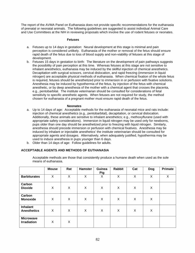
The report of the AVMA Panel on Euthanasia does not provide specific recommendations for the euthanasia of prenatal or neonatal animals. The following guidelines are suggested to assist individual Animal Care and Use Committees at the NIH in reviewing proposals which involve the use of rodent fetuses or neonates. Fetuses
a. Fetuses up to 14 days in gestation: Neural development at this stage is minimal and pain perception is considered unlikely. Euthanasia of the mother or removal of the fetus should ensure rapid death of the fetus due to loss of blood supply and non-viability of fetuses at this stage of development.
b. Fetuses 15 days in gestation to birth: The literature on the development of pain pathways suggests the possibility of pain perception at this time. Whereas fetuses at this stage are not sensitive to inhalant anesthetics, euthanasia may be induced by the skillful injection of chemical anesthetics. Decapitation with surgical scissors, cervical dislocation, and rapid freezing (immersion in liquid nitrogen) are acceptable physical methods of euthanasia. When chemical fixation of the whole fetus is required, fetuses should be anesthetized prior to immersion in or perfusion with fixative solutions. Anesthesia may be induced by hypothermia of the fetus, by injection of the fetus with chemical anesthetic, or by deep anesthesia of the mother with a chemical agent that crosses the placenta, e.g., pentobarbital. The institute veterinarian should be consulted for considerations of fetal sensitivity to specific anesthetic agents. When fetuses are not required for study, the method chosen for euthanasia of a pregnant mother must ensure rapid death of the fetus.
Neonates
a. Up to 14 days of age: Acceptable methods for the euthanasia of neonatal mice and rats include: injection of chemical anesthetics (e.g., pentobarbital), decapitation, or cervical dislocation. Additionally, these animals are sensitive to inhalant anesthetics; e.g., methoxyflurane (used with appropriate safety considerations). Immersion in liquid nitrogen may be used only for newborns, pups older than one day should be anesthetized prior to freezing with liquid nitrogen. Similarly, anesthesia should precede immersion or perfusion with chemical fixatives. Anesthesia may be induced by inhalant or injectable anesthetics’ the institute veterinarian should be consulted for appropriate agents and dosages. Alternatively, when adequately justified, hypothermia may be used to induce anesthesia in pups younger than 6 days.
b. Older than 14 days of age: Follow guidelines for adults. ACCEPTABLE AGENTS AND METHODS OF EUTHANASIA
Acceptable methods are those that consistently produce a humane death when used as the sole means of euthanasia.
Mouse Rat Hamster Guinea
Pig Rabbit Cat Dog Primate
Barbiturates
X X X X X X X X
Carbon Dioxide
X
X
X
X
X
X
X
Carbon Monoxide
X
X
X
X
X
X
X
Inhalant Anesthetics
X
X
X
X
X
X
X
Microwave Irradiation
X
X
82
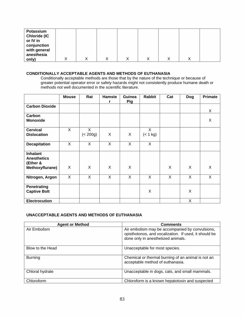
Potassium Chloride (IC or IV in conjunction with general anesthesia only)
X
X
X
X
X
X
X
CONDITIONALLY ACCEPTABLE AGENTS AND METHODS OF EUTHANASIA
Conditionally acceptable methods are those that by the nature of the technique or because of greater potential operator error or safety hazards might not consistently produce humane death or methods not well documented in the scientific literature.
Mouse Rat Hamste
r Guinea
Pig Rabbit Cat Dog Primate
Carbon Dioxide X
Carbon Monoxide
X
Cervical Dislocation
X X (< 200g)
X
X
X (< 1 kg)
Decapitation
X X X X X
Inhalant Anesthetics (Ether & Methoxyflurane)
X
X
X
X
X
X
X
Nitrogen, Argon
X X X X X X X X
Penetrating Captive Bolt
X
X
Electrocution X UNACCEPTABLE AGENTS AND METHODS OF EUTHANASIA
Agent or Method Comments Air Embolism Air embolism may be accompanied by convulsions,
opisthotonos, and vocalization. If used, it should be done only in anesthetized animals.
Blow to the Head Unacceptable for most species.
Burning Chemical or thermal burning of an animal is not an acceptable method of euthanasia.
Chloral hydrate Unacceptable in dogs, cats, and small mammals.
Chloroform Chloroform is a known hepatotoxin and suspected
83
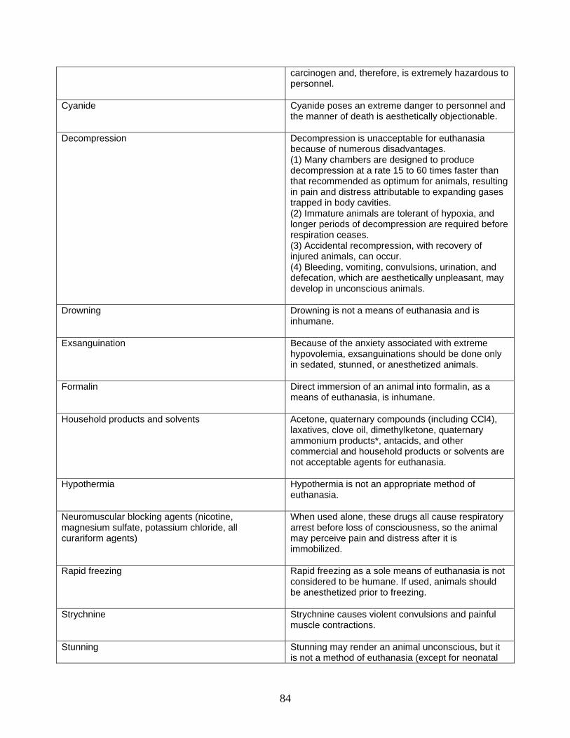
carcinogen and, therefore, is extremely hazardous to personnel.
Cyanide Cyanide poses an extreme danger to personnel and the manner of death is aesthetically objectionable.
Decompression Decompression is unacceptable for euthanasia because of numerous disadvantages. (1) Many chambers are designed to produce decompression at a rate 15 to 60 times faster than that recommended as optimum for animals, resulting in pain and distress attributable to expanding gases trapped in body cavities. (2) Immature animals are tolerant of hypoxia, and longer periods of decompression are required before respiration ceases. (3) Accidental recompression, with recovery of injured animals, can occur. (4) Bleeding, vomiting, convulsions, urination, and defecation, which are aesthetically unpleasant, may develop in unconscious animals.
Drowning Drowning is not a means of euthanasia and is inhumane.
Exsanguination Because of the anxiety associated with extreme hypovolemia, exsanguinations should be done only in sedated, stunned, or anesthetized animals.
Formalin Direct immersion of an animal into formalin, as a means of euthanasia, is inhumane.
Household products and solvents Acetone, quaternary compounds (including CCl4), laxatives, clove oil, dimethylketone, quaternary ammonium products*, antacids, and other commercial and household products or solvents are not acceptable agents for euthanasia.
Hypothermia Hypothermia is not an appropriate method of euthanasia.
Neuromuscular blocking agents (nicotine, magnesium sulfate, potassium chloride, all curariform agents)
When used alone, these drugs all cause respiratory arrest before loss of consciousness, so the animal may perceive pain and distress after it is immobilized.
Rapid freezing Rapid freezing as a sole means of euthanasia is not considered to be humane. If used, animals should be anesthetized prior to freezing.
Strychnine Strychnine causes violent convulsions and painful muscle contractions.
Stunning Stunning may render an animal unconscious, but it is not a method of euthanasia (except for neonatal
84

animals with thin craniums). If used, it must be immediately followed by a method that ensures death.
Tricaine methane sulfonate Should not be used for euthanasia of animals intended as food.
85