buddingandfission yeast casein kinase i isoforms have dual ...repository.cshl.edu/31403/1/kuret...
TRANSCRIPT
Molecular Biology of the CellVol. 5, 877-886, August 1994
Budding and Fission Yeast Casein Kinase I IsoformsHave Dual-specificity Protein Kinase ActivityMerl F. Hoekstra,*t Namrita Dhillon,* Gilles Carmel,*Anthony J. DeMaggio,* Richard A. Lindberg,§111 Tony Hunter,and Jeff Kuret*
*Icos Corporation, Bothell, Washington 98290; tCold Spring Harbor Laboratory, Cold SpringHarbor, New York 11724; §Department of Biology, University of California at San Diego, La Jolla,California 92093; and IlMolecular Biology and Virology Laboratory, The Salk Institute for BiologicalStudies, San Diego, California 92186
Submitted February 28, 1994; Accepted June 27, 1994Monitoring Editor: Michael H. Wigler
We have examined the activity and substrate specificity of the Saccharomyces cerevisiaeHrr25p and the Schizosaccharomyces pombe Hhpl, Hhp2, and Ckil protein kinase isoforms.These four gene products are isotypes of casein kinase I (CKI), and the sequence of theseprotein kinases predicts that they are protein serine/threonine kinases. However, each ofthese four protein kinases, when expressed in Escherichia coli in an active form, was rec-ognized by anti-phosphotyrosine antibodies. Phosphoamino acid analysis of 32P-labeledproteins showed phosphorylation on serine, threonine, and tyrosine residues. The E. coliproduced forms of Hhpl, Hhp2, and Ckil were autophosphorylated on tyrosine, and bothHhpl and Hhp2 were capable of phosphorylating the tyrosine-protein kinase syntheticpeptide substrate polymer poly-E4Yl. Immune complex protein kinases assays from S. pombecells showed that Hhpl-containing precipitates were associated with a protein-tyrosinekinase activity, and the Hhpl present in these immunoprecipitates was phosphorylatedon tyrosine residues. Although dephosphorylation of Hhpl and Hhp2 by Ser/Thr phos-phatase had little effect on the specific activity, tyrosine dephosphorylation of Hhpl andHhp2 caused a 1.8-to 3.1-fold increase in the Km for poly-E4Y, and casein. These datademonstrate that four different CKI isoforms from two different yeasts are capable ofprotein-tyrosine kinase activity and encode dual-specificity protein kinases.
INTRODUCTION
Casein kinase I (CKI) is a serine/threonine protein ki-nase activity that is detected in all eukaryotic cells (re-viewed in Tuazon and Traugh, 1991). Unlike most otherprotein kinases, CKI is acidotrophic, preferring acidicresidues in its substrates (Flotow and Roach, 1991).Substrates containing the motif Ser/Thr(PO4)-Xaa-Xaa-Ser/Thr also are recognized by CKI (Flotow et al., 1990),suggesting that prior phosphorylation can direct sub-strate specificity in vivo. This links the activity of CKIin a hierarchical protein phosphorylation relay wherebyCKI can amplify or dampen signal transduction and
tCorresponding author.liPresent address: AMGEN, Thousand Oaks, CA 91320-1789
allows for the recruitment of CKI into second messen-ger-dependent phosphorylation cascades (Roach, 1991).The sizes of CKI activities range from 25 to 60 kDa,
but CKI has been most frequently described as a single'30-kDa subunit (Tuazon and Traugh, 1991). Thebroad size distribution for CKI activity is most easilyexplained by the existence of multiple isoforms encodedby distinct genes and by alternative splice variants. Thisnotion has been confirmed by the cloning and analysisof CKI cDNAs from several eukaryotic species that en-code polypeptides ranging between 34 and 62 kDa.Saccharomyces cerevisiae has four different genes,HRR25, CKI1, CKI2, and CKI3 (Hoekstra et al., 1991;Robinson et al., 1992; Wang et al., 1992; Hoekstra,Dhillon, and DeMaggio, unpublished data) that encodeproteins with molecular weight between 55 and 62 kDa.
C) 1994 by The American Society for Cell Biology 877

M.F. Hoekstra et al.
Schizosaccharomyces pombe has at least four CKI genes,hhpl+, hhp2+, ckil+, and cki2+ (Dhillon and Hoekstra;1994, Wang et al.; 1994b), and the proteins encoded bythese genes range from 45 to 50 kDa. In vertebrates,four closely related CKI isoforms (a,fA, y, 5) have beenreported and these range from 34 to 49 kDa (Rowles etal., 1991; Zhai et al., 1992; Graves et al., 1993).
Several studies suggest where CKI might play an im-portant role in cellular metabolism and regulation. CKIshows cell cycle-dependent localization to mitotic spin-dles (Brockman et al., 1992), implicating a role for theenzyme in mitosis. Also, a CKI-like activity can be stim-ulated by insulin in a dose-dependent fashion and byIL-1 or tumor necrosis factor (Cobb and Rosen, 1983;Guy et al., 1991; Guesdon et al., 1993), and membrane-associated CKI activity can be inhibited by phosphati-dylinositol 4,5-bisphosphate (Brockman and Anderson,1991). Furthermore, sites phosphorylated by CKI bothin vivo and in vitro have been determined for cAMPresponsive element modulator (CREM) (de Groot et al.,1993), SV40 large T antigen (Cegielska and Virshup,1993), glycogen synthase (Roach, 1991), and p53 (Milneet al., 1992). In CREM, the phosphorylation by CKI af-fects the DNA-binding activity of the transcription fac-tor. For SV40, the residues phosphorylated by CKI areimportant for T antigen-driven replication capacity. Inglycogen synthase, CKI phosphorylation is involved ininhibiting enzyme activity. For p53, the role of CKIphosphorylation in regulating p53 activity is not un-derstood.The majority of eukaryotic protein kinases have been
classified into two groups based on their specificity forthe hydroxyamino acid phosphorylated in substrateproteins. One class, the protein serine/threonine ki-nases, modifies either Ser, Thr, or both. The other class,the protein-tyrosine kinases, phosphorylate Tyr. Re-cently, a number of protein kinases that phosphorylateboth at Tyr and at Ser/Thr (so called "dual-specificity"enzymes) have been described (Lindberg et al., 1992).In S. cerevisiae, examples of these enzymes includeMcklp and Spklp (Dailey et al., 1990, Stern et al., 1991;Lim et al., 1993, Zheng et al., 1993). Although the genestructure and classical characterization of CKI activityhas revealed that CKI is most closely related to serine/threonine protein kinases, several experiments havesuggested that CKI might have a promiscuous substratespecificity. A preparation of CKI from human eryth-rocytes was found to catalyze the phosphorylation ofprotein tyrosine in a variety of substrates such as an-giotensin-II, tyrosine-containing peptides, alkylatedbovine serum albumin (BSA), band 3, and ankyrin (Luand Tao, 1986).The recent identification of CKI-encoding genes in S.
cerevisiae and S. pombe has allowed an examination ofthe significance of CKI activity. In this report we extendthe biochemical description of yeast CKI (DeMaggio etal., 1992; Vancura et al., 1993; Carmel et al., 1994) and
report a novel characteristic of S. cerevisiae and S. pombeforms of CKI, namely the ability to phosphorylate serine,threonine, and tyrosine residues.
MATERIALS AND METHODS
MaterialsAntiphosphotyrosine antibodies were monoclonal antibody 4G10 fromUpstate Biotechnology (Lake Placid, NY) or rabbit polyclonal anti-phosphotyrosine serum (Kamps and Sefton, 1988) kindly providedby Bart Sefton (The Salk Institute, San Diego, CA). Recombinant pro-tein phosphatases were the generous gift of David Barford (Cold SpringHarbor Laboratory, Cold Spring Harbor, NY). Escherichia coli expres-sion plasmids pRSET:: HRR25 and pRSET:: hrr25-KR38 containHRR25 fused at its translation initiation codon to the bacteriophageT7 gene 10 initiation codon. Details of the construction of these plas-mids are outlined in DeMaggio et al. (1992). Expression of the hhpl+gene in E. coli for the experiments shown in Figure 2 used the T7gene 10-based expression plasmid pRSET6B (Invitrogen, La Jolla, CA).This construct and the HA epitope-tagged construct were describedin Dhillon and Hoekstra (1994). In vivo labeling of Hrr25p and Hhpl,as shown in Figures 1 and 2, exactly followed the conditions andmethods described in DeMaggio et al. (1992) and Lindberg et al. (1993).Anti-phosphotyrosine antibody probing of E. coli lysates was as de-scribed by Lindberg et al. (1993), and the in vivo labeling of Hrr25pwas identical to that shown in DeMaggio et al. (1992). For experimentsusing rabbit anti-phosphotyrosine antibody, Western blots wereprobed as described by Kamps and Sefton (1988).
For protein overproduction, the cDNAs for hhpl+ and hhp2+ weremodified by adding useful restriction sites (Nde I at the initiation codonand Xho I just after the termination codon) using polymerase chainreaction (PCR) as described previously (Wang et al., 1992). The re-sulting PCR fragments were digested with Nde I/Xho I and ligatedinto the Nde I/Xho I sites of expression vector pET-15b. This derivativeof the T7 expression system (Studier et al., 1990) drives the overpro-duction of proteins fused to an N-terminal, 20-residue peptide (Met-Gly -Ser-Ser-His-His-His-His -His - His -Ser -Ser -Gly -Leu -Val -Pro -
Arg-Gly-Ser-His-) that allows affinity purification on immobilizednickel columns (Hochuli et al., 1987). The final constructs (pET-15b/hhpl, pET-15b/hhp2) were transformed into BL21(DE3) cells to createthe strains used for protein overproduction. Expression and purificationof poly-His tagged CKI were as described by Carmel et al. (1994).
E. coli cells containing the pET-15b-based plasmids were grown inLB broth containing 200 jig/ml ampicillin at 37°C to an A600nm of 1.0,at which point isopropyl f-D-thiogalactopyranoside (IPTG) was addedto a final concentration of 1 mM. After 3 h of induction, cells wereharvested by centrifugation (20 min at 3000 X g, 4°C), washed withSTE buffer (10 mM tris(hydroxymethyl)aminomethane (Tris)-HCl pH8.0, 100 mM NaCl, 1 mM EDTA), and stored at -70°C until used. A3-1 growth typically yielded 8 g (wet weight) of cells.
Purification of Polyhistidine-tagged Hhpl and Hhp2All steps were carried out at 4°C and were identical for Hhpl andHhp2. Frozen cells were thawed, resuspended in 5 vol of lysis buffer(20 mM Tris-HCl pH 7.9, 0.5 M NaCl, 5 mM imidazole, 10% glycerol,1 mM phenylmethylsulfonyl fluoride [PMSF], and 5 Ag/ml each ofleupeptin, aprotinin, and pepstatin), and ruptured by two passesthrough a French press operated at 1000 psi. The resulting homogenatewas sonicated briefly (= 15 s) to shear nucleic acids, made 0.1% Brij35, and centrifuged (100 000 X g, 1 h) to yield a clear supernatant(crude extract).
After filtration through a 0.45-,um filter, the extract was loadeddirectly onto a 4-ml Ni2+-nitrolotriacetate-agarose column preequili-brated in lysis buffer containing 0.1% Brij 35. The column was washedwith 200 ml (50 bed volumes) of lysis buffer containing 0.05% Brij35 and was developed with sequential 20-ml steps of lysis buffercontaining 0.02% Brij 35 and 10, 20, 40, and 60 mM imidazole. Frac-
Molecular Biology of the Cell878

Dual-specificity of Casein Kinase I
tions containing casein kinase activity (eluting at 40 mM imidazole)were pooled, brought to 75% saturation with solid (NH4)2SO4, stirred20 min, and then centrifuged 20 min at 27 000 X g. The resultingpellet was resuspended in Buffer A (10 mM N-2-hydroxyethylpiper-azine-N'-2-ethanesulfonic acid [HEPES] pH 7.5, 250 mM NaCl, 0.1mM ethylene glycol-bis(f3-aminoethyl ether)-N,N,N`,N'-tetraacetic acid[EGTA]) to a final volume of 4 ml and loaded directly onto a 180-mlcolumn (1.6 X 91 cm) of Sephacryl S-100 HR gel filtration mediumequilibrated and run in Buffer A at 30 ml/h. Fractions containingcasein kinase activity were pooled, diluted fivefold with 0.02% Brij35 (to reduce the NaCl concentration to 50 mM), and loaded onto aMonoQ 5/5 high-pressure liquid chromatography column equilibratedin Buffer B (10 mM Tris-HCl pH 7.5, 0.02% Brij 35, 0.1% 2-mercap-toethanol) containing 50 mM NaCl. The gradient was washed with5 bed volumes of this buffer and developed with a 20 ml linear gradientof increasing NaCl (from 50 to 400 mM). Fractions containing caseinkinase activity were pooled, concentrated by dialysis against storagebuffer (50% glycerol, 10 mM 3-(N-morpholino) propanesulfonic acid[MOPS] pH 7.0, 250 mM NaCl, 0.1 mM EGTA, 0.02% Brij 35, 1 mMdithiothreitol [DTT]), and stored at -20°C.
Phosphoamino Acid AnalysisPhosphoamino acid analysis using acid hydrolysis were carried outon proteins bound to Immobilon as described by Lindberg et al. (1993).High voltage, two-dimensional, thin-layer electrophoresis of hydro-lyzed phosphoprotein samples was performed as described by Boyleet al. (1991).
Analysis of Hhpl from S. pombeNative protein lysates were prepared from logarithmically growingcultures (at an approximate cell density of 5 X 10⁶ cells ml-') of S.pombe in synthetic media. Briefly, 1 x 10' cells were resuspended in20 ,ul of lysis buffer (25 mM MOPS pH 7.2, 60 mM ,B-glycerolphos-phate, 15 mM MgCl2, 15 mM EGTA, 1 mM DTT, 0.1 mM Na3VO4,1% Triton X-100, 1 mM PMSF, 20 M ml-' leupeptin, and 2% Trasylol[Miles, Kankakee, IL]) and 5 vol of acid-washed glass beads wereadded to the cell suspension. Cells were vortexed five to six times for30 sec at 2-min intervals, and the lysate was separated from the glassbeads by piercing a hole through the microcentrifuge tube followedby a brief centrifugation into another microcentrifuge tube at 100 Xg for 1 min. Lysates were cleared at 16 000 X g for 10 min at 4°C.Supernatants were assayed for total protein content using Bradford'sreagent, and equivalent amounts of protein from each strain wereused for immunoprecipitation reactions. Pellets from the clearing spinwere resuspended in a small volume (20 jAl) of boiling buffer (10 mMTris-HCl pH 8.0, 1% sodium dodecyl sulfate [SDS]) and heated to100°C for 3 min. The volume was brought up to 0.5 ml with lysisbuffer, and this fraction, designated as the pellet fraction, was usedfor subsequent immunoprecipitation reactions and Western blot anal-ysis.
Immunoprecipitation reactions were performed in a reaction volumeof 0.5 ml in lysis buffer. Five micrograms of affinity-purified mono-clonal antibody (12CA5) against the HA epitope (BabCo, Berkley,CA) were incubated with -0.25-0.5 mg of total protein on ice for60 min, followed by the addition of rabbit anti-mouse antisera (5 ,g)for a further 45 min. Reactions were precipitated with protein A aga-rose (Repligen, Minneapolis, MN) with end-over-end shaking for 60min at 4°C. The immunoprecipitates were washed six times in 1.0 mlof lysis buffer, and the protein A pellets were boiled in an equalvolume of SDS loading buffer for SDS-polyacrylamide gel electro-phoresis (PAGE). For in vitro kinase assays, immunoprecipitates wereincubated in 1X kinase buffer (50 mM Tris-HCl pH 7.5, 12 mM MgCl2,100 mM NaCl, 1 mM DTT) and [y-32P]ATP at a final concentrationof 1 MuM at 30°C for 10 min before boiling in SDS loading buffer andelectrophoresis.
Dephosphorylation ReactionsTo dephosphorylate phosphotyrosine residues, purified Hhpl andHhp2 were incubated at 37°C for 30 min with recombinant T-cell
protein tyrosine phosphatase (500:1 wt/wt) in T cell protein-tyrosinephosphatase (PTPase) buffer (10 mM Tris-HCl pH 7.5, 0.1% 2-mer-captoethanol, 0.02% Brij 35). Reactions were terminated by the ad-dition of an equal volume of 2 mM Na3VO4. To dephosphorylatephosphoserine and phosphothreonine residues, purified Hhpl andHhp2 were incubated at 37°C for 30 min with recombinant phos-phoprotein phosphatase 1 (5:1 wt/wt) in PrP buffer (10 mM Tris-HCl pH 7.4, 50 mM NaCl, 0.5 mM MnCl2, 0.1% 2-mercaptoethanol,0.02% Brij 35). Reactions were terminated by the addition of an equalvolume of 0.6 mM okadaic acid (OA). Terminated reactions weresubjected to SDS-gel electrophoresis or phosphotransferase assay asdescribed below.
Autophosphorylation of Hhpl on TyrosinePurified Hhpl was dephosphorylated by PTPase treatment as de-scribed above and diluted into phosphotransferase buffer (25 mM 2-(N-morpholino) ethanesulfonic acid [MES] pH 6.5, 50 mM NaCl, 15mM MgCl2, 2 mM EGTA) containing 1 mM Na3VO4. Autophos-phorylation was initiated at 37°C by the addition of ATP to a finalconcentration of 25 MM. The time course of tyrosine autophosphor-ylation (25 Mg/ml Hhpl) was determined by removing aliquots (100ng Hhpl) at 0, 5, 10, 15, and 30 min for phosphotyrosine quantitation(described below). The concentration dependence of tyrosine auto-phosphorylation was determined by incubating various concentrationsof Hhpl (5, 10, 25, and 50Mg/ml) for 15 min. Phosphotyrosine contentwas determined on 10-ng samples as described below.
Western Assay for PhosphotyrosineAutophosphorylation reactions were terminated by boiling (5 min) inthe presence of SDS sample buffer (3% SDS, 50 mM Tris-HCl pH6.8, 8% glycerol, 2% 2-mercaptoethanol). Samples (10-100 ng Hhpl)were electrophoresed through 10% polyacrylamide gels and trans-ferred to nitrocellulose membranes as described previously (Wang etal., 1992). Phosphotyrosine was labeled with monoclonal antibody4G10 (1 Mg/ml) and detected by enhanced chemiluminescence(Amersham, Arlington Heights, IL) using sheep anti-mouse IgG con-jugated to horseradish peroxidase. Images were collected on X-rayfilm and quantified in arbitrary absorbance units by laser densitometry(Molecular Dynamics, Sunnyvale, CA).
Analytical MethodsPhosphotransferase assays with casein or poly-E4Y, as protein sub-strate were performed as described previously (Racker, 1991; Vancuraet al., 1993). Initial velocity measurements were carried out in duplicatewith casein (0.45, 0.6, 1.0, 2.0, and 4.0 mg/ml) or poly-E4Y1 (10, 25,100, 200, 500 Mug/ml) as the varied substrate. When necessary, thephosphatase inhibitors Na3VO4 (1 mM) or OA (0.3 mM) were included.Estimates of Km, Vmax, and their SE were calculated as described byWilkinson (1961). Protein-bound phosphate was determined in trip-licate as described by Carmel et al. (1994) and reported as mean ±SD.SDS-polyacrylamide gels were prepared as described previously(Vancura et al., 1993). Molecular mass markers included BSA (66.2kDa), ovalbumin (42.7 kDa), bovine carbonic anhydrase (29.0 kDa),and a-lactoglobulin (18.4 kDa).The percentage of CKI molecules containing phosphotyrosine was
estimated after immunoprecipitation with antiphosphotyrosine an-tibody 4G10. A constant amount of protein kinase (160 nM) wasincubated with increasing amounts of 4G10 (0, 0.08, 0.16, 0.8, 1.6,MM) in 20 Ml on ice for 1 h and precipitated with an equal volume ofprotein-A agarose (50% slurry). Kinase activity remaining in the su-pernatant was assayed in duplicate as described above. The resultantdata was fit to a binding isotherm to calculate the maximum amountof immunoprecipitable kinase ± SE of the estimate.
Vol. 5, August 1994 879
M.F. Hoekstra et al.
RESULTSBaker's Yeast Hrr25p Is a Dual-specificityProtein KinaseTo characterize the substrate specificity of CKI, selectedyeast isoforms of the enzyme were expressed in E. coliand assayed for the presence of phosphotyrosine. Thegoal was to determine whether the observation by Luand Tao (1986) that erythrocyte CKI contains tyrosinekinase activity could be extended to recombinant yeastisoforms of CKI. E. coli was used as a host for theseexperiments because many CKI isoforms are efficientlyexpressed in bacteria (DeMaggio et al., 1992; Zhai et al.,1992; Graves et al., 1993; Carmel et al., 1994), and be-cause E. coli contains little if any endogenous proteintyrosine kinase activity (Lindberg et al., 1993).
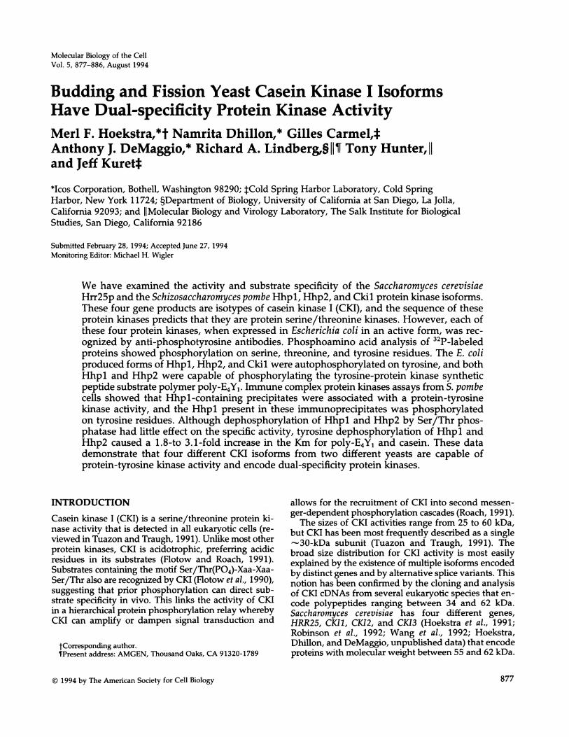
First, we examined the ability of Hrr25p, a buddingyeast CKI isoform, to react with an anti-phosphotyrosineantibody. As shown in Figure 1, extracts containing fullyactive, wild-type Hrr25p show a strong anti-phospho-tyrosine immunoreactivity centered around 60 kDa.Conversely, this immunoreactivity was not observed incontrol extracts containing either catalytically inactiveHrr25p-K38R (Figure 1) (DeMaggio et al., 1992) or thecatalytic subunit of the mammalian cAMP-dependentprotein kinase. In parallel comparisons, the anti-phos-photyrosine immunoreactive staining is as robust as thatobserved with E. coli extracts containing the PYT dual-specificity protein kinase or LYN tyrosine protein kinases(Lindberg et al., 1993), indicating that the anti-phos-photyrosine reactivity is specific to extracts containingactive Hrr25p. We conclude that Hrr25p contains phos-photyrosine residues as a result of autophosphorylation.
Second, to confirm the presence of phosphotyrosine,32P-radiolabelled E. coli protein extracts prepared fromtransformants containing either HRR25, hrr25-K38R, orvector alone were fractionated by gel electrophoresis,transferred to membranes, and subjected to autoradi-ography. As shown in Figure 1, wild-type Hrr25p, butnot Hrr25p-K38R or the vector control, catalyzed thetransfer of phosphate to endogenous E. coli proteins inaddition to autophosphorylation. Phosphoamino acidanalysis performed on autophosphorylated Hrr25pconfirmed the presence of phosphotyrosine that, byCerenkov counting, comprised 10% of the total 32pbound to Hrr25p (Figure 1).To determine if Hrr25p could phosphorylate sub-
strates other than itself on tyrosine, four radiolabeledproteins, including Hrr25p, were subjected to phos-phoamino acid analysis (Figure 1). The results revealedthat in addition to phosphoserine and phosphothreo-nine, three of the four proteins contained phospho-tyrosine. These observations lead us to conclude thatHrr25p is a dual specificity kinase.
Fission Yeast CKI Isoforms Are Dual-specificityKinasesLike budding yeast, fission yeast has a four-gene familyof CKI isoforms. Two of these genes, hhpl+ and hhp2+,
1 2 3 4 5 6
Figure 1. Hrr25p has dual-*3t specific protein kinase activity.
E. coli transformants were125 68 grown to mid-log phase and88 induced with IPTG. Lysates
were prepared as previously65 4_ 5 described (Lindberg et al.,56 * 1993). Lanes 1-3 are samples
that were transferred to Im-mobilon and probed with a
38 30 rabbit anti-phosphotyrosine34 antibody. The samples in lanesit w 4-6 are from in vivo-labeled
21 phosphoproteins as shown inDeMaggio et al. (1992). Pres-
pSer tained molecular weight mark-
pTh ers (Sigma) were used as sizestandards. The relative migra-*O pTyr tion of Hrr25p is indicated byan arrow on the left hand sideof lane 1. Lanes 1 and 4, vectoralone; lanes 2 and 5, wild-type
HRR25; lanes 3 and 6, catalytically inactive hrr25-K38R. The bottomleft panel shows phosphoamino acid analysis of in vivo-labeledHrr25p, and the right panel shows the migration pattern of ninhydrin-stained standards. Additional phosphoproteins that were examinedfor phosphoamino acids are indicated in lane 5. *, proteins that containphosphoserine, phosphothreonine, and phosphotyrosine. 0, proteinthat contains phosphoserine and phosphothreonine.
encode protein kinases that are homologous to Hrr25pand participate in DNA damage repair (Dhillon andHoekstra, 1994). The other two genes, ckil+ and cki2+,are more distantly related to Hrr25p and are found ex-clusively in the cytoplasm (Wang et al., 1994b). To de-termine whether fission yeast CKI isoforms share dualspecificity protein kinase activity, Hhpl, Hhp2, and Ckilwere expressed in E. coli and examined for the presenceof phosphotyrosine.
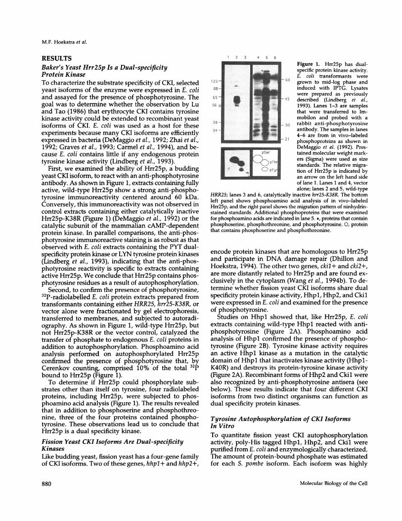
Studies on Hhpl showed that, like Hrr25p, E. coliextracts containing wild-type Hhpl reacted with anti-phosphotyrosine (Figure 2A). Phosphoamino acidanalysis of Hhpl confirmed the presence of phospho-tyrosine (Figure 2B). Tyrosine kinase activity requiresan active Hhpl kinase as a mutation in the catalyticdomain of Hhpl that inactivates kinase activity (Hhpl-K4OR) and destroys its protein-tyrosine kinase activity(Figure 2A). Recombinant forms of Hhp2 and Ckil werealso recognized by anti-phosphotyrosine antisera (seebelow). These results indicate that four different CKIisoforms from two distinct organisms can function asdual specificity protein kinases.
Tyrosine Autophosphorylation of CKI IsoformsIn VitroTo quantitate fission yeast CKI autophosphorylationactivity, poly-His tagged Hhpl, Hhp2, and Ckil werepurified from E. coli and enzymologically characterized.The amount of protein-bound phosphate was estimatedfor each S. pombe isoform. Each isoform was highly
Molecular Biology of the Cell880
Dual-specificity of Casein Kinase I
kDaA
97.4-
68-
43-
29-
18.4-
B kDa
1 2 3 4 5 6
0- EELk
1 2
Figure 2. Hhpl has dual-spedfic protein kinase activity. (A) Westernblot analysis of lysates prepared from E. coli cells expressing eitherwild-type Hhpl (lanes 1 and 4), Hhp1-K40R (lanes 2 and 5), or vectoralone (lanes 3 and 6) that had been induced with IPTG (lanes 1-3)or from uninduced cells (lanes 4-6). Lysates were resolved on a 10%SDS-polyacrylamide gel, transferred to nitrocellulose, and probed withanti-phosphotyrosine polyclonal antiserum. (B) E. coli cells carryingthe vector alone (lane 1) or wild-type hhpl+ (lane 2) were labeledwith orthophosphate, and lysates prepared from these cultures wereanalyzed on a 10% SDS-polyacrylamide gel. The right panel showsthe phosphoamino acid content of the 45-kDa phosphoprotein cor-responding to autophosphorylated Hhpl.
phosphorylated, containing 4.8 ± 0.4 (Hhpl), 13.1 ± 1.2(Hhp2), and 12.3 ± 0.6 (Ckil) mol phosphate/mol en-zyme. The percentage of molecules containing phos-photyrosine was estimated by immunoprecipitation ofeach purified enzyme with antiphosphotyrosine anti-body 4G10. The results revealed that 52.7 ± 0.6% ofrecombinant Hhpl, 36.9 ± 0.4% of Hhp2, and 23.9+ 2.8% of Ckil molecules contained phosphotyrosineas isolated from E. coli. We conclude that each of thesethree fission yeast CKI homologues contain significant
AM 2 3 4
B2 34
66.2- -_-42.7 -
29.0 -
18.4 -
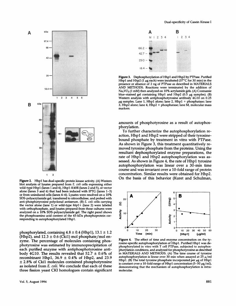
Figure 3. Dephosphorylation of Hhpl and Hhp2 by PTPase. PurifiedHhpl and Hhp2 (1 jg each) were incubated (37°C for 30 min) in thepresence or absence of 2 ng of PTPase as described in MATERIALSAND METHODS. Reactions were terminated by the addition ofNa3VO4 (1 mM) then analyzed on 10% acrylamide gels. (A) Coomassieblue-stained gel containing Hhpl and Hhp2 (0.5 yg samples). (B)Western analysis with antiphosphotyrosine antibody 4G10 on 0.25ug samples. Lane 1, Hhpl alone; lane 2, Hhpl + phosphatase; lane3, Hhp2 alone; lane 4, Hhp2 + phosphatase; lane M, molecular massmarkers.
amounts of phosphotyrosine as a result of autophos-phorylation.To further characterize the autophosphorylation re-
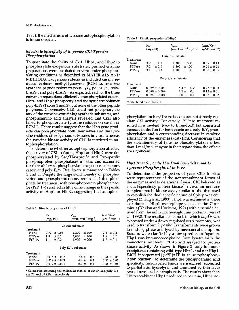
action, Hhpl and Hhp2 were stripped of their tyrosine-bound phosphate by treatment in vitro with PTPase.As shown in Figure 3, this treatment quantitatively re-moved tyrosine phosphate from the proteins. Using theresultant dephosphorylated enzyme preparations, therate of Hhpl and Hhp2 autophosphorylation was as-sessed. As shown in Figure 4, the rate of Hhpl tyrosineautophosphorylation was linear over a 30-min timecourse and was invariant over a 10-fold range of proteinconcentration. Similar results were obtained for Hhp2.On the basis of this behavior (Kuret and Schulman,
2
-
.2t
0
0
0 10 20 30Time (min)
st 5 -
E
4.
3.
3! -
2
1
u -1 .
0 10 20
[Hhpl]30 40 50
(igIml)60
Figure 4. The effect of time and enzyme concentration on the ty-rosine-specific autophosphorylation of Hhpl. Purified Hhpl was de-phosphorylated in vitro with T cell PTPase, subjected to autophos-phorylation conditions, and analyzed for phosphotyrosine as describedin MATERIALS AND METHODS. (A) The time course of tyrosineautophosphorylation is linear over 30 min when assayed at 25 ,u/mlHhpl. (B) The total tyrosine phosphate incorporated per Mg of Hhplis constant over a 10-fold range of Hhpl concentration (5-50 ,g/ml),demonstrating that the mechanism of autophosphorylation is intra-molecular.
Vol. 5, August 1994
B
0
11
881
M.F. Hoekstra et al.
1985), the mechanism of tyrosine autophosphorylationis intramolecular.
Substrate Specificity of S. pombe CKI TyrosinePhosphorylationTo quantitate the ability of Ckil, Hhpl, and Hhp2 tophosphorylate exogenous substrates, purified enzymepreparations were incubated in vitro under phosphor-ylating conditions as described in MATERIALS ANDMETHODS. Exogenous substrates included casein, re-duced carboxy methyl-lysozyme (RCM-L), and thesynthetic peptide polymers poly-E1Yl, poly-E4Y1, poly-E1A1Yj, and poly-E6A3Y1. As expected, each of the threeenzyme preparations efficiently phosphorylated casein.Hhpl and Hhp2 phosphorylated the synthetic polymerpoly-E4Y1 (Tables 1 and 2), but none of the other peptidepolymers. Conversely, Ckil could not phosphorylateany of the tyrosine-containing synthetic substrates, andphosphoamino acid analysis revealed that Ckil alsofailed to phosphorylate tyrosine residues on casein orRCM-L. These results suggest that the Hhp gene prod-ucts can phosphorylate both themselves and the tyro-sine residues of exogenous substrates in vitro, whereasthe tyrosine kinase activity of Ckil is restricted to au-tophosphorylation.To determine whether autophosphorylation affected
the activity of CKI isoforms, Hhpl and Hhp2 were de-phosphorylated by Ser/Thr-specific and Tyr-specificphosphoprotein phosphatases in vitro and examinedfor their ability to phosphorylate exogenous substratescasein and poly-E4Y1. Results are summarized in Tables1 and 2. Despite the large stoichiometry of phospho-serine and phosphothreonine, removal of this phos-phate by treatment with phosphoprotein phosphatase-1y (PrP-1'y) resulted in little or no change in the specificactivity of Hhpl or Hhp2, suggesting that autophos-
Table 1. Kinetic properties of Hhpl
Km Vmax kcat/Kma(mg/ml) (nmol min-' mg-') (gM-1 min1)
Casein substrateTreatmentNone 0.77 ± 0.05 2,200 ± 100 2.8 ± 0.2PTPase 1.8 ± 0.2 3,000 ± 200 1.6 ± 0.2PrP-ly 1.1 ± 0.2 1,900 ± 200 1.7 ± 0.4
Poly-E4YI substrate
TreatmentNone 0.015 ± 0.002 7.4 ± 0.2 0.66 ± 0.09PTPase 0.028 ± 0.003 6.4 ± 0.2 0.31 ± 0.03PrP-1ly 0.012 ± 0.001 6.1 ± 0.1 0.68 ± 0.06
a Calculated assuming the molecular masses of casein and poly-E4YIare 22 and 30 kDa, respectively.
Table 2. Kinetic properties of Hhp2
Km Vmax kcat/Kma(mg/ml) (nmol min' mg-') (LM-' min-')
Casein substrateTreatmentNone 3.9 ± 1.1 1,300 ± 300 0.35 ± 0.13PTPase 7.2 ± 2.0 1,800 ± 400 0.26 ± 0.20PrP-ly 3.1 ± 0.3 1,100 ± 100 0.37 ± 0.05
Poly-E4Yl substrate
TreatmentNone 0.029 ± 0.003 5.4 ± 0.2 0.27 ± 0.03PTPase 0.089 ± 0.009 7.1 ± 0.4 0.12 ± 0.01PrP-l1y 0.025 ± 0.001 10.0 ± 0.1 0.57 ± 0.02
a Calculated as in Table 1.
phorylation on Ser/Thr residues does not directly reg-ulate CKI activity. Conversely, PTPase treatment re-sulted in a modest (two- to three-fold) but consistentincrease in the Km for both casein and poly-E4Yl phos-phorylation and a corresponding decrease in catalyticefficiency of the enzymes (kcat/Km). Considering thatthe stoichiometry of tyrosine phosphorylation is lessthan 1 mol/mol enzyme in the preparations, the effectsare significant.
hhpl from S. pombe Has Dual Specificity and IsTyrosine Phosphorylated In VivoTo determine if the properties of yeast CKIs in vitrowere representative of the nonrecombinant forms ofthe enzymes and to determine if yeast CKI behaved asa dual-specificity protein kinase in vivo, an immunecomplex protein kinase assay similar to the that usedto establish the dual-specific nature of Spklp was em-ployed (Zheng et al., 1993). Hhpl was examined in theseexperiments. Hhpl was epitope-tagged at the C-ter-minus (Dhillon and Hoekstra, 1994) with a peptide de-rived from the influenza hemaglutinin protein (Tyers etal., 1992). The resultant construct, in which hhpl+ wasexpressed under a down-regulated nmtl promoter, wasused to transform S. pombe. Transformants were grownto mid-log phase and lysed by mechanical disruption.Extracts were clarified by a low speed centrifugation.Hhpl was immunoprecipitated from lysates with themonoclonal antibody 12CA5 and assayed for proteinkinase activity. As shown in Figure 5, only immuno-precipitates containing wild-type Hhpl, and not Hhpl-K40R, incorporated [,y-32P]ATP in an autophosphory-lation reaction. To determine the phosphoamino acidspecificity, radiolabeled bands were excised, subjectedto partial acid hydrolysis, and examined by thin-layertwo-dimensional electrophoresis. The results show that,like recombinant Hhpl produced in bacteria, Hhpl iso-
Molecular Biology of the Cell882
Dual-specificity of Casein Kinase I
kDa
200 -
97-
68 -
43 -
29 -
..1. .4"
a
1 2
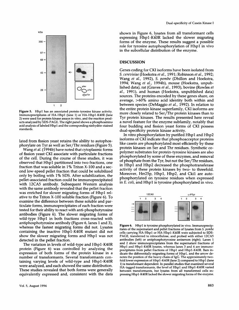
Figure 5. Hhpl has an associated protein-tyrosine kinase activity.Immunoprecipitates of HA-Hhpl (lane 1) or HA-Hhpl-K40R (lane2) were used for protein kinase assays in vitro, and the reaction prod-ucts analyzed by SDS-PAGE. The right panel shows a phosphoaminoacid analysis of labeled Hhpl and the corresponding ninhydrin-stainedstandards.
lated from fission yeast retains the ability to autophos-phorylate on Tyr as well as Ser/Thr residues (Figure 5).Wang et al. (1994b) have noted that cytoplasmic forms
of fission yeast CKI associate with particulate fractionsof the cell. During the course of these studies, it wasobserved that Hhpl partitioned into two fractions, onefraction that was soluble in 1% Triton X-100 and a sec-ond low-speed pellet fraction that could be solubilizedonly by boiling with 1% SDS. After solubilization, thepellet-associated fraction could be immunoprecipitatedwith 12CA5 antibody. Subsequent Western analysiswith the same antibody revealed that the pellet fractionwas enriched for slower migrating forms of Hhpl rel-ative to the Triton X-100 soluble fraction (Figure 6). Toexamine the difference between these soluble and par-ticulate forms, immunoprecipitates of each fraction weretested for their ability to react with anti-phosphotyrosineantibodies (Figure 6). The slower migrating forms ofwild-type Hhpl in both fractions cross-reacted withantiphosphotyrosine antibody (Figure 6, lanes 1 and 3),whereas the fastest migrating forms did not. Lysatescontaining the inactive Hhpl-K40R mutant did notshow the slower migrating forms and Hhpl was notdetected in the pellet fraction.The variation in levels of wild-type and Hhpl-K40R
protein (Figure 6) was controlled by analyzing theexpression of both forms of the protein kinase in anumber of transformants. Several transformants con-taining varying levels of wild-type and Hhpl-K40Rwere analyzed, and each showed the same distribution.These studies revealed that both forms were generallyequivalently expressed and, consistent with the data
shown in Figure 6, lysates from all transformant cellsexpressing Hhpl-K40R lacked the slower migratingforms of the enzyme. These results suggest a possiblerole for tyrosine autophosphorylation of Hhpl in vivoin the subcellular distribution of the enzyme.
DISCUSSION
Genes coding for CKI isoforms have been isolated fromS. cerevisiae (Hoekstra et al., 1991; Robinson et al., 1992;Wang et al., 1992), S. pombe (Dhillon and Hoekstra,1994; Wang et al., 1994b), mouse (Hoekstra, unpub-lished data), rat (Graves et al., 1993), bovine (Rowles etal., 1991), and human (Hoekstra, unpublished data)sources. The proteins encoded by these genes share, onaverage, >60% amino acid identity both within andbetween species (DeMaggio et al., 1992). In relation tothe entire protein kinase superfamily, CKI isoforms aremore closely related to Ser/Thr protein kinases than toTyr protein kinases. The results presented here reveala novel feature for the enzyme subfamily, notably thatfour budding and fission yeast forms of CKI possessdual-specificity protein kinase activity.
In vitro phosphorylation by purified Hhpl and Hhp2isoforms of CKI indicate that phosphoacceptor proteinslike casein are phosphorylated most efficiently by theseprotein kinases on Ser and Thr residues. Synthetic co-polymer substrates for protein-tyrosine kinases are alsophosphorylated by some of these enzymes, and removalof phosphate from the Tyr, but not the Ser/Thr residues,in Hhpl and Hhp2 decreased the phosphotransferaseactivity of these protein kinases by two- to threefold.Moreover, Hrr25p, Hhpl, Hhp2, and Ckil are auto-phosphorylated on tyrosine residues when expressedin E. coli, and Hhpl is tyrosine phosphorylated in vivo.
12CA5 c-PTyr
1 2 3 4 1 2 3 4
Figure 6. Hhpl is tyrosine phosphorylated in vivo. Immunoprecip-itates of the supematant and pellet fractions of lysates from S. pombecells carrying HA-Hhpl or HA-Hhpl-K40R were subjected to SDS-PAGE, transferred to nitrocellulose, and probed with either 12CA5antibodies (left) or antiphosphotyrosine antiserum (right). Lanes 1and 2 show immunoprecipitates from the supernatant fractions ofHhpl and Hhpl-K40R lysates, whereas lanes 3 and 4 are immuno-precipitates from pellet fractions of Hhpl and Hhpl-K40R. Bars in-dicate the differentially migrating forms of Hhpl, and the arrow de-notes the position of the heavy chain of IgG. The approximately two-fold lower expression of Hhpl-K40R (lane 2) compared to Hhpl (lane1) is transformant dependent. In parallel studies that examined severalHA-tagged transformants, the level of Hhpl and Hhpl-K40R variedbetween transformants, but lysates from all transformed cells ex-pressing Hhpl-K40R lacked the slower migrating forms of the enzyme.
Vol. 5, August 1994 883

M.F. Hoekstra et al.
Hhpl activity is required for this modification, sug-gesting that the tyrosine phosphorylation of Hhpl invivo is because of autophosphorylation. Thus, tyrosinephosphorylation of Hhpl may have a physiological sig-nificance, and consistent with this suggestion we finddifferential fractionation profiles for the tyrosine-phos-phorylated form.
In addition to demonstrating that yeast forms of CKIare dual-specificity protein kinases, tyrosine dephos-phorylation of these enzymes affects their in vitro ac-tivity. Dephosphorylation of Hhpl and Hhp2 with aserine/threonine phosphatase has little effect on theability to phosphorylate casein or poly-E4Y1. Treatmentof Hhpl and Hhp2 with T cell PTPase quantitativelyremoves phosphotyrosine, and the dephosphorylatedenzymes autophosphorylate on tyrosine in vitro. Thetyrosine-dephosphorylated enzymes show an increasedKm and reduced catalytic efficiency for casein and poly-E4Y1, suggesting that tyrosine phosphorylation of theseenzymes might positively regulate their activity. Cou-pled with the observation that tyrosine-phosphorylatedHhpl shows differential fractionation, these resultssuggest that the tyrosine kinase activity of these en-zymes might be important for their optimum activity invivo.Hhpl, Hhp2, Ckil, and Hrr25p join a growing list of
protein kinases in yeast that have been implicated asdual-specificity kinases. Based on their primary se-quence, these protein kinases are structurally classifiedwith the Ser/Thr protein kinase subfamily. The Mcklpprotein kinase from baker's yeast was first identified byvirtue of its ability to phosphorylate poly(E, Y) (Daileyet al., 1990). The Mcklp gene product is important foryeast kinetochore function in mitosis and for earlymeiotic gene expression (Neigeborn and Mitchell, 1991;Shero and Hieter, 1991). The Spklp protein kinase isan essential S-phase-specific gene product that is a nu-clear Ser/Thr/Tyr kinase (Zheng et al., 1993). It is sug-gested that Spklp plays an important role in regulatingDNA synthesis. In the mating type cascade in bothyeasts, mitogen-activated protein kinases (MAPKs) andtheir activators have been identified. These protein ki-nases include Fus3p, Ksslp, and Ste7p in S. cerevisiaeand Byrl and Byr2 in S. pombe (Neiman et al., 1993;Zhou et al., 1993). Regulation of MAPK activity is viaphosphorylation of a sequence motif, -TEY-, seven res-idues N-terminal to the conserved kinase domain VIII(-APE-), and phosphorylation of both Thr and Tyr isessential for MAPK signal transduction activity.Of particular note for yeast dual-specificity protein
kinases is that these enzymes are important for signalamplification. CKI isoforms are acidotrophic enzymesthat are characterized by their ability to recognize priorphosphorylated substrates and consequently feed intosecond messenger pathways. MAPK isoforms are criticalfor the rapid amplification of external signals that stim-ulate transcription and proliferation. Given the diverse
but common feature of signal amplification for thesedual-specificity protein kinases, perhaps the capacity tophosphorylate Ser, Thr, and Tyr was independently ac-quired by a number of enzymes.Hhpl joins glycogen synthase kinase-3 (GSK-3), cdc2,
and MAPK as protein serine/threonine kinases that aretyrosine phosphorylated in vivo (Featherstone andRussell, 1991; Davis, 1993; Hughes et al., 1993). GSK-3 protein requires tyrosine phosphorylation for function,and, when purified from tissues, GSK-3 contains phos-photyrosine. Phosphorylation of Tyr on GSK-3,B ap-pears to activate GSK-3f and Ser/Thr phosphorylationinactivates (Wang et al., 1994a). Further, the activity ofGSK-3 for stimulating transcription is dependent upontyrosine phosphorylation and a mutation in GSK-3 thatabolishes tyrosine phosphorylation also alleviates theinhibition effect of GSK-3 on c-Jun activity (Hughes etal., 1993). Hhpl shows tyrosine phosphorylation invivo, and this phosphorylation is required for Hhplsubcellular distribution as assessed by cell fractionation(Figure 6). The tyrosine phosphorylation of Hhpl ap-pears to be autophosphorylation, and this suggests thatHhpl might regulate its distribution within the cellthrough its ability to regulate its own phosphorylationstate. Perhaps the biological function of CKI Tyr phos-phorylation may not be direct regulation of enzyme ac-tivity but indirect regulation via control of cellular lo-calization. Indeed, Anderson and coworkers (Brockmanet al., 1992) have shown that human CKI isoforms mightparticipate in cell cycle progression as the CKIa isoformrelocalizes from vesicular structures in G2 phase cellsto the centrosome and mitotic spindles in mitotic cells.One important question arising form the discovery
that yeast forms of CKI can have associated protein-tyrosine kinase activity is whether human or other me-tazoan forms of this ubiquitous protein kinase showsimilar activity. To date, there is little experimental ev-idence regarding this notion. One report (Lu and Tao,1986) has shown that an erythrocyte form of CKI iscapable of protein-tyrosine kinase activity. In this study,CKI was purified to apparent homogeneity from a hu-man erythrocyte cytosolic fraction. It was shown thatthe enzyme preparation could phosphorylate syntheticpeptide substrates for tyrosine protein kinases like poly-EY, angiotensin II, and alkylated BSA on tyrosine. LikeHhpl and Hhp2, the erythrocyte CKI preferred poly-E4Y1 over poly-E,Yl, and the erythrocyte enzyme wasalso capable of tyrosine phosphorylating band 3 proteinand ankyrin. Clearly, one important challenge will bethe elucidation of in vivo substrates for CKI.
ACKNOWLEDGMENTSWe want to thank D. Barford (Cold Spring Harbor Labs) for proteinphosphatases and Bart Sefton (Salk Institute) for anti-phosphotyrosineantibody. We also thank Paul Keamey for technical assistance andHelen Kim for helpful discussions. Supported in part by NationalInstitutes of Health grant 42816 (J.K.).
Molecular Biology of the Cell884

Dual-specificity of Casein Kinase I
REFERENCESBoyle, W.J., van der Geer, P., and Hunter, T. (1991). Phosphopeptidemapping and phosphoamino acid analysis by two-dimensional sep-aration on thin-layer cellulose plates. Methods Enzymol. 201, 110-149.
Brockman, J.L., and Anderson, R.A. (1991). Casein kinase I is regulatedby phosphatidylinositol 4,5-bisphosphate in native membranes. J. Biol.Chem. 266, 2508-2512.Brockman, J.L., Gross, S.D., Sussman, M.R., and Anderson, R.A.(1992). Cell cycle-dependent localization of casein kinase I to mitoticspindles. Proc. Natl. Acad. Sci. USA 89, 9454-9458.
Carmel, G., Leichus, B., Cheng, X., Patterson, S.P., Mizra, U., Chait,B.T., and Kuret, J. (1994). Expression, purification, crystallization, andpreliminary x-ray analysis of casein kinase-I from Schizosaccharomycespombe. J. Biol. Chem. 269, 7304-7309.
Cegielska, A., and Virshup, D.M. (1993). Control of simian virus 40DNA replication by the HeLa cell nuclear kinase casein kinase I. Mol.Cell. Biol. 13, 1202-1211.
Cobb, M.H., and Rosen, O.M. (1983). Description of a protein kinasederived from insulin-treated 3T3-L1 cells that catalyzes the phos-phorylation of ribosomal protein S6 and casein. J. Biol. Chem. 258,12472-12481.
Dailey, D., Schieven, G.L., Lim, M.Y., Marquardt, H., Gilmore, T.,Thorner, J., and Martin, G.S. (1990). Novel yeast protein kinase (YPK1gene product) is a 40-kilodalton phosphotyrosyl protein associatedwith protein-tyrosine kinase activity. Mol. Cell. Biol. 10, 6244-6256.
Davis, R. (1993). The mitogen-activated protein kinase signal trans-duction pathway. J. Biol. Chem. 268, 14553-14556.
de Groot, R.P., den Hertog, J., Vandenheede, J.R., Goris, J., andSassone-Corsi, P. (1993). Multiple and cooperative phosphorylationevents regulate the CREM activator function. EMBO J. 12, 3903-3911.
DeMaggio, A.J., Lindberg, R.A., Hunter, T., and Hoekstra, M.F. (1992).The budding yeast HRR25 gene product is a casein kinase I isoform.Proc. Natl. Acad. Sci. USA 89, 7008-7012.
Dhillon, N., and Hoekstra, M.F. (1994). Characterization of two proteinkinases from Schizosaccharomyces pombe involved in the regulation ofDNA repair. EMBO J. 13, 2777-2788.
Featherstone, C., and Russell, P. (1991). Fission yeast plO7weel mitoticinhibitor is a tyrosine/serine kinase. Nature 349, 808-811.
Flotow, H., Graves, P.R., Wang, A., Fiol, C.J., Roeske, R.W., and Roach,P.J. (1990). Phosphate groups as substrate determinants for caseinkinase I action. J. Biol. Chem. 265, 14264-14269.
Flotow, H., and Roach, P.J. (1991). Role of acidic residues as substratedeterminants for casein kinase I. J. Biol. Chem. 266, 3724-3727.
Graves, P.R., Haas, D.W., Hagedom, C.H., DePaoli-Roach, A.A., andRoach, P.J. (1993). Molecular cloning, expression, and characterizationof a 49-kilodalton casein kinase I isoform from rat testis. J. Biol. Chem.268, 6394-6401.
Guesdon, F., Freshney, N., Waller, R.J., Rawlinson, L., and Saklatvala,J. (1993). Interleukin 1 and tumor necrosis factor stimulate two novelprotein kinases that phosphorylate the heat shock protein hsp27 andbeta-casein. J. Biol. Chem. 268, 4236-4243.
Guy, G.R., Chua, S.P., Wong, N.S., Ng, S.B., and Tan, Y.H. (1991).Interleukin 1 and tumor necrosis factor activate common multipleprotein kinases in human fibroblasts. J. Biol. Chem. 266, 14343-14352.
Hochuli, E., Dobeli, H., and Schacher, A. (1987). New metal chelateadsorbent selective for proteins and peptides containing neighbouringhistidine residues. J. Chromatogr. 411, 177-184.
Hoekstra, M.F., Liskay, R.M., Ou, A.C., DeMaggio, A.J., Burbee, D.G.,and Heffron, F. (1991). HRR25, a putative protein kinase from budding
yeast: association with repair of damaged DNA. Science 253, 1031-1034.Hosey, M.M., and Tao, M. (1977). Protein kinases of rabbit and humanerythrocyte membranes. Solubilization and characterization. Biochim.Biophys. Acta 482, 348-357.
Hughes, K., Nikolokabi, E., Plyte, S.E., Totty, N.F., and Woodgett,J.R. (1993). Modulation of the glycogen synthase kinase-3 family bytyrosine phosphorylation. EMBO J. 12, 803-808.
Kamps, M.P., and Sefton, B.M. (1988). Identification of multiple novelpolypeptide substrates of the v-src, v-yes, v-fps, v-ros, and v-erb-Boncogenic tyrosine protein kinases utilizing antisera against phos-photyrosine. Oncogene 2, 303-315.Kuret, J., and Schulman, H. (1985). Mechanism of autophosphorylationof the multifunctional Ca2+/calmodulin-dependent protein kinase.J. Biol. Chem. 260, 6427-6433.
Lim, M-Y., Dailey, D., Martin, G.S., and Thomer, J. (1993). YeastMCK1 protein kinase autophosphorylates at tyrosine and serine butphosphorylates exogenous substrates at serine and threonine. J. Biol.Chem. 268, 21155-21164.
Lindberg, R.A., Fischer, W.H., and Hunter, T. (1993). Characterizationof a human protein threonine kinase isolated by screening an expres-sion library with antibodies to phosphotyrosine. Oncogene 8, 351-359.Lindberg, R.A., Quinn, A.M., and Hunter, T. (1992). Dual-specificityprotein kinases: will any hydroxyl do? Trends Biochem. Sci. 17, 114-119.
Lu, P.-W., and Tao, M. (1986). Phosphorylation of protein tyrosineby human erythrocyte casein kinase A. Biochem. Biophys. Res. Com-mun. 139, 855-860.
Milne, D.M., Palmer, R.H., Campbell, D.G., and Meek, D.W. (1992).Phosphorylation of the p53 tumour-suppressor protein at three N-terminal sites by a novel casein kinase I-like enzyme. Oncogene 7,1361-1369.
Neigebom, L., and Mitchell, A.P. (1991). The yeast MCK1 gene encodesa protein kinase homolog that activates early meiotic gene expression.Genes& Dev. 5, 533-548.Neiman, A.M., Stevenson, B.J., Xu, H.-P., Sprague, G.F., Jr.,Herskowitz, I., Wigler, M., and Marcus, S. (1993). Functional homologyof protein kinases required for sexual differentiation in Schizosac-charomyces pombe and Saccharomyces cerevisiae suggests a conservedsignal transduction module in eukaryotic organisms. Mol. Biol. Cell4, 107-120.
Racker, E. (1991). Use of synthetic amino acid polymers for assay ofprotein-tyrosine and protein-serine kinases. Methods Enzymol. 200,107-111.
Roach, P.J. (1991). Control of glycogen synthase by hierarchal proteinphosphorylation. FASEB J. 4, 2961-2968.
Robinson, L.C., Hubbard, E.J.A., Graves, P.R., DePaoli-Roach, A.A.,Roach, P.J., Jung, C., Haas, D.W., Hagedom, C.H., Goebl, M., Cul-bertson, M.R., and Carlson, M. (1992). Yeast casein kinase I homo-logues: an essential gene pair. Proc. Natl. Acad. Sci. USA 89, 28-32.Rowles, J., Slaughter, C., Moomaw, C., Hsu, J., and Cobb, M.H. (1991).Purification of casein kinase I and isolation of cDNAs encoding mul-tiple casein kinase I-like enzymes. Proc. Natl. Acad. Sci. USA 88,9548-9552.
Shero, J.H., and Hieter, P. (1991). A suppressor of a centromere DNAmutation encodes a putative protein kinase (MCK1). Genes & Dev. 5,549-560.
Stem, D.F., Zheng, P., Beidler, D.R., and Zerillo, C. (1991). Spkl, anew kinase from Saccharomyces cerevisiae, phosphorylates proteinson serine, threonine, and tyrosine. Mol. Cell. Biol. 11, 987-1001.
Vol. 5, August 1994 885

M.F. Hoekstra et al.
Studier, F.W., Rosenberg, A.H., and Dunn, J.J. (1990). Use of T7 RNApolymerase to direct expression of cloned genes. Methods Enzymol.185, 60-89.
Tuazon, P.T., and Traugh, J.A. (1991). Casein kinase I and II-multi-potential serine protein kinases: structure, function, and regulation.Adv. Second. Messenger Phosphoprotein Res. 23, 123-164.
Tyers, M., Tokiwa, G., Nash, R., and Futcher, B. (1992). The Cln3-Cdc28 kinase complex of S. cerevisiae is regulated by proteolysis andphosphorylation. EMBO J. 11, 1773-1784.
Vancura, A., O'Connor, A., Patterson, S.D., Mirza, U., Chait, B.T.,and Kuret, J. (1993). Isolation and properties of YCK2, a Saccharomycescerevisiae homolog of casein kinase-1. Arch. Biochem. Biophys. 305,1-7.
Wang, Q.M., Fiol, C.J., DePaoli-Roach, A.A., and Roach, P.J. (1994b).Glycogen synthase kinase-3,B is a dual specificity kinase differentiallyregulated by tyrosine and serine/threonine phosphorylation. J. Biol.Chem. 269, 14566-14574.
Wang, P.C., Vancura, A., Desai, A., Carmel, G., and Kuret, J. (1994b).Cytoplasmic forms of fission yeast casein kinase- 1 associate primarily
with the particulate fraction of the cell. J. Biol. Chem. 269, 12014-12023.
Wang, P.-C., Vancura, A., Mitcheson, T.G.M., and Kuret, J. (1992).Two genes in Saccharomyces cerevisiae encode a membrane-boundform of casein kinase-1. Mol. Biol. Cell 3, 275-286.
Wilkinson, G.N. (1961). A statistical estimation in enzyme kinetics.Biochem. J. 80, 324-332.
Zhai, L., Graves, P.R., Longenecker, K.L., DePaoli-Roach, A.A., andRoach, P.J. (1992). Recombinant rabbit muscle casein kinase I alphais inhibited by heparin and activated by polylysine. Biochem. Biophys.Res. Commun. 189, 944-949.Zheng, P., Fay, D.S., Burton, J., Xiao, H., Pinkham, J.L., and Stem,D.F. (1993). SPK1 is an essential S-phase-specific gene of Saccharomycescerevisiae that encodes a nuclear serine/threonine/tyrosine kinase.Mol. Cell. Biol. 13, 5829-5842.
Zhou, Z., Gartner, A., Cade, R., Ammerer, G., and Errede, B. (1993).Pheromone-induced signal transduction in Saccharomyces cerevisiaerequires the sequential function of three protein kinases. Mol. Cell.Biol. 13, 2069-2080.
Molecular Biology of the Cell886