bulk structural autogenous grafts and allografts for reconstruction of the acetabulum in total hip...
TRANSCRIPT

Bulk Structural Autogenous Grafts and Allografts for Reconstruction of the Acetabulum in Total Hip Arthroplasty. Sixteen-Year-Average Follow-up*
by ANDREW A. SHINAR, and WILLIAM H. HARRIS
J Bone Joint Surg AmVolume 79(2):159-68
February 1, 1997
©1997 by The Journal of Bone and Joint Surgery, Inc.

Fig. 1 Graph of the percentage of acetabular components that were supported by autogenous graft (diamonds) or by allograft (squares) and that were not revised, plotted against time.
ANDREW A. SHINAR, and WILLIAM H. HARRIS J Bone Joint Surg Am 1997;79:159-68
©1997 by The Journal of Bone and Joint Surgery, Inc.

Fig. 2 Graph of the percentage of acetabular components that were supported by autogenous graft (diamonds) or by allograft (squares) and that remained rigidly fixed, plotted against time.
ANDREW A. SHINAR, and WILLIAM H. HARRIS J Bone Joint Surg Am 1997;79:159-68
©1997 by The Journal of Bone and Joint Surgery, Inc.

Fig. 3 Kaplan-Meier28 survivorship curves representing the rate of survival, with revision as the end point, for the hips with an autogenous graft (diamonds) and those with an allograft
(squares), plotted against time.
ANDREW A. SHINAR, and WILLIAM H. HARRIS J Bone Joint Surg Am 1997;79:159-68
©1997 by The Journal of Bone and Joint Surgery, Inc.

Figs. 4-A through 4-E: Radiographs of a thirty-three-year-old woman who had total congenital dislocation of the left hip.
ANDREW A. SHINAR, and WILLIAM H. HARRIS J Bone Joint Surg Am 1997;79:159-68
©1997 by The Journal of Bone and Joint Surgery, Inc.

Fig. 4-B: Anteroposterior radiograph made after application of an autogenous graft from the femoral head, which covered 40 per cent of the forty-millimeter cemented cup and was fixed with
two bolts.
ANDREW A. SHINAR, and WILLIAM H. HARRIS J Bone Joint Surg Am 1997;79:159-68
©1997 by The Journal of Bone and Joint Surgery, Inc.

Fig. 4-C: Anteroposterior radiograph, made 2.9 years postoperatively, showing displacement of the socket associated with mild superior and lateral resorption of the graft.
ANDREW A. SHINAR, and WILLIAM H. HARRIS J Bone Joint Surg Am 1997;79:159-68
©1997 by The Journal of Bone and Joint Surgery, Inc.

Fig. 4-D: Radiograph made after revision with a forty-millimeter cup inserted with cement.
ANDREW A. SHINAR, and WILLIAM H. HARRIS J Bone Joint Surg Am 1997;79:159-68
©1997 by The Journal of Bone and Joint Surgery, Inc.

Fig. 4-E: Radiograph made after a repeat revision with a forty-six-millimeter cup inserted without cement 13.2 years after the first revision.
ANDREW A. SHINAR, and WILLIAM H. HARRIS J Bone Joint Surg Am 1997;79:159-68
©1997 by The Journal of Bone and Joint Surgery, Inc.

Figs. 5-A, 5-B, and 5-C: Radiographs of a forty-two-year-old woman who had severe dysplasia of the left hip and mild dysplasia of the right hip.
ANDREW A. SHINAR, and WILLIAM H. HARRIS J Bone Joint Surg Am 1997;79:159-68
©1997 by The Journal of Bone and Joint Surgery, Inc.

Fig. 5-B: Anteroposterior radiograph of the left hip, made immediately after reconstruction with an autogenous graft from the femoral head that covered 30 per cent of the forty-four-millimeter
cup and was fixed with two bolts.
ANDREW A. SHINAR, and WILLIAM H. HARRIS J Bone Joint Surg Am 1997;79:159-68
©1997 by The Journal of Bone and Joint Surgery, Inc.
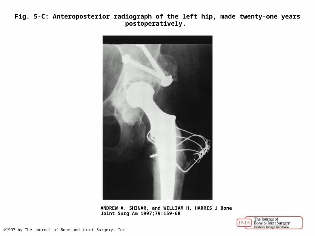
Fig. 5-C: Anteroposterior radiograph of the left hip, made twenty-one years postoperatively.
ANDREW A. SHINAR, and WILLIAM H. HARRIS J Bone Joint Surg Am 1997;79:159-68
©1997 by The Journal of Bone and Joint Surgery, Inc.