bulletin of the british museum (natural history) - qut of the british museum ... the bulletin of the...
TRANSCRIPT

Bulletin of the British Museum (Natural History)
The anomalous ant-attended mealybugs (Homoptera: Pseudococcidae) of south-east Asia
D. J. Williams
Entomology series Vol 37 No 1 25 May 1978

A22354638B
The Bulletin of the British Museum (Natural History), instituted in 1949, is issued in four scientific series, Botany, Entomology, Geology and Zoology, and an Historical series.
Parts are published at irregular intervals as they become ready. Volumes will contain about four hundred pages, and will not necessarily be completed within one calendar year.
Subscription orders and enquiries about back issues should be sent to: Publications Sales, British Museum (Natural History), Cromwell Road, London SW7 5BD, England.
World List abbreviation: Bull. Br. Mus. nat. Hist. (Ent.)
) Trustees of the British Museum (Natural History), 1978
Entomology series Vol 37 No 1 pp 1-72
Issued 25 May 1978

The anomalous ant-attended mealybugs |Li (Homoptera: Pseudococcidae) of south-east Asia *~ D. J. Williams Commonwealth Institute of Entomology, c/o British Museum (Natural History), Cromwell Road, London SW7 5BD
Contents Synopsis. . . . . . . . . . . . . . 1 Introduction 1 Habit 2 Distribution 4 Morphology . . . . . . . . . . . . . 6 Classification . . . . . . . . . . . . . 1 3 Acknowledgements 13 Taxonomic treatment . . . . . . . . . . . 1 3
Pseudococcinae . . . . . . . . . . . . 1 4 Allomyrmococcini trib. n. . . . . . . . . . 1 4
Key to genera of Allomyrmococcini (adult females) . . . . . 1 4 Key to genera of Allomyrmococcini (immature instars) . . . . . 1 5 Allomyrmococcus Takahashi, 1941 . . . . . . . . 1 5 Hippeococcus Reyne, 1954 . . . . . . . . . . 2 0 Malaicoccus Takahashi, 1950 . . . . . . . . . 3 0 Paramyrmococcus Takahashi, 1941 . . . . . . . . 5 4
Rhizoecinae. . . . . . . . . . . . . 6 2 Key to adult females of the anomalous ant-attended genera of Rhizoecinae . 62 Eumyrmococcus Silvestri, 1926 . . . . . . . . . 6 3 Xenococcus Silvestri, 1924 . . . . . . . . . . 6 3
References 70 Index 71
Synopsis The morphology, habits and taxonomy of 12 unusual myrmecophilous mealybugs from south-east Asia are discussed. Unusual characters of some of the species are long antennae, an anal ring without pores, a dense covering of minute setae on the dorsum, long anal lobe setae and large protruding ostioles. The ostioles are unlike any others in the Pseudococcidae and are compared with the siphunculi of aphids. Allomyrmococcus, Hippeococcus, Malaicoccus and Paramyrmococcus are aerial genera and are assigned to the subfamily Pseudococcinae. These four genera have many characters in common and a new tribe Allomyrmococcini is erected for them. Two new species of Malaicoccus and one new species of Para-myrmococcus are described. Eumyrmococcus and Xenococcus are subterranean and are placed in the sub-family Rhizoecinae.
Introduction Mealybugs are easy to recognize in life by an elongate-oval body usually covered with mealy or cottony white wax. This wax often extends around the sides to form a series of short filaments but a pair of much longer filaments may be present at the posterior end of the body. Most species have legs which rarely protrude beyond the lateral margins and movement is usually slow. Oviparous females become sedentary and may secrete a noticeable white cottony ovisac.
Bull. Br. Mus. not. Hist. (Ent.), 37 (1): 1-72 Issued 25 May 1978
1

In common with all female scale insects, species can be identified only from microscope slide mounts, by the arrangements of body pores and setae. Mealybugs are separated from related groups in possessing at least one of the major characters, cerarii, ostioles, circulus, trilocular pores and tubular ducts which are not cupped at the interior end.
As in all Homoptera, scale insects suck the sap of plants with long stylets and, so far as is known, all mealybug excretion is in the form of honeydew. This substance is excreted in all the families of Coccoidea except the Diaspididae, Conchaspididae, Halimococcidae and certain groups at present assigned to the family Asterolecaniidae. When palatable, honeydew forms an important part of ants' diet and a strong association has been built up between some species of ants and certain Homoptera. The literature discussing this association is extensive and in recent years Nixon (1951), Way (1963) and Wilson (1971) have discussed the subject at some length. Way has defined mutualism as any association beneficial to both the ant and the other insect. Myrmecophilous insects (according to Way) are those which benefit from ants and are more or less adapted to live with them but the relationship need not be obligatory or mutually beneficial.
The majority of myrmecophilous mealybug species having a mutual association with ants are those which a coccidologist would regard as normal and without visible signs of special adaptation. In south-east Asia some myrmecophilous mealybugs have developed most unusual characters and habits and it is the purpose here to describe and discuss this group. At present the group comprises the genera Allomyrmococcus Takahashi, Eumyrmococcus Silvestri, Hippeo-coccus Reyne, Malaicoccus Takahashi, Paramyrmococcus Takahashi and Xenococcus Silvestri.
Habit Silvestri (1924) described the first of these mealybugs under the name Xenococcus annandalei. It is an elongate-oval species but the abdomen tapers abruptly at the posterior end, and the long antennae are strongly geniculate with a special articulatory mechanism between the first and second segments. One of the most noticeable characters is the dense covering of minute setae on the dorsum which at the time of description had not been seen in mealybugs before. This is a subterranean species found in the nests of the ant Acropyga acutiventris Roger. Silvestri described the habit as living on the rootlets of Ficus sp. When the soil is damp and warm both ants and mealybugs remain just below the surface under stones but in cold, dry weather they retire deep into the ground. The ants carry away mealybugs in their mandibles when the nest is disturbed and when ants leave the nest, each female carries a female mealybug in her mandibles to new nests elsewhere.
Later Silvestri (1926) described Eumyrmococcus smithii from Macao and Shanghai as living under stones with the ant Acropyga (Rhizomyrma) sauteri Forel. Recorded later in Taiwan with the same ant, Takahashi (1934) mentioned that females and workers carry the mealybugs in the mandibles. This species is also densely covered with minute setae and the abdomen is abruptly narrowed but the antennae are short.
Above ground in Thailand were found Allomyrmococcus acariformis and Paramyrmococcus chiengraiensis described by Takahashi (1941). Both were covered with dense clusters of the ant Dolichoderus sp. [=Hypoclinea]. Malaicoccus riouwensis described by Takahashi (1950) from the Riouw Is (now known as Riau Is) and M. formicarii described by Takahashi (1951) from Malaya were both attended by large numbers of Polyrhachis sp. This ant carried M. formicarii in its mandibles at the constricted part of the thorax. A new species described here as M. moundi is also carried at the head or prothorax by the ant Hypoclinea sp. (Fig. IB). Specimens of the mealybug examined in alcohol were firmly grasped in the ants' mandibles which had to be prised apart to release the mealybugs.
Species of Hippeococcus described by Reyne (1954) have an even more unusual habit. These species are also aerial, feeding on stems and suckers of various plants, and are covered with ants of the genus Dolichoderus (= Hypoclinea). Mealybugs of H. rappardi Reyne are gleaming without any trace of wax and are very agile. When the ants are disturbed the mealybugs cling to the body, sitting crosswise on the ants' back (Fig. 1 A). Immature H. wegneri Reyne feeding on young shoots and fruit of Diospyros kaki cling to the thorax and other parts of the ant's body. A Hypoclinea
2

Fig. 1 (A) Ant, Hypoclinea sp., carrying Hippeococcus sp. on its back. Redrawn from Reyne (1954). (B) Ant, Hypoclinea sp., carrying Malaicoccus khooi in its mandibles. Drawn specially for this work by Mrs Linda Huddleston of the British Museum (Natural History).
3
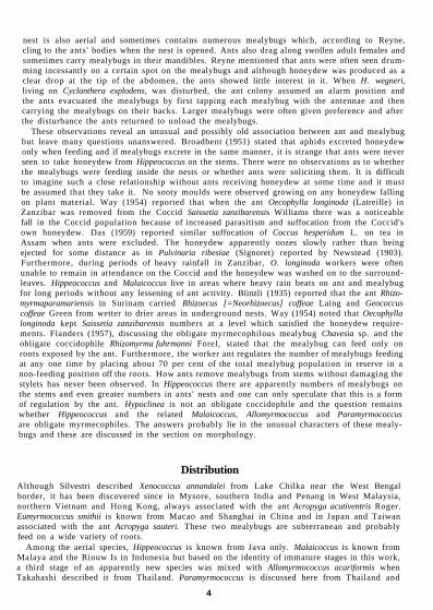
nest is also aerial and sometimes contains numerous mealybugs which, according to Reyne, cling to the ants' bodies when the nest is opened. Ants also drag along swollen adult females and sometimes carry mealybugs in their mandibles. Reyne mentioned that ants were often seen drumming incessantly on a certain spot on the mealybugs and although honeydew was produced as a clear drop at the tip of the abdomen, the ants showed little interest in it. When H. wegneri, living on Cyclanthera explodens, was disturbed, the ant colony assumed an alarm position and the ants evacuated the mealybugs by first tapping each mealybug with the antennae and then carrying the mealybugs on their backs. Larger mealybugs were often given preference and after the disturbance the ants returned to unload the mealybugs.
These observations reveal an unusual and possibly old association between ant and mealybug but leave many questions unanswered. Broadbent (1951) stated that aphids excreted honeydew only when feeding and if mealybugs excrete in the same manner, it is strange that ants were never seen to take honeydew from Hippeococcus on the stems. There were no observations as to whether the mealybugs were feeding inside the nests or whether ants were soliciting them. It is difficult to imagine such a close relationship without ants receiving honeydew at some time and it must be assumed that they take it. No sooty moulds were observed growing on any honeydew falling on plant material. Way (1954) reported that when the ant Oecophylla longinoda (Latreille) in Zanzibar was removed from the Coccid Saissetia zanzibarensis Williams there was a noticeable fall in the Coccid population because of increased parasitism and suffocation from the Coccid's own honeydew. Das (1959) reported similar suffocation of Coccus hesperidum L. on tea in Assam when ants were excluded. The honeydew apparently oozes slowly rather than being ejected for some distance as in Pulvinaria ribesiae (Signoret) reported by Newstead (1903). Furthermore, during periods of heavy rainfall in Zanzibar, O. longinoda workers were often unable to remain in attendance on the Coccid and the honeydew was washed on to the surround-leaves. Hippeococcus and Malaicoccus live in areas where heavy rain beats on ant and mealybug for long periods without any lessening of ant activity. Biinzli (1935) reported that the ant Rhizo-myrmaparamariensis in Surinam carried Rhizoecus [=Neorhizoecus] coffeae Laing and Geococcus coffeae Green from wetter to drier areas in underground nests. Way (1954) noted that Oecophylla longinoda kept Saissetia zanzibarensis numbers at a level which satisfied the honeydew requirements. Flanders (1957), discussing the obligate myrmecophilous mealybug Chavesia sp. and the obligate coccidophile Rhizomyrma fuhrmanni Forel, stated that the mealybug can feed only on roots exposed by the ant. Furthermore, the worker ant regulates the number of mealybugs feeding at any one time by placing about 70 per cent of the total mealybug population in reserve in a non-feeding position off the roots. How ants remove mealybugs from stems without damaging the stylets has never been observed. In Hippeococcus there are apparently numbers of mealybugs on the stems and even greater numbers in ants' nests and one can only speculate that this is a form of regulation by the ant. Hypoclinea is not an obligate coccidophile and the question remains whether Hippeococcus and the related Malaicoccus, Allomyrmococcus and Paramyrmococcus are obligate myrmecophiles. The answers probably lie in the unusual characters of these mealy-bugs and these are discussed in the section on morphology.
Distribution Although Silvestri described Xenococcus annandalei from Lake Chilka near the West Bengal border, it has been discovered since in Mysore, southern India and Penang in West Malaysia, northern Vietnam and Hong Kong, always associated with the ant Acropyga acutiventris Roger. Eumyrmococcus smithii is known from Macao and Shanghai in China and in Japan and Taiwan associated with the ant Acropyga sauteri. These two mealybugs are subterranean and probably feed on a wide variety of roots.
Among the aerial species, Hippeococcus is known from Java only. Malaicoccus is known from Malaya and the Riouw Is in Indonesia but based on the identity of immature stages in this work, a third stage of an apparently new species was mixed with Allomyrmococcus acariformis when Takahashi described it from Thailand. Paramyrmococcus is discussed here from Thailand and
4
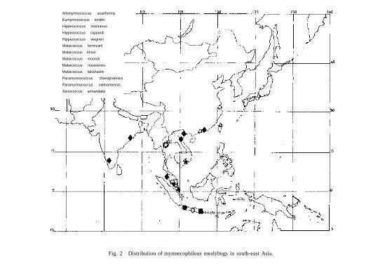
Allomyrmococcus acariformis
Eumyrmococcus smithii
Hippeococcus montanus
Hippeococcus rappardi
Hippeococcus wegneri
Malaicoccus formicarii
Malaicoccus khooi
Malaicoccus moundi
Malaicoccus riouwensis
Malaicoccus takahashii
Paramyrmococcus chiengraiensis
Paramyrmococcus vietnamensis
Xenococcus annandalei
Fig. 2 Distribution of myrmecophilous mealybugs in south-east Asia.

Vietnam. These aerial species seem to be attended only by ants of the genera Hypoclinea and Polyrhachis which are widespread in south-east Asia. The mealybugs will probably have a distribution throughout the area wherever the ants are present. Since this paper went to press the writer has received a new species of Malaicoccus attended by Hypoclinea sp. from Sarawak near the border with Sabah.
Morphology Body In life the body shape tends to be clavate with the head and thorax dilated and with the abdomen narrowing abruptly at about the third segment. Only in Malaicoccus is the abdomen broadly rounded. The body of Xenococcus is widest at about the first abdominal segment but the body still has a clavate appearance by the enormous basal segments to the antennae. Paramyrmococcus is more turbinate. In South America Chavesia has a similar body shape to that of Eumyrmococcus. Chavesia seems to have developed along similar lines to the genera in south-east Asia and there is strong evidence that Xenococcus and Eumyrmococcus are closely related to Chavesia but that Allomyrmococcus, Paramyrmococcus, Malaicoccus and Hippeococcus form a separate group. Coccidologists become used to studying flattened insects on slides, discussing only dorsal and ventral surfaces and forgetting that species have lateral sides. In all the genera from south-east Asia and South America the abdomen curves upwards at the posterior end, as shown by Silvestri (1924; 1926) and by Balachowsky (1957). It is not clear what advantage this gives to the mealybug or the ant. When an ant strokes a mealybug the abruptly narrowed abdomen may resemble an ant petiole and so the mealybug may be more readily accepted. Ants seem to carry these mealy-bugs near the head and prothorax whereas ants carry mealybugs of more normal shape at about the first abdominal segment. As Biinzli (1935) has remarked, the mandible marks may disappear but sometimes they may persist. Malaicoccus has developed heavy sclerotization on the head at the point where they are carried.
Legs
The legs of all species are well developed and reach their greatest length in Allomyrmococcus when they are longer than the body. Reyne (1954) has mentioned that Hippeococcus moves quickly yet Strickland (1950) stated that in West Africa the mealybugs associated with ants have shorter legs than the active longer legged species that are not associated with ants. The leg setae are well developed on each species and sometimes they are abundant. The claw gives some clue that these mealybugs belong to two distinct groups. In Xenococcus and Eumyrmococcus the claw is long and slender with a pair of short setose digitules at the base but in the other four genera the claw is stout with a pair of large flattened digitules which are usually twice as long as the claw. Reyne (1954) has suggested that Hippeococcus uses the digitules to cling to the ants' bodies and this seems a reasonable deduction. It should be remembered that these large flattened digitules are present in Allomyrmococcus, Paramyrmococcus and Malaicoccus also but these mealybugs apparently do not cling to ants' bodies. The tarsus of each species has a single cam-paniform sensillum at the outer proximal end, a structure discussed recently by Koteja (19746).
Antennae
Eumyrmococcus has short 2-segmented antennae but in Xenococcus the antennae are 4-segmented of a most unusual type. These are long and strongly geniculate with a large basal segment. The articulation between the first and second segments is so well developed that the antennae can bend from a forward position to one lying along the back of the mealybug. Helping in this movement are small teeth on the outer proximal corner of the second segment which fit into grooves on the outer distal corner of the first segment. Long pointed setae are present on all the segments and on the third and fourth segments some setae are as long as the segments. As Xenococcus lives underground it is probably advantageous for the mealybug to streamline its body by folding
6

the antennae along the back but in other circumstances the antennae may be used for recognizing an ant.
The antennae of the other four genera are 6-segmented and are unlike those of any other mealy-bugs. In each species and in all stages, the second segment is short and does not articulate with the third segment. In the first stage larvae, long setae form whorls mainly at the distal ends of the segments but in Paramyrmococcus vietnamensis the fifth and sixth segments have numerous short setae also. Short setae are present on all second instars and they become more numerous and longer on each successive stage so that the adult antennae are densely covered. The antennae in most adult females of this group are about as long as the body. It is difficult to understand why these insects have such long and well-developed antennae. It is usual for an ant to feel or stroke a mealybug before either attending one or carrying one away. Mealybugs of Hippeococcus and possibly those of related genera may equally seek out or recognize an ant with their long antennae.
Setae
One of the striking characters of nearly all these mealybugs and the South American genus Chavesia is the dense covering of minute setae over the entire body or at least over the dorsum. In many species the setae are short and are so numerous that the distance between the setal bases is much shorter than the length of a single seta. These mealybugs are without the usual protection of mealy or flocculent wax and the numerous setae may act as an alternative protective covering. By trapping air between the setae, the body surface of underground species would be protected from excessive moisture. The aerial species are exposed to heavy rains for long periods and the dense covering of setae may have a waterproofing effect.
Some species do not have such dense coverings. Adult Hippeococcus have, in addition to minute pointed setae, small blunt setae of uncertain function. Longer clavate and fleshy setae, which in H. rappardi are present in the first stage only, persist in all stages in H. wegneri. In many of the immature stages of Malaicoccus there are minute setae in varying numbers, each with the seta shorter than the diameter of the setal base. Immature Paramyrmococcus have body setae which are short, cylindrical and blunt but these are replaced by numerous short pointed setae in the adult. A few setae which are flat and variously shaped are present on the head and thorax of Xenococcus. There is little difference, apart from abundance, in the arrangement of the setae in the stages of Allomyrmococcus but immature stages have extra long setae on parts of the body. Adult mealybugs, therefore, usually have some characters such as the addition of special body setae or the loss of long setae, which distinguish them from the immature stages. Ants may be able to recognize these differences by quickly stroking the mealybug's body.
Anal areas
This section includes the anal ring, anal lobes, anal lobe setae and any other structures at the apex of the abdomen. The anal ring is usually sclerotized with 6 setae of various lengths and always without pores except for a few minute pore-like openings in Malaicoccus. In the more usual mealybugs, of which Pseudococcus obscurus Essig is an example, a short wax tube is secreted from pores around the anal ring. Honeydew droplets are, therefore, excreted a short distance from the body surface. A colony of P. obscurus on a potato tuber at times ejected honeydew for a distance of 2-4 cm. This honeydew is clear and sticks to the surrounding foliage, but each droplet of honeydew of the cochineal insect Dactylopius coccus Costa is coated with fine wax and falls directly to the ground without sticking to the plant. Broadbent (1951) has mentioned that the aphid Hayhurstia atriplicis (L.) excreted globules coated with wax from contact with the wax covering of the aphid. There is always the possibility that wax coating may affect the palatability to ants. No observations have yet been made on the honeydew of the myrmecophilous mealybugs.
Reyne (1954) has stated that Hippeococcus rappardi had usually a clear drop of liquid between widely diverging silvery hairs, which are presumably the anal hairs. In Chavesia, Beardsley (1970) has described the anal lobes as protuberant and sclerotized on the ventral surface. The anal ring is in a membranous depression on the dorsal surface of the lobes. These lobes also contain
7
numerous setae and the whole structure forms what Beardsley has called a honeydew basket. A similar structure is present in Allomyrmococcus and Malaicoccus but the anal ring may be terminal or situated a short distance from the apex. In Hippeococcus and Xenococcus the anal ring lies at the apex of the dorsum which projects beyond the ventral anal lobes. Wherever the anal ring is placed it is within easy access for the ant and in the absence of pores it seems likely that honeydew remains at the apex of the abdomen until the ant removes it. Broadbent (1951) has described how the aphid Hyalopterus prurti (Geoffroy) kicks away globules with the hind legs. Normally the legs of mealybugs are too short to reach the anus but in the absence of ants most of the myrmecophilous mealybugs in this discussion could remove globules with their legs if necessary but this has not been observed.
Long anal lobe setae are present in all genera except Paramyrmococcus. Sometimes they are numerous and are longer than the body. Their function is uncertain and their absence in Para-myrmococcus does not seem to affect the myrmecophilous habit.
As previously stated, honeydew is probably produced only when the mealybugs are feeding. Furthermore excretion rates may be increased by ants attending mealybugs and removing honey-dew. It is usual for an ant to stimulate the mealybug into giving up a drop of honeydew by pal-pating the abdomen, but the mechanism of this stimulation is not understood. In the Coccidae, Way (1954) and Smith (1942) have discussed how ants solicit species of Saissetia by dragging the tips of the antennae back and forth over the caudal end of the body. Saissetia and Udinia are two related genera often solicited by ants in the tropics but species of both genera have long pri-mary discal setae on the anal plates which when touched may induce the Coccid to open the plates and excrete some honeydew. By stroking the anal ring setae, ants may induce mealybugs to excrete a droplet.
Body pores An important feature of the myrmecophilous mealybugs is the almost complete absence of pores and ducts. All species have been described without surface wax, and Reyne (1954) described Hippeococcus as having a gleaming body. Eumyrmococcus and Xenococcus are completely without body wax pores and in all the other genera the only pores present in the female stages are trilocular and a few simple pores except in some species of Malaicoccus which have a few oral collar ducts in the mid-thorax and anterior abdominal segments. In most species the trilocular pores are quite sparse but in Hippeococcus and certain Malaicoccus species they are more numerous. It is only the second stage male which has the more usual pores associated with mealybugs. In H. wegneri and M. moundi the second stage males have multilocular disc pores and oral collar tubular ducts and it is assumed that these secrete the waxy covering for the pupal stage. Nixon (1951) has given a few examples of ants removing wax coverings of mealybugs. For instance, Le Pelley (1943) noted that ants removed wax completely from certain areas of the body of Pseudococcus [ =Plano-coccus] lilacinus (Cockerell) in the Philippines. It is not clear how male mealybug pupae could retain wax coverings in the presence of attending ants. Nevertheless, the gleaming and waxless condition of myrmecophilous mealybugs is probably the result of specialization in the continual presence of ants. Recently Delage-Darchen et al. (1972) have noted an unusual association between Diaspidid scale insects and ants. The scale insects live in tunnels in woody stems and are attended by ants in so far as ants remove the scale coverings. One species of these scale insects is completely without dorsal ducts so that no scale could be secreted anyway. Similar observations with scale insects have also been made by Prins et al. (1975). Although ants may remove wax for food it is also possible that in time species that would normally secrete a wax covering evolve without even any means of producing one. It is difficult to understand how ants benefit from species not producing honeydew.
Labium
The labium of Pseudococcidae is conical and 3-segmented and its morphology has been studied by Koteja (1974a) whose terminology is used here. The basal segment is small with a pair of rounded sclerites, often fused medially with the medial segment, and contains 2 pairs of short
8

setae and one longer pair. On the medial segment there is a basic number of 1 pair of setae. The apical segment has 2 or 3 pairs of posterior setae on the posterior or under surface, a pair of short apical setae, 4 pairs of subapical setae, one pair of lateral setae and 2 pairs of anterior setae situated near the base. In many species there are supernumerary setae anterior to the lateral setae.
Koteja recognized the four subfamilies Trabutininae, Pseudococcinae, Rhizoecinae and Sphaerococcinae. The Trabutininae is separated from the others, using the character of the labium only, in possessing 3 pairs of posterior setae. All the species studied in this work have 2 pairs of posterior setae and the species are separated from the Sphaerococcinae in having a well-developed basal segment not fused totally with the medial segment. The labium of Rhizoecinae, according to Koteja, differs from that of the Pseudococcinae in being small and narrow with the third subapical setae distinctly stouter than the others and with the lateral setae about twice as long as the other labial setae. Williams (1969) has already placed Eumyrmococcus in the Rhizoe-cinae on other grounds but the labium of this genus has a width-length ratio of 1 : 2, there are 14 pairs of setae and the subapical setae become progressively longer towards the base. Xeno-coccus has a labial width-length ratio of 1 : 1 -87 when flattened on the slide and 1 : 2-27 when not flattened. The number of setae varies from 12 to 13 and the subapical setae are remarkable in extending more than half way to the base of the apical segment. As in Eumyrmococcus these subapical setae become progressively longer towards the base of the segment. For reasons to be given later under classification, Xenococcus should be assigned also to the Rhizoecinae.
In the genera Hippeococcus, Malaicoccus and Paramyrmococcus the width-length ratio of the labium varies considerably from 1:1-4 in P. chiengraiensis to 1 : 1-92 in Malaicoccus khooi. Despite this range in size there is reason to believe that these genera with Allomyrmococcus belong to the Pseudococcinae. In Allomyrmococcus and Malaicoccus the total number of setae varies from 20 to 32 pairs in the adult female but in the first and second stages they become reduced to the basic number of 14 pairs.
Circulus
The circulus, when present, is a round or oval sclerotized area, surrounded by a rim, usually lying on venter between the fourth and fifth abdominal segments. Occasionally it may lie within the borders of the fourth segment and some species may have several circuli on different segments. In the Rhizoecinae the circulus is usually small and round but heavily sclerotized, conical and often with an irregular surface. Pesson (1939a) and Lloyd & Martini (1957) have shown that adult females of the genera Pseudococcus or Phenacoccus use the circulus as an adhesive organ when viewed in profile against a glass surface. The circulus is exserted and then adpressed to the surface allowing the insect to make exploratory leg movements, the body actually pivoting at the circulus. This use seems to be restricted to the adult female because Lloyd & Martini observed that the three nymphal instars were able to ascend or descend with ease by leg actions presumably with the help of the claw digitules.
No similar research has been done on the circulus of the Rhizoecinae but Silvestri (1924), working on alcohol material of Xenococcus annandalei, suggested that the circulus of this species secreted some kind of liquid possibly attractive to ants. Whatever its function it does seem that the circulus of the Rhizoecinae has been derived from those of the other groups of the Pseudo-coccidae.
Ostioles
Among the Coccoidea the Pseudococcidae are unique in usually possessing 2 pairs of transverse slit-like organs on the dorsal surface, commonly named ostioles. In Allomyrmococcus, Hippeococcus, Malaicoccus and Paramyrmococcus the ostioles reach an unusual development and this seems an appropriate place to discuss their structure and to compare them with siphunculi or cornicles in aphids.
Occasionally some species of such genera as Rhodania Goux and Ehrhomia Ferris are without ostioles but they may be present in some of the immature stages where they are limited to the posterior pair only. In adult females of Antonina, ostioles may be present as a poorly developed
9

posterior pair only. The absence or reduction of ostioles is correlated with the amount of external wax secreted from the pores, the term wax being used here in its general sense. Usually those species producing large amounts of dorsal wax or those which become almost enclosed in a felted ovisac, as in Antonina, have reduced ostioles.
According to Ferris & Murdock (1936) the anterior pair belong to the pronotum and the posterior pair to the sixth abdominal segment. Pesson (1939b), however, after an overall study of the musculature, concluded that the anterior pair are placed slightly forward to the anterior edge of the prothorax and the posterior pair lie between the fifth and sixth abdominal segments.
Viewed from above each ostiole is composed of two membranous lips which often contain numerous setae and trilocular pores. The inner edges of the lips are usually sclerotized and sometimes these inner edges are the only parts visible.
Sulc (1909) observed in Phenacoccus farinosus (de Geer) that blood corpuscles oozed from the ostioles after pressure was applied to the body. He also observed some muscles parallel to and perpendicular to the lips of the ostioles.
In histological studies Ferris & Murdock (1936) found no muscle attachments on these struc-tures and concluded that they led directly into the haemocoele. When the insect was stimulated by an application of violence, a globule of liquid appeared which quickly hardened on exposure to air. The globule, according to Ferris & Murdock, was merely a portion of the body fluids.
In more detailed histological studies, Pesson (1939b) found that in a transverse section, the edges of the slit are continued into the body cavity as a chitinized depression, the median part of which forms an orifice leading directly to the haemocoele. From the lateral inner ends of the depression in each anterior ostiole are inserted two muscles, the anterior or inner muscle inserting itself obliquely to the inner edge of and lateral to the clypeus, and the posterior or outer muscle attached a little anterior to the first leg. There are also two similar muscles in each posterior ostiole which are attached ventrally. If an ostiole is regarded as a valve then normally it should prevent blood loss when at rest but after an application of some body violence near an ostiole the muscles contract and open the lips slightly to emit a droplet of blood or other contents of the haemocoele. This emission, according to Pesson, is nothing more than reflex-bleeding or auto-haemorrhage.
Sulc (1909) noted that the expelled wax, on hardening in contact with air, could have a protect-ing roll by coating the oral parts of an attacker. Pesson never witnessed this in Pseudococcus adonidum (L.) [=P. longispinus (Targioni-Tozzetti)] and indicated that the liquid expelled is from the free wax occurring in the blood as corpuscles. After repeated excitation of the surround-ing cuticle young wax-producing cells may be emitted. Wheeler (1921), in his studies of the myrme-cophyte Tachigalia and the attending behaviour of beetles of the genus Coccidotropus on Pseudo-coccus bromeliae (Bouche) [= Dysmicoccus brevipes (Cockerell)], observed that the beetles were interested only in the honeydew excreted and were never immobilized or impeded by any secretion from the ostioles. He stated that the beetles may be attracted by some fascinating aroma secreted by the mealybug but did not actually state that this was emitted from the ostioles.
After Coccoidea are macerated in potash as a first step in the mounting technique there are often observed certain internal wax globules varying in quantity as to the family. It is in the Pseudococcidae that this internal wax is most strikingly abundant and it is apparently this free wax which is expelled through the ostioles.
The posterior ostioles have often been homologized with the cornicles or siphunculi of aphids. Little work has been done so far on ostioles but in recent years work on siphunculi has been extensive. A resume of recent work on siphunculi in aphids is given here and it is hoped this may stimulate further work on ostioles.
It was shown by Hottes (1928) that cornicles are situated on the dorsolateral surfaces of the sixth abdominal segment. In their most conspicuous form they are cylindrical and longer than wide. Usually the longer cornicles are movable but at the tip of all cornicles there is a slit-like opening which may be opened or closed by a valve moved by a muscle attached near the median-free portion of the valve. The valve is attached at the apex by a flexible hinge. Hottes further indicated that, although cornicles may be movable, the possibility that droplets can be aimed is practically nil. The function of the cornicles, according to Hottes, was not a defence mechanism
10
but was part of the metabolic processes carried out by the aphid and that the cornicles have an excretionary function. Furthermore, the presence or absence of cornicles may be correlated with the presence or absence of wax pores or a lower reproduction rate. Hottes stated that as aphids have often evolved along with their hosts the change in structure of the cornicles may be the result of the nature of the food consumed.
Despite Hottes' rejection of the defence mechanism function of cornicles, Dixon (1958) observed that if the larva of the coccinelid Adalia decempunctata (L.) was smaller than the aphid Microsiphum evansi (Theobald) and seized an appendage, the aphid could escape by pulling the appendage free. If the coccinelid larva and aphid were about the same size the aphid could escape if the siphunculus nearest the appendage swung over and placed a drop of oily liquid on the coccinelid's head where the drop would spread over the mouthparts and solidify.
Reflex-bleeding discussed by Hottes was confirmed in aphids by Edwards (1966) who stated that when the cornicle valve is opened, the aphid released material from the haemocoele. As no solvent of the internal material was detected by gas chromatography he explained that the fluid is in a stable liquid-crystalline state within the aphid and changes to the solid crystal phase with a seeding nucleus. He further stated that the rapid crystallization on contact with a solid surface, a hair or duct, suggests that the liquid was in a supercooled state and that foreign material provides a seeding nucleus. The melting point of the waxes of three species of aphids ranged from 37-5 °C to 48 °C so that the waxes should crystallize on seeding at normal summer tempera-tures. Studying the cornicle area of various species of aphids, Wynn & Boudreaux (1972) found that in Cinara and Longistigma a multicellular sac possessing a wall, formed from a layer of flat cells with compressed nuclei, extends into the cornicle. This sac is surrounded by vacuolated fat cells and a mass of lipid is found in the sac. All the aphids studied possessed a muscle inserting on the cornicle valve and originating on the venter below the cornicle. The authors suggest that this muscle, in addition to opening the valve at the distal end of the cornicle, may also assist in elevat-ing the cornicle. They also suggest that a dorsoventral muscle just anterior to the cornicle appears to be used in association with other dorsoventral muscles in compressing the body contents causing the emission of a droplet. In each case the cornicle communicates with the haemolymph and blood cells including fat cells escape. The colour of the cornicle droplet corresponds with the colour of the aphid and its haemolymph.
In contrast, Lindsay (1969), studying the cornicles of the pea aphid Acyrtosiphonpisum (Harris), stated that the term reflex-bleeding as applied to cornicle exudation is inapplicable since, although a small amount of haemolymph may escape, the primary exudate is the cornicle cells. Analysis of these cornicle cells indicates a close similarity to those of the fat-body cells in composition. Lindsay suggested that the fat-body cells and cornicle cells are homologous, the fat body being a precursor of the cornicle cells and that there is a greater tendency of the cornicle cells to crystallize compared with those of the fat-body cells.
Evidence that haemolymph is not extruded through the cornicles was presented by Strong (1967) who stated that lipids were the principal constituents of the hardened cornicle droplets and were composed solely of triglycerides. Histological preparations by Chen & Edwards (1972) showed that the cornicle secretionary cells are present inside a sac separated by a thin acellular lamina from the haemolymph. The sac is formed by an invagination of the epidermal basal lamina reflected back from the tip of the cornicle. The enclosed secretionary cells are comparable with epidermal gland cells or subepidermal oenocytes. Fluid in the sac differs in composition from the haemolymph and the fluids released through the cornicle are thus not a direct loss of haemo-lymph but are presumably derived from the haemolymph.
Referring to the triglycerides in the cornicle secretions, Callow et al. (1973) found that the secretions in any one species are the same and that different species of aphids taken from the same host plant have different secretions. The authors found it was easier to obtain droplets from young or small aphids rather than large aphids and they presumed that the larger aphids have less need of a defence mechanism. This is interesting because as already stated ostioles may be present in immature mealybugs and absent in adults although defence mechanisms in mealy-bugs have not been proved there may be a greater need for ostioles in immatures for different reasons. Analysis by Greenway & Griffiths (1973) of the body triglycerides showed them to contain
11
the same fatty acid radicles as in the cornicles but in many species there are greater proportions of some triglycerides in the body. When an aphid is overstimulated the composition of the cornicle secretions gradually becomes the same as the body contents.
Speculative suggestions that odours may be emitted by mealybug ostioles have not so far been confirmed but Dahl (1971) reported that odours from crushed aphids repelled other apids. Kislow & Edwards (1972) found that certain aphids are repelled by the odour from cornicle droplets and squashed specimens of the same species. These authors proposed that when there is a slow release of odour it may cause intraspecific spacing of aphids, prevent the influx of other species or even induce aphids to migrate. The alarm pheromone was identified as trans-/3-farnesene or TBF in some aphids by Bowers et al. (1972). Nault et al. (1973) showed that the alarm pheromone is known to be interspecific and that the receptor of this repellent odour is the primary sensorium on segment 6 of the antenna. The existence of multiple alarm pheromones was suggested by Nault & Bowers (1974) who found that Hydaphis erysimi (Kaltenbach) does not respond to pure TBF and they suggest that the aphid produces one or more other compounds in addition to TBF to produce alarm activity.
Before referring once more to ostioles in mealybugs it is interesting that Dixon & Stewart (1975) after blocking the openings of siphunculi of the sycamore aphid Drepanosiphum platanoides (Schrank) found that this occlusion had no adverse effect on the number of offspring produced or on the weight of the aphid. Furthermore, aphids with occluded siphunculi do not space out differently from aphids with normal siphunculi and that the aphid takes most avoiding action when the smell of the pheromone is accompanied by vibrations associated with a struggling aphid.
There is obviously a need for much further research on ostioles. Even a cursory glance at the ostioles ofAllomyrmococcus, Paramyrmococcus and Hippeococcus shows that they are much more highly developed than in normal mealybugs. The sclerotized lips have their greatest development in any mealybugs known to the author. Although in the illustrations they are located on the edges, this is due to dorsoventral flattening when specimens are prepared on microscope slides but even in specimens preserved in alcohol they are unusually large and conspicuous with the sclerotized lips protruding on the dorsolateral areas of the body. Their most unusual development is found in Malaicoccus. In this genus the anterior pair are often located on the ventral surface on slide preparations. Each ostiole appears to have a fixed anterior sclerotized lip but the posterior lip is a large hinged semicircular flap often containing numerous setae. This flap is often seen completely closed but in some specimens it is open and raised at a right-angle.
In the four genera there is in each ostiole a membrane, easily taking up stain, which completely covers the opening of the lips. It is not certain, working only from microscope preparations, whether there is any opening on this membrane. There is in some specimens a hole or slit on the surface but this may be due to tearing during the mounting technique. The membrane may normally be internal and may be exserted as a result of pressure on the body when flattened.
Why these species should possess such complicated ostioles is still uncertain but the mealybugs, as explained earlier, are transported when the ants are disturbed. Although ants are attracted to excretions of honeydew it is doubtful if honeydew has any odour over long distances but any odour from the ostioles would attract ants and thus help them to transport mealybugs back to the preferred host plants. This is conjecture but any work on pheromones must be done in areas where the mealybugs are common. It is easy to bring aphids into the laboratory for study but much more difficult to experiment with the most interesting of mealybugs which inhabit the tropics. If there is an odour from ostioles of these myrmecophilous mealybugs to attract ants, then there is possibly a similar odour emitted, as suggested by Wheeler, from those mealybugs with a less obligatory association with ants. Any research on this odour could lead to an alteration of the relationship between ants and mealybugs and could possibly help in controlling mealybugs. Mutual association with ants in the Palaearctic Region is common in such mealybugs as Chnauro-coccus subtenaneus (Newstead) and Euripersia europaea (Newstead), species which may be useful for research, and in the sugar-cane areas mealybugs living in association with ants are common enough to afford the means for easy research. It is doubtful, for instance, whether alarm pheromones are emitted by mealybugs as in aphids but any work on mealybug ostioles that may determine if their function is different from reflex-bleeding usually propounded would be useful.
12

Classification Xenococcus and Eumyrmococcus seem to belong to the subfamily Rhizoecinae. Williams (1969) placed Eumyrmococcus in this group because it possesses long slender claws with setose digitules and 3 pairs of long anal lobe setae. Xenococcus has similar long claws and also 2 circuli which are round, sclerotized and slightly conical. The antennae are not typical of the Rhizoecinae but the reduction in the number of segments suggests a relationship. The long slender labium of both genera each have a width-length ratio of 1 : 2 and the assignment of the genera to the Rhizoecinae agrees with research of the group made by Koteja (1974a; 1974c). Beardsley (1970) tentatively placed the genus Chavesia in the Rhizoecinae and this is probably correct. Some of these species have 4-segmented antennae but of a different shape to those of Xenococcus. The labium of Chavesia has a width-length ratio varying from 1 : 1-33 to 1 : 200 but Beardsley has described it as being 2-segmented only so that the addition of the basal segment would alter the width-length ratio. The labium of C. trinidadensis Beardsley has a similar shape to the labium of Xenococcus.
The four genera Allomyrmococcus, Hippeococcus, Malaicoccus and Paramyrmococcus have many characters in common. The antennae and claws are remarkably similar and so unusual that a new tribe is here erected for the genera within the subfamily Pseudococcinae. In the absence of adult males it is not possible to give the relationship of the tribe.
Separation of the subfamilies Pseudococcinae and Rhizoecinae in this work is based mainly on the shape of the claw and the width-length ratio of the labium but it must be stressed that the species under discussion belong to aberrant genera which are not typical of the subfamilies.
Acknowledgements Most of the material discussed in this work is housed in the collections of the British Museum (Natural History). Further specimens including holotypes and type-material have been kindly made available for study by Mr Charles Chia-chu Tao of the Taiwan Agricultural Research Institute, Taipeh; Dr J. P. Duffels of the Instituut voor Taxonomische Zoologie (Zoblogisch Museum), Amsterdam; Dr P. H. van Doesburg of the Rijksmuseum van Natuurlijke Historie, Leiden; Dr Khoo Soo Ghee of the University of Malaya, Kuala Lumpur; Dr A. Tranfaglia of the Istituto di Entomologia Agraria dell'Universita di Napoli, Portici and Dr D. R. Miller of the USDA, Beltsville, who sent specimens from the United States National Museum, Washington.
The writer is much indebted for useful information on aphids given by Dr V. F. Eastop and Dr R. E. Blackman of the British Museum (Natural History).
Abbreviations of the type-depositories mentioned in the text are as follows.
BMNH, London British Museum (Natural History), London IEAUN, Portici Istituto di Entomologia Agraria dell'Universita di Napoli, Portici ITZ, Amsterdam Instituut voor Taxonomische Zoologie, Amsterdam RNH, Leiden Rijksmuseum van Natuurlijke Historie, Leiden TARI, Taipeh Taiwan Agricultural Research Institute, Taipeh UM, Kuala Lumpur University of Malaya, Kuala Lumpur USNM, Washington United States National Museum, Washington
Taxonomic treatment An ideal classification of Coccoidea should be based on the taxonomy of adult males but so few males have been studied that identification is possible only with adult females which are neotenic. Female Pseudococcidae usually have three immature instars and males two immature and two pupal instars. There is often little to distinguish immatures from adult females in life apart from size. It is the experience of the writer, who has been engaged in identification of Coccoidea for many years, that identification of immature stages is often just as important to the collector as identification of the adult. When a new species is named, the name applies to all stages, not just to the stage mentioned in the description. In the present work available immature stages are discussed along with adult females.
13

PSEUDOCOCCINAE
ALLOMYRMOCOCCINI trib. n.
Type-genus: Allomyrmococcus Takahashi, 1941.
Pseudococcinae with body of adult female variously shaped, turbinate, or with abdomen rounded or abruptly narrowed at third segment. Head either membranous or heavily sclerotized when there is a deep constriction at about the prothorax. Anal lobes either wanting, in which case the anal lobe setae are short, or well developed with long anal lobe setae about as long as body. Anal ring usually sclerotized, terminal or situated a short distance from apex, with 6 setae but without pores except in one genus with minute pore-like structures. Antennae 6-segmented, stout, well developed and often as long as body, of a distinctive shape. The first segment on dorsum when mounted on slide, often large and wide; second segment short and not articulating with third segment. Remaining segments long, widest at distal end except the last segment, which is often depressed near middle of segment and variously curved. Fourth to sixth segments with abundant short setae, the distance between setal bases shorter than length of a seta. First and second segments with or without abundant setae. Longer and stouter sensory setae present on last segment. Legs well developed, long, the tarsus much shorter than tibia, often with numerous setae. Claw stout, each with a pair of flattened and expanded digitules, these about twice as long as claw and about same width. Labium conical, with 3 segments and with basic number of 14 pairs of setae but sometimes with as many as 32 pairs. Ostioles situated on edges of body when mounted on slide or lying on ventral surface, with an unusual development with heavily sclerotized and wide projecting lips, without setae or with posterior lip in form of sclerotized flap with numerous setae, the inner edges of lips joined by a membrane. Circulus present or absent. Body setae short, often abundant when they may cover the entire body surface except for intersegmental areas. When less abundant they may be accompanied with short blunt setae. Body pores often restricted to trilocular pores and simple pores but occasionally a few oral collar ducts may be present in mid-thoracic and anterior abdominal segments.
DISCUSSION. Based on the characters of the adult female, the tribe is one of the most distinctive in the family Pseudococcidae. As mentioned earlier in the section on classification, no adult males have been studied to give a clue to the relationship of the tribe. The evidence suggests that males exist and there is a pressing need for further observations in the areas where the tribe is known.
DISTRIBUTION. Thus far the tribe occurs in Thailand, West Malaysia (Malaya), Sarawak, Indonesia and Vietnam but the distribution may be extended after further study is made of nests of the ants Hypoclinea and Polyrhachis.
Key to genera of Allomyrmococcini (adult females) 1 Body without recognizable anal lobes, posterior end of body rounded, narrow, with short
setae in normal positions of lobes. Body setae abundant on dorsal and ventral surfaces PARAMYRMOCOCCUS (p. 54)
- Body with anal lobes, either poorly or well developed, with anal lobe setae about as long as body 2 2 Head heavily sclerotized and body constricted at prothorax. Long anal lobe setae and marginal
setae on at least first preceding segment. Ostioles with posterior lip in form of a sclerotized flap which opens and closes against an apparently fixed sclerotized anterior lip. Anal ring narrow with minute pore-like structures MALAICOCCUS (p. 30)
- Head membranous, body not constricted at prothorax. Long setae on anal lobes only or, if long marginal setae are present they are not on first preceding segment. Ostioles each with two sclerotized lips, the posterior lip similar to anterior lip. Anal ring with broad sclerotized rim without pores 3
3 Anal ring situated a distance about its own length from apex of abdomen, in a membranous depression in middle of sclerotized anal lobes. Body setae all pointed, densely covering dorsal and ventral surfaces, the distance between setal bases shorter than length of one seta. Legs about as long as body with abundant setae . . . ALLOMYRMOCOCCUS (p. 15)
- Anal ring terminal, at apex of dorsal lobe-like structure between ventral anal lobes. Body setae of two main types, one short and pointed and the other short and blunt, slightly clavate, the distance between setal bases greater than length of single seta. Legs shorter than body length with numerous setae but not densely covered; sometimes these setae are long and fleshy
HIPPEOCOCCUS (p. 20)
14

Key to genera of Allomyrmococcini (immature instars) 1 Body elongate-turbinate, without recognizable anal lobes, posterior end of body rounded with
short setae in normal positions of lobes. Body setae mainly short and cylindrical on dorsum but with longer pointed setae on venter and around margins . PARAMYRMOCOCCUS(p. 54)
- Anal lobes developed to some extent even though poorly developed in some stages. Long anal lobe setae as long as body 2
2 Head with some sclerotization even if only around the eyes, this sclerotization becoming more extensive in each successive stage. Long marginal setae on at least first preceding segment from anal lobes often as far forward as sixth preceding segment.
Body setae pointed but sometimes there are present also minute setae with the seta shorter than diameter of setal base. Anterior ostioles with posterior lip in form of flap
MALAICOCCUS (p. 30) - Head always membranous. If long marginal setae are present they are not on first preceding
segment from anal lobes . . . . . . . . . . . . 3 3 Anal ring situated at least its own length from apex of body in a small membranous depression
in middle of sclerotized anal lobes. Legs about as long as body (first instar not seen) ALLOMYRMOCOCCUS (p. 15)
- Anal ring terminal at apex of sclerotized dorsal lobe-like structure between sclerotized ventral lobes. Legs long but shorter than bodv. Body setae pointed or long, fleshy and clavate
HIPPEOCOCCUS (p. 20)
ALLOMYRMOCOCCUS Takahashi, 1941 Allomyrmococcus Takahashi, 1941:201. Type-species: Allomyrmococcus acariformis Takahashi, by
original designation and monotypy.
Only a single species is known so far. The genus has some peculiar characters among which are the unusually long stout legs about as long as the body. The turbinate shape, the well-developed anal lobes and dense covering of minute setae are some of the other most important characters. There is little information on its biology except that colonies are covered with a dense cluster of the ant Dolichoderus [=Hypoclinea] and the insects feed on young shoots. There is every indication that a honeydew channel or basket is formed between the anal lobes. The anal ring lies some distance from the apex of the abdomen and the space between the lobes is in the form of a concave depression which presumably retains honeydew.
DESCRIPTION. A broadly oval to turbinate body form with well-developed anal lobes each terminating with a group of stout setae about as long as body. Anal ring sclerotized, without pores but with 6 setae lying just outside the ring. Antennae 6-segmented, about as long as body, the second segment short and not articulated with the third; all segments densely covered with slender setae. Legs stout, as long as body or longer, covered with slender setae, tibia about four times as long as tarsus. Claw with a pair of flat expanded digitules, these about as wide as claw at base and twice as long. Body densely covered with minute pointed setae. Ostioles situated laterally, prominent, with well-developed sclerotized lips, without setae or trilocular pores. Labium stout but longer than wide with 30 or more pairs of setae in adult female, ventral or posterior surface with 2 pairs of posterior setae. Eyes present. Trilocular pores present but sparse.
Immature instars similar to adult female in form but smaller and with fewer body setae. No first instar available but second and third instars described here and illustrated.
DISTRIBUTION. At present the genus is known only from Thailand.
Allomyrmococcus acariformis Takahashi, 1941 Allomyrmococcus acariformis Takahashi, 1941 : 201. Holotype ?, THAILAND: Mt Sutep, near Chiengrai,
on plant of family Leguminosae, associated with ant Dolichoderus sp. [ = Hyploclinea sp.] (TARI, Taipeh).
Allomyrmococcus acariformis Takahashi; Takahashi, 1942 : 15.
Adult Female (Fig. 3)
DESCRIPTION. External appearance described originally as 'Dark reddish brown and blackish with no wax.'
15

Fig. 3 Allomyrmococcus acariformis Takahashi. Adult female. Specimen from Thailand, Mt Sutep.
16

When mounted on the slide a broadly oval to turbinate species about 1.85 mm long and 1.4 mm wide, broad at anterior end and tapering posteriorly to a well-developed and protruding pair of rounded anal lobes. Antennae 1.81-1.92 mm long thus about as long as body; with 6 segments which on the longest antenna the first segment is 170 µm long, the second 150 um, third 450 µm, fourth 320 µm, fifth 380 µm and sixth 450 µm, the last segment slightly curved, narrow at base but becoming 65 µm at widest point a quarter of length from base before tapering to apex. Each segment densely covered with slender setae which on first segment are about 32 µm long and commonly 70 µm long on last segment. These setae also accompanied with a few long stout setae on apices of fifth and sixth segments. Legs quite long and well developed. Hind trochanter + femur 800-810 µm long, hind tibia 750-850 µm long and hind tarsus 170-190 µm long, often curved inwards at an angle to tibia. All segments including coxae densely covered with slender setae, those on femur about 20 µm long and on tibia about 48 µm long. Claw stout 44µ m long, each with a pair of stout flat digitules about twice as long as claw. Labium about 200 µm long but width difficult to determine in available specimens. With 30-32 pairs of setae. Apical segment with 2 pairs ventral posterior setae, 1 pair minute apical setae, 4 pairs of subapical setae each 28 µm long, 10 pairs of lateral and anterior setae the longest 40 µm, 10-12 pairs of medial setae the longest 48 µm, 3 pairs of basal setae the longest pair each 40 µm long and 2 pairs each 24-32 µm long. Clypeolabral plate with about 80 setae. Anal ring on dorsal surface about twice its diameter from posterior end of body, without pores but with 6 setae each about 80 µm long. Ostioles large with thick heavily sclerotized and protruding lips, without setae. Circulus absent. Anal lobes sclerotized on both surfaces, this sclerotization extending forwards almost to level of vulva. Median dorsal surface between lobes membranous, depressed and forming a 'honeydew basket'. Each lobe with 6 dorsal and 4 ventral stout apical setae each about 1-5 mm long.
Body surface densely covered with slender pointed setae each about 16-20 µm long, covering almost entire surface except for intersegmental areas. Spaces between setal bases much shorter than length of one seta, the spaces closer on dorsal surface than on ventral surface. The setae lie in various directions giving the surface a wavy appearance. Setal bases of two sizes, the larger size much fewer. A pair of stout setae each about 500 µm long present on lateral edges of sixth abdominal segment. Trilocular pores sparse.
Third Instar Female (Fig. 4) Similar to adult female but differing mainly in size. Body broadly oval and not so turbinate as in adult, 1-5 mm long and 1-25 mm wide. Anal lobes moderately developed. Antennae 6-segmented 1-35-1-42 mm long, the longest antenna with first segment 150 µm long, second 120 µm, third 300 µm, fourth 220 µm, fifth 270 µm and sixth 360 µm. All segments densely covered with slender setae but not so numerous as in adult, the setae ranging in length from 24 µm on first segment to 60 fxm on sixth segment. Legs as in adult but shorter. Hind trochanter + femur 600-630 µm long, hind tibia 550-570 µm long and hind tarsus 150 µm long. Claw stout, 40 µm long with a pair of wide flat digitules about twice as long as claw. Labium 176 µm long and 108 µm wide, with 30-32 pairs of setae, distributed as in adult but differing in size. Lateral setae each about 40 µm long, longest anterior seta 32 µm long, longest medial seta 40 µm long, 3 pairs of basal setae, the longest pair each 40 µm long and the other 2 pairs each 20 µm long. Clypeolabral plate with about 55 setae. Ostioles well developed, as in adult. Anal ring on dorsum about twice diameter from apex of body about 44 µm wide with 6 setae each about 60 µm long. Anal lobes sclerotized on dorsal and ventral surfaces, each lobe with about 9 stout apical and subapical setae 1-55 mm long. Median dorsal area between lobes forming a channel or 'honeydew basket'. Body setae slender, abundant and densely covering almost entire surface except for clear intersegmental areas, about 16-20 µm long, the spaces between setal bases much shorter than length of one seta, dorsal setae tending to be closer together than ventral setae. A pair of lateral marginal setae present on sixth segment each about 400 µm long. Trilocular pores not numerous.
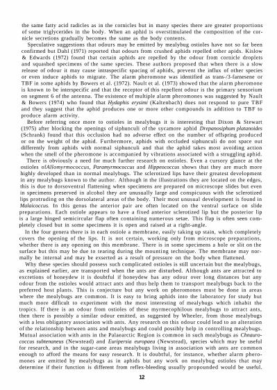
Second Instar Female (Fig. 5) This instar has similar characters to those of adult and third instar female but can easily be distinguished in possessing a pair of lateral setae on sixth abdominal segment each about 360 µm long, a pair on dorsal lateral edge of fourth segment each about 550 (xm long and a submarginal pair on dorsum of prothorax each about 500 fxm long, the last 2 pairs with sclerotized areas around setal bases. Body ovoid, about 1-35 mm long and 10 mm wide, anal lobes moderately developed. Antennae 0-9 mm long, the first segment 110 µm long, the second 80 µm long, third 180 µm, fourth 130 µm, fifth 160 µm and sixth 240 µm long. Setae ranging from 24 µm long on first segment to 36 µm long on last segment. Legs with hind trochanter+femur 450 µm long, hind tibia 390 µm and hind tarsus 150 µm; all segments densely covered with slender setae, those on disc of femur 16-20 µm long and on disc of tarsus 32 µm long. Claw 32 µm long. Anal ring 40 µm in diameter with 6 setae each 52 µm long; situated about twice diameter
17

Fig. 4 AUomyrmococcus acariformis Takahashi. Third instar female.
18
Fig. 5 Allomyrmococcus acariformis Takahashi. Second instar female.
19

from apex of body. Labium 132 µm long and 116 µm wide, with numerous setae as in third and adult instars but obscured in specimen available. Clypeolabral shield with numerous setae but difficult to count in available specimen. Ostioles with well-developed sclerotized lips, the anterior pair lying on ventral surface when mounted on slide. Anal lobes sclerotized, each with 11 apical and subapical setae about 1 -5 mm long, their positions varying on dorsal and ventral surfaces but probably with 4 on dorsal surface and 7 on ventral surface. Body setae slender, about 16 µm long, covering almost entire surface except for intersegmental spaces, the distance between setal bases shorter than length of one seta. Trilocular pores few, scattered.
MATERIAL EXAMINED. THAILAND: 2, 'Siam', Mt Sutep, 5.iv.l940 (R. Takahashi; 1 third instar $ and 1 second instar $, same data as holotype (TARI, Taipeh).
This material has the same data as the holotype and it is assumed it is part of the original. The specimens have been kindly made available for study by Mr Charles Chia-chu Tao of the Taiwan Agricultural Research Institute, Taipeh.
HIPPEOCOCCUS Reyne, 1954 Hippeococcus Reyne, 1954 : 237. Type-species: Hippeococcus rappardi Reyne, by original designation. This genus is known only from Java. It has some peculiar and unusual features connected with its mode of life. According to Reyne the species are invariably associated with ants of the genus Dolichoderus (=Hypoclinea). They are known to crawl on to ants' backs and are carried away when the ants are disturbed but large individuals may be carried in the ants' jaws. Found on the suckers and stems of the host, colonies are often covered with dense crowds of ants.
Reyne has given detailed descriptions of the species but there has been a pressing need for modern illustrations. The following descriptions, therefore, serve to supplement Reyne's and to accentuate the principal characters.
In common with most mealybugs there are four female instars and all of these are described for H. wegneri Reyne. Although Reyne mentioned only three instars it is possible that his descriptions of the second instar may include characters of the third also. Only three female instars are available here for H. rappardi Reyne; nevertheless, one or two specimens are at hand of what is presumed to be the second instar male and this is described. Reyne described the adult female before preparation as 'Body gleaming, without a coating of wax'. DESCRIPTION OF ADULT FEMALE. Body broadly turbinate, seventh and posterior segments narrow. Venter of last segment folded on either side near the mid-line giving it the appearance, when viewed through the stereomicroscope, of forming two parallel tubes, the apices forming the anal lobes containing 5-7 long setae which may be almost as long as the body. The two tube-like parts of the last segment are covered dorsally with a triangular lobe containing setae of various lengths and which bears the anal ring at the apex. This ring is sclerotized, without pores, crescentic and contains 6 setae which increase in length and thickness posteriorly. The anal ring is usually on level with apices of the anal lobes but in one species the anal lobes are produced latero-posteriorly for a distance about same length again as last segment. Antennae 6-segmented, first segment normal, second quite small and usually wider than long, both segments with few setae. Third to sixth segments stout and elongate, containing numerous slender pointed setae, the total length of antenna about half length of body. Ostioles well developed, lying at sides of body, each with heavily sclerotized lips, the sclerotization surrounding anterior lips of first pair also extending around eyes. Without membranous lips, setae or trilocular pores. Setae on both surfaces of body abundant, mainly of two types, one of which is minute, clavate, usually 6-8 µm long, almost transparent, and the other type normal and pointed but short, about 20 µm long. Other setae, present on the posterior abdo-minal segments and legs, may be stout and long with a blunt tip or swollen apically and of a fleshy appearance. Only the basal part of these setae is sclerotized, the remainder becoming more transparent towards the apex. Claw stout, bearing a pair of quite wide and flat digitules each usually about as wide as the claw and about twice as long. Trilocular pores present. Circulus oval.
DESCRIPTION OF IMMATURE INSTARS. Normally turbinate but becoming wider at each instar. Anal lobes and dorsal triangular lobe containing anal ring present in all stages. Minute transparent clavate setae absent. Short pointed setae absent in first instar but becoming more numerous in each successive stage. Antennae of first instar with thickened long setae only. Second instar with abundant short setae on third to sixth antennal segments and these setae become more numerous in successive instars.
20

Fig. 6 Hippeococcus rappardi Reyne. Adult female. Specimen from Java, Mt Kawai.
21

Key to species of Hippeococcus (adult females) 1 Anal lobes projecting about the length of last segment from level of anal ring montanus Reyne (p. 22) - Anal lobes terminating at about same level as anal ring 2 2 Ventral surface of abdomen with numerous stout or expanded setae in addition to long setae
on seventh and posterior segments. Anal lobes each with 6 long apical setae wegneri Reyne (p. 26) - Ventral surface of abdomen without numerous expanded setae, short pointed setae present
only except for long setae on seventh and posterior segments. Anal lobes each with 5 long apical setae rappardi Reyne (p. 22)
Immature instars A few instars have not been available for study and it is not possible to give a workable key. The first instars of H. rappardi and H. wegneri have thick, fleshy and clavate setae on the dorsal and ventral surfaces but in H. rappardi there are 2 long pointed setae on each side of sixth and seventh segments. In H. wegneri there is 1 long seta and 1 short blunt seta in these positions. The thick clavate setae persist in the second and third instars of H. wegneri but in H. rappardi they are replaced by pointed setae. Instars are mainly distinguished by an increase in antennal and leg lengths. The second instar males are easily recognized by the presence of multilocular disc pores and oral collar tubular ducts.
Hippeococcus montanus Reyne, 1954
22
Adult Female (Fig. 6) Body broadly turbinate, about 2-1 mm long and 1-45 mm wide, posterior segments tapering. Anal lobes heavily sclerotized ventrally and with the dorsal surface forming lobe covering median area of last segment, with anal ring at apex. Seventh and eighth segments sclerotized mid-dorsally, and areas of sclerotization present around the well-developed ostioles and around the eyes. Entire surface with minute sclerotized dots that give the body a slightly dark appearance even when not stained. Antennae about 1-55 mm long, the third to sixth segments densely covered with slender setae, those on the third segment 52-60 µm long and those on sixth segment thicker and usually 100 µm long. Legs well developed, hind trochanter + femur 530 µm long, hind tibia + tarsus 540-560 µm long, hind coxa with a few translucent pores. Setae on legs of various sizes, on the hind tarsus they range from stiff pointed setae 40-100 µm long on the inner edge to stout fleshy setae, pointed, 52-160 µm long on the outer edge. Claw about 44 (µm long. Labium 180 µm long and 140 µm wide with 14 pairs of setae. Apical segment with two pairs of posterior setae, 1 pair of minute apical setae, 4 pairs of subapical setae each 32 µm long, 1 pair lateral setae each 52 µm long and 2 pairs of anterior setae each 68 µm long. Medial segment with a single pair of medial setae each 60 µm long and basal segment with 3 pairs basal setae, 1 pair 32 (xm long and 2 pairs each 20 µm long. Circulus oval, about 74 µm wide. Anal ring at apex of abdomen, about 88 µ.m wide, with 6 setae, these becoming distinctly longer in a posterior direction, the posteriormost quite thick.
Dorsal setae of two main types. On all segments anterior to eighth there are numerous minute clavate setae with a fleshy appearance, each about 6 µm long. Other dorsal setae pointed, numerous and usually 26 µm long. On the posterior abdominal segments the setae become longer. Trilocular pores not numerous.
There are no specimens available of this species as Reyne (1954) named it from certain drawings made by P. van der Goot possibly in 1915. The illustration has the appearance of being accurately executed. Although obviously a Hippeococcus species, it differs from H. rappardi and H. wegneri in having quite long and prominent anal lobes that are set rather wide apart and protrude about the same distance as the length of base or dorsal lobe covering them. In the other two species the anal lobes are about the same length as the dorsal lobe.
Other than living in Java there is no further information on its locality but from the illustration reproduced by Reyne, it should be easy to identify the insect when collected again.
Hippeococcus rappardi Reyne, 1954

Fig. 7 Hippeococcus rappardi Reyne. Third instar female.
23

Fig. 8 Hippeococcus rappardi Reyne. (A) First instar. (B) Second instar male.
24

Ventral surface with numerous minute blunt setae as on dorsum but they are absent between antennae and on eighth and posterior segments. Other setae pointed, about 26 µm long except for longer setae near abdominal margins and for long setae 180-200 µm long on the seventh and eighth segments. Tri-locular pores tending to be more numerous than on dorsum but absent on head.
Third Instar Female (Fig. 7) Body about 1 -75 mm long and 0-75 mm wide, elongate, widest on thorax, abdomen tapering. Seventh and eighth segments with distinct sclerotization in mid-regions. Anal lobes and dorsal median lobe heavily sclerotized. Antennae 1-2 mm long, third to sixth segments with numerous short setae. Hind trochanter + femur 450 µm long, hind tibia + tarsus 460 µm long. Claw about 36 fxm long. Labium 148 µm long and 100 µm wide with same number of setae as in adult and about same size. Circulus oval, 56 µm wide. Ostioles present at edges of body, similar to those of adult but smaller. Body setae short and pointed only, with a similar distribution to those of adult female, but less numerous; mid-dorsal setae about 40 fxm long and a typical short ventral seta about 40 µm long. Seventh and eighth segments sclerotized over most of the area. On the seventh segment most of the setae lie in the sclerotized area and on the eighth segment all the setae lie within the sclerotized area. Some long ventral setae on segments 7 and 8 stout and longer than the segments.
Second Instar Male (Fig. 8B) The writer agrees with Reyne (1954) that this is the second instar male rather than the female because the body has a vestiture of numerous tubular ducts and multilocular disc pores. Although second instar females of other mealybugs may possess similar pores and ducts they usually become more numerous in the third and adult instars. In this species they are absent completely in the third and adult female instars. Numerous pores and ducts in the second instar male are present to secrete the pupal covering.
Body elongate, abdomen tapering, about 1-35 mm long and 0-55 mm wide. Antennae 800 µm long, the third to sixth segments densely covered with short slender setae. Legs well developed, hind trochanter + femur 300 fxm long, hind tibia + tarsus 340 µm long. Claw 28 fxm long. Labium 120 µm long and 100 µm wide with 14 pairs of setae, common to the species. Dorsal labial setae shorter than those of previous stages, subapical setae each 24 µm long, lateral setae 36 µm long, anterior setae each 40 µm long. Medial segment with medial setae 52 µm long and basal segment with 1 pair basal setae 32 fxm long and 2 pairs each 16 µm long. Circulus oval, about 20 fxm wide. Ostioles well developed, on edges of body. Anal ring 64 µm wide.
Dorsal setae all pointed, numerous, a mid-dorsal seta usually about 24 µm long. Longer setae present also on the lobe-like structure above anal lobes. Multilocular disc pores present, mainly in submedian areas of thorax and anterior abdominal segments. Oral collar tubular ducts, each with the internal collar flange-shaped and occupying about half length of duct, situated among the multilocular disc pores and extending to margin. Trilocular pores sparse.
Ventral surface with numerous pointed setae tending to be longer than those on dorsum, a common length being about 32 um. Two pairs of sclerotized areas on seventh and eighth segments each with a pair of long pointed setae. Multilocular disc pores in mid-regions of thorax, anterior abdominal segments and near spiracles. Tubular ducts present near the multiloculars and towards the margins. Trilocular pores few.
First Instar (Fig. 8A) Body narrowly turbinate, about 1 -2 mm long and 0-4 mm wide at the thorax. Anal lobes and dorsal surface forming lobe, heavily sclerotized. Anal ring about 60 µm wide, without pores but with 6 setae at anterior or dorsal half of the ring. These setae become progressively thicker and longer posteriorly, the longest about 160 µm. Anal lobes with 2 long dorsal setae and 3 ventral setae, each about 0-95 mm long. Antennae 6-segmented, about 630 µm long with a few long setae slightly expanded distally, on each segment except last where they are pointed. Legs well developed, hind trochanter + femur 220 µm long, hind tibia + tarsus about 310 µm long. Claw 28 um long. Labium 120 µm long and 88 µm wide, the dorsal setae tending to be shorter than in previous stage; subapical setae 24 µm long, lateral setae 36 µm long, anterior setae each 32 µm long, medial setae 36 fxm long and basal setae with 1 pair 32 µm long and 2 pairs each 12 µm long. Ostioles well developed, without setae but with lips heavily sclerotized. Circulus small and oval about 20 µm wide.
Dorsal setae slightly expanded towards apex, each with a fleshy appearance and about 56 µm long on mid-abdomen where there are about 8 across a segment. Eighth segment with 2 pairs of long pointed setae reaching beyond anal lobes. Trilocular pores few, forming a single submedian row and with one or two around margins.
25

Ventral surface with setae similar to those on dorsum but tending to be longer, up to 60 fxm, except the submarginal setae on seventh and eighth segments which are long and pointed, in pairs, on lightly scleroed areas. Trilocular pores few, in submedian areas. Simple pores few on submargins, each about same size as a trilocular pore but usually oval in shape.
MATERIAL EXAMINED. Hippeococcus rappardi Reyne, holotype $, JAVA: East, Yang Mts, Gondang Plantation (RNH, Leiden).
JAVA: Immatures with same data as holotype; immature instars, Mt Kawi, Gaden, on young shoots of Litsea sp. (Lauraceae), ii.1951 (F. W. Rappard) (RNH, Leiden); 1 $ and immatures, same data but labelled from Litsea sp., Eugenia sp. [Myrtaceae] and Rubus sp. [Rosaceae] (BMNH, London).
Recorded also by Reyne (1954) from JAVA: Gondang, on Ficus variegata, on suckers of Eugenia sp. and Litsea confusa; from Gaden, on stems of Rubus sp. and Eupatorium sp. (Com-positae).
Hippeococcus wegneri Reyne, 1954 Hippeococcus wegneri Reyne, 1954:255. Holotype $, JAVA: West, Tjibodas, Botanical Garden, from
nest of Dolichoderus ( = Hypoclinea) gibbifer Emery, i.1953 (RNH, Leiden) [examined].
Adult Female (Fig. 9) Body broadly turbinate, attaining a length of 2-75 mm and a width of 2-0 mm, abdomen narrowing abruptly. Anal lobes and dorsal surface of eighth segment heavily sclerotized. Sclerotization also in dorsal median areas of fifth to seventh segments and in ventral submedian areas of seventh and eighth segments. Anal lobe setae comprising three dorsal and four ventral, each about 1-5 mm long. Entire surface with minute sclerotized dots giving the membranous body the appearance of being lightly sclerotized. Antennae about 1-5 mm long with abundant slender setae on third to sixth segments. Setae on third segment about 60 µm long, becoming progressively longer to sixth segment where they are thicker and 90-100 µm long. Legs well developed, hind trochanter + femur 570-590 µm long, hind tibia + tarsus 600-620 µm long. Claw 40 µm long. Setae on legs of different shapes; they are long clavate and thick with a fleshy appearance, or short or long and pointed, the longest on the outer side of tibia and tarsus. On the tarsus the pointed setae are 40-160 µm long and the stout fleshy setae 40-180 µm long. Hind coxae with a few translucent pores. Labium 184 [xm long and 120 µm wide, with 14 pairs of setae. Apical segment with 2 pairs of posterior setae, a pair of minute apical setae, 4 pairs of subapical setae each 28 µm long, a pair of slender lateral setae each 48 µm and 2 pairs of thick fleshy anterior setae each 80 µm long. Medial segment with 1 pair of fleshy setae each 92 µm long and basal segment with 2 pairs of short pointed setae each 20 (xm long and a pair of fleshy setae each 44 µm long. Circulus about 68 µm wide. Anal ring 84 µm wide, on level with apices of anal lobes, the anterior pair of setae 100 µxm long, the median pair 140 µm long and the posterior pair 180 µm long, these setae becoming thicker as they become longer.
Dorsal surface anterior to seventh segment with numerous minute clavate setae each 6-8 µm long, of a fleshy appearance, barely taking up the stain. Short pointed setae each about 20 µm long, evenly distributed. Posterior abdominal segments with a few clavate or thick setae ranging in length from 30 to 120 µm, the dorsal lobe covering anal lobes with a few thick setae about 60 µm long. Trilocular pores in moderate numbers, evenly distributed.
Ventral surface with similar minute clavate and pointed setae as on dorsum, on sixth and anterior segments. Long clavate or thick and bluntly pointed setae present across the abdominal segments ranging in length from about 60 to 90 µm except for some on seventh, eighth and anal lobe segments that may be up to 260 µm long. Trilocular pores numerous with an even distribution.
Third Instar Female (Fig. 10) Body a similar turbinate shape to that of adult female but narrower, 2-0 mm long and 1-0 mm wide. Sixth and posterior segments sclerotized with the 3 dorsal and 3 ventral anal lobe setae about 1-5 mm long. Antennae 1-13 mm long, the third to sixth segments densely covered with short setae which become longer towards the last segment. Hind legs with trochanter + femur 450 µm long, hind tibia + tarsus 480 µm long. Claw about 30 µm long. Labium 152 µm long and 120 µm wide, with 14 pairs of setae but these shorter than in adult. Subapical setae each 26 µm long, lateral setae each 36 µm long, the 2 pairs of anterior setae each 56 µm long, thick and fleshy. Medial segment with medial setae thick, each 68 µm long and basal segment with 2 pairs of short pointed setae each 16 µm long and 1 pair of thick setae each 56 µm long. Circulus oval, 68 µm wide. Anal ring about 68 µm wide. Body setae of two main types,
26

Fig. 9 Hippeococcus wegneri Reyne. Adult female. Specimen from Java, Tjibodas.
27

Fig. 10 Hippeococcus wegneri Reyne. Third instar female.
28

Fig. 11 Hippeococcus wegneri Reyne. (A) First instar. (B) Second instar female.
29
a clavate or expanded type, thick and fleshy, ranging in length from 50 to 100 µm except at posterior end of body where some are longer on ventral surface, and short pointed setae, which are numerous and interspersed among the fleshy setae. The minute clavate setae are absent in this instar. Trilocular pores sparse.
Second Instar Female (Fig. 11B) Body narrowly turbinate, abdomen tapering, 1.4 mm long and 0.7 mm wide. Sclerotization at posterior end of body on anal lobes, on dorsal surface of eighth segment and in median areas of seventh and sixth segments. Legs with hind trochanter + femur 370 µm long, hind tibia + tarsus 390 µm long. Claw about 28 µm long. Antennae 900 µm long, third to sixth segments with numerous slender setae. Ostioles less developed than in previous stage. Circulus about 40 µm wide. Labium with 14 pairs of setae. Apical segment with 4 pairs of subapical setae each 24 µm long, a pair of slender lateral setae each 36 µm long and 2 pairs of thick fleshy anterior setae each 48 µm long. Medial segment with 1 pair of fleshy setae each 56 µm long and basal segment with 2 pairs of slender setae 12 µm long and a pair of thick setae each 44 µm long. Body setae with similar distribution of clavate type to those in third stage but short pointed setae less numerous especially on head and ventral thorax, the short dorsal setae about 24 µm long and the ventral setae about 32-40 µm long. The clavate setae range in length from 50 to 100 µm long except at posterior end of body where some are 160 µm long. Anal lobe setae difficult to measure but all are over 1.0 mm long. Trilocular pores sparse on both surfaces.
First Instar (Fig. 11 A) Body narrowly turbinate, about 1.35 mm long and 0.7 mm wide, anal lobes each with 3 dorsal and 2 ventral setae at apices, about 1.3 mm long. Anal lobes and dorsal lobe-like structure covering anal lobes heavily sclerotized. Antennae 630 µm long with clavate setae on all segments except last, which has long pointed setae. Legs well developed, hind trochanter + femur 300 µm long, hind tibia + tarsus 320 µm long. Claw 24 µm long. Labium 108 µm long and 80 µm wide, with 14 pairs of setae, all slender except for medial setae which are thick and fleshy. Apical setae each 24 µm long, lateral setae each 28 µm long, anterior setae each 40 µm long. Medial segment with medial setae 56 µm long and basal segment with 2 pairs of basal setae 12 µm long and 1 pair 32 µm long. Ostioles small but with lips heavily sclerotized. Circulus oval, about 20 µm wide. Anal ring 44 µm wide with 6 setae which become progressively longer posteriorly, the second pair twice as long as anterior pair and posterior pair half as long again as median pair.
Dorsal surface without short pointed setae but with long clavate setae ranging in length from 40 to 68 µm. Eighth segment with 4 stout pointed setae as long as anal lobes. Trilocular pores sparse.
Ventral surface with similar clavate setae to those on dorsum but some are longer, approaching 80 µm long. Long stout clavate to pointed setae present also on small sclerotized areas on seventh and eighth segments. Trilocular pores few.
MATERIAL EXAMINED. Hippeococcus wegneri Reyne, holotype $, JAVA : West, Tjibodas, Botanical Garden, i.1953 (A. M. R. Wegner) (RNH, Leiden).
JAVA: immatures, same data as holotype; paratypes and immatures, same data, 1 $ on young shoots and fruit of Diospyros kaki [Ebenaceae] (RNH, Leiden), 1 $ (ITZ, Amsterdam); 6 $ and immatures, i.1953 {A. M. R. Wegner); 2 ?, vi.1953 (A. M. R. Wegner); 1 $ Tjibodas, 22.x. 1973 (B. Bolton) (BMNH, London).
Recorded also by Reyne (1954) on Cyclanthera explodens (Cucurbitaceae).
MALAICOCCUS Takahashi, 1950 Malaicoccus Takahashi, 1950: 65. Type-species: Malaicoccus riouwensis Takahashi, by original desig-
nation.
This is an unusual genus of the Pseudococcidae in that the known species have the prothorax constricted and most of the head is heavily sclerotized. In life they have a strong resemblance to aphids and may often be mistaken for them. All are associated with ants of the genera Polyrhachis and Hypoclinea but little is known of their life history or habits. The mealybugs are attended by the ants which often carry them in their mandibles at the constriction on the prothorax.
DESCRIPTION OF ADULT FEMALE. Body form ovoid to globular with a definite constriction on prothorax opposite first coxae. Head heavily sclerotized around antennal bases, the sclerotization often extending to
30

first pair of ostioles; the front of head on ventral surface almost completely sclerotized forward from clypeus. Antennae 6-segmented, often longer than entire body and densely covered with slender setae; the first segment arising from dorsal surface. Second segment short and not articulating with third segment. Legs well developed, often with numerous setae of different sizes. Claws stout, with flattened digi-tules which are greater in size than claw itself. Labium conical with 21-32 pairs of setae, always with 2 pairs of posterior setae, a pair of minute apical setae, 4 pairs of subapical setae and 1 pair of lateral setae. The supernumerary setae occur in the anterior and medial groups. Circulus present, usually well developed with deep lateral constrictions. Ostioles of a distinctive type, the anterior pair each with an apparently fixed anterior lip which is arc-shaped. A heavily sclerotized semi-circular lower lip containing a few setae is hinged to close over to the anterior lip. These ostioles often lie on ventral surface on slides due to convexity of body or more often on margins so that they are viewed in lateral aspect. Posterior ostioles with anterior lip apparently fixed and with the posterior lip narrower than in anterior ostiole, with or without small setae. Anal ring terminal, narrow, with a few minute pores and 6 setae. Two crescentic or triangular areas of sclerotization containing setae present at either side of anal ring or just anterior to it. The anal ring is depressed behind the crescentic areas so that apex of abdomen forms a small 'honeydew basket'. Setae on margins of last 2-4 segments about as long as body and often accompanied with 1 or 2 short setae. Body setae minute, always numerous and sometimes densely covering the surface in which case the distance between the setal bases is shorter than the length of a seta. Other setae stout, of various lengths. Posterior end of body with sclerotized areas on both sides of anal lobes and on ventral surface at margin of some preceding segments.
DESCRIPTION OF IMMATURE INSTARS. Body oval but head angular in first instar and becoming more rounded in successive instars. Prothorax with a slight constriction in first instar but constriction increasingly apparent in second and third instars. Sclerotization on head starting around eyes in first instar and increasing in each instar to around base of antenna and in an area forward from clypeus. Anal lobes always developed to some extent. Antenna 6-segmented, often as long as body. In the first instar long pointed setae form whorls usually at distal end of segments but in the second and third instars the segments become densely covered with short pointed setae. Legs well developed, claw stout, with 2 flat, wide digitules even in the first instar. Labium with 14 basic pairs of setae but this number often increasing in third instar. Ostioles similar in shape to those of adult but without setae. Anal ring usually narrow with a few minute pores. Setae about as long as body on anal lobes and on at least first segment preceding lobes, often present as far forward as second segment or sixth preceding lobes, always on sclerotized areas. Body setae long on first instar but second and third instars with increasing numbers of short setae. In some species there are numerous minute setae with the seta shorter than diameter of setal base. Trilocular pores and simple pores present only except in second instar male which has multilocular disc pores and oral collar tubular ducts also.
DISCUSSION. The genus is probably more closely related to Allomyrmococcus than to any other, differing mainly in the sclerotized head, the presence of a circulus and the terminal anal ring. DISTRIBUTION. Malaya and the Riouw Is in Indonesia. A third instar female of an undescribed species is known among material of Allomyrmococcus acariformis collected by Takahashi in Thailand. See also the record from Sarawak on p. 6.
Key to species of Malaicoccus (adult females) 1 A distinct tuft of short slender setae opposite each first spiracle, the space between these setal
bases shorter than the length o f a seta . . . . . . . . . . 2 - Tuft of short setae opposite each first spiracle absent . . formicarii Takahashi (p. 33) 2 Setae in median area of ventral thorax all short and slender except for an occasional long seta
khooi sp. n. (p. 39) - Setae in median area of ventral thorax with numerous long stout setae in addition to short
slender setae . . . . . . . . . . . . . . 3 3 Long marginal setae each nearly 1.0 mm long on last 4 segments. Long setae in median areas
o f anterior ventral abdominal segments absent . . . . . moundi sp. n . (p. 44) - Long marginal setae nearly 1.0 mm long on last 2 segments only. Marginal setae on preceding
segments much shorter. Long setae present in median areas of all abdominal segments . 4 4 Hind tibia with setae 40-60 µm long, the spaces between setal bases often longer than a seta.
A seta 80 µm long on seventh segment in addition to long setae on eighth segment and anal lobes takahashii sp. n. (p. 52)
31

Fig. 12 Malaicoccus formicarii Takahashi. Adult female. Specimen from Malaya, Fraser's Hill.
32

- Hind tibia with numerous setae about 28 µm long, the spaces between setal bases shorter than a seta. Single setae 200-400 µm long on margins of sixth and seventh segments in addition to long setae on eighth segment and anal lobes . . . riouwensis Takahashi (p. 50)
Immature Instars It is hoped that the immature instars described here may be recognized easily from the illustrations and descriptions but in the absence of suitable material of some species it is not possible to present a workable key. Some of the following distinguishing characters may prove useful. In M. takahashii there are long setae on the anal lobes and margins of the eighth abdominal segment only but in the other species studied, long marginal setae reach as far forward as the second abdominal segment or sixth preceding segment from anal lobes. M. khooi, M. moundi and M. takahashii have unusually short setae in the second and third instars, each seta much shorter than the diameter of the setal bases. The characters of the adult female are reflected in the immature instars. For instance, in M. khooi there is a short wide terminal segment on the antenna in the adult, third and second instars and there is an absence of long setae on the ventral surface of thorax in the adult and third instars. The labium of the first and second instars has the basic number of 14 pairs of setae. In M. moundi this number persists in the third instar but in the other species studied the number increases to as many as 18 pairs in the third instar. The labium of the second and third instars of M. khooi has a primary medial seta which is clavate and digitule-like. Second instar males are easy to recognize by the multilocular disc pores and oral collar tubular ducts on both surfaces.
Malaicoccus formicarii Takahashi, 1951
33
Adult Female (Fig. 12) External appearance described as 'brownish black, without wax'.
On the slide body quite rotund except for a constriction on prothorax. Head and thorax heavily sclerotized around bases of antennae, in a broad band posterior to first pair of ostioles and ventrally in an area forward from clypeus to margins, there being a separate area running from the dorsal to ventral surface enclosing eye. The extent of the sclerotization varies slightly in different specimens. Antennae 2.3-2.5 mm long, the second segment longer than wide and all segments densely covered with slender setae which become longer towards apical segment. In an antenna 2.45 mm long the first segment is 300 um long, second 140 µm, third 320 µm, fourth 340 µm, fifth 370 µm and sixth 950 µm, the last segment strongly curved. Legs well developed, hind trochanter + femur 610-670 µm long, hind tibia + tarsus 670-760 µm long, always longer than trochanter + femur. Hind coxae with translucent pores towards outer edge. All segments densely covered with short setae. Claw 48 µm long. Circulus 150 um wide, wider than long with a constriction on either side. Anal ring elongate and becoming quite narrow at the point of constriction, easily distorted, with a few minute pores. Ostioles well developed, the anterior pair each with a narrow sclerotized lip which is apparently fixed, the posterior lip in the form of a hinged plate containing short setae which, when not flat against anterior lip, opens to almost right-angles to body. Posterior pair similar but posterior lip narrower, with a few setae. Labium 210 µm long and 120 urn wide, with 23-30 pairs of setae. Apical segment with 2 pairs of posterior setae, a pair of minute apical setae, 4 pairs of subapical setae each 28 µm long, 1 pair of lateral setae each 72 µm long, 3-7 pairs of anterior setae, the longest 60 µm. Medial segment with 9-12 pairs of setae, the longest 72 µm long. Basal segment with a pair of short setae each 24 µm long and a pair each 32 µm long. Body setae all pointed. Long marginal setae in four distinct groups on either side of abdomen, all about 1.0 mm long. Anal lobe setae comprising a pair on dorsal surface on a small triangular area of sclerotization and a ventral pair on a larger subrectangular sclerotized area. A pair of long setae also present on ventral margin of eighth segment at base of large sclerotized area. A pair also located on margin of seventh segment and a single seta on margin of sixth segment. Dorsal setae abundant, covering most of surface except parts of thorax and head where there are naked areas. The setae lie in definite waves or directions on each segment, a common length being about 16 µm except for longer setae on posterior abdominal segments and on two sclerotized crescentic areas anterior-lateral to anal ring. Ventral setae also abundant but not so numerous as on dorsum, mainly 20 um long. Trilocular pores very few on both surfaces except for a noticeable concentration near inner edge of first coxa and around the labium. A few minute circular pores also present, each with a diameter much smaller than a trilocular pore or setal base. Tubular ducts absent.

Fig. 13 Malaicoccus formicarii Takahashi. Third instar female.
34
Fig. 14 Malaicoccus formicarii Takahashi. Second instar female. Specimen from Selangor, Ula Langat.
35

Fig. 15 Malaicoccus formicarii Takahashi. First instar.
36

Fig. 16 Malaicoccus khooi sp. n. Adult female. Specimen from Malaya, Genting Highlands, on Poikilospermum cordifolium.
37

Third Instar Female (Fig. 13) This instar has many of the features of the adult female but the body is less broadly oval, 1 -75 mm long and 1-2 mm wide and with a smaller constriction opposite first coxae. Antennae 1-70-1-75 mm long, the first two segments with numerous short setae and the third to sixth segments densely covered with slender setae. Long marginal setae on abdomen numbering 6 pairs, about 1 -0 mm long on irregular sclerotized areas. Short body setae less numerous than in previous stage, the dorsal setae 20-28 µm long and some of the ventral slightly longer. Other body setae present much longer than the dorsal, ranging from 60 to 150µm long and ventral setae distributed across the segments 60-180 µm long. Sclerotization not so extensive as in adult. In addition to areas around marginal setae and behind the antennae there is a distinctive sclerotized area just below first pair of ostioles, and two triangular areas between antennal bases on dorsum, these containing setae. Circulus about 88 µm wide with a constriction on either side. Trilocular pores and smaller simple pores present but few. Anal ring narrow with lateral constrictions and becoming distorted during the preparation of the insect on the slide, with a few minute pores. Legs with a few short setae and some long pointed setae, hind trochanter + femur 430-450 µm long, hind tibia + tarsus 500-510 µm long. Claw 44 µm long. Labium 160 µm long and 100 µm wide, with 17-18 pairs of setae. Apical segment with 4 pairs of subapical setae each 28 µm long, a pair of slender lateral setae each 36 µm long and 2-3 pairs anterior setae, the longest 68 µm. Medial segment with 3-4 pairs, the longest 68 µm long, basal segment with 2 pairs basal setae each 24 µm long and a pair each 60 µm long.
Second Instar Female (Fig. 14) Body oval, about 1-35 mm long and 0-8 mm wide, with anterior end angled and with slight constrictions on the thorax opposite the first coxae. Antennae about 1-0 mm long, with long pointed setae and with a few short slender setae on first segment and numerous such setae on third to sixth segments. Legs with hind trochanter + femur 320 µm long, hind tibia + tarsus 350 µm long. Claw about 36 µm long. Labium 130 µm long and 80 µm wide with 14 pairs of setae. Apical segment with subapical setae each 24 |xm long, a pair of slender lateral setae each 32 µm long and 2 pairs of anterior setae, the longest 72 µm long. Medial segment with a single pair of medial setae each 56 µm long and basal segment with 3 pairs of setae, a long pair each 52 µm long and 2 shorter pairs each 20 m long. Anal ring sclerotized with 6 setae about 80 µm long, with a few minute pores at anterior end and one or two only at sides. Circulus 64 µm wide, oval with a slight notch on either side. Marginal setae on abdomen reaching over 1 0 mm in length. On the anal lobes there are 2 dorsal and 2 ventral setae, the dorsal setae on an irregular-shaped area of sclerotization and the ventral pair on a larger area that extends inwards over most of segment. The anterior long marginal setae either on venter or dorsum when mounted on slide depending on convexity of body. Each long seta accompanied with a much shorter seta, their bases on an irregular-shaped sclerotized area. Other sclerotized areas at bases of shorter setae on thorax, behind the antennae and on either side of anterior end of anal ring, the latter areas crescentic or triangular containing a few setae. Other body setae less numerous than in third stage, thick pointed setae present up to 120 µm long, also short pointed setae which on dorsum are 16-20 µm long and on venter about 40 µm long. Ostioles well developed, the anterior pair with a movable hinged lip.
First Instar (Fig. 15) Body oval, about 1 -05 mm long and 0-6 mm wide. Anal lobes poorly developed, anal ring on apex of abdomen, projecting further than anal lobes. Anal lobes with 2 ventral apical setae about 1-1 mm long, 1 dorsal seta about same length and another about half the length. The five preceding segments each with marginal ventral setae, these also about 1 0 mm long, all of these setae including those on anal lobes, on irregular-shaped sclerotized areas which, anterior to anal lobes, also contain a much shorter seta. Sclerotized areas present on head around the eyes and in a crescentic patch on either side of anal ring. Antennae 620 um long, with long pointed setae only on each segment. Ostioles prominent with heavily sclerotized lips. Circulus apparently absent. Anal ring sclerotized with 6 setae each about 44 µm long and a few minute pores at anterior edge only. Legs well developed, hind trochanter+femur 220 µm long, hind tibia + tarsus 260 µm long. Labium 110 µm long and 70 µm wide with 14 pairs of setae. Subapical setae each 20 µm long, a single pair of lateral setae each 40 µm long and 2 pairs of anterior setae the longest 60 µm long. Medial setae present as a single pair each 64 µm long. Basal segment with 2 pairs of setae each 20 µm long and a pair 44 µm long. Trilocular pores and simple circular pores in submedian areas, quite sparse.
38

WEST MALAYSIA: 17 $ and immatures, same data as lectotype (BMNH, London); 7 <j>, Selangor, Kepong, on Bauhinia sp. [Leguminosae], 29.viii.1943 (R. Takahashi); 2 $ and immatures, Selangor, Ula Langat, on 'jungle plant', 1928 (H. T. Pagden); immatures, Fraser's Hill, on Languas sp. [=Alpinia] [Zinziberaceae], 6.vi.l943 (R. Takahashi) (BMNH, London); 3 $, Cameron Highlands, on Rubus sp. [Rosaceae], x. 1944 (R. Takahashi) (UM, Kuala Lumpur); 2 ? and immatures, Gombak, on Mikania scandens [Compositae], 4.xi.l971 (Y. P. Tho) (UM, Kuala Lumpur). DISCUSSION. The adult female is unlike any other in the genus in lacking a tuft of short setae on the venter opposite the first spiracles and in having the short body setae so densely covered that the distance between setal bases is shorter than the length of a seta.
Malaicoccus khooi sp. n.
Adult Female (Fig. 16) External appearance not known. Body broadly oval, about 2-35 mm long and 2-17 mm wide with a deep constriction opposite first coxae. Head heavily sclerotized around antennal bases on dorsum and encircling the eyes, this area continuing to ventral surface just above anterior ostioles. Ventral head region anterior to clypeus almost completely sclerotized. Antennae about 1-88 mm long, first segment 250 µm, second 130 µm, third 360 µm, fourth 300 µm, fifth 260 µm and sixth 560 µm long, the last segment rather short and squat compared with that in other species. All segments densely covered with slender setae. Labium 208 µm long and 108 µm wide with 27 pairs of pointed setae. Apical segment with a pair of posterior setae, a pair of minute apical setae about 4µm long, 4 pairs of subapical setae each 32 µm long, a pair of lateral setae 52 µm long and 2 pairs of anterior setae, the longest 72 urn. Medial segment with 11 pairs of setae, the longest 64 µm long. Basal segment with 3 pairs of setae, the basal pair 16 µm long and the longest 56 µm. Circulus rather large with a deep constriction at either side, about 230 µm at its widest at posterior end. Legs stout, well developed, hind trochanter + femur 520 µm long, hind tibia + tarsus 460 µm long and thus shorter than trochanter+femur. Hind coxa with a few translucent pores on outer dorsal surface, short setae on outer ventral surface and longer pointed setae on inner ventral surface. Long setae also present on outer margin of trochanter and a few present on femur, tibia and tarsus: a few short setae also present on proximal end of femur. Claw 40 µm long. Anal ring reduced to a narrow rim with 6 setae each about 64 µm long, and a few minute pores. Ostioles well developed, the anterior pair lying on ventral surface when mounted on the slide due to convexity of body. Both pairs with edge of anterior lip heavily sclerotized and probably fixed, the posterior lips forming a hinged semi-circular plate which is capable of folding over to anterior lip, this plate with numerous short setae on anterior ostioles but without setae in posterior pair.
Marginal setae on abdomen long, on anal lobes and three preceding segments. Anal lobes with 1 long dorsal and 2 long ventral setae each 850 µm long, the dorsal surface also with a shorter seta about one-third length on an elongate-oval area of sclerotization. Ventral anal lobe setae at apex of a subrectangular sclerotized area. Marginal setae on three preceding segments slightly ventral in position. Eighth segment with a single seta 950 µm long and a much shorter seta on a subrectangular area of sclerotization. Seventh segment with a seta 480-550 µm long and a shorter seta on a small sclerotized patch. Sixth segment with a seta 490-600 µm long and also a short seta on a small sclerotized patch. Dorsal surface of head lateral to each antennal base with a long pointed seta about 32 µm long. A few other setae 12-18 µm long also within the sclerotized areas of head, on abdominal margins and in mid-regions of eighth segment on dorsum and seventh and eighth segments on venter. A few setae present on a sclerotized crescentic area on either side of anal ring. Other body setae numerous, ranging from 8 to 16 µn long on dorsum, the larger setae easily noticeable by the larger setal bases, evenly distributed except on head and thorax where there are a few completely bare areas. Ventral body setae usually longer than dorsal, 12-20 µm long, the longer setae being the most numerous. A distinct tuft of short setae present near margin opposite each first spiracle. Trilocular pores not numerous, evenly distributed but a few concentrated near labium. Minute circular pores sparse, each with a diameter less than a setal base. Tubular ducts each with a diameter smaller than a trilocular pore, represented by one or two on inner side of second and third coxae.
Third Instar Female (Fig. 17) Body similar in shape to that of adult female with deep constriction opposite first pair of coxae, 1-4 mm long and 0-9 mm wide. Antennae 1-25-1-30 mm long, the longest with first segment 180 µm long and 160 µm wide, second 90 µm long, third 260 fxm, fourth 230 µm, fifth 200 µm and sixth 430 µm. All seg-
39

Fig. 17 Malaicoccus khooi sp. n. Third instar female.
40

Fig. 18 Malaicoccus khooi sp. n. Second instar female.
41

Fig. 19 Malaicoccus moundi sp. n. Adult female. Specimen from Malaya, Genting Highlands, on stems of Uncaria sp.
42

ments densely covered with slender setae ranging in length from 30 to 40 µm on first segment to 45 µm on last segment. Legs well developed, stout. Hind trochanter + femur 390-400 µm long, hind tibia + tarsus 360-370 µm long. All legs with pointed setae of different sizes but second and hind legs each with a pair of knobbed digitule-like setae on outer proximal end of tibia, the first legs with similar setae at distal end of femur and along outer edge of tibia. Labium 160 µm long and 100 µm wide, with 17 pairs of setae. Apical segment with a pair of posterior setae, 1 pair of minute apical setae, 4 pairs of subapical setae each 24 µm long, 1 pair of lateral setae each 44 µm long, 2 pairs of anterior setae the longest 68 µm long. Medial segment with 4 pairs of setae, one of the pairs knobbed and digitule-like, 48 µm long. Basal segment with 2 pairs 16 µm long and 1 pair 52 µm long. Circulus constricted, widest at posterior end, 120 µm wide. Anal ring slender with a few minute pores and 6 setae each 64 µm long. Ostioles as in adult female, the anterior pair lying on ventral surface when mounted on slide but these ostioles without setae. Anal lobes sclerotized on both surfaces. Dorsal surface of each lobe with a seta 0-85 mm long and a shorter 330 fxm long. Ventral surface of lobe with apical seta 0-85 mm long and another probably shorter but broken in available specimens. Stout marginal setae on 6 anterior segments. On the eighth segment these setae are 0-85 mm long, on the seventh 350 µm, sixth 450 µm, fifth 450 µm, fourth 500 µm and third 550 µm long.
Dorsal surface with setae of different sizes. A pair of stout marginal setae each 350 (xm long present just behind first antennal segment. Other setae about 50-60 µm long, few on prothorax, head and last two abdominal segments and singly on margins of most segments. On last segments they are present on two crescentic sclerotized areas anterior-lateral to anal ring. Short slender setae numerous, each 6-8 µm long. Minute setae abundant, each with seta much shorter than setal base. Ventral setae of similar sizes to those on dorsum but a pair 300 µm long on head margin. Other long setae, the longest of which are 85 µm, present in transverse rows on abdomen, head and prothorax. Slender setae 4-12 µm long, few on abdomen but numerous in median areas of head and thorax. Minute setae present in moderate numbers, each much shorter than setal base. Trilocular pores and simple circular pores not numerous, scattered.
Second Instar Female (Fig. 18) Body oval, about 1 -0 mm long and 0-6 mm wide, sclerotized on head around eyes and on a few setal bases. Antennae 840-880 µm long. On longest antenna first segment 110 µm long, second 60 µm, third 160 µm, fourth 150 µm, fifth 120 µm and sixth 280 µm. Slender setae numerous but on each of first and second segments there are one or two knobbed digitule-like setae. Legs well developed with hind trochanter + femur 270-280 µm long, hind tibia + tarsus 270 µm long. Knobbed setae present at distal end of first and second femora and on outer edge of all tibiae. Other leg setae all pointed except for tarsal digitules. Claw stout 28 µm long. Labium 120 µm long and 80 µm wide, with 14 pairs of setae. Apical segment with minute pair of apical setae, 4 pairs of subapical setae each 20 µm long, 1 pair of lateral setae each 32 µm long and 2 pairs of anterior setae, the longest 56 µm long. Medial segment with 1 pair of knobbed setae 52 µm long. Basal segment with 2 pairs of short setae 12 µm long and 1 pair 36 µm long. Circulus constricted, 68 µm wide. Ostioles rather large for size of body, the anterior pair lying behind eye on ventral surface when mounted on slide, without setae. Anal lobes sclerotized, each with a dorsal seta 0-8 mm long, another 270 µm long and a ventral apical seta 0-8 mm long with another 450 µm long. Anterior six segments with long marginal setae, the bases surrounded by sclerotized areas. Eighth segment with marginal setae 0-8 mm long, seventh 250 µm, sixth 350 µm, fifth 490 µm, fourth 550 µm and third 550 µm long. Anal ring with a few minute pores and 6 setae each 60 µm long.
Dorsal surface with stout setae on thorax and posterior abdominal segments 24-50 µm long. On remainder of abdomen the setae are small and slender, about 12 µm long. A pair of long setae behind first antennal segment each about 210 µm long, the area about the setal bases sclerotized. Minute setae numerous, each with seta much shorter than diameter of setal base. Ventral setae of different sizes, the longest about 50 µm accompanied with short slender setae and a few of the minute type. Trilocular pores and simple circular pores sparse.
Holotype $, WEST MALAYSIA: Malaya, Genting Highlands, on Poikilospermum cordifolium [Urticaceae], attended by ant Hypoclinea sp., 4.x. 1973 (L. A. Mound) (BMNH, London).
Paratypes. WEST MA
LAYSIA: 3 $ and immatures, same data as holotype; 1 $, Genting Highlands, on low herbaceous plant, attended by ant Hypoclinea sp., 4.x. 1973 (B. Bolton) (BMNH, London); 2 ?, Gombak F. S., on bamboo [Gramineae], 26.x. 1971 (Khoo Soo Ghee) (UM, Kuala Lumpur). DISCUSSION. This species is similar to M. moundi in having setae about 1.0 mm long on anal lobes and margins of the three preceding segments but it differs in the setae on mid-ventral thorax
43

which are short and slender, rarely more than 20 [xm long whereas in M. moundi there are numerous setae up to 130 jam long in this area. The sixth antennal segment in M. khooi is about 560 µm long but in M. moundi it is more slender and 710 µ.m long.
Malaicoccus moundi sp. n.
Adult Female (Fig. 19) Body broadly oval, with a distinct constriction on margins opposite first coxae, about 1-75 mm long and 1-15 mm wide. Head sclerotized around antennal bases on dorsal surface and over entire apex of venter as far as clypeus and almost to constriction on margin. Antennae densely covered with short setae which become longer towards last segment. Total length of antennae 212 mm and longer than body in available specimen; first segment 260 µm long, second 120 µm, third 370 µm, fourth and fifth each 330 |xm and sixth segment 710 µm long. Legs well developed, hind trochanter + femur 640 µm long, hind tibia 4- tarsus 630 µm long hence slightly shorter than trochanter + femur. All segments with moderately long setae, hind coxa with a few translucent pores on dorsal outer half. Claw 56 µm long. Labium 212 µm long and 124 µm wide, with 22 pairs of pointed setae. Apical segment with 2 pairs of posterior setae, 1 pair of apical setae about 16 µm long, 4 pairs of subapical setae each about 28 µm long, 1 pair of slender lateral setae each 64 µm long and 2 pairs of anterior setae, the longest 92 µm. Medial segment with 8 pairs of setae, the longest 84 µm. Basal segment with 4 pairs of setae, the shortest 22 µm long and the longest 68 µm long. Circulus large with a deep constriction on either side, the posterior edge wider than anterior edge, 240 |xm at widest. Anal ring elongate and narrow, constricted, but distorted in available specimen; with 6 setae, each 72 µm long and a row of minute pores. Ostioles well developed, each with a fixed sclerotized anterior lip and a hinged sclerotized posterior lip which may completely close the ostiole. Anterior pair on ventral margin on slide probably due to convexity of body, the posterior lips with a few setae.
Marginal setae on abdomen, on anal lobes and three preceding segments, the long setae all about 1 -2 mm long. Anal lobe setae represented by 2 on dorsum, one thinner than the other, at apex of an elongate area of sclerotization. Ventrally each lobe with a pair of long setae and a large subrectangular area of sclerotization. Sixth to eighth segments with long setae on ventral margins, those on sixth segment accompanied with a shorter seta about 130 jxm long, on the seventh segment with a shorter seta about 130 µm long and on the eighth segment with a seta about 400 (xm long. Stout setae 50-130 µm long present dorsally on head, prothorax, abdominal margins, median areas of eighth and posterior segments and in a cluster on a sclerotized crescentic area on each side of anal ring. Similar setae present ventrally on head, in median areas of thorax and in mid-region of seventh and posterior segments of abdomen. One or two longer setae 130-300 µm long, in median area of dorsal and ventral eighth abdominal segment. Body setae abundant except on dorsum of head and prothorax where there are extensive bare areas. These setae extremely slender, 6-24 µm long, the majority being the smallest. Ventral surface opposite first spiracles with a distinctive tuft of short setae. Trilocular pores not numerous, evenly distributed but absent on head; there are, however, noticeable concentrations around labium. Simple circular pores, jach with a diameter smaller than a trilocular pore, in moderate numbers between second and third coxae.
Third Instar Female (Fig. 20) Body broadly-oval with a constriction opposite first coxae giving the body a pyriform appearance, about 1-4 mm long and 0-95 mm wide. Head sclerotized on dorsum between antennal bases, on venter between :lypeus and anterior margins, and surrounding eyes. Anal lobes moderately developed. Antennae about 1-57-1-58 mm long, the first segment 170 fxm long, second 100 [xm, third and fourth each 250 (xm, fifth 260 |xm and sixth 550 µm long, each segment with numerous slender setae which on first segment are ibout 32 |xm long and on sixth segment about 36 µm long. Legs with hind trochanter + femur 460-470 µm long, hind tibia + tarsus about 460 µm long, stout. Labium 160 (µm long and 100 |xm wide. Apical segment with 2 pairs of posterior setae, a pair of minute apical setae, 4 pairs of subapical setae each 24 µm long, 1 pair of lateral setae each 40 µm long and 2 pairs of anterior setae 60 and 80 [xm long. Medial segment with 2 pairs of setae 40 and 60 µm long. Basal segment with 2 pairs of short setae each 24 µm long and a pair of longer setae each 68 µm long. Anal ring slender, easily distorted, with a few minute pores and 5 setae each 88 µm long. Circulus constricted, 130 µm wide. Ostioles as in adult female, the anterior pair jften lying on ventral surface when mounted on slide but without setae. Anal lobes sclerotized on dorsal md ventral surfaces, each lobe with a dorsal and ventral seta about 1-3 mm long, a ventral seta 0-8 mm ong and a dorsal 0-6 mm long. Ventral margins of third to eighth segments with sclerotized areas each containing one long and one short seta, third and fourth segments with the longer seta 0-7 mm long,
44

Fig. 20 Malaicoccus moundi sp. n. Third instar female.
45

Fig. 21 Malaicoccus moundi sp. n. Second instar female.
46
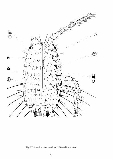
Fig. 22 Malaicoccus moundi sp. n. Second instar male.
47

Fig. 23 Malaicoccus moundi sp. n. First instar.
48

fifth 0-6 mm, sixth 0-7 mm, seventh 0-6 mm and eighth 1-3 mm long. The short marginal seta on eighth segment about 330 µm long, the others on anterior segments shorter.
Body setae of different sizes. Dorsum with stout setae, most about 85 fxm long, in single transverse rows but a median pair on last segment about 150 µm long. A pair of sclerotized crescentic areas present anterior to anal ring, each with a few setae of different sizes. Stout ventral setae also of different sizes, a common length being about 100 µm. Slender setae scattered on both surfaces, 8-28 µm long. Other minute setae numerous, each shorter than the diameter of its setal base. Trilocular pores not numerous. Simple circular pores smaller than a trilocular pore, scattered.
Second Instar Female (Fig. 21) This instar differs from previous instars in having shorter and fewer characters. Body about 1 -25 mm long and 0-85 mm wide. Head with 2 elongate sclerotized areas between antennae on dorsum. More extensive sclerotization present around eyes and almost reaching anterior ostioles. Antennae 1-12 mm long; first segment 130 µm long, second 60 fxm, third 180 µm, fourth 180 µm, fifth 170 µm and sixth 400 µm. All segments with numerous setae. Legs as in previous stage, hind trochanter + femur 330 µm long, hind tibia + tarsus 350 µm long. Claw 36 µm long. Labium 128 µm long and 78 µm wide, with 14 pairs of setae. Apical segment with 2 pairs of posterior setae, a pair of minute apical setae, 4 pairs of subapical setae each 28 µm long, a pair of lateral setae each 36 µm long and 2 pairs of anterior setae 40 and 50 µm long. Medial segment with a single pair of setae 60 µm long. Each lobe of basal segment with a pair of short setae each 20 µm long and 1 long seta 72 µm long. Circulus 80 fxm wide. Ostioles as in previous stage, the anterior pair lying on ventral surface when mounted on slide, without setae. Anal ring with 1 or 2 minute pores and 6 setae each about 60 µm long. Anal lobes each with dorsal and ventral elongate areas of sclerotization, with 2 dorsal setae, one 1 0 mm long, the other 550 µm long and 2 ventral setae, one 1 -0 mm long and the other 800 µm long. Long marginal setae present on 6 preceding segments, each setal base surrounded by sclerotized areas. Eighth segment with seta 1 0 mm long and a shorter about 180 µm long, seventh with seta 300 fxm long, sixth seta 370 µm long, fifth and fourth each 500 µm and third 630 µm long.
Dorsum with stout pointed setae in single transverse rows, usually 44-88 µm long except in mid-region of eighth segment where there is a pair 112 µm long. Similar setae present in a sclerotized crescentic area anterior-lateral to anal ring. Other dorsal setae more numerous and minute, each with seta much shorter than diameter of base. Ventral surface with similar long setae to those on dorsum except on head margin where there is a pair 100µm long and behind first ostioles where there is a pair each about 120 µm long, these on sclerotized bases. On abdomen there are shorter pointed setae each about 8 µm long and minute setae of two types. One of these is similar to the dorsal minute setae but the other type is longer 2-4 µm but always shorter or same length as setal base. Trilocular pores on both surfaces, not numerous. Simple circular pores present, sparse, smaller than trilocular pores.
Second Instar Male (Fig. 22) Body elongate, head margin tending to be straight, 1-12 mm long and 0-65 mm wide. Antennae with 6 segments about 745 µm long, the first segment 90 µm long, second 45 µm long, third 120 µm, fourth and fifth each 110 µm long and sixth segment 270 µm long. Fourth to sixth segments with slender setae ranging in length from 36 to 52 µm. Legs with hind trochanter+femur 280 µm long, tibia+tarsus 300 µm long. Claw 28 µm long. Labium 120 µm long and 68 µm wide, with 13 or 14 pairs of setae. Apical segment with 2 pairs of posterior setae, a minute apical pair, 4 pairs of subapical setae each 20 µm long, a lateral pair each 36 µm long, 1 or 2 pairs of anterior setae, the longer 64 µm and the shorter when present 40 µm. Medial segment with 1 pair each 60 µm long and basal segment with 2 pairs 18 µm long and a pair 72 µm long. Anal ring with a few minute pores and 6 setae. Anal lobes sclerotized on both surfaces, the dorsal surface with 1 seta 0-9 mm long and another 500 µm long, the ventral surface with one 0-9 mm long and another 650 µm long. Long marginal setae on 6 anterior segments with bases surrounded by sclerotized areas, on eighth segment these setae 0-9 mm long, on seventh segment 220 µm, on sixth 310 µm, on fifth 400 µm, on fourth 500 µm and on third 550 µm long.
Dorsal and ventral setae similar to those of second instar female but minute setae absent on venter and sparse on dorsum. Trilocular pores few. Simple circular pores quite sparse, each smaller than a trilocular pore. Multilocular disc pores each with 8 loculi, present on dorsum in small submedian groups on fifth abdominal segment and anterior segments to prothorax, an occasional pore also present on anterior abdominal segments and mid-thorax. Oral collar tubular ducts present, each slightly larger in diameter than a trilocular pore and only slightly longer than wide with deep sclerotized internal collar. On dorsum they tend to lie in single transverse rows but on head and thorax they become more scattered. On venter they are present mainly on head and thorax and become sparse on abdomen.
49

First Instar (Fig. 23) Body oval, 1-0 mm long and 0-65 mm wide, head sclerotized around eyes. Antennae 565-700 µm long, one of largest with first segment 80 [xm long, second 60 µm, third 90 fxm, fourth 100 jxm, fifth 100 µm and sixth 270 µm long. Each segment with a few long pointed setae at distal end but more numerous setae present on last segment. Legs well developed with hind trochanter+femur 230-250 µm long, hind tibia + tarsus 250-270 µm long. Claw 28 fxm long. Circulus apparently absent. Labium 92 pun long and 52 µm wide, with 14 pairs of setae. Apical segment with 2 pairs of posterior setae, a minute pair of apical setae, 4 pairs of subapical setae each 20 µm long, a lateral pair 38 µm long and 2 pairs of anterior setae 20 and 56 µm long. Medial segment with 1 pair of setae 52 µm long and basal segment with 2 pairs 12 µm long and 1 pair 44 µm long. Anal ring slender with a few minute pores and 6 setae each 52 µm long. Anal lobes sclerotized on both surfaces, each with 2 dorsal setae, one 1 0 mm long, the other 460 µm long and 2 ventral setae, one 1 -0 mm long and the other 700 µm long. Long marginal setae present on 6 preceding segments, the bases surrounded by sclerotized areas. All of these setae stout, measuring on eighth segment 1-0 mm long, seventh segment 160 µm, sixth segment 220 µm, fifth segment 280 µm, fourth segment 280 µm and on third segment 340 µm.
Body setae 28-44 fxm long on dorsum but mid-region of eighth and last segment with pair of setae each 90 µm long on sclerotized patches. Ventral setae usually longer, about 80 µm long. Trilocular pores and simple circular pores sparse.
Holotype ?, MALAYSIA: Malaya, Genting Highlands, 50 km E. of Kuala Lumpur, on stems of Uncaria sp. [Naucleaceae], attended by the ant Hypoclinea sp., 28.ix.1973 (L. A. Mound) (BMNH, London).
Not included in type-series. MALAYSIA: 2 third instar $, 3 second instar $, 3 second instar <$ and 9 first instars, same data as holotype (BMNH, London).
DISCUSSION. The nearest species is M. khooi but it differs in the characters given in the discussion of that species. It differs from M. riouwensis and M. takahashii in having 4 pairs of long marginal setae on the abdomen instead of only 2 pairs. The short dorsal and ventral setae are similar to those of M. riouwensis and M. takahashii in that the distance between the setal bases is greater than the length of a seta but it differs from M.formicarii, which has the distance between the setal bases shorter than the length of a seta.
Malaicoccus riouwensis Takahashi, 1950 Malaicoccus riouwensis Takahashi, 1950 : 66. Holotype, ?, INDONESIA: Riouw Is, Rempang, in association
with ants of genus Polyrhachis sp. (probably lost).
Adult Female (Fig. 24) External appearance described originally as 'Shining blackish brown, with no wax, in life, but dark reddish brown and a little shining when dry.'
Body of adult female pear-shaped with a constriction opposite first coxae. Anal lobes moderately developed. Anterior end of body with extensive sclerotization, which on the dorsal surface, extends around the antennal bases, then laterally and posteriorly around the first pair of ostioles. Ventrally the sclerotization extends over anterior end of head to just above clypeus. Circulus folded in available specimen but described by Takahashi as having deep constrictions at the sides and widest at anterior end. It is possible that Takahashi studied specimens that were distorted and that in reality the circulus is widest at posterior end. Labium 180 µm long, with 21 pairs of setae. Apical segment with 2 pairs of posterior setae, a pair of minute apical setae, 4 pairs of subapical setae each 28 µm long, 1 pair of lateral setae each 60 µm long and 2 pairs of anterior setae, the longest 80 µm long. Basal segment with 2 pairs short setae and 1 pair longer setae. Antennae 6-segmented, 2-33 mm long, thus longer than body; first segment 280 µm long, the second 120 µm, third 390 µm, fourth 370 µm, fifth 320 µm and sixth 850 µm and distinctly curved. Each segment densely covered with short setae ranging from 24 µm long on the proximal segments to about 40 µm towards terminal segment. Other slender setae 120 µm long on second to sixth segments. Thick setae present on all segments, these about the length of the shortest slender setae but much fewer. Legs well developed, hind trochanter+femur 640 µm long, hind tibia+tarsus 650µm long. Hind coxa with translucent pores on outer dorsal half. Short setae present on all leg segments, those on tibia numerous, about 28 fxm long, the distance between setal bases shorter than length of one seta. Claw 48 µm long. Anal ring slender with 6 setae about 56 µm long and a few minute pores. Ostioles well developed, of the type common to the genus, the anterior pair with setae on the lower lips, the posterior pair without setae. Long
50
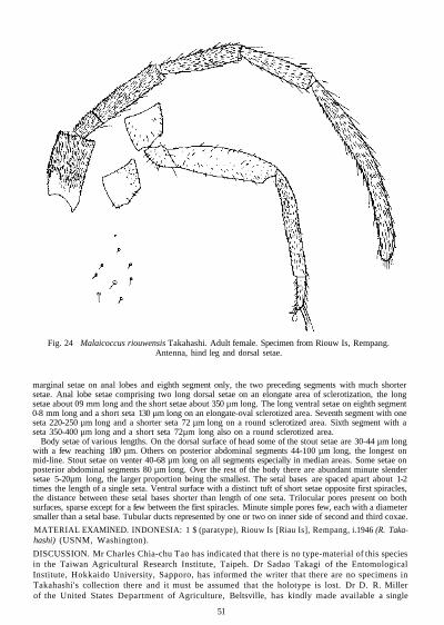
Fig. 24 Malaicoccus riouwensis Takahashi. Adult female. Specimen from Riouw Is, Rempang. Antenna, hind leg and dorsal setae.
marginal setae on anal lobes and eighth segment only, the two preceding segments with much shorter setae. Anal lobe setae comprising two long dorsal setae on an elongate area of sclerotization, the long setae about 09 mm long and the short setae about 350 µm long. The long ventral setae on eighth segment 0-8 mm long and a short seta 130 µm long on an elongate-oval sclerotized area. Seventh segment with one seta 220-250 µm long and a shorter seta 72 µm long on a round sclerotized area. Sixth segment with a seta 350-400 µm long and a short seta 72µm long also on a round sclerotized area.
Body setae of various lengths. On the dorsal surface of head some of the stout setae are 30-44 µm long with a few reaching 180 µm. Others on posterior abdominal segments 44-100 µm long, the longest on mid-line. Stout setae on venter 40-68 µm long on all segments especially in median areas. Some setae on posterior abdominal segments 80 µm long. Over the rest of the body there are abundant minute slender setae 5-20µm long, the larger proportion being the smallest. The setal bases are spaced apart about 1-2 times the length of a single seta. Ventral surface with a distinct tuft of short setae opposite first spiracles, the distance between these setal bases shorter than length of one seta. Trilocular pores present on both surfaces, sparse except for a few between the first spiracles. Minute simple pores few, each with a diameter smaller than a setal base. Tubular ducts represented by one or two on inner side of second and third coxae.
MATERIAL EXAMINED. INDONESIA: 1 $ (paratype), Riouw Is [Riau Is], Rempang, i.1946 (R. Takahashi) (USNM, Washington).
DISCUSSION. Mr Charles Chia-chu Tao has indicated that there is no type-material of this species in the Taiwan Agricultural Research Institute, Taipeh. Dr Sadao Takagi of the Entomological Institute, Hokkaido University, Sapporo, has informed the writer that there are no specimens in Takahashi's collection there and it must be assumed that the holotype is lost. Dr D. R. Miller of the United States Department of Agriculture, Beltsville, has kindly made available a single
51

paratype from the collections of the United States National Museum. This specimen is not in good condition and it has not been possible to illustrate it completely here.
The species is closely related to M. takahashii in having only 2 pairs of marginal setae nearly 1-0 mm long on abdomen on eighth segment and anal lobes. Anterior marginal setae to these are only 200-400 µm long. The most distinctive character separating this species from M. takahashii is the arrangement of the setae on the hind tibiae. These are short, about 28 µm long, and the distance between the setal bases on the disc of tibia is shorter than the length of one seta.
Malaicoccus takahashii sp. n.
Adult Female (Fig. 25) An oval species, 1-7 mm long and 1-1 mm wide, with a deep constriction opposite first coxae. Anal lobes moderately developed. Anterior end of body heavily sclerotized; on the dorsum this sclerotization encircles antennal base and curves laterally and posteriorly to completely surround anterior ostioles. The sclerotization on ventral surface of head occupies the area immediately in front of clypeus to anterior margin but curves forward laterally before reaching ostiole. Antennae with the first segment 250 [xm long, second 150 µm, third 360 µm, fourth 360 µm, fifth 350 µm and sixth 720µm, the last segment curved. With a total length of 2-19 mm the antennae are longer than body. All antennal segments densely covered with short setae which range in length from 28 µm on the first segment to 48 µm on the last segment. Legs well developed, hind trochanter + femur 560 µm long, hind tibia + tarsus 550 µm long. All segments with quite stout setae in moderate numbers, the smallest on tarsus about 44 µm long with the space between setal bases usually same or greater than length of an average seta. Hind coxa with translucent pores on dorsal outer half. Claw 48 µm long. Circulus notched at either side, 188 µm wide. Labium 200 µm long and 130 µm wide with 20 pairs of setae, many of which are broken but their presence determined by setal bases. Apical segment with 2 pairs of posterior setae, a pair of minute apical setae, 4 pairs of subapical setae, 1 pair of lateral setae and 2 pairs of anterior setae. Medial segment with 7 pairs of setae and basal segment with 2 pairs of short setae and 1 pair of longer setae. Ostioles well developed, the anterior pair each with sclerotized posterior lip containing a few setae. Anal ring rather thin and easily distorted, with a few minute pores on periphery and 6 setae, each about 68 µm long. Long marginal setae each about 780 µm long on anal lobes and eighth segment only. Dorsal surface of anal lobes with an oval sclerotized area. Long marginal setae of eighth segment at apex of an elongate sclerotized area that also contains a seta about 80 µm long, each on a small sclerotized patch. Stout setae on dorsum confined to head, prothorax, eighth and posterior segments. On the head they are present mainly within the sclerotized areas but on the anal lobe segment they are present in two sclerotized crescentic areas anterior-lateral to anal ring. Although most of the stout setae are about 40 µm long some on eighth segment may reach 80 µm and others on head attain a length of 100 µm. Stout setae present on venter on all segments but on abdomen they are present in median areas only. These setae are about 40 µm long and on posterior segments of abdomen they are up to 90 µm long. Other body setae short, slender and numerous, evenly distributed over dorsal and ventral surfaces but absent on dorsal parts of head and thorax. A marginal tuft of short setae present opposite first spiracles, the distance between setal bases shorter than length of one seta. All the short setae range in length from 5 to 24 µm but the shorter setae are most numerous. Trilocular pores few but there is a noticeable concentration between the two first spiracles. Tubular ducts represented by one or two on inner side of second and third coxae. Simple circular pores minute, sparse.
Immature Instars The three following instars are represented by poor distorted material and it has not been possible to illustrate them. Nevertheless, the long marginal setae are present only on the anal lobes as in the adult.
Third Instar Female Body oval 1 -25 mm long and 0-8 mm wide, constricted opposite first coxae. Head sclerotized around antennal bases, between clypeus and anterior head margin and around eyes on venter. Antennae 1-55 mm long, first segment 180 µm long, second segment 100 µm, third 250 µm, fourth 250 µm, fifth 200 µm and sixth 570 µm long; all segments with numerous slender setae. Legs with hind trochanter + femur 400 µm long, hind tibia + tarsus 410 jxm long. Claw 40 µm long. Circulus 128 |xm wide. Anal ring with a few
52

Fig. 25 Malaicoccus takahashii sp. n. Adult female. Specimen from Malaya, Kuala Lumpur.
53

minute pores and 6 setae each 60 m long. Labium 160 µm long and 90 [xm wide, with 16 pairs of setae. Apical segment with 2 pairs of posterior setae, a pair of minute apical setae, 4 pairs of subapical setae each 20 µm long, 1 pair of lateral setae each 56 µm long and 2 pairs of anterior setae, the longest 64 µm long. Medial segment with 3 pairs of setae, the longest 56 µm long. Each lobe of basal segment with a pair of short setae 20 µm long and one seta 44 µm long. Anal lobes sclerotized, each with one dorsal seta 0-8 mm long and a shorter seta 120 µm long, the ventral surface with one seta 0-8 mm long and another 280 fxm long. Eighth segment with marginal setae 0-7 mm long and anterior marginal setae much shorter, each about 100 µm long, all on sclerotized areas. Dorsal and ventral setae numerous, of two types, a slender pointed type and a minute type with the seta much shorter than diameter of setal base.
Second Instar Female Body 1-0 mm long and 0-55 mm wide, sclerotized on head between antennae on dorsum and around eyes on venter. Antennae 1-04 mm long, first segment 120 µm long, second 60 µm, third 170 µm, fourth and fifth each 160 µm and sixth 370 µm long. All segments with numerous slender setae. Hind trochanter+ femur 290 µm long, hind tibia + tarsus 320 µm long. Claw 34 µm long. Circulus 80 µm wide. Anal ring slender with a few minute pores and each of the 6 setae 48 µm long. Labium 130 µm long and 70 µm wide, with 14 pairs of setae. Apical segment with 2 pairs of posterior setae, a pair of minute apical setae, 4 pairs of subapical setae each 20 µm long, 1 pair of lateral setae 40 µm long and 2 pairs of anterior setae the longest 56 µm. Medial segment with a single pair of setae 44 µm long. Each lobe of basal segment with a pair of short setae 16 µm long and one seta 36 µm long. Anal lobes each with 4 setae, a dorsal and ventral 0-8 mm long, one dorsal 150 µm long and one ventral seta 270-320 µm long. Eighth segment with marginal setae 0-8 mm long. Anterior marginal setae 100 µm long on sclerotized areas. Trilocular pores, simple circular pores and minute setae very few.
First Instar Body oval, 0-85 mm long and 0-45 mm wide. Antennae 680 fxm long, the first segment 70 µm long, second 40 µm, third 100 µm, fourth 110 µm, fifth 100 µm and sixth 260 µm long. Hind trochanter + femur 220 µm long, hind tibia + tarsus 240 µm long. Claw 28 µm long. Anal ring setae 40 µm long. Anal lobes each with a dorsal and apical seta 0-85 mm long and a short dorsal and ventral seta 220 µm long. Eighth segment with marginal setae 0-85 mm long and anterior marginal setae 100-120 µm long, all on sclerotized areas. Labium 110 µm long and 60 µm wide with 14 pairs of setae. Apical segment with a pair of minute apical setae, 4 pairs of subapical setae each 20 µm long, a pair of lateral setae 32 µm long and 2 pairs of anterior setae, the longest 60 µm long. Medial segment with 1 pair of setae 52 µm long and each lobe of basal segment with a pair of short setae 16 µm long and 1 seta 40 µm long.
Holotype?, MALAYSIA: Malaya, Selangor, Kuala Lumpur, ll.viii.1943 (JR. Takahashi) (BMNH, London).
Not included in type-series. MALAYSIA: 1 third instar $, 9 second instar ? and 2 first instars, same data as holotype (UM, Kuala Lumpur).
DISCUSSION. This is probably the undescribed species from Kuala Lumpur, on Palaquium obova-twn (Sapotaceae), mentioned by Takahashi (1951) when discussing M. formicarii. Specimens were kindly made available by Dr Khoo Soo Ghee.
The most distinctive features of the adult female are the long pairs of setae present only on the anal lobes and eighth segment. These are about 780 µm long. The only other marginal setae, in addition to the normal body setae, are present on the seventh segment but these are only 80 µm long. A major character separating this species from M. riouwensis is the arrangement of the tibial setae, which although longer than those in M. riouwensis, are sparser, and on the tibial disc the distances between the setal bases are the same as or longer than the length of one seta.
PARAMYRMOCOCCUS Takahashi, 1941
Paramyrmococcus Takahashi, 1941 : 204. Type-species: Paramyrmococcus chiengraiensis Takahashi, by original designation and monotypy.
DESCRIPTION. Adult female with turbinate to oval body, tapering to a rounded posterior end, anal lobes obsolete. Antennae 6-segmented, about as long as body, first and second segments each with 1 or 2 stout setae, third to sixth segments densely covered with slender setae; second segment quite small, either shorter or just slightly longer than wide. Legs well developed, setae varying in size and number, claw
54

Fig. 26 Paramyrmococcus chiengraiensis Takahashi. Adult female. Specimen from Thailand, Chiengrai.
55
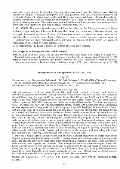
stout with a pair of wide flat digitules. Anal ring sclerotized with 6 setae but without pores. Ostioles situated on margins, of unusual development with large prominent lips that are heavily sclerotized but are devoid of setae. Circulus normal, usually oval. Body setae minute and slender, of unusual abundance, covering almost entire surface except for intersegmental areas, lying in definite directions giving the surface a wavy appearance. Other setae stout, situated mainly around margins; anal lobe setae not much more than twice diameter of anal ring in length. Trilocular pores few.
DISCUSSION. This genus is easily separated from the others in the Allomyrmococcini in being without recognizable anal lobes and in having short setae, only about twice diameter of anal ring in length, in normal positions of lobes. The immature instars are about the same shape as the adult but they tend to be more slender. Distinctive characters of the immature instars, based on P. vietnamensis, are short cylindrical and blunt setae on dorsum at least, which are replaced completely in the adult by short pointed setae. DISTRIBUTION. The genus is known so far from Thailand and Vietnam.
Key to species of Paramyrmococcus (adult females) Setae on hind tibial disc sparse, the distance between setal bases longer than length of a single seta.
Marginal stout setae on head and thorax attaining a length of 40 µm chiengraiensisTakahashi (p. 56) Setae on hind tibial disc numerous, the distance between setal bases shorter than length of one seta.
Marginal stout setae on head and thorax attaining a length of 80 µm vietnamensis sp. n. (p. 61)
Paramyrmococcus chiengraiensis Takahashi, 1941
(Fig. 26) Paramyrmococcus chiengraiensis Takahashi, 1941:204. Holotype ?, THAILAND: Chiengrai, Lampang,
on undetermined vine, attended by ant Dolichoderus sp. [ = Hypoclinea sp.] (probably lost). Paramyrmococcus chiengraiensis Takahashi; Takahashi, 1942 : 16.
Adult Female (Fig. 26) External appearance in life not noted. On the slide, adult female turbinate or broadly oval, widest at mesothorax, posterior end of body tapering, rounded, about 2.0 mm long and 1.25 mm wide. Antennae about 1.78 mm long, first segment 170 µm, second 90 µm, third 430 µm, fourth 320 µm, fifth 270 µm and sixth 500 µm long. Second segment about 80 (µm wide and thus slightly longer than wide. Sixth segment widest at proximal half, which then narrows before becoming slightly swollen. The first two segments with 1 or 2 stout setae only, the remaining segments densely covered with slender setae about 32 µm long on third segment and as long as 44 µm on sixth segment. Legs well developed, hind trochanter + femur 520 µm long, femur at its widest about 150 µm; hind tibia + tarsus 450-460 µm long, the tarsus 80 µm at its widest and distinctly shorter than trochanter + femur. Slender setae on dorsal and ventral surfaces of tibia quite sparse, about 40 µm long, the spaces between setal bases much greater than length of one seta. Claw 40 µm long. Labium 160 µm long and 140 µm wide, with 14 pairs of setae. Apical segment with two pairs of posterior setae, a pair of minute apical setae and four pairs of subapical setae each 28 µm long except the second pair, which are more slender and shorter, 20 µm long. Lateral setae and 2 pairs of anterior setae present, each 40 µm long. Medial segment with 1 pair of setae each 40 µm long and each lobe of basal segment with 2 pairs of setae 20 µm long and 1 pair 40 µm long. Circulus oval to subrectangular, 60-104 µm wide. Ostioles well developed, on the margins, with protruding sclerotized lips, without setae. Anal ring 72-80 fxm wide with 6 setae 70-80 µm long.
Body setae mainly slender, about 16 µm long but with a smaller proportion of longer setae about 20 µm long evenly distributed among the smaller, covering almost entire surface except for intersegmental areas; the distance between the setal bases much shorter than the length of one seta. On the dorsum the setae are even closer together than on venter. Other setae present are stout, those on margins of head and thorax on both surfaces 40-50 µm long, on margins of abdomen 60-80 µm long except on eighth segment where they are 112 µm long and the two ventral anal lobe setae about 120 µm long. Mid-ventral setae about 40 µm long. Trilocular pores sparse, apparently on venter only. Round pores on dorsal margins few, each slightly smaller than a trilocular pore and with obscure structure.
MATERIAL EXAMINED. THAILAND: 2 $, 'Siam', Chiengrai, 16.iv.1940 (R. Takahashi) (TARI, Taipeh).
56

Fig. 27 Paramyrmococcus vietnamensis sp. n. Adult female. Specimen from Vietnam, Da Lat.
57

Fig. 28 Paramyrmococcus vietnamensis sp. n. Third instar female.
58

Fig. 29 Paramyrmococcus vietnamensis sp. n. Second instar female.
59

Fig. 30 Paramyrmococcus vietnamensis sp. n. First instar.
60

DISCUSSION. The two specimens examined seem to belong to Takahashi's original type-series. They were kindly made available by Mr Charles Chia-chu Tao of the Taiwan Agricultural Research Institute, Taipeh.
Paramyrmococcus vietnamensis sp. n.
Adult Female (Fig. 27) External appearance not known. When mounted on the slide, body turbinate, widest at mesothorax, posterior end narrow and rounded, length 2.1 mm, width 1.3 mm. Antennae 1.9-2.0 mm long and thus about as long as body. On an antenna measuring 1.9 mm long the segment lengths are: first 200 µm, second 60 µm, third 470 µm, fourth 290 µ|m, fifth 290 µm and sixth 590 µm. The second segment 80 µm wide and, therefore, wider than long; sixth segment with a constriction at about middle but basal part wider than terminal part. First and second segments each with one or two stout setae only, remaining segments densely covered with slender setae ranging from about 40 µm long on third segment to 60 µm long on sixth segment. Legs well developed, hind trochanter + femur 560-570 µm long, the widest part of femur about 150 µm at distal end; hind tibia + tarsus 560-590 µm long, thus same length as trochanter+ femur or longer, widest part of tarsus 80 µm. Legs with 1 or 2 stout blunt setae and covered with slender setae about 40 µm long, which on the disc of hind tibia are set close together on dorsal and ventral surfaces so that spaces between are shorter than length of one seta. Claw 40 µm long. Ostioles well developed, situated on margins, each with prominent heavily sclerotized lips without setae. Circulus oval, 56-60 µm wide but sometimes with slight constrictions on either side, situated between fourth and fifth segments. Labium 172 µm long and 144 µm wide with 14 pairs of setae. Apical segment with 2 pairs of posterior setae, a pair of minute apical setae, 4 pairs of subapical setae each 28 µm long, 1 pair of lateral setae 40 µm long and 2 pairs of anterior setae, the longest 60 µm. Medial segment with 1 pair of setae each 44 µm long and basal segment with 2 pairs of setae 24 (xm long and 1 pair 40 µm long. Anal ring 68-72 µm wide, with 6 setae about 80 µm long. Body setae abundant and covering almost entire surface except for inter-segmental spaces. For the most part the setae are slender, about 16 µm long on dorsum and 16-20 µm long on venter, the spaces between the setal bases much shorter than length of one seta but even so these spaces are shorter on dorsum than on venter. Some of the slender setae are a little longer and are easily discernible by the larger setal bases. The directions of the slender setae form definite patterns giving the surface a wavy appearance. Stout setae, easily recognized by the large setal bases, present in small numbers around margins of both surfaces and across ventral abdominal segments, about 25-40 µm long but on the normal positions of the lobes there are usually 2 dorsal and 2 ventral setae about 120 µm long and on margins of eighth segment there are 1 or 2 setae about 100 µm long. Trilocular pores apparently confined to venter where they are sparse. Dorsal marginal and submarginal areas with a few round pores slightly smaller in diameter than a trilocular pore but with an obscure structure.
Third Instar Female (Fig. 28) Body pyriform, 1.7 mm long and 0.7 mm wide. Antennae 1.57 mm long, the sixth segment with a con-striction in the middle. First and second segments with a few stout setae only, third to sixth segments with numerous slender pointed setae but not so numerous as in adult. Labium 160 µm long and 120 µm wide with same number of setae as in adult. Apical segment with 4 pairs of subapical setae each 24 µm long, 1 pair of lateral setae each 32 µm long and 2 pairs of anterior setae 40 µm long. Medial setae 40 µm long and basal setae 20 µm and 36 µm long. Legs with hind trochanter + femur 450 µm long, hind tibia + tarsus 460 µm long. Apart from stout setae 40.55 µm long there are also numerous slender pointed setae 28-32 µm long. Claw 36 µm long. Ostioles prominent with wide sclerotized lips. Circulus about 60 µm wide with a distinct notch at either side. Anal ring 64 µm wide with the 6 setae about 72 µm long.
Dorsal surface with numerous setae of different sizes. Long stout marginal setae present about 60 µm long, each usually with a blunt or irregular apex and often showing a discharge. Shorter pointed setae on margins of head and thorax and on last segment. Evenly distributed over dorsum are two types of minute setae, blunt with parallel sides. One type is extremely thin and about 6 µm long, the other type about twice as thick, about 5 µm long and interspersed with the thin type, both types being present in about equal numbers. Ventral setae only occasionally represented by the minute blunt type, replaced by numerous pointed setae 16-20 µm long, these also accompanied with long thick blunt setae. Trilocular pores few, on ventral surface only but they are more numerous in median areas of thorax. Simple circular pores, sparse, around dorsal margins.
61
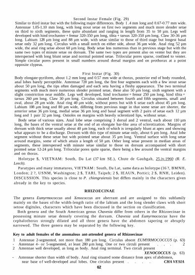
Second Instar Female (Fig. 29) Similar to third instar but with the following major differences. Body 1 .4 mm long and 0.67-0.77 mm wide. Antennae 1.05-1.10 mm long, with long stout setae on first two segments and much more slender setae on third to sixth segments, these quite abundant and ranging in length from 35 to 50 µm. Legs well developed with hind trochanter + femur 320-350 µm long, tibia + tarsus 320-350 µm long. Claw 30-36 µm long. Labium 128 µm long and 88 µm wide, with setae similar to those of third stage but longest basal setae only 32 µm long. Circulus with a small notch on either side, about 36 µm wide. Anal ring 52 µm wide, the anal ring setae about 60 µm long. Body setae less numerous than in previous stage but with the same two types of minute setae on dorsum. The same two types are present also on venter but they are interspersed with long blunt setae and normal pointed setae. Trilocular pores sparse, confined to venter. Simple circular pores present in small numbers around dorsal margins and on prothorax at a point opposite clypeus.
First Instar (Fig. 30) Body elongate-pyriform, about 1.2 mm long and 0.57 mm wide at thorax, posterior end of body rounded, anal lobes barely perceptible. Antennae 720 µm long, the first four segments each with a few stout setae about 50 µm long, the tips often damaged and each seta having a fleshy appearance. The two terminal segments with much more numerous slender pointed setae, these also 50 µm long; sixth segment with a slight constriction near middle. Legs well developed, hind trochanter + femur 250 µm long, hind tibia + tarsus 260 µm long. Claw 30 µm long. Circulus situated between fourth and fifth segments, small and oval, about 28 µm wide. Anal ring 40 µm wide, without pores but with 6 setae each about 45 µm long. Labium 108 µm long and 80 µm wide, differing from previous stage in that some setae are shorter, the anterior setae 36 µm long, medial setae 36 µm long and basal segment with 2 pairs of setae each 12 µm long and 1 pair 32 µm long. Ostioles on margins with heavily sclerotized lips, without setae.
Body setae of various sizes. Anal lobe setae comprising 3 dorsal and 2 ventral, each about 110 µm long, the bases of the ventral setae at the end of an elongate bar-like area of sclerotization. Margins of dorsum with thick setae usually about 40 µm long, each of which is irregularly blunt at apex and showing what appears to be a discharge. Dorsum with thin type of minute setae only, about 6 µm long. Anal lobe segment without these setae but with pointed setae about 35 µm long. Ventral surface with long setae around margins, some of which attain a length of 50 µm. Other long setae present in median areas of segments, these interspersed with minute setae similar to those on dorsum accompanied with short pointed setae 12-24 µm long. Trilocular pores quite sparse, there being a few around the ventral margins and on thorax.
Holotype $, VIETNAM: South, Da Lat (37 km SE.), Chute de Gauhgah, 25.iv.1960 (R. E. Leech).
Paratypes and many immatures, VIETNAM : South, Da Lat, same data as holotype (16 ?, BMNH, London; 2 ?, USNM, Washington; 2 $, TARI, Taipeh; 2 $, IEAUN, Portici; 2 $, RNH, Leiden). DISCUSSION. This species is close to P. chiengraiensis but differs mainly in the characters given already in the key to species.
RHIZOECINAE
The genera Eumyrmococcus and Xenococcus are aberrant and are assigned to this subfamily mainly on the bases of the width-length ratio of the labium and the long slender claws with short setose digitules, characters which have been discussed in the section on classification.
Both genera and the South American genus Chavesia differ from others in the Rhizoecinae in possessing minute setae densely covering the dorsum. Chavesia and Eumyrmococcus have the cephalothorax strongly dilated and all three genera have the abdomen tapering or abruptly narrowed. The three genera may be separated by the following key.
Key to adult females of the anomalous ant-attended genera of Rhizoecinae 1 Antennae 2-segmented, not more than 180 µm long. Circulus absent EUMYRMOCOCCUS (p. 63) - Antennae 4- or 5-segmented, at least 280 µm long. One or two circuli present . . . 2 2 Antennae well developed, as long as body. Anal ring terminal. Two circuli present
XENOCOCCUS (p. 63) - Antennae shorter than width of body. Anal ring situated some distance from apex of abdomen,
near base o f well-developed anal lobes. One circulus present . . . . CHA VESIA 62
EUMYRMOCOCCUS Silvestri, 1926 Eumyrmococcus Silvestri, 1926:271. Type-species: Eumyrmococcus smithii Silvestri, by original desig
nation and monotypy. Eumyrmococcus Silvestri; Williams, 1969: 336. Eumyrmococcus Silvestri; Williams, 1970: 138.
The description of this genus is virtually the same as the type-species given already by Williams (1970). The genus was placed in the tribe Rhizoecini by Williams (1969) and this assignment has been confirmed here by a further study of the labium which has a width-length ratio of about 1:2.
Eumyrmococcus smithii Silvestri, 1926
(Fig. 31) Eumyrmococcus smithii Silvestri, 1926: 273. LECTOTYPE $, CHINA: Macao [on roots of plants, attended
by ant Acropyga (Rhizomyrma) sauteri Forel] (IEAUN, Portici), here designated [examined]. Eumyrmococcus smithii Silvestri; Silvestri, 1927: 254. Eumyrmococcus smithii Silvestri; Takahashi, 1934: 2. Eumyrmococcus smithii Silvestri; Williams, 1970: 138.
Williams (1970) has described and illustrated this species recently and the illustration from the Bulletin of Entomological Research is reproduced with permission but with slight modifications. The labium was too distorted in the material studied at the time but further specimens of the type-series have become available through the kindness of Dr A. Tranfaglia and it is now possible to modify the description of the labium. There is no specimen with a perfect labium and the following description of this structure remains tentative.
Length 156-160 µm and 80 µm wide, apical segment with 2 pairs of posterior setae, 1 pair of minute apical setae, 4 pairs of subapical setae. One pair of lateral setae and 2 pairs of anterior setae. The sub-apical setae become longer and stouter towards the base. Medial segment with 1 pair of medial setae and basal segment with 2 pairs of short setae and 1 longer pair. Almost all the setae are curled in the specimens examined and the lengths are difficult to determine.
MATERIAL EXAMINED. Eumyrmococcus smithii Silvestri, lectotype $, CHINA: Macao (IEAUN, Portici).
CHINA: 4 $, same data as lectotype (IEAUN, Portici) (1 ?, BMNH, London); 1 $, Shanghai (IEAUN, Portici); 4 ?, Taipo Market, 26.xii.1924 (F. Silvestri) (2 $, BMNH, London; 2 $, IEAUN, Portici) (mounted from Silvestri's alcohol material). TAIWAN: 4 $, Taichu, on sugarcane, 24.ii.1933 (M. Yangihara) (TARI, Taipeh).
DISCUSSION. The width-length ratio of 1 : 2 of the labium confirms the assignment of the genus to the Rhizoecinae agreeing also to the definition of the subfamily by Koteja (1974a).
XENOCOCCUS Silvestri, 1924
Xenococcus Silvestri, 1924: 312. Type-species: Xenococcus annandalei Silvestri, by original designation and monotypy.
Xenococcus Silvestri; Silvestri, 1926: 275. Xenococcus Silvestri; Silvestri, 1927 : 253. DESCRIPTION. Mealybugs with broadly-oval body, abdomen tapering to narrow sclerotized terminal segment. Apex with dorsal anal ring projecting beyond ventral lobes. Anal ring with 8 setae, the anterior 2 pairs slender and the posterior pair thick and long, the posteriormost often on ventral surface. Anal lobes with numerous long stout setae. Antennae 4-segmented, nearly as long as body, the first segment long and wide, articulating with much narrower second segment containing small teeth at proximal end fitting into grooves at distal end of first segment. All segments with long pointed setae. Legs long and well developed with long slender claw, at base of which are 2 short setose digitules. Labium long and slender with a width-length ratio in excess of 1: 2. Subapical setae reaching more than half distance to base of apical segment. Two circuli present in adult, circular and slightly conical. Body setae densely covering dorsum. Ventral setae longer and not so numerous. Pores and ducts absent.
Immature instars similar in shape to adult but with fewer setae and with only a single circulus.
63

Fig. 31 Eumyrmococcus smithii Silvestri. Adult female. Specimen from Japan.
64
Fig. 32 Xenococcus annandalei Silvestri. Adult female. Specimen from India, Barkuda I.
65

DISCUSSION. The most distinctive character of this genus is the peculiar pair of antennae which are unlike any known in the Pseudococcidae. An anal ring projecting beyond ventral anal lobes, the long stout anal lobe setae and the dense covering of dorsal pointed setae, are characters shared with some of the Allomyrmococcini but the long slender claw with short setose digitules, the round sclerotized circuli and the 4-segmented antennae are characters which seem to belong to the Rhizoecini of the subfamily Rhizoecinae.
De Lotto (1977) has recently described Xenococcus scorpioides, an interesting species from South Africa, from the nests of Acropyga sp. and the first anomalous species from the African continent. In possessing short 4-segmented antennae, shorter legs with thick claws, it does not seem to be congeneric with X. annandalei but probably lies somewhere between Chavesia and Xenococcus. DISTRIBUTION. At present the genus is known from India, Malaya, Vietnam and Hong Kong.
Xenococcus annandalei Silvestri, 1924 Xenococcus annandalei Silvestri, 1924: 312. LECTOTYPE ?, INDIA: Barkuda I. [Chilka Lake, Madras
District (now Orissa Province), on roots of Ficus obtusa, with ants Acropyga acutiventris Roger] (IEAUN, Portici), here designated [examined].
Xenococcus annandalei Silvestri; Silvestri, 1926 : 275. Xenococcus annandalei Silvestri; Silvestri, 1927 : 253.
Adult Female (Fig. 32) Adult female in alcohol ovoid, abdomen tapering, posterior end curved slightly upwards, antennae strongly geniculate, lying in a posterior direction almost parallel to body margins.
On the slide, when flattened, broadly oval, 1.6-1.7 mm long and 1.1-1.4 mm wide, abdomen tapering to a narrow sclerotized last segment almost as long as broad with the anal ring at apex of dorsum pro-jecting beyond a pair of ventral lobes that curve inwards almost to mid-line. Antennae with 4 segments, of a distinctive shape and structure, 1.35-1.54 mm long, attached to dorsal surface of head. One of longest antennae with first segment 380 µm long and 230 µm at widest. Remainder of antenna much narrower, the second segment 440 µm long, the third 200 µm long, and last segment tapering, 520 µm long. All segments with numerous long slender setae of different sizes, the longest on third and fourth segments. Length of longest seta, on third segment 500 µm. Joint between first and second segments distinctive, there being small tooth-like projections on proximal lower corner of second segment which fit into small grooves on posterior distal edge of first segment. Legs well developed, hind trochanter + femur 410-470 µm long, hind tibia + tarsus 470-550 µm long. Tarsus tapering to a distance of about half its length which then becomes narrow and subparallel towards distal end, a small notch also present on inner margin at point between tapering part and narrow extremity. Each leg segment with setae of various lengths but mostly long and slender, the longest on femur about 180 µm long. Claw unusually long and narrow with a length of 40-44 µm and with a pair of setose digitules, each 16 µm long. Two circuli present situated in middle of third and fourth segments. Each circulus round, 40 µm in diameter, with a sclerotized outer rim and with the inner surface forming a cup. Labium unusually elongate; in alcohol specimens 250 µm long and 110 µm wide but, when flattened on slide, about 128 µm wide with 12 or 13 pairs of setae. Apical segment tending to be lanceolate instead of the usual conical shape, with 2 pairs of posterior setae, a pair of apical setae 20 µm long, 4 pairs of subapical setae extending towards base of segment, the distal setae about 20 µm long and the others becoming longer towards the base. Anterior setae represented by a single pair each 100 µm long but a single lateral pair each 80 µm long may be present or absent. Medial segment with a single pair of medial setae each about 75 µm long. Basal segment with a pair of setae each 10 µm long and a single setae 64 µm long on each lobe. Anal ring situated mainly on dorsal surface but often curving to ventral surface, sclerotized, about 120 µm wide at widest point, without pores but apparently with 4 pairs of setae around perimeter, there being 2 pairs of slender anterior setae each about 110 µm long and an outer pair of stouter setae each 400 µm long. A pair of stout setae about 0.8 mm long usually on ventral surface seem to belong to ring. Vulva occupying almost total width of its segment. Posterior setae stout, each about 1.0 mm long, there being 4 such pairs, 1 pair on lateral edges of dorsum anterior to anal ring and 3 pairs on posterior edge of ventral lobes. An anterior pair of setae on inner edges of these lobes much shorter and more slender.
Dorsal setae abundant, covering almost entire surface except for head and intersegmental areas, the spaces between setal bases much shorter than length of one seta. The most abundant are slender, 20—28 µm long but there are some, interspersed with these, about 40 µm long and which may be detected by the
66

Fig. 33 Xenococcus annandalei Silvestri. Third instar female.
67
Fig. 34 Xenococcus annandalei Silvestri. First instar. Specimen from India, Mysore, on coconut roots.
68
slightly larger setal bases. On prothorax and mesothorax there are short flat setae in varying numbers but usually few. Each is variously shaped and often peculiarly angled. Setae on last segment longer than usual body setae and just anterior to anal ring a fringe of long setae present comprising 2-3 median pairs each about 480 |xm long and a lateral pair usually stouter about 560 µm long. Other long setae, some attaining a length of 240 µm, present on head and around margins of abdomen. Ventral setae numerous, of different sizes, but many are long, the longest about 200 µm. Minute setae present from about fifth segment as far forward as prothorax in a zone extending to spiracles. Body pores and tubular ducts absent.
Third Instar Female (Fig. 33) Body ovoid, 1.15 mm long, tapering to last segment. Posterior end of body rounded, last segment sclero-tized at least on dorsum, the ventral surface curved inwards to form a pair of lobes. Antennae with 4 seg-ments, 1•10 -1.23 mm long; on longest antenna, first segment 350 µm long, second 360 µm, third 180 µm and fourth 340 µm, narrower than other segments. Joint between first and second segments similar to that in adult female and with minute teeth on outer posterior angle of second segment fitting into small grooves on outer distal angle of first segment. Each segment with long slender setae of different lengths. Legs similar in shape to those in adult female but shorter, hind trochanter + femur 280-310 µm long, hind tibia + tarsus 350-400 µm long. Labium as in adult but 205 µm long and 88 µm wide, with 13 pairs of setae. Apical segment with 2 pairs of posterior setae, a pair of minute apical setae 12 µm long, 4 pairs of subapical setae set wide apart so that proximal pair at mid-point of segment, the setae ranging in length from 12 µm at apex to 68 µm on mid-segment. Also present 1 pair of lateral setae and 1 pair of anterior setae each 68 µm long. Medial segment with 1 pair of setae 60 µm long. Basal segment with each lobe containing 2 short setae each 8 µm long and 1 seta 36 µm long. One circulus present, circular, about 32 µm in diameter, situated in middle of fourth segment. Anal ring at apex of dorsal surface about 56 µm wide, with narrow sclerotized rim and surrounded by 4 pairs of setae. The anterior median pair are slender and only slightly longer than diameter of ring, outer 2 pairs stouter, the third pair about 210 µm long and the fourth pair 440 µm long. Anal lobes each with one dorsal and two ventral stout setae about 1 0 mm long, there being also a slender seta about 150 µm long on inner margin of lobe.
Dorsal setae abundant anterior to fourth abdominal segment, these mainly slender 12-16 µm long, the distance between setal bases usually shorter than length of one seta or about same length. Minute flat setae with varying distal shape, scattered among slender setae on prothorax and mesothorax. Dorsal setae on fourth and posterior segments fewer and of different sizes, the longest about 180 µm. Ventral setae mainly long and slender on abdomen, the longest about 160 µm but from first segment to prothorax small slender setae, similar to those on anterior dorsum extend inwards from margin to spiracles. Pores and tubular ducts absent.
First Instar (Fig. 34) Body oval, 0.7-0.9 mm long, membranous except for anal lobe segment which on dorsum is sclerotized on margin extending to a band anterior to anal ring. Anal lobes present ventrally, these also sclerotized. Antennae with 4 segments similar in shape to those of previous stages, 0.85-0.90 mm long, the longest antenna with first segment 240 µm long, the second 320 µm, third 110 µm and fourth 230 µn long. Each segment with slender setae varying in length, the longest about 300 µm. Joint between first and second segments similar to those in other stages. Legs as in previous stages except that tarsus longer than tibia whereas in other stages the tibia is longer than tarsus. Hind trochanter-!-femur about 220 µm long, hind tibia + tarsus 240-260 µm long. Claw slender, 28 µm long. Labium 180 µm long and 80 µm wide with similar setal distribution to that in previous stages but setae longer than in third stage. Apical segment with a pair of apical setae each 8 µm long, 4 pairs of subapical setae extending to a point more than half length of segment and ranging In length from 12 µm distally to an inner pair 76 µm long. Also present are a lateral pair and an anterior pair each 76 µm long. Medial segment with either 1 or 2 pairs of setae, the longest 72 µm. Basal segment with a pair of setae each 8 µm long and 1 seta 72 µm long on each lobe. Circulus round, about 28 µm in diameter, situated within borders of fourth segment. Anal ring entirely on dorsum, with narrow rim and 4 pairs of setae around outer margin, there being 2 pairs of inner slender setae each about 60 µm long, a third stouter pair about 360 µm long and a stout posterior pair about 750 µm long. The last setae resemble anal lobe setae which are of similar shape and length, there being on each lobe, a pair on dorsal margins and a single seta at apex of lobe on venter.
Dorsal setae mainly short and slender on anterior abdominal segments, thorax and head, each about 12 µm long. A few flat and irregularly shaped setae present on prothorax each about 8 µm long. Ab-dominal setae all slender, of different lengths, the longest always in transverse rows, about 120 µm long.
69

Ventral setae on abdomen similar to those on dorsum of abdomen but fewer. Setae sparse on thorax and head. Pores and tubular ducts absent.
DISCUSSION. This is an unusual species that will probably be found underground throughout south-east Asia, attended by the ant Acropyga acutiventris. The peculiar antennae, with the unusual articulation between the first and second segments, are similar in all stages but as yet no second stage has been available.
References Balachowsky, A. S. 1957. Sur un nouveau genre aberrant de cochenille radicicole myrmecophile nuisible
au cafeier en Colombie. Revue Path. veg. Ent. agric. Fr. 36 : 157-164, 1 fig. Beardsley, J. W. 1970. Three new species of Chavesia Balachowsky from Tropical America (Homoptera :
Coccoidea). Proc. Hawaii, ent. Soc. 20 : 509-520, 5 figs. Bowers, W. S., Nault, L. R., Webb, R. E. & Dutky, S. R. 1972. Aphid alarm pheromone: isolation, identi
fication, synthesis. Science, N.Y. 177: 1121-1122. Broadbent, L. 1951. Aphid excretion. Proc. R. ent. Soc. Lond. (A) 26: 97-103. Biinzli, G. H. 1935. Untersuchungen uber coccidophile Ameisen aus den Kaffeefeldern von Surinam.
Mitt, schweiz. ent. Ges. 16: 453-593, 51 figs. Callow, R. K., Greenway, A. R. & Griffiths, D. C. 1973. Chemistry of the secretion from the cornicles of
various species of aphids. J. Insect Physiol. 19 : 737-748, 3 figs. Chen, Su-Wan & Edwards, J. S. 1972. Observations on the structure of secretory cells associated with
aphid cornicles. Z. Zellforsch. mikrosk. Anat. 130 : 312-317, 3 figs. Dahl, M. L. 1971. Uber einen Schreckstoff bei Aphiden. Dt. ent. Z. (N.F.) 18 : 121-128. Das, G. M. 1959. Observations on the association of ants with Coccids of tea. Bull. ent. Res. 50 : 437- 448,
6 figs. Delage-Darchen, B., Matile-Ferrero, D. & Balachowsky, A. S. 1972. Sur un cas aberrant de symbiose.
Cochenilles x Fourmis. C. r. hebd. Seanc. Acad. Sci., Paris (D) 275 : 2359-2361, 1 pl. De Lotto, G. 1977. On some African mealybugs (Homoptera: Coccoidea: Pseudococcidae). J. ent. Soc.
sth.Afr. 40 : 13-36, 6 figs. Dixon, A. F. G. 1958. The protective function of the siphunculi of the nettle aphid, Miciolophium evansi
(Theob.) (Hem., Aphididae). Entomologist's mon. Mag. 94: 8. & Stewart, W. A. 1975. Function of the siphunculi in aphids with particular reference to the syca
more aphid, Drepanosiphum platanoides. J. Zool., Lond. 175 : 279-289, 4 figs. Edwards, J. S. 1966. Defense by smear: supercooling in the cornicle wax of aphids. Nature, Lond. 211:
73-74. Ferris, G. F. & Murdock, G. E. 1936. Contributions to the knowledge of the Coccoidea (Homoptera).
III. Microentomology 1: 115-122, 3 figs. Flanders, S. E. 1957. The complete interdependence of an ant and a Coccid. Ecology 38: 535-536. Greenway, A. R. & Griffiths, D. C. 1973. A comparison of triglycerides from aphids and their cornicle
secretions. J. Insect Physiol. 19 : 1649-1655, 2 figs. Hottes, F. C. 1928. Concerning the structure, function, and origin of the cornicles of the family Aphididae.
Proc. biol. Soc. Wash. 4 1 : 71-84, 30 figs. Kislow, C. J. & Edwards, L. J. 1972. Repellent odour in aphids. Nature, Lond. 235: 108-109. Koteja, J. 1974a. Comparative studies on the labium in the Coccinea (Homoptera). Zesz. nauk. Akad.
roln. Krakow 27 : 1-162, 78 figs. 1974b. The occurrence of a campaniform sensillum on the tarsus in the Coccinea (Homoptera).
Polskie Pismo ent. 44 : 243-252, 3 figs. 1974c. On the phylogeny and classification of the scale insects (Homoptera, Coccinea) (discussion
based on the morphology of the mouthparts). Acta zool. cracov. 19: 267-325, 5 figs.
70

Le Pelley, R. H. 1943. An oriental mealybug {Pseudococcus lilacinus Ckll.) and its insect enemies. Trans. R. ent. Soc. Lond. 93: 73-93, 7 figs, 4 pls.
Lindsay, K. L. 1969. Cornicles of the pea aphid, Acyrtosiphon pisum; their structure and function. A light and electron microscope study. Ann. ent. Soc. Am. 62: 1015-1021, 6 figs.
Lloyd D. C. & Martini, E. 1957. A note on the circulus as an adhesive organ in some Pseudococcidae. Can. Ent. 89: 46-48.
Nault, L. R. & Bowers, W. S. 1974. Multiple alarm pheromones in aphids. Entomologia exp. appl. 17: 455-457.
Edwards, L. J. & Styer, W. E. 1973. Aphid alarm pheromones: secretion and reception. Envir. Ent. 2:101-105, 3 figs.
Newstead, R. 1903. Monograph of the Coccidae of the British Isles. 2. viii+ 270 pp., 4 figs, 52 pls. London. Nixon, G. 1951. The association of ants with Aphids and Coccids. 36 pp. London. Pesson, P. 1939a. Une glande exsertile ventrale chez les Coccides. Bull. Soc. zool. Fr. 64: 144-152, 4 figs.
1939b. Ostioles et autohemorrhee chez les Coccides. Bull. Soc. zool. Fr. 64: 152-159, 2 figs. Prins, A. J., Ben-Dov, Y. & Rust, D. J. 1975. A new observation on the association between ants
(Hymenoptera: Formicidae) and armoured scale insects (Homoptera: Diaspididae). J. ent. Soc. sth. Afr. 38:211-216, 6 figs.
Reyne, A. 1954. Hippeococcus a new genus of Pseudococcidae from Java with peculiar habits. Zool. Meded. Leiden 32 : 233-257, 53 figs.
Silvestri, F. 1924. A new myrmecophilous genus of Coccidae (Hemiptera) from India. Rec. Indian Mus. 26:311-315, 5 figs.
1926. Descrizione di un novo genere di Coccidae (Hemiptera) mirmecofilo della Cina. Boll. Lab. Zool. gen. agr. R. Scuola Agric. Portici 18 (1925): 271-275, 2 figs.
1927. Descriptions of two species of myrmecophilous Coccidae (Hemiptera Insecta). China J. 7 : 253-254, 2 figs.
Smith, M. R. 1942. The relationship of ants and other organisms to certain scale insects on coffee in Puerto Rico. J. Agric. Univ. P. Rico 26 : 21-27.
Strickland, A. H. 1951. The entomology of Swollen Shoot of cacao. I - the insect species involved, with notes on their biology. Bull. ent. Res. 41 : 725-748, 1 fig.
Strong, F. E. 1969. Observations on aphid cornicle secretions. Ann. ent. Soc. Am. 60: 668-673, 4 figs. Sulc, K. 1909. Zur Anatomie der Cocciden. Zool. Anz. 34: 164-172, 4 figs. Takahashi, R. 1934. Observations on the Coccidae of Formosa. Part IV. Rep. Govt res. Inst. Dep. Agric.
Formosa 63 : 1-38, 24 figs. 1941. Two new myrmecophilous Coccidae from Thailand (Siam). Tenthredo 3: 201-207, 2 figs. 1942. Some injurious insects of agricultural plants and forest trees in Thailand and Indo-China,
II. Coccide. Rep. Govt Res. Inst. Dep. Agric. Formosa 81: 1-56, 26 figs. 1950. Some species of Coccidae from the Riouw Islands. Part I. Insecta matsum. 17: 65-72, 5 figs. 1951. Three new myrmecophilous scale insects from Malay Peninsular (Homoptera). Mushi 22:
1-8, 4 figs. Way, M. J. 1954. Studies on the association of the ant Oecophylla longinoda (Latr.) (Formicidae) with the
scale insect Saissetia Zanzibarensis Williams (Coccidae). Bull. ent. Res. 45 : 113-134, 8 figs. 1963. Mutualism between ants and honeydew-producing Homoptera. A. Rev. Ent. 8: 307-344.
Wheeler, W. M. 1921. A study of some social beetles in British Guiana and of their relations to the ant-plant Tachigalia. Zoologica, N.Y.3: 35-126, 12 figs, 5 pls.
Williams, D. J. 1969. The family-group names of the scale insects (Hemiptera : Coccoidea). Bull. Br. Mus. nat.Hist. (Ent.) 23: 315-341.
1970. The mealybugs (Homoptera, Coccoidea, Pseudococcidae) of sugar-cane, rice and sorghum. Bull. ent. Res. 60 : 109-188, 29 figs.
Wilson, E. O. 1971. The insect societies. xii + 548 pp. Cambridge, Massachusetts. Wynn, G. G. & Boudreaux, H. B. 1972. Structure and function of aphid cornicles. Ann. ent. Soc. Am.
65:157-166, 13 figs.
Index
acariformis, 15 brevipes, 10 Dysmicoccus, 10 Allomyrmococcini, 14 Allomyrmococcus, 14, 15 Chavesia, 6, 7, 13, 62 Ehrhornia, 9 annandalei, 66 chiengraiensis, 56 Eumyrmococcus, 62, 63 Antonina, 9, 10 Chnaurococcus, 12 Euripersia, 12
71

QUT Library europaea, 12
farinosus, 10 formicarii, 31, 33
Hippeococcus, 14, 15, 20, 22
khooi, 31, 39
lilacinus (Planococcus), 8 lilacinus (Pseudococcus), 8 longispinus, 10
Malaicoccus, 14, 15, 30, 31
montanus, 22 moundi, 31, 44
obscurus, 7
Paramyrmococcus, 14, 15, 54 Phenacoccus, 10 Pseudococcinae, 9, 14 Pseudococcus, 7, 8, 10
rappardi, 22 Rhizoecinae, 9, 62 Rhodania, 9 riouwensis, 33, 50
scorpioides, 66 smithii, 63 Sphaerococcinae, 9 subterraneus, 12
takahashii, 31, 52 Trabutininae, 9 trinidadensis, 13
vietnamensis, 56, 61
wegneri, 22, 26
Xenococcus, 62, 63
72

British Museum (Natural History) Monographs & Handbooks The Museum publishes some 10-12 new titles each year on subjects including zoology, botany, palaeontology and mineralogy. Besides being important reference works, many, particularly among the handbooks, are useful for courses and students' background reading.
Lists are available free on request to:
Publications Sales British Museum (Natural History) Cromwell Road London SW7 5BD
Subscriptions placed by educational institutions earn a discount of 10% off our published price.