bupivacaine myotoxicity is mediated by mitochondria · of padova and the consiglio nazionale delle...
TRANSCRIPT

Bupivacaine Myotoxicity is Mediated by Mitochondria*
William Irwin‡, Eric Fontaine‡§, Laura Agnolucci‡, Daniele Penzo‡, Romeo Betto¶, Susan
Bortolotto‡‡, Carlo Reggiani‡‡, Giovanni Salviati‡ ¶ §§ and Paolo Bernardi‡ ¶¶ //
From the Departments of ‡Biomedical Sciences and ‡‡Anatomy and Physiology, University
of Padova and the Consiglio Nazionale delle Ricerche Units for the Study of ¶Muscle
Pathophysiology and ¶¶Biomembranes, Viale Giuseppe Colombo 3, I-35121 Padova, Italy
and the §Laboratoire de Bioénergétique Fondamentale et Appliquée, Université J. Fourier, F-
38041 Grenoble, France
Proofs to be sent to:
Prof. Paolo Bernardi Dipartimento di Scienze Biomediche Sperimentali Viale Giuseppe Colombo 3 I-35121 Padova Italy Phone +39 049 827 6365 Fax +39 049 827 6361 E-mail: [email protected]
Running title: Mitochondrial Bupivacaine Myotoxicity
Copyright 2002 by The American Society for Biochemistry and Molecular Biology, Inc.
JBC Papers in Press. Published on January 14, 2002 as Manuscript M108938200 by guest on A
ugust 29, 2018http://w
ww
.jbc.org/D
ownloaded from

2
FOOTNOTES
*This work was supported by Grants from Telethon-Italy (Grants 847 and 1141 to P.B.), the
Consiglio Nazionale delle Ricerche (Dotazione Centri Biomembrane e Fisiopatologia
Muscolare). W.I. and D.P. were supported by Fellowships from Telethon-Italy and E.F. was
supported by the European Economic Community Fellowship ERBFMBICT961385.
§§Deceased March 12, 1998
//To whom correspondence should be addressed: Dipartimento di Scienze Biomediche
Sperimentali, Viale Giuseppe Colombo 3, I-35121 Padova, Italy
1The abbreviations used are:
PTP, Mitochondrial Permeability Transition Pore; CsA, Cyclosporin A; FDB, Flexor
Digitorum Brevis; EDL, Extensor Digitorum Longus; PN, pyridine nucleotides; EGTA,
ethylene-bis(oxoethylenenitrilo) tetraacetic acid; BSA, bovine serum albumin; FCCP,
carbonylcyanide-p-trifluoromethoxyphenyl hydrazone; TMRM, tetramethyl rhodamine
methyl ester; Rh123, Rhodamine 123; ∆ψ, mitochondrial membrane potential difference; LI,
Localization Index; BAPTA, 1,2-bis-(2-Aminophenoxy)ethane-N,N,N’N’-tetraacetic acid;
MHC, Myosin heavy chain.
by guest on August 29, 2018
http://ww
w.jbc.org/
Dow
nloaded from

3
We have investigated the effects of the myotoxic local anesthetic bupivacaine on
rat skeletal muscle mitochondria and isolated myofibers from flexor digitorum brevis
(FDB), extensor digitorum longus (EDL), soleus and from the proximal, striated portion
of the esophagus. In isolated mitochondria, bupivacaine caused a concentration-
dependent mitochondrial depolarization and pyridine nucleotide oxidation, which were
matched by an increased oxygen consumption at bupivacaine concentrations of 1.5 mM
or less at pH 7.4, while respiration was inhibited at higher concentrations. As a
consequence of depolarization, bupivacaine caused opening of the permeability
transition pore (PTP), a cyclosporin A-sensitive inner membrane channel that plays a
key role in many forms of cell death. In intact FDB fibers bupivacaine caused
mitochondrial depolarization and pyridine nucleotides oxidation that were matched by
increased concentrations of cytosolic free Ca2+, release of cytochrome c and eventually
hypercontracture. Both mitochondrial depolarization and cytochrome c release were
inhibited by cyclosporin A, indicating that PTP opening rather than bupivacaine as such
was responsible for these events. Similar responses to bupivacaine were observed in the
soleus, which is highly oxidative. In contrast, fibers from the esophagus (which we show
to be more fatiguable than FDB fibers), and from the highly glycolytic EDL didn’t
undergo pyridine nucleotide oxidation upon the addition of bupivacaine, and were
resistant to bupivacaine toxicity. These results suggest that active oxidative metabolism
is a key determinant in bupivacaine toxicity; that bupivacaine myotoxicity is a relevant
model of mitochondrial dysfunction involving the PTP and Ca2+ dysregulation; and that
it represents a promising system to test new PTP inhibitors that may prove relevant in
spontaneous myopathies where mitochondria have long been suspected to play a role.
by guest on August 29, 2018
http://ww
w.jbc.org/
Dow
nloaded from

4
Bupivacaine is a local anesthetic that induces rapid degeneration of skeletal muscle
fibers (1,2). As is the case for muscular dystrophies, the pathogenesis of bupivacaine-induced
muscle cell death remains unclear. Solving this problem is of interest for the understanding of
degenerative muscular diseases because the sequence of fiber breakdown induced by
bupivacaine is similar to that of progressive muscular dystrophy (3). It is also striking that the
same types of muscle fibers are spared by both Duchenne muscular dystrophy and
bupivacaine toxicity (4,5).
It has been suggested that bupivacaine may disrupt Ca2+ homeostasis in vivo,
triggering Ca2+-activated cellular death pathways that include proteolysis (4,6). This
suggestion is supported by the findings (i) that bupivacaine affects sarcoplasmic reticulum
function in vitro (3); (ii) that extracellular Ca2+ omission delays the morphological changes
(6) and decreases the protein degradation rate (7) that are observed in isolated rat soleus
muscle exposed to bupivacaine; and (iii) that bupivacaine uncouples isolated rat liver and
heart mitochondria (8-12) and decreases mitochondrial membrane potential and oxygen
consumption both in cultured fibroblasts (13,14) and Ehrlich tumor cells (15,16).
Mitochondrial dysfunction results in ATP depletion (14) and in turn is expected to have a
major impact on intracellular Ca2+ homeostasis (17).
The importance of mitochondria in the pathways to cell death is largely recognized
even if the exact mechanism(s) in specific experimental paradigms may not be easy to
identify (18). A potential mechanism is represented by opening of the PTP1, a high
conductance channel located in the inner mitochondrial membrane that can be inhibited by
CsA (19). PTP opening in vitro leads to collapse of the protonmotive force, disruption of
ionic homeostasis, mitochondrial swelling, and release of cytochrome c (20). This sequence of
events has drawn considerable attention to the PTP as a potential effector in the pathways to
cell death through at least three mechanisms, i.e. decreased levels of cellular ATP (21-23);
by guest on August 29, 2018
http://ww
w.jbc.org/
Dow
nloaded from

5
increase of cytosolic Ca2+ (24); and release of apoptotic factors such as cytochrome c (25,26),
apoptosis inducing factor (27), Smac-Diablo (28) and endonuclease G (29).
With the long term goal of defining the role of mitochondria in the pathways to muscle
cell death (30), we have investigated the effects of bupivacaine both on rat skeletal muscle
mitochondria and on isolated mouse FDB, EDL, soleus and esophagus fibers. We found that
bupivacaine causes depolarization, PN oxidation and PTP opening in isolated skeletal muscle
mitochondria. Measurements on isolated FDB fibers indicated that bupivacaine also induced
mitochondrial depolarization that was significantly delayed by CsA in situ, indicating that
depolarization was due to PTP opening rather than to the uncoupling effects of bupivacaine as
such. Consistent with this data, bupivacaine caused CsA-inhibitable release of cytochrome c
in situ. Fibers from glycolytic, non resistant to fatigue muscles such as EDL and esophagus
were instead strikingly resistant to bupivacaine toxicity, suggesting that bupivacaine toxicity
selectively affects oxidative muscles. Thus, bupivacaine toxicity is a relevant model of
mitochondrial dysfunction involving the PTP and Ca2+ dysregulation, and represents a
promising system to test new PTP inhibitors that may prove relevant in spontaneous
myopathies where mitochondria have long been suspected to play a role, like Duchenne’s
muscular dystrophy (31).
Material and Methods
Rat skeletal muscle mitochondria were prepared according to (32) with slight
modifications. Albino Wistars rats weighing 250-350 g were killed by decapitation and the
gastrocnemius muscles were rapidly excised and transferred into the isolation medium (150
mM sucrose, 75 mM KCl, 50 mM Tris-HCl, 1 mM KH2PO4, 5 mM MgCl2, 1 mM EGTA, pH
7.4). Muscles were minced with scissors and trimmed clean of visible fat and connective
tissues. Muscle pieces were transferred to 30 ml of isolation medium supplemented with 0.2%
by guest on August 29, 2018
http://ww
w.jbc.org/
Dow
nloaded from

6
BSA and 0.2 mg x ml-1 Nagarse (Fluka, Buchs). After 1 minute, muscles were homogenized
using a motor-driven Plexiglas/Plexiglas potter, transferred to 120 ml of isolation medium
supplemented with 0.2% BSA and centrifuged at 700 x g for 10 min. The supernatant was
decanted and centrifuged at 10,000 x g for 10 min. The resulting pellet was resuspended in a
medium containing 250 mM sucrose, 0.1 mM EGTA-Tris, 10 mM Tris-HCl, pH 7.4 and
centrifuged at 7,000 x g for 6 min. The final mitochondrial pellet was resuspended in 0.5 ml
of the same medium at a final protein concentration of about 20 mg x ml-1. All procedures
were carried out at 0-4 °C.
Mitochondrial oxygen consumption was measured polarographically at 25 °C using a
Clark-type electrode. Measurements of membrane potential and PN oxidation-reduction status
were carried out fluorimetrically with a Perkin-Elmer 650-40 spectrofluorimeter equipped
with magnetic stirring and thermostatic control. Membrane potential was measured in the
presence of 0.1 µM Rh123 as described (33)(excitation-emission: 548-573 nm). The PN
oxidation-reduction status was evaluated based on endogenous NAD(P)H fluorescence
(excitation-emission: 345-450 nm).
Isolated muscle fibers were prepared from FDB, EDL, soleus muscle and from the
upper region of the esophagus of C57BL/10ScSn mice according to (34) with slight
modifications. Mice were killed by cervical dislocation and the muscles were incubated for 1h
at 4°C in Tyrode solution containing 135 mM NaCl, 4 mM KCl, 1 mM CaCl2, 1 mM MgCl2,
0.33 mM KH2PO4, 10 mM glucose and 10 mM Hepes (pH 7.3) supplemented with 0.3%
collagenase type I and 0.2% BSA. The temperature was raised to 37°C and the incubation
continued for a further one hour. The muscle mass was then removed and washed twice in
Tyrode solution, and single myocytes were dispersed by passing the muscle repeatedly
through a wide-pore Pasteur pipette. Myocytes suspended in Tyrode solution were plated on
glass coverslips and allowed to attach for at least 1 hour prior to experiment. Myocytes were
by guest on August 29, 2018
http://ww
w.jbc.org/
Dow
nloaded from

7
then rinsed and placed in 1 ml Tyrode solution and loaded with the indicated concentrations
of TMRM for 20 min at 37 °C. Myocytes were then placed on the stage of the confocal
microscope, maintaining temperature at 37 °C. In some experiments, myocytes were also
loaded with 2 µM Fluo-3-AM.
Imaging was performed with either a real time confocal system (Nikon, RCM 8000)
on a Nikon Diaphot-300 microscope with a 40X, 1.3 NA oil immersion objective or on a
Zeiss Axiovert 100TV inverted fluorescence microscope. For the Nikon setup excitation
wavelength/detection filter were 488/525 ± 25 nm bandpass and 568/585 longpass for Fluo-3
and TMRM, respectively. In some experiments, Fluo-3 and TMRM fluorescence emissions
were collected simultaneously by using two separate color channels on the detector assembly.
In most of the experiments sequential confocal images were acquired and stored typically at
60 sec intervals during 20-45 min. Time course of ∆ψ and [Ca2+] (measured in arbitrary
fluorescence units) were performed using the Nikon RCM8000 Real Time Confocal System
data acquisition software. Skeletal fibers were identified as regions of interest, and
background was identified as an area without cells. For the Zeiss setup, a 10x objective was
used. TMRM was excited with 546 ± 5 nm and the emission was monitored at 580 ± 15 nm
with a 560 nm dichroic mirror. NAD(P)H was excited at 365 ± 15 nm and the emission was
monitored at 460 ± 25 nm. The data was analyzed with the MetaMorph MetaFluor Imaging
Software.
Cytochrome c release was monitored exactly as described in (35). FDB fibers were
treated with vehicle or with 1 mM bupivacaine as specified in the legend to Fig. 8, and then
washed. Fibers were fixed for 30 min at room temperature with 3.7% (v/v) ice-cold
formaldehyde, permeabilized for 20 min with 0.01% (v/v) ice-cold Nonidet P-40, incubated
for 15 min with a 0.5% solution of BSA and then for 15 min at 37°C with a mouse
monoclonal anti-cytochrome c antibody (Pharmingen, CA, clone 6H2.B4) and with an affinity
by guest on August 29, 2018
http://ww
w.jbc.org/
Dow
nloaded from

8
purified rabbit antibody against the rat bc1 complex (a generous gift of Prof. Roberto Bisson,
Padova). Fibers were then sequentially incubated for 15 min at 37°C with TRITC-conjugated
goat anti-mouse IgG and with FITC-conjugated goat anti-rabbit IgG. Cellular fluorescence
images were acquired with a Nikon Eclipse E600 microscope equipped with a BioRad MRC-
1024 laser scanning confocal imaging system. For cytochrome c and bc1 complex detection,
red and green channel images were acquired simultaneously using two separate color
channels on the detector assembly of the Nikon Eclipse E600 microscope equipped with
488/522 ± 25 nm bandpass and 568/605 longpass filter settings, and a 60X, 1.4 NA oil
immersion objective (Nikon). Using the BioRad LaserSharp analysis program, a set of lines
was drawn across the cells and the fluorescence intensity of each pixel along the lines in both
the green and the red channel was measured. The localization index, LI, is defined as the ratio
of the standard deviation of the fluorescence intensity divided by the total fluorescence for
each channel: (S.D./Σ)red / (S.D./ Σ)green. A punctate distribution (which is typical of
mitochondria) results in a higher S.D., and normalization allows correction for different
fluorescence intensities in the two channels. The normal LI must be 1, which indicates that the
bc1 complex and cytochrome c have the same pattern of intracellular distribution. A LI < 1
indicates that the distribution of cytochrome c is more homogeneous than that of the bc1
complex, i.e. that cytochrome c has diffused away from mitochondria (35).
Fiber typing of FDB, EDL, esophagus and soleus muscles was based on
electrophoretic separation of MHC isoforms, which can be used as molecular markers of fiber
type (36). Muscle samples were immersed in Laemmli solution (37) and MHC isoforms were
resolved by the SDS-PAGE method as described (38). Briefly, 8% polyacrylamide slab gels
containing 30% glycerol were run for 24h at 275V in the cold room (4oC). The gels were
removed and stained with Coomassie Brilliant Blue G. Densitometry was performed using
1D Image Analysis Software (Kodak Digital Science).
by guest on August 29, 2018
http://ww
w.jbc.org/
Dow
nloaded from
9
Muscle fatigability was used as an index of their dependence on glycolytic or
oxidative metabolic supply (39,40). The muscles were mounted in a myograph and perfused
with oxygenated Krebs solution (temperature 20°C). The muscles were allowed to equilibrate
for 10 minutes then the frequency (Fmax) at which maximum isometric tension was obtained
was identified. Fatigue was induced by repetitive stimulation at Fmax. The muscles were
stimulated to contract isometrically for 0.5 s every other second (duty cycle 25%). The ratio
between the tension developed after 30 s and after 60 s of stimulation and the initial tension
(time zero) was taken as a fatigue index.
Bupivacaine and collagenase were purchased from Sigma; Rh123, TMRM and Fluo-3
AM were purchased from Molecular Probes; and CsA was a gift from Novartis (Basel,
Switzerland). All other chemicals were of the highest purity commercially available.
Results
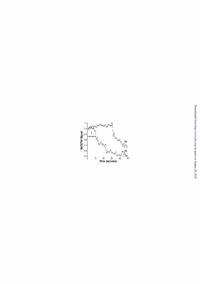
Effects of bupivacaine on rat skeletal muscle mitochondria. Bupivacaine is an
uncoupler in isolated liver and heart mitochondria (8), and causes complex effects on
respiration in cultured cells (13-16). The PTP is a voltage-dependent channel that can be
opened by depolarization with uncouplers and respiratory inhibitors (41). We have therefore
first characterized the effects of bupivacaine on rat skeletal muscle mitochondria in the
presence of CsA (in order to prevent PTP opening). The experiments of Fig. 1 document a
biphasic effect of bupivacaine on the rate of oxygen consumption and on the PN oxidation-
reduction status. Respiration increased linearly up to a concentration of about 1.5 mM
bupivacaine and it then declined as the concentration was increased further (Fig. 1, triangles).
The reduction levels of PN were a mirror image of the respiratory changes, with increased
oxidation matching uncoupling and increased reduction matching respiratory inhibition (Fig.
1, squares).
by guest on August 29, 2018
http://ww
w.jbc.org/
Dow
nloaded from

10
Fig. 2, panel A reports the effects of increasing bupivacaine concentrations on oxygen
consumption by skeletal muscle mitochondria at pH 7.4 (triangles) and pH 7.0 (squares).
While the overall pattern remained biphasic, at pH 7.4 the concentrations of bupivacaine
required for 50% stimulation and inhibition of respiration were approximately 0.75 and 2.2
mM, respectively. At pH 7.0 these values became approximately 1.5 mM and 4.2 mM
bupivacaine, i.e. about twice the values obtained at pH 7.4. When the data were replotted as a
function of the calculated concentration of the de-protonated form of bupivacaine (BPo, pKa
8.1) it became apparent that the effects of the drug correlated with BPo (Fig. 2, panel B).
Although the pKa of bupivacaine in the membrane is not known, these findings suggest that
BPo is responsible for both uncoupling and inhibition of respiration, and indicate that the
concentration of BPo required for maximal stimulation of respiration may be as low as 0.25
mM (Fig. 2B).
We next investigated the effects of bupivacaine on the membrane potential (Fig. 3,
panel A) and respiration (Fig. 3, panel B) maintained by isolated skeletal muscle
mitochondria. Mitochondria were energized with complex I substrates and loaded with a
small amount of Ca2+ in the presence of Pi, an optimal condition to reveal PTP opening by
depolarization (42). It can be seen that the addition of 1 mM bupivacaine was readily
followed by a fast but partial mitochondrial depolarization, followed within a few minutes by
a further depolarization (panel A, trace a). These changes were matched by a transient
stimulation of respiration followed by respiratory inhibition (panel B, trace a). The nature of
these complex changes was elucidated when the experiment was repeated in the presence of
CsA. Under these conditions, the addition of 1 mM bupivacaine was only followed by the
fast, partial depolarization (panel A, trace b), while uncoupling was not followed by
respiratory inhibition (panel B, trace b). Thus, both the late depolarization and the delayed
inhibition of respiration were due to PTP opening. Indeed, inhibition of respiration could be
by guest on August 29, 2018
http://ww
w.jbc.org/
Dow
nloaded from

11
prevented by the addition of exogenous NADH (panel B, trace b) (43). Thus, by acting on
three key sites of PTP regulation [i.e. membrane potential (41), NAD(P)H levels (44) and
electron flux through complex I (33)] bupivacaine is a trigger for PTP opening in isolated
skeletal muscle mitochondria.
Effects of bupivacaine on FDB mitochondria in situ. We next addressed the question
of whether bupivacaine could decrease the mitochondrial membrane potential per se and/or
through PTP opening in FDB muscle fibers. Fig. 4, panel A, shows the typical preparation
used for these experiments as a fluorescence image after TMRM loading. Panel B documents
that within 40 minutes of the addition of 1 mM bupivacaine the TMRM signal was largely
lost while the fiber shortened considerably (panel B). The experiment of Fig. 5 illustrates the
time course of the fluorescence changes of single TMRM-loaded FDB fibers elicited by the
addition of 1 mM bupivacaine. The signal decreased within about 30 minutes (squares, trace
a), and depolarization was prevented by CsA (closed circles, trace b) indicating that PTP
opening rather than bupivacaine as such was responsible for complete depolarization under
these conditions (Fig. 5). Finally, Fig. 5 shows that mitochondrial depolarization was favored
by Ca2+. Indeed, it could be delayed but not prevented by treatment with dantrolene, an
inhibitor of Ca2+ release from the sarcoplasmic reticulum (triangles, trace c) or BAPTA-AM,
a permeant Ca2+ chelator (open circles, trace d). The role of intracellular Ca2+ in bupivacaine
toxicity was investigated further in direct measurements with the fluorescent indicator Fluo-3.
The experiments of Fig. 6, panel A, show that mitochondrial depolarization induced by
bupivacaine (circles, trace a) was mirrored by an increase of [Ca2+]c (squares, trace b), which
was observed also in nominally Ca2+-free media (not shown). Fig. 6, panel B, shows that in a
Ca2+-free medium ryanodine was able to prevent the rise of [Ca2+]c (squares, trace b) but not
mitochondrial depolarization (circles, trace a). These experiments suggests that the latter is
the cause rather than the consequence of dysregulation of Ca2+ homeostasis.
by guest on August 29, 2018
http://ww
w.jbc.org/
Dow
nloaded from

12
The depolarization due to the addition of bupivacaine prior to onset of PTP opening
was clearly detectable in isolated mitochondria (Fig. 3A) but not in FDB fibers (Fig. 5).
However, the TMRM fluorescence changes of mitochondria following depolarization in situ
may be complex [see (45,46) for recent reviews]. We therefore also studied the changes of the
oxidation-reduction state of endogenous PN, which reflects the rate of electron flux within the
respiratory chain. The experiments depicted in Fig. 7 demonstrate that the addition of
bupivacaine was followed by the expected oxidation of PN in situ (squares, trace a). As was
the case for depolarization, however, also PN oxidation was inhibited by CsA (circles, trace
b). These results indicate that, at this concentration, bupivacaine has little direct (i.e., PTP-
independent) effects on the membrane potential.
Bupivacaine releases cytochrome c from mitochondria in FDB fibers. PTP opening
plays a role in several paradigms of cell death that depend on cytochrome c release, which
may be followed by caspase activation. We have studied whether bupivacaine causes
cytochrome c release in FDB fibers with a very sensitive in situ method that was recently
developed in one of our laboratories (35). The experiments of Fig. 8 report the distribution of
cytochrome c (red) and of the bc1 complex (green) in an untreated FDB fiber (panel A) or
after 30’ of treatment with 1 mM bupivacaine in the absence (panel B) or presence (panel C)
of CsA. The quantitative analysis of the fluorescence pattern of the two channels (35)
revealed a modest but significant difference in the distribution of cytochrome c after treatment
with bupivacaine. This redistribution was fully prevented by CsA implying PTP opening as a
causative event (panel D).
EDL and esophagus fibers are resistant to bupivacaine toxicity. Fig. 9 shows that
within two minutes of the addition of 2 mM bupivacaine FDB fibers hypercontracted (panel
A’, compare with panel A). This event was followed by the formation of blebs in the
sarcolemma and eventually by permeability changes that compromised cell viability (not
by guest on August 29, 2018
http://ww
w.jbc.org/
Dow
nloaded from

13
shown). Fig. 9 also shows that the same concentration of bupivacaine was ineffective at
inducing contracture of striated fibers from the esophagus after 20 minutes (panel B’,
compare with panel B), while a modest effect was seen after 40 minutes (panel B’’).
Prompted by this observation, we investigated whether the myotoxic effects of bupivacaine
are dependent on the fiber type.
Fig. 10 shows that addition of bupivacaine caused a deep oxidation of PN in the soleus
fibers, an event that was readily followed by hypercontracture (panel A, trace a). Strikingly,
the same concentration of bupivacaine was ineffective in fibers from the EDL (panel A, trace
b) and from the esophagus (panel A, trace c). In order to explore whether this diversity could
be traced to fiber composition we studied the pattern of MHC isoform expression, which can
be used as a molecular marker of the fiber type (36). Fig. 10, panel B, shows that in the
esophagus and in the EDL the predominant isoform was IIb (esophagus: 57% IIb and 43 %
IIx(d); EDL: 65% Iib, 23% IIx(d) and 12% IIa) , whereas FDB expressed predominantly
myosin IIa (50%) and IIx(d) (41%) with low levels of myosins IIb (5%) and I or slow (4%).
By comparison soleus expressed equal amounts of IIx(d), IIa and I. Expression of IIb myosin
generally associates with glycolytic metabolism, whereas expression of IIa and IIx(d) myosins
associates with oxidative metabolism (47).
To further characterize the metabolic profile of the four muscles we measured
resistance to fatigue, which is dependent on the ability to produce ATP with oxidative
metabolism avoiding intracellular accumulation of H+ and Pi. The fatigue index, which
represents the active force still developed after a period of contractile activity, was determined
after 30 s and 60 s of repetitive stimulation (see Methods). The fatigue indexes (values refer
to 30 and 60 s of stimulation, respectively, and are expressed as mean ± standard error) were:
0.90 ± 0.02 and 0.84 ± 0.02 for the FDB, 0.90 ± 0.003 and 0.79 ± 0.04 for the soleus, 0.81 ±
0.01 and 0.76 ± 0.01 for the esophagus, and 0.61 ± 0.06 and 0.40 ± 0.06 for the EDL. The
by guest on August 29, 2018
http://ww
w.jbc.org/
Dow
nloaded from

14
higher fatigue resistance of FDB and soleus and the presence of energetically less expensive
myosin isoforms (mainly IIa and IIx(d)) indicates an active oxidative metabolism. The
abundance of the fast and energetically expensive myosin IIb and the glycolytic metabolism
cooperate to make EDL and esophagus less resistant to fatigue.
Discussion
Multiple effects of bupivacaine on mitochondrial energy metabolism. The mechanism
through which the local anesthetic bupivacaine uncouples oxidative phosphorylation has been
the subject of considerable debate in Bioenergetics (8,10-12,48). This is due both to the
intrinsic interest of clarifying the mechanisms of uncoupling by hydrophobic amines, which
are largely used as anesthetics; and to an effort to understand the basis of bupivacaine toxicity
(49,50) which is exploited to induce cell death in studies of muscle regeneration (1,2). Despite
initial controversies, it appears now established that bupivacaine is a bona fide protonophore
(8) although it can also form ion pairs with lipophilic anions (11,48). Since we discovered that
bupivacaine can induce the PTP both in isolated mitochondria and intact muscle fibers, it was
essential to preliminarly reinvestigate the effects of bupivacaine on mitochondrial energy
metabolism under conditions where a contribution of the PTP itself could be excluded, a
question that had not been addressed in previous studies on the mechanisms of uncoupling by
bupivacaine. The issue is important because PTP opening can cause both uncoupling and
respiratory inhibition through depletion of matrix PN (33). Our results with isolated
mitochondria demonstrate that bupivacaine is a mitochondrial uncoupler at concentrations of
the free base below 0.25 mM, while higher concentrations cause respiratory inhibition, with
matching changes of the oxidation-reduction status of mitochondrial PN (Figs. 1 and 2).
The PTP-inducing effects of bupivacaine in isolated mitochondria can be explained
within the framework of pore regulation by the membrane potential (41), the oxidation-
by guest on August 29, 2018
http://ww
w.jbc.org/
Dow
nloaded from

15
reduction state of PN (51) and the electron flux through Complex I (33). Indeed, low
concentrations of bupivacaine can increase the probability of pore opening through membrane
depolarization (Fig. 3A), which would synergize with NADH oxidation and increased
electron flux secondary to uncoupling (Fig. 1). What remains more difficult to understand is
why no measurable changes of TMRM and PN fluorescence can be detected upon addition of
bupivacaine before onset of the permeability transition (Figs. 5-7). One possibility is that the
small depolarization induced by bupivacaine is offset by an increased flux through glycolysis
(15) and/or the Krebs cycle, or that glycolytic ATP can be used to sustain the membrane
potential (16). This would mean that the major mechanism through which bupivacaine
induces PTP opening in situ is through increased electron flux, which indeed is a key
determinant in regulation of the PTP in skeletal muscle mitochondria (33). In any case, our
results demonstrate that PTP opening is the major underlying cause of mitochondrial
depolarization induced by bupivacaine in isolated FDB fibers, and that this event is essential
for the subsequent dysfunction that leads to hypercontracture and cell death.
The mechanism of bupivacaine-dependent cell death. The mechanisms of bupivacaine-
dependent cell death downstream of mitochondria remain unclear. It is reasonable to
hypothesize that depletion of ATP and the rise of cytosolic Ca2+ concentration play a
prominent role. Damage could then be amplified by increased production of superoxide anion
following mitochondrial release of cytochrome c (52), and additional factors would be
activation of proteases and possibly caspases. It has recently been shown that cytosols from
human skeletal muscle lack the ability to activate type-II caspases despite the presence of
procaspases 3 and 9, a finding that could be explained by the lack of cytosolic Apaf-1 (53).
These findings have been taken to indicate that human skeletal muscle cells should be
refractory to mitochondria-mediated proapoptotic events (53). We have not directly addressed
whether cytochrome c release activates caspase 9 in FDB fibers from the mouse. However,
by guest on August 29, 2018
http://ww
w.jbc.org/
Dow
nloaded from

16
other proapototic factors are likely to be released when the permeability transition takes place
including Apoptosis Inducing Factor (27) and endonuclease G (29), which both cause nuclear
degradation independent of caspase activation.
Our experiments have shown that the oxidative FDB and soleus fibers are very
sensitive to bupivacaine toxicity while the glycolytic EDL and esophagus fibers are strikingly
resistant. Like the EDL, esophagus fibers appear to be mostly glycolytic based on both
resistance to fatigue and on the prevailing myosin types. These findings therefore suggest that
oxidative fibers are the main target of bupivacaine. The mechanistic basis for this striking
difference is under active investigation, but these results already have interesting implications
for muscle pathophysiology.
Implications for muscle pathophysiology. The structural basis of muscular dystrophies
has been traced to defects of the dystrophin-glycoprotein complex linking the extracellular
matrix to the cytoskeleton (54). In Duchenne’s muscular dystrophy and in its mouse mdx
model the molecular defect resides in dystrophin, while other proteins of the complex are
missing or altered in other forms of muscular dystrophy (55). Although the pathogenesis
remains unclear, Ca2+ dysregulation appears to be a key factor in disease onset and
progression. This could be due to both decreased fiber resistance to mechanical stress
resulting in sarcolemmal damage (56) and to increased Ca2+ flux through mechanosensitive
channels prior to sarcolemmal damage (57). The resulting increase of cytosolic [Ca2+] would
cause damage by activation of proteases and/or by increased oxidative stress eventually
leading to cell death (58). Specific muscles are spared by the disease, however, and these
include the extraocular muscles (59) and the esophagus (60). In the case of extraocular muscle
the adaptive mechanism appears to be at least in part linked to upregulation of the closely
related utrophin, yet selected fibers are resistant to death even in mice lacking both dystrophin
and utrophin (61). Furthermore, extraocular muscles are also spared in mice carrying the
by guest on August 29, 2018
http://ww
w.jbc.org/
Dow
nloaded from

17
targeted deletion of both the γ- and δ-sarcoglycan genes, a murine model of limb girdle
muscular dystrophy where utrophin cannot provide compensation for the defect (62). This
finding led Porter and Coworkers to contend that « the disruption of dystroglycan complex
organization does not inevitably produce myofiber death but suggests that there are inherent
properties of extraocular muscle that permit its protection from the dystrophic process » (62).
Strikingly, both extraocular muscle (4) and esophagus fibers (Fig. 9) are resistant to
bupivacaine toxicity as well. Mitochondrial dysfunction has long been suspected to be a key
determinant in the biochemical events downstream of the genetic lesion of muscular
dystrophy (31). It is established that Ca2+ overload is a key factor for mitochondrial damage
(24). If mitochondria prove to be the link between Ca2+ dysregulation and cell death,
bupivacaine toxicity may provide a model to test mitochondrial drugs of potential relevance to
these diseases.
References
1. Milburn, A. (1976) J. Neurocytol. 5, 425-446
2. Hall-Craggs, E. C. (1980) Br. J. Exp. Pathol. 61, 139-149
3. Nonaka, I., Takagi, A., Ishiura, S., Nakase, H., and Sugita, H. (1983) Acta
Neuropathol. Berl. 60, 167-174
4. Porter, J. D., Edney, D. P., McMahon, E. J., and Burns, L. A. (1988) Invest.
Ophthalmol. Vis. Sci. 29, 163-174
5. Kaminski, H. J., al, H. M., Leigh, R. J., Katirji, M. B., and Ruff, R. L. (1992) Ann.
Neurol. 32, 586-588
by guest on August 29, 2018
http://ww
w.jbc.org/
Dow
nloaded from

18
6. Steer, J. H., Mastaglia, F. L., Papadimitriou, J. M., and Van Bruggen, I. (1986) J.
Neurol. Sci. 73, 205-217
7. Steer, J. H. and Mastaglia, F. L. (1986) J. Neurol. Sci. 75, 343-351
8. Dabadie, P., Bendriss, P., Erny, P., and Mazat, J. P. (1987) FEBS Lett. 226, 77-82
9. van Dam, K., Shinohara, Y., Unami, A., Yoshida, K., and Terada, H. (1990) FEBS
Lett. 277, 131-133
10. Terada, H., Shima, O., Yoshida, K., and Shinohara, Y. (1990) J. Biol. Chem. 265,
7837-7842
11. Sun, X. and Garlid, K. D. (1992) J. Biol. Chem. 267, 19147-19154
12. Schönfeld, P., Sztark, F., Slimani, M., Dabadie, P., and Mazat, J. P. (1992) FEBS Lett.
304, 273-276
13. Grouselle, M., Tueux, O., Dabadie, P., Georgescaud, D., and Mazat, J. P. (1990)
Biochem. J. 271, 269-272
14. Sztark, F., Tueux, O., Erny, P., Dabadie, P., and Mazat, J. P. (1994) Anesth. Analg. 78,
335-339
15. Floridi, A., Barbieri, R., Pulselli, R., Fanciulli, M., and Arcuri, E. (1994) Oncol. Res.
6, 593-601
16. Pulselli, R., Arcuri, E., Paggi, M. G., and Floridi, A. (1996) Oncol. Res. 8, 267-271
17. Bernardi, P. (1999) Physiol. Rev. 79, 1127-1155
by guest on August 29, 2018
http://ww
w.jbc.org/
Dow
nloaded from

19
18. Bernardi, P., Scorrano, L., Colonna, R., Petronilli, V., and Di Lisa F. (1999) Eur. J.
Biochem. 264, 687-701
19. Crompton, M. (1999) Biochem. J. 341, 233-249
20. Petronilli, V., Nicolli, A., Costantini, P., Colonna, R., and Bernardi, P. (1994)
Biochim. Biophys. Acta 1187, 255-259
21. Imberti, R., Nieminen, A. L., Herman, B., and Lemasters, J. J. (1993) J. Pharmacol.
Exp. Ther. 265, 392-400
22. Pastorino, J. G., Snyder, J. W., Serroni, A., Hoek, J. B., and Farber, J. L. (1993) J.
Biol. Chem. 268, 13791-13798
23. Duchen, M. R., McGuinness, O., Brown, L. A., and Crompton, M. (1993) Cardiovasc.
Res. 27, 1790-1794
24. Duchen, M. R. (1999) J. Physiol. Lond. 516, 1-17
25. Pastorino, J. G., Chen, S. T., Tafani, M., Snyder, J. W., and Farber, J. L. (1998) J.
Biol. Chem. 273, 7770-7775
26. Scorrano, L., Penzo, D., Petronilli, V., Pagano, F., and Bernardi, P. (2001) J. Biol.
Chem. 276, 12035-12040
27. Susin, S. A., Lorenzo, H. K., Zamzami, N., Marzo, I., Snow, B. E., Brothers, G. M.,
Mangion, J., Jacotot, E., Costantini, P., Loeffler, M., Larochette, N., Goodlett, D. R.,
Aebersold, R., Siderovski, D. P., Penninger, J. M., and Kroemer, G. (1999) Nature
397, 441-446
28. Chai, J., Du, C., Kyin, S., Wang, X., and Shi, Y. (2000) Nature 406, 855-862
by guest on August 29, 2018
http://ww
w.jbc.org/
Dow
nloaded from

20
29. Li, L. Y., Luo, X., and Wang, X. (2001) Nature 412, 95-99
30. Bernardi, P. (1999) Ital. J. Neurol. Sci. 20, 395-400
31. Wrogemann, K. and Pena, S. D. (1976) Lancet 1, 672-674
32. Madsen, K., Ertbjerg, P., and Pedersen, P. K. (1996) Anal. Biochem. 237, 37-41
33. Fontaine, E., Eriksson, O., Ichas, F., and Bernardi, P. (1998) J. Biol. Chem. 273,
12662-12668
34. Liu, Y., Carroll, S. L., Klein, M. G., and Schneider, M. F. (1997) Am. J. Physiol. 272,
C1919-C1927
35. Petronilli, V., Penzo, D., Scorrano, L., Bernardi, P., and Di Lisa F. (2001) J. Biol.
Chem. 276, 12030-12034
36. Schiaffino, S. and Reggiani, C. (1996) Physiol Rev. 76, 371-423
37. Laemmli, U. K. (1970) Nature 227, 680-685
38. Talmadge, R. J. and Roy, R. R. (1993) J. Appl. Physiol. 75, 2337-2340
39. Fitts, R. H. (1994) Physiol Rev. 74, 49-94
40. Westerblad, H., Allen, D. G., Bruton, J. D., Andrade, F. H., and Lannergren, J. (1998)
Acta Physiol Scand. 162, 253-260
41. Bernardi, P. (1992) J. Biol. Chem. 267, 8834-8839
42. Petronilli, V., Cola, C., and Bernardi, P. (1993) J. Biol. Chem. 268, 1011-1016
by guest on August 29, 2018
http://ww
w.jbc.org/
Dow
nloaded from

21
43. Vinogradov, A., Scarpa, A., and Chance, B. (1972) Arch. Biochem. Biophys. 152, 646-
654
44. Haworth, R. A. and Hunter, D. R. (1980) J. Membr. Biol. 54, 231-236
45. Nicholls, D. G. and Ward, M. W. (2000) Trends Neurosci. 23, 166-174
46. Bernardi, P., Petronilli, V., Di Lisa F., and Forte, M. (2001) Trends Biochem. Sci. 26,
112-117
47. Pette, D. and Staron, R. S. (1990) Rev. Physiol Biochem. Pharmacol. 116, 1-76
48. Garlid, K. D. and Nakashima, R. A. (1983) J. Biol. Chem. 258, 7974-7980
49. Eledjam, J. J., de la Coussaye, J. E., Brugada, J., Bassoul, B., Gagnol, J. P., Fabregat,
J. R., Masse, C., and Sassine, A. (1989) Anesth. Analg. 69, 732-735
50. de la Coussaye, J. E., Bassoul, B., Albat, B., Peray, P. A., Gagnol, J. P., Eledjam, J. J.,
and Sassine, A. (1992) Anesth. Analg. 74, 698-702
51. Costantini, P., Chernyak, B. V., Petronilli, V., and Bernardi, P. (1996) J. Biol. Chem.
271, 6746-6751
52. Cai, J. and Jones, D. P. (1998) J. Biol. Chem. 273, 11401-11404
53. Burgess, D. H., Svensson, M., Dandrea, T., Gronlund, K., Hammarquist, F., Orrenius,
S., and Cotgreave, I. A. (1999) Cell Death Differ. 6, 256-261
54. Ervasti, J. M. and Campbell, K. P. (1991) Cell 66, 1121-1131
55. Campbell, K. P. (1995) Cell 80, 675-679
56. Mokri, B. and Engel, A. G. (1975) Neurology 25, 1111-1120
by guest on August 29, 2018
http://ww
w.jbc.org/
Dow
nloaded from

22
57. Franco, O. A. J. and Lansman, J. B. (1994) J. Physiol. Lond. 481, 299-309
58. Brown, R. H. (1995) Curr. Opin. Neurol. 8, 373-378
59. Porter, J. D. and Baker, R. S. (1996) Neurology 46, 30-37
60. Karpati, G. and Carpenter, S. (1986) Am. J. Med. Genet. 25, 653-658
61. Porter, J. D., Rafael, J. A., Ragusa, R. J., Brueckner, J. K., Trickett, J. I., and Davies,
K. E. (1998) J. Cell Sci. 111, 1801-1811
62. Porter, J. D., Merriam, A. P., Hack, A. A., Andrade, F. H., and McNally, E. M. (2001)
Neuromuscul. Disord. 11, 197-207
by guest on August 29, 2018
http://ww
w.jbc.org/
Dow
nloaded from

23
Figure legends
Figure 1. Effect of bupivacaine on the oxygen consumption rate and pyridine nucleotides
oxidation-reduction state of rat skeletal muscle mitochondria. The incubation medium
contained 250 mM sucrose, 20 mM KCl, 10 mM K+ phosphate, 5 mM glutamate-Tris, 5 mM
pyruvate-Tris, 2.5 mM malate-Tris, and 5 mM MgCl2. Final volume 2 ml, pH 7.4, 25 °C. The
experiments were started by the addition of 0.6 mg of skeletal muscle mitochondria (not
shown) followed by the indicated concentrations of bupivacaine, whose structure is depicted
in the figure. Triangles, oxygen consumption; Squares, oxidation-reduction level of pyridine
nucleotides. Values on the ordinate refer to the ratio between the respiratory rate following
and preceding the addition of bupivacaine (Oxygen Utilization) or to arbitrary units (for
NAD(P)H Levels). Measurements were performed in parallel incubations of the same
mitochondrial preparation. For further details see the Materials and Methods Section.
Figure 2. Effects of bupivacaine on mitochondrial respiration at pH 7.4 and 7.0. Panel A,
the experimental conditions were exactly as in Fig. 1, except that the final pH was 7.4
(triangles) or 7.0 (squares). In panel B the data have been replotted as a function of the
calculated concentration of deprotonated bupivacaine (pKa 8.1). Values on the ordinate are as
in Fig. 1.
Figure 3. Effects of bupivacaine and CsA on mitochondrial membrane potential and
respiration. Skeletal muscle mitochondria (0.4 mg) were incubated in 250 mM sucrose, 10
mM Tris-MOPS, 10 mM Pi-Tris, 5 µM EGTA-Tris, 5 mM glutamate-Tris, 5 mM pyruvate-
Tris, 2.5 mM malate-Tris. Final volume 2 ml, 25°C. Panel A, the medium was supplemented
with 0.2 µM Rh123; traces b (both panels) 1 µM CsA was present. Where indicated 20 µM
by guest on August 29, 2018
http://ww
w.jbc.org/
Dow
nloaded from

24
Ca2+, 1 mM bupivacaine, 2 mM NADH and 2 µM rotenone were added. Panel A, changes of
membrane potential (∆ψ), as measured from the changes of Rh123 fluorescence; Panel B,
oxygen consumption.
Figure 4. Effect of bupivacaine on mitochondrial TMRM staining in isolated skeletal
muscle fibers. The mitochondrial membrane potential of intact mouse FDB muscle fibers
was monitored by TMRM fluorescence at 100X magnification in Tyrode buffer supplemented
with 25 mM Hepes at pH 7.3. FDB fibers were previously loaded with 10 nM TMRM at 37°
C for 10 minutes in Tyrode buffer. The figure on the right is the same FDB fiber 40 minutes
after the addition of 1 mM bupivacaine, which induced the loss of mitochondrial membrane
potential and hypercontracture.
Figure 5. Effects of CsA, dantrolene and BAPTA-AM on bupivacaine-dependent
changes of mitochondrial TMRM fluorescence in isolated FDB muscle fibers.
Experimental conditions were as in Fig. 4. In all cases, 1 mM bupivacaine was added at T = 4
minutes. Additions of CsA (2 µM, closed circles, trace b), dantrolene (0.1 mM, triangles,
trace c) or BAPTA-AM (5 µM, open circles, trace d), were made at T = 0 minutes (not
shown). Squares (trace a), only bupivacaine was added. Values on the ordinate refer to the
normalized TMRM fluorescence signals.
Figure 6. Effect of bupivacaine on [Ca2+]c and mitochondrial TMRM fluorescence in
FDB fibers. The experimental conditions were as in Fig. 4. The incubation medium was
complete Tyrode in Panel A, and Ca2+-free Tyrode supplemented with 100 µM ryanodine in
Panel B. Isolated FDB fibers were stained with 10 nM TMRM and 2 µM Fluo-3 AM, and the
fluorescence of TMRM (circles, traces a) and Fluo-3 (squares, traces b) was followed over
by guest on August 29, 2018
http://ww
w.jbc.org/
Dow
nloaded from

25
time by laser confocal microscopy. Where indicated 2 mM bupivacaine was added. Values on
the ordinate refer to the normalized TMRM or Fluo-3 fluorescence signals. In both panels an
asterisk denotes onset of hypercontracture.
Figure 7. Effect of CsA on the changes of the NAD(P)H signal induced by bupivacaine in
FDB Fibers. The NAD(P)H signal from single mouse FDB fibers was monitored by
fluorescence microscopy in Tyrode buffer with 25 mM HEPES. Where indicated 0.5 mM
bupivacaine was added at T = 5 minutes. In the experiment denoted by circles the fiber had
been treated with 4 µM CsA at T = 0 minutes. Values on the ordinate refer to the normalized
NAD(P)H signal. Asterisks denote onset of hypercontracture.
Figure 8. Effects of bupivacaine and CsA on cytochrome c distribution in FDB fibers.
FDB fibers were fixed and treated with anti cytochrome c (red) and anti bc1 complex
antibodies (green) as described in the Materials and Methods Section. Prior to fixation, the
fibers were incubated in the absence of additions (Panel A), or treated with 1.0 mM
bupivacaine for 30 min in the absence (Panel B) or presence (Panel C) of 2 µM CsA. Panel D
reports an analysis of the distribution of cytochrome c relative to the bc1 complex carried out
as described in the materials and methods section [see also (35)] after the addition of
bupivacaine for the indicated periods of time in the absence (open bars) or presence (hatched
bars) of 2 µM CsA. Grey bar, no bupivacaine addition. A localization index of 1.0 signifies
that cytochrome c has the same distribution of the bc1 complex, while an index lower than 1
means that the distribution fo cytochrome c is more homogenoeus than that of the bc1
complex.
by guest on August 29, 2018
http://ww
w.jbc.org/
Dow
nloaded from

26
Figure 9. Effect of bupivacaine on FDB versus esophagus muscle fibers. Two FDB
(Panels A, A’ and A’’) and one esophagus (Panels B, B’, B’’) muscle fibers were incubated in
Tyrode buffer supplemented with 25 mM Hepes at pH 7.3 and 25° C. A set of optical
transmission microscopy pictures was taken (magnification 100x) before (Panels A and B)
and 2 minutes (Panel A’), 20 minutes (Panel B’), or 40 minutes (Panels A’’ and B’’) after the
addition of 2 mM bupivacaine.
Figure 10. Responses of soleus, EDL and esophagus fibers to bupivacaine: Correlation to
fiber MHC composition. Panel A. Soleus (closed squares, trace a), EDL (open squares, trace
b) and esophagus muscle fibers (closed circles, trace c) were incubated in Tyrode buffer
supplemented with 25 mM Hepes (pH 7.3). Where indicated, 1 mM (trace c) or 2 mM (traces
a and b) bupivacaine and 5 µM FCCP were added. Values on the ordinate refer to the relative
NAD(P)H fluorescence, and the asterisk denotes onset of fiber hypercontracture. Panel B.
Electrophoretic separation of MHC isoforms of EDL, esophagus, FDB and soleus muscles.
MHC isoforms are used as molecular markers of fiber type, and the position of the MHC
isoforms is denoted by arrows.
by guest on August 29, 2018
http://ww
w.jbc.org/
Dow
nloaded from


esophagus F DB soleus
MHCIIaMHCIIx
MHCIIb
MHC I
B
A
EDL
by guest on August 29, 2018
http://ww
w.jbc.org/
Dow
nloaded from

Bortolotto, Carlo Reggiani, Giovanni Salviati and Paolo BernardiWilliam Irwin, Eric Fontaine, Laura Agnolucci, Daniele Penzo, Romeo Betto, Susan
Bupivacaine myotoxicity is mediated by mitochondria
published online January 14, 2002J. Biol. Chem.
10.1074/jbc.M108938200Access the most updated version of this article at doi:
Alerts:
When a correction for this article is posted•
When this article is cited•
to choose from all of JBC's e-mail alertsClick here
by guest on August 29, 2018
http://ww
w.jbc.org/
Dow
nloaded from