bute's ultrasound-based measuring technique and model for gait
TRANSCRIPT
FACTA UNIVERSITATISSeries: Physical Education Vol. 1, No 6, 1999, pp. 1 - 13
Scientific Paper
BUTE'S ULTRASOUND-BASED MEASURING TECHNIQUEAND MODEL FOR GAIT ANALYSIS
UDC 796.421
Laszlo Kocsis1, Rita M. Kiss1, Zsolt Knoll2, Mihaly Jurak1
1Department of Applied Mechanics, Budapest University of Technology and Economics,Hungary
2MEDICaMENTOR Foundation, Budapest, Hungary
Abstract. The paper focuses on the presentation of a new 3D motion analysis techniquefor treadmill walking. An ultrasound-based 3D measurement system and a developedmeasuring arrangement were used to measure and determine gait parameters duringtreadmill walking. The model considers each limb segment to be a rigid body, linked toeach other by a joint. This paper also presents a new 3D motion analysis softwarepackage for treadmill walking and introduce the DataManager developed. We studiedknee kinematics and temporal-distance gait measurement parameters (step length,stride length, stride width, etc.) to be obtained from treadmill walking. Thesemeasurements were highly correlated with and not significantly different to those inliterature. Treadmill walking allows for the analysis of several cycles of each subject.On the basis of the analysis the standard deviation of temporal-gait parameters and theknee kinematics data of each subject can be established. The 3D movement analysissystem presented is a suitable and standardized procedure for quick gait analysis.
Key words: motion analysis, gait analysis, knee joint, treadmill walking
1. INTRODUCTION
Gait analysis can be described as a field of biomechanical engineering dealing withthe subject of human locomotion. By means of different measuring techniques available(for example video recording), human gait data are captured (i.e. the gait pattern is re-corded as a function of time) and further analysis and calculations are done in order toobtain all the data required for evaluating the quality of the subject's gait, including basicgait parameters (stride length, cadence, velocity, etc.), forces and moments occurring inthe joints, muscle activity during each gait cycle, velocity and acceleration of each seg-ment of the limb, etc. Received June 28, 2003

2 L. KOCSIS, R. M. KISS, Z. KNOLL, M. JURAK
Since the measuring and recording techniques for capturing gait patterns have devel-oped very much in the last decades, gait analysis is now frequently used in every-daypractice of those involved in the rehabilitation of human movement. Therefore gait analy-sis has its application now in almost all considerable fields of human locomotion, bothhealthy and pathological: rehabilitation medicine, orthopaedics, kinesiology, sports sci-ence, and other related fields (Medved, 2001). But there are also many other fields inwhich gait analysis can be successfully applied sports or athletic applications (e.g. vol-leyball, soccer, tennis, horse racing, golf, etc.), post-injury assessment, disability evalua-tions, forensic research analysis of injuries sustained in football, gymnastics, horse-racingand running shoes, space research, industrial applications of product design, analysis,improvement, rehabilitation medicine etc. (Medved 2001).
There are many available software packages on the market such as BioWare/Gaitway(KISTLER), Ariel Performance Analysis System APAS/APASGait (Ariel Dynamics),StepPC (Median Systems), BTSwin/GaitELICLINIC (BTS Bioengineering Technology&Systems), KinTrak/OrthoTrak (MotionAnalysis Corporation), MOTUS (Peak Perform-ance Technologies), Vicon 250/ 512 System (VICON), SIMI Motion (DataForce), An-thropo (SIMI) (Rishi, 1998). A number of 3D methods and techniques are specificallydesigned for the study of walking (Marzani et al., 2001; Stiehl et al., 2001; Alexander andAndriacchi 2001).
Most of kinematics models assume that the body is composed of rigid segments thatare connected by ideal links (Andrews, 1995). Gait analysis systems use active or passivemarkers attached to the skin of the investigated body segments. At the recording phasethese markers must be seen from both sides, perpendicular to the sagittal plane of themotion, meaning that segments could be positioned only on the external surfaces of thebody. In these arrangements – as the displacements of the markers perpendicular to thesagittal plane are smaller than the components lying in the sagittal plane - , the relativeerrors of these components are usually not negligible and this fact influences the accuracyof gait parameters. Because of some technical problems (e.g. the space of the measure-ment is limited), usual gait measurements investigate only one ore two steps. The use oftreadmills can only solve this problem, but this type of measurement is not widely known(Alton et al., 1998). Initially, it may seem that suitable tools for the quantitative study ofhuman movement are available and that routine applications are feasible for instance inthe treatment of motor disorders.
Nowadays clinical motion analysis is a usual method.More and more laboratories offer their facilities and investigation is used for sup-
porting doctors in their decisions. The hardware and software packages available on themarket are with very different prices and calculation results. In the 21st century only thosemethods and calculation techniques have economical value which can meet the followingrequirments:
• Preparation for the measurement and the whole procedure takes up to one hour.• No need for more than two persons' assistance during the measurement.• The procedure for the post-processing of the measured data and printing doesn't re-
quire more than one additional hour from one expert person.• The earlier usual errors caused by skin movements and positioning of devices and
marker clusters could be reduced to minimum or eliminated.• The evaluation technique takes several gait cycles (min 5-15) into consideration be-
cause their differences can significantly characterize the gait investigated.
Bute's Ultrasound-Based Measuring Technique and Model for Gait Analysis 3
• The model used can be easily changed and adopted to the requirements.• The required space (room) is less than 20 m2.Unfortunately the most frequently used gait analysis methods do not fulfill the re-
quirements mentioned. We really appreciate the long and tiring work that has to be doneby scientists getting usable results from the measured data, but more economical tech-niques must be used if we would like to apply gait analysis for every-day clinical practiceas MRI or CT are used.
A real problem is the errors of the measured data. The software packages used can an-swer almost all the questions, but the results sometimes are very far from reality.
There are very precise active marker systems (infrared, ultrasound, and magnetic),able to determine really accurately the spatial position of the markers. These systemshave to be used.
Another problem is the errors caused by skin movements. The markers are mostlymounted on the skin of the subject. When, during a movement, the skin slides comparedto the bone, this unambiguous relation between marker and body segment is lost, whichmay have consequences for the validity of measured results (Knoll et al., 2002). The tra-ditional marker placement scheme, where the body segment axis in lower extremities isderived from skin marker positions on the anatomical landmark of the ankle, knee andhip, is not ideal in this respect (Cappozzo, 1991; Zahedi, 1987). Such inaccuracies haveinspired the development of other marker placement schemes, where markers aremounted on relatively stable skin locations and the joint rotation axes and centers aresubsequently derived through a calibration procedure or calculation (Alexander and An-driacchi, 2001; Lu and O'Connor, 1999). Further problems relate to the models for datareduction, movement representation and the not fully automatic calculation of differentparameters (Woltring, 1994).
This paper describes a special method and technique for measuring gait, raw data cor-rections, post-processing of measured data, and determining all possible parameters ofhuman gait developed at the Biomechanical Laboratory of the Budapest University ofTechnology and Economics.
This method and technique are objective and quantitative and solve almost all theproblems previously mentioned.
2. METHOD AND SUBJECT
2.1. Method
A very simple idea or trick can absolutely eliminate or neglect the errors caused byskin movements: Let's use external marker clusters on rigid plates, to be fixed on theinvestigated body segment by a method which insures a fix and stabile position of theclusters during motion. During the calibration phase of the measurement, anatomicalpoints should be added to these clusters as points of the same rigid body determined bythe cluster. During the measurement, from the spatial coordinates of the markers posi-tioned on the cluster, the coordinates of the added anatomical points can be easily calcu-lated from the following equations.
Denoting the markers positioned on the cluster and their position vectors by 1, 2, 3and R1, R2 and R3 the unit vectors (eξ , eη , eζ )of the local coordinate system fixed to thecluster will be calculated by:

4 L. KOCSIS, R. M. KISS, Z. KNOLL, M. JURAK
)(
)(
12
12
RR
RRe
−
−=ξ
))(5.0(
))(5.0(
213
213
RRR
RRRe
+−
+−=η ηξς ×= eee
Denoting, in the local coordinate system, the constant position vector by
PPPPPPeee ξ+η+ξ=ρ ξ
its scalar coordinates can be determined by:
ξ•+−=ξ eRRR PP*
2*
1** ))(5.0(
µ•+−=η eRRR PP*
2*
1** ))(5.0(
ς•+−=ς eRRR PP*
2*
1** ))(5.0(
Here * denotes the values obtained from the calibration phase.During the measurement the global coordinates (RP) of the anatomical point P will be
calculated by:
PP RRR ρ++= )(5.0 21
By the technique described, any number of anatomical points can be positioned to agiven cluster; and by an on-line system, the spatial coordinates of the anatomical pointscan be calculated and presented on the screen already during the measurement.
If the cluster is fixed correctly to the body segment, no arbitrary skin movement underthe "hypothetical anatomical point" has any effect on the result because the anatomicalpoints are fixed as rigid body points to the cluster.
Three years ago, at the start of an EU5 research project, we used the criteria describedfor choosing an appropriate system for the measurement of the upper limb. An ultrasoundbased on-line active marker system came into consideration, which used special clustersnamed triplets (three small microphones were mounted on a plastic plate and could be cor-rectly fixed on the investigated body segment). A special pointer existed already to specifythe spatial coordinate of an invisible point using two microphones mounted on the visiblepart of the pointer. During the project Zebris GmbH- the owner of the system mentioned -developed a special software (ArmModel) that follows the technique described earlier to fixthe anatomical points to the triplets. The measurements proved the idea, and by this methodthe errors of spatial coordinates reduced to 1 mm, neglecting the effect of skin movements.This gives us never supposed possibilities for precise measurements.
Later we extended the method described for gait analysis, but in this case we also de-veloped a new measuring arrangement:
Generally the cameras, measuring heads, etc. are positioned perpendicular to the sag-ittal plane. The arm movements sometimes hide the markers or clusters, or in case of us-ing treadmill for measuring more gait cycles, the handlebars at both side of the treadmillcan also cause problems. Our arrangement also solves all these problems.
The Zebris measuring system has a special head consisting of three small ultrasoundsources. They can be connected directly to the basic processing unit. Three ultrasoundsources work sequentially and are mounted at a predefined distance to each other. Themeasuring head sensors send steady pulses to the marker and the delay of the signals is

Bute's Ultrasound-Based Measuring Technique and Model for Gait Analysis 5
measured. The absolute coordinates in space are calculated by triangulation.The new measuring arrangement developed for gaitis as follows: the patient is walk-
ing on the treadmill, and the measuring head is positioned at the back of the treadmill.The triplets are attached to the sacrum, the thighs, and the calves, and are positionedbackward (Figure 1). During motion, the system is measuring the coordinates of the trip-lets and the connected computer is calculating on-line the spatial coordinates of the in-vestigated anatomical points virtually linked by a pointer to the appropriate triplet. In thiscase the anatomical points can be positioned at the "invisible " side of the body segments,because only the ultrasound heads must see the measuring triplets. Therefore the patientshould stand on the treadmill within the reach of the sensors of the measuring head. Dur-ing the calibration - by pointing the tip of the pointer to each anatomical point (on thesurface of the skin) and pressing the button on the pointer - their location in space (withrespect to the defined global coordinate system) is registered by scanning the signals ofthe pointer's two ultrasound microphones (Figure 2). The order of the anatomical points
Triplet 1 (right calf),Markers 1-3
Triplet 2 (right thigh),Markers 4-6
Triplet 3 (left calf),Markers 7-9
Triplet 4 (left thigh),Markers 10-12
Triplet 5 (sacrum),Markers 13-15Ultrasound measuring
head
Fig. 1. A subject with triplets on the manual treadmill,with only one measuring sensor positioned at the back of the subject
Measuring head
Basic unit ofCMS-HS
Calibrationof anatomical pointsby pointer
Measuring triplets
Treadmill
Fig. 2. Arrangement of the measurement and the markers during the calibration phase

6 L. KOCSIS, R. M. KISS, Z. KNOLL, M. JURAK
is fixed according to the biomechanical model applied and has to be considered whenentering them to the program. For investigating the fine motion of the knee joint, a spe-cial model was developed using 19 points (Figure 3).
Fig. 3. Position of anatomical points and triplets for the 19-point-model: (1) right medialmalleolus, (2) right heel, (3) right lateral malleolus, (4) right tibial tubercule,(5) right fibula head, (6) right lateral femoral epicondyle, (7) right medial femoralepicondyle (8) right great trochanter, (9) right asis, (10) left medial malleolus,(11) left heel, (12) left lateral malleolus, (13) left tibial tubercule, (14) left fibulahead, (15) left lateral femoral epicondyle, (16) left medial femoral epicondyle(17) right great trochanter, (18) left asis, (19) sacrum. (I-III) triplet on the rightcalf, (IV-VI) triplet on the right thigh (VII-IX) triplet on the left calf, (X-XII)triplet on the left thigh, (XII-XV) triplet on the sacrum)
We use a treadmill allowing for recording multiple gait cycles, and the statisticalanalysis of the disturbances. Walking on the treadmill can initially be an unfamiliar expe-rience. This, in turn, can influence the measured parameters. Therefore, the measurementstarts after 6 minutes of familiarization time (Alton et al., 1998; Matsas et al., 2000).
After calibration, the patient can start the walking. The patient does not know the startand the end of the measurement and arbitrary gait cycles can be measured for further sta-tistical analysis of the gait parameters.
2.2. Subjects
The study population consisted of thirty-one men and twenty women. The averageage was 31.70±4.1 years; the mean height 1.71±0.12 meters and the mean weight72.1±25.2 kilograms. For inclusion, subjects were not to have any pathology that wouldaffect gait and had to be unfamiliar with treadmill walking. Each subject provided in-formed consent before participation.

Bute's Ultrasound-Based Measuring Technique and Model for Gait Analysis 7
2.3. Assessment parameters
The ArmModel software package is able to determine only the spatial coordinates ofthe anatomical points predefined in the program. The raw data are further refined, fil-tered, and post-processed by the newly developed DataManager.
DataManager is a macro-program for use with MS Excel, developed at BUTE for theinternational research project "REHAROB" (Kocsis, 2002). It is subdivided in severalparts and enables the analysis of data measured by ArmModel (Figures 4 and 5). Themain program allows the preparation of the measured data (e.g. elimination of errors) forfurther analysis by other modules. Automated batch processing of multiple data files withthe same processing parameters is also possible.
Input/output
Data correction Coordinatetransformation
Data series Automatic dataprocess
Fig. 4. Opening page of Data Manager
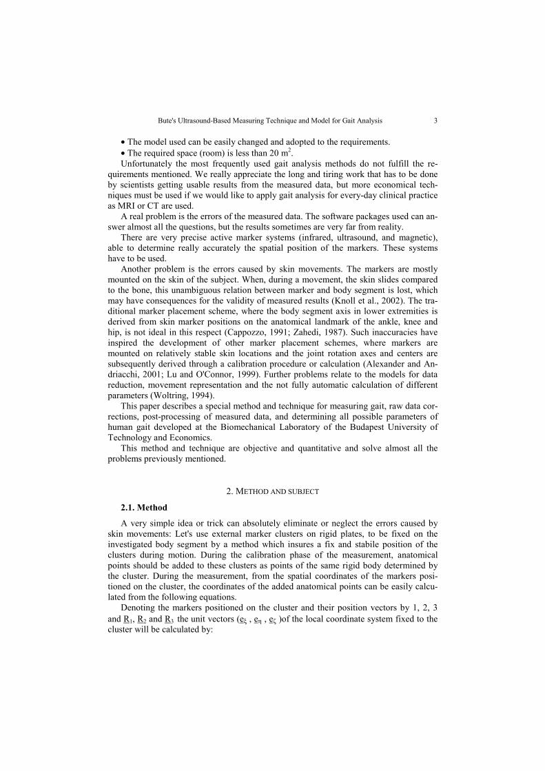
The package allows for dividing the motion into periodic gait cycles (Figure 6) anddemonstrates all these gait parameters as a discrete function in each cycle. The measuredgait coordinates and calculated parameters can be displayed in a diagram from 0 to 100%of the gait cycle. The parameters can be represented either relative to the right or to theleft side cycle.
Temporal and distance gait parameters are also commonly obtained in clinical settingsas they are easily measured and useful in the evaluation of pathological gait (Whittle,1991). Temporal-distance gait parameters may be analyzed with individuals walking on atreadmill, and compared with normal values in literature. The following temporal-dis-tance parameters are calculated from the coordinates of the anatomical points: stridelength (the distance traveled during one stride measured between one heel strike to thenext heel strike on the same side), step length (distance between the heel strike of onefoot to the heel strike of the other foot), step width (mediolateral distance between thefeet), stride time (duration of a single stride).
8 L. KOCSIS, R. M. KISS, Z. KNOLL, M. JURAK
View of measureddata
Data process window forautomatic calculation
Fig. 5. Automatic calculation tool
Fig. 6. Co-ordinates of anatomical points, vertical reaction components,and EMG envelope curves presented as a function of the gait cycle

Bute's Ultrasound-Based Measuring Technique and Model for Gait Analysis 9
Knee angles of the gait cycle play a majorrole with regard to the energy expended duringwalking and are commonly affected by patho-logical disorders (Whittle, 1991). The kneeangle is defined as the angle between a straightline joining the lateral malleolus - fibula headand a straight line joining the lateral femoralepicondyle – great trochanter (Figure 7). Foreach subject, knee angles were calculated atfour positions including initial contact, mid-stance, and peak values of extension and flex-ion. Midstance was defined as the point whenthe knee joint had attained maximum flexionafter initial contact.
The behaviour of the knee depends on themovement of ligaments. The motion could bedescribed by the newly defined relative liga-ment-points-movement parameter, which is therelative maximum displacement between thetwo characterized points of the knee. The twocharacterized points of the knee were chosen ina manner so that the line between these twopoints is closely parallel with the investigatedligament. For example, the anterior cruciate ligament - (ACL) movement - parameter isdefined as the maximum relative displacement between the tibial tubercule and the lateralfemoral epicondyle (Figure 8) divided by the minimum distance between those two points.
Fig. 8. Definition of the ACL movement parameter. The ACL movement is definedas the maximum displacement between the tibial tubercule and the lateralfemoral epicondyle.
Treadmill walking allows to determine the average and the deviation of all parametersfrom six complete gait cycles for each subject.
180-α
Fig. 7. Definition of the knee angle (α).The knee angle is defined as theangle between a straight linejoining the lateral malleolus -fibula head and a straight linejoining the lateral femoralepicondyle – great trochanter.

10 L. KOCSIS, R. M. KISS, Z. KNOLL, M. JURAK
3. RESULTS AND DISCUSSION
The validation of the new technique is shown on a few selected spatial-temporal pa-rameters and knee joint kinematics, which are either very common or newly defined.
Spatial - temporal variables, such as the stride time, the stride and the step length, andthe walking base, are derived from the temporal and spatial coordinates. For each subject,the average and the standard deviation of parameters were determined from six completegait cycles. Figure 9 shows as an example, the stance, swing and double support phases forone subject's steps. As can be seen, these parameters differ at each gait cycle analyzed forone subject. The other parameters show similar differences, which are not significant(p>0.47). Table 1 summarizes the average values and standard deviation of these quantitiesat healthy female and male subjects. No significant differences were found between spatial- temporal variables of the left and right sides of one subject (p> 0.37) and between thesevariables of subjects (p>0.41). However, on the basis of the results we can establish that thestep length and the walking base of the right step are greater (5-10%), than these of the left,and the step length, the walking base and the stride length of female-subjects are smallerthan those of males. The spatial – temporal parameters presented in this study compare fa-vorably with values found in literature (Whittle, 1992). The spatial – distances parameterscorrelated to values found in literature (Whittle, 1992).
Fig. 9. The stance, swing and double support phases shown at one subject's steps
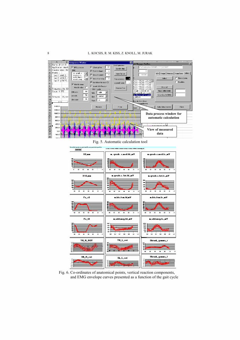
Figure 10 shows a graphical representation of one subject's knee angles. As can beseen, the knee angles differ at each gait cycle, similarly to spatial - temporal parameters.The average values and the standard deviation of the peak values of flexion and extensionat 51 adults are summarized in Table 1. No statistical differences were found between theleft and right sides of one subject (p>0.55) and between these values of subjects (p>0.39).The definition of the knee angle presented in this study does not evaluate frontal andtransverse plane components. This is important in some pathological gait, where abnor-malities occur essentially in these planes. The knee angle is not zero at extended legs, atheel-up phase of gait (Figure 10), because the knee angle models also the anatomical an-gle between the femur and the tibia in frontal plane.
The relative ligament - movement parameters are closely the same at each gait cycle.The average and standard deviance of all four ligament-movement parameters are sum-
Bute's Ultrasound-Based Measuring Technique and Model for Gait Analysis 11
marized in Table 1. On the basis of the results we can establish that the relative ligamentmovement parameters of male subjects are closely equivalent to values of females'. Nodifference was found between the values of the right and left sides. The relative ligament-movement parameters were used to quantify the tibial translation into the direction ofligaments. Let us see the characteristics of the relative ACL - movement parameter,which describes the condition of the anterior cruciate ligament (ACL). At heel strike withthe knee at full extension, the tibia is at its maximum anterior position during the gaitcycle. The next key event occurs at terminal extension where the tibia is located posterioragain while the knee is near full extension. During the swing phase with the limb un-loaded, the tibia moves to its maximum posterior position at maximum knee flexion, andthen moves forward rapidly and the knee extends prior to heel strike. The overall range,the maximum displacement between the tibial tubercule and the lateral femoral epicon-dyle was used to quantify the characteristics of dynamic stability and the differences ob-served between normal subjects and patients of knees with anterior cruciate ligament de-ficiency. The measured data represent that the relative-ligament-movement parameters donot depend on gender and the dominant side. The values of these parameters depend onlyon the movement of the tibia with respect to femur and on the translation-motion of thefemur's condylus, which depends only on the anatomical state of the knee.
0
10
20
30
40
50
60
0% 20% 40% 60% 80% 100%
Percent of gait cycle
Kne
e an
gle
[deg
ree]
Fig. 10. One subject's knee angle function in time divided into gait cycles

12 L. KOCSIS, R. M. KISS, Z. KNOLL, M. JURAK
Table 1. The average values and deviation of gait parameters
Parameters Average Standard deviation Parameters in literatureStep length [mm] 513.3 26.6 350-790Stride length [mm] 1012 25.47 600-1700Walking base [mm] 40.9 8.2 9-75Stride time [msec] 984 47.2 800-1780Knee angle at initial contact(peak value in swing phase) [degree]
5.5 0.21 2-34
Knee angle at midstance(peak value in stance phase) [degree]
21.47 0.42 10-40
Knee angle at heel rise(peak value of extension) [degree]
6.47 0.14 0-11
Knee angle in middle of swing phase(peak value of flexion) [degree]
55.87 0.71 25-90
Relative ACL-points-movementparameter [-]
0.25 0.014 -
Relative PCL-points movementparameters
0.34 0.016 -
Relative LCL-points movementparameters
0.32 0.032 -
Relative MCL-points movementparameters
0.062 0.0044 -
4. CONCLUSIONS
The method suggested needs less space for gait measurement and uses only onemeasuring head positioned at the back of the treadmill. In this case the errors of the dis-placements perpendicular to the sagittal plane are reduced and the handlebars of thetreadmill and the arm movements cause no disturbances.
The described powerful and very advanced measurement method for the analysis ofthe lower limb has been well established in biomechanics research and clinical applica-tions for a long time. The 19-point-model is verified by measuring knee movement andtemporal-distance gait parameters during treadmill walking. These measurements ob-tained after 6 minutes of treadmill walking were highly correlated with and not signifi-cantly different to those given in literature.
Treadmill walking allows to analyze several gait cycles of one subject and to deter-mine the deviation of parameters for each subject, which are calculated automatically byDataManager.
The 3D motion analysis presented in this paper is suitable for clinical gait analysis.
Acknownledgements. This work was supported in part by the Hungarian Scientific Fund T034130,T037272 by the István Széchenyi Award (Rita Kiss, László Kocsis) and by the MEDICaMENTORFoundation.

Bute's Ultrasound-Based Measuring Technique and Model for Gait Analysis 13
REFERENCES
1. Alexander, E.J. and Andriacchi, T.P. (2001). Correcting for deformation in skin-based marker system.Journal of Biomechanics, (34), 355-361.
2. Alton, F., Baldey, L., Caplan, S. and Morrissey, M.C. (1998). A kinematic comparison of over groundand treadmill walking. Clinical Biomechanics, (13), 434-440.
3. Andrews, J.G. (1995). Euler's and Lagrange's equations for linked rigid-body models of three-dimen-sional human motion. In: Allard, P., Stokes, I.A.F., Blanchi, J.P. (Eds.) Three-Dimensional Analysis ofHuman Movement. Human Kinetics, Chanoaign, 145-175.
4. Cappozzo, A. (1991). Three-dimensional analysis of human walking: Experimental methods and associ-ated artifacts. Hum Mov Sci. (10), 598-602.
5. Knoll, Zs., Kiss, R. and Kocsis, L, (2003). Gait patterns before and after anterior cruciate ligament re-construction. Knee Surgery, Sports Traumatology, Arthroscopy (during printing).
6. Kocsis, L. (2002). More precise measurement method for 3D gait analysis. Proceedings of Third Con-ference on Mechanical Engineering, Springer Verlag, Budapest. (1), 215-218.
7. Lu, T.W. and O'Connor, J.J. (1999). Bone position estimation from skin marker co-ordinates using globaloptimization with joint constraints. Journal of Biomechanics, (32), 129-134.
8. Matsas, A., Taylor, N. and McBurney, H. (2000). Knee joint kinematics from familiarized treadmillwalking can be generalized to over ground walking in young unimpaired subjects. Gait and Posture, (11,)46-53.
9. Marzani, F., Calais, E., and Legrand, L. (2001). A 3D marker-free system for the analysis of movementdisabilities. IEEEE Trans Inf Technol Biomed, (5), 18-26.
10. Medved, V. (2001). Measurement of human locomotion. CRC Press LLC, New York 11. Rishi, T, (1998), 3-D analysis of the human body with special regard to the changing of the mass mo-
ments of inertia during the interval of motion. B.Sc. Diploma Thesis. Budapest: Technical University ofBudapest, Department of Applied Mechanics.
12. Stiehl, J.B., Komistek, R., and Dennis, D.A. (2001). A novel approach to knee kinematics. Am J Orthop,(30,) 287-293.
13. Whittle, M. (1991). Gait analysis – An introduction. Butterworth – Heinemann, Oxford 14. Woltring, H.J. (1994). 3D attitude representation of human joints: a standardization proposal. Journal of
Biomechanics, (27), 1399-1414. 15. Zahedi, M.S., Spence, W.D., Solomonides, S.E. and Paul, J.P. (1987). Repeatability of kinetic and kine-
matic measurements in gait studies of the lower limb amputee. Prosthet Orthot Int, (11), 55-64.
BUTS-OVA ULTRAZVUČNA OSNOVAMERNE TEHNIKE I MODEL ZA ANALIZU HODA
Laszlo Kocsis, Rita Kiss, Zsolt Knoll, Mihaly Jurak
Rad se fokusiran na prezentaciju nove 3D analize tehnike hodanja na tredmilu. Ultrazvučnaosnova 3D sistema merenja i razvijeni aranžman kretanja su korišćeni da izmere i utvrdeparametre hoda tokom hodanja na tredmilu. Model podrazumeva da je svaki rasčlanjeni segmentuda odvojen deo, povezan jedan sa drugim zglobovima. Ovaj rad takođe prikazuje novi paketsoftvera za 3D analizu kretanja hodanjem na tredmilu koji je Datamenadžer razvio. Mi smoproučavali kinematiku kolena i vremensku razliku merenih parametara hoda (dužina koraka,dugačak korak, širinu koračanja, itd) dobijenih hodanjem na tredmilu. Ova merenja su visokokorelantna i ne značajno različita od onih u literaturi. Hodanje na tredmilu omogućava analizu unekoliko ciklusa za svakog subjekta. Na osnovu analize kod svakog subjekta se može utvrditistandardna devijacija parametara vremena hoda i podataka kinematike kolena. 3D sistemskaanaliza pokreta prezentovana je kao odgovarajuća i standardna procedura za analizu brzog hoda.
Ključne reči: analiza pokreta, analiza hoda, koleni zglob, hod na tredmilu