by dr. nahidah ibraheim
TRANSCRIPT

CARDIOVASCULAR SYSTEM 1
by
Dr. Nahidah Ibraheim
College of pharmacy /Dep. Clinical laboratory Science
stage 1 Date:21/11/2020
By the end of this lecture, students are expected to:
1. List and describe the layers of the hart.
2. Outline the histological features of the pericardium.
3. Outline the different types of epithelium simple squamous cells found in heart.
4. Summarize the functional and histological structure of Purkinje fibres.
The circulatory system pumps and directs blood cells and substances carried in blood to
all tissues of the body. It includes both the blood and lymphatic vascular systems, and in
an adult the total length of its vessels is estimated at between 100,000 and 150,000
kilometers.
This system consists of the heart, major
arteries, arterioles, capillaries, venules, and
veins.
The main function of this system is to
deliver oxygenated blood to cells and
tissues and to return venous blood to the
lungs for gaseous exchange
Dr. Nahidah Ibrahim College of Pharmacy /Dep. of clinical laboratory Science 1st stage Date:21/11/2020
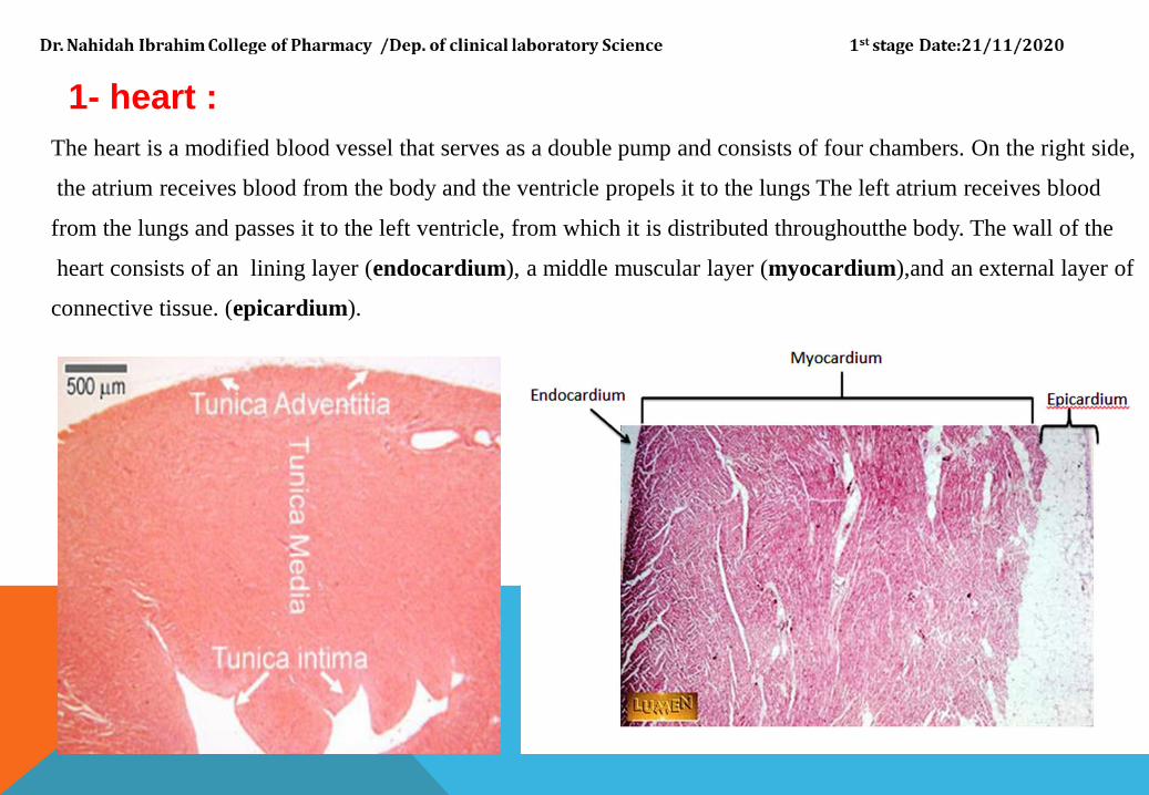
1- heart :
The heart is a modified blood vessel that serves as a double pump and consists of four chambers. On the right side,
the atrium receives blood from the body and the ventricle propels it to the lungs The left atrium receives blood
from the lungs and passes it to the left ventricle, from which it is distributed throughoutthe body. The wall of the
heart consists of an lining layer (endocardium), a middle muscular layer (myocardium),and an external layer of
connective tissue. (epicardium).
1-ENDOCARDIUM :
The endocardium forms the inner lining of the atria and ventricles and is continuous
with and comparable to the inner lining of blood vessels. It consists of a single layer of polygonal
squamous (endothelial) cells with oval or rounded nuclei .The endothelial cells rest on a continuous layer of fine
collagen fibers, separated from it by a basement membrane.The fibrous layer is called the subendothelial layer.
Deep to it is a thick layer of denser connective tissue hat forms the bulk of the endocardium and contains elastic
fibers and some smooth muscle cells. a loose connective tissue constituting the subendocardial layer binds the
endocardium to the underlying heart muscle and contains collagen fibers, elastic fibers, and blood vessels. In the
ventricles it also contains the specialized cardiac muscle fibers of the conducting system (purkinje fibers ).

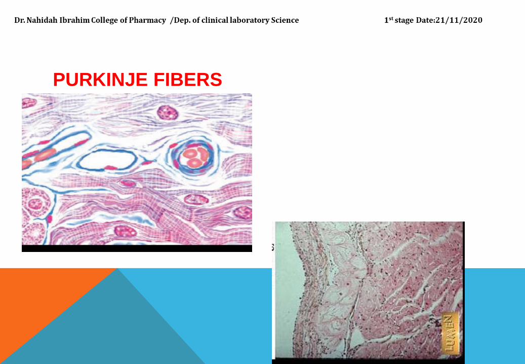
PURKINJE FIBERS
Purkinje fibers are thicker and larger than cardiac muscle fibers and contain a greater amount of
glycogen. They also contain fewer contractilefilaments. Purkinje fibers are part of the conduction
system of the heart. These fibers are located beneath the endocardium on either side of the
interventricular septum and are recognized as separate tracts. Because Purkinje fibers branch
throughout the myocardium, they deliver continuous waves of stimulation from the atrial nodes to
the rest of the heart musculature via the gap junctions. This produces ventricular contractions
(systole) and ejection of blood from both ventricular chambers.
PURKINJE FIBERS
2-MYOCARDIUM
The myocardium is the middle layer of the heart and consists primarily of cardiac muscle. The myocardium is a
very vascular tissue. The myocardium is the thinnest in the atria and thickest in the left ventricle. It consist of
cardiac muscle cells = myocytes .Different from smooth or skeletal muscle cells due to placement of
nuclei, cross striations, and intercalated disks. Intercalated disks Junctional complexes that contain
fascia adherens, desmosomes, and gap junction to provide connection and communication. Bind
myocytes and allow ion exchange to facilitate electrical impulses to pass.
The capillaries completely surround individual cardiac myocytes and are held in close apposition to
them by the enveloping delicate connective tissue that occurs between individual muscle cells. The
myocardium is arranged in layers that form complex spirals about the atria and
ventricles. In the atria, the cardiac muscle cells are smaller and contain a number of dense
granules not seen elsewhere in the heart. These myocytes have properties associated
with endocrine cells. They are most numerous in the right atrium and release the secretory granules
when stretched
MYOCARDIUM
Atrial Natriuretic Hormone
Certain cardiac muscle fibers in the atria exhibit dense granules in their cytoplasm. These granules contain
atrial natriuretic hormone, a chemical that is released in response to atrial distension or stretching. The
main function of this hormone is to decrease blood pressure by regulating blood volume. This action is
accomplished by inhibiting the release of renin by the specialized cells in the kidney and aldosterone from
the adrenal gland cortex. This induces the kidney to lose more sodium and water (diuresis).As a result, the
blood volume and blood pressure are reduced, and the distension of the atrial wall is relieved.
Gap junctions:
functionally couple all cardiac muscle fibers to rapidly spread the stimuli for contraction of the heart muscle.
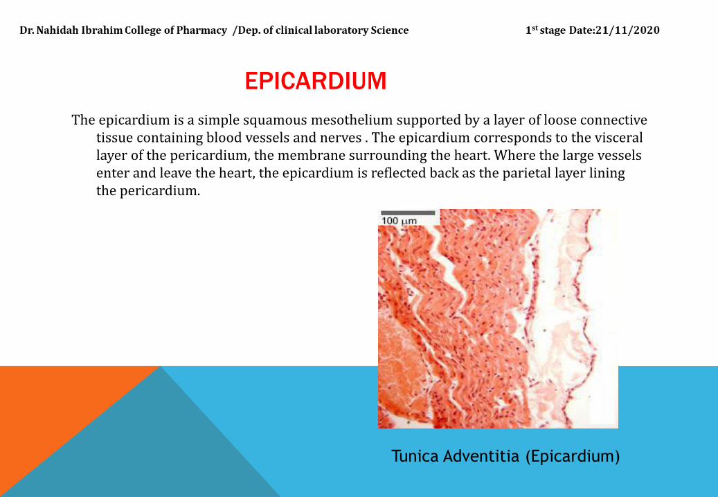
EPICARDIUM
The epicardium is a simple squamous mesothelium supported by a layer of loose connective tissue containing blood vessels and nerves . The epicardium corresponds to the visceral layer of the pericardium, the membrane surrounding the heart. Where the large vessels enter and leave the heart, the epicardium is reflected back as the parietal layer lining the pericardium.
Tunica Adventitia (Epicardium)
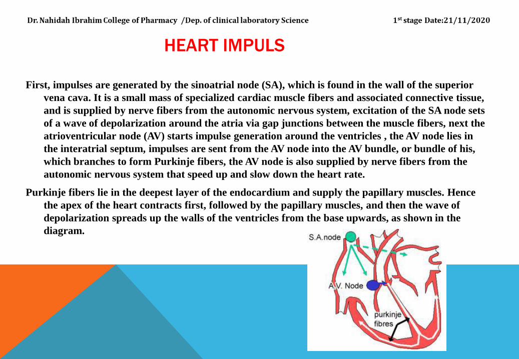
HEART IMPULS
First, impulses are generated by the sinoatrial node (SA), which is found in the wall of the superior
vena cava. It is a small mass of specialized cardiac muscle fibers and associated connective tissue,
and is supplied by nerve fibers from the autonomic nervous system, excitation of the SA node sets
of a wave of depolarization around the atria via gap junctions between the muscle fibers, next the
atrioventricular node (AV) starts impulse generation around the ventricles , the AV node lies in
the interatrial septum, impulses are sent from the AV node into the AV bundle, or bundle of his,
which branches to form Purkinje fibers, the AV node is also supplied by nerve fibers from the
autonomic nervous system that speed up and slow down the heart rate.
Purkinje fibers lie in the deepest layer of the endocardium and supply the papillary muscles. Hence
the apex of the heart contracts first, followed by the papillary muscles, and then the wave of
depolarization spreads up the walls of the ventricles from the base upwards, as shown in the
diagram.
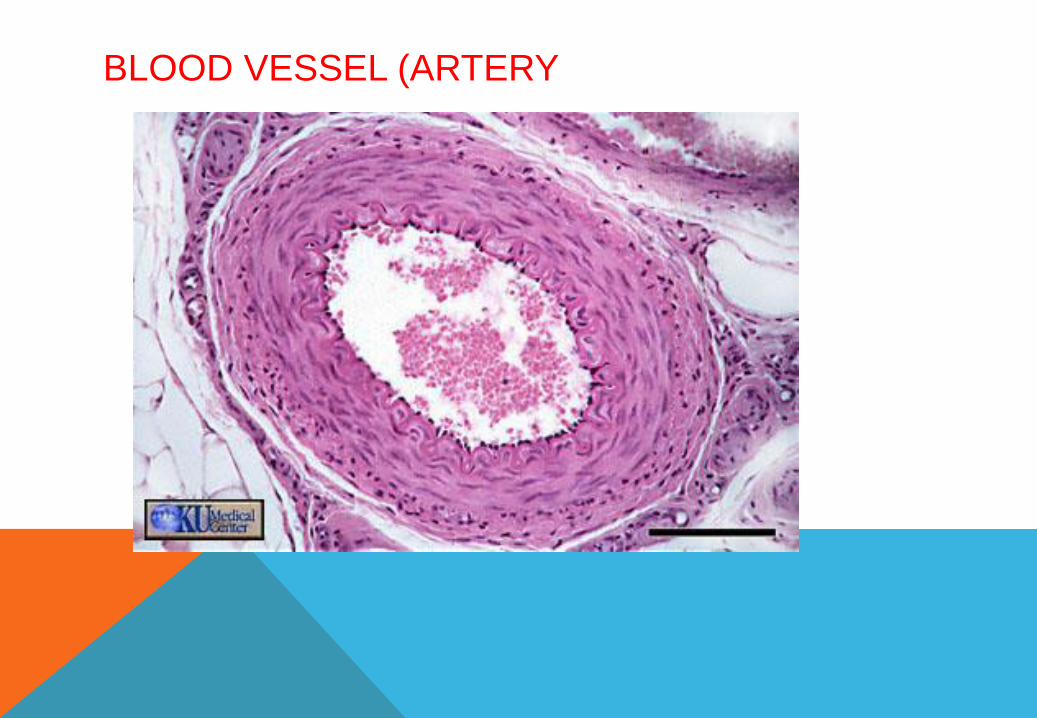
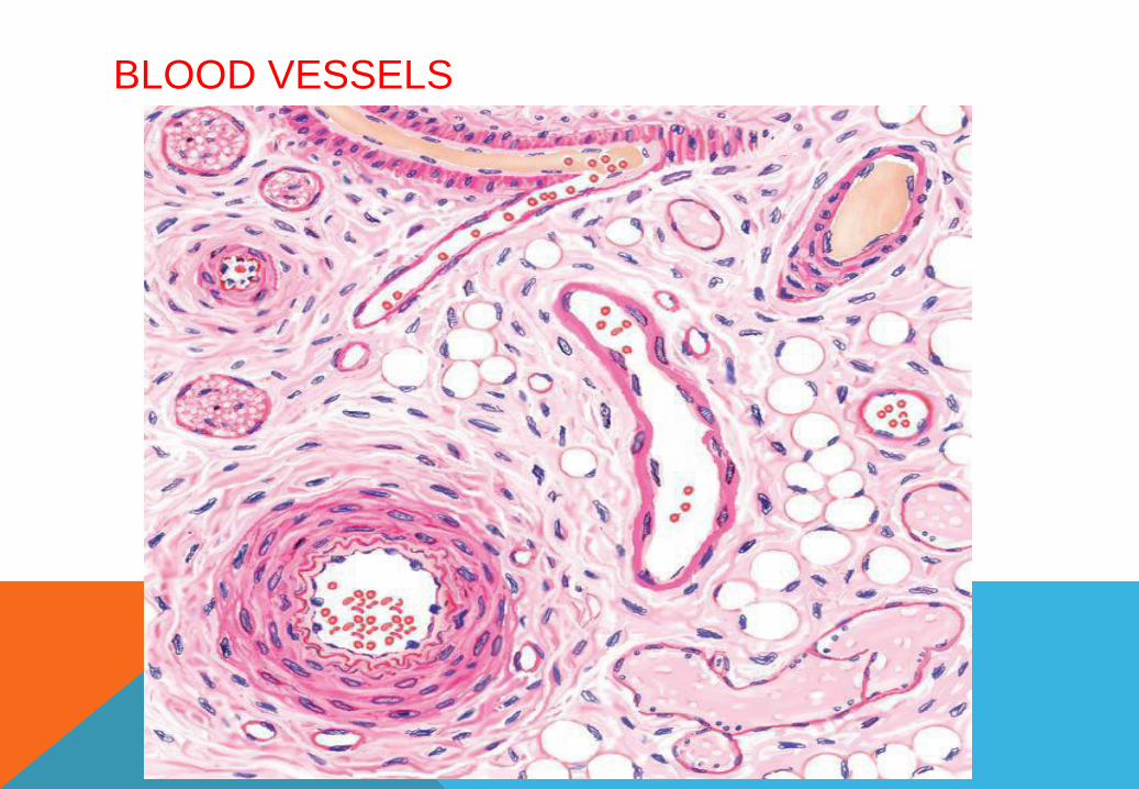
2- BLOOD VESSELS
:
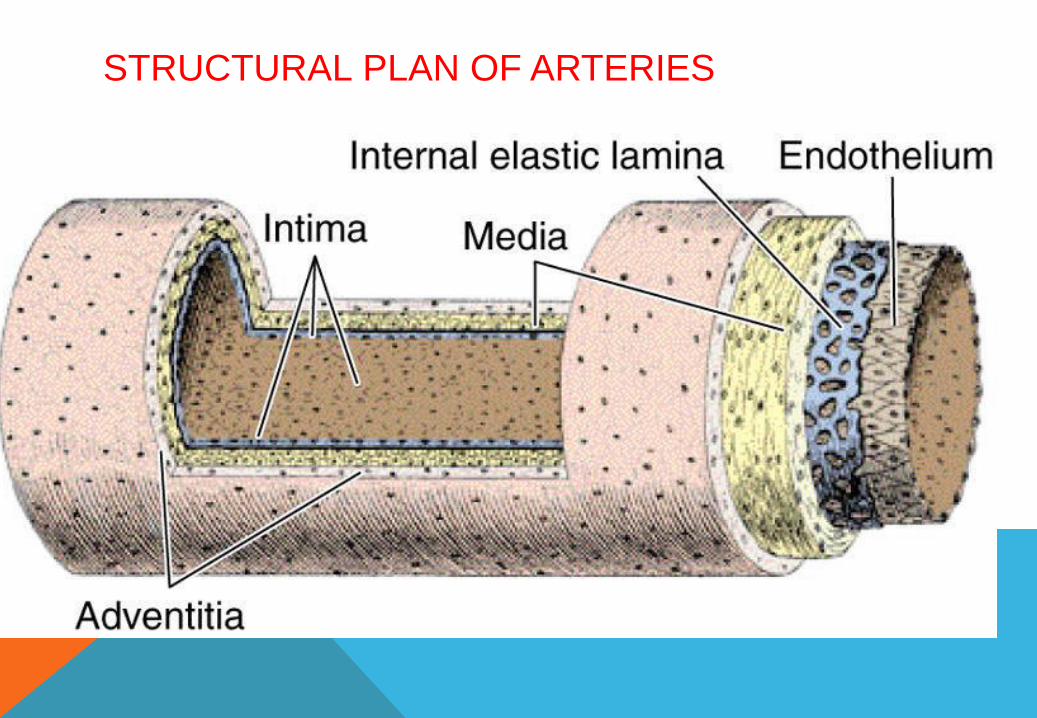
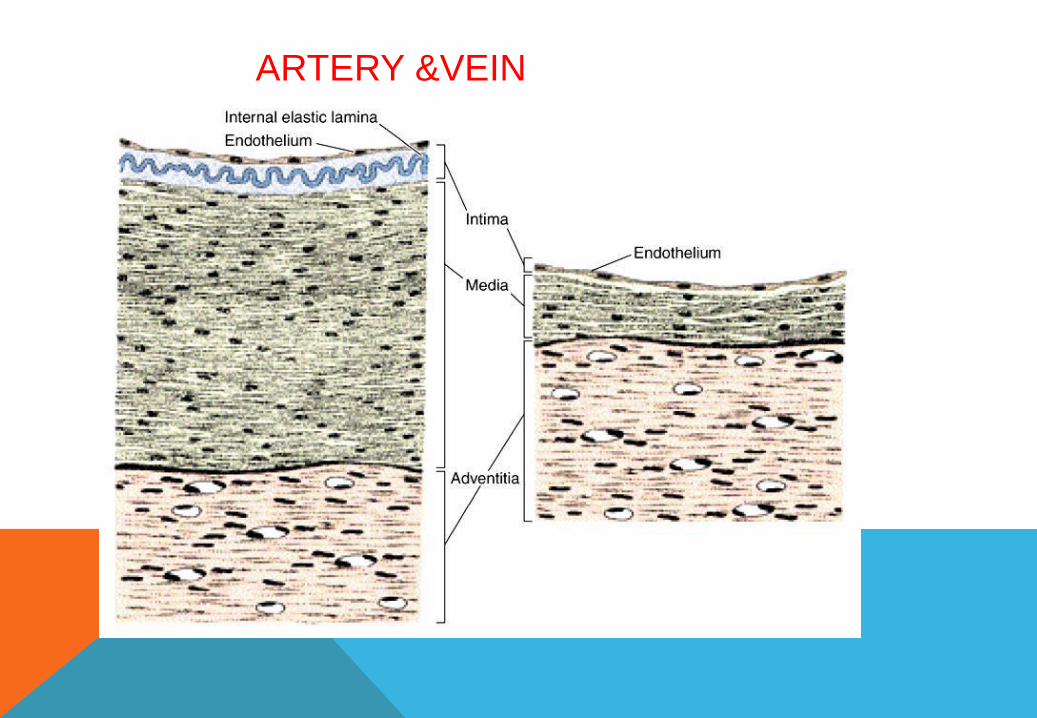
blood vessels differ in size, distribution,and function, structurally they share many common
features. As in the heart, the walls of blood vessels consist of three major coats or tunics.
From the lumen outward, the wall of a blood
vessel consists of :
1-tunica intima,
2- tunica media, and
3- tunica adventitia
CONT.
1- TUNICA INTIMA :
The tunica intima corresponds to and is continuous with the endocardium of the heart.
It consists of an endothelium of flattened squamous cells resting on a basal lamina and
is supported by a subendothelial connective tissue.
2- TUNICA MEDIA :
The tunica media is the equivalent of the myocardium of the heart and is the layer most
variable both in size and structure. Depending on the function of the vessel, this layer
contains variable amounts of smooth muscle and elastic tissue.
3- TUNICA ADVENTITIA :
The tunica adventitia also varies in thickness in different parts of the vascular circuit. It consists
mainly of collagenous connective tissue and corresponds to the epicardium of the heart, but
it lacks mesothelial cells.
BLOOD VESSEL (ARTERY
TYPES OF ARTERIES :
Three classes of arteries can be distinguished:
large elastic or conducting arteries,
medium-sized muscular or distributing arteries,
and small arteries and arterioles.
A characteristic feature of the entire arterial side of the blood vasculature system is the
prominence of smooth muscle in the tunica media.
Arteries that leave the heart to distribute the oxygenated blood exhibit progressive
branching. With each branching, the luminal diameters of the arteries gradually
decrease, until the smallest vessel, the capillary, is formed.
STRUCTURAL PLAN OF ARTERIES

ARTERY
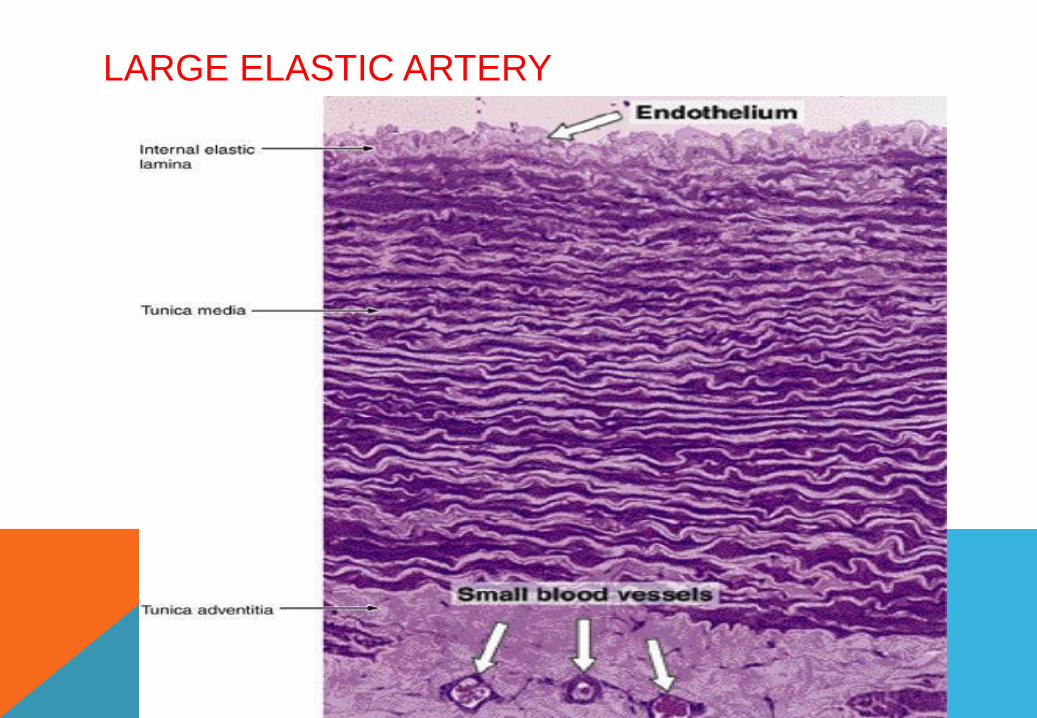
1- ELASTIC ARTERIES :
Elastic arteries are the largest blood vessels in the body and include the pulmonary
trunk and aorta with their major branches, the brachiocephalic, common carotid,
subclavian, vertebral, pulmonary, and common iliac arteries. The walls of these
vessels are primarily composed of elastic connective tissue fibers. These fibers provide
great resilience and flexibility during blood flow.
The tunica intima is relatively thick and is lined by a single layer of flattened, polygonal
endothelial cells that rest on a complete basal lamina, about one-fourth of the total thickness
of the intima is formed by the subendothelial layer, a layer of loose connective tissue that
contains elastic fibers and a few smooth muscle cells
The tunica media is the thickest layer and consists largely of elastic tissue. Smooth muscle
cells are the only cells present in the media of elastic arteries and synthesize and maintain the
elastic fibers and collagen. The tunica adventitia is relatively thin andcontains bundles of collagen
fibers (type 1) and a few elastic fibers,
LARGE ELASTIC ARTERY
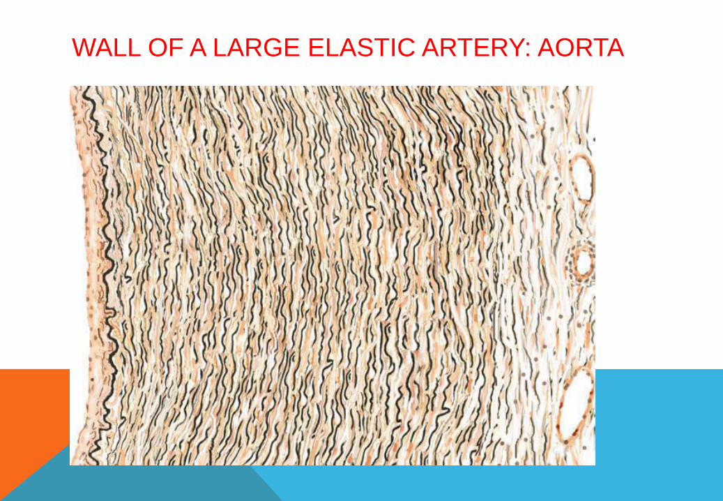
WALL OF A LARGE ELASTIC ARTERY: AORTA
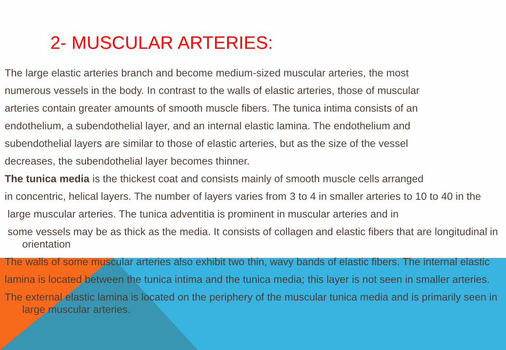
2- MUSCULAR ARTERIES:
The large elastic arteries branch and become medium-sized muscular arteries, the most
numerous vessels in the body. In contrast to the walls of elastic arteries, those of muscular
arteries contain greater amounts of smooth muscle fibers. The tunica intima consists of an
endothelium, a subendothelial layer, and an internal elastic lamina. The endothelium and
subendothelial layers are similar to those of elastic arteries, but as the size of the vessel
decreases, the subendothelial layer becomes thinner.
The tunica media is the thickest coat and consists mainly of smooth muscle cells arranged
in concentric, helical layers. The number of layers varies from 3 to 4 in smaller arteries to 10 to 40 in the
large muscular arteries. The tunica adventitia is prominent in muscular arteries and in
some vessels may be as thick as the media. It consists of collagen and elastic fibers that are longitudinal in
orientation
The walls of some muscular arteries also exhibit two thin, wavy bands of elastic fibers. The internal elastic
lamina is located between the tunica intima and the tunica media; this layer is not seen in smaller arteries.
The external elastic lamina is located on the periphery of the muscular tunica media and is primarily seen in
large muscular arteries.
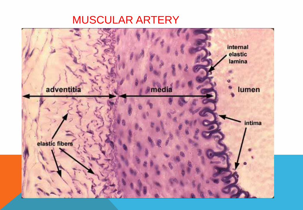
MUSCULAR ARTERY
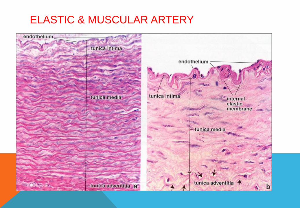
ELASTIC & MUSCULAR ARTERY
FUNCTIONAL CORRELATIONS :The elastic arteries transport blood from the heart and move it along the systemic
Vascular path. The presence of an increased number of elastic fibers in their walls
allows the elastic arteries to greatly expand in diameter during systole (heart
contraction), when a large volume of blood is forcefully ejected from the ventricles
into their lumina. In contrast, the muscular arteries control blood flow and blood pressure
through vasoconstriction or vasodilation of their lumina. Vasoconstriction and vasodilation,
owing to a high proportion of smooth muscle fibers in the artery walls, are controlled by
unmyelinated axons of thesympathetic division of the autonomic nervous system. Similarly,
by autonomic constriction or dilation of their lumina, the smooth muscle fibers in smaller
muscular arteries or arterioles regulate blood flow into the capillary beds .
During diastole (heart relaxation), the expanded elastic walls recoil upon the volume
of blood in their lumina and force the blood to move forward through the vascular channels.
As a result, a less variable systemic blood pressure is maintained, and blood flows more evenly
through the body during heart beats.
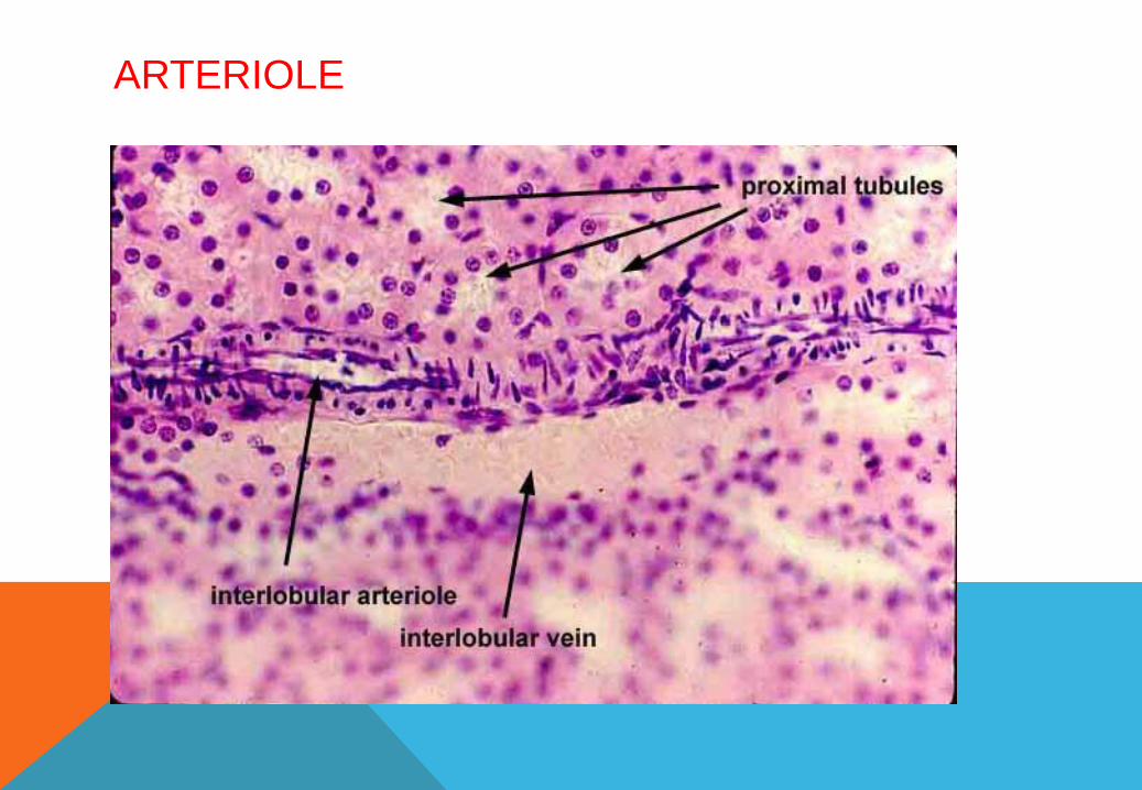
3- ARTERIOLES :
Arterioles are the smallest branches of the arterial system. Their walls consist of one to
five layers of smooth muscle fibers. tunica intima consists only of endothelium and a
fenestrated internal elastic lamina. Tunica adventitia also decreases in thickness,
becoming extremely thin in the smallest arterioles.
Arterioles deliver blood to the smallest blood vessels, the capillaries. Capillaries connect
arterioles with the smallest veins or venules
ARTERIOLE
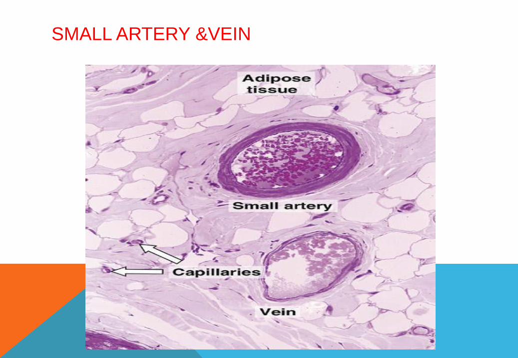
SMALL ARTERY &VEIN
CAPILLARY
Metarterioles are intermediate between capillaries and arterioles and regulate the flow of
blood through capillary beds. They also are called capillary sphincter areas or precapillary
arterioles,their lumina generally are wider than those of the capillaries .

STRUCTURAL PLAN OF VEINS
Capillaries unite to form larger blood vessels called venules; venules usually accompany
arterioles. The venous blood initially flows into smaller postcapillary venules and then into
veins of increasing size. The veins are classified as small, medium, and large. Compared
with arteries, veins typically are more numerous and have thinner walls, larger diameters, and
greater structural variation. Small-sized and medium-sized veins, particularly in the
extremities, have valves. The presence of valves in veins assists venous blood flow by
preventing back flow. When blood flows toward the heart, pressure
in the veins forces the valves to open. As the blood begins to flow backward, the valve flaps
close the lumen and prevent back flow of blood. Valves are absent in veins of the central
nervous system, the inferior and superior venae cavae, and viscera. The walls of the veins, like
the arteries, also exhibit three layers or tunics. However, the muscular layer is much less
prominent. The tunica intima in large veins exhibits a prominent endothelium and subendothelial
connective tissue. In large veins, the muscular tunica media is thin, and the smooth muscles
intermix with connective tissue fibers . In large veins, the tunica adventitia is the thickest and
best-developed layer . bundles of smooth muscle fibers are common in the connective tissue of
this layer. Vasa vasorum are present and may extend into the Media.
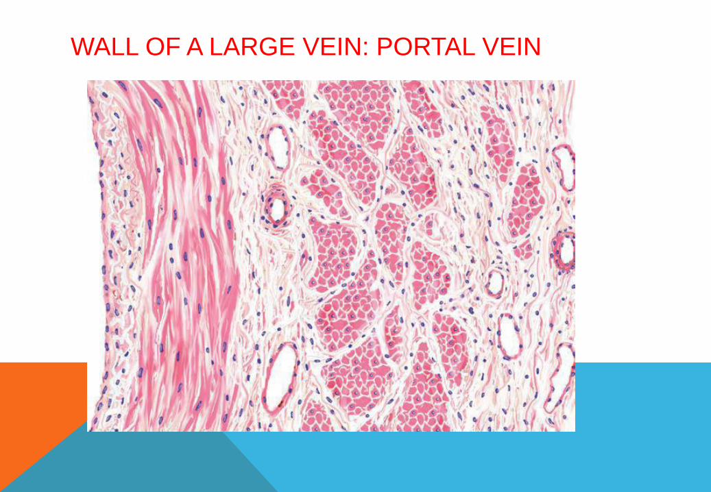
WALL OF A LARGE VEIN: PORTAL VEIN

ARTERY &VEIN
ARTERY &VEIN
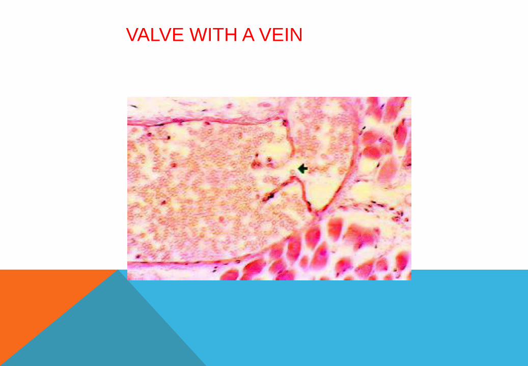
VALVE WITH A VEIN
\FUNCTIONAL CORRELATIONS :In veins, blood pressure is lower than in the arteries.As a result, venous blood flow is
passive. Venous blood flow in the head and trunk is primarily owing to negative
pressures in the thorax and abdominal cavities resulting from respiratory movements.
Venous blood return from the extremities is aided by surrounding muscle contractions
and prevented from flowing back by numerous valves in the large veins of the
extremities.

MEDIUM VEINS
The medium size veins, includes most of the named veins of gross anatomy except for
major trunks. The thin tunica intima consists of endothelial cells resting on a basal
lamina, but a narrow subendothelial layer may be present. but a poorly defined internal
elastic lamina is formed only in the larger vessels. In most medium veins, the tunica
media, is thinner than in corresponding arteries. The thick tunica adventitia forms the
bulk of the wall and is larger than the tunica media. It consists of collagen and elastic
fibers and contains smooth muscle cells. Vasa vasorum are present in the larger vessels
of this class . Valves are present in most of the medium size veins
VENULES :
Venules arise from the union of several capillaries to form vessels . The junctions
between venules and capillaries are important sites of fluid exchange between tissues
and blood. The tunica intima consists of a thin,continuous endothelium. The tunica
media is missing in the smallest venules, and the relatively thin adventitia contains a few
collagen fibers,
VASA VASORUM :
The walls of larger arteries and veins are too thick to receive nourishment by direct
diffusion from their lumina. As a result, these walls are supplied by their own small blood
vessels called the vasa vasorum (vessels of the vessel). The vasa vasorum allows for
exchange of nutrients and metabolites with cells in the tunica adventitia and tunica media
BLOOD VESSELS
TYPES OF CAPILLARIES :
Capillaries are the smallest blood vessels. Their
size, is about the size of an erythrocyte (red blood
cell). There are three types of capillaries: ---
continuous capillaries,
fenestrated capillaries,
sinusoids.
These structural variations in capillaries allow for
different types of metabolic exchange between
blood and the surrounding tissues.
Regardless of the type, the basic structure of capillaries is similar and represents an
extreme simplification of the vessel wall. The tunica intima consists of endothelium and a
basal lamina; the tunica media is absent and the tunica adventitia is greatly reduced
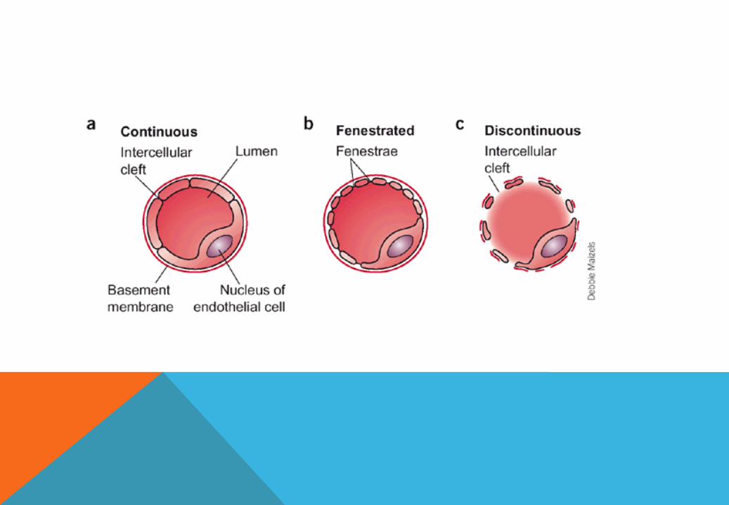

1- CONTINUOUS CAPILLARIES :
Continuous capillaries are the most common. They are found in muscle, connective tissue,
nervous tissue, skin, respiratory organs, and exocrine glands. In these capillaries, the endothelial cells
are joined and form an uninterrupted, solid endothelial
lining. Pericytes are irregular, branched, isolated cells that occur at intervals along capillaries,
enclosed by the basal lamina of the endothelium. The cells resemble fibroblasts
2- FENESTRATED CAPILLARIES :
Fenestrated capillaries are characterized by fenestrations (pores) in the cytoplasm of
endothelial cells designed for a rapid exchange of molecules between blood and tissues.
Fenestrated capillaries are found in endocrine tissues and glands, small intestine, and kidney
glomeruli.
3- SINUSOIDAL CAPILLARIES :
are blood vessels that exhibit irregular,tortuous paths . Their much wider diameters slow down the
flow of blood. Also, the cells may be separated by gaps and rest on a discontinuous basal lamina.
A direct exchange of molecules occurs between blood contents and cells. Sinusoidal capillaries
are found in the liver, spleen, and bone marrow

ARTERIOVENOUS ANASTOMOSES :
In addition to their capillary connections, arteries and veins may unite by shunts
called arteriovenous anastomoses. Generally these arise from side branches of
arterioles that pass directly to venules. They are thick-walled, muscular vessels of small
caliber that usually are coiled and surrounded by a connective tissue sheath
They are plentiful in the plantar and palmar surfaces, fingertips, toes, lips, and nose and
also occur in the thyroid. When open, the anastomoses shunt blood around the capillary
bed and thus regulate the blood supply to many tissues. In the skin they function primarily in the
regulation of body temperature.