by milan a. logan (from the biochemical laboratory ... except that the titration of excess barium...
TRANSCRIPT
COMPOSITION OF CARTILAGE, BONE, DENTIN, AND ENAMEL
BY MILAN A. LOGAN
(From the Biochemical Laboratory, Harvard Medical School, and the
Forsyth Dental Injirmary, Boston)
(Received for publication, May 10, 1935)
Analyses made by Gabriel (1) and Klement (2) on bone ash and analyses made by Gassman (3) indicate that the alkali metal con- tent of bones and teeth is 2 to 3 times as great as the alkali metal content of other tissues of the body and is usually at least as great as the magnesium content of calcified structures. Other investi- gations made to consider the inorganic composition of bone ha.ve, asfar as I am aware, considered among the basic constituents, only the calcium and magnesium.
For an understanding of the process by which calcification takes place, it is important to know the nature of the insoluble inorganic salt contained in calcified structures (4) and to know whether the composition is constant or varies according to the tissue in which it is deposited. In this respect the equivalence between acid and basic constituents is an essential consideration, but the situation existing in the fresh tissue cannot be determined from the analyses of bone ash, because the carbonate content may be altered during the ashing (5).
The analyses reported here were made on fresh tissues, and the alkali metals are considered, not alone that the equivalence (or lack of it) between acid and basic constituents may be more accurately known, but, since these constituents by themselves form no insoluble precipitates with phosphate and carbonate, that an understanding of how they came to be concentrated far above the level in the blood plasma might throw some light on the process by which the insoluble constituents accumulate in calcified structures. Calcifying cartilage was investigated to see what bearing the com- position of this tissue may have on the base content of the bone which replaces it.
375
by guest on June 13, 2018http://w
ww
.jbc.org/D
ownloaded from

376 Cartilage, Bone, Dentin, and Enamel
Procedure
Preparation of Tissue. Bone-Under ether or amytal anesthesia, the bone was dissected from the animal. It was scraped clean, split open, the trabecuhe scraped out, and freed of adhering mar- row with the aid of a clean damp cloth. Portions of the diaphysis, epiphysis, or line of proliferation were crushed or cut with steel instruments to particles not larger than 1 mm. in diameter, and weighed immediately in small glass weighing bottles. Crushing to a fine powder is undesirable because it causes cloudiness in the trichloroacetic acid filtrate.
Teeth-After removal from patients or dogs, the teeth were split and the pulp removed. As much of the crown as could be definitely identified as dentin was collected with the aid of a dental burr. A layer near the dentin and enamel junction was discarded and the remaining enamel crushed with steel instruments for analysis.
Cartihge from calves (nasal septum), and from calf embryos (epiphyses of long bones) was frozen with solid carbon dioxide, the perichondrium scraped off, and while frozen, the tissue cut into slices approximately 0.1 mm. thick, weighed, and added to 10 volumes of 10 per cent trichloroacetic acid. For purposes of analysis, the water content of cartilage was assumed to be 80 per cent (6). Samples of cartilage for carbon dioxide determination were frozen in sealed bottles.
Carbon Dioxide-The procedure described by Van Slyke (7) was employed, except that the titration of excess barium hydroxide was carried out without filtering off the precipitated barium carbonate. The method was adapted to the determination of small quantities. For the determination of amounts greater than 5 cc. of 0.1 N carbon dioxide, the tube containing the weighed sample of bone was dropped into the inside tube of the apparatus described by Van Slyke, and the air replaced by carbon dioxide-free air. The stopper was removed just far enough to admit the tip of a pipette, and an amount of 0.1 N barium hydroxide approximately 50 per cent in excess of the estimated carbonate was admitted to the flask. The apparatus was evacuated and N hydrochloric acid run onto the bone, drop by drop, time being allowed after each addition until bubbling had almost ceased. The amount eventually added was sufficient to cover the specimen. After the mixture was
by guest on June 13, 2018http://w
ww
.jbc.org/D
ownloaded from

M. A. Logan 377
shaken the reaction was allowed to proceed overnight and the vacuum was replaced with carbon dioxide-free air. The stopper was removed, the inside tube washed down with 40 cc. of distilled water, 2 drops of 1 per cent phenolphthalein were added, and then from a burette, 0.1 N hydrochloric acid was run in with constant rotation of the flask until the indicator turned a pale pink.1
Samples of cartilage tested by this procedure were kept frozen up to the point of admitting the N hydrochloric acid.
For samples containing less than 3 cc. of 0.1 N carbon dioxide, the determination was carried out in an apparatus made from a 200 cc. Pyrex Erlenmeyer flask, and the titration of excess barium hydroxide made with 0.02 N hydrochloric acid, without removing the inside tube of the apparatus. A depression in the inside tube was provided to admit the long accessory tip of a 5 cc. burette to the Erlenmeyer flask.
To test the procedure, four weighed portions of Kahlbaum’s calcium carbonate (zur Analyse), equivalent to 10 cc. of 0.1 N
carbon dioxide, were analyzed. The following amounts were found: 9.90, 9.95, 10.03, 10.00 cc. of 0.1 N carbon dioxide. Three portions equivalent to 5 cc. of 0.1 N carbon dioxide each were tested with the following recoveries: 5.04, 4.97, 5.00 cc. of 0.1 N carbon dioxide. Five 1 cc. portions of 0.1 N sodium carbonate solution (Merck’s Blue Label) were analyzed with the following recoveries: 1.03, 1.01, 1.01, 0.99, 0.99 cc. of 0.1 N carbon dioxide.
Calcium-The residue left from the determination of carbon dioxide was transferred quantitatively to a volumetric flask (usu- ally 100 or 200 cc.) with the aid of 10 per cent trichloroacetic acid solution (prepared from the redistilled acid) and made up to volume with the same solution. Aliquots of this, containing approxi- mately 1 mg. of calcium, were taken for the determination of cal- cium by the method of Fiske and Logan (8).
Inorganic phosphorus was determined on aliquots of the tri- chloroacetic acid filtrate by the method of Fiske and Subbarow (9).
Base Other Than Calcium-An aliquot of the trichloroacetic acid filtrate containing 10 to 40 mg. of phosphorus was evaporated to dryness in a lipped Pyrex test-tube (200 X 25 mm.), with the aid of apparatus previously described (10). 0.5 cc. of 10 N sulfuric
1 Thymol blue proved to be equally good but no better as an indicator. The titration must be carried to the pale green of the indicator.
by guest on June 13, 2018http://w
ww
.jbc.org/D
ownloaded from

378 Cartilage, Bone, Dentin, and Enamel
acid was added and the organic matter destroyed or hydrolyzed with the aid of nitric acid by heating in a bath of molten salts at a temperature not greater than 250” until a colorless or light yellow residue remained.2s3 The residue was dissolved and diluted with distilled water so as to contain about 1 mg. of phosphorus per cc. 0.25 cc. of 2.5 per cent oxalic acid was added for each mg. of cal- cium present (or each 0.5 mg. of phosphorus). A small drop of 0.04 per cent alcoholic thymol blue was added and the tube heated to 90’ in a boiling water bath. The solution was brought to pH 1 with redistilled (5 N) ammonium hydroxide, at which point precipi- tation usually started. After the solution was shaken and heated in the water bath for 10 minutes, it was brought to pH 3 with 5 N
ammonium hydroxide added drop by drop, and permitted to remain in the water bath for an additional 10 minutes. A drop of saturated alcoholic methyl red was then added, and the solution adjusted to pH 5 with N ammonium hydroxide, drop by drop, and heated for 30 seconds between additions. The solution was permitted to cool, and in approximately 3 hours filtered through a tight ashless filter paper. The precipitate was washed with three 2 cc. portions of 1 per cent ammonium oxalate. The filtrate was evaporated to dryness and the oxalic acid present destroyed with the aid of nitric acid in the ammonium sulfate-sulfuric acid bath.3 The residue was dissolved and made to volume in a flask of such size that the solution contained not more than 1 mg. of phosphorus per cc. From an aliquot of this solution the phosphate was pre- cipitated and the rest of the determination of total base carried out by the Fiske (12) “First method.” The values for Na + K were obtained by subtracting the magnesium values from the values obtained for total base. This method rather than deter- mination of the individual alkali metals was chosen, for the purpose of the investigation, because the sum of the two is the value of greater significance and can be determined in small quantities with far greater accuracy than is possible for determination of the individuals separately.
2 Heating to high temperature to destroy the last trace of organic matter
is unnecessary and undesirable because it may result in the production of an insoluble residue.
3 Directions for preparmg the bath of molten salts is included, together
with the directions for the determinations of magnesium in the Folin Manual (11) and will be published in detail shortly.
by guest on June 13, 2018http://w
ww
.jbc.org/D
ownloaded from

M. A. Logan 379
The directions described above were arrived at after several preliminary experiments. To test the procedures, solutions of pure salts similating bone in composition were made up and the magnesium and total base determined on the solution obtained after removing the calcium as oxalate (Table I). The following are typical results. The magnesium was precipitated as mag- nesium ammonium phosphate and the phosphorus content of the precipitate determined calorimetrically in all cases.3 The results so obtained were checked in several cases by precipitation of the magnesium with 8-hydroxyquinoline, ignition of the precipitate to magnesium oxide, and estimation of the magnesium by alkali-
TABLE I
Analysis of Known Mixture of Salts of Composition Similar to Bone
The mixture contained, per cc., 4 mg. of calcium (as chloride), 1.86 mg. of phosphorus (as (NH&HPOb), 0.0655 mg. of magnesium (as lactate),
0.115 mg. of sodium (as chloride), in 0.1 N nitric acid. Calcium was pre- cipitated and analysis made of the filtrate. Each result is an average of two determinations agreeing within 2 per cent.
Amount of solution taken
0.1 N Mg
Present I Found
cc.
10
20
0.1 i-i base
Present Found
cc. I cc.
1.039
I
1.052
2.078 2.080
metric titration.3 A comparison of the phosphate method and alkalimetric titration for magnesium is given in Table II.
Chloride-To a weighed portion of the bone (about 1 gm.), suspended in 20 cc. of distilled water, 2 cc. of 0.1 N silver nitrate were added. The organic matter was then destroyed by acidifying with nitric acid and adding chloride-free potassium permanganate (saturated solution), drop by drop, to the boiling solution, until the purple color remained permanently. A drop of chloride-free oxalic acid solution was added to decolorize the solution, 2 cc. of 25 per cent ferric ammonium sulfate in 5 N nitric acid were added, and the excess silver was titrated with 0.1 N ammonium thio- cyanate from a microburette.
Results obtained by the above procedure on cartilage were com-
by guest on June 13, 2018http://w
ww
.jbc.org/D
ownloaded from

380 Cartilage, Bone, Dentin, and Enamel
pared with results obtained from a dilute nitric acid filtrate of the tissue and showed excellent agreement. 10 volumes of cold 0.3 N
nitric acid were added to weighed samples of the sliced tissue. After standing at least 1 hour, the solution was filtered, silver ni- trate added, the solution evaporated to approximately one-fourth its volume, permanganate and nitric acid added to destroy the small amount of organic matter present, and the excess silver titrated as described above.
Total Sulfate-The total sulfate of cartilage was determined by the Fiske (13) benzidine method on a solution of the tissue made by autoclaving with 2 volumes of 5 N hydrochloric acid for 1 hour at 20 pounds pressure.
TABLE II
Comparison of Determination of Magnesium in Bone by Means ofMagnesium
Ammonium Phosphate and by Alkalimetric Titration
Each result is an average of two determinations agreeing within 1 per cent.
Experiment No.
1 2 3 4
Mg found
Alkalimetric Phosphate
mg. per gm. mg. per gm.
9.26 9.24 5.42 5.52 3.55 3.55 4.28 4.28
Acid hydrolysates of bone gave no precipitate with benzidme after removing the phosphate. The precipitation of such large amounts of phosphate was well as the presence of relatively large amounts of organic matter, however, introduced two elements of uncertainty. Therefore, total sulfate on bone was determined by weighing the barium sulfate obtained by adding 4 cc. of 5 per cent barium chloride to a solution of the ash of 10 gm. of bone in 30 cc. of 5 N hydrochloric acid. The results would presumably in- clude unoxidized sulfur, of which, however, bone contains very little. Until a better procedure is devised for separating the small amount of sulfate present in bone from the organic matter, this method appears most suited to the purpose at hand.
by guest on June 13, 2018http://w
ww
.jbc.org/D
ownloaded from

M. A. Logan 381
Results
Samples of bone were examined from twelve cats, the ages of which ranged from 24 hours to maturity, and included the bones of the mother of the youngest animal examined. Portions of the diaphysis were compared with trabeculs and the epiphysis of the same bone in two instances each, and with the line of proliferation in one instance. Diaphyses of femurs of seven rabbits, ranging from 24 hours of age to maturity and including the mother of the youngest, were likewise examined. The same was repeated on femurs from six rats, including one comparison each of the dia-
TABLE III
Phosphate and Carbonate Content of Rat Bone in Relation to Calcium
Rat No. Weight of rat
gm.
24.1 25.0 79.5 95.0
187.5 238.0
-
. _
-
per cant of Ca
10.7 10.7 12.5 12.6 13.7 15.8
-
_-
-
Phosphate
per cent of Ca
95.3 98.8 94.2 94.0 90.0 39.3
1 wk. old 2 wks. “
Mother of Rats 1 and 2
The results are expressed in relation to the calcium instead of to the total base, because sufficient bone, freed from soft tissue, was not obtained from Rats 1 and 2 to determine total base and magnesium.
physis with the epiphysis and line of proliferation, and that of the mother with the offspring, 1 week and 2 weeks old.
The results showed no definite differences in composition of the inorganic part of the bone in respect to the part of the bone taken for analysis. In rat bone, the carbonate content expressed in stoichiometric equivalents in relation to the calcium increased pro- gressively from 10.7 per cent, at 1 week of age, to 15.8 per cent at maturity, and the inorganic phosphate (calculated as a tribasic acid) correspondingly decreased from 95.3 to 89.3 per cent of the calcium (Table III). This change has previously been noted in rat bone by Kramer and Shear (14). The analysis of cat bone showed a more nearly constant composition in this respect; the carbon
by guest on June 13, 2018http://w
ww
.jbc.org/D
ownloaded from

382 Cartilage, Bone, Dentin, and Enamel
dioxide was stoichiometrically equivalent to 11.6 to 13.8 per cent of the calcium (10.7 to 13.0 per cent of the total base). Theolder
animals did, however, on the whole, have slightly higher carbonate content and lower phosphate content relative to the total base (Table IV). In the case of rabbits, the carbonate content varied between 10.3 and 15.2 per cent of the calcium (9.6 to 14.4 per cent of the total base) without respect to age. Representative results showing typical variations are given.
Samples of dentin and enamel from human teeth of seven individuals ranging from 6 to 65 years of age were analyzed. Most
TABLE IV
Inorganic Composition of Diaphyses of Femurs
Ani- mal NO.
1
2
“2” animal
gm.
Kitten, 24 149 hrs. old
Cat, mother 3650 of Animal 1
Cat, male 4000 Young rat 80 Rabbit 1224
“ 2850
-
s
-
“2 0.1 N Mg
0.1 N ““Kf
--
x. per cc. per gm. gm.
87.4 2.77
-
1 cc. per gm.
4.43
wr ten total base
86.3
139.2 3.28 3.97 83.0
119.0 3.52 1.38 86.5 79.5 3.14 3.13 87.7
106.5 5.24 0.87 88.3 125.8 3.56 3.17 85.1
- 7
co2
‘9 cm l&d base
11.6
12.5
13.0 11.5
9.6 14.4
0.19
0.19
0.30 0.30
Total lssei m m of acids deter- mined
1.03
1.04
1.00 1.01
1.02 1.00
* Calculated as a tribasio acid.
of these teeth showed some degree of caries and all were taken from persons in which other teeth showed caries. It is difficult to obtain teeth from persons who do not show some degree of dental defect; and if a change in composition of the inorganic part of the teeth is associated with the production of caries, one might expect to find the change in any of the teeth of a person susceptible to caries whether or not the particular tooth examined was carious. Con- sequently, analysis of normal dog teeth are given for comparison.
It is evident from the results on human and dog teeth (Table V) that the magnesium of the dentin, in relation to the total base, is 3 to 4 times as high as the magnesium of the enamel. No signifi-
by guest on June 13, 2018http://w
ww
.jbc.org/D
ownloaded from

M. A. Logan 383
cant difference is apparent between the magnesium content of normal dog enamel or dentin, and that found in carious human teeth. The carbonate content of dentin and enamel of the normal dog’s teeth is essentially the same, and in relation to the total base, amounts to approximately 70 per cent of the carbonate content of the bone from the same animal. The carbonate content of the enamel of the carious human teeth is only slightly less than that
TABLE V
Inorganic Composition of Human Teeth and Doa Teeth Compared with Bone from Same Animals
Subject
Human (‘ C‘ “
Dog 1 “ 2 “ 1 “ 2 “ 1 ” 2
Age
15 yrs. 55 “ 15 “ 55 ‘(
4 mos. 6 ‘< 4 (‘ 6 ‘I 4 ‘I 6 ‘I
of2 0.1 N Mg
~--
c&per cc.per vm. llm.
Dentint 131.5 6.82 “ $ 125.6 7.05
Enamelf 179.0 2.11 “ $ 168.0 3.67
cc. per l7m.
1.34 1.32 3.10 3.95
Dentin 116.9 5.90 2.46 ‘I 114.8 9.60 1.33
Enamel 160.5 2.68 2.46 ‘I 156.1 3.72 2.45
Bone 106.7 2.12 2.12 ‘I 114.9 1.95 2.52
T
0.1 N Ng+
-7
Mg
_- aer ten total base
4.89
5.26 1.15 2.09
4.71 7.65 1.62 2.29 1.91 1.63
-
hf-
gsp4 cot
'W cm total base
87.8 88.9 88.7 88.0
,er cm fotd base
10.3 10.2 6.8 7.0
92.8 8.2 91.9 8.0 94.7 7.9 88.4 8.2 88.8 11.6 83.3 12.8
Total ,aSei rum of acid.5 deter- mined
1.02 1.01 1.04 1.05
0.98 1.00 0.97 1.04 0.99 1.04
* Calculated as tribasic acid. t Impacted molar, not carious. $ Carious incisor.
of the enamel of the dog, but the carbonate of the dentin is higher, more nearly approaching that of bonee4
Results based on analyses of combined dentin and enamel have been cited to support the contention that carious teeth contain more magnesium than normal teeth (15). The futility of attempt-
* The above results were presented at a meeting of the American Associa- tion for the Advancement of Science, December, 1933 (Logan, M. A., J. Am. Coil. Dent., 1, 46 (1934)).
by guest on June 13, 2018http://w
ww
.jbc.org/D
ownloaded from

384 Cartilage, Bone, Dentin, and Enamel
ing to settle the question by that means is evident from the differ- ence in composition between dentin and enamel.
The differences found between the composition of the inorganic part of bone, dentin, and enamel are so large as to preclude the possibility of excluding them from consideration concerning the means by which calcification takes place. If, as the results of x-ray analysis of bone indicate (4, 16), the inorganic part of bone is composed of minerals of the apatite series of the general formula 3Ca3(P0&.CaX9 in which X represents *CO8 chiefly, it is neces- sary to consider that conditions under which calcification takes place are such as to cause a substitution of sodium and potassium for part of the calcium, and .to consider that the difference in condition under which deposition takes place in dentin and enamel of teeth is sufficient to cause a 300 per cent difference between the amount of magnesium similarly substituted, and a 30 per cent difference in the carbonate.
The results of analyses of twenty-four specimens of bone, in which the total inorganic base and at least phosphate and car- bonate were determined, showed that the total base was equivalent to the sum of the acids determined within 1 per cent in fifteen specimens, three agreed within 2 per cent, and the remaining six showed from 3 to 6 per cent more base than acid. Three of these latter six came from a mother cat and two of its offspring. Of nine samples of dentin, the total inorganic base agreed with the sum of the acid in six, and the remaining three agreed within 2 per cent. Of nine samples of enamel, one showed 3 per cent more acid than base, two showed 1 per cent agreement between the base and acids, one agreed within 2 per cent, and five showed 4 to 5 per cent more base than acid. Chloride determinations were made in ten samples and included in the calculation when made, but affected the results usually by about 0.2 per cent, and never more than 0.5 per cent. To the extent that chemical analysis of these inorganic constituents can contribute to the question, the essential equiva- lence of the acid and basic constituents in the majority of deter- minations on bone and dentin supports the contention that the bases exist in the bone and dentin essentially as tertiary phosphates and carbonates.
As far as the determinations are concerned, the results are con- sidered to be accurate to 1 per cent. In this connection, it is
by guest on June 13, 2018http://w
ww
.jbc.org/D
ownloaded from

M. A. Logan
pertinent to note that all the inorganic bases are determined but this is not necessarily true of the acids. The results on a few specimens of bone and the majority of samples of enamel leave room for consideration as one possibility that anions to the extent of 3 to 5 per cent of the total are not included in the analyses. As becomes evident from the analyses of cartilage, however, an ele- ment of uncertainty concerns the base-combining power of the organic matrix of the bone, which, constituting 35 to 50 per cent of the tissue, may or may not affect the results by more than 1 per cent.
Although an appreciable quantity of the base of bone may be combined with organic constituents of the tissue, the results of Gabriel’s analyses (1) indicated that most of the alkali metals exist as constituents of the inorganic portion relatively insoluble in water. He found that the amount of sodium far exceeded the amount of potassium in bone, and in some experiments he removed the organic matter from bone by boiling it with a glycerol-potas- sium hydroxide mixture. The results obtained on the insoluble residue left after washing out the glycerol-potassium hydroxide with water were compared with analyses made on bone ash. Es- sentially the same amount of sodium was found byeitherprocedure. Klement (2) determined the amount of carbonate, phosphate, and calcium dissolved by conductivity water from bone residues prepared according to the procedures of Gabriel. He thought that the magnesium, sodium, and Ijotassium as carbonates owed their presence in bone to serum in the Jficellen des Knockengewebes. His results indicated that the magnesium and alkali metals re- mained in the residue obtained after extracting the bone with glycerol-potassium hydroxide and presumably washing out the added potassium hydroxide with water. How the bases could escape being removed from the residue by such a procedure unless they originally existed as components of the inorganic structure is difficult to understand. The ratio of alkali metals to calcium is essentially the same in bone as in the enamel of teeth. In the enamel of teeth, as much as 90 per cent of the weight of the fresh tissue can be accounted for by calcium combined as phosphate and carbonate. If the other bases existed as carbonates apart from this portion of the structure, they would of necessity constitute 25 per cent of the remaining tissue.
by guest on June 13, 2018http://w
ww
.jbc.org/D
ownloaded from
386 Cartilage, Bone, Dentin, and Enamel
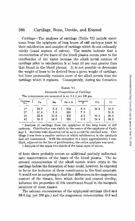
Cartilage-The analyses of cartilage (Table VI) include speci- mens from the epiphysis of long bones of calf embryos prior to their calcification and samples of cartilage which do not ordinarily calcify (nasal septum of calves). The results indicate that a concentration of the bases of the blood plasma occurs prior to the calcification of the tissue because the alkali metal content of cartilage prior to calcification is at least 50 per cent greater than that found in the blood plasma. It is not possible to determine the weight of bone to be derived from a given weight of cartilage, butt bone presumably contains more of the alkali metals than the cartilage which it replaces. Consequently, during the formation
TABLE VI
Inorganic Composition of Cartilage
The components are measured in cc. 0.1 N per 100 gm.
“%i:“” C& Mg Na + K
1* 19.7 8.2 213 2* 17.8 7.5 200 3t 22.6 15.8 402 4t 25.2 14.0 321
-
Inorric co2
6.2 14.3 7.5 3.2 10.4 3.5 14.4
Cl
53.0 51.8 46.0 46.2
* Analysis of cartilage from the epiphyses of the long bones of calf embryos. Calcification was visible in the center of the epiphysis of Carti- lage 1. Sections were dissected out so as to avoid the calcified area. Car- tilage 2 was from a smaller embryo in which calcification in the epiphysis had not commenced. With the exception of a layer approximately 1 mm. thick, adjacent to the line of proliferation, the entire epiphysis was used.
t Analysis of the upper two-thirds of the nasal septa of calves.
of bone there probably occurs an additional partially indiscrimi- nate concentration of the bases of the blood plasma. The in- creased concentration of the alkali metals which exists in the cartilage before the formation of bone could, however, be expected to favor the inclusion of those constituents in the final structure. It would not be surprising to find that differences in the magnesium content of the tissues, from which dentin and enamel develop, influence the proportion of this constituent found in the inorganic structure of these tissues.
The calcium concentration of the epiphyseal cartilage (35.6 and 39.4 mg. per 100 gm.) and the magnesium concentration (9.9 and
by guest on June 13, 2018http://w
ww
.jbc.org/D
ownloaded from
M. A. Logan 387
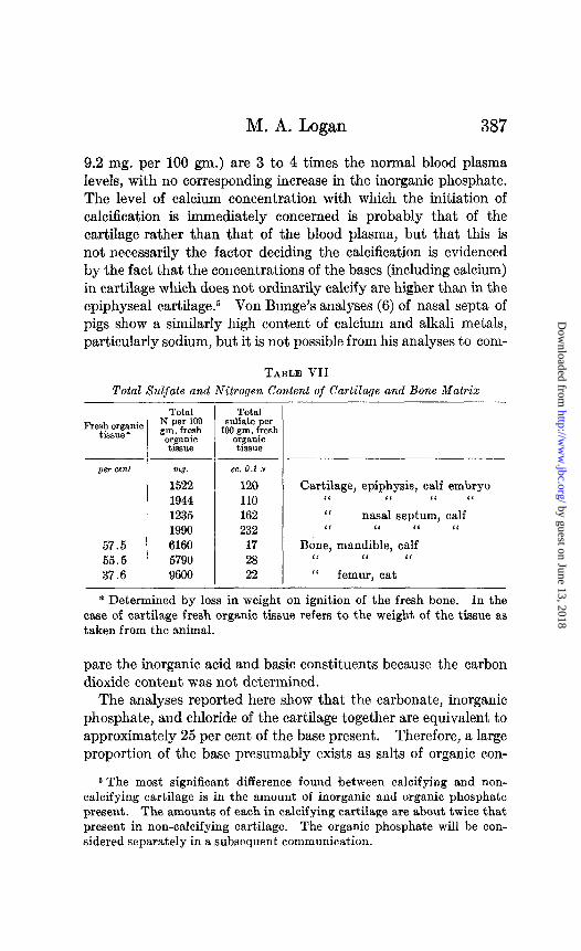
9.2 mg. per 100 gm.) are 3 to 4 times the normal blood plasma levels, with no corresponding increase in the inorganic phosphate. The level of calcium concentration with which the initiation of calcification is immediately concerned is probably that of the cartilage rather than that of the blood plasma, but that this is not necessarily the factor deciding the calcification is evidenced by the fact that the concentrations of the bases (including calcium) in cartilage which does not ordinarily calcify are higher than in the epiphyseal cartilage.6 Von Bunge’s analyses (6) of nasal septa of pigs show a similarly high content of calcium and alkali metals, particularly sodium, but it is not possible from his analyses to com-
TABLE VII
Total Sulfate and Nitrogen Content of Cartilage and Bone Matrix
57.5 55.5 37.6
-
-
m3. ce. 0.1 N
1522 120 1944 110 1235 162 1990 232 6160 17 5790 28 9600 22
-
-
Total sulfate per
100 gm. fresh organic
tissue
Cartilage, epiphysis, calf embryo ‘I ‘I ‘I “ ‘( nasal septum, calf ‘I “ I‘ ‘I
Bone, mandible, calf ‘I I< ‘I I‘ femur, cat
* Determined by loss in weight on ignition of the fresh bone. In the case of cartilage fresh organic tissue refers to the weight of the tissue as taken from the animal.
pare the inorganic acid and basic constituents because the carbon dioxide content was not determined.
The analyses reported here show that the carbonate, inorganic phosphate, and chloride of the cartilage together are equivalent to approximately 25 per cent of the base present. Therefore, a large proportion of the base presumably exists as salts of organic con-
6 The most significant difference found between calcifying and non- calcifying cartilage is in the amount of inorganic and organic phosphate present. The amounts of each in calcifying cartilage are about twice that present in non-calcifying cartilage. The organic phosphate will be con- sidered separately in a subsequent communication.
by guest on June 13, 2018http://w
ww
.jbc.org/D
ownloaded from
388 Cartilage, Bone, Dentin, and Enamel
stituents of the cartilage. It is not possible, however, to predict the base-combining power of the organic matrix of bone from the composition of cartilage, because, as determinations of total nitrogen and total sulfate on cartilage and bone matrix (Table VII) show, a change in composition takes place in the conversion of the former into the latter. The total sulfate drops from over 100 cc. of 0.1 N to about 20 cc. of 0.1 N sulfate per gm. of fresh organic tissue, whereas the total nitrogen shows at least a S-fold increase in the change from cartilage to organic matrix.6 This can only be accounted for by the loss from the tissue of part of the chondroitin sulfuric acid, and possibly other constituents of relatively low nitrogen content. The nature of the change (loss of a potentially acid constituent) is such as to suggest that it may favor the precipitation of the inorganic salts.
SUMMARY
1. Relative to the total amount of base present, the magnesium content of dentin is higher than that of the enamel or bone, and the carbonate content of bone is higher than that of enamel or dentin.
2. Analysis of bone and dentin indicates that the inorganic bases are present essentially as carbonates and tertiary phosphates.
3. Concentration of the bases of the blood plasma in cartilage precedes calcification.
4. The conversion of cartilage to organic matrix of bone is characterized by loss of organic sulfates and gain of nitrogenous constituents.
BIBLIOGRAPHY
1. Gabriel, S., Z. physiol. Chem., 18, 257 (1894). 2. Klement, R., Z. physiol. &em., 184, 132 (1929); 196, 140 (1931). 3. Gassman, T., Z. physiol. Chem., 66, 455 (1908); 63, 397 (1909); 70, 161
(1910). 4. Roseberry, H. H., Hastings, A. B., and Morse, J. K., J. Biol. Chem., 90,
395 (1931). 5. Bogert, L. J., and Hastings, A. B., J. BioZ. Chem., 94,473 (193132). 6. von Bunge, G., Z. physiol. Chem., 28,452 (1899).
6 If 1 hydrogen atom of the chondroitin sulfuric acid is available for com- bination with base, it could account for about 25 per cent of the base present in cartilage.
by guest on June 13, 2018http://w
ww
.jbc.org/D
ownloaded from

M. A. Logan 389
7. Van Slyke, D. D., J. Biol. Chem., 36,351 (1918). 8. Fiske, C. H., and Logan, M. A., J. Biol. Chem., 93,211 (1931). 9. Fiske, C. H., and Subbarow, Y., J. Biol. Chem., 66,375 (1925).
10. Logan, M. A., J. Biol. Chem., 66, 761 (1930). 11. Folin, O., Laboratory manual of biological chemistry, New York and
London, 5th edition (1934). 12. Fiske, C. H., J. BioZ. Chem., 61, 55 (1922). 13. Fiske, C. H., J. BioZ. Chem., 47, 59 (1921). 14. Kramer, B., and Shear, M. J., J. BioZ. Chem., 79,147 (1923). 15. Kaushinsky, L. I., Dent. Cosmos, 74, 468 (1932). 16. Taylor, N. W., and Sheard, C., J. BioZ. Chem., 31,479 (1929).
by guest on June 13, 2018http://w
ww
.jbc.org/D
ownloaded from
Milan A. LoganDENTIN, AND ENAMEL
COMPOSITION OF CARTILAGE, BONE,
1935, 110:375-389.J. Biol. Chem.
http://www.jbc.org/content/110/2/375.citation
Access the most updated version of this article at
Alerts:
When a correction for this article is posted•
When this article is cited•
alerts to choose from all of JBC's e-mailClick here
tml#ref-list-1
http://www.jbc.org/content/110/2/375.citation.full.haccessed free atThis article cites 0 references, 0 of which can be
by guest on June 13, 2018http://w
ww
.jbc.org/D
ownloaded from