by - usm research and...
TRANSCRIPT

PHARMACOKINETICS AND METABOLISM STUDIES OF ANTIFILARIAL DRUGS
DERIVATIVES OF BENZIMIDAZOLE CARBAMATE
by
SURASH RAMANATHAN
Thesis submitted in fulfilment of the requirements for the degree of Doctor of Philosophy •
..
November 1996
v
TABLE OF CONTENTS
Page Numbers
Acknowledgement... . . . . . . . . . . . . . . . . . . . . . . . . . . . . . . . . . . . . . . . . . . . . . . . . . . . . . . . . . . . . . . . . . . . . . . . . . . . . ii
Publications ................................................................................... XII
List of Conferences .............................................................................. xiii
List of Abbreviation ............................................................................. xiv
Abstrak ............................................................................................. xv
Abstract. ........................................................................................... xvii
Chapter 1: INTRODUCTION .................................................................. 1
1.1 General Introduction ......................................................... ... 1
1.2 The Pathology and Clinical Manifestation of Lymphatic
Filarasis ............................................................................ 3
1.2.1 The filarial life cycle .................................................... .3
1.2.2 Clinical manifestation of lymphatic filariasis infection ............. 7
1.3 Chemotherapy of Antifilarial Drugs ............................................ 8 .. 1.3.1 Introduction ............................................................... 8
1.3.2 DEC and ivermectin ..................................................... 9
1.3.3 Suramin .... :·~--- ...................................................... ... 11
1.3.4 Benzimidazole carbamate .............................................. 11
1.4 Mode of Action for Anti filarial Drugs ........................................ 13
1.4.1 Introduction ............................................................. 13
Ill

1.4.2 Benzimidazole carbamate ............................................. 14
1.4.3 Diethylcarbamazine .................................................. 15
1.4.4 Suramin ................................................................. 16
1.4.5 Ivermectin ............................................................... 17
1.5 The Pharmacokinetic and Metabolism of Anti filarial Drugs .............. 19
1.5.1 Introduction ............................................................. 19
1.5.2 Mebendazole ............................................................. 20
1.5.3 Albendazole ............................................................. 23
1. 5.4 Flubendazole ......................................................... 27
1.5.5 Ivermectine .......................................................... 27
1.5.6 Diethylcarbamazine ................................................... 28
1.5. 7 Suramin ................................................................ 28
' 1.6 Drug Toxicity and Metabolism ................................................. 29
1. 7 Racemate and Drug Disposition ................................................ 31
1.8 General Principles of Pharmacokinetics and Drug Metabolism ............ .32
1.8.1 Introduction ............................................................ 32
1.8.2 Drug absorption ........................................................ 33
1.8.3 Drug distribution ...................................................... 34
1.8.4 Drug elimination ........................................................ 34
1.8.5 Drug excretion .... ·.;..· ................................................... 36
1.9 Pharmacokinetics Parameters .................................................. .37
1.9.1 Elimination half-life ................................................... 37
1.9.2 Clearance ................................................................ 38
1. 9.3 Volume of distribution ................................................ 40
lV . ----

1.10 The Scope of This Study ...................................................... 41
Chapter 2: CHEMICALS, INSTRUMENT AND STANDARD ....................... .42
2.1 Chemicals ...................................................................... .42
2.2 Equipment ....................................................................... 43
2.3 Preparation of Standard Solutions and buffer. ............................. .44
2.3.1 Standards .............................................................. 44
2.3.2 Extraction Buffer .................................................... .45
2.4 Chemical Purity of Compounds .......................................... .45
2.5 Radiolabelled Flubendazole ................................................ .45
2.6 Solubility ofUMF-078 and UMF-289 in various solvents ............. .46
Chapter 3: DETERMINATION OF THE ANTIFILARIAL DRUG UMF-058 AND
ITS METABOLITE, MEBENDAZOLE IN WHOLE BLOOD BY
IDGH-PERFORMANCE LIQUID CEROMATOGRAPHY ........... .48
3.1 Introduction ................................................................... 48
3.2 Determination of Approximate pKa value of UMF-058 ................ .49
3.3 UV Absorption ofUMF-058 and MBZ .................... : ............... 50
3.4 HPLC Method Development ................................................ .50
3.4.1 Chromatography ..................................................... .50
3.4.2 Extraction procedure ................................................ 51 .. -
3.4.3 ·Detector linearity ..................................................... 51
3.4.4 Standard curve ....................................................... .51
3.4.5 Analytical recovery, within-day and day-to- day
precision ............................................................. 52
v
----------------····-------·--·-·-

3.4.6 Quality control ofUMF-058 and MBZ analysis ................. 52
3.5 Results and Discussion ....................................................... 53
3.5. I Determination of approximate pKa value of
UMF-058 ............................................................... 53
3.5.2 UV absorption of UMF-058 and MBZ ................. .' ....... 53
3.5.3 HPLC method development.. ....................................... 54
3.6 Conclusion ..................................................................... 55
Chapter 4: DETERMINATION OF THE ANTIFILARIAL DRUG UMF-078
AND ITS METABOLITES UMF-060 AND FLUBENDAZOLE IN
WHOLE BLOOD USING IDGH PERFORMANCE
LIQUID CHROMATOGRAPHY. ............................................ . 64
4.1 Introduction ...................................................................... 64
4.2 Determination of approximate pKa value ofllM:F-078 ................... 65
4.3 UV Absorption ofUMF-078, UMF-060 and FBZ ........................ 65
4.4 HPLC method development. .................................................. 66
4.4.1 Chromatography ....................................................... 66
4.4.2 Extraction procedure ................................................... 66
4.4.3 DeteCtor linearity ....................................................... 67
4.4.4 Standard curve .......................................................... 67
4.4.5 Analytical recovery, within-day and day-to-day precision ...... 67 --4.4.6 Quality control ofUMF-078, FBZ and UMF-060
analysis ................................................................... 68
4.5 Results and Discussion ......................................................... 68
4.5.1 Determination of approximate pKa value ofUMF-078 ........... 68
VI

4.5.2 UV absorption ofUMF-078, UMF-060 and FBZ .................. 68
4.5.3 HPLC method development. ......................................... 69
4.6 Conclusion ........................................................................ 70
ChapterS: THE DETERMINATION OF DECARBOMETHOXY METABOLITES
OF UMF-078, UMF-060 AND FLUBENDAZOLE IN WHOLE
BLOOD USING IDGH PERFORMANCE LIQUID
CHROMATOGRAPHY ...................................................... .... 79
5.1 Introduction .................................................................... 79
5.2 UV Absorption of The Various Decarbomethoxy Metabolites ......... 80
5.3 HPLC Method Development ................................................. 80
5.3.1 Chomatography ........................................................ 80
5.3.2 Extraction procedure ................................................. 81
5.3.3 Detectorlinearity .......................... ·~ ........................ 81
5.3.4 Standard curve ......................................................... 81
5.3.5 Analytical recovery, within-day and day-to-day
precision ............................................................... 82
5.3.6 Quality control.ofD-UMF-078, D-FBZ and
D-.ilJMF-060 analysis ................................................ 82
5.4 Results and Discussion ....................................................... 83
5.4.1 UV absorption ofD-UMF-078, D-FBZ and --. D-UMF-060 .......................................................... 83
5.4.2 HPLC method development.. ...................................... 83
5.5 Conclusion ...................................................................... 84
vii

Chapter 6: PHARMACOKINETIC AND EFFICACY STUDIES OF UMF-058 IN
BRUGIA MALAJ71NFECTED MONKEYS (PRESBYTIS
CRISTATA) ........................................................................... 93
6.1 Introduction ...................................................................... 93
6.2 Material and Methods ........................................................... 94
6.2.1 Study design in infected monkeys ................................... 94
6.2.2 Blood sampling ......................................................... 94
6.2.3 Analysis of Blood ..................................................... 95
6.2.4 Pharmacokinetic calculation ......................................... 95
6.2.5 Data analysis ............................................................ 96
6.3 Results .................................................................... · ........ 96
6.3.1 Pharmacokinetics ofUMF-058 in infected monkeys following
a single and multiple oral doses ofUMF-058 ..................... 96
6.3.2 The efficacy of single and multiple oral doses of UMF-058
against B.malayi in infected monkeys .............................. 97
6.4 Discussion ........................................................................ 98
6.5 Conclusion ...................................................................... 100
Chapter7: PHARMACOKINETIC STUDY OF UMF-078 IN HEALTHY
MONKEYS (MACACA FASCICULARJS): EFFECT OF
FORMULATION, ROUTES AND DOSE SIZE ........................... 107
7.1 Introduction .................................................................... 107
7.2 Materials and Methods ........................................................ 107
7.2.1 Study design in healthy monkeys ................................. 107
7.2.2 Drug administration ................................................ 108
Vlll
- --------------------· ---

7.2.3 Blood sampling ................................................... .108
7.2.4 Analysis of plasma ................................................ I 09
T2.5 Pharmacokinetic calculations ..................................... 109
7.2.6 Data Analysis ....................................................... 109
7.3 Results .......................................................................... 109
7.3.1 Effect of formulation on UMF- 078 drug absorption .......... 110
7.3.2 Effect of dose size on UMF-078 drug absorption ............ 110
7.3.3 Effect of routes on UMF-078 drug absorption .................. 112
7.4 Discussion ..................................................................... 113
7.5 Conclusion.................................................................. 116
Chapter 8: PHARMACOKJNETIC STUDY OF UMF-078 IN HEALTHY
DOGS (BEAGLES): EFFECT OF FORMULATION AND
DOSE SIZE ................................................ : .. ................... 128
8.1 Introduction ................................................................... 128
8.2 Materials and Methods ....................................................... 129
8.2.1 Study design in dogs .................................................. 129
8.2.2 Blood sampling ....................................................... 130
8.2.3 Analysis of plasma .................................................... 131
8.2.4 Pharmacokinetic calculations ...................................... 131
8.2.5 Statistical analysis ................................................... 131
8.3 Results .. :' ...................................................................... 132
8.3.1 The pharmacokinetic properties ofUMF-078 following
intravenous administration of 20 mglkg UMF-078 salt in
healthy beagles ....................................................... 132
IX

8.3.2 Effect of formulation on UMF-078 drug absorption in healthy
beagles ................................................................. 132
8.3.3 Effect of various dose regimens ofUMF-078 base on
UMF-078 drug absorption in healthy beagles .................... 133
8.3.4 Effect of oral dose increment ofUMF-078 base from
150 mglk:g to 300 mglk:g on UMF-078 drug absorption in
healthy beagles ...................................................... 134
8.4 Discussion .................................................................... : .135
8.5 Summary ........................................................................ 139
Chapter 9: THE DETERMINATION OF DECARBOMETHOXY METABOLITES
OF UMF-078, FBZ AND UMF-060 IN BLOOD AFTER ORAL
ADMINISTRATION OF UMF-078IN RATS ..................... ......... 155
9.1 Introduction ........................................... : . ...................... 155
9.2 Materials and Methods ....................................................... 156
9.2.1 Animals ................................................................. 156
9.2.2 Blood sampling ........................................................ 156
9.2.3 Blood assay procedure .............................................. 157
9.2.4 Data·'Elhalysis ......................................................... 157
9.3 Results .......................................................................... 157
9.4 Discussion ..................................................................... .158
9.5 Conclu5ion ..................................................................... 160
X

Chapter 10: A PRELIMINARY INVESTIGATION OF UMF-078 AND
FLUBENDAZOLE METABOLISM IN RATS ......................... 165
10.1 Introduction ................................................................. 165
10.2 Material and Method ...................................................... 166
10.2.1 Study Design ....................................................... 166
10.2.2 Surgical procedures for intravenous study ..................... 167
10.2.3 Drug administration and blood sampling ..................... 167
10.2.4 Drug analysis ...................................................... 168
10.2.5 Data Analysis ...................................................... 169
10.3 Results ....................................................................... 169
10.4 Discussion ................................................................... 171
10.5 Conclusion .................................................................. 174
Chapter 11: CONCLUDINGDISCUSSION ...................... : ...................... 183
References ........................................................................................ 191
...
Xl
PUBLICATIONS
I. Ramanathan, S., Nair, N.K., Mansor, S.M. & Navaratnam, V. (1993). Determination of a new antifilarial drug, UMF-058, and mebendazole in whole blood by high performance liquid chromatography. Journal of Chromatography 615, 303-307.
2. Ramanathan, S., Nair, N.K., Mansor, S.M. & Navaratnam, V. (1994). Determination of the antifilarial drug UMF-078 and its metabolites UMF-060 and flubendazole in whole blood using high performance liquid chromatography. Journal of Chromatography 655,269-273.
3. The pharmacokinetic of UMF-078 in healthy dogs: Effect of dose size and formulation. (In preparation).
4. The pharmacokinetic of UMF-078 in healthy monkeys: Effect of dose size, formulation and route. (In preparation).
5. The metabolic products of a new candidate antifilarial drug UMF-078 in rats. (In preparation).
. ...
Xll
LIST OF CONFERENCES
1. Ramanathan, S., Mansor, S.M., Mak. J. W., Mohamad, M., Nair, N.K. and Navaratnam, V. ( 1992). Determination of UMF-058 and mebendazole in whole blood using high performance liquid chromatography for application in pharmacokinetic study. 28th. Annual Scientific Seminar of the Malaysian Society of Parasitology and Tropical Medicine, Kuala Lumpur, Malaysia.
2. Ramanathan, S., Mak, J. W., Mansor, S.M., Nair, N.K., Navartnam, V. and Wernsdorfer, W.H. (1992). Pharmacokinetics of candidate antifilarial drug, UMF-058, in leaf monkey infected with Brugia malayi, 5th World Conference in Clinical Pharmacology and Therapeutics, Yokohama, Japan.
3. Ramanathan, S., Mansor, S.M., Nair, N.K. and Navaratnam, V. (1994). Pharmacokinetics of UMF-078 and its metabolites in dogs: effects of dose size and formulation. First International Congress of Parasitology and Tropical Medicine, Kuala Lumpur.
4. Ramanathan, S., Mansor, S.M., Mak, J.W., Nair, N.K. and Navaratnam, V. (1994). Pharmacokinetics of UMF-078 and its metabolites in monkeys: effects of formulation dose and route. First International Congress of Parasitology and Tropical Medicine, Kuala Lumpur, Malaysia.
5. Mansor, S.M., Ramanathan, S., Navaratnam, V. (1996). The metabolic products of a new candidate antifilarial drug UMF-078 in rats. XIVth International Congress for Tropical Medicine and Malaria, Nagasaki, Japan .
. ..
Xlll
% ± ABZ AUC oc Cl
Cmax cv DEC D-FBZ D-UMF-060 -D-UMF-078 -F FBZ g HPLC LID.
l.V.
Kg L M MBZ mg mf mm ml n ng p QC r S.D. S.E.M. Tl/2 Tmax
)ll )lg uv Vd
LIST OF ABBREVIATIONS
Percentage Plus and minus Albendazole Area under the curve Celcius Clearance Maximum plasma drug concentration Coefficient of variation Diethylcarbamizine Decarbomethoxy metabolite ofFBZ Decarbomethoxy metabolite ofUMF-060 Decarbomethoxy metabolite ofUMF-078 Female Flubendazole Acceleration due to gravity. Its value is 9.81 meter/second2
High performance liquid chromatography Intramuscular Intravenous Kilogram Litre Male Mebendazole Milligrammes Microfilarial Minutes Mil litre Number of observation Nanogrammes Level of significance Quality Control Correlation coefficient .. Standard Deviation Standard Error of the Mean Half-life Time to reach maximum drug con~~ntration Micro litre Micro gramme Ultraviolet Volume of distribution
XLV

KAJIAN FARMAKOKINETIKDAN METABOLISME UBAT-UBATAN
ANTIFILARIA TERBIT AN BENZIMIDAZOLA KARBAMAT
ABSTRAK
Farmakokinetik dan metabolisme dua jenis drug ani filaria baru UMF-078 dan UMF-058
telah dikaji dalam model haiwan yang sesuai dengan perekaan bagi aspek penyerapan
drug dan metabolisme. Kaedah-kaedah kromatografi cecair keupayaan tinggi yang
spesifik dan sensitif bagi UMF-078, UMF-058 dan metabolitnya telah
diperkembangkan. Kaedah-kaedah ini digunakan untuk mengkaji kesan formulasi,
regimen dos dan cara pemberian drug ke atas penyerapan dalam model haiwan monyet
dan anjing. Dalam monyet sihat, formulasi garam UMF-078 menunjukkan penyerapan
yang lebih tinggi daripada bagi UMF-078 bes. Dalam kajian peningkatan dos,
penyerapan drug sebanyak 3 112 kali ganda diperhatikan apabila dos ditngkatkan
daripada 100 mg/kg ke 200 mg/kg. Penyerapan drug (AUC) melalui laluan otot
didapati lebih kecil dan eratik dari laluan oral. Kesan formulasi dan regimen dos ke
atas penyerapan drug UMF-078 juga dikaji bagi model anjing. Dalam anjing sihat, .. penyerapan drug UMF-078 bagi formulasi garam dan bes tidak berbeza. Penyerapan
drug UMF-078 didapati bertarnbah apabila drug UMF-078 diberi sebagai pecahan dos
·~~ ~
berbanding (150 mglkgx 2; 50 mglkg x b.i.d. x 3) dengan dos tunggal (300 mg/kg).
Kajian keseluruhan dengan monyet dan anjing juga menunjukkan bahawa UMF-060
dan flubendazola adalah metabolit minor. Farmakokinetik dan efikasi drug UMF-078
dalarn monyet yang dijangkiti dengan cacing Brugia malayi juga dikaji. kedua-dua
regimen dose tunggal dan berganda (50 mglkg x 5) menunjukkan penyerapan drug
XV

(AUCo-t) yang hampir sama. kedua-dua regimen dos tunggal dan berganda didapati tidak
menunjukkan aktiviti antifilaria ke atas cacing dewasa. Walau bagaimanapun, regimen
dose berganda menunjukkan aktiviti antifilaria ke atas microfilaria.
Metabolisme drug UMF-078 juga dikaji dalam model haiwan tikus. UMF-078 dan
metabolitnya UMF-060, FBZ mengalami hidrolisis karbamat. Kepentingan metabolit
adalah dalam susunan berikut D-UMF-078 < D-UMF-060 < FBZ < UMF-060 < D-FBZ.
Metabolisme drug UMF-078 dan FBZ juga dibandingkan dengan menggunakan model
haiwan tikus. Dalam metabolisme drug UMF-078, metabolit seperti D-UMF-060,
UMF-060 dan D-FBZ, di kesan dalam darah tikus yang diberi drug UMF-078.
Sebaliknya produk-produk ini tidak dijumpai dalam tikus yang diberi drug FBZ.
Perhatian ini mencadangkan bahawa, ada kemungkinan metabolit ini terbit daripada
tapak jalan metabolisme UMF-078 yang lain dan bukannya daripada flubendazola.
Kajian pra-klinikal ini keseluruhannya menambahkan lagi pengetahuan asas tentang
drug UMF-078 yang boleh membantu dalam penyelidikan seterusnya .
. ..
XVI

ABSTRACT
The pharmacokinetics and metabolism of two new antifilarial drugs, UMF-078 and
UMF-058 have been investigated in suitable animal models with special emphasis on
drug absorption and drug metabolism, the latter was to determine the possible
metabolic products follO\ving metabolism in rats. Specific and sensitive high
performance liquid chromatography techniques (HPLC) for UMF-078, UMF-058 and
their respective putative metabolites has been developed. The availability of these
HPLC techniques further facilitated investigation of the effects of dose regimens,
formulation and routes on drug absorption in monkeys and dogs. In healthy monkeys,
UMF-078 absorption was relatively better for UMF-078 salt than of UMF-078 base.
With respect to dose escalation study of UMF-078 base a 3 1/2 fold increment in UMF-
078 absorption (AUC) was encountered when the dose was doubled from 100 to 200
mglkg. The effect of routes (i.m. vs. oral) on drug absorption in monkeys was also
investigated Following i.m. administration UMF-078 absorption was observed to be
poor and erratic when compared to oral administration of the same dosing schedule
animals.
. .. Studies were also initiated in dogs to investigate the effect of formulation and dose size
on drug absorption. The overall absorption of UMF-078 is similar in group of animals
receiving either UMF-078 salt or UMF-078 base .UMF-078 absorption was found to
increase when the drug was given as divided dose (150 mglkg x 2; 50 mglkg x b.i.d. x
3) as compared to those administered with a single bolus dose of 300 mglkg of UMF-
078. In addition studies in dogs and monkeys also tend to suggest that FBZ and UMF-
060 were not the major metabolites ofUMF-078.
XVIl

The pharmacokinetics and efficacy of UMF-058 in monkeys infected with Brugia
malayi were also investigated. The overall UMF-058 oral absorption (AUCo-t) was
similar for both single (250 mg/kg) and multiple dose regimen (50 mg/kg x 5). Neither
single nor multiple oral dose regimens demonstrated adultcidal activity against B.
malayi in infected monkeys. However, multiple dose regimen of UMF-058 was
microfilaricidal against B. malayi. The metabolism of UMF-078 was investigated in
rats. UMF-078 and its metabolites FBZ and UMF-060 all undergo carbamate hydrolysis.
The importance ofthese metabolites were found in the order ofD-UMF-078 < D-UMF-
060 < FBZ < UMF-060 < D-FBZ. A comparative metabolism study of UMF-078 and
FBZ in rats were also initiated. D-UMF-060, UMF-060 and D-FBZ were encountered in
blood of rats having received UMF-078 but not in animals treated with FBZ. This tend
to suggest that these metabolites probably derived from other alternative pathway of
UMF-078 rather than via FBZ. The pre-clinical information described has improved our
understanding of UMF-078 and thus provides the basis for further evaluation of this
new candidate antifilarial compound
...
XVII!

CHAPTER 1
INTRODUCTION·
1.1 General Introduction
Filariasis was probably known as early as 600 - 250 BC as people with
elephantiasis were excluded from the Buddhist priesthood (Laurence, 1967).
Microfilariae probably that of Wuchereria bancrofti origin was first noted in the
hydrocoele fluid of a man who originated from Havana (Demarquay, 1863)
subsequently in the urine of haematuric patients in Brazil (Wucherer, 1868) and in
human blood in India (Lewis, 1872). Manson, (1899) also first observed the
characteristic of nocturnal periodicity in microfilaria of W bancrofti and subsequently
describe the localization of microfilaria in lungs and cardiac muscles during the day.
Manson, ( 1878) demonstrated the occurrence of micro filariae in the blood of patients
with elephantiasis and latter associated this and other clinical signs of lymphatic
obstruction with the infection. Low (1900) on the basis of histological studies on
infected mosquitos was the first to propose correctly that human are infected following
a mosquito bite. Filariasis is a disease caused by nematodes belonging to the
superfamily filariodea, family onchocercidae. Parasite known to cause human
infections belong mainly to genera Wuchereria, Brugia, Onchocerca, Dipetalonema, ~) .,..-:'-
Mansonella, Loa and Dirofilaria. (Manson et a!., 1982~ Ottesen, 1987). Only
Wuchereria bancrofti, Brugia malayi and Brugia timori are known to cause human
lymphatic filariasis. The adults of these three species normally live in the lymphatic
system and thus are commonly said to produce lymphatic filariasis. W. bancrofti and
B. malayi are mainly responsible for human filariasis in Malaysia and the surrounding
countries.
Filariasis is a morbid condition disease which leads to considerable socto
economic effects. Clearly, people with gross elephantiasis are severely impaired both
physically and socially and the prevalence of elephantiasis alone makes lymphatic
filariasis an important public health problem. Lymphatic filariasis infection is most
prominent in subtropical and tropical regions of the world. To date an estimated 78.9
million are infected, as evidenced by microfilariae in blood samples and clinical
symptoms. Of this figure, 72.8 million are infected by W bancrofii and 5.8 million
with B. malayi or B. timori (Ramachandran, 1994). Ideally for any control programme
to be effective, the community must be acquainted with not only the modem tools and
techniques used but also an acceptable way of thinking about the causation of the
disease and its treatment. A successful programme for the control of lymphatic
filariasis is usually based on a thorough understanding of its distribution and the
dynamics of the disease in the aimed populations. However, the diverse demographic
environmental and socioeconomic characteristics of population in endemic areas of
lymphatic filariasis as well as differences in vector, parasite and disease parameters, do ... not allow a simple, uniform approach for control programmes to be implemented.
Hence, each of the control strategies need to be tailored to the unique circumstances of
-a particular affected PQpulatiori' and also its environment and ecology. Unfortunately, in
most of the endemic areas ofthe world, there is at present no effective filariasis control.
The main reasons are filariasis is not accepted as a public health priority by health
officials and most control programmes are too complicated and costly to be sustained.
Methods used to control this vector borne disease such as the use of insecticides have
2

~·· demonstrated vague results (Chiang et al., 1989). This being mainly due to the
resistance developed by these mosquitos towards insecticides. Biological control which
is an innovative method of controlling this vector borne disease is still in the early
stages. For instance Bacillus sphericus a biological insecticide is currently effectively
used agains Culex species for large scale control (Arunachalam et a/., 1991 ). Still more
work need to be done to assess the possibility of using this method to disrupt the filarial
vector in the remaining endemic areas. However, it might be more appropriate to
emphasize on the development and implementation of simple, cost-effective and
sustainable programmes for morbidity control. In view of this, chemotherapy plays a
key role in controlling lymphatic filariasis. The situation has stimulated research into
better and more effective usage of the currently available drugs and development of
new antifilarial agents.
1.2 The pathology and clinical manifestation of lymphatic filariasis.
1.2.1 The filarial life cycle
In order to develop control strategies for this disease it is essential to delineate the host,
parasite and vector relationship by knowing the lifecycle of ftlariasis (Fig.l.l ). The life
cycle of B. malayi and W bancrofti are essentially similar. The adult worms normally 0 ••
resides in the lymphatic systems of infected humans. Following mating, female worms
release blood circulating microfilariae into surrounding tissue. The microfilaremic host
then serves as the source of infection to vector mosquitoes which depending on the
locality belong to either or a combination of the genera mansonia, anopheles, culex and
aedes. Following mosquito bite, microfilariae is taken up to the thoracic muscles of the
mosquito, where the parasite that undegoes development becomes thicker and shorter
than its initial size. The first stage of larva (Ll) at 2.5 days has an average length of
3

143.7 ± 3.8 J.Lm and a maximum width of 16.5 ± 0.7J.lm. The development of L1 to
second stage larva (L2) occurs at about day 5. The length and maximum width of this
L2 at day 5.5 are 441 ± 27 J.Lm and 29.4 ± 1.8 J.Lm respectively. The L2 moults again to
become L3 or infective stage by the 9th to 1Oth day of entry following mosquito bite.
L3 has a mean length of 1503 ± 40.6J.Lm and a maximum width of 30.5 ± 0. 7 J.lm at
day 10.5. L3 at maturity, migrates mainly to the proboscis but can be seen also in
another parts of the insect body. It is responsible for the propogation of disease and
may take several months to a year to become an adult worm. When an infective
mosquito bites a susceptible host, L3 actively migrate into the wound and travel to the
different lymphatics and subcapsular sinus. In the final host, L3 moults again to the
fourth stage (L4) by 9 to 10 days post-entry . In experimentally infected leaf monkey
Preshytis melalophos at two weeks post infections, B. malayi female larvae U have a
mean length of 4710 ± 214.3 J.Lm and a maximum width of38.6 ± 2.7 J.Lm. The male
U larvae measures 4236 ± 120.8 J.lm and 37.4 ± 1.9 J.Lm respectively (Mak, 1983).The
final development of U to adult worms occurs approximately 35 - 40 days after initial
infection (Edeson & Buckley, 1959). B. malayi juvenile adult worms in P. melalophos
have an average lengths of 13.7 ± 0.3 mm and 12.6 ± 0.5 mm in females and males
respectively (Mak, 1983). Sexual maturity occurs two to three months after infection in ..•
B. malayi. Variability in mean prepatent period are noted in both B. malayi and W.
bancrofti species of filarial parasite in different hosts. For instance, the prepatent peiod
for B. matayi in P. melatophos-is about 67 days, whereas in cats and jirds it is 80 days
(Edeson & Wharton, 1957; Ash & Riley, 1970). The prepatent period for W. bancrofti
in monkeys is about 8 - 18 months (Cross et at., 1979). W. bancrofti demonstrated a
longer prepatent period in man (11 months) (Colwell et at., 1970) than B. matayi which
4

is about 3.5 months (Edeson & Wharton, 1957). However, variation in mean prepatent
period were observed in both B. malayi and W bancrofti.
-..
5

Infective larvae
Adult worms
!/ pre -L1--
Man (definitive host)
~-. Microfllarlae
Insect --- (Intermediate host)
-.. Fig. 1.1 Life cycle of the filarial parasite
6
1.2.2. Clinical manifestation of lymphatic filariasis infection.
The general sequence of events following filarial infection are: as prepatent
period, asymptomatic microfilaremia, acute and finally chronic clinical filariasis.
Lymphatic filariasis causes various acute, occult and chronic manifestations. The acute
manifestation involves episodic attacks of adenolymphangitis associated with fever and
malaise. The attack may last a week or more and incapacitate the patient for several
days. It would appear that acute attacks are a significant cause of morbidity and loss of
workdays. The most prevalent chronic manifestation of lymphatic filariasis are
hydroceles and lymphoedema of the extremites, including its most advanced and feared
state, elephantiasis. Milky urine, which is painless but could result in weight loss is
also noted Chyluria occurs when lymph flow is obstructed in the thoracic duct, above
the lymphatic branches of the kidney. The sedi.ment may contain micro filariae and red
blood cells. Lymphoedema and elephantiasis more commonly observed in brugian
filariasis than in cases of bancroftian filariasis. In brugian filariasis leg, arm, scrotum,
vulva and breast are effected. In the contrast to the brugian type, bancroftian
elephantiasis occurs beyond the knee or elbow, affecting the whole leg or arm.- Occult
filariasis is another clinical manifestation of lymphatic filariasis infection which occurs
when microfilaria are P!~duced and destroyed by the host immune response (Lie, 1962).
The destruction of the microfilaria is thought to be responsible for the clinical
syndrome. The clinical features include lymph node enlargement, usually affecting a .... ,~·
single group of glands or generalised lymphadenopathy. These glands are painless,
fimi, movable and may reach a diameter of 5 em. Tropical pulmonary eosinophilic
(TPE) is also a form of occult filariasis and is believed to be due to a hyperimmune
response to filarial parasite mainly microfilariae.
7
1.3 Chemotherapy of antifilarial drugs.
1.3.1 Introduction
In situations where vector control is relatively complex and unaffordable the
first approach to control lymphatic filariasis is by use of antifilarial drugs that kill the
infecting parasites such as Wbancrofti and B. malayi in human host. The currently
available drugs are primarily microfilaricidal, with still poorly defined degrees of
macrofilaricidal activity. However, the pursuit of filariasis control using only
microfilaricidal drugs is still appropriate for at least two reasons: first, because the
clearance of microfilariae from the blood can be expected to have a positive effect
toward reducing transmission of the infection; and the other reason is that decreasing
microfilaremia in a community yields a positive "clinical effect" on infected subjects
(i.e.; decreasing incidence of clinical lymphoedema and adenolymphangitis attacks).
The choice of various drugs and regimens for filariasis control must certainly depend on
the relative effectiveness of the regimens, adverse reaction induced, as well as the
relative cost for controlling programmes and also widely used upon. Drugs currently
available and widely used for the treatment of lymphatic filariasis are ivermectin and
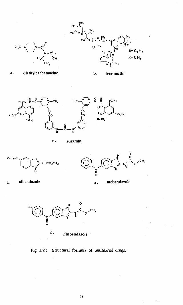
diethylcarbamazine (DEC). The structure of these compounds are shown in Fig. 1.2.
These two drugs are described mainly in this chapter since they are widely used in the -..
treatment of lymphatic filariasis in human. The usage of other antifilarial drugs such
as mebendazole (MBZ), flubendazole (FBZ) and suramin for the treatment of lymphatic
filariasis will be desc~bed briefly since their efficacy studies in human and animal
models are limited (Fig. 1.2).
8

1.3.2. DEC and Ivermectin
Currently five drug regimens are available for the control of lymphatic filariasis.
The administration of"WHO-recommended standard course" of DEC 6mg/kg per day x
12 days for W.bancrofti and x 6 days for B. malayi were found to be effective in
treating lymphatic filarial infections (Ottesen, 1985). However, such regimes are
expensive, require patients compliance and causes possible adverse reactions (usually,
fever, local inflammatory reactions, gastrointestinal symptoms) (Ottesen, 1987) making
them impractical for most control programmes.
The administration of single dose (spaced dose ) of DEC given at weekly,
monthly, 6 monthly, or yearly intervals has been advanced for many years by a number
of public health workers in W. bancrofti endemic areas. (Laigret et a/., 1980). A more
frequent yearly single dose of DEC (about 6 mglkg) regimens are found to be effective
in decreasing microfilarial prevalence and density in both B. malayi and W. bancrofti
infected-communities; Tahiti (Laigret eta/:, -1980);·-- --India-tpanicker eta/., 1991);
Samoa (Kimura eta/., 1985) andFrench polynesia (Cartel et a/., 1992).
The chemical stability of DEC permits its use as a DEC fortified salt in both ...
cooking and other flavouring. The effectiveness of DEC-fortified salt in decreasing the
microfilarial prevalence of both bancroftian and brugian filariasis in large population in
China, Taiwan and India are wel1 reviewed by Gelband (1994). Similar observation
was made in an individual patient in whom prevalence of W. bancrofti microfilaraemia
decreased by 97.8% after 4 months of using DEC- salt and whose corresponding
microfilarial ( mf) density fell even more dramatically greater than 99% (J ingyuan,
1992). However, the effectiveness of this strategy primarily determined by the
9

exclusive availability of salt fortified with DEC as well as its consumption ts
determined by choice within the households.
Ivermectin, a new antifilarial drug has been long employed effectively for
control of onchocerciasis (Aziz et at., 1982; Upp et at., 1986; Albiez et at., 1988).
Recently its effectiveness against microfilariae of both W. bancrofti and B. malayi has
been evaluated in individual patients for a period of 12 - 24 months following drug
administration (Richards eta!., 1991; Addis eta!., 1993; Kazura eta!., 1993; Eberhard
et al., 1992). It is well recognised that a single dose of ivermectin 400 uglkg yields
superior microfilaricidal activity (Richards et al., 1974). At this dose level there were
36- 70% and 86- 99% decrease of mf prevalence and mf densities respectively after
12 months post-treatment.
The combination of single dose of DEC together with ivermectin seems to
offer a more effective drug regimen than either single dose DEC or single dose
ivermectin. The results of using-this--regimerr-mainly-comes-from individual patients
with W. bancrofti infections receiving.this ivermectin!DEC-combination (Richards-et
a!., 1991 ; Addison eta!., 1993 ). Studies comprising a total of 33 patients demonstrated
a decrease in mf prevalence of 45 - 70% and mf density of 96 - 99% after 12 and 24 ..• months of post-treatment. In this study the dose of ivermectin used in the
ivermectin!DEC combination was only 20 Jlg/kg (along with 6 mglkg DEC). In
-addition safety trials have already been successfully carried out to demonstrate the
feasibility of using ivermectin/DEC combination (Navaratnam et al., 1992; Glaziou et
at., 1994)
10

, ~.:
f' 1.3.3 Suramin '
Suramin is a potent filaricide used for the treatment of onchocerciasis
(Hawking, 1978). However, its toxicity and the need for intravenous dosing under
medical supervision for up to six weeks greatly limit its use, (Katzung, 1985). The
antifilarial properties of suramin against human lymphatic parasite have not been
extensively studied, since, DEC and ivermectin were the better choice of drugs in the
treatment of brugian and bancroftian filariasis. However, investigators have reported
the filaricidal activity of suramin against lymphatic parasite in experimentally infected
animal models. Denham and co-workers in their study demonstrated the adultcidal
activity of suramin against B. pahangi in cats at five weeks post-treatment (Denham &
Me Greevy, 1977). On the other hand tertiary screening of potential filaricides, in
Brugia malayi - Presbytis cristata animal model has identified suramin to possess
adultcidal activity (Mak eta/., 1990). Intravenous suramin at 10 mglkg daily x 5 days
or 17mg/k:g weekly x 5 weeks substantially reduced the recovery of live adult worms to
50.6% and 13.6% of control respectively. ~ But no microfilaricidal activity was
accounted. However, the suramin as~ antifilariaLagent for lymphatic filariasis obviously
depends on envisaged preclinical and clinical evaluation, while DEC and ivermectin
still remain as the drugs of choice . . ..
1.3.4 Benzimidazole carbamate ...,..
MBZ and FBZ .. derivatives of benzimidazole carbamate are widely used in the
treatment of onchocerciasis (Domiquez-vazques et al., 1993; Rivas-Alcala et al., 1981
a, b). In the treatment of 0. volvulus infections patients receiving 1 gm of MBZ twice
daily for 28 days had a 50% decrease in their skin microfilariae counts for over 6
months (Domiquez-Vazques eta/., 1983). However, poor oral absorption of MBZ as
11
well as prolonged dosing regimen do not seem to be a suitable regimen for routine
practice. On the other hand, intramuscular (i.m.) injection ofFBZ 750 mg/week for 5
weeks demonstrated a profound reduction of skin microfilariae in patients compared to
patients receiving DEC 200 mg/day for 2 weeks (Rivas-Alcala et a!.. 1981 ). However,
ulceration at the site of injections currently limit the usage of FBZ in the treatment of
onchocerciasis. Both MBZ and FBZ are known well for their limited solubility and
poor absorption from the gastrointestinal (GI) tract (Brugmans et al., 1971 ~ Michiels et
al., 1982), thus making them suitable for intestinal helminth treatment (Keystone &
Murdoch, 1979; Van den Bossche eta!., 1982).
The filaricidal activity of MBZ and FBZ against lymphatic parasite have been
demonstrated both in animal and humans. Denham eta!. (1978) showed the adultcidal
activity ofMBZ against B. pahangi in experimentally infected jirds and cats. A similar
observation was also noted following filaricidal screening of FBZ in B. pahangi
infectedjirds and cats (Denham eta!., 1979). However, no microfilaricidal activity was
observed in FBZ studies above.In clinical trials, the macrofilaricidal action ofMBZ was
reported in few patients suffering from B. malayi and W bancrofti infection. These
patients were treated with large doses of mebendazole 500 mg three times daily for 21
days (WHO 1984). Ho~~ver, this high doses are not recommended because its
absorptions is erratic, it is teratogenic in some animals and it can be toxic in large
doses. A successful therapy for systemic infections disease such as lymphatic filariasis -requires a sufficient quantity of drug to be absorbed to achieve a therapeutic plasma
concentration. In view of this poorly soluble drugs such as MBZ and FBZ do not seem
to offer a viable alternative to current drugs (DEC, ivermectin) in the treatment of this
disease. In accordance with this new benzimidazole carbamate derivatives with better
solubility need to be synthesized. In this thesis the bioavailability of two new
12
benzimidazole carbamate derivatives UMF-078 and UMF-058 have been investigated in
suitable animal models.
1.4 Mode of Action for antifilarial drugs
1.4.1 Introduction
Generally, extensive studies on the mode of action of parasite drugs were done
mainly on anthelmintic agents especially benzimidazole carbamates, which have shown
themselves to be highly effective and safe for the treatment of the majority of intestinal
helminth infections both in human and veterinary medicine (Van Den Bossche et al.,
1982). Knowledge of the mechanism by which anti filarial drugs are absorbed and their
mechanism of action on filarial parasites are vital in the discovery of new drugs and
development of more effective delivery system. However, present studies are mainly
focused on large nematodes such as Ascaris sp (Thompson et al., 1993). This probably
might be due to its wide distribution, its importance in both human and veterinary
medicine as well as the fact that it is large enough to study using a variety of techniques.
Little is known on the mechanism of action of antifilarial drugs on lymphatic
parasites such as B. malayi and W. bancrofti. The studies on the antifilarial mechanism
mode of action of drugs such as DEC, ivermectin, suramin and benzimidazole
carbamate on filarial parasite is still in the early stages. This probably is due in part to -the lack of knowledge regarding these organisms as well as lack of suitable animal
model for the study of these parasites in the laboratory. Indeed parasites material are
also scarce thus limiting the scope of physiological and biochemical studies on these
worms which may reveal promising targets for research. However, the antifilarial
mechanism of these drugs will be discussed briefly herein.
13

1.4.2 Benzimidazole carbamate
The mode of action of benzimidazole carbamates are usually assayed in terms of
its antitubulin activity against microtubulin polymerization in parasite (Lacey, 1990).
Tubulin is the functional subunit of microtubules which participate in several important
cell functions e.g. the transport of materials within cells. Microtubules exist in dynamic
equilibrium with tubulin and are being controlled by a range of endogenous regulatory
proteins and co-factors. The equilibirum can be interrupted both in vivo and in vitro by
exogenous substances known as microtubule inhibitors. Most of the inhibitors exert
their action by binding to tubulin to prevent the self-association of sub-units onto the
growing microtubules. This results in "capping" of the microtubule at the associating
end with a net loss of microtubule length. The disintegration of the microtubule led to
impaired cell functions such as mitosis, secretion and regulation of cell form.
Both MBZ and FBZ induced the disappearance of the cytoplasmic microtubules of
the tegumental or intestinal cells of cestodes and nematodes, which results in blocking
the transport of secretary vessicles(Van Den Bossche, 1976; Van Den Bossche, 1986).
This subsequently lead to impaired coating of the membranes thus decreasing the
digestion and absorptio~ .• of nutrients. This probably might be the basis of MBZ
induced impairment of glucose absorption in nematodes and cestodes both in vivo and
in vitro studies (Van Den Bossche, 1976; Van Den Bossche 1986). The antitubulin
activity was also demonstrated by other congeners ofMBZ such as albendazole (ABZ),
parbendazole, oxibendazole and fenbendazole (Van Den Bossche eta/., 1982). Further
evidence of tubulin dependent benzimidazole action was obtained in charcoal binding
stability study. Colchicine, a microtubule inhibitor forms a tight pseudoirreversible
complex with tubulin that enables the colchicine - tubulin complex to survive
14
extraction with charcoal. In addition, studies with benzimidazole resistant isolates
parasites demonstrated a reduced charcoal-stable [H3] mebendazole binding (Lacey,
1988). This observation further supported tubulin as the site of action of benzimidazole
carbamate. It is also important to note that other postulated mode of action was also
been proposed. McCracken & Stillwell ( 1991) showed that benzimidazole anthelmintic
activity in part may be due to bioenergectic disruptions of natural membrane system
resulting from transmembrane proton discharge. However, it is not yet possible to
determine whether the action of these group of drugs on microtubules or on energy
producing systems represents their primary mode of action. Further study is necessary
to determine the exact mode of action of these chemotherapeutic agents.
1.4.3 Diethylcarbamazine (DEC)
DEC is the oldest and the most effective antifilarial drug used in the treatment
of lymphatic filariasis. However, the exact mechanism of action is still unknown.
Fujimak eta/. (1988) proposed that DEC inhibited the development of Brugia pahangi
larvae cultured in vitro in the presence of feeder cells (LLC-M cells). It was discovered
recently by Fujimaki and co-workers that DEC also inhibits proliferation of LLC-MK2
cells, disrupts the cytop~~mic microtubules complex, inhibits the assembly of
rnicrotubules in vitro and induces the disassembly of the performed microtubules in
vitro (Fujimaki et a/., 1990). They suggested that the inhibition of larvae development
by DEC probably might be due to the antitubulin effect of DEC on feeder cells and
finally these cells lose their supporting function for filarial larvae.
Subsequent study by Fujimaki et al. (1990b) also showed that B. pahangi
larvae exposed to DEC in vitro were retarded in their development in jirds. These
15

result indicate that DEC has a direct action against the infective larvae of B. pahangi.
The other postulated mode of action was that DEC might enhance the host parasite
defence system by increasing the adherence of leukocytes to microfilariae mediated by
antibodies reacting with the surface of the worms (Willy et a!., 1979). Other
investigators suggested that DEC might affect the neuromuscular system and surface
layers of the larvae (Hawking, 1979). Further studies need to be done since knowledge
regarding the physiological and biochemical aspects of the neuromuscular system are
insufficient.
1.4.4. Suramin
The antifilarial action of suramin upon B. pahangi has been investigated in vivo
and in vitro (Howells eta/., 1983). With regard to its polyanioninature, suramin was
found to be bound to the surface of worms in vitro. Subsequent in vivo studies with
jirds fail to demonstrate the ability of suramin to alter the rates of glucose utilization,
uptake rates of glucose, leucine and adenosine. However, ultrastructural changes were
noted in the intestinal epithelium of worms from suramin treated jirds study, thus
suggesting the intestinal epithelium itself might be the site of action of the drug.
Further studies are needed to investigate suramins effect on filarial gut system since the ..•
knowledge regarding the pysiological aspects of the gut of filarial worm are still
lacking.
-
16

1.4.5 Ivermectin
The exact mechanism of action of ivermectin remains unknown but is thought to
involve the activation of y-aminobutyric acid (GABA) pathways in the parasite via an
effect on the GABA receptor-chloride ion channel complex (Vande Wan, 1991).
Ivermectin potentiates the release and binding of GABA at postsynaptic sites on the
neuromuscular junction thus paralysing the nematode worms (Gutaffson et a!., 1987).
However, ivermectin is not filaricidal at 100-200 ug!k:g dose level as recurrence of
microfilaraemia was encountered in polynesians infected with W bancrofti following
ivermectin efficacy study (Cartel et a!., 1993). In addition, recent study in Brazil
showed that high dose of ivermectin ( 400 ug!k:g) has no observable macrofilaricidal
effect on adult W. bancrofti, although it is know to be a potent microfilaricidal agent
(Dreyer eta!., 1995).
. ..
l7
a. diethylcarbamazine b. ivennectin
tbSO, ~-g~CH, H,C~g-n SO,Nl
·~ .. rw-..;;;: NH NH I I I ~ -&
JbO,S CO ;5 , ., SO,,h
1-1150 & NaSO, • r .~
::-... I W ::-... I N-C-1'1 H H
c. suramin
d. albendazole e. mebendazole
f. ¥flnbendazole
Fig 1.2: Structural formula of antifilarial drugs.
18
1.5 The Pharmacokinetic and Metabolism Of Antiftlarial Drugs
1.5.1 Introduction
The primary objective of pharmacokinetic and drug metabolism studies in
development of new drugs is to understand how the drug molecule is handled by the
living animal, which involves the absorption, distribution, metabolism and excretion of
the drug in the living system. Before reaching the stage of clinical evaluation the new
compound, potentially a new drug is usually evaluated in numerous animal models in
order to determine its pharmacological potency, toxicological potential and metabolic
fate. Frequently, several animal species are used in the evaluation. Metabolism studies
conducted in living animals provide the ultimate information regarding the
pharmacokinetic properties and metabolic pathway of the drug molecule which can be
used to correlate or interpret efficacy and toxicity data collected in the same species.
Work in this thesis describe the pharmacokinetic and metabolism of new
benzimidazole carbamate derivatives UW'-078 and UW'-058. In view of this it is
essential to review the pharmacokinetic and metabolism of benzimidazole carbamate
related drugs for our reference to the current status of this compound in this aspect In
addition the metabolic fate of other important antifilarial drugs such as DEC, ... ivermectin and suramin will also be discussed briefly herein. Clinical results were
presented wherever essentiaL However, when human studies are not available, results
of animal experiment from literature were used to described the pharmacokinetic and
metabolism of the drugs.
Benzimidazole carbamates are important broad-spectrum drugs primarily employed in
the treatment of infections of helminth parasite. In human clinical practice only three
19

benzimidazole compounds; MBZ, FBZ and ABZ are currently used. However, their
poor solubility in gastrointestinal tract limited their usage in the treatment of systemic
disease such as lymphatic filariasis. Nevertheless, the antifilarial activity of MBZ
(WHO 1984) as well as FBZ (Denham eta/. 1979) in limited animal and human studies
have been demonstrated. This section will discuss some of the pharmacokinetic and
metabolism aspect ofMBZ, FBZ and ABZ.
1.5.2 Mebendazole
MBZ is poorly absorbed after oral administration due to its poor aqueous
solubility. Following administration of MBZ tablets (1.5 gm) to three fasting
volunteers, plasma levels of MBZ remained below 17 nmol/L in two volunteers and a
peak concentration of 17 nmol/L was noted in the third subject. When the same dose
regimen was employed with a fatty meal, plasma concentration mounted up to 91,112
and 142 nmol!L within 2 to 4 hr in the three treated subjects (Munst eta/., 1980). The
increase in the absorption was probably due to the ability of the fatty meal to aid
dissolution, through solution of the drug in the fatty media, from which it is partitioned
into the aqueous GI contents. Dawson et a/. ( 1985) in their attempt to determine the
absolute bioavailability of MBZ utilised a solution of radio labelled MBZ in dimethyl-..•
sulphoxide (DMSO) (0.25%) for oral and intravenous studies in volunteers. Following
intravenous administration of tracer dose radio labelled MBZ ( 1.18 ug), the average
distribution half life, elimination half-life (T 112) and rate of clearance (Cl) were 0.2 hr,
1.12 hr and 1.163 llmin respectively. After oral administration of the similar solution
the mean T112, Cl and time to achieve the maximum concentration (Tmax) were 0.93 h,
0.846 1/min and 0.42 hr respectively. The bioavailability of MBZ was about 22%.
Comparisons of area under the curve (AUC) data for two MBZ metabolites after
20
administration of the parent drug by each route indicate that absorption of MBZ from
the GI tract at this dose is almost complete. The low bioavailability observed following
oral administration at this subtherapeutic dose level is due to high first pass elimination.
However, at higher dosages the poor bioavailability ofMBZ is due to a combination of
high first pass metabolism and very low solubility of the drug. (Dawson eta/., 1985)
In patients with hydatid disease, oral administration of MBZ 10 mglkg yield a
plasma concentration time curves which differed considerably among subjects. The
T112, Cmax and Tma.x ranged from 2.8 to 9.0 hr, 17.5 to 500 ug!L and 1.5 to 7.3 hr
respectively (Witassek et. a!., 1981). Greater values of half lives were encountered
following oral administration of large doses of MBZ compared to intravenous admin-
istration of MBZ. This difference is probably due to the absorption rate limitation of
MBZ from GI tract This is further evident, when the T 112 (0.92 hr) value of MBZ
following a subtherapeutic oral administration of MBZ solution compares favourably
with the T112 (L 12 hr) value of MBZ after an intravenous treatment as described
previously in this section (Dawson eta!., 1985).
The metabolic tr~Q-Sformation of MBZ in vitro and in vivo has been reported
in literature. The in vitro metabolic pattern ofMBZ in rat, dog and pig liver has been
described by Meuldermans et a!. (1976) (Fig. 1.3). In all the three species studied
hydroxy metabolite (methyl [5-(a-hydroxy-a phenylmethyl)-H benzimidazole-2-yl)
carbamate was the major biotransformation product resulting from reduction of the
ketone of the drug molecule. The major hydroxy metabolite of MBZ was also formed
by the three different subcellular fractions (10,500 g supernatant, 100,500g supernatant,
and microsomal enzyme) of rat, dog and pig liver. The relative activity of both soluble
21

and cellular microsomal enzyme differed for the three species. A much greater part of
MBZ was reduced by the dog enzyme preparations than by the rats or pigs. A species
differences was encountered for a minor amine metabolite of MBZ in the 10,000 g
supernatant fraction and the microsomal fraction of pig liver. This metabolite (2-
amino-1-H benzimidazole-5yl) phenylmethane was produced from carbamate
hydrolysis and was not noted in the incubates with the rat or dog liver enzyme
preparations. The carbamate hydrolysis of MBZ was inhibited significantly by SKF-
525A indicating the involvement of the microsomal mixed function oxidation enzyme
system. Conversely the formation of hydroxy metabolite of MBZ was only slightly
influenced in the presence of SKF-525A inhibitor. From the overall study they
conclude the ketone reduction ofMBZ was to be the most important in vitro metabolic
pathway ofMBZ in dog, rat and pig liver preparations (Meuldermans eta/., 1976).
Allan eta/. (1982) successfully identified three biliary metabolites ofMBZ in
the rats after intravenous administration of a mixture of MBZ and
pentadeuteromebendazole. These metabolites are methyl-5(6)-(a-hydroxybenzyl)-2 -
benzimidazole carbamate, 2-amino-5(6)-a-hydroxybenzyl benzimidazole and 2-amino
5(6)-benzoylbenzimidazole which were obtained after enzymic conjugate hydrolysis . . .. They conclude that the major route of metabolism for MBZ was carbonyl or ketone
reduction followed by conjugation to form glucoronides and sulphates. Conjugation
~
appeared to be extensive since only unmetabolized MBZ could be detected in bile
extracts taken before enzyme hydrolysis. Subsequent intravenous metabolic study in
rats using eH]-MBZ and [ 14C]- MBZ further ascertain the ketone reduction ofMBZ to
be the more important metabolic route in MBZ biotransformation (Allan et al., 1983).
The carbamate hydrolysis remain as a trivial pathway in MBZ metabolism in rats. The
22
major hydroxy MBZ accounted for about 77% of the the total recovered and 99% of it
was the conjugate metabolite (Allan et al., 1983 ).
1.5.3. Albendazole
There is little data available on the pharmacokinetics of ABZ in man because it
1s largely undetectable in human plasma due to its low GI absorption and rapid
metabolism (Penicaut eta/., 1983; Marriner eta/., 1986). When ABZ is taken orally
either as tablets or a 2% suspension ( 400 rng) the plasma concentration of active
metabolite ABZ sulfoxide peaked in the range of 0.22 to 0.25 mg/L at 2.0 to 3.0 hours
post dose. The elimination T 112 ofthis metabolite was 8.5 hours (Penicaut et al., 1983).
Marriner et a/. (1986) reported an inconsistent increase in ABZ sulphoxide
concentration when ABZ was administered with oil and milk. Lange et a/. ( 1988) in
their study demonstrated a significant increase of ABZ sulphoxide concentration in
patients with echinococcosis when ABZ was given with a fatty breakfast (42.5%
relative to fasting patients).
ABZ undergoes extensive metabolism. The metabolic pathway noted in man
(Penicaut eta/., 1983), cattle, sheep, rats and mice (Gyurik eta/., 1981) was similar; the ..• hydrolysis of the carbamate moiety and oxidation of the sulphur atom, alkyl side chain
and aromatic ring (Fig. 1.4). Sulphoxide and sulphone are the major metabolites of _.
ABZ biotransformation in the Urine and plasma of all species and the proportion varies
considerably among species (Delatour eta/., 1991; Lanusse eta/., 1992). However, the
parent compound was detected in minor amount in the urine in all species (Gyurik et
al., 1981). Microsomal incubation studies of ABZ demonstrated the formation of
sulfoxide metabolite. The formation was mediated by cytochrome P-450 and/or FAD
23

containing monooxygenase depending on the system used; rat (Fargetton et a/., 1986),
sheep (Galtier et al .. 1986), pig (Souhaili-El Amri et a/., 1987) or human (Rolin et a!.,
1989) liver microsomes. Unlike ABZ sulfoxidation, the oxidation to ABZ sulfone has
been exclusively related to cytochrome P-450 in perfused rat liver preparation. ABZ is
also known to induce its own metabolism (Souhail-El Amri et al., 1988) .
. ..
24