by: zewdu seyoum(dvm, msc, mtah)
TRANSCRIPT
Myiasis causing fliesBy:
Zewdu Seyoum(DVM, MSc, MTAH)

Learning objectives
At the end of this lesson students will be able to:
explain what mean by myiasis
identify those flies/larvae that cause myiasis
explain their pathogenic and economic significances
design treatment, control and prevention techniques of
myiasis

Myiasis
Is the infestation of living organs and tissues of host animals
by the larvae of dipteran flies, usually known as maggots or
grubs
The fly larvae feed directly upon the host’s living or The fly larvae feed directly upon the host’s living or
dead/necrotic tissue or, in the case of intestinal myiasis, on
the host’s ingested food for at least a period
Host: mammals, birds, amphibians or reptiles
Myiasis
Myiasis: could be classified into different groups based on:
Anatomical localization/site of infestation: in or on the
animal
Host-parasite relationship: biological interest
• Based on anatomical/site of infestation, it could be:
cutaneous/dermal or sub-dermal(example: Lucilia)
nasal: e.g., Oestrus in nasal
somatic: e.g., Hypoderma

Myiasis• Based on host-parasite relationship, it could be:
Facultative/optional: e.g., Calliphorids
Obligatory/strict parasite: e.g., Oestridis
Accidental: e.g., Muscids- Musca group
• Accidental or Pseudomyiasis:• Accidental or Pseudomyiasis:
– invade inappropriate host by chance
– insects in the family Muscidae
– Larval stage is the problem stage
– enteric, accidental, rectal, and urinary

Types of Myiasis
• Facultative:
– Can develop in both living and dead organic matter
– maggots are free-living, but can become parasitic
– attack carcasses, may attack living host– attack carcasses, may attack living host
• Obligatory: must have living host to complete their
development
– maggots live on a live host for part of their life
– are always parasitic; unable to survive without living host

Life cycle of myiasis producing flies
• Complex metamorphosis with egg, larval stages (instars),
pupa, and adults;
• Separate sexes: with adult females laying eggs or larvae on
host or in environment;host or in environment;
• Larvae hatch from eggs; three larval stages (maggots);
• Third larval stage pupates with adults emerging from pupae.

Myiasis Producing Flies
• Three families of flies are involved in myiasis occurrence:
1. Calliphoridae
2. Oestridae
3. Sarcophagidae

Family of Oestridae
• Contains flies commonly known as bots and warble
• Are obligate parasites
• most show a high degree of host specificity
• Adult flies: are large, usually hairy, have primitive non-• Adult flies: are large, usually hairy, have primitive non-
functional mouthparts and short lived
• Larvae: highly host and site specific, and obligatory parasites
spend considerable time: in feeding and developing in
their host
Family of Oestridae
• develop in the nasopharyngeal cavities or skin
boils(warbles) of mammals
• include four important genera:
Hypoderma Hypoderma
Oestrus
Gastrophilus
Dermatobia

Hypoderma: warble flies/cattle grubs
• Larvae live as subcutaneous parasites; are relatively host-
specific
• Host: primarily cattle and sometimes equines, sheep, man
• Species: Hypoderma bovis and H. lineatum: affect cattle• Species: Hypoderma bovis and H. lineatum: affect cattle
• Life cycle: egg maggot ( L1 L2 L3) pupa(on leaves)
adult: complete metamorphosis
• Distribution: WW
Morphology
1. Adults: H. bovis and H. lineatum
• Body: is covered with dense yellow-orange hairs with a
broad band of black hairs around the middle
• Mouthparts: non-functional or vestigial• Mouthparts: non-functional or vestigial

Adult Hypoderma fly or warble fly

Morphology
2. Larvae: mature/third stage larvae:
are thick and somewhat barrel shaped, tapering
anteriorly
have hooks and posterior spiracle plate that completely have hooks and posterior spiracle plate that completely
surrounded by small spines
Segmented: most segments bear short spines
Colour: dirty white when newly emerged from host; then
turns to dark brown

Larvae of warble fly in skin section

Newly emerged larvae

Life cycle
• Adult females: deposit their eggs on hairs of the lower
regions of the legs and lower parts of the body;
• Eggs hatch and release L1 then the larvae crawl down the
hairs and enter into skin or hair follicles then migrate hairs and enter into skin or hair follicles then migrate
through connective tissue towards the region of the
diaphragm by the aid of a pair of mouth hooks and secretion
of proteolytic enzymes ;
Life cycle
• The L1 feed as they travel to the resting sites: wall of
oesophagus and epidural fat in the spinal canal here L1
moult in to L2 L2 then migrate to underneath of skin of the
back here L2 moult to L3, which cause swellings(‘warbles’) back here L2 moult to L3, which cause swellings(‘warbles’)
of skin L3 then make cutaneous opening for breathing
activities by inserting its spiracles into opening after 4-6
weeks L3 emerge and fall to the ground then the larvae
pupate adults emerge

Life cycle of Hypoderma species

Pathogenic significances
• Economic loss: down-grading values and condemnation of
hides perforated by larvae, reduction of milk yield and
weight gain
• Migratory behavior of larvae via tissue causes yellow or • Migratory behavior of larvae via tissue causes yellow or
greenish, gelatinous, oedematous areas due to eosinophils
infiltration: known as ‘butchers jelly’
• Reduced carcass value: due to tissue inflammation

Pathogenic significances
• Hypersensitivity reaction: may cause anaphylactic shock
particularly if the larvae die in the animal tissue
• Egg laying activity of the fly produces self buzzing noise:
results in avoidance behavior of the host known as
‘gadding’ or panic or running away aimlessly to escape

Pathogenic significances
• Gadding behavior: resulting in self-inflicted wounding: since
animals injure themselves on posts, barbed wire and other
obstacles
• Flies noisy behavior: also results in animal feeding • Flies noisy behavior: also results in animal feeding
interruption: results in spontaneous abortion, retarded
growth, reduced milk yield and reduced weight gains

Clinical signs and diagnosis
• Signs: nodular lesion on back area; running away aimlessly
• Diagnosis: clinical signs: larvae in cysts or lumps under skin
of the back indicate warble infestation; eggs may be found on of the back indicate warble infestation; eggs may be found on
hair of the animal’s legs

Nodules on the back due to Hypoderma larvae

Treatment, Control and Prevention
• Mechanical removal of 3rd stage larvae: mature larvae may
be squeezed out of the warble swelling
• Insecticide treatment: Use active organophosphorous and
ivermectin systemicallyivermectin systemically
• Local fly eradication: SIT, chemicals
• Use vaccine
• Integrated management: SIT, insecticide,
Oestrus ovis
• Commonly known as ‘nasal bots’;
• Larvae: are obligate parasites; spend their parasitic period
in the air passage of hosts: nasal passages, frontal and nasal
sinuses;sinuses;
• Have high degree of host and site specificity;
• have posterior spiracular plate with numerous small pores
• Adult flies: have non-functional mouthparts, and are
viviparous (lay larvae: L1 )

Oestrus ovis
• Host: sheep, goat, rarely human beings
• Site: nasal passages and nasal sinuses
• Species: Oestrus ovis: have veterinary significance
• Distribution: WW

Morphology
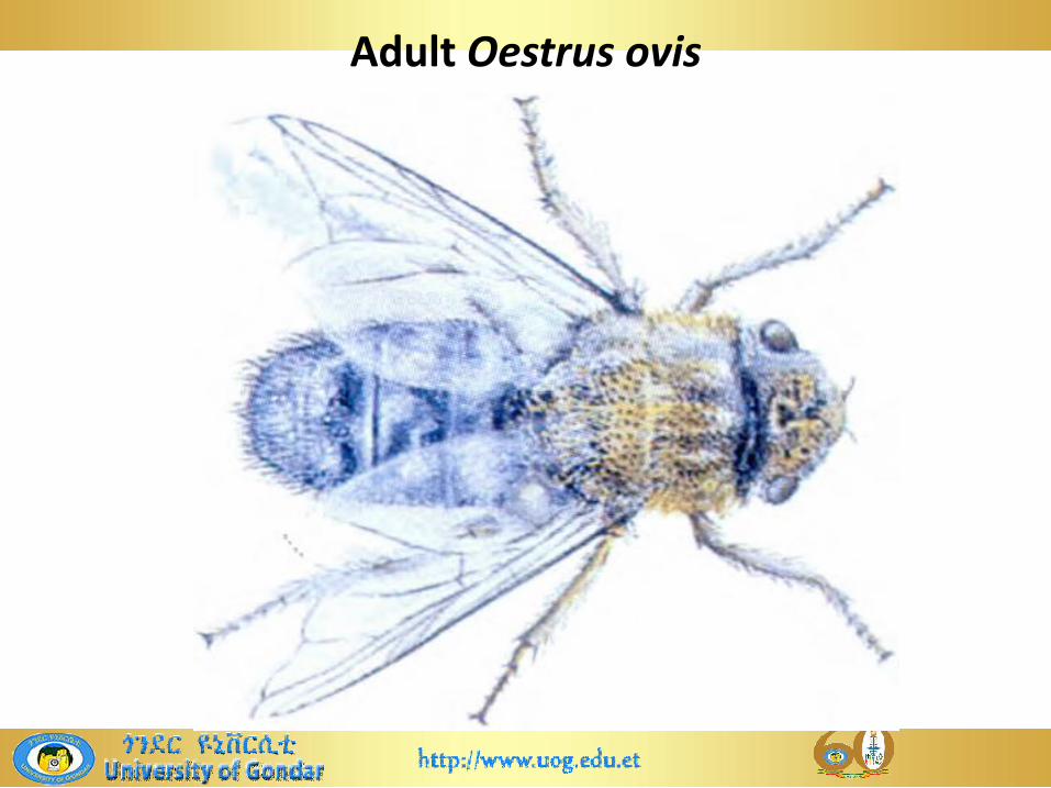
1. Adults flies:
Body: greyish-brown, with many small black spots on
the thorax and abdomenthe thorax and abdomen
Head: broad, with small eyes
Mouthparts: reduced to small knobs: nonfunctional

Morphology
2. Mature larvae: L3
Found in nasal passages
Colour: yellow-white, tapering anteriorly
Segmented: each segment has a dark transverse band Segmented: each segment has a dark transverse band
dorsally
Ventral surface of each segment bears a row of small
spines
Adult Oestrus ovis

Oestrus ovis: larva in the sinus

Oestrus ovis larvae

Larvae that penetrate into the gingival sulcus
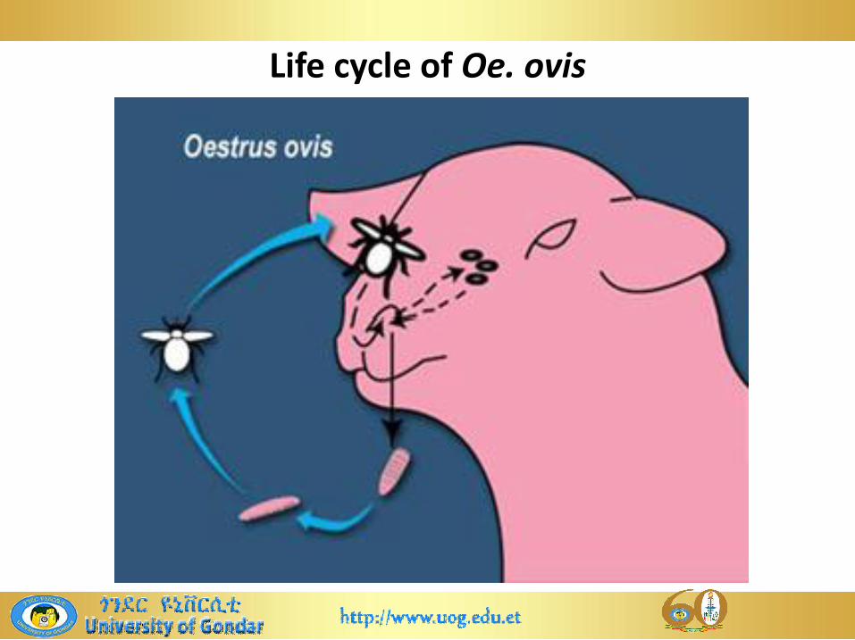
Life cycle
Undergo complete metamorphosis
Female flies are viviparous, depositing/squirting a jet of liquid
containing first-stage larvae at the nostrils at a time in or on
the nostrils of the host, during the hottest part of the day the nostrils of the host, during the hottest part of the day
when flies fly
Then the larvae crawl up the nasal cavity to nasal sinuses and
attach to the mucous membranes with its hooks; feeding on
mucus: this secretion is stimulated by their movement

Live cycle
• Then the larvae enter the frontal sinuses where they
develop into 2nd and dark brown third-stage larvae
• Subsequently, the third-stage larvae crawl out of the nostrils
or are sneezed out by the host; pupate in the ground; adultsor are sneezed out by the host; pupate in the ground; adults
emerge 3–6 weeks later
Life cycle of Oe. ovis

Pathogenic significances
• Adults flies: egg laying activity can be lead to considerable
disturbance and panic in a flock
• As adult flies deposit larvae: the activity of adult flies may
annoy or panic sheep: stamp their feet, bunch together and
press their nostrils into each others’ fleeces and against the
ground: leading to a loss of grazing time, reduced weight
gain, loss of condition
• Most infections are light: sheep showing nasal discharge,
sneezing. and rubbing their noses on fixed objects

Pathogenic significances
• Heavy infections: unthriftiness and sheep may circle and
show in coordination: often termed as ‘false gid’
• Dead larvae in the sinuses: allergic and inflammatory
responses: sneezing and 2⁰ry bacterial infection: cerebral
involvementinvolvement
• Larvae cause irritation and excessive secretion: oral hooks
and spines effect
• Larvae cause purulent rhinitis or sinusitis leading to head
shaking, restlessness, snorting or sneezing

Clinical signs
• mild discomfort, nasal discharge, sneezing, nose rubbing or
head shaking, circling, in coordination, head pressing
• Decreased appetite, restlessness, weight loss, fly worry
• Impaired respiration: by larvae and the thickening of the • Impaired respiration: by larvae and the thickening of the
nasal mucosa
• Larvae may penetrate nasal plate and subsequently enter
the brain: causing injury, ataxia, circling/stagger and head
pressing

Nasal discharge due to Oe. ovis infestation
Diagnosis
• Clinical signs: sneezing and nasal discharges; CNS disorders:
restlessness, false gid, head shaking; season, keeping their
muzzle near to the ground
• Observation: of dark brown larvae dropping out of nostrils • Observation: of dark brown larvae dropping out of nostrils
after severe sneezing attack
• Postmortem examination: by sawing skull and separate it in
to half part longitudinally, then rinsing key areas with water:
examine the water for larvae with lens
Management
• In heavy infection: use nitroxynil, tricleabendazole,
rafoxanide and ivermectin are highly effective as are the
organophosphates, trichlorfon and dichlorvos
• Strategic treatment: at the beginning of summer to kill
newly deposited larvae and midwinter to kill overwintering
larvae
• Fly repellant as prophylaxis

Gasterophilus: bot flies
• Learning objectives: at the end of this lesson students will be able:
illustrate their morphology
identify and distinguish them using their morphology
describe their life cycle and factors that influence breeding
and survival of them
explain their pathological effect and economic significance
design their control and prevention mechanisms

General features
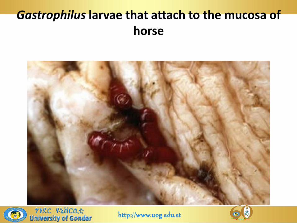
• Are commonly referred to as ‘bot flies’: horse stomach
bot
• Are obligate parasites of horses, donkeys, mules,
zebras, elephants and rhinoceroseszebras, elephants and rhinoceroses
• Larvae, termed ‘bots’: spend most of their time
developing in the stomach of equines, but they are
generally considered of little pathogenic significance
General features
• Host: horses, donkeys, mules, zebras, elephants and
rhinoceroses
• Major species: G. intestinalis, G. nasalis, G. haemorrhoidalis
• Distribution: WW

Morphology
• Adult bot flies:
resemble to honey bee with a long curved ovipositor
carried beanth the abdomen
have non functional mouthparts
are robust dark/brown flies; about 1-2cm long
Body: is densely covered with yellowish hairs
Wings: in most common species, there are transverse
bands or dark patches; have no cross-venation

Gasterophilus species: adult fly

Adult flies of Gastrophilus

Morphology
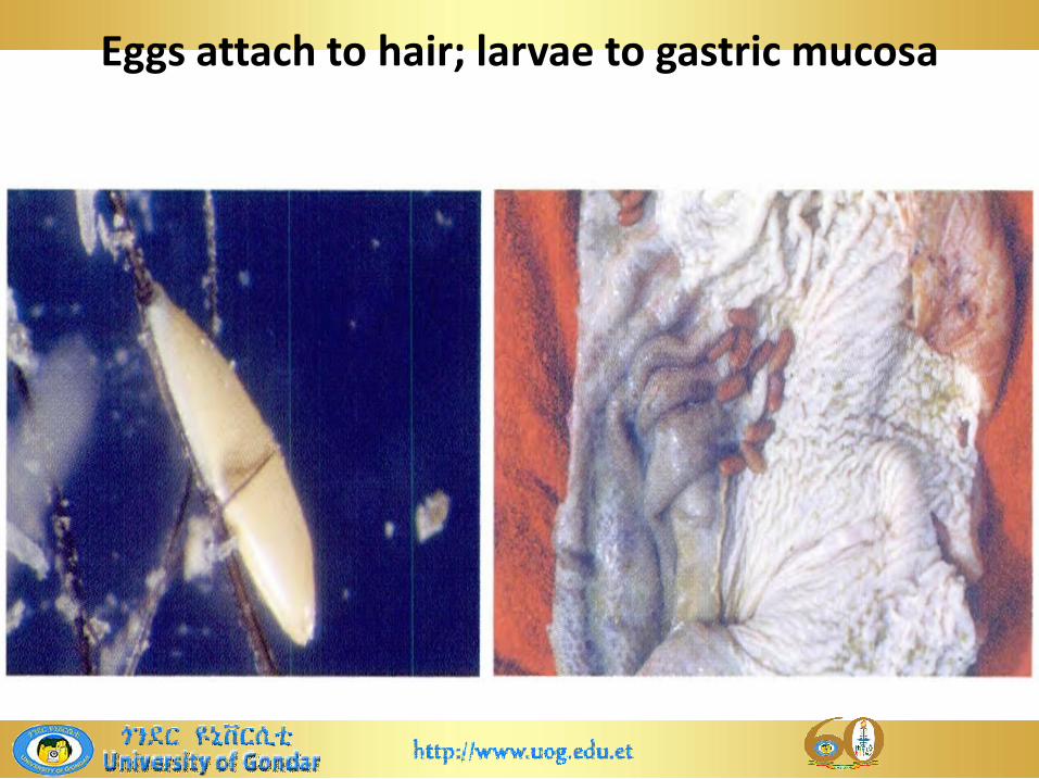
• Mature /third stage larvae:
present in the stomach or passed in faeces :
are cylindrical, 1.6-2cm long and feed on tissue not
on bloodon blood
reddish-orange with posterior spiracles
segmented: bears spines

Larvae of Gastrophilus
Life cycle
• Undergo complete metamorphosis
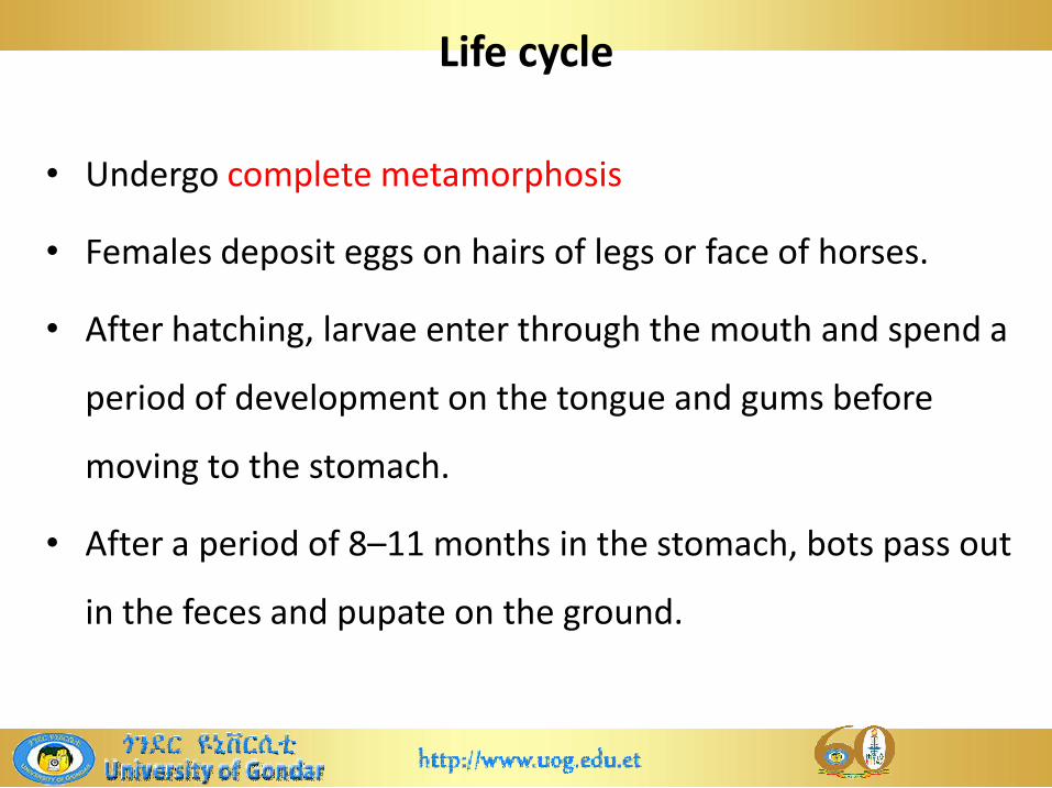
• Females deposit eggs on hairs of legs or face of horses.
• After hatching, larvae enter through the mouth and spend a
period of development on the tongue and gums before period of development on the tongue and gums before
moving to the stomach.
• After a period of 8–11 months in the stomach, bots pass out
in the feces and pupate on the ground.

Eggs that glued on the hairs of horse

Horse bot eggs deposited on the skin of the legs and chest
Eggs attach to hair; larvae to gastric mucosa
Gastrophilus larvae that attach to the mucosa of horse
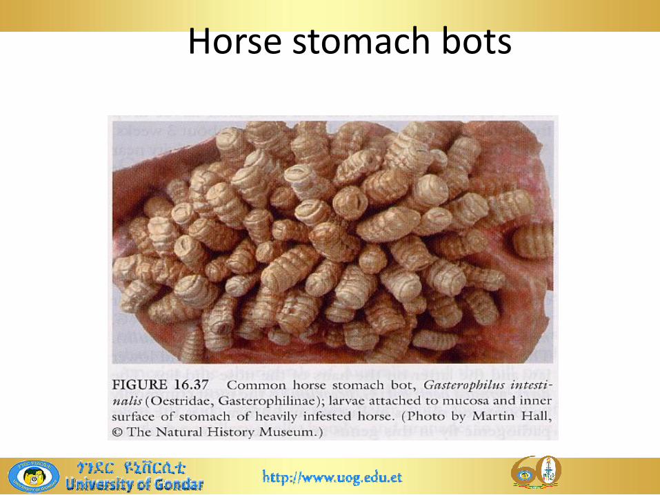
Horse stomach bots

Life cycle of horse bot fly

Pathogenic significances
• Induce great annoyance or panic to horses when adults
approach horses to lay their eggs
• Bots(larvae): may cause obstruction to the food passing
from the stomach to intestine,
• Irritation by the larvae during migration: dermatitis,
inflammation of the pharynx, oesophagus, stomach or
rectum(rectal prolapse)

Pathogenic significances
• Larvae penetration with its hooks at the site of attachment:
may result in stomatitis with ulceration of the tongue,
erosions, ulcers, nodular mucosal proliferation, stomach
perforation, gastric abscesses, peritonitis: inflammatory perforation, gastric abscesses, peritonitis: inflammatory
reactions
• Heavy infections: can cause general debilitation and even
rectal prolapse
• Light infections are tolerable well

Clinical signs and diagnosis
• Signs: mild gastritis, stomatitis, pain on eating , annoyance
• Diagnosis: difficult, so it could be made by demonstration of
larvae in the faeces; observation of the cream-white bot larvae in the faeces; observation of the cream-white bot
eggs on the animal’s hair

Management
• Insecticide application: carbon disulphide and trichlorfon;
the broad spectrum insecticide/anthelmintics
• Dichlorvos and ivermectin are also very effective against
botsbots
• Fly repellant and frequent grooming
• Integrated management: stimulate the eggs on the hair coat
with warm water containing insecticides during summer and
autumn; strategic treatment

Family Calliphoridae
• Are medium to large flies
• Almost all have a metallic-blue or green sheen
• Majority of the species are saprophages: living in decaying
organic materialorganic material
• Important genera that cause myiasis under this family
include:
Cochliomyia, Lucilia and Cordylobia

Life cycle
• All are oviparous and except Cordylobia species, eggs are
laid in wounded, infected or faecally soiled skin of warm
blooded vertebrate hosts
• Larvae pass through three instars while feeding on the host • Larvae pass through three instars while feeding on the host
tissues,
• causes cutaneous or traumatic myiasis
• Mature larvae drop on the ground and pupate in
substrate adult emerge

Cochliomyia: Screw worm
• Are green to violet /bluish-green blowflies with three
prominent black, longitudinal stripes on the thorax, short
palps and orange-brown eyes
• Are obligate ectoparasite• Are obligate ectoparasite
• Infest almost all warm blooded livestock, wildlife and
humans
• Important species: C. hominivorax and C. macellaria
Cochliomyia: Screw worm
• Host: wild and domestic animals and humans;
• occur primarily in tropical areas;
• lay their eggs on wounds;
Larvae characteristically feed as a colony and penetrate• Larvae characteristically feed as a colony and penetrate
the tissues; creating a large and foul-smelling lesion.

Morphology
• Adult fly:
Colour: deep greenish-blue metallic with a yellow, orange
or reddish face and three dark stripes on the dorsal
surface of thorax between the wings
• Larvae:
tapered
segmented: swollen ridges encircle each segments: hence
the larvae look somewhat like screws

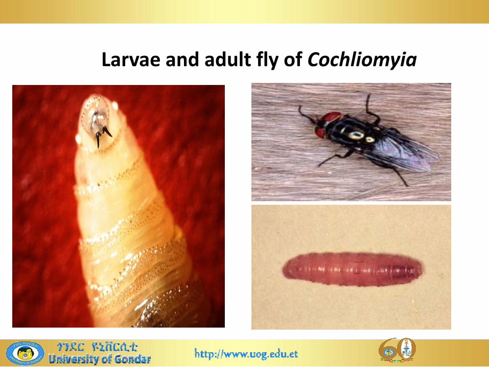
Larvae and adult fly of Cochliomyia

Larvae of Screw worm
Life cycle
• Undergo complete metamorphosis
• Female flies lay eggs on the skin of the host near open
wounds: scratches, sores, barbed wire cuts, scabs, injuries
from brand marks, dehorning/castration
• Eggs then hatched and release larvae/maggots
• Larvae start to feed invasively on secretions and living tissue
• Following completion of development, larvae fall to the
ground and pupate in soil and adult emerge after 7-12 days

Pathogenic effects
• If untreated, repeated infestations may quickly lead to the
death of the host
• Economic loss: use of insecticide, damage and disfigurement
of skinof skin
• Putrefied smelling discharges and ulcerations
• Treatment should be immediate
• Irrigate infested areas with ethanol + veggie oil
• May require surgery

Clinical signs
• Ragged, foul-smelling lesion containing larvae (maggots)
• Constant licking of the lesion by the animal
• Secondary infections and strikes are common
• Fever
• Lethargy and loss of appetite • Lethargy and loss of appetite
• Debilitation
• Decreased growth rate
• Mortality rate in newborn calves from navel strike may be as
high as 30-50%

Management
• Use insecticide: spraying or dipping livestock with
coumaphous against the larvae
• Ivermectin
• Eradication: SIT

Blow flies: Lucilia
• Cause cutaneous myiasis and are scavenger on dead
animals, decaying vegetable matter or garbage
• Important species: L. sericata and L. cuprina
• Are facultative ectoparasites: strike living wild and farm • Are facultative ectoparasites: strike living wild and farm
animals(sheep) that have sores or wounds or are soiled
with manure
• Larvae infest and feed on living tissues of warm
blooded vertebrates: sheep
Blow flies: Lucilia
• Infestation by these species known as blowfly strike
• Host: mainly sheep( mainly diarrheic sheep), sometimes
wild and domestic animals and humanswild and domestic animals and humans
• Distribution: WW

Morphology
• Adults: - up to 1cm in length; have black legs
- all have a metallic blue or green sheen on the body
• Larvae:
Are white to yellowish, smooth, segmented and blunt at Are white to yellowish, smooth, segmented and blunt at
the rear and tapers towards the head
possess a pair of black oral hooks for tearing flesh,
spiracles on the anterior segment, and stigmatic plates
also bearing spiracles

Colour of adult blow flies
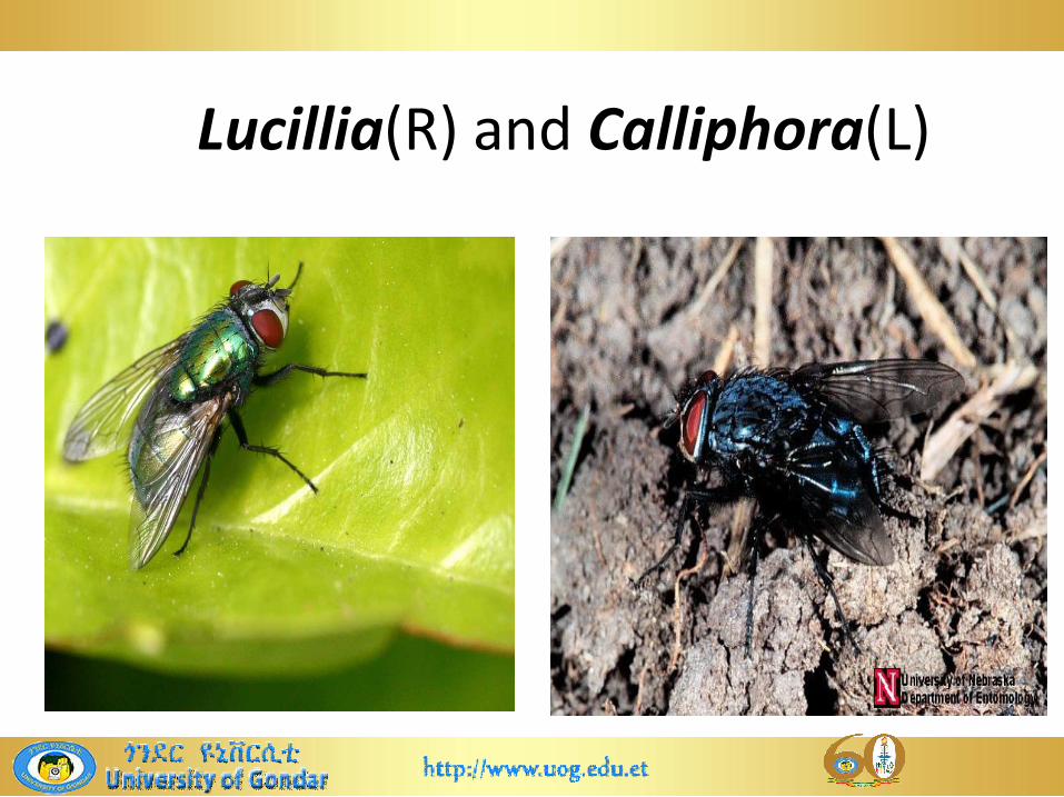
Lucillia(R) and Calliphora(L)
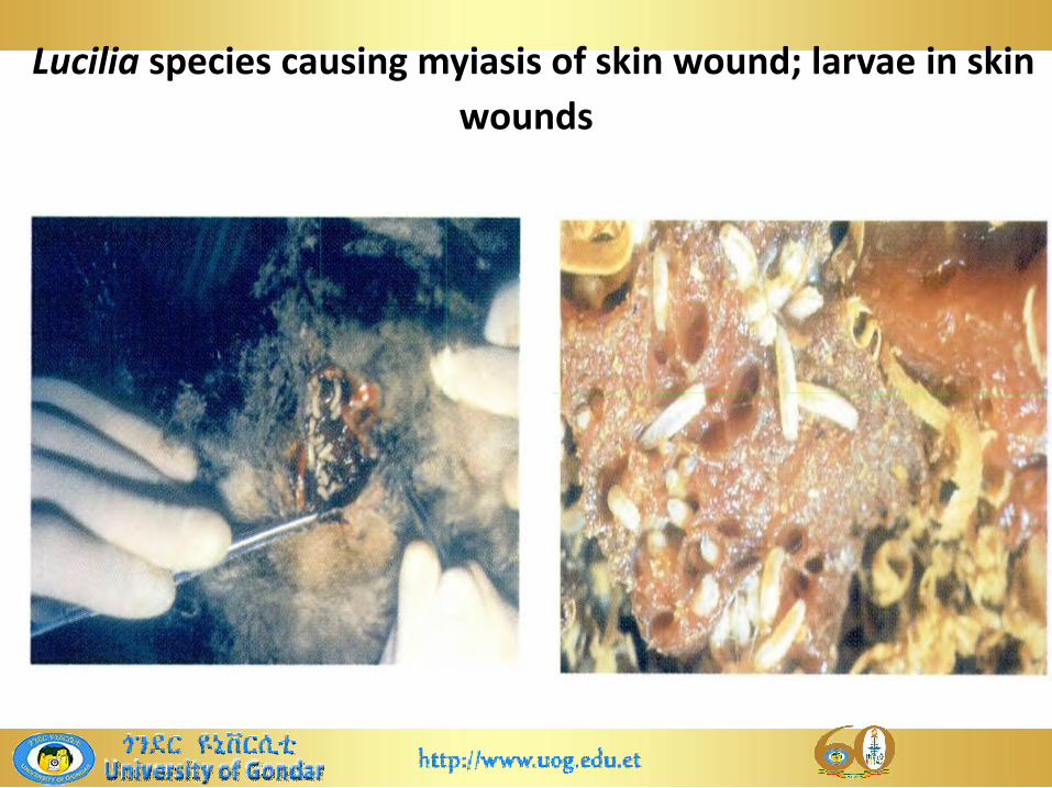
Lucilia species causing myiasis of skin wound; larvae in skin
wounds
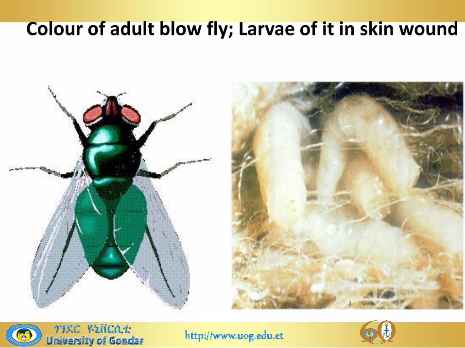
Colour of adult blow fly; Larvae of it in skin wound

Larvae of blow flies
Life cycle
• Undergo complete metamorphosis
• Gravid female flies lay eggs on wounds, soiled fleece or dead
animals, being attracted by the odour of the decomposing
matter
• Then the eggs hatch and release larvae
• The larvae feed on necrotic tissue, grow rapidly and moult
twice to become fully mature maggots in 3-10days then
drop to the ground and pupate adults emerge in 3-7days

Epidemiology
• Occurrence of cutaneous myiasis in sheep depends on:
temperature; flock size; stock density, rainfall
host susceptibility: if the host has got putrefactive odours
on the fleece due bacterial decomposition of organic
matter. It can be caused by soiling of the hindquarters due matter. It can be caused by soiling of the hindquarters due
to urine or diarrhoea and injuries due to shearing, fighting
or barbed wire
Breed type: example Merino sheep
sex: ram with narrow penile sheath

Pathogenic significances
• Larvae lacerate skin with their oral hooks and proteolytic
enzymes, which digest and liquefy skin tissues
• Secondary blow fly strikes: can occur and are strongly
associated with faecal soiling, bacterial dermatophilosis and
bacterial fleece rot(superficial dermatitis)bacterial fleece rot(superficial dermatitis)
• Secondary bacterial infection can happen
• Extreme irritation and distress can happen when larvae
crawl over animals’ skin or into wounds: extremely
debilitating and sheep can rapidly lose condition
Clinical signs and diagnosis
• Increase body temperature and respiratory rate; weight
loss and anorexia
• Anaemia and suffer toxaemia; inflammatory reaction
• Loss of fertility • Loss of fertility
• If untreated death
• Diagnosis: based on the clinical signs and recognition
of maggots in the lesion

Management
Good management: separate infected animals; avoid/clip
hairs that surrounding the lesion; infested animals should be
treated promptly
Use suitable insecticides
Control: prophylactic insecticide; sanitation or proper
disposal of organic matters; worm management and wound disposal of organic matters; worm management and wound
management;
Buried dead animals and other organic matter since blow
flies can be attracted to dead animals
Avoid injury during shearing, performing docking and
castration
Sarcophagidae: flesh-flies
• Read morphology, lifecycle, pathogenic effects, management