c h. 18 t he k nee. o bjectives describe the functions of the knee describe the ligament structure...
TRANSCRIPT

CH. 18 THE KNEE

OBJECTIVES
Describe the functions of the knee Describe the ligament structure of the knee Explain the function of the patellofemoral
joint List and define various sports-related injuries
of the knee
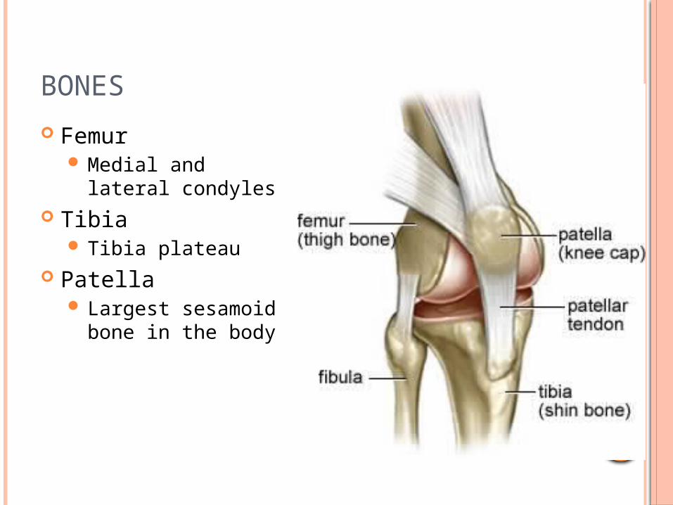
BONES
Femur Medial and lateral
condyles Tibia
Tibia plateau Patella
Largest sesamoid bone in the body

JOINTS
Tibiofemoral joint is a hinge joint composed of a joint capsule and several ligaments Main motions are flexion and extension
Patellofemoral joint is the articulation between the patella and femur Gives greater mechanical advantage in knee
extension Increases quadriceps force by 33% to 50%

CARTILAGE
Articular cartilage covers ends of both tibia and femur
Menisci are located between femoral condyle and tibial plateau
Shock absorption, force distribution, improve stability
Synovial membrane and fluid
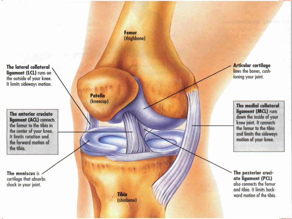

COLLATERAL LIGAMENTS
Medial collateral ligament (MCL) Attaches femur to
tibia Lateral collateral
ligament (LCL) Attaches femur to
fibula Provides medial and
lateral stability of the knee

CRUCIATE LIGAMENTS
‘Cruciate’ means cross
Anterior cruciate ligament (ACL) restricts anterior
translation of tibia Posterior cruciate
ligament (PCL) restricts posterior
translation of tibia Both control rotation
and help with stability

MUSCLES
Quadriceps Vastus medialis Vastus lateralis Vastus intermedius Rectus femoris
Powerful knee extensor
Forms patella tendon which attaches to tibia at tibial tubercle

MUSCLES
Hamstrings Biceps femoris Semimembranosus Semitendinosus
Activated during knee flexion
Semitendinosus tendon used during ACL repair

MUSCLES
Pes Anserine-assist with knee flexion Sartorius Gracilis Semitendinosus
Sartorius is the longest muscle in the body