c report weine’s 3 in cvek’s 4 using biodentine: a case...
TRANSCRIPT
Khursheed O et al. Weine’s 3 in Cvek’s 4 using Biodentine.
180
Journal of Advanced Medical and Dental Sciences Research |Vol. 2|Issue 3| July-September 2014
Case Report
Weine’s 3 in Cvek’s 4 using Biodentine: A Case Report Obaid Khursheed1, Sonal Gupta1, Irfana Khursheed2, Ruchika Bansal3, Tajinder Bansal4
Departments of 1Pedodontics, K.D.Dental college and Hospital, Mathura, 2GDC Srinagar, 3Conservative & Endodontics, 4Oral Medicine& Radiology Swami Devi Dyal Dental College and Hospital. Corresponding Author:
Dr. Ruchika Bansal,
Department of Conservative &
Endodontics,
Swami Devi Dyal Dental College
& Hospital, Barwala, Haryana,
India. Pin Code 134118
E mail: [email protected]
Received: 27-06-2014
Revised: 29-07-2014
Accepted: 12-08-2014
This article can be cited as: Khursheed O, Gupta S, Khursheed I, Bansal R, BansaL T. Weine’s 3 in Cvek’s 4 using Biodentine: A Case Report. J Adv Med Dent Scie Res 2014;2(3):180-184.
Introduction The major challenges associated with endodontic treatment of teeth with open apices are achieving complete debridement, canal disinfection and optimal sealing of the root canal system.1 Despite the clinical success of the calcium hydroxide apexification technique, it has many disadvantages. The treatment requires high patient compliance, multiple appointments extending over a long period of time and relatively high pH of the material that compromise the strength of the root dentin.2 An alternative to this multi-appointment
apexification procedure has been a single-step apical barrier technique. The rationale is to establish an apical stop that would enable root canal to be filled immediately. There is no attempt at root end closure, rather an artificial apical stop is created.3 Several materials have been proposed for use as an apical barrier, recently, MTA (ProRoot, Dentsply Tulsa Dental, Tulsa, OK), has been advocated as an apexification material by Shabahang et al 4 because it permits an adequate seal of the canal and was found to have less cytotoxic effects and better results, giving it more clinical success over
Abstract: The immature tooth with apical periodontitis presents numerous challenges. Several procedures utilizing different materials have been recommended to induce the root end barrier formation. Conventional treatment with calcium hydroxide for such cases is associated with certain difficulties, such as they take very long treatment time, possibility of tooth fracture owing to its highly alkaline pH & incomplete calcification of the bridge. Since the mid 1990s, use of Mineral Trioxide Aggregate (MTA) apical plug appears to be a promising alternative due to its high biocompatibility, superior sealing ability & reduced treatment time. Biodentine is a cement of the same class as MTA, new novel dentin substitute material that septodont claim is set to revolutionise the world of dentistry. The purpose of this article is to report the flow characteristic of this novel dentin substitute and the clinical procedure for using biodentine in teeth with open apices, successfully treated with biodentine apical plugs. Key-words: Immature teeth, single visit apexification, Biodentine, apical barrier, Weine’s 3.
Khursheed O et al. Weine’s 3 in Cvek’s 4 u
Journal of Advanced Medical and Dental Sciences Research
traditional root-end fillNevertheless, MTA has some drawbacks:setting time is long, difficult at times, compressive anstrengths are much lower than those of dentin, highly alkaline pH and it is quite costly. Biodentine, in contrast, offers similar properties to those of MTA minus the prolonged setting time and pH. It is available as powder in a liquid in a pipette form. The powder mainly contains tricalcium silicate, calcium carbonate, and dicalcium silicate, the principal components of MTA. Zirconium oxide serves as the radiopacifier. The liquid consists of calcium chloride in aqueousolution with an admixture of polycarboxylate. The powder is mixed with the liquid in a capsule in a triturator for 30 seconds, can be spatulated manuallyonce mixed, its setting time is close tominutes. Because of its material properties, biodentine was considered to be an interesting alternative to conventional rootend filling materials that would benefit from improved radiopacity, reduced setting time and alkalinity.7 This article presents a caseopen apices were managedapical barrier technique using biodentine
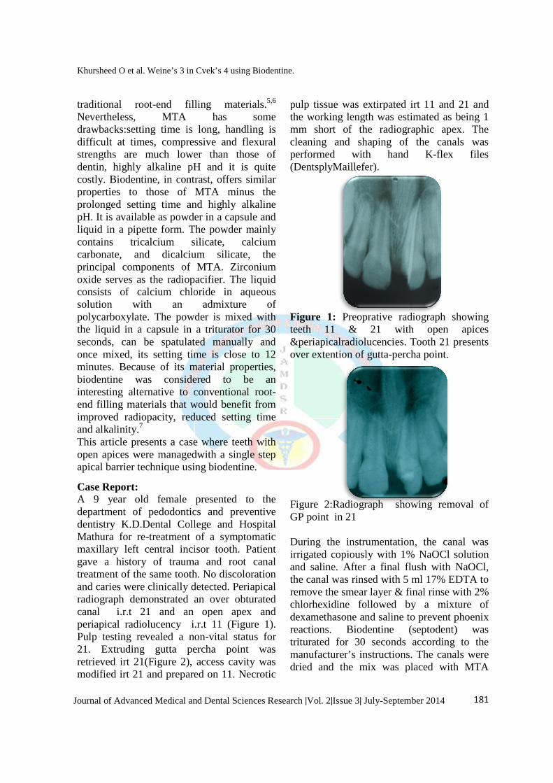
Case Report: A 9 year old female presented to the department of pedodontics and preventive dentistry K.D.Dental College and Mathura for re-treatment of a symptomatic maxillary left central incisor tooth. Patient gave a history of trauma and root canal treatment of the same tooth. No discoloration and caries were clinically detected. Periapical radiograph demonstrated an over obturated canal i.r.t 21 and an open apex and periapical radiolucency i.r.t 11 (Pulp testing revealed a non21. Extruding gutta percha point was retrieved irt 21(Figure 2), access cavity was modified irt 21 and prepared on 11. Necrotic
Weine’s 3 in Cvek’s 4 using Biodentine.
Journal of Advanced Medical and Dental Sciences Research |Vol. 2|Issue 3| July-September 2014
end filling materials.5,6 Nevertheless, MTA has some drawbacks:setting time is long, handling is
compressive and flexural strengths are much lower than those of dentin, highly alkaline pH and it is quite
Biodentine, in contrast, offers similar properties to those of MTA minus the prolonged setting time and highly alkaline
powder in a capsule and . The powder mainly
contains tricalcium silicate, calcium carbonate, and dicalcium silicate, the principal components of MTA. Zirconium oxide serves as the radiopacifier. The liquid consists of calcium chloride in aqueous solution with an admixture of polycarboxylate. The powder is mixed with the liquid in a capsule in a triturator for 30
can be spatulated manually and d, its setting time is close to 12
minutes. Because of its material properties, tine was considered to be an
interesting alternative to conventional root-end filling materials that would benefit from improved radiopacity, reduced setting time
s article presents a case where teeth with open apices were managedwith a single step
using biodentine.
9 year old female presented to the department of pedodontics and preventive
ollege and Hospital treatment of a symptomatic
incisor tooth. Patient gave a history of trauma and root canal treatment of the same tooth. No discoloration and caries were clinically detected. Periapical radiograph demonstrated an over obturated canal i.r.t 21 and an open apex and
ncy i.r.t 11 (Figure 1). Pulp testing revealed a non-vital status for 21. Extruding gutta percha point was
2), access cavity was modified irt 21 and prepared on 11. Necrotic
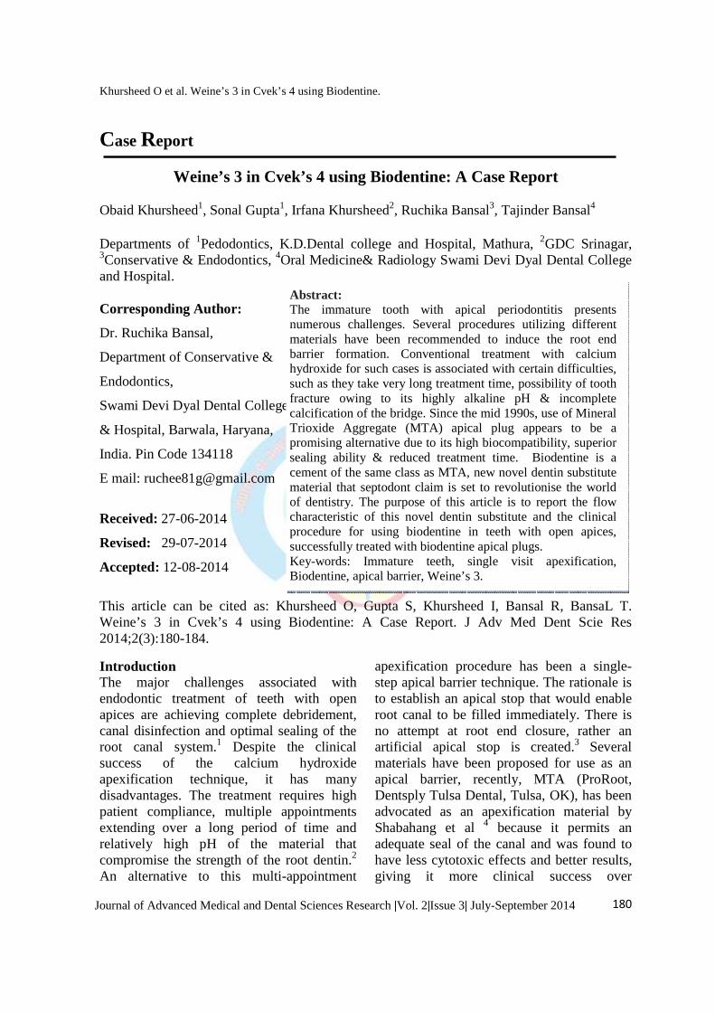
pulp tissue was extirpated irt 11 and 21 and the working length was estimated as being 1 mm short of the radiographic apex. The cleaning and shaping of the canals was performed with hand K(DentsplyMaillefer). Figure 1: Preoprative teeth 11 & 21 with open apices &periapicalradiolucencies.over extention of gutta- Figure 2:Radiograph showing removal of GP point in 21 During the instrumentation, the canal was irrigated copiously with 1% NaOCl solutionand saline. After a final flush with NaOCl, the canal was rinsed with 5 mremove the smear layer & final rinse with 2% chlorhexidine followed by a mixture of dexamethasone and saline to prevent phoenix reactions. Biodentinetriturated for 30 seconds according to the manufacturer’s instructions.dried and the mix was placed with MTA
181 September 2014
pulp tissue was extirpated irt 11 and 21 and the working length was estimated as being 1 mm short of the radiographic apex. The cleaning and shaping of the canals was performed with hand K-flex files
Preoprative radiograph showing teeth 11 & 21 with open apices &periapicalradiolucencies. Tooth 21 presents
-percha point.
2:Radiograph showing removal of
During the instrumentation, the canal was copiously with 1% NaOCl solution . After a final flush with NaOCl,
the canal was rinsed with 5 ml 17% EDTA to remove the smear layer & final rinse with 2% chlorhexidine followed by a mixture of dexamethasone and saline to prevent phoenix
s. Biodentine (septodent) was triturated for 30 seconds according to the manufacturer’s instructions. The canals were dried and the mix was placed with MTA

Khursheed O et al. Weine’s 3 in Cvek’s 4 u
Journal of Advanced Medical and Dental Sciences Research
carrier in the apical portion of the cIncrements were condensedpaper points (size-80) which provide a better propioceptive control (Figure Figure 3: Radiograph showingBiodentine apical plugs, During the master cone selection Weine’s 3 type apical end formation was evident, confirming the flowable this novel dentin substitute. cone selection (Figure 4), obturation was done by cold lateral condensation technique (Figure 5). Access cavity was sealed with composite. The contact with the patient was lost but the patient did return after 3 months (Figure 5) and 6 months with a chipped of build up (Figure 6), on clinical examinationthe tooth was asymptomatic. IOPARrevealed positive changes, root end formation, radiolucent area was absent and the trabecular bone was forming. Figure 4: Master cone radiograph showing positive Biodentine apical plugs.
Weine’s 3 in Cvek’s 4 using Biodentine.
Journal of Advanced Medical and Dental Sciences Research |Vol. 2|Issue 3| July-September 2014
carrier in the apical portion of the canal. Increments were condensed using butt end of
80) which provide a better Figure 3).
: Radiograph showing placement of
During the master cone selection Weine’s 3 type apical end formation was evident, confirming the flowable characteristics of this novel dentin substitute. After master
4), obturation was done by cold lateral condensation technique
vity was sealed with contact with the patient was
id return after 3 months and 6 months with a chipped of
clinical examination was asymptomatic. IOPAR
Weine’s 3 type of radiolucent area was
absent and the trabecular bone was forming.
Master cone radiograph showing positive Biodentine apical plugs.
Figure 5: Post obturation radiograph
Figure 6: 6month follow up Discussion: The response to trauma can be varied. Some pulps remain apparently normal with no adverse effects, whereas others become necrotic. When treating non vital teeth, main issue is eliminating bacteria from the root canal system. As instruments cannbe used properly in teeth with open apices, cleaning and disinfection of the root canal system rely on the chemical action of NaOClas an irrigant.8 In the past, several different materials such as amalgam, reinforced zinc oxide eugenol cements (interimmaterial [IRM], super ethoxy benzoic acid (EBA), glass-ionomer cement,resin were used.9 Recently, MT“Portland cement,10 was found to have less cytotoxic effects and better results with biocompatibility and microleakprotection, giving it more clinical success
182 September 2014
Post obturation radiograph
6month follow up
The response to trauma can be varied. Some pulps remain apparently normal with no adverse effects, whereas others become necrotic. When treating non vital teeth, main issue is eliminating bacteria from the root canal system. As instruments cannot be used properly in teeth with open apices, cleaning and disinfection of the root canal system rely on the chemical action of NaOCl
In the past, several different materials such as amalgam, reinforced zinc oxide eugenol cements (interim restorative material [IRM], super ethoxy benzoic acid
ionomer cement, and composite Recently, MTA, a refined
was found to have less cytotoxic effects and better results with biocompatibility and microleakage protection, giving it more clinical success
Khursheed O et al. Weine’s 3 in Cvek’s 4 using Biodentine.
183
Journal of Advanced Medical and Dental Sciences Research |Vol. 2|Issue 3| July-September 2014
over traditional root-end filling materials.11 Nevertheless, MTA has some drawbacks: setting time is long, handling is difficult at times, compressive and flexural strengths are much lower than those of dentin and it is quite costly. Biodentine, introduced in 2010 as a material for crown and root dentin repair treatment, repair of perforations, apexifications, resorption repair and root-end fillings12offers similar properties to those of MTA minus the high alkalinity, prolonged setting time and high cost.It is available as powder in a capsule and liquid in a pipette.The main component is a highly purified tricalcium silicate powder that contains small amounts of dicalcium silicate and calcium carbonate, the principal components of MTA and a radioopacifier.13
The liquid consists of calcium chloride in aqueous solution with an admixture of polycarboxylate. The powder is mixed with the liquid in a capsule in a triturator for 30 seconds, and once mixed, its setting time is around 12 minutes, making single sitting procedure a reality. Biodentine fulfills all the requirements for a suitable root-end filling material in that it exhibits biocompatibility, long-term sealing of the cavity, antimicrobial properties, and the ability to induce hard-tissue regeneration; it is also stable, insoluble, non-resorbable, hydrophilic, and easy to prepare and place.7 The interfacial properties of dentin-biodentine interface were studied under microscope and tag-like microstructures were detected. The flowable consistency of biodentine penetrates dentinal tubules and helps in the mechanical properties of the interface.13 Investigation of the bioactivity of Biodentine, MTA and a new Tricalcium silicate cement revealed that all three cements allowed the deposition of hydroxyapatite on the surface. This shows that all three materials are bioactive.14 The action mechanism of calcium silicate cements such as biodentine involves the release of calcium hydroxide with a pH lower
than calcium hydroxide, in addition, impervious dentin--‐material interfaces, as well as a dissolution resistance that does not involve any re-intervention. Despite the low clinical hindsight, on account of the recent availability of the material, the available studies in the animal model lead us to expect excellent results in terms of preserving pulpal vitality, dentin bridge formation and absence of complications (internal resorption). The clinical case reported here demonstrates that when biodentine is used as an apical plug in necrotic teeth with immature apices, the canals can be effectively sealed and a desirable root end anatomy(Weine’s 3) can be achieved in a single sitting, which would otherwise take months, courtesy flowable characteristics of this novel dentin substitute. Conclusion Studies to understand the flow characteristics of biodentine in a better way are needed. Because of its material properties, biodentine should be an interesting alternative to conventional root-end filling materials owing to its flow characteristics, faster setting, easy handling, moderate pH and improvedradiopacity . References 1. Andreasen JO, Flores MT. Injuries to
developing teeth. In: Andreasen JO, Andreasen FM, Andersson L, editors. Textbook and color atlas of traumatic injuries to the teeth. 4th ed. Copenhagen: Munksgaard; 2007. p. 542-76.
2. Weisenseel JA, Hicks ML, Pelleu GB. Calcium hydroxide as an apical barrier. J Endodon 1987;13:1–5.
3. Rafter M. Apexification: a review. Dent Traumatol 2005;21:1–8.
4. Shabahang S, Torabinejad M, Boyne PJ, Abedi HH, McMillan P. Apexification in immature dog teeth using osteogenic protein-1, mineral trioxide aggregate, and
Khursheed O et al. Weine’s 3 in Cvek’s 4 using Biodentine.
184
Journal of Advanced Medical and Dental Sciences Research |Vol. 2|Issue 3| July-September 2014
calcium hydroxide. J Endodon 1999;25:1–5.
5. Bodrumlu E. Biocompatibility of retrograde root filling materials: A review. Aust Endod J. 2008;34(1):30-35.
6. Roberts HW, Toth JM, Berzins DW, Charlton DG. Mineral trioxide aggregate material use in endodontic treatment: A review of the literature. Dent Mater. 2008;24(2):149-164.
7. About I, Laurent P, Tecles O. Bioactivity of Biodentine: a Ca3SiO5--‐based Dentin Substitute. Oral session, IADR Congress 2010 July, Barcelona Spain.
8. Siqueira JF Jr, Guimarães-Pinto T, Rôças IN. Effects of chemomechanical preparation with 2.5% sodium hypochlorite and intracanal medication with calcium hydroxide on cultivable bacteria in infected root canals. J Endod. 2007;33(7):800-5.
9. Bodrumlu E. Biocompatibility of retrograde root filling materials: A review. Aust Endod J. 2008;34(1):30-35.
10. Dammaschke T, Gerth HUV, Züchner H, Schäfer E. Chemical and physical surface and bulk material characterization of white ProRoot MTA and two Portland cements. Dent Mater. 2005;21(8):731-738.
11. Roberts HW, Toth JM, Berzins DW, Charlton DG. Mineral trioxide aggregate material use in endodontic treatment: A review of the literature. Dent Mater. 2008;24(2):149-164.
12. Trond Bjorvik Osen, Ina Iselin Astrup, Carl Haavard Knutsson, Biodentine as a root-end filling, 2012.
13. A.R.Atmeh, E.Z.Chong, G.Richard, F.Festy, T.F.Watson, Dentin-cement interfacial interaction: Calcium silicates and Polyalkenoates. J Dent Res 2012; 91(5): 454-459.
14. Camilleri J, Sorrentino F, Damidot D, Investigation of the hydration and bioactivity of radiopacified tricalcium silicate cement, Biodentine and MTA Angelus. Academy of Dent materials 2013;29(5):580-593.
Source of support: Nil Conflict of interest: None declared