c u l a r b io m a t , o ior in journal of moleular ... · additionally, according to roskams, the...
TRANSCRIPT

Research Article OMICS International
Journal of Molecular Biomarkers & DiagnosisJo
urna
l of M
olecular Biomarkers & Diagnosis
ISSN: 2155-9929
El-Sabaawy et al., J Mol Biomark Diagn 2017, 8:5DOI: 10.4172/2155-9929.1000352
Volume 8 • Issue 5 • 1000352J Mol Biomark Diagn, an open access journalISSN:2155-9929
*Corresponding author: Maha El-Sabaawy, Departments of Hepatology, National Liver Institute, Menoufia University, Egypt, Tel: 020482223216; E-mail: [email protected]
Received May 27, 2017; Accepted June 26, 2017; Published June 28, 2017
Citation: El-Sabaawy MM, Abdelsameea E, Abdallah AR, El-Refaey A, Soltan M, et al. (2017) Hepatic Progenitor Cells and Cells Resistant to Apoptosis in Chronic HCV. J Mol Biomark Diagn 8: 352. doi: 10.4172/2155-9929.1000352
Copyright: © 2017 El-Sabaawy MM, et al. This is an open-access article distributed under the terms of the Creative Commons Attribution License, which permits unrestricted use, distribution, and reproduction in any medium, provided the original author and source are credited.
Hepatic Progenitor Cells and Cells Resistant to Apoptosis in Chronic HCV Maha M El-Sabaawy1*, Eman Abdelsameea1, Ayat R Abdallah2, Ahmed El-Refaey3, Mervat Soltan3 and Nemine Ehsan3
1Departments of Hepatology, National Liver Institute, Menoufia University, Egypt2Epidemiology and Preventive Medicine, National Liver Institute, Menoufia University, Egypt3Department of Pathology, National Liver Institute, Menoufia University, Egypt
AbstractBackground: Hepatic progenitor cells (HPC) as a hepatic regeneration reservoir, are now signified as one of
the promising therapeutics. However, connection to cells resistant to apoptosis in pathogenesis of chronic hepatitis C virus (HCV) is still evolving.
Aim: title relationship between HPC and cells resistant to apoptosis in HCV along with liver disease severity and fibrosis progression. Methods: liver biopsies of 91 chronic HCV patients were immunohistochemically examined. Both demographic and clinical characteristics were sourced from the data registries. METAVIR scoring was unified for both Grading and staging. Immunostaining with CK7, Ki67, and bcl2 antibodies was done.
Results: Transaminases, platelets and prothrombin time exhibited significant relation with Ki67, CK7 both isolated and ductular and bcl2 both LPT and LAH. CK7 ductular showed association with fibrosis and necroinflammatory activity (P< 0.05), while non-significant relation was noticed with the CK7 isolated form and Ki67 (P> 0.05). Moreover, bcl2 both (LPT) and (LAH) demonstrated association with fibrosis and necroinflammatory activity (P< 0.05). Positive correlation between immunoexpression of HPCs both isolated (r=0.547, <0.001) and ductular with bcl2 LAH (r=0.476, p<0.001) was reported. bcl2 H showed positive correlation with the CK7 isolated form (r=0.476, p <0.001), with no correlation between it and the CK7 ductular (r=0.298, p= 0.003).
Conclusion: Hepatic progenitor cells and cells resistant to apoptosis are conversely interrelated to HCV pathogenesis with a pivotal role in disease severity and progression. It is a notion to be considered in development new therapies of HCV-related chronic liver disease.
Keywords: Hepatic progenitor cells; Cells resistant to apoptosis; HCV; Liver fibrosis
IntroductionHPCs in humans present the potential reservoir of cholangiocyte/
hepatocyte differentiation [1,2]. Defective mature hepatocyte regeneration is claimed to be an activation stimulus to HPCs proliferation implying an auxiliary role in parenchymal regeneration [3]. The proliferation of HPC is incremented in the progression of the majority of chronic liver diseases including genetic mutations, viral infections and metabolic disorders [4]. It has been reported that activation of HPC in chronic hepatitis C virus infection (CHC) is coupled to the severity of inflammation, stage of fibrosis along with the concomitant clinical status of liver disease [5]. However, the mechanisms underlying this process with their relation to HCV pathogenesis are still unclear.
Additionally, according to Roskams, the activated HPC in chronic HCV might be the target cell population for carcinogenesis as the telomerase shortened hepatocytes are senescent in advanced cirrhosis [6]. Facts implying an extensive concern on their therapeutic based impacts.
A modulation of proapoptotic and antiapoptotic proteins which is the consequence of ignited immune responses by infections has been considered the decisive stance in mapping liver disease prognosis [7]. Persistent inflammation with the resultant lymphocytes resistant to apoptosis had been mentioned to be a major backer to both chronicity and severity of liver disease [8]. Most studies considering apoptosis in chronic HCV infection; had validated its pathogenic role as an integrator of host immune response against viral infections [9]. Eminently enhanced hepatocyte apoptosis was found to be interconnected to the inflammation severity, stages of fibrosis, transaminases levels and viral load in CHC patients [10]. Apoptotic markers; Fas, Fas ligand, Fas-associated death domain, caspases 3, 8, and 9, and in-situ apoptosis; were
almost always the most studied items in this issue [7]. The BCL proteins as antiapoptotic mediators participating substantially in the apoptotic process; have not been systematically and thoroughly discussed in liver tissue of chronic hepatitis C virus infection (CHC) [9].
Investigating the linkage between the HPCs proliferation and lymphocytes resistant to apoptosis in CHC patients might help in intensifying therapeutic based options offered for those patients. In this study, a semi-quantitative and topographical immuno-histological evaluation of HPCs along with portal and hepatic parenchymal lymphocytes resistant to apoptosis was conducted on liver tissues of patients with CHC. Results were correlated with different demographic and clinicopathologic variables to better understand the pathogenic contribution of HPCs and cells resistant to apoptosis to CHC.
Materials and MethodsSpecimen collection
This study included 91 chronic HCV patients. Clinical, demographic, and laboratory data were assembled from the data registry of the outpatient clinics of Hepatology department, National Liver Institute) NLI), Menoufia University, Egypt, from the period

Citation: El-Sabaawy MM, Abdelsameea E, Abdallah AR, El-Refaey A, Soltan M, et al. (2017) Hepatic Progenitor Cells and Cells Resistant to Apoptosis in Chronic HCV. J Mol Biomark Diagn 8: 352. doi: 10.4172/2155-9929.1000352
Page 2 of 5
Volume 8 • Issue 5 • 1000352J Mol Biomark Diagn, an open access journalISSN:2155-9929
between January 2013 until December 2014. Inclusion criteria were: 1- age from 18 years up. 2- Positive HCV-PCR. 3. Genotype 4. 4-. Liver biopsies containing at least 6 portal tracts.
Exclusion criteria
HCV co-infection with hepatitis B, schistosomiasis, and/or non-alcoholic steatohepatitis virus. Paraffin blocks and routinely stained slides from liver biopsies of patients included in this study were retrieved from the archives of the department of pathology of NLI. Adapting to the ethical guidelines of the 1975 Declaration of Helsinki, a written consent was a prerequisite to endorsement in this study along with the approval of the ethical committee of NLI, Menofia University. Histological evaluation of liver biopsies from patients with chronic hepatitis included grading of necroinflammation and staging of fibrosis based on the scoring system proposed by Ishak et al. [11]. Grades 1 to 3 was ascribed to minimal, grades 4 through 8 as mild, and grades 9 through 12 as moderate chronic hepatitis. No cases revealed necroinflammatory score more than 12. Stages 1–2 indicated mild fibrosis, stages 3–4 moderate fibrosis, stages 5–6 severe fibrosis/cirrhosis. Steatosis, lymphoid aggregates, bile duct injury, and vascular changes were identified by their presence or absence [11].
Immunohistochemistry
Being placed on positively charged slides, tissue sections were deparaffinized in xylene, rehydrated in a graded series of ethanol, and then incubated with 3% hydrogen peroxide. The method used for immunostaining was streptavidin–biotin amplified system. Slides were rinsed in phosphate-buffered saline (PBS) and then exposed to heat-induced epitope retrieval in citrate buffer solution (pH 6) for 20 minutes. After cooling, the slides were incubated overnight at room temperature with monoclonal CK7 (CODE MS-1352-P Thermoscientific OV-TL 12/30), or monoclonal Ki67 (CODE IR626, Dako MIB-1) or monoclonal bcl-2 (CODE MS-123-P1 Thermoscientific 100/D5). (100 µg concentrated and diluted with PBS in a dilution 1:100. Positive tissue controls were bile ducts in liver tissue (built in control) for CK7, pancreatic adenocarcinoma for Ki67 and lymphoma for bcl2. Detection of immunoreactivity was carried out using the ultra-vision detection system, ready-to-use anti-polyvalent horseradish peroxidase/diaminobenzidine (NeoMarkers, LabVision, California, USA). Finally, the reaction was visualized by an appropriate substrate/ chromogen (diaminobenzidine) reagent. A counter stain was carried out using Mayer's hematoxylin. The staining procedure included negative controls obtained by substitution of primary antibodies with phosphate-buffered saline (17). CK7(+) and Ki67 (+) HPCs were identified as isolated progenitor cells (IPC), and as isolated ductular structures (IDS) according to the classification and terminology proposed by Roskams et al. [12] with modifications. CK7(+) immunostaining in HPC was strongly cytoplasmic (Table 1). The ductular reaction was assessed semi-quantitatively on a three-grade scale based on its extent around the limiting plate; grade 1 represented focal discontinuous or continuous ductular reaction in less than 1/3 of the portal tract circumference. Grade3 was defined by continuous ductular reaction rimming more than 1/3 of the portal tract circumference. Grade 2 was intermediate between grades 1 and 3 [6-8,10,13,14]. The positive
reaction was expressed by brownish coloration of the cytoplasm for bcl-2. Under a microscope, lymphocyte percentage was calculated (the number of positive immunoreaction cells in ratio to 100 cells) in five portal tracts; lymphocytes related to portal tracts (LPT) and five fields of hepatic parenchyma; lymphocytes associated hepatic parenchyma (LHP) at magnification x400 per slide. Immuno-reactive lymphocytes for bcl-2 were scaled from 1 to 3. Number of immunoreactive cells less than 10 were scored as 1, from 11 to 50 scored as 2 and number of positive cells more than 50 were scored 3 (Table 1) [15].
Statistical analysisData has been collected and entered into the computer using SPSS
(Statistical Package for Social Science) program for statistical analysis, (version 20; Inc., Chicago. IL). Two types of statistics were done:
1. Descriptive statistics: Where quantitative data has been shown as mean, and SD.
2. Analytical statistics: Student t-test has been used to compare mean and SD of 2 sets of quantitative normally distributed data, while Mann-Whitney test was used when this data is not normally distributed. Spearman's correlation has been used to study the correlation between two variables having not normally distributed data. P-value was considered statistically significant when it is less than 0.05.
ResultsClinical, laboratory and histopathological characteristics
The entire cohort (91 patients) was mainly formed of males (62.6%) with a mean age of 40.06 ± 8.99 years. Liver function tests were within normal ranges, except for mild elevations of transaminases (AST: 46.9 ± 40.6; ALT: 47.1 ± 35.9 IU/ml) with positive HCV-RNA with a level of viremia ranged from 837449.95 ± 1672800.563 IU/ml.
Histopathological evaluation of liver biopsy: most cases exhibited mild necroinflammatory activity (68.7%), mild fibrosis (82.4%), and no steatosis (87.5%).
Immuno-histochemical studies
This study demonstrated the relationship between CK7, Ki67 and bcl-2 immunoreactive cells with the laboratory variables of all patients. Hepatic progenitor cell markers CK7 and Ki67 showed negative correspondence with any of laboratory data including transaminases (AST, ALT) platelets and prothrombin time (p>0.05). However, Bcl2 both LPT and LHP showed significant converse connection with prothrombin time, INR, and platelets (p<0.05).
A ductular form of HPCs revealed 74.1%, and 25.9% in mild and moderate fibrosis respectively, while the isolated form of HPC revealed 76.9% and 23.1% in mild and moderate fibrosis respectively with significant relation (p-value <0.005) (Table 2 and Figure 1).
Regarding Ki67 immunoreaction, it presented in 72.7% and 27.3% in mild and moderate fibrosis respectively with no significant difference (p=0.543) (Table 2 and Figure 2).
In relation to necroinflammation; HPCs were found to be expanded in most patients of the ductular form, while the isolated form expanded in all patients. The ductular expansion in higher grades of necroinflammation was 16%, 69%, 13.6% and 1.2% in minimal, mild, moderate and severe necroinflammation, respectively, with (p=0.525). However, the isolated form of HPCs expansion increased significantly with grades of the necroinflammatory activity Isolated form of HPCs were 15.4%, 71.4%, 12.1%, and 1.1% in minimal, mild, moderate and severe necroinflammation, respectively (Table 2, Figures 1 and 2).
Antibody Code Company Experimental Tool CloneKi-67 IR626 Dako Monoclonal mouse MIB-1CK7 MS-1352-P Thermoscientific Monoclonal mouse OV-TL 12/30
BCL-2 MS-123-P1 Thermoscientific Monoclonal mouse 100/D5
Table 1: Antibodies used in the current study.
Citation: El-Sabaawy MM, Abdelsameea E, Abdallah AR, El-Refaey A, Soltan M, et al. (2017) Hepatic Progenitor Cells and Cells Resistant to Apoptosis in Chronic HCV. J Mol Biomark Diagn 8: 352. doi: 10.4172/2155-9929.1000352
Page 3 of 5
Volume 8 • Issue 5 • 1000352J Mol Biomark Diagn, an open access journalISSN:2155-9929
Ki67 showed a non-significant relation to necroinflamatory grades (13.6%, 70.5%, 15.9% and zero) in minimal, mild, moderate and severe necroinflamation, respectively (p value>0.005) (Table 2 and Figure 2).
Relation of antiapoptotic cells with fibrosis and necroinflamma-tory activity
Bcl2 immunoreaction in LPT and LHP were 20.4%, 73.4%, 6.3%, zero and 25.5%, 70.6%, 3.9% and zero in minimal, mild, moderate and severe necroinflammation. Bcl2 LPT and LHP were found to be significantly correlated with the necroinflammatory activity according to Ishak score (p-value 0.007, and0.002). The more advanced necroinflammation the less the presence of bcl2 LPT and bcl2 LHP (Table 2 and Figure 3).
Regarding bcl-2 immunoreaction relation to fibrosis stage, bcl2 was highly significantly associating with different stages of fibrosis (p<0.001,
Vari-ables CK7 ductular P-
value CK7 Isolated P-value BCL2 LPT P-value BCL2 LHP P-
value Ki 67 P-value
<0.1 ≥ 0.1 <0.3 ≥ 0.3 Absent Present < 2 ≥ 2 Absent Present
Mean+SD Mean+SD Mean+ SD Mean+SD Mean+SD Mean+SD Me an+SD Mean+SD Mean+SD Mean+SD
Albu-min 4.4+0.42 4.2+0.37 0.048 -- 4.2+0.38 -- 4.3+3.6 4.1+0.42 0.061 4.3+0.35 4.1+0.41 0.089 4.3+0.37 4.2+0.40 0.345
PT* 93.5+3.9 89.9+9.9 0.252 -- 90.2+9.5 -- 91.2+9.3 88.2+9.9 0.183 92.3+8.6 87.5+10.1 0.018 89.3+9.4 91.1+9.7 0.381INR 1.1+0.03 1.1+0.12 0.281 -- 1.1+0.11 -- 1.1+0.11 1.1+0.12 0.084 1.1+0.08 1.1+0.13 0.005 1.1+0.11 1.0+0.11 0.609
Plate-lets 220.1+42.5 199.3+55.5 0.258 -- 201.6+54.4 -- 211.1+49.9 179.0+58.9 0.009 209.4+49.7 191.5+59.1 0.12 200.3+51.3 202.9+58.1 0.819
ALT* 59.5+19.4 61.9+46.3 0.295 -- 61.7+44.1 -- 53.1+38.5 82.0+50.1 0.001 52.5+29.8 73.3+55.7 0.128 65.2+34.7 67.5+52.1 0.42AST* 69.6+67.3 50.3+32.3 0.286 -- 52.4+37.6 -- 45.2+29.0 69.4+49.3 0.005 42.3+15.7 65.2+51.5 0.045 48.8+36.7 56.2+38.6 0.217
ISHAK activ-ity:
0.525 0 (0.0) 0.002 13 (20.3) 1 (3.7) 0.007 13 (25.5) 13 (25.5) 0.002 8 (17.0) 6 (13.6)
-Mini-mal 0 (0.0) 14 (15.4) 47 (73.4) 18 (66.7) 36 (70.6) 36 (70.6) 34 (72.3) 31 (70.5)
-Mild 1 (10.0) 13 (16.0) 0 (0.0) 65 (71.4) 4 (6.3) 7 (25.9) 2 (3.9) 2 (3.9) 4 (8.5) 7 (15.9) -Mod-erate 9 (90.0) 56 (69.1) 0 (0.0 11 (12.1) 0 (0.0) 1 (3.7) 0 (0.0) 0 (0.0) 1 (2.1) 0 (0.0) 0.543
-Se-vere 0 (0.0) 11 (13.6) 1 (1.1)
0 (0.0) 1 (1.2)ISHAK fibro-sis:
0.108 0 (0.0) <0.001 56 (87.5) 14 (51.9) <0.001 49 (96.1) 49 (96.1) 0.001 38 (80.9) 32 (72.7) 0.358
-Mild 10 (100.0) 0 (0.0 8 (12.5) 13 (48.1) 2 (3.9) 2 (3.9) 9 (19.1) 12 (27.3)-Mod-erate 0 (0.0) 60 (74.1) 70 (76.9)
21 (25.9) 21 (23.1)*PT: Prothrombin, AST: Aspartate Transaminase ALT: Alanine Transaminase
Table 2: Association of laboratory and histopathological data to the HPCs and cells resistant to apoptosis patients (n=91).
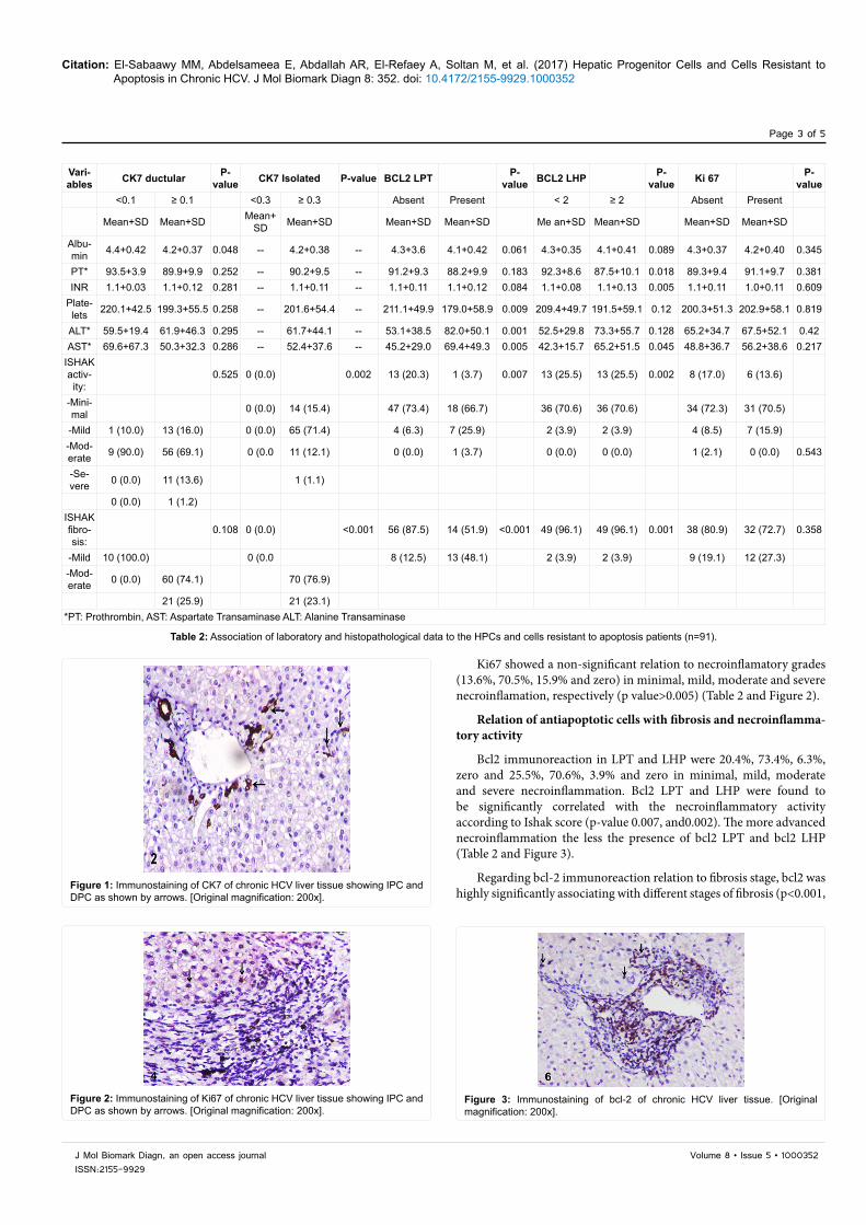
Figure 1: Immunostaining of CK7 of chronic HCV liver tissue showing IPC and DPC as shown by arrows. [Original magnification: 200x].
Figure 2: Immunostaining of Ki67 of chronic HCV liver tissue showing IPC and DPC as shown by arrows. [Original magnification: 200x].
Figure 3: Immunostaining of bcl-2 of chronic HCV liver tissue. [Original magnification: 200x].

Citation: El-Sabaawy MM, Abdelsameea E, Abdallah AR, El-Refaey A, Soltan M, et al. (2017) Hepatic Progenitor Cells and Cells Resistant to Apoptosis in Chronic HCV. J Mol Biomark Diagn 8: 352. doi: 10.4172/2155-9929.1000352
Page 4 of 5
Volume 8 • Issue 5 • 1000352J Mol Biomark Diagn, an open access journalISSN:2155-9929
and 0.001). Bcl-2 expression in LPT was absent in 87.5 and 12.5 in mild and moderate fibrosis. Also, bcl2 expression in LHP was absent in 96.1, and 47.5 in mild and moderate fibrosis (Table 2 and Figure 3).
Correlation studies between HPC and antiapoptotic markers: a positive correlation was substantiated between HPC and markers of antiapoptotic cells and was verified as followed: BCl2LAH was positively correlated with CK7isolated (r=0.547, p<0.001), and with CK7ductular (r=0.476, p<0.001). BCl2 H also was positively correlated with CK7 isolated (r=0.476, p<0.001), while bcl2 was found to be negatively correlated with CK7ductular (r=0.289, p=0.003) (Table 3).
DiscussionStudying the pathogenic events evolving in HCV chronicity was,
still and will be the most appealing issue attracting hepatologists allover. In spite of the high incidence of HCV in Egypt; data assigning the virus pathogenesis in this special population are insufficient. Hepatocyte regeneration added to apoptosis is two eminent elements portraying chronic HCV natural history events. However, the interconnection linking both axioms is still unclear.
Expansion of HPC was ascribed by Spee et al., with the predilection towards the hepatocytic lineage, suggesting the state of these cells during this chronic lower degree of hepatocyte damage [16].
The relationship between HPCs and the grade of necroinflammatory activity has been a subject of controversy. Some authors reported a significant association between HPCs and grade of activity [17-21]. In this study, despite being insignificant, HPCs were found to be expanded in most patients in the ductular form, while the isolated form was expanded in all patients. The ductular, and isolated expansion along with ki67 were reported to be augmented in a mild grade of necroinflammation according to Ishak grading (69%, 71.4%, and 70.5%), respectively. It is claimed that the inflammatory pathway mediates the response of HPCs in HCV [22]. Likewise, the three markers of HPCs expansion were found to be boosted in patients with moderate fibrosis than those with milder stages (74.1, % and 25.9%), (76.9%, and 23.1%), and (72.7% and 27.3%) respectively. Consequently, we could conclude that the number of HPCs is increased significantly with the progression of fibrosis. This agrees with several investigators [4,8,19,23]. It is hypothesized that blocking of the proliferative activity of HCV-infected hepatocytes leads to activation of HPCs which in turn promote the fibrogenesis cascade [7]. Another explanation based on the hypothesis implying that replication exhaustion of hepatocytes in association with diminished liver mass in severe fibrosis and cirrhosis might be the endorsing stimulus of HPC activation [6]. Clouston et al. [9] found a similar correlation between the extent of the DR and the number of IHPCs (r = 0.624, P = 0.0001) in adults with CHC. These suppositions suggest that IHPCs form is the earlier stage of HPCs expansion before the formation of the DR form. Theories might support our verification of the significant linkage between the isolated form of HPC with liver fibrosis and necroinflammation. These findings are inconsistent with El-Araby et al. and Svegliati-Baroni et al. who found a close relationship between necroinflammatory activity and both DR and IHPCs [13,24]. Knight et al. [23] had adopted a postulation of the presence of inflammatory cellular infiltration and localized
cytokine production were common features of liver pathologies in which the HPCs compartment is active. A process suggesting intervening mediation between pro-inflammatory pathways and the HPCs response during chronic liver injury. The involvement of HPC in HCV fibrogenesis, suggested by Clouston et al. and Richardson et al. cannot be denied [9,25]. Moreover, El-Araby et al. had verified the link between HPCs with both the severity of hepatitis and the stage of fibrosis which might be justified by the hypothesized role of HPCs in disease progression [13]. Data regarding phenotype and topography of HPC activation vary among different studies. A ductular reaction at the limiting plate of portal tracts and septa has been invariably identified [1,8,12,26,27]. Diverse observations have been also reported regarding the intraparenchymal HPC [1].
No association between the number of HPCs and steatosis was detected. Similar results have been obtained by Delladetsima et al. [5] and El-Araby et al. [5,13] indicating that steatosis had no role in the activation of HPCs.
In spite of being hypothesized, the interconnection between apoptosis and chronic HCV is still mystifying. Either being direct or indirect, the apoptotic mechanisms evolved in chronic HCV are proposed to be the initiative step of HCV abolition [7]. Bcl2 proto-oncogene as an antiapoptotic protein had the ability to halt the programmed cell death. Our results showed that markers of cells resistant to apoptosis bcl2 H and bcl2 LAH were found to be significantly connected to the necroinflammatory activity and fibrosis staging according to Ishak score (p-value 0.007 and 0.002 respectively). The more advanced the necroinflammation the less the presence of bcl2 H and bcl2 LAH. A significant absence of bcl2 H and bcl2 LAH was extremely coupled to the presence of advancing fibrosis. These results come in concordance Delladetsima et al. and Ozaras et al. who ascertained the interconnection between different apoptotic parameters and inflammation and fibrosis [5,7]. Supporting this theory is the evident Bcl2 expression in liver of cirrhotic patients with concealed presence in the liver of hepatitis C patients detected by [28-31]. This hypothesis was sourced to rationalize the high incidence of hepatocellular carcinoma in cirrhotic HCV patients supported by the anti-apoptotic/oncogenic probability of bcl-2 [29]. El-Bendary et al. had studied apoptosis in HCV infection associated with bilharziasis spotting light on the presence of apoptotic (fas) and antiapoptotic (Bcl2) markers [32]. In spite of being statistically insignificant Bcl2 was found to be lessened accompanying disease progression and severity in HCV patients.
To be genuine, in our cohort the converse interplay between the two elements of this study: HPC expansion and cells resistant to apoptosis was eminently evident. This complementary harmonized counter connection might be potentially incriminated in chronic HCV pathogenesis. Nevertheless, the mechanisms involved in such an association are not yet clear. o elucidates the mechanism by which interferon alpha treatment reduced the numbers of intermediate hepatobiliary cells present in the chronic hepatitis C patients, we examined the effects of pegylated interferon alpha 2b on the proliferation, apoptosis, and differentiation of two well-characterized murine oval cell lines. The results suggest that interferon may exert direct effects on hepatic progenitor cells, reducing their rate of cell growth as well as stimulating them to undergo apoptosis [33]. This is in accordance with the observed effects of interferon on human HCC-derived cell lines, in which interferon alpha caused delayed S-phase progression through inhibition of cyclin-dependent kinases [34] and induced caspase-dependent apoptosis [35].
VariablesBCl2LAH
P-valueBCl2H
P-valueCorrelation Coefficient (r)
Correlation Coefficient (r)
CK7 isolated 0.547 <0.001 0.476 <0.001CK7 Ductular 0.476 <0.001 0.289 0.003
Table 3: Correlation between HPC and markers of anti-apoptosis.

Citation: El-Sabaawy MM, Abdelsameea E, Abdallah AR, El-Refaey A, Soltan M, et al. (2017) Hepatic Progenitor Cells and Cells Resistant to Apoptosis in Chronic HCV. J Mol Biomark Diagn 8: 352. doi: 10.4172/2155-9929.1000352
Page 5 of 5
Volume 8 • Issue 5 • 1000352J Mol Biomark Diagn, an open access journalISSN:2155-9929
Hh signal released by dying hepatocyte could activate the compensatory outgrowth of hepatic progenitors, which are involved in liver regeneration [35]. As a Hh target, osteopontin is highly expressed in fibrotic liver tissue and influences the function of hepatic progenitors (34). HOWEVER, neutralization of osteopontin could suppress progenitor cell response and attenuate liver fibrosis in CCl4, methionine-choline-deficient diet (MCD) and 3,5-diethoxycarbonyl-1,4-dihydrocollidine diet (DDC) mice. These suppositions might open new horizons in HCV pathogenesis understanding [36].
Conclusion As a reservoir of hepatic tissue, the substantial role of HPC in liver
cells renewal is unquestionable especially in the era of direct antiviral drugs which are mainly concerned with viral eradication rather than restoration of liver vitality. The hypothesized linkage between HPC expansion and cells resistant to apoptosis in chronic HCV patients should be more emphasized for a better advantageous revival of therapeutic options in these cohorts. A new role for antiapoptotic along with HPC antagonists might be advocated; paving the way for halting the predestined progression of a HCV-related chronic liver disease. However, further research is recommended to investigate the mechanisms underlying such an association.
References1. Shin S, Kaestner KH (2014) The origin, biology, and therapeutic potential of
facultative adult hepatic progenitor cells. Curr Top Dev Biol 107: 269-292.
2. Glaser SS, Gaudio E, Miller T, Alvaro D, Alpini G (2009) Cholangiocyte proliferation and liver fibrosis. Expert Rev Mol Med 11: e7.
3. Franchitto A, Onori P, Renzi A, Carpino G, Mancinelli R, et al. (2013) Expression of vascular endothelial growth factors and their receptors by hepatic progenitor cells in human liver diseases. Hepatobiliary Surg Nutr 8: 328-336.
4. Nobili V, Carpino G, Alisi A, Franchitto A, Alpini G, et al. (2012) Hepatic progenitor cells activation, fibrosis and adipokines production in pediatric nonalcoholic fatty liver disease. Hepatology 56: 2142-2153.
5. Delladetsima J, Alexandrou P, Giaslakiotis K, Psichogiou M, Hatzis G, et al. (2010) Hepatic progenitor cells in chronic hepatitis C: a phenomenon of older age and advanced liver disease. Virchows Arch 457: 457-466.
6. Roskams T (2006) Liver stem cells and their implication in hepatocellular and cholangiocarcinoma. Oncogene 25: 3818-3822.
7. Ozaras R, Tahan V, Ozbay G, Ozturk R, Yenice N, et al. (2015) Hepatic apoptotic markers are not predictors of the virological response to interferon-based therapy in chronic hepatitis C patients. Eur J Gastroenterol Hepatol 27: 1057-1062.
8. Anatol P, Danuta P, Janusz D, Bozena P (2005) Expression of bcl-2 protein in chronic hepatitis C: Effect of interferon-alpha 2b with ribavirin therapy. World J Gastroenterol 11: 2949-2952.
9. Clouston A, Jonsson R, Powell E (2009) Hepatic progenitor cell–mediated regeneration and fibrosis: Chicken or egg? Hepatol 5: 1424-1426.
10. Brost S, Zimmermann A, Koschny R, Sykora J, Stremmel W, et al. (2014) Hepatocyte expression of TRAIL pathway regulators correlate with histopathological and clinical parameters in chronic HCV infection. Pathol Res Pract 210: 83-91.
11. Ishak K, Baptista A, Bianchi L, Callea F, De Groote J, et al. (1995) Histological grading and staging of chronic hepatitis. J Hepatol 22: 696-699.
12. Roskams TA, Theise ND, Balabaud C, Bhagat G, Bhathal PS, et al. (2004) Nomenclature of the finer branches of the biliary tree: canals, ductules, and ductular reactions in human livers. Hepatology 39: 1739-1745.
13. El-Araby HA, Ehsan NA, Konsowa HA, Abd-Elaati BM, Sira AM (2015) Hepatic progenitor cells in children with chronic hepatitis C: correlation with histopathology, viremia, and treatment response. Eur J Gastroenterol Hepatol 27: 561-569.
14. Carpino G, Renzi A, Onori P, Gaudio E (2013) Role of hepatic progenitor cells in nonalcoholic fatty liver disease development: cellular cross-talks and molecular networks. Int J Mol Sci 14: 20112-201130.
15. Jackson P, Blythe D (2008) Immuno-histochemical techniques. In: Theory and Practice of Histological Techniques. Teoksessa Bancroft JD & Gamble M (Eds) 6: 433-472.
16. Spee B, Carpino G, Schotanus BA, Katoonizadeh A, Vander Borght S, et al. (2010) Characterisation of the liver progenitor cell niche in liver diseases: Potential involvement of Wnt and Notch signaling. Gut 59: 247-257.
17. Lim R, Knight B, Patel K, McHutchison JG, Yeoh GC, et al. (2006) Antiproliferative effects of interferon alpha on hepatic progenitor cells in vitro and in vivo. Hepatology 43: 1074-1083.
18. Novo E, Marra F, Zamara E, Valfrè di Bonzo L, Monitillo L, et al. (2006) Overexpression of Bcl-2 by activated human hepatic stellate cells: resistance to apoptosis as a mechanism of progressive hepatic fibrogenesis in humans. Gut 55: 1174–1182.
19. Tsamandas AC, Syrokosta I, Thomopoulos K, Zolota V, Dimitropoulou D, et al. (2006) Potential role of hepatic progenitor cells expression in cases of chronic hepatitis C and their relation to response to therapy: A clinicopathologic study. Liver Int 26: 817-826.
20. Chu C, Shyu W, Liaw Y (2008) Comparative studies on the expression of alpha-smooth actin in hepatic stellate cells in chronic hepatitis B and C. Dig Dis Sci 53: 1364-1369.
21. Capuron L, Miller A (2011) Immune system to brain signaling: neuropsychopharmacological implication. Pharmacol Ther 130: 226-238.
22. Săndulescu L, Rogoveanu I, Ciurea T, Comănescu MV, Streba CT, et al. (2001) Immunohistochemical study of stellate cells in patients with chronic viral hepatitis C genotype 1. Rom J Morphol Embryol 52: 137-143.
23. Knight B, Matthews VB, Olynyk JK, Yeoh GC (2005) Jekyll and Hyde: evolving perspectives on the function and potential of the adult liver progenitor (oval) cell. Bioessays 27: 1192-1202.
24. Svegliati-Baroni G, Faraci G, Fabris L, Saccomanno S, Cadamuro M, et al. (2011) Insulin resistance and necroinflammation drives the ductular reaction and epithelial-mesenchymal transition in chronic hepatitis C. Gut 60: 108-111.
25. 25. Richardson MM, Jonsson JR, Powell EE, Brunt EM, Neuschwander-Tetri BA, et al. (2007) Progressive fibrosis in nonalcoholic steatohepatitis: Association with altered regeneration and a ductular reaction. Gastroenterology 133: 80-90.
26. Murphy D, Detjen KM, Welzel M, Wiedenmann B, Rosewicz S (2001) Interferon-alpha delays S-phase progression in human hepatocellular carcinoma cells via inhibition of specific cyclin-dependent kinases. Hepatology 33: 346-356.
27. Sokal EM, Bourgois A, Stéphenne X, Silveira T, Porta G, et al. (2010) Peginterferon alfa-2a plus ribavirin for chronic hepatitis C virus infection in children and adolescents. J Hepatol 52: 827-831.
28. Nakamoto Y, Kaneko S, Kobayashi K (2002) Increased susceptibility to apoptosis and attenuated Bcl-2 expression in T lymphocytes and monocytes from patients with advanced chronic hepatitis C. J Leuko Biol 72: 49-55.
29. Li Y, Zhang Q, Liu Y, Luo Z, Kang L, et al. (2012) Hepatitis C virus activates Bcl-2 and MMP-2 expression through multiple cellular signaling pathways. J Virol 86: 12531e43.
30. Chakraborty J, Oakley F, Walsh M (2012) Mechanisms and biomarkers of apoptosis in liver disease and fibrosis. Int J Hepato 64: 915.
31. Frommel TO,Yong S, Zarling EJ (2012) Immunohistochemical evaluation of Bcl-2 gene family expression in liver of hepatitis C and cirrhotic patients: A novel mechanism to explain the high incidence of hepatocarcinoma in cirrhotics. Am J Gastroenterol 94: 178-182.
32. El-Bendary M, Hawas S, Elhammady D, Al-Hadidy AH, Rizk H (2014) Expression of Fas (CD95) and Bcl-2 in peripheral blood mononuclear cells in patients with chronic HCV and schistosomiasis. (EJBAS) 3: 136-143.
33. Thyrell L, Erickson S, Zhivotovsky B, Pokrovskaja K, Sangfelt O, et al. (2002) Mechanisms of Interferon-alpha induced apoptosis in malignant cells. Oncogene 21: 1251-1262.
34. Coombes JD, Swiderska-Syn M, Dollé L, Reid D, Eksteen B, et al. (2015) Osteopontin neutralization abrogates the liver progenitor cell response and fibrogenesis in mice. Gut 64: 1120-1131.
35. Jung Y, Witek RP, Syn WK, Choi SS, Omenetti A, et al. (2010) Signals from dying hepatocytes trigger growth of liver progenitors. Gut 59: 655-665.
36. Ehsan NA, Elsabaawy MM (2016) Pathogenesis of HCV and liver fibrosis. Gastroenterol Hepatol 5: 141.