calcaneus fractures: a review article - kzoofas.comkzoofas.com/download/calcaneus fractures - a...
TRANSCRIPT

10 (2005) 463–489
Calcaneus Fractures: A Review Article
John D. Maskill, MDa, Donald R. Bohay, MDb,c,*,
John G. Anderson, MDb,c
aGrand Rapids Medical Education and Research Center/Michigan State University Orthopaedic
Surgery Residency Program, 300 Lafayette, Suite 3400, Grand Rapids, MI 49503, USAbDepartment of Orthopaedic Surgery, College of Human Medicine, Michigan State University,
1111 Leffingwell NE, Suite 100, Grand Rapids, MI 49525, USAcOrthopaedic Associates of Grand Rapids, P.C., Foot and Ankle Division, 1111 Leffingwell NE,
Suite 100, Grand Rapids, MI 49525, USA
Calcaneus fracture management has been a source of controversy for at least
the last century. These fractures present many obstacles to the surgeon. The ir-
regular bony anatomy, the complicated joint mechanics between the tarsal bones,
and the delicate soft tissue envelope in which they sit have made these fractures
a challenge. Many classification schemes, operative techniques, and postopera-
tive regimens have been proposed, but a true consensus has not been reached. De-
spite significant advances in diagnostic and therapeutic tools, many topics of
debate still arise. Definitive management has been slow to progress secondary to
the lack of standardization in fracture classification and evaluation [1–5]. Ex-
perienced surgeons do acknowledge a significant learning curve [5,6], yet con-
servative management often times is wrought with functional impairment and
disability [4–11].
Since the original description of these fractures, they have been recognized
as being problematic. There has been one fact upon which all have been in
agreement—the socioeconomic impact is large [4,12–15]. These fractures
typically occur in the young and middle-aged male industrial worker and results
in significant economic importance [4,15]. In 1916, Cotton and Henderson [16]
stated, ‘‘ordinarily speaking, the man who breaks his heel bone is ddoneT so far as
his industrial future is concerned.’’ This impact propelled the orthopedic com-
munity to seek methods to obtain better outcomes.
Foot Ankle Clin N Am
1083-7515/05/$ – see front matter D 2005 Elsevier Inc. All rights reserved.
doi:10.1016/j.fcl.2005.03.002 foot.theclinics.com
* Corresponding author. Department of Orthopaedic Surgery, College of Human Medicine,
Michigan State University, 1111 Leffingwell NE, Suite 100, Grand Rapids, MI 49525.
E-mail address: [email protected] (D.R. Bohay).

maskill et al464
History
In 1856, Malgaigne first drew out the complex anatomy of the calcaneus
fracture in his atlas [3,12,15]. It was drawn with such precision as to compare
favorably with biomechanical and clinical studies using CT more than a century
later [6,17]. Initially Cotton and Wilson [18] and McLaughlin [19] believed that
operative fixation was extraordinarily difficult and recommended closed re-
duction. This was done with a hammer to reduce the lateral wall in attempts to
‘‘reimpact’’ the fracture. This technique was given up and they went on to man-
age the malunions after the fractures had healed [18].
In 1931, Bfhler [12] first described the pathomechanics behind the fracture.
He advocated anatomic reduction by placing the foot over a ‘‘wooden wedge.’’
The foot was then plantarflexed, and skeletal traction was used to reduce the
tuberosity. The talus was lifted off the calcaneus to restore the joint space
between the two. The patient was placed in a plaster cast to hold the reduction.
He used the tuber-joint angle (Bfhler’s angle, Fig. 1) to aid in diagnosis and to
assess postreduction films.
In 1934, Westhues, of Germany, came up with a technique for percutaneous
pin placement into the posterior tuberosity for reduction [2]. He recommended
holding the reduction with plaster immobilization. This idea was popularized
later in the United Stated by Gissane [2].
In 1935, Conn [14] was unhappy with the results of closed reduction tech-
niques and commented on the ‘‘serious disabling injuries in which the end results
continue to be incredibly bad.’’ He noted the traumatic flatfoot deformity which
consisted of ‘‘pronated heels, planus arches, and valgus forefoot with pain.’’ His
approach was to take the healed, malunited fracture and by performing a triple
arthrodesis, restored position and alignment. He reported excellent results. A
decade later, in 1943, Gallie [20] recommended only a subtalar arthrodesis for
fractures that had healed.
Palmer [21] was unimpressed with closed and delayed treatment of these
fractures. In his work published in 1948, he proposed performing an open
Fig. 1. (A) Bfhler’s angle. (B) Angle of Gissane. (C) Thalamic portion of the calcaneus. (D) The
‘‘neutral triangle.’’

calcaneus fractures: a review 465
reduction of the joint surface supplemented with bone graft through the use of a
standard Kocher approach for acute calcaneus fractures. All of the patients were
back to their previous work 4 to 8 months later; 90% reported excellent results.
Essex-Lopresti [2] described similar findings. He determined that displaced
articular fragments were joint depression fragments or tongue-type fragments. He
continued with the use of Westhues and Gissane’s method of percutaneous re-
duction for the tongue-type fragments, but recommended open reduction for
the joint depression types using Palmer’s approach. In this patient group, 91%
returned to work in less than 1 year.
Unfortunately, not many could duplicate the results of Palmer. Thus, in the
middle of the twentieth century, high infection rates and inadequate fixation
predominated in the treatment of acute calcaneus fractures. Surgeons moved from
open reduction to double and triple arthrodeses as advocated by Conn [14] and
Gallie [20]. Lindsay and Dewar [22] compared nonoperative and operative results
in 1958 and showed superior results in those who were treated nonoperatively.
This article placed suspicion on the operative treatment of calcaneus fractures
once again. The result was a trend in the 1960s and 1970s that favored non-
operative management [3,6,19,23].
With the advent of CT scanning, Arbeitsgemeinschaft fur Osteosynthesefur-
gen (AO) principles of internal fixation, and introduction of antibiotics, surgeons
have been able to obtain better outcomes with operative intervention [5,6,13,
24–26]. Open reduction with internal fixation (ORIF) is the preferred method of
management for displaced intra-articular fractures of the calcaneus. Although
methods continue to improve, the treatment is challenging and complications
are frequent.
Anatomy
Understanding the anatomy of these fractures is a prerequisite to under-
standing the operative management. The calcaneus is the largest bone of the foot,
and makes up the essential posterior portion of the longitudinal arch of the foot
and lateral column. The bony architecture of the calcaneus is that of an irregularly
shaped rectangle with four facets, three superiorly and one anteriorly. The largest
facet, posteriorly, is oval, convex, and runs distal and slightly lateral at 458 in the
sagittal plane [27]. It is separated from the other two facets by the calcaneal
sulcus, which forms the inferior border of the tarsal canal medially and the sinus
tarsi laterally. The sinus tarsi houses the interosseous ligament complex between
the talus and calcaneus. The middle facet is concave and the anterior facet is flat.
The middle and anterior facets are fused together in approximately 20% of cases
[28]. The anterior process provides a biconcave joint surface to articulate with
the cuboid. It functions as a saddle-type joint, and is part of Chopart’s midtarsal
joint. This structure acts as a solid buttress to the lateral column of the foot [27].
Medially, the sustentaculum tali emerges from the medial wall and is the most
stable portion of the calcaneus. It is connected firmly to the talus by strong
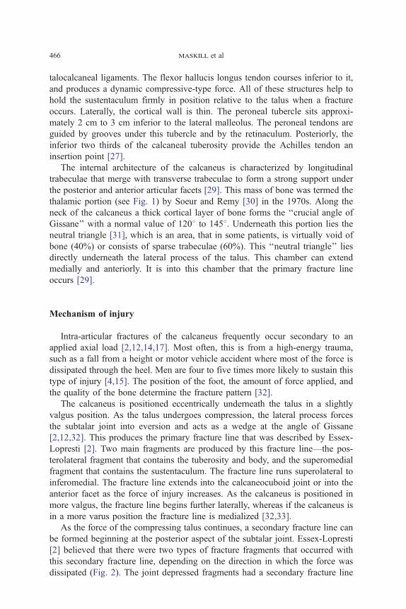
maskill et al466
talocalcaneal ligaments. The flexor hallucis longus tendon courses inferior to it,
and produces a dynamic compressive-type force. All of these structures help to
hold the sustentaculum firmly in position relative to the talus when a fracture
occurs. Laterally, the cortical wall is thin. The peroneal tubercle sits approxi-
mately 2 cm to 3 cm inferior to the lateral malleolus. The peroneal tendons are
guided by grooves under this tubercle and by the retinaculum. Posteriorly, the
inferior two thirds of the calcaneal tuberosity provide the Achilles tendon an
insertion point [27].
The internal architecture of the calcaneus is characterized by longitudinal
trabeculae that merge with transverse trabeculae to form a strong support under
the posterior and anterior articular facets [29]. This mass of bone was termed the
thalamic portion (see Fig. 1) by Soeur and Remy [30] in the 1970s. Along the
neck of the calcaneus a thick cortical layer of bone forms the ‘‘crucial angle of
Gissane’’ with a normal value of 1208 to 1458. Underneath this portion lies the
neutral triangle [31], which is an area, that in some patients, is virtually void of
bone (40%) or consists of sparse trabeculae (60%). This ‘‘neutral triangle’’ lies
directly underneath the lateral process of the talus. This chamber can extend
medially and anteriorly. It is into this chamber that the primary fracture line
occurs [29].
Mechanism of injury
Intra-articular fractures of the calcaneus frequently occur secondary to an
applied axial load [2,12,14,17]. Most often, this is from a high-energy trauma,
such as a fall from a height or motor vehicle accident where most of the force is
dissipated through the heel. Men are four to five times more likely to sustain this
type of injury [4,15]. The position of the foot, the amount of force applied, and
the quality of the bone determine the fracture pattern [32].
The calcaneus is positioned eccentrically underneath the talus in a slightly
valgus position. As the talus undergoes compression, the lateral process forces
the subtalar joint into eversion and acts as a wedge at the angle of Gissane
[2,12,32]. This produces the primary fracture line that was described by Essex-
Lopresti [2]. Two main fragments are produced by this fracture line—the pos-
terolateral fragment that contains the tuberosity and body, and the superomedial
fragment that contains the sustentaculum. The fracture line runs superolateral to
inferomedial. The fracture line extends into the calcaneocuboid joint or into the
anterior facet as the force of injury increases. As the calcaneus is positioned in
more valgus, the fracture line begins further laterally, whereas if the calcaneus is
in a more varus position the fracture line is medialized [32,33].
As the force of the compressing talus continues, a secondary fracture line can
be formed beginning at the posterior aspect of the subtalar joint. Essex-Lopresti
[2] believed that there were two types of fracture fragments that occurred with
this secondary fracture line, depending on the direction in which the force was
dissipated (Fig. 2). The joint depressed fragments had a secondary fracture line

Fig. 2. (A) Lateral drawing of the classic Essex-Lopresti tongue-type fracture. The fracture line ex-
tends directly posterior to produce a large tongue-like fragment through the tuberosity. (B) Lateral
drawing of the classic Essex-Lopresti joint depression–type fracture. The fracture line only extends just
beyond the posterior facet. This piece is free to depress down into the ‘‘neutral triangle’’ zone. (From
Fitzgibbons TC. Fractures and dislocations of the calcaneus. In: Bucholz RW, Heckman JD, editors.
Fractures in adults. 5th edition. Philadelphia: JB Lippincott Company; p. 2152; with permission).
calcaneus fractures: a review 467
posterior and superior to the posterior facet. The fracture line ran downward only,
essentially not involving the tuberosity. This created a free superolateral fragment
that was displaced inferiorly into the cancellous deficient neutral triangle. The
tongue-type fragment, however, showed a secondary fracture line that extended
longitudinally into the tuberosity. The anterior portion of the fragment was de-
pressed down into the neutral triangle with the posterior portion elevated, which
essentially reversed Bfhler’s angle.These observations have been validated by other studies in which calcaneus
fractures were created [17,34]. The primary and secondary fracture lines were
seen consistently (Fig. 3). One portion of the primary fracture line divided the
calcaneus into medial and lateral halves. The other divided it into anterior and
posterior halves. Depending on the amount of force and the position of the foot,
the primary fracture line extended anteriorly and creating an anterolateral
fragment. Multiple joint-depressed and tongue-type fragments were noted.
Radiographic assessment
When a patient is evaluated initially, plain radiographs always should be done.
If a intra-articular fracture is identified, a CT scan should be obtained. For the
calcaneus, many different radiographs have been described and can be difficult to
interpret consistently [4]. The CT scan of the calcaneus has brought new di-

Fig. 3. 1, Primary fracture line dividing in the coronal plane; 2, primary fracture line dividing
longitudinally in the sagittal plane. Superomedial (SM) fragment contains the sustentaculum; the
lateral fragment contains the tuberosity. In the joint depression–type fracture, a superolateral (SL)
fragment is created. With more energy, the anterolateral (AL) fragment is created. (From Carr JB.
Mechanism and pathoanatomy of the intra-articular calcaneal fracture. Clin Orthop 1993;290:37;
with permission.)
maskill et al468
agnostic and prognostic ability to the operating surgeon [1,5,6,17]. Because of
the association of calcaneus fractures with injuries of the spine, radiographs of the
lumbar spine should be considered if clinically appropriate.
The plain radiograph series that should be obtained includes a lateral radio-
graph of the hindfoot, an anteroposterior (AP) radiograph of the foot, and a Harris
axial radiograph of the heel (Fig. 4) [35]. The lateral radiograph of the hindfoot
(see Fig. 1) allows evaluation of the height of the calcaneus, Bfhler’s angle, andthe angle of Gissane. Small avulsion fractures off the anterior process or posterior
tuberosity also can be seen on this view. The diagnosis of a calcaneus fracture
should be confirmed on this view. Bfhler’s angle [12] usually is depressed and
the angle of Gissane [36] is widened. The angle of Gissane only increases when
the posterior facet is separated from the superomedial fragment. If only the lateral
half of the facet is fractured and depressed, a ‘‘double density’’ sign is noted.
In this case, Bfhler’s angle also will be normal [35]. The depressed articular
surface usually is rotated 908 relative to the remainder of the joint surface. The
lateral radiograph also indicates whether the fracture is a joint-depressed–type or
a tongue-type [2]. The AP radiograph of the foot shows possible extension of
the fracture line into the calcaneocuboid joint and any associated midfoot pa-
thology. The Harris axial radiograph is useful to look at the lateral wall com-
minution and position of the heel. Only the central portion of the posterior
articular facet is able to be visualized. The rest of the joint is superimposed with
other structures and cannot be relied on to evaluate the articular surface in its

Fig. 4. Normal roentograms. (A) AP view. (B) Hindfoot lateral view. (C) Harris axial heel view.
calcaneus fractures: a review 469
entirety [27]. This view is difficult to get in the acute setting secondary to pain.
Broden’s projection I (Fig. 5) [37] is an excellent way to visualize the entire
joint surface [4,5,35]. This view is obtained by laying the patient supine with
the radiograph cassette under the leg and ankle. The foot is in neutral flexion,
and the leg is rotated internally 308 to 408. The x-ray beam is centered over
the lateral malleolus and four radiographs are taken with the tube angled at 408,308, 208, and 108. This shows the articular surface of the posterior facet from
anterior to posterior and is most helpful in the operating room when evaluating
joint reduction.
With the advent of CT scanning, our understanding of these fractures has
increased significantly [5,6,33,38]. Coronal and transverse images are obtained
using 2-mm sections (Fig. 6). The coronal views are to be perpendicular to the

Fig. 5. Broden’s projection I. (A) Diagram depicting proper positioning with the x-ray beam. (B) A
Broden’s view at 208 showing the posterior articular facet. (From Sanders R, Fortin P, DiPasquale T,
et al. Operative treatment in 120 displaced intraarticular calcaneus fractures. Clin Orthop 1993;290:89;
with permission.)
maskill et al470
posterior articular facet, whereas the transverse views are parallel to the foot. All
cuts are to be at least 2 mm in thickness to evaluate the calcaneus properly. The
coronal views show the number and location of the articular fragments. Sanders
et al [6] showed this to be of prognostic significance and is the basis of his clas-
sification scheme. The calcaneal body can be evaluated for widening and
shortening, and the tuberosity for positioning (varus, valgus). The peroneal ten-
dons can be identified and impingement can be evaluated. The transverse images
also show the lateral wall blowout, because it shows comminution of the sus-
tentaculum and calcaneocuboid joint surface. The anteroinferior posterior articu-
lar facet also is seen best on these cuts. Three-dimensional CT scanning has been
studied over the past decade—and although the technology is improving—the
cost-benefit ratio is high. In a recent study, it was recommended that surgeons
who are not completely familiar with the three-dimensional anatomy of the
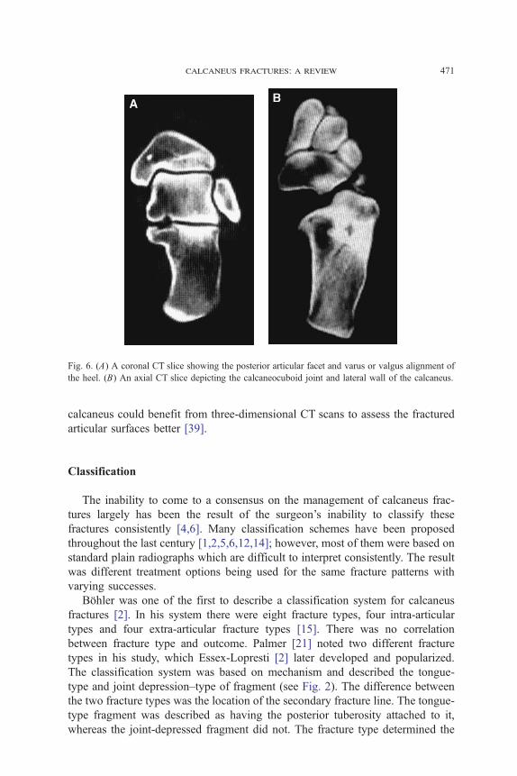
Fig. 6. (A) A coronal CT slice showing the posterior articular facet and varus or valgus alignment of
the heel. (B) An axial CT slice depicting the calcaneocuboid joint and lateral wall of the calcaneus.
calcaneus fractures: a review 471
calcaneus could benefit from three-dimensional CT scans to assess the fractured
articular surfaces better [39].
Classification
The inability to come to a consensus on the management of calcaneus frac-
tures largely has been the result of the surgeon’s inability to classify these
fractures consistently [4,6]. Many classification schemes have been proposed
throughout the last century [1,2,5,6,12,14]; however, most of them were based on
standard plain radiographs which are difficult to interpret consistently. The result
was different treatment options being used for the same fracture patterns with
varying successes.
Bfhler was one of the first to describe a classification system for calcaneus
fractures [2]. In his system there were eight fracture types, four intra-articular
types and four extra-articular fracture types [15]. There was no correlation
between fracture type and outcome. Palmer [21] noted two different fracture
types in his study, which Essex-Lopresti [2] later developed and popularized.
The classification system was based on mechanism and described the tongue-
type and joint depression–type of fragment (see Fig. 2). The difference between
the two fracture types was the location of the secondary fracture line. The tongue-
type fragment was described as having the posterior tuberosity attached to it,
whereas the joint-depressed fragment did not. The fracture type determined the
maskill et al472
treatment, but not the prognosis. These terms are still used to describe the frac-
ture morphology.
In the 1970s, Soeur and Remy [30] came up with a classification system which
was based on the number of articular fracture fragments. Plain radiographs
(lateral hindfoot, AP, Harris axial view) were used to asses the posterior articular
facet. First degree fractures were nondisplaced. Second degree fractures showed
secondary fracture lines that resulted in three fragments. Third degree fractures
were severely comminuted fractures and were unable to be classified. There was
no mention as to whether the comminution was of the calcaneal body or of the
articular facet. Their work did not correlate results with outcomes, but served as a
stepping stone for modern classification systems.
The precise assessment of fracture fragments by CT scan provides a con-
siderable advantage for modern classification schemes [1,5,6,38]. The most
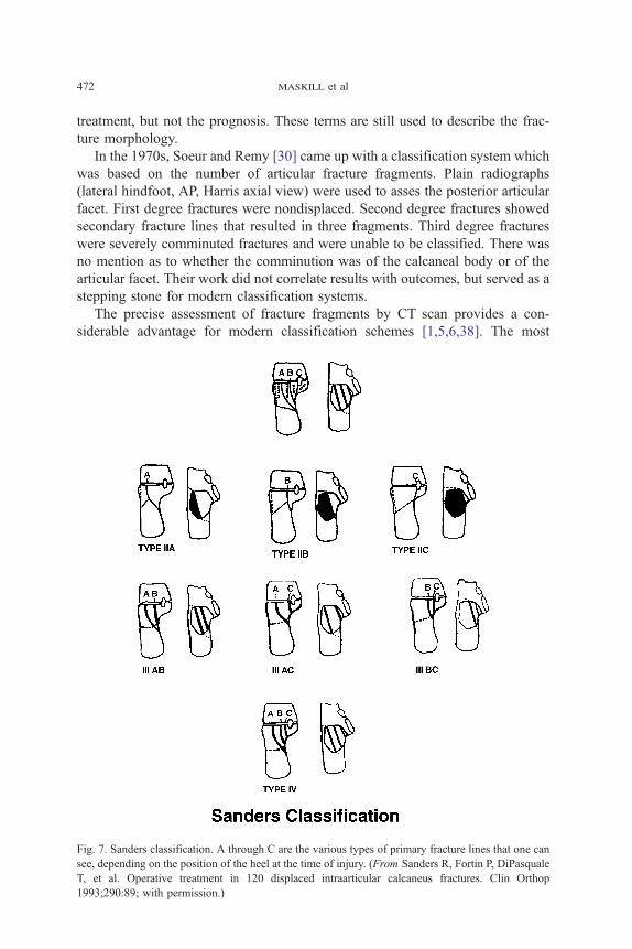
Fig. 7. Sanders classification. A through C are the various types of primary fracture lines that one can
see, depending on the position of the heel at the time of injury. (From Sanders R, Fortin P, DiPasquale
T, et al. Operative treatment in 120 displaced intraarticular calcaneus fractures. Clin Orthop
1993;290:89; with permission.)
calcaneus fractures: a review 473
widely used classification system is that of Sanders et al (Fig. 7) [6]. This system
bases its classification on the number of fracture fragments that is identified on a
semicoronal CT image. The image used is the one that displays the widest
undersurface of the posterior facet of the talus. Sanders et al described the talus as
being divided into three columns by two lines. These lines divided the posterior
articular facet into three potential pieces: a medial, a central, and a lateral frag-
ment. The addition of a third line that is located just medial to the medial edge
makes for a fourth possible fracture piece, the sustentaculum portion. All non-
displaced fractures (regardless of the number of fracture lines) are classified as
type I; one fracture line is a type II; two fracture lines is a type III; and three or
more fracture lines is a type IV. The lines are lettered according to placement on
the facet. Lateral fracture lines are type A, central lines are type B, and medial
lines are type C. This system has been useful in terms of determining treatment,
and was shown to correlate well with prognosis and level of operative difficulty.
Clinical evaluation
Clinical evaluation of a fractured hindfoot is critical. Because the soft tissue
envelope is fragile, treatment must be performed accordingly. The displacement
of the fracture and the degree of soft tissue injury are directly proportional to the
amount of force that is seen at the time of injury [2,4,15,17]. In severe cases, an
open fracture may occur. In subtle cases, the medial spike of the superomedial
fragment breaks the skin barrier with evidence of a small medial puncture wound.
A high index of suspicion is needed to avoid neglecting this dangerous fracture.
The surgeon must be careful not to overlook other fractures that might have
occurred in the foot. One also must be cognizant of the correlation of lumbar
spine fractures with this injury [40]. A thorough neurologic examination should
be performed.
Typical features of calcaneus fractures are significant swelling and hematoma
formation within the hindfoot. Frequently, the patient is unable to bear weight on
that extremity. Often, the foot has hindfoot bulging from the lateral wall blowout.
The peroneal tendons should be palpated along the posterior aspect of the lateral
malleolus to be sure that they have not dislocated. If the patient is seen after
6 hours, the swelling usually is so severe that the skin creases have disappeared.
Swelling may be so severe that blisters form—intradermal (clear fluid) and full
thickness (serosanguinous fluid) types [41,42]. Some fracture fragments might
be tenting the skin with the eminent risk of full-thickness skin necrosis [2,15].
Pain often is severe and compartment syndrome always should be ruled out.
Compartment syndrome is noted often in high-energy injuries or crush-type
injuries. Manoli and Weber [43] described nine compartments in the foot. Three
compartments run the entire length of the foot, whereas six are confined to the
forefoot or the hindfoot. The four interosseous compartments and the adductor
compartment make up the forefoot compartments and the calcaneal compartment

maskill et al474
lies in the hindfoot. The calcaneal compartment contains the quadratus plantae
and the lateral plantar nerve. This compartment is the one that is affected most
commonly in this type of fracture. The calcaneus—an extremely vascular bone—
bleeds into the compartment and affects the pulse pressure to the point where
arterial flow is compromised. If compartment syndrome is believed to be a pos-
sibility, the pressures should be measured. Because there is no current literature to
prove that the foot can withstand greater pressures than other fascial compart-
ments in the body, the current recommendation is to perform fasciotomies with
pressures greater than 30 mm Hg, or within 10 mm Hg to 30 mm Hg of the dia-
stolic blood pressure. These pressures have been found to occur in 10% of calca-
neus fractures [44]. If the diagnosis is missed, the patient can go on to develop
intrinsic contractures, claw toe deformities of the lesser toes, sensory abnor-
malities, stiffness, chronic aching, and atrophy with weakness [43,44].
Options for treatment
Treatment options can be broken down into the following categories: emer-
gent, nonoperative, minimally invasive ORIF, standard open reduction with in-
ternal fixation, and primary arthrodesis.
Emergency procedures
Emergent procedures are performed only in cases in which the soft tissue
envelope is compromised. Situations in which this might occur are a foot that
develops compartment syndrome, an open fracture, or severe tenting of the skin
by displaced bony fragments.
Compartment syndrome that has been confirmed with elevated pressures that
are greater than 30 mm Hg or within 10 mm Hg to 30 mm Hg of the diastolic
pressure should be dealt with emergently by performing a fasciotomy [43,44]. To
do this, a medial incision that starts 4 cm anterior to the posterior heel and 3 cm
superior to the plantar surface of the foot is made. Typically, the incision is ap-
proximately 6 cm in length. The medial compartment is opened and the abductor
hallucis muscle is elevated until the medial intermuscular septum is seen (Fig. 8).
This fascia is opened to release the deep calcaneal compartment. The lateral
plantar nerve is at risk with this procedure because it lies just lateral to the medial
septum. The dorsal compartments may be released as needed clinically, de-
pending on the type of injury seen.
Open calcaneus fractures are much less common than closed fractures.
Standard irrigation and debridement of the opened areas should be performed
[4,15]. Most often, this is a small puncture wound medially from the spike of the
superomedial fragment. The wound is covered with a standard dressing or a
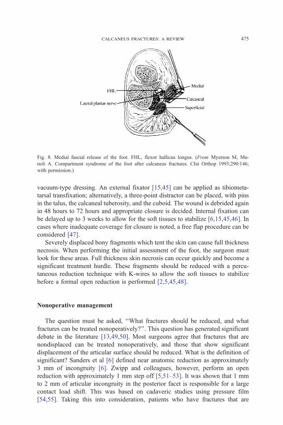
Fig. 8. Medial fascial release of the foot. FHL, flexor hallicus longus. (From Myerson M, Ma-
noli A. Compartment syndrome of the foot after calcaneus fractures. Clin Orthop 1993;290:146;
with permission.)
calcaneus fractures: a review 475
vacuum-type dressing. An external fixator [15,45] can be applied as tibiometa-
tarsal transfixation; alternatively, a three-point distractor can be placed, with pins
in the talus, the calcaneal tuberosity, and the cuboid. The wound is debrided again
in 48 hours to 72 hours and appropriate closure is decided. Internal fixation can
be delayed up to 3 weeks to allow for the soft tissues to stabilize [6,15,45,46]. In
cases where inadequate coverage for closure is noted, a free flap procedure can be
considered [47].
Severely displaced bony fragments which tent the skin can cause full thickness
necrosis. When performing the initial assessment of the foot, the surgeon must
look for these areas. Full thickness skin necrosis can occur quickly and become a
significant treatment hurdle. These fragments should be reduced with a percu-
taneous reduction technique with K-wires to allow the soft tissues to stabilize
before a formal open reduction is performed [2,5,45,48].
Nonoperative management
The question must be asked, ‘‘What fractures should be reduced, and what
fractures can be treated nonoperatively?’’. This question has generated significant
debate in the literature [13,49,50]. Most surgeons agree that fractures that are
nondisplaced can be treated nonoperatively, and those that show significant
displacement of the articular surface should be reduced. What is the definition of
significant? Sanders et al [6] defined near anatomic reduction as approximately
3 mm of incongruity [6]. Zwipp and colleagues, however, perform an open
reduction with approximately 1 mm step off [5,51–53]. It was shown that 1 mm
to 2 mm of articular incongruity in the posterior facet is responsible for a large
contact load shift. This was based on cadaveric studies using pressure film
[54,55]. Taking this into consideration, patients who have fractures that are

maskill et al476
nondisplaced or have less than 2 mm of displacement could be treated non-
operatively [4,5,13,25,49,56]. Patients who have diabetes mellitus (DM), periph-
eral vascular disease, or traumatic injury are not candidates for surgery. If the soft
tissue envelope is believed to be questionable in any way (eg, those who have
significant blistering or massive edema), the window of operative opportunity
may pass [4,5,15].
Nonoperative treatment typically includes rest, ice, elevation, and a posterior
splint until the swelling decreases. A compression dressing or stocking is helpful
with swelling. Early motion is started immediately. After the swelling is down,
the patient can be fitted with a removable boot or splint fixed at 908 to prevent an
equinus contracture [4]. Weight-bearing status is controversial. Zwipp et al [5]
restrict the patient immediately to a 20-kg limit on the affected extremity for
6 weeks to 10 weeks. Conversely, Sanders et al [4] recommend a nonweight-
bearing status for 12 weeks. Many investigators’ recommendations are in be-
tween these two [24,26,57–63].
Most investigators do not recommend formal reduction of these intra-articular
fractures [4,13,25,50]; however, Omoto and Nakamura [64] describe a technique
of repetitive squeezing combined with strong longitudinal traction while the
patient is anesthetized. The patient is then placed in a short-leg walking cast with
the heel in 458 of equinus. This method relies on ligamentotaxis. They reported
good to excellent results in 89 of 102 fractures; patients who had severe tongue-
types and comminuted joint depressed–types could not be reduced successfully.
The criteria for adequate reduction were not given, nor were the fractures
classified by CT.
The malunion that can follow nonoperative treatment can result in significant
clinical problems [4,9,11,15,65–67]. A reduction of the articular surface never is
obtained and carries the risk of arthritis in the subtalar joint, the calcaneocuboid
joint, and even the tibiotalar joint with chronic dorsiflexion of the talus [4]. The
patient could develop problems with shoe wear because the heel often is wide,
short, and in a varus position. The peroneal tendons can be a source of pain
secondary to impingement or chronic dislocation as a result of the lateral
wall blowout.
Minimally invasive options
Minimally invasive techniques are attractive in the management of intra-
articular calcaneus fractures. The benefit of such techniques are less soft tissue
trauma, and possibly, reduced cost. The risk involved, however, is the possible
acceptance of an incongruent reduction from lack of visualization. Indirect closed
reduction with percutaneous fixation has been done since Westhues introduced
the idea in 1934 [2,15]. Gissane popularized this method in the United States
during the next decade [2]. Essex-Lopresti [2] reported using this technique
to reduce tongue-type fracture fragments. He mentioned that joint depressed
fragments had to be opened because adequate control of the depressed frag-
calcaneus fractures: a review 477
ment was unable to be obtained. These investigators used plaster to immobilize
their reductions.
Today, this type of technique is useful to some investigators. Tornetta [48] de-
scribed the use of this technique; instead of Steinmann pins he used percutaneous
screws. Rammelt et al [51] have used this type of reduction and fixation with
Sanders type IIC fractures. This fracture fragment is essentially the entire pos-
terior facet because the fracture line is far medial. Therefore, the surgeon can
reduce the joint by facilitating reduction of the fracture fragment. In a recent
article, however, Rammelt et al [51] mentioned the idea of using a 1.9-mm/08arthroscope to view the reduction. This allows the indications for percutaneous
reduction to be advanced to Sanders types IIA and IIB fractures. If one could
visualize the joint surface and the reduction there would be no need to open.
Zwipp attempted percutaneous operative fixation on 21 Sanders types IIA and
IIB intra-articular fractures [52]. Three were unable to be reduced by way of
percutaneous method and thus, were discarded. The remaining 18 were able to be
reduced anatomically because they were visualized arthroscopically. Time to
operation was an average of 6 days postinjury to allow for stabilization of the soft
tissues. The patients were placed in a lateral decubitus position on the noninjured
side. Subtalar arthroscopy was performed first by way of the anterior or pos-
terolateral portal, depending on the fracture location. Any small bony avulsions or
cartilaginous bodies were removed through a second portal (posterolateral or
anterior). Reduction of the tuberosity was undertaken with a percutaneously placed
Steinmann pin as in open reduction with internal fixation (ORIF). The impacted
fragment was loosened with varus/valgus stress and the pin pulled downward to
restore the height. Percutaneous screws were placed into the tuberosity and into the
superomedial fragment. This was controlled by way of fluoroscopy. K-wires were
placed to assist in manipulation of the depressed fragment if needed. If impacted,
a pestle was inserted percutaneously to tap the joint surface back to position.
After the reduction was visualized arthroscopically and found to be adequate,
percutaneous screws were placed into the thalamus of the calcaneus parallel to the
posterior facet. Anterior process fractures also were fixed percutaneously unless
there was significant comminution. Patients were treated with physical therapy
starting on postoperative day 1 with active range of motion exercises.
One year postoperatively, 15 patients were available for review. Subjectively,
all were satisfied and the average American Orthopaedic Foot and Ankle Society
(AOFAS) Ankle/Hindfoot Score was 94.1. Twelve patients experienced no pain
during activities of daily living and work, whereas 3 reported occasional pain.
Return to work was noted at an average of 10.9 weeks. No wound complications
were seen (wound sloughs, deep infection, or hematoma). Bohler’s angle was
improved from a mean of 13.18 to a mean of 25.88 postoperatively. It was
concluded that Sanders type II fractures could undergo a percutaneous closed
reduction; IIA and IIB types require arthroscopic assistance. In skilled hands, this
could minimize scar formation, and thus, stiffness, and decrease the incidence of
wound problems. This cohort could not be compared with the cohort that under-
went ORIF because of the incorporation of more complex fracture patterns [15].

maskill et al478
Open reduction internal fixation
If a surgeon elects to perform an ORIF, the timing is critical. Massive swelling
is a contraindication to surgery on a closed fracture [65,68]. Time must be allotted
for this to recede. Sanders [4] recommended looking for a positive wrinkle test
(Fig. 9). This test is performed by direct palpation of the skin over the lateral
aspect of the calcaneus and by visual evaluation of this area when the patient
dorsiflexes and everts the foot. If skin wrinkling is seen and no pitting edema is
present, the patient is said to have a positive test and the operation may be
performed. Some studies showed good results with the use of foot pumps or
compression stockings to eliminate edema [69,70]. Generally, if longer than
3 weeks has elapsed since the time of injury, nonoperative intervention is the best
option because the fragments most likely have started to consolidate [4,13,15,25].
After the operative timing has been determined, the approach to the fracture
needs to be addressed. Several approaches have been advocated throughout the
years. These include the medial [33,57], sustentacular [15], sinus tarsi [71], and
extended lateral approaches [4,5,24,46,59,63]. As operative intervention became
popular again in the mid- 1970s, investigators focused more on the restoration of
the shape of the calcaneus rather than on the posterior facet. This led McReynolds
and Burdeaux to use the medial approach so as to obtain a solid reduction of the
tuberosity to the superomedial fragment [33,57]. The posterior facet was reduced
through the fracture under fluoroscopic guidance. There was no direct visuali-
zation of the facet from this approach. This led to inadequate reduction of the
joint surface. In his 21-year review, Burdeaux [57] reported the need for a lateral
incision to assist in reduction of the posterior facet in 14 of 63 fractures.
Stephenson [62,72] then reported on the combined (medial and lateral) approach,
for which he advocated going medially only if the tuberosity was unable to be
reduced adequately. A sustentacular approach was mentioned in the literature for
obtaining reduction of isolated sustentaculum fractures [15]. A 3-cm to 5-cm
incision is made directly over the palpable sustentaculum (2 cm below and 1 cm
distal to the medial malleolus). After reduction of the medial facet, 3.5-mm
Fig. 9. A positive wrinkle test. Wrinkles occur when the swelling has gone down indicating that it is
safe to proceed with ORIF.
calcaneus fractures: a review 479
screws can be placed aiming slightly plantar so as to avoid the sinus tarsi and the
posterior facet.
Currently. most investigators prefer the ‘‘extended lateral approach’’ with the
‘‘no-touch’’ technique for displaced intra-articular fractures of the posterior facet
[4,5,24,46,59,63]. The advantages are in obtaining an excellent view of the
posterior facet and lateral wall of the calcaneus. The original lateral, or Kocher,
approach initially was popularized by Palmer [21] in the 1940s, but results were
not able to be duplicated. Wound sloughs and a high infection rate were frequent,
owing to the fragile soft tissue envelope laterally and the watershed area in this
region. Letournel [59] modified Palmer’s approach by placing the incisions more
posteriorly and inferiorly using a full thickness skin flap so as not to disrupt the
peroneal tendons, sural nerve, or calcaneofibular ligament. This approach has
been used successfully in several large studies [1,4,5,13,24,26,46,50,59,60,63].
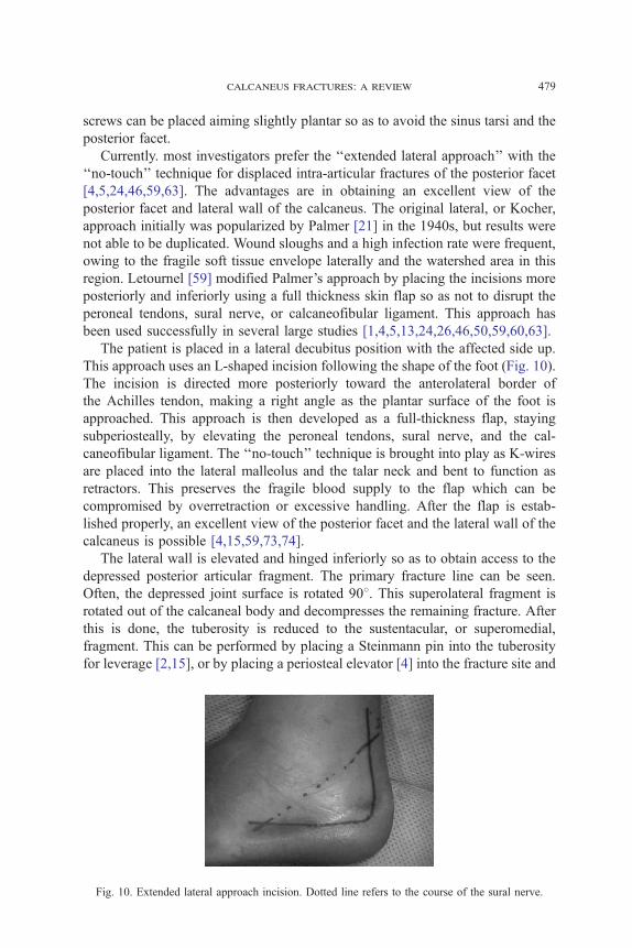
The patient is placed in a lateral decubitus position with the affected side up.
This approach uses an L-shaped incision following the shape of the foot (Fig. 10).
The incision is directed more posteriorly toward the anterolateral border of
the Achilles tendon, making a right angle as the plantar surface of the foot is
approached. This approach is then developed as a full-thickness flap, staying
subperiosteally, by elevating the peroneal tendons, sural nerve, and the cal-
caneofibular ligament. The ‘‘no-touch’’ technique is brought into play as K-wires
are placed into the lateral malleolus and the talar neck and bent to function as
retractors. This preserves the fragile blood supply to the flap which can be
compromised by overretraction or excessive handling. After the flap is estab-
lished properly, an excellent view of the posterior facet and the lateral wall of the
calcaneus is possible [4,15,59,73,74].
The lateral wall is elevated and hinged inferiorly so as to obtain access to the
depressed posterior articular fragment. The primary fracture line can be seen.
Often, the depressed joint surface is rotated 908. This superolateral fragment is
rotated out of the calcaneal body and decompresses the remaining fracture. After
this is done, the tuberosity is reduced to the sustentacular, or superomedial,
fragment. This can be performed by placing a Steinmann pin into the tuberosity
for leverage [2,15], or by placing a periosteal elevator [4] into the fracture site and
Fig. 10. Extended lateral approach incision. Dotted line refers to the course of the sural nerve.
maskill et al480
levering the tuberosity down while shifting it medially. This restores the height
and length of the calcaneus and brings the heel out of varus.
After the height and length have been restored, attention can be focused on the
joint reduction. The joint is reduced from medial to lateral, using the supero-
medial fragment as the stable piece [4,15,75]. The anterolateral corner of the
superolateral fragment should line up with the posterolateral corner of the
anterolateral fragment to restore Gissane’s angle properly [4]. After the articular
surface is reapproximated, 3.5-mm cortical screws are placed from lateral to me-
dial into the sustentacular bone. Broden’s views are an excellent way to asses the
reduction of the posterior facet using intraoperative fluoroscopy [4,15,35]. After
the joint surface is reduced, the body of the calcaneus is ready for fixation. At this
point, there is most likely a large defect from the impaction of the cancellous
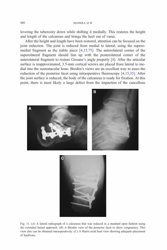
Fig. 11. (A) A lateral radiograph of a calcaneus that was reduced in a standard open fashion using
the extended lateral approach. (B) A Broden view of the posterior facet to show congruency. This
view also can be obtained intraoperatively. (C) A Harris axial heel view showing adequate placement
of hardware.
calcaneus fractures: a review 481
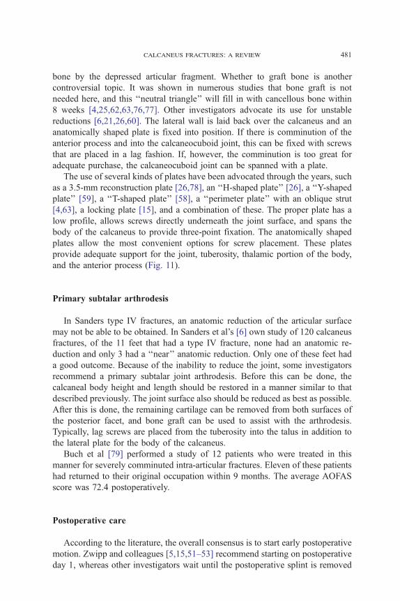
bone by the depressed articular fragment. Whether to graft bone is another
controversial topic. It was shown in numerous studies that bone graft is not
needed here, and this ‘‘neutral triangle’’ will fill in with cancellous bone within
8 weeks [4,25,62,63,76,77]. Other investigators advocate its use for unstable
reductions [6,21,26,60]. The lateral wall is laid back over the calcaneus and an
anatomically shaped plate is fixed into position. If there is comminution of the
anterior process and into the calcaneocuboid joint, this can be fixed with screws
that are placed in a lag fashion. If, however, the comminution is too great for
adequate purchase, the calcaneocuboid joint can be spanned with a plate.
The use of several kinds of plates have been advocated through the years, such
as a 3.5-mm reconstruction plate [26,78], an ‘‘H-shaped plate’’ [26], a ‘‘Y-shaped
plate’’ [59], a ‘‘T-shaped plate’’ [58], a ‘‘perimeter plate’’ with an oblique strut
[4,63], a locking plate [15], and a combination of these. The proper plate has a
low profile, allows screws directly underneath the joint surface, and spans the
body of the calcaneus to provide three-point fixation. The anatomically shaped
plates allow the most convenient options for screw placement. These plates
provide adequate support for the joint, tuberosity, thalamic portion of the body,
and the anterior process (Fig. 11).
Primary subtalar arthrodesis
In Sanders type IV fractures, an anatomic reduction of the articular surface
may not be able to be obtained. In Sanders et al’s [6] own study of 120 calcaneus
fractures, of the 11 feet that had a type IV fracture, none had an anatomic re-
duction and only 3 had a ‘‘near’’ anatomic reduction. Only one of these feet had
a good outcome. Because of the inability to reduce the joint, some investigators
recommend a primary subtalar joint arthrodesis. Before this can be done, the
calcaneal body height and length should be restored in a manner similar to that
described previously. The joint surface also should be reduced as best as possible.
After this is done, the remaining cartilage can be removed from both surfaces of
the posterior facet, and bone graft can be used to assist with the arthrodesis.
Typically, lag screws are placed from the tuberosity into the talus in addition to
the lateral plate for the body of the calcaneus.
Buch et al [79] performed a study of 12 patients who were treated in this
manner for severely comminuted intra-articular fractures. Eleven of these patients
had returned to their original occupation within 9 months. The average AOFAS
score was 72.4 postoperatively.
Postoperative care
According to the literature, the overall consensus is to start early postoperative
motion. Zwipp and colleagues [5,15,51–53] recommend starting on postoperative
day 1, whereas other investigators wait until the postoperative splint is removed
maskill et al482
[6,25,26,46,59,63]. Weight bearing also is controversial. Some investigators start
patients off immediately with partial weight bearing in their own shoes [5,16].
Sanders [4] recommends a boot fixed in neutral flexion so as to prevent equinus
contractures, followed by a nonweight-bearing status for 12 weeks. No studies
have been done to validate either end of the spectrum.
Complications
The complications of calcaneus fractures can be divided into two groups:
operative and nonoperative. Superficial wound edge necrosis is the most
frequently observed operative complication [4,15,26,65,81]. This has been seen
in up to 14% of cases after the standard extended lateral approach [15,26,46,
65,74], and has been as high as 27% with the combined (medial and lateral)
approaches [62]. This increased sensitivity to dehiscence is secondary to presence
of the watershed zone in this area. Often, the incision closes easily, but subse-
quently dehisces as late as 4 weeks postoperatively [4,65,74]. If this occurs,
motion should be stopped and the wound should be treated with daily whirlpools
and saline dressings to allow for secondary closure. The use of vacuum dressing
also is useful in this scenario. Although results are anecdotal, it was found to
enhance the formation of healthy granulation tissue in the wound by enhancing
the oxygenation. With the use of this dressing, the authors have been able to
avoid the need for flap coverage, even in the diabetic smoker. If this is still un-
successful, a fasciocutaneous flap may be needed [46,82]. The incidence of deep
infection is far less common—1.3% to 7% [15,26,47,74]. Most patients do not
have diffuse osteomyelitis, but the superficial type, as a result of direct extension
from an adjacent source [4]. In this type, the hardware may be retained, but
the wound bed should be cleaned thoroughly. The patient should be placed on
6 weeks of intravenous antibiotics. If the osteomyelitis is diffuse, hardware re-
moval is necessary with adequate debridement of bone [4,15,74]. Open fractures,
smoking, delay in surgery more than 14 days, and obesity are risk factors for deep
infections [4,65,83]. Injuries to the cutaneous nerves frequently affect the sural
nerve because of the popularity of the lateral approach [4,74,84]. Medially, the
calcaneal branch of the posterior tibial nerve is affected most often [4]. Numbness
of the area is mostly observed which is treated nonoperatively. A painful neu-
roma can develop which should be excised with burial of the stump in deep
tissue [4]. Peroneal tendonitis may occur secondary to prominent hardware, or
disruption of the tendon sheath and scarring. With the former Kocher approach,
the tendon sheaths were violated and the tendons were dislocated to gain better
access to the subtalar joint. With the extended lateral approach, a subperiosteal
dissection is advocated so as not to disturb the peroneal tendon bed, but rather,
elevate them as a whole [59]. If scarring has occurred, an operative release with
hardware removal is recommended [4]. As with any surgery, a failed attempt
to reduce the joint can lead to arthrosis secondary to incongruency from a
malunion. This ability to obtain an adequate reduction is dependent on the

calcaneus fractures: a review 483
surgeon and on the fracture type. The incidence of nonunion is rare after stable
internal fixation. Bone graft and the use of larger screws can aid in healing [15].
Many surgeons treat calcaneus fractures conservatively, either because of lack
of familiarity with operative techniques or because they fear the surgical com-
plications; however, complications from nonoperative treatment can be just as
troubling. Malunions can be responsible for painful subtalar arthritis, malposition
of the talus which leads to tibiotalar impingement and ankle pain, shortening or
widening of the hindfoot, fibulocalcaneal impingement, varus or valgus mal-
alignment, impingement or subluxation of the peroneal tendons, or sural or pos-
terior tibial neuritis [3,7,8,20,56,85–87]. Although painful subtalar joint arthritis
Fig. 12. (A) A Harris view of a calcaneus fracture malunion in varus. The wide heel from the lateral
wall blowout and the varus tuberosity cause significant morbidity. (B) A hindfoot lateral view showing
significant loss of height and reduction of Bfhler’s angle, and an increase in the angle of Gissane. No
significant subtalar arthrosis is seen. (C) An AP view showing decreased joint space in the
calcaneocuboid joint.

maskill et al484
can be treated with an isolated subtalar fusion [20], the deformity of the calcaneus
must be corrected to restore adequate function to the hindfoot (Fig. 12).
As early as 1921, Cotton [81] noted the maladies that were associated with
malunions and recommended decompression of the lateral wall and the lateral
aspect of the joint to relieve abutment. Carr et al [88] were the first to suggest
subtalar distraction bone block arthrodesis to re-establish calcaneal height and
relieve tibiotalar impingement. Romash [89] suggested adding a corrective osteo-
tomy along the former fracture line and reported favorable results in 90% of
cases. Stephens and Sanders [86] derived a prognostic classification system for
malunions. Type I include a large lateral exostosis with or without extremely
lateral subtalar arthrosis. Type II include a calcaneus with a lateral exostosis,
combined with arthrosis across the width of the subtalar joint, and type III has a
lateral exostosis with severe arthrosis of the subtalar joint and malalignment of
the heel in varus or valgus. Treatment is tailored to the type of deformity: lateral
wall decompression, peroneal tenolysis, and an extremely lateral joint resection
for type I; additional in situ subtalar arthrodesis for type II; and an additional
calcaneal osteotomy to correct height and varus/valgus malalignment for type III.
Results of operative treatment
Many clinical studies deal specifically with operative treatments of intra-
articular calcaneus fractures. It is still difficult to draw conclusions because many
variables exist between them, such as different classification systems, functional
outcome measurements, and overall relative low patient numbers [4,13,15]. Five
large (N100 patients) studies of intra-articular fractures that were classified by
way of CT scanning and treated with ORIF showed good to excellent results in
60% to 85% of cases [5,6,14,59,63]. These studies, however, used different
outcome measurements. Sanders et al [6] concluded in their study on 120 cal-
caneal fractures that: (1) an anatomic articular reduction is needed to obtain an
excellent or good result; (2) an anatomic articular reduction cannot ensure a good
to excellent result, most likely because of the cartilaginous damage that is
incurred at the time of injury; (3) a reproducible operative technique is surgeon
dependent and 35 to 50 cases are required to pass the learning curve; and
(4) Sanders type IV fractures are so severe that primary arthrodesis is warranted.
Several studies compared nonoperative treatments with operative treatments
for displaced intra-articular fractures of more than 2 mm [13,25,49,50,80]. Most
of these are retrospective. Although some show significantly greater functional
results with ORIF [10,24,25,49,60], others observed no significant difference
between the groups [13,50,80]. Two studies [50,80] that showed no significance
between operative and nonoperative treatments did show that in patients who
were treated operatively, anatomic reduction of the intra-articular surface cor-
responded with a better clinical score than no reduction or a ‘‘less than adequate
reduction.’’ The numbers of patients in these studies were not large. One pro-
spective, randomized trial that was evaluated by Thordarson and Krieger [25]
calcaneus fractures: a review 485
looked at 15 operative cases and 11 nonoperative cases at an average of 17 and
14 months follow-up, respectively. All fractures were Sanders types II and III.
They showed 12 good to excellent results in the operative group versus 4 in the
nonoperative group. Another larger prospective, randomized, controlled multi-
center trial by Buckley et al [13], which involved 471 displaced intra-articular
calcaneal fractures, showed that without stratification of the groups, the func-
tional results between operative and nonoperative care were insignificant. When
the data were stratified, and when those who were receiving Workman’s Com-
pensation benefits were removed from the study, the outcomes were better in
the group that was treated operatively. These results also were obtained from
many different surgeons. Sanders’ classification system was used to stratify the
fractures; it was confirmed that those who had less comminution (type II) were
three times more likely to score above the mean on the Short Form–36 and Visual
Analog Scale when treated operatively. There was no difference between opera-
tive and nonoperative treatment in patients who had more comminution (type
IV). Postoperatively, Bohler’s angle was found to be prognostic. It also was noted
that significantly less subtalar fusions were necessary after operative treatment.
Among patients who were not receiving Workman’s Compensation benefits,
women, in general; younger patients (b29 years old); and patients who had a
moderately lower Bohler angle, an anatomic reduction, or step off of less than
2 mm scored much higher when treated surgically.
Summary
Calcaneus fractures are a significant burden to society. Assessment and treat-
ment of these injuries has improved significantly over the past 2 decades with the
use of CT scanning. It has allowed us greater understanding of the pathologic
anatomy of these fractures, and has provided us with a prognostic classification
system with respect to outcome. Nonoperative treatment is effective for fractures
that are nondisplaced or minimally displaced (b2 mm). ORIF is the standard
therapy for fractures that are displaced greater than 2 mm, with 65% to 80% good
to excellent results. To obtain these results, the soft tissues always must be re-
spected. Compartment syndrome always should be ruled out clinically and open
fractures should be treated aggressively. A minimally invasive approach with the
assistance of subtalar arthroscopy is an attractive option for fracture types with
minimal comminution (Sanders type II), but should be reserved for the more
experienced surgeon. An extended lateral approach respects the local anatomy
and provides the best opportunity to restore the congruity of the joint surface.
Anatomic restoration of the articular surface and restoration of the original shape
of the calcaneus (Bohler’s angle) are of prognostic value. These measures can be
assessed intraoperatively with the Harris axial view, the lateral hindfoot view, and
the Broden view with fluoroscopy. Patients who have severely comminuted
fractures (Sanders type IV) can be treated with an ORIF of the body of the
calcaneus combined with primary subtalar arthrodesis.
maskill et al486
References
[1] Crosby LA, Fitzgibbons T. Computerized tomography scanning of acute intra-articular fractures
of the calcaneus. A new classification system. J Bone Joint Surg Am 1990;72:852–9.
[2] Essex-Lopresti P. The mechanism, reduction technique, and results in fractures of the os calcis.
Br J Surg 1952;39:395–419.
[3] Sanders R. Intra-articular fractures of the calcaneus: present state of the art. J Orthop Trauma
1992;6:252–65.
[4] Sanders R. Displaced intra-articular fractures of the calcaneus. J Bone Joint Surg Am 2000;82:
225–50.
[5] Zwipp H, Tscherne H, Thermann H, et al. Osteosynthesis of displaced intra-articular fractures of
the calcaneus. Results in 123 cases. Clin Orthop 1993;290:76–86.
[6] Sanders R, Fortin P, DiPasquale A, et al. Operative treatment in 120 displaced intra-articular
calcaneal fractures. Results using a prognostic computed tomographic scan classification. Clin
Orthop 1993;290:87–95.
[7] Carr JB, Hansen S, Benirschke S. Subtalar distraction bone block fusion for late complications of
os calcis fractures. Foot Ankle 1988;9:81–6.
[8] James ET, Hunter GA. The dilemma of painful old os calcis fractures. Clin Orthop 1983;177:
112–5.
[9] Myerson M, Quill G. Late complications of fractures of the calcaneus. J Bone Joint Surg Am
1993;75:331–41.
[10] Catani F, Benedetti MG, Simoncini L. Analysis of function after intra-articular fracture of the os
calcis. Foot Ankle Int 1999;20:417–21.
[11] Sangeorzan B. Salvage procedures for calcaneus fractures. Instr Course Lect 1997;46:339–46.
[12] Bfhler L. Diagnosis, pathology, and treatment of fractures of the os calcis. J Bone Joint Surg
1931;13:75–89.
[13] Buckley R, Tough S, McCormack R. Operative compared with nonoperative treatment of
displaced intra-articular fractures: a prospective, randomized, controlled multicenter trial. J Bone
Joint Surg Am 2002;84:1733–44.
[14] Conn HR. The treatment of fractures of the os calcis. J Bone Joint Surg 1935;17:392–405.
[15] Rammelt S, Zwipp H. Calcaneus fractures: facts, controversies and recent developments. Injury
2004;35:443–61.
[16] Cotton FJ, Henderson FF. Results of fractures of the os calcis. Am J Orthop Surg 1916;14:
290–8.
[17] Carr JB, Hamilton JJ, Bear LS. Experimental intra-articular calcaneal fractures: anatomical basis
for a new classification. Foot Ankle 1989;10:81–7.
[18] Cotton FJ, Wilson LT. Fractures of the os calcis. Boston Med J 1908;159:559–65.
[19] McLaughlin HL. Treatment of complications after os calcis fractures. Clin Orthop 1963;30:111–5.
[20] Gallie WE. Subastragalar arthrodesis in fractures of the os calcis. J Bone Joint Surg 1943;25:
731–6.
[21] Palmer I. The mechanism and treatment of fractures of the calcaneus. J Bone Joint Surg Am
1948;30:2–8.
[22] Lindsay WRN, Dewar FP. Fractures of the os calcis. Am J Surg 1958;95:555–76.
[23] Rowe CR, Sakellarides HT, Freeman PA, et al. Fractures of os calcis. A long term follow-up
study of one hundred forty-six patients. JAMA 1963;184:920–3.
[24] Crosby LA, Fitzgibbons TC. Open reduction and internal fixation of type II intra-articular
calcaneus fractures. Foot Ankle Int 1996;17:253–8.
[25] Thordarson DB, Krieger LE. Operative vs. nonoperative treatment of intra-articular fractures of
the calcaneus: a prospective randomized trial. Foot Ankle Int 1996;17:2–9.
[26] Benirschke SK, Sangeorzan BJ, Hansen ST. Results of operative treatment of calcaneal fractures
of the foot. Surgical management of calcaneal fractures. Clin Orthop 1993;292:128–34.
[27] Hall RL, Shereff MJ. Anatomy of the calcaneus. Clin Orthop 1993;290:27–35.
[28] Sarrafian SK. Anatomy of the foot and ankle. Philadelphia7 Lippincott; 1993.

calcaneus fractures: a review 487
[29] Sabry FF, Ebraheim NA, Mehalik JN, et al. Internal architecture of the calcaneus: implications
for calcaneus fractures. Foot Ankle Int 2000;21:114–8.
[30] Soeur R, Remy R. Fractures of the calcaneus with displacement of the thalamic portion. J Bone
Joint Surg Br 1975;57:413–21.
[31] Harty M. Anatomic consideration in injuries of the calcaneus. Orthop Clin North Am 1973;4:
179–83.
[32] Carr JB. Mechanism and pathoanatomy of the intra-articular calcaneal fracture. Clin Orthop
1993;290:36–40.
[33] Burdeaux BD. Reduction of calcaneal fractures by the McReynolds medial approach technique
and its experimental basis. Clin Orthop 1983;177:87–103.
[34] Wqlker N, Zwipp H. Fracture anatomy of the calcaneus with axial loading. Foot Ankle Surg
1986;2:155–62.
[35] Koval KJ, Sanders R. The radiologic evaluation of calcaneal fractures. Clin Orthop 1993;290:
41–6.
[36] Gissane W. News notes: Proceedings of the British Orthopedic Association. J Bone Joint Surg
1947;29:254–5.
[37] Broden B. Roentgen examination of the subtaloid joint in fractures of the calcaneus. Acta Radiol
1949;31:85–91.
[38] Janzen DL, Connell DG, Munk PL, et al. Intraarticular fractures of the calcaneus: value of CT
findings in determining prognosis. American Journal of Roentgenology 1992;158:1271–4.
[39] Prasartritha T, Sethavanitch C. Three-dimensional and two-dimensional computerized tomo-
graphic demonstration of calcaneus fractures. Foot Ankle Int 2004;25:262–73.
[40] Hansen Jr S. Foot injuries. In: Skeletal trauma. 2nd edition. Philadelphia: W.B Saunders
Company; 1998. p. 2405–38.
[41] Giordano CP, Koval KJ, Zuckerman JD, et al. Fracture blisters. Clin Orthop 1994;307:214–21.
[42] Giordano CP, Koval KJ. Treatment of fracture blisters: a prospective study of 53 cases. J Orthop
Trauma 1995;9:171–6.
[43] Manoli AD, Weber TG. Fasciotomy of the foot: an anatomical study with special reference to
release of the calcaneal compartment. Foot Ankle 1990;10:267–75.
[44] Myerson M, Manoli A. Compartment syndromes of the foot after calcaneal fractures. Clin
Orthop 1993;290:142–50.
[45] Baumgaertel FR, Gotzen L. Two stage operative treatment of comminuted os calcis fractures:
primary indirect reduction with medial external fixation and delayed lateral plate fixation. Clin
Orthop 1993;290:132–41.
[46] Bezes H, Massart P, Delvaux D, et al. The operative treatment of intraarticular calcaneal
fractures. Indications, technique, and results in 257 cases. Clin Orthop 1993;290:55–9.
[47] Brenner P, Rammelt S, Gavlik JM, et al. Early soft tissue coverage after complex foot trauma.
World J Surg 2001;25:603–9.
[48] Tornetta III P. The Essex-Lopresti reduction for calcaneal fractures revisited. J Orthop Trauma
1998;12:469–73.
[49] Randle JA, Kreder HJ, Stephen D, et al. Should calcaneal fractures be treated surgically? Clin
Orthop 2000;377:217–27.
[50] Buckley RE, Meek RN. Comparison of open versus closed reduction of intraarticular calcaneal
fractures: a matched cohort in workmen. J Orthop Trauma 1992;6:216–22.
[51] Rammelt S, Gavlik JM, Barthel S, et al. The value of subtalar arthroscopy in the management of
intra-articular calcaneus fractures. Foot Ankle Int 2002;23:906–16.
[52] Gavlik JM, Rammelt S, Zwipp H. Percutaneous, arthroscopically-assisted osteosynthesis of
calcaneus fractures. Arch Orthop Trauma Surg 2002;122:424–8.
[53] Gavlik JM, Rammelt S, Zwipp H. The use of subtalar arthroscopy in open reduction and internal
fixation of intra-articular calcaneal fractures. Injury 2002;33:63–71.
[54] Mulcahy DM, McCormack DM, Stephens MM. Intra-articular calcaneal fractures: effect of open
reduction and internal fixation on the contact characteristics of the subtalar joint. Foot Ankle Int
1998;19:842–8.
maskill et al488
[55] Sangeorzan BJ, Ananthakrishnan D, Tencer AF. Contact characteristics of the subtalar joint after
a simulated calcaneus fracture. J Orthop Trauma 1995;9:251–8.
[56] Crosby LA, Fitzgibbons TC. Intra-articular calcaneal fractures. Results of closed treatment. Clin
Orthop 1993;290:47–54.
[57] Burdeaux BD. Fractures of the calcaneus: open reduction and internal fixation from the medial
side, a 21-year prospective study. Foot Ankle Int 1997;18:685–92.
[58] Laughlin RT, Carson JG, Calhoun JH. Displaced intra-articular calcaneus fractures treated with
the Galveston plate. Foot Ankle Int 1996;17:71–8.
[59] Letournel E. Open reduction and internal fixation of calcaneal fractures. Clin Orthop 1993;290:
60–7.
[60] Leung KS, Yuen KM, Chan WS. Operative treatment of displaced intra-articular fractures of the
calcaneum. Medium-term results. J Bone Joint Surg Br 1993;75:196–201.
[61] Paley D, Hall H. Intra-articular fractures of the calcaneus. A critical analysis of results and
prognostic factors. J Bone Joint Surg Am 1993;75:342–54.
[62] Stephenson JR. Treatment of displaced intra-articular fractures of the calcaneus using medial and
lateral approaches, internal fixation, and early motion. J Bone Joint Surg Am 1987;69:115–30.
[63] Thordarson DB, Latteier M. Open reduction and internal fixation of calcaneal fractures with a
low profile titanium calcaneal perimeter plate. Foot Ankle Int 2003;24:217–21.
[64] Omoto H, Nakamura K. Method for manual reduction of displaced intra-articular fracture of the
calcaneus: technique, indications and limitations. Foot Ankle Int 2001;22:874–9.
[65] Abidi NA, Dhawan S, Gruen GS, et al. Wound healing risk factors after open reduction and
internal fixation of calcaneal fractures. Foot Ankle Int 1998;19:856–61.
[66] Burton DC, Olney BW, Horton GA. Late results of subtalar distraction fusion. Foot Ankle Int
1998;19:197–202.
[67] Chandler JT, Bonar SK, Anderson RB, et al. Results of in situ subtalar arthrodesis for late
sequelae of calcaneus fractures. Foot Ankle Int 1999;20:18–24.
[68] Folk JW, Star AJ, Early JS. Early wound complications of operative treatment of calcaneus
fractures: analysis of 190 fractures. J Orthop Trauma 1999;13:369–72.
[69] Gardner AM, Fox RH, Lawrence C, et al. Reduction of post-traumatic swelling and compartment
pressure by impulse compression of the foot. J Bone Joint Surg Br 1990;72:810–5.
[70] Myerson MS, Henderson MR. Clinical application of a pneumatic intermittent impulse com-
pression device after trauma and major surgery to the foot and ankle. Foot Ankle Int 1996;
14:198–203.
[71] Ebraheim NA, Elgafy H, Sabry FF, et al. Sinus tarsi approach with trans-articular fixation for
displaced intra-articular fractures of the calcaneus. Foot Ankle Int 2000;21:105–13.
[72] Stephenson JR. Surgical treatment of displaced intraarticular fractures of the calcaneus:
a combined lateral and medial approach. Clin Orthop 1993;290:68–75.
[73] Gould N. Lateral approach to the os calcis. Foot Ankle 1984;4:218–20.
[74] Harvey EJ, Grujic L, Early JS, et al. Morbidity associated with ORIF of intra-articular calcaneus
fractures using the lateral approach. Foot Ankle Int 2001;22(11):868–73.
[75] Stephenson JR. Displaced fractures of the os calcis involving the subtalar joint: the key role of
the superomedial fragment. Foot Ankle 1983;4:91–101.
[76] Longino D, Buckley RE. Bone graft in the operative treatment of displaced intra-articular
calcaneal fractures: is it helpful. J Orthop Trauma 2001;15:280–6.
[77] O’Farrell D, Jim OB, McCabe JP, et al. Fractures of the os calcis: improved results with internal
fixation. Injury 1993;24:263–5.
[78] Tile M. Fractures of the calcaneus. In: Schatzker J, Tile M, editors. The rationale of operative
fracture care. Berlin7 Springer-Verlag; 1996. p. 589–603.
[79] Buch BD, Myerson MS, Miller SD. Primary subtalar arthrodesis for the treatment of comminuted
calcaneal fractures. Foot Ankle Int 1996;17:61–70.
[80] Kundel K, Funk E, Brutscher M, et al. Calcaneal fractures: operative versus nonoperative
treatment. J Trauma 1996;41:839–45.
[81] Levin LS, Nunley JA. The management of soft-tissue problems associated with calcaneal
fractures. Clin Orthop 1993;290:151–6.

calcaneus fractures: a review 489
[82] Brenner P, Rammelt S, Gavlik JM, et al. Early soft tissue coverage after complex foot trauma.
World J Surg 2001;25:603–9.
[83] Attinger C, Cooper B. Soft tissue reconstruction for calcaneal fractures or osteomyelitis. Orthop
Clin North Am 2001;32:135–70.
[84] Paley D, Hall H. Intra-articular fractures of the calcaneus. A critical analysis of results and
prognostic factors. J Bone Joint Surg Am 1993;75:342–54.
[85] Kitaoka HB, Schaap EJ, Chao EY, et al. Displaced intra-articular fractures of the calcaneus
treated non-operatively. Clinical results and analysis of motion and ground-reaction and temporal
forces. J Bone Joint Surg Am 1994;76:1531–40.
[86] Stephens HM, Sanders R. Calcaneal malunions: results of a prognostic computed tomography
classification system. Foot Ankle Int 1996;17:395–401.
[87] Cotton FJ. Old os calcis fractures. Ann Surg 1921;74:294–303.
[88] Carr JB, Hansen S, Benirschke S. Subtalar distraction bone block fusion for late complications of
os calcis fractures. Foot Ankle 1988;9:81–6.
[89] Romash MM. Reconstructive osteotomy of the calcaneus with subtalar arthrodesis for malunited
calcaneal fractures. Clin Orthop 1993;290:157–67.