calcium dependence of membrane sealing at the cut end of ... · department of physiology, ... in...
TRANSCRIPT

0270.6474/85/0506-1626$02.00/O Copyright 0 Soaety for Neuroscience Printed m U.S.A.
The Journal of Neuroscience Vol. 5, No. 6, pp. 1626-1632
June 1985
Calcium Dependence of Membrane Sealing at the Cut End of the Cockroach Giant Axon’
HIROMU YAW0 AND MOTOY KUNO’
Department of Physiology, Kyoto University, Faculty of Medicine, Kyoto 606, Japan
Abstract
Following transection of a nerve fiber, the ruptured mem- brane must be resealed before regeneration from the proxi- mal cut end. However, it is not certain how fast the cut end is resealed or how it is resealed. We have examined the membrane sealing process following transection of the cock- roach giant axon, using recovery of decreased membrane potential and input resistance as criteria. The cable analysis and the injury current profile measured under voltage clamp suggested that the injured site is resealed by a structure with high electrical resistances 5 to 30 min after the transection. This recovery of membrane properties roughly coincided in time with the appearance of a partition-like structure formed near the cut end. The membrane resealing process was highly dependent upon temperature and extracellular Ca*+. Substitution of Ca2+ by equimolar Mg*‘, Mn*+, or SP’ failed to induce the membrane sealing following transection. How- ever, even in a Ca2+-free solution, the membrane resealing ocurred if Ca2+ was injected into the axoplasm near the cut end. Thus, the membrane resealing appears to require Ca2+ entry into the axoplasm. The resealing process was not affected by colchicine or cytochalasin. However, the reseal- ing was prevented by the application of phospholipase A2 inhibitors. These results suggest that the membrane reseal- ing is triggered by phospholipase A2, which is activated by excess Ca2+ entering the axoplasm at the injured site.
Since the original observations by Engelmann (1877), it has been known that membrane depolarization (demarcation potential) pro- duced by injury of cardiac muscle recovers within a few minutes (Rothschuh, 1951; Weldmann, 1952; De Mello et al., 1969; DBIBze, 1970; De Mello, 1972; Nishiye, 1977). Cable analysis of the electro- tonic potential near the cut end of Purkinje fibers has suggested that the recovery of the injury potential (“healing-over”) in cardiac muscle IS due to the formation of a new diffusion barrier with high electrical resistances at the injured site and hence, presumably, to membrane resealing (Weidmann, 1952; DBIBze, 1970; also, see De Mello et al., 1969; Nishiye, 1977). The rapid membrane resealing following lesion, however, is not a universal phenomenon among
Received August 20, 1984; Revised November 5, 1984; Accepted November 8, 1984
’ We wish to thank Dr. Yasunobu Okada for his helpful criticism. This work was supported by Grants-in-Aid from the Ministry of Education, Science, and Culture of Japan (564 40 026) and was carried out during the tenure of a
postdoctoral fellowshlp (to H. Y.) from the Japan Society for the Promotion of Science.
*To whom correspondence should be addressed.
excitable cells. In striated muscle fibers, for example, the injury potential persists for several hours (Rothschuh, 1950).
A transected nerve fiber regenerates from the proximal cut end within days to weeks. Before outgrowth of the proximal cut end, the ruptured nerve membrane must be resealed. However, how fast the cut end is resealed is rather controversial in nerve fibers. The injury potential recorded near the cut end of the frog sciatic nerve slowly vanishes within a few hours (Lorente de N6, 1947; also see Engle- mann, 1877). The injury current measured with an extracellular vibrating probe after transection of the lamprey spinal cord disap- pears largely within an hour (Borgens et al., 1980). By contrast, decreases in the membrane potential and the input resistance observed near the cut end of the cockroach giant axon show no recovery for about 8 days (Meiri et al., 1981). This result is puzzling, since ultrastructural observations have shown that the membrane at the severed edge of nerve fibers is resealed within 1 to 2 days after lesion in the cockroach (Meiri et al., 1983), as well as in the vertebrate (Zelenh et al., 1968; Martinez and Friede, 1970). The apparently inconsistent results might be due to differences in species and/or experimental conditions.
We have examined the conditions that influence the resealing process at the cut end of the cockroach giant axon, using recovery of decreased membrane potential and input resistance as criteria. The membrane resealing process was found to be highly dependent upon temperature and extracellular calcium ions. Under optimum conditions the membrane sealing occurred within 5 to 40 min after transection. The present results suggest that the resealing of the ruptured nerve membrane is triggered by phospholipase AP which is activated by Ca’+ entering the axoplasm at the site of injury. A preliminary report on parts of this work has appeared (Yawo and Kuno, 1983).
Materials and Methods
Adult male cockroaches, Periplaneta americana, were anesthetized with CO* and decapitated. The nerve cords (connectives) and attached ganglia from the first (A,) through sixth (As) abdominal segments were excised and placed in a chamber filled with a solution of the following composition (mM): NaCI, 200; KCI, 3.1; Ca&, 9; NaHC03, 1; glucose, 16.7; Tris, 5; PIPES, 3. The pH, adjusted by Tris base, was maintalned between 7.20 and 7.25 at 5 to 25°C.
In the cockroach, each nerve cord contains four ventral giant axons and three dorsal giant axons (Hess, 1958). Their cell bodies lie in the As ganglion (Harris and Smyth, 1971; Harrow et al., 1980; Daley et al., 1981), and the axons project centripetally through the thoracic ganglia without synaptic interruption (Spira et al., 1969). In the present study, the three largest axons of the ventral group were used. The three axons, respectively designated as axons I, II, and Ill (Harris and Smyth, 1971; Harrow et al., 1980; Daley et al., 1981), were 40 to 50 pm in diameter. The nerve cords were desheathed between the fourth (A4) and fifth (As) abdominal ganglia, and the giant axons were isolated, leaving their rostra1 and caudal connections intact. This procedure was facilitated by pretreatment with 0.05% Pronase and 0.05% collagenase (Parnas et al., 1969). The enzymatic treatment showed no apparent deleterious effects on the axons. The axons pretreated with the enzymes continued to maintain normal action potentials in vitro during the
1626

The Journal of Neuroscience Membrane Sealing in Injured Axons 1627
observation periods up to 12 hr. Also, their resting (mean, -81 mV) and action potential (mean, 112 mV) were comparable to the values reported previously by Yamasaki and Narahashi (1959) and Pichon (1974), who used no enzymes.
During the experiment, the preparation was super-fused with the above- mentioned solution equilibrated with air. The preparation was stabilized by pinning the connective tissues around the ganglia and by applying Vaseline over the nerve cords. Intracellular recordings from the isolated giant axons were made in a chamber mounted on the stage of an inverted phase-contrast microscope. The glass micro-electrodes were filled with 3 M KCI in which 1 M potassium acetate was dissolved. The electrodes had resistances between 12 and 25 megohms. While intracellular potentials of a given giant axon were recorded from one electrode, another electrode was inserted within 30 pm from the recording site in order to apply current pulses for measuring the input resistance (Fig. 1). In some experiments, a third micro-electrode was
f/gure 7. TransectIon of an Isolated giant axon. Phase contrast. A, After penetration with recording (V) and current InjectIon (I) electrodes, and axon was pressed with a glass capillary (P) lmmedlately before severance by a tungusten knife (K). 13, Immediately after transection of the axon by the tungsten knife (K). C, 20 min after the transection. The arrowhead indicates a partition-like structure which was newly formed at about 150 pm from the cut end. Also, note numerous vacuolar structures which were absent imme- diately after transection in /3.
mV
-40
-60
I
“1
-80
-100 lTiTl lm
1 I I I 1 1
0 5 10 15 20 25 /- 55 60 Time after section. min
figure 2. Changes in the membrane properties near the cut end of an acutely transected giant axon. Inset, A schematic diagram illustrating the posItIons of two recording electrodes (V,, V,) and a current injection electrode relative to the site of transection. VI and VP, membrane potentials before and after axonal transection (at time = 0) recorded at 0.5 and 1.5 mm from the cut end. Vertical lines superimposed on these records are potential changes induced by hyperpolanzing current pulses (10 nA, 120 msec) applied through the current electrode inserted within 30 pm from V,. In this experiment, TEA (50 mM) was added to the superfusion solution to reduce the delayed rectification.
used to record intracellular potentials simultaneously at two sites (Fig. 2, inset).
After insertion of two or three micro-electrodes, the axon was acutely severed at about 0.5 to 1 .O mm from the recording site. Figure 1 illustrates the procedure of the axonal transection. To avoid dislodgment of the micro- electrodes, the axon was gently pressed to the bottom of the recording chamber with a glass capillary (Fig. 1 A, P) Immediately before transection. The axon was then severed with a polished tungsten knife (Fig. 1 A, B; K) mounted on a micro manipulator. Pressing with the glass capillary itself might have caused some injuries to the axon. In fact, this procedure alone produced a transient depolarization which disappeared within 1 or 2 min. A similar transient depolarization was also observed following insertion of the second micro-electrode near the site of penetration with the first electrode. When the axon was partially damaged by glass capillaries with various tip diameters, the recovery time of injury potentials showed a constderable variation, depending upon varying degrees of injuries. In order to avoid these compli- cations, the observations were made only on those axons which were completely transected by the tungsten knife within 20 set after pressing. In a number of experiments, the axon was cut with a pair of fine scissors, and intracellular potentials were subsequently recorded near the cut end. Under these conditions, the recovery time of injury potential was similar to that observed following transection after pressing with the glass capillary. There- fore, it was assumed that some injuries produced by the pressing capillary did not affect the relatively slow time course (>5 min) of membrane sealing following the transection. Additional technical details are given in the appro- priate section of “Results.”
Results
Resealing of ruptured nerve membrane. Our previous study
showed that in the cockroach giant axon, the changes in the membrane potential and input resistance produced by transection recover gradually to near-normal levels (Yaw0 and Kuno, 1983). To test whether this recovery is due to the development of a new barrier
at the cut end, sequential changes in the cable parameters of the giant axon were measured before and after transection. In the experiment illustrated in Figure 2, intracellular potentials were re-
corded at two sites (V,, V,) which were 1 mm apart from each other,
1628 Yaw0 and Kuno Vol. 5, No. 6, June 1985
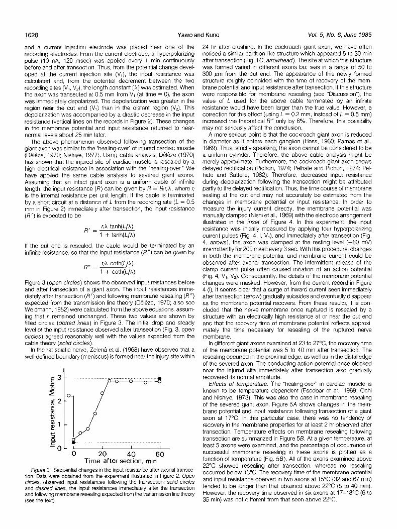
and a current injection electrode was placed near one of the recording electrodes. From the current electrode, a hyperpolarizing pulse (10 nA, 120 msec) was applied every 1 min continuously before and after transection. Thus, from the potential change devel- oped at the current injection site (V,), the input resistance was calculated and, from the potential decrement between the two recording sites (V,, V,), the length constant (X) was estimated. When the axon was transected at 0.5 mm from V, (at time = 0), the axon was immediately depolarized. The depolarization was greater in the region near the cut end (V,) than in the distant region (V,). This depolarization was accompanied by a drastic decrease in the input resistance (vertical lines on the records in Figure 2). These changes in the membrane potential and input resistance returned to near- normal levels about 25 min later.
The above phenomenon observed following transection of the giant axon was similar to the “healing-over” of injured cardiac muscle (Deleze, 1970; Nishiye, 1977). Using cable analysis, Deleze (1970) has shown that the injured site of cardiac muscle is resealed by a high electrical resistance in association with the “healing-over.” We have applied the same cable analysis to severed giant axons. Assuming that an intact giant axon is a uniform cable of infinite length, the input resistance (R) can be given by R = %r,X, where r, is the internal resistance per unit length. If the cable is terminated by a short circurt at a distance of L from the recording site (L = 0.5 mm in Figure 2) immediately after transection, the input resistance (R’) is expected to be
R, = r,X tanh(L/A) 1 + tanh(L/X)
If the cut end is resealed, the cable would be terminated by an infinite resistance, so that the input resistance (R”) can be given by
R,, = rA coth(W
1 + coth(L/X)
Figure 3 (open circles) shows the observed input reistances before and after transection of a giant axon. The input resistances imme- diately after transection (R’) and following membrane resealing (R”) expected from the transmission line theory (Deleze, 1970; also see Weidmann, 1952) were calculated from the above equations, assum- ing that r, remained unchanged. These two values are shown by filled circles (doffed lines) in Figure 3. The initial drop and steady level of the input resistance observed after transection (Fig. 3, open circles) agreed reasonably well with the values expected from the cable theory (solid circles).
In the rat sciatic nerve, Zelena et al. (1968) have observed that a well-defined boundary (meniscus) is formed near the injury site within
Time after section, min Figure 3. Sequential changes in the input resistance after axonal transec-
tron. Data were obtained from the experiment illustrated in Figure 2. Open circles, observed input resistances following the transection: solid circles and dashed lines, the input resistances immediately after the transection and following membrane resealing expected from the transmission line theory (see the text).
24 hr after crushing. In the cockroach giant axon, we have often noticed a similar partition-like structure which appeared 5 to 30 min after transection (Fig. 1 C, arrowhead). The site at which this structure was formed varied in different axons but was in a range of 50 to 300 pm from the cut end. The appearance of this newly formed structure roughly coincided with the time of recovery of the mem- brane potential and input resistance after transection. If this structure were responsible for membrane resealing (see “Discussion”), the value of L used for the above cable terminated by an infinite resistance would have been larger than the true value. However, a correction for this effect (using L = 0.2 mm, instead of L = 0.5 mm) increased the theoretical R” only by 6%. Therefore, this possibility may not seriously affect the conclusion.
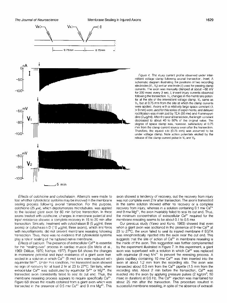
A more serious point is that the cockroach giant axon is reduced in diameter as it enters each ganglion (Hess, 1960; Parnas et al., 1969). Thus, strictty speaking, the axon cannot be considered to be a uniform cylinder. Therefore, the above cable analysis might be merely approximate. Furthermore, the cockroach giant axon shows delayed rectification (Pichon, 1974; Pelhate and Pichon, 1974; Pel- hate and Sattelle, 1982). Therefore, decreased input resistance during depolarization following the transection might be attributed partly to the delayed rectification. Thus, the time course of membrane sealing at the cut end may not accurately be estimated from the changes in membrane potential or input resistance. In order to measure the injury current directly, the membrane potential was manually clamped (Nishi et al., 1969) with the electrode arrangement illustrated in the inset of Figure 4. In this experiment, the input resistance was initially measured by applying four hyperpolarizing current pulses (Fig. 4, I, V,), and immediately after transection (Fig. 4, arrows), the axon was clamped at the resting level (-80 mV) intermittently for 200 msec every 3 sec. With this procedure, changes in both the membrane potential and membrane current could be observed after axonal transection. The intermittent release of the clamp current pulse often caused initiation of an action potential (Fig. 4, V,, V,). Consequently, the details of the membrane potential changes were masked. However, from the current record in Figure 4 (I), it seems clear that a surge of inward current seen immediately after transection (arrow) gradually subsides and eventually disappear as the membrane potential recovers. From these results, it is con- cluded that the nerve membrane once ruptured is resealed by a structure with an electrically high resistance at or near the cut end and that the recovery time of membrane potential reflects approxi- mately the time necessary for resealing of the ruptured nerve membrane.
In different giant axons examined at 23 to 27°C the recovery time of the membrane potential was 5 to 40 min after transection. The resealing occurred in the proximal edge, as well as in the distal edge of the severed axon. The conducting action potential once blocked near the injured site immediately after transection also gradually recovered its normal amplitude.
Effects of temperature. The “healing-over” in cardiac muscle is known to be temperature dependent (Escobar et al., 1969; Ochi and Nishiye, 1973). This was also the case in membrane resealing of the severed giant axon. Figure 5A shows changes in the mem- brane potential and input resistance following transection of a giant axon at 17’C. In this particular case, there was no tendency of recovery in the membrane properties for at least 2 hr observed after transection. Temperature effects on membrane resealing following transection are summarized in Figure 55. At a given temperature, at least 5 axons were examined, and the percentage of occurrence of successful membrane resealing in these axons is plotted as a function of temperature (Fig. 58). All of the axons examined above 22°C showed resealing after transection, whereas no resealing occurred below 13°C. The recovery time of the membrane potential and input resistance oberved in two axons at 15°C (32 and 67 min) tended to be longer than that obtained above 22°C (5 to 40 min). However, the recovery time observed in six axons at 17-18°C (6 to 35 min) was not different from that seen above 22°C.
The Journal of Neuroscience Membrane Sealing in Injured Axons
- 0.75mm -- 0.75mm -
nA I
mV -6O-
V -80 -
-lOO-
-5 min
Effects of colchicine and cytochalasin. Attempts were made to
test whether cytoskeletal systems may be involved in the membrane sealing process following axonal transection. For this purpose, colchicine (25 PM), which depolymerizes microtubules, was applied
to the isolated giant axon for 60 min before transection. In three axons treated with colchtcine, changes in membrane potential and input resistance showed a complete recovery in 15 to 20 min after
transection. Similarly, treatment with cytochalasin B (5 fig/ml; three axons) or cytochalasin D (10 Kg/ml; three axons), which interferes with neurofilaments, did not prevent membrane resealing following
transectIon. Thus, there was no evidence that cytoskeletal systems play a role in sealing of the ruptured nerve membrane.
Effects of calcium. The presence of extracellular Ca*’ is essential for the “healing-over” process in cardiac muscle (De Mello et al., 1969; Deleze, 1970; Nishiye, 1977). Figure 6A shows the changes
in membrane potential and input resistance of a giant axon tran-
sected in a solution in which Ca’+ (9 mM) ions were replaced with equimolar Mn’+. Under this condition, the transected axon showed no sign of recovery for at least 2 hr at 23 to 27%. Similarly, when
extracellular Ca”+ was substituted by equimolar Sr’+ or Mg’+, the transected axon consistently failed to seal its cut end. Thus, the
membrane resealing process appears to require specifically Ca’+. Figure 6/3 shows the results obtained from a giant axon which was transected in the presence of 0.5 mM Ca”+ and 9 mM Mg*+. This
1629
Figure 4. The injury current proflle observed under inter- mittent voltage clamp following axonal transection. Inset, A schematic diagram illustrating the positions of two recording electrodes (V,, V,) and an electrode (I) used for passing clamp currents. The axon was manually clamped at about -80 mV for 200 msec every 3 sec. I, inward injury currents observed following the transection. VI, changes in the membrane poten- tial at the site of the intermittent voltage clamp. V,, same as VI, but at 0.75 mm from the site at which the clamp currents were applied. Axons with a relatively large space constant (X > 9 mm) were used for this series of experiments, and delayed rectification was minimized by TEA (50 mM) and 4-aminopyri- dine (3 pg/ml). After the axonal transection, the length constant decreased to about 40 to 50% of the original value. The degree of space clamp was, however, satisfactory at 0.75 mm from the clamp current source even after the transection. Therefore, the injured site (0.75 mm) was assumed to be under voltage clamp. Note action potentials elicited by the release of the clamp current pulse in V, and V2.
axon showed a tendency of recovery, but the recovery from injury
was not complete even 2 hr after transection. The axons transected in the same solution showed either no recovery or a complete recovery from injury, whereas in a solution containing 0.1 mM Ca*’
and 9 mM Mg”+, the axon invariably failed to seal its cut end. Thus, the minimum concentration of extracellular Ca*’ required for the membrane resealing seems to be about 0.1 to 0.5 mM.
Our previous study (Yaw0 and Kuno, 1983) showed that even when a giant axon was sectioned in the presence of 9 mM Ca*’ at 23 to 27’C, the axon failed to seal its injured membrane if EGTA
was ionophoretically injected into the axon near the cut end. This suggests that the site of action of Ca*’ in membrane resealing is the inside of the axon. This suggestion was further complemented
by the experiment illustrated in Figure 7. In this experiment, a giant axon was superfused with a solution in which Ca*’ was replaced
with equimolar (9 mM) Mn2+ to prevent the resealing process. A glass capillary containing 10 mM Ca*’ was then inserted into the
axon at about 1.2 mm from the recording site. The axon was transected about 0.3 mm from the Ca*’ pipette (1.5 mm from the
recording site). About 2 min before the transection, Ca*’ was injected into the axon by applying pressure pulses (2 kg/cm”; 50 msec in duration) at 0.5 Hz. The Ca*’ injection was maintained for
about 25 min after the transection. This procedure resulted in successful membrane resealing, in spite of the absence of extracel-
Yaw and Kuno Vol. 5, No. 6, June 1985 1630
mV
OL , -o--o/ I I I I I
5 10 15 20 25 30 Temperature, ‘C
Figure 5. Effects of temperature on membrane resealing. A, Changes in the membrane potential and input resistances (vertical lines on the record) of a giant axon transected (arrow) at 17°C. B, Percentage of successful membrane resealing following transection as a function of temperature.
lular Ca’+. From these results, it seems reasonable to conclude that sealing of the ruptured nerve membrane requires Ca2’ entry into the axoplasm.
Possible involvement of phospholipase A2 in membrane reseal- ing. The Ca2’ dependence of membrane resealing observed in the cockroach giant axon resembles Ca”+-dependent activation of phos- pholipase A2 in plasma membranes in three aspects. First, the two processes are very specific for Ca’+ compared with the other divalent metal ions tested (Pieterson et al., 1974; Van den Bosch, 1980). Second, the two processes require relatively high concentra- tions of Ca2’ (0.05 to 0.5 mM; Van den Bosch, 1980). Third, the two processes require a rise in cytoplasmic free Ca2+ concentration (Van den Bosch, 1980). Because of these similarities, the possible involve- ment of phospholipase A, in membrane resealing was examined.
The above possibility was tested by the application of two widely used phospholipase A2 inhibitors; one is p-bromophenacyl bromide (Volwerk et al., 1974; Gan-Elepano and Mead, 1978; Erman et al.,
mV
1983) and the other is mepacrine (Markus and Ball, 1969; Vargaftig and Dao Hai, 1972; Erman et al., 1983). The effects of p-bromo- phenacyl bromide on membrane resealing are shown in Figure 8. The axon was superfused with the standard solution containing 2 pM (Fig. 8A) or 10 pM (Fig. 88) of the inhibitor for 30 min before transection. The inhibitor had no significant effects on the resting and action potentials or on the input resistance. In the presence of 2 PM of the inhibitor, the axon showed a tendency of recovery following the transection at 24°C although the recovery was incom- plete even 2 hr after the injury (Fig. 8A). At a concentration of 10 PM of the inhibitor, there was no tendency of recovery of the membrane potential and input resistance after the transection (Fig. 813). The effects of p-bromophenacyl bromide were examined in a range of 1 to 20 pM. The inhibitor prevented membrane sealing following transection in a concentration-dependent fashion, The concentration at which 50% of the axons tested failed to recover from the injury was 5 pM. Essentially the same results were obtained with another inhibitor, mepacrine (see Figure 25 in Yawo and Kuno, 1983). The effects of mepacrine were tested at a concentration of 0.1 to 5 mM; its 50%inhbition dose was 1 mM. These results were consistent with the assumption that activation of phospholipase A2 plays a central role in membrane resealing after injury.
Discussion
From the present results, there seem to be some features in common between the “healing-over” in cardiac muscle and the membrane resealing of axotomized cockroach giant axons. Thus, the injured site appears to be resealed by a structure with high electrical resistances. Also, the recovery from injury is Ca’+-depend- ent and temperature-dependent.
The presence of extracellular Ca2+ above 0.1 to 0.5 mM was essential for successful membrane sealing following transection of the cockroach giant axon. However, even in the presence of high extracellular Ca2+ (9 mM), no membrane sealing occurred after the injury if the expected increase in the intracellular Ca’+ concentration near the cut end was prevented by intra-axonal injections of EGTA (Yawo and Kuno, 1983). Also, even in a Ca2+-free solution, mem- brane resealing did occur when Ca2’ was injected into the axon near the cut end (Fig. 7). Therefore, the membrane resealing must be triggered by excess Ca2’ entering the axoplasm at the injured site.
Phospholipase A2 is considered to be embedded in the inner layer of the plasma membrane, and its activation requires Ca” (see review by Van den Bosch, 1980). When phospholipase A2 was
A OmMCa-9mMMn
Figure 6. Effects of extracellular Ca” on membrane resealing. A, Changes In the membrane potential and input resistance of a -80 1 giant axon transected (arrow) in a solution in which Ca*’ was replaced with equimolar Mn”. B, Same as in A, but in a solution containing 0.5 mM Ca*’ plus 9 mM Mg”+. The bathing solution
,,,V
was maintalned at 25“C in both A and B.
-60 t
-8OL -,,-,-/ i!!?!%
I , I I ,
B 0.5mMCa-9mMMg

The Journal of Neuroscience Membrane Sealing in Injured Axons 1631
-60
-80 t I I I
0 10 20 310 4!k--e Time, min
mV Or
-20 A pBPB. 2x10-%
0
c
B pBPB, 10x 10-6M
-20
10 min
Figure 8. Effects of phospholtpase A2 inhibitors on membrane resealing. A, Changes in the membrane potential and input resistance of a giant axon transected in the presence of 2 PM p-bromophenacyl bromide. Note a partial recovery from the injury. B, Same as in A, but with 10 PM of the inhibitor. Note the lack of tendency of recovery from the injury. Temperature, 24°C in both A and B.
inhibited by p-bromophenacyl bromide or mepacrine, the axon failed to reseal its cut end. No other membrane properties were affected by the application of the inhibitors, These results strongly suggest that the membrane resealing is induced by phospholipase A2 which may be activated by Ca” entering the axon at the injured site. Phospholipase A2 catalyzes the hydrolysis of certain membrane phospholipids to arachidonate and lysolecithin. Howell and Lucy (1969) have shown that lysolecithin can induce membrane fusion of erythrocytes. Since then, the role of lysolecithin as a fusogen has been investigated in a variety of preparations (Poste and Allison, 1973; Lucy, 1978; Papahadjopoulos, 1978). We attempted to test whether the application of lysolecithin alone may facilitate membrane resealing at the injured site without activation of phospholipase AP. For this purpose, the phospholipase action was inhibited by mepa- crine (2 mM), and lysophosphatidylcholine (20 pg/ml dissolved in
figure 7. Membrane resealing induced by intra- axonal injections of Ca” in a giant axon transected in a Ca”-free solution. Inset, A schematic diagram illus- trating the arrangement of a recording electrode, a current Injection electrode for measuring the input re- sistance and a pipette containing 10 mM Ca2+ (dis- solved in 0.12 M potassium acetate plus 0.08 M K,SO,; pH = 7.0) used for pressure injections. Heavy horizon- tal line, the period during which Ca’+ was injected In the axon. Bottom record, changes in the membrane potential and input resistance of a giant axon tran- sected (arrow) in a solution in which Ca*+ was replaced with equimolar Mn*+. Temperature, 24°C.
467 mM manitol and 5 mM Tris-Cl; pH = 7.0) was injected into the axon near the cut end 5 min after the transection, using the pressure pulse method (1 kg/cm*; 300 msec at 1 Hz). Immediately after the injection, the membrane potential and input resistance showed a tendency of recovery. The partial recovery could be maintained as long as the injection was held. However, soon after interruption of the application, the axon was again depolarized. The explanation for this transient, partial recovery of membrane properties by lysolecithin remains uncertain.
The membrane resealing occurred in the cut proximal end as well as in the cut distal end. Thus, the resealing is a local event of the nerve membrane, being independent of the cell body. In the present study, there was no evidence that cytoskeletal systems play a role in the membrane resealing. The membrane sealing induced in skeletal muscle by isotonic Ca’+ solution was not affected by trypsin or chymotrypsin, suggesting that the membrane proteins are not involved in this process (De Mello, 1973). In the cockroach giant axon, the critical temperature for successful membrane resealing was in a range of 13-22’C. This range of temperatures is similar to the phase transition temperatures of certain membrane lipids (Shis- chick and McConnell, 1973; Wisnieski et al., 1974; Chapman, 1975). Above their transition temperatures, lipids are known to be more susceptible to fusion (Papahadjopoulos, 1978). Thus, the membrane resealing may be achieved by fusion of lipids forming the membrane. However, it should be noted that the action of phospholipase A2 on phosphatidylcholine is exerted only above the phase transition tem- perature of the lipid (Wilschut et al., 1978). Therefore, it remains uncertain whether the failure of membrane resealing at low temper- atures is due to the nonfluid state of the membrane lipids (Papahad- jopoulos, 1978) or to a lack of the phospholipase A2 action which is essential for the membrane resealing.
A newly formed partition-like structure observed near the cut end under a phase-contrast microscope (Fig. 1 C, arrowhead) might be responsible for membrane resealing. Our preliminary observations showed that the diffusion of fluorescein sodium injected into the axon is interrupted exactly at the site of this partition-like structure. Also, when the partition-like structure happened to be formed be- tween the two micro-electrodes placed near the cut end, current injections from one electrode produced no detectable potential changes in the other side (H. Yawo and M. Kuno, unpublished observation). Therefore, there is little doubt that this structure acts as a diffusion barrier. This structure was not formed when the axon failed to recover its membrane potential and input resistance follow- ing the transection. Thus, this structure appears to be a double layer membrane formed as a result of membrane fusion (see Bennett, 1969). Alternatively, it may be an axoplasmic “plug” formed by the Ca*+-dependent surface precipitation reaction (Heilbrunn, 1956). Electron microscopic observations which are now under way would provide some clue about the nature of this structure.
1632 Yaw0 an
References
Bennett, H. S. (1969) The cell surface: Movements and recombinations. In Handbook of Molecular Cytology, A. Lima-de-Faria, ed., pp. 1294-l 319, Elsevier-North Holland Publishing Co., Amsterdam.
Borgens, Ft. B., L. F. Jaffe, and H. J. Cohen (1980) Large and persistent electrical currents enter the transected lamprey spinal cord. Proc. Natl. Acad. Sci. U. S. A. 77: 1209-1213.
Chapman, D. (1975) Phase transitions and fluidity characteristics of lipids and cell membranes. Q. Rev. Brophys. 8: 185-235.
Daley, D. L., N. Vardi, B. Appignanr, and J. M. Camhi (1981) Morphology of the giant interneurons and cereal nerve projections of the American cockroach. J. Comp. Neurol. 796: 41-52.
Deleze, J. (1970) The recovery of resting potential and input resistance in sheep heart injured by knife or laser. J. Physiol. (Lond.) 208: 547-562.
De Mello, W. C. (1972) The healing-over process in cardiac and other muscle fibres. In Electrical Phenomena in the Heart, W. C. DeMello, ed., pp. 323- 351, Academic Press, Inc., New York.
De Mello, W. C. (1973) Membrane sealing in frog skeletal muscle fibers. Proc. Natl. Acad. Sci. U. S. A. 70: 982-984.
De Mello, W. C., G. E. Motta, and M. Chapeau (1969) A study on the healing- over of myocardial cells of toads. Circ. Res. 24: 475-487.
Engelmann, Th. W. (1877) Vergleichende Untersuchungen zur Lehre von der Muskel- und Nervenelectricitat. Pfliigers Arch. 75: 116-148.
Erman, A., R. Azuri, and A. Raz (1983) Phospholipase A:, inhibitors: Differential inhibition of fatty acid acylation into kidney lipids by mepacrine and p- bromophenacyl bromide. Brochem. Pharmacol. 32: 2083-2087.
Escobar, I., W. C. De Mello, and B. Perez (1969) Temperature and myocardial healing-over. Fed. Proc. 28: 328-331.
Gan-Elpano, M., and J. F. Mead (1978) The functron of phospholrpase AP in the metabolism of membrane lipids. Biochem. Biophys. Res. Commun. 83: 247-251.
Harris, C. L., and T. Smyth (1971) Structure details of cockroach giant axons revealed by injected dye. Comp. Biochem. Physiol 40A: 295-303.
Harrow, I. D., B. Hue, M. Pelhate, and D. B. Sattelle (1980) Cockroach giant interneurones stained by cobalt-back filling of dissected axons. J. Exp. Biol. 84: 341-343.
Heilbrunn, L. V. (1956) The Dynamics of Living Protoplasm, Academic Press, Inc., New York.
Hess, A. (1958) Experimental anatomical studies of pathways in the severed central nerve cord of the cockroach. J. Morphol. 703: 479-501.
Howell, J. I., and J. A. Lucy (1969) Cell fusion induced by lysolecithin. FEBS Lett. 4: 147-l 50.
Lorente de No, R. (1947) A study of nerve physiology: Part 1. Stud. Rockefeller Institute 737: l-496.
Lucy, J. A. (1978) Mechanisms of chemically induced cell fusion. In Mem- brane fusion, G. Poste and G. L. Nicolson, eds., pp. 267-304, Elsevier- North Holland Biomedical Press, Amsterdam.
Markus, H. B., and E. G. Ball (1969) Inhibition of lipolytic processes in rat adipose tissue by antimalarial drugs. Biochim. Biophys. Acta 787: 486- 491.
Martinez, A. J., and R. L. Friede (1970) Accumulation of axoplasmic organ- elles in swollen nerve fibers. Brain Res 79: 183-198.
Meiri, H., M. E. Spira, and I. Parnas (1981) Membrane conductance and action potential of a regenerating axon tip. Science 27 7: 709-712.
Merr, H., A. Dormann, and M. E. Spira (1983) Comparison of ultrastructural
d Kuno Vol. 5, No. 6, June 1985
changes in proximal and distal segments of transected grant fibers of the cockroach Periplaneta americana. Brain Res. 263: l-l 4.
Nishi, S., H. Soeda, and K. Koketsu (1969) Unusual nature of ganglionic slow EPSP studied by a voltage-clamp method. Life Sci. (Part I) 8: 33-42.
Nishiye, H. (1977) The mechanism of Ca” action on the healing-over process in mammalian cardiac muscles: A kinetic analysrs. Jpn. J. Physiol. 27: 451-466.
Ochi, R., and H. Nishiye (1973) Temperature dependence of the healina- over in mammalian cardiac muscle. ‘Proc. Jpn. Acad. 49: 372-375. -
Papahadiopoulos, D. (1978) Calcium-induced phase chanaes and fusion in natural and model membranes. In Membrane Fusion, GI Poste and G. L. Nicolson, eds., pp. 765-790, Elsevier-North Holland Biomedical Press, Amsterdam.
Pelhate, M., and Y. Pichon (1974) Selective inhibition of potassium current in the grant axon of the cockroach. J. Physiol. (Lond.) 242: 90-91 P.
Pelhate, M., and D. B. Satelle (1982) Pharmacological properties of insect axons: A review. J. Insect Physiol. 28: 889-903.
Pichon, Y.(1974) Axonal conduction in insects. In Insect Neurobiology, J. E. Treherne, ed., pp. 73-l 17, Elsevier-North Holland Publishing Co., Amster- dam.
Pieterson, W. A., J. J. Volwerk, and G. H. De Haas (1974) Interaction of phospholipase AP and its zymogen with divalent metal ions. Biochemistry 73: 1439-1445.
Poste, G., and A. C. Allison (1973) Membrane fusion. Biochim. Biophys. Acta 300: 421-465.
Rothschuh, K. E. (1950) Uber elektrische Entlandungsvorgange an der verletzten Skelemusckelfaser und ihre Beziehungen zum Vorgang der Degeneration, der Regeneration und des Absterbens. Pflugers Arch. 252: 445-467.
Rothschuh, K. E. (1952) Uber den funktionelllen Aufbau des Herzens aus elektrophysiologischen Elementen und tiber den Mechanismus der Erre- gungsleitung im Herzen. Pflijgers Arch. 253: 238-251.
Shimshick, E. J., and H. M. McConnell (1973) Lateral phase separation in phospholipid membranes. Biochemistry 72: 2351-2360.
Soira. M. E.. I. Parnas, and F. Beraman (1969) Histoloaical and electrophvs- iological studies on the giant axons of the cockroach, feriplaneta am&- cana. J. Exp. Biol. 50: 629-634.
Van den Bosch, H. (1980) Intracellular phospholipase A. Biochim. Biophys. Acta 604: 191-246.
Vargaftrg, B. B.. and N. Dao Hai (1972) Selective inhibition by mepacrine of the release of “rabbit aorta contracting substance” evoked by the admin- istration of bradvkinin. J. Pharm. Pharmacol. 24: 159-161,
Volwerk, J. J., W.-A. Preterson, and G. H. De Haas (1974) Histidine at the active site of phospholrpase A*. Biochemistry 73: 1446-1454.
Weidmann, S. (1952) The electrical constants of Purkinje fibers. J. Physiol. (Lond.) 7 78: 348-360.
Wilschut, J. C., J. Regts, H. Westenberg, and G. Scherphof (1978) Action of phospholipase A2 on phosphatidylcholine bilayers. Biochim. Biophys. Acta 508: 185-l 96.
Wisnreski, B. J., J. G. Parkes, Y. 0. Huang, and C. F. Fox (1974) Physical and physiological evidence for two phase transitions in cytoplasmic mem- branes of animal cells. Proc. Natl. Acad. Sci. U. S. A. 77: 4381-4385.
Yamasaki, T., and T. Narahashi (1959) Electrical properties of the cockroach giant axon. J. Insect Physiol. 3: 230-242.
Yawo, H., and M. Kuno (1983) How a nerve fiber repairs its cut end: Involvement of phospholipase A2. Science 222: 1351-l 353.
Zelena, J., L. Lubinska, and E. Gutmann (1968) Accumulation of organelles at the ends of interrupted axons. Z. Zellforsch. 91: 200-219.