calcium hydroxide delivery
DESCRIPTION
dghdghTRANSCRIPT

DTR
ATpiscfmtaplwponcqil
KCn
Es1
PaWm0
Ed
Basic Research—Technology
6
elivery of Calcium Hydroxide: Comparison of Four Fillingechniquesichard M. Simcock, DDS* and M. Lamar Hicks, DDS, MS*
Cfwsm
dqst
ct#sacff
eCamt
dtia
cratOtmmap
(r
bstracthis study compared the weight and radiographic ap-earance of Ca(OH)2 delivered into standardized, min-
mally and fully prepared canals using (a) an injectionystem, (b) an endodontic Flex-O file rotated counter-lockwise, (c) a lentulo spiral, and (d) a .04 rotary NiTiile rotated in reverse. Two extracted human secondandibular premolars with single canals were prepared
o an MAF #25 or an MAF #40. A weight measurementnd radiograph were made for each filling. Three inde-endent examiners evaluated the radiographs. Regard-
ess of technique, only about 45% of the optimaleight of Ca(OH)2 was delivered into the minimallyrepared canal. Radiographically the filling quality wasnly 1 to 2 (10 scale). In contrast, all delivery tech-iques delivered near optimal weight of Ca(OH)2 in theompletely prepared canal with a radiographic fillinguality of 8.8 to 9.3. The results indicate that complete
nstrumentation is needed to obtain near optimal de-ivery of Ca(OH)2. (J Endod 2006;32:680–682)
ey Wordsa(OH)2 delivery, complete instrumentation needed forear optimal delivery, testing of four techniques
From the *I. B. Bender Division of Endodontics, Albertinstein Medical Center, Philadelphia, Pennsylvania. Currenttatus of Dr. Simcock: Private Practice of Endodontics, 130 S.5th Street, Suite 101, Mount Vernon, WA 98274.
Address requests for reprint to Dr. M. Lamar Hicks, Clinicalrofessor, Department of Endodontics, Operative Dentistry,nd Prosthodontics, University of Maryland at Baltimore, 6601est Baltimore Street, Baltimore, MD 21201. E-mail address:[email protected].
099-2399/$0 - see front matterCopyright © 2006 by the American Association of
ndodontists.oi:10.1016/j.joen.2006.01.009
t
80 Simcock and Hicks
alcium hydroxide (Ca(OH)2) is used extensively as an intracanal medication (1). Itinhibits the growth of bacteria between appointments (2–5). Bystrom et al. (6)
ound that Ca(OH)2 effectively eliminated all microorganisms in infected root canalshen the dressing was maintained for 4 wk. Because the goal of cleaning the root canal
ystem is to remove necrotic tissue and eliminate bacteria, Ca(OH)2 has been recom-ended as an intracanal medication after cleaning and shaping the canal system (7).
To maximize the antibacterial properties of Ca(OH)2, it is important to have aense, homogenous filling to the root apex (8). Although several studies investigated theuality of Ca(OH)2 placement in large immature canals (9 –11), they typically de-cribed apexification procedures. However, when dealing with mature canals, ques-ions remain as to the best placement technique to use.
Because of limited chair time, Ca(OH)2 often is placed in minimally instrumentedanals. Sigurdsson et al. (12) found that radiographically the lentulo spiral producedhe highest quality filling when canals were instrumented to a master apical file (MAF)25. However, in fully prepared canals (MAF #50), Staehle et al. (13) found that ayringe system gave the best results radiographically and in ground sections. Torres etl. (14) found the greatest radiodensity of Ca(OH)2 in the apical 1 mm of a simulatedanal (44 degree curvature) prepared to an MAF #40 when a lentulo was used. There-ore, it may be important to determine the degree of canal preparation needed to allowor optimal placement of Ca(OH)2.
Delivering the largest amount of Ca(OH)2 into the root canal should enhance thelimination of bacteria. No study has used a quantitative method to assess the amount ofa(OH)2 placed in a canal. Because a radiographic image is two-dimensional, clinicallyssessing the quality of a filling is difficult. It would be beneficial to assess the weight ofaterial delivered for a given technique. This would allow for an objective and quan-
itative evaluation.The aim of this study was to compare the weights of Ca(OH)2 delivered by four
ifferent techniques into minimally and fully prepared canals in a standardized split-ooth model (15). In addition, the radiographic appearance was evaluated to determinef a correlation exists between the weight of Ca(OH)2 delivered and the radiographicppearance.
Materials and MethodsThis study used two extracted human second mandibular premolars with single
anals. After cleaning, the teeth were accessed with a high speed, water-cooled #4ound bur. The working length was determined by visualizing a #15 K-file through thepical foramen and subtracting 1 mm. The teeth were prepared with a crown-downechnique using .04 rotary nickel-titanium (NiTi) files (Tulsa Dental Products, Tulsa,K). The canals were irrigated between files with 2 ml of 17% REDTA (Roth Interna-
ional, Chicago, IL) followed by a 5-ml flush of 5.25% sodium hypochlorite. A #15 K-fileaintained canal patency throughout instrumentation. One tooth was prepared to aaster apical file (MAF) #25 (minimally prepared canal) to simulate an emergency
ppointment and the other to an MAF #40 (fully prepared) to simulate a completedreparation. After final irrigation, the canals were dried.
Two custom boxes were fabricated to provide a matrix for orthodontic resinCaulk/Dentsply, Milford, DE), which was mixed according to the manufacturer’s di-ections. After pouring the resin into each box, the teeth were inserted root end first into
he center of the resin to the buccal CEJ. After the resin polymerized, two alignmentJOE — Volume 32, Number 7, July 2006
hn
c(ba
.Tdcttm
tFw.aa
U
twpc
C
fro
L
bpsw
R
#pro
bb
R
tfc
at
Kp
eCwohom
nAmdbn
ct2cseCg
ogp
ssimsD
ii
T#
D
D
(
*
�
Basic Research—Technology
J
oles were drilled in the resin on the mesial side of the root taking careot to penetrate the root.
Each resin block was then longitudinally sectioned through theenter of the root canal using an Isomet Buehler low-speed sawBuehler LTD., Evanston, IL). After sectioning, the two halves of thelock were reassembled and secured with threaded bolts placed in thelignment holes
The reassembled blocks were individually weighed to the nearest0001 g on a Mettler H20 scale (Mettler-Toledo, Inc., Columbus, OH).o establish a target weight for maximum capacity, both blocks wereisassembled and Ca(OH)2 firmly condensed into each half using aement spatula. After condensation, the spatula leveled the Ca(OH)2 tohe cut root surface. The blocks were then placed together, secured withhe bolts, and weighed independently three times to obtain the maxi-
um (optimal) average weight.Ultracal (Ultradent, South Jordan, UT) was used for each delivery
echnique tested: (a) an injection system (Ultracal), (b) an endodonticlex-O file (Maillefer, Ballaigues, Switzerland) rotated counter-clock-ise, (c) a lentulo spiral (Maillefer, Ballaigues, Switzerland), and (d) a
04 rotary NiTi file (Tulsa Dental Products) rotated counterclockwise atconstant 150 rpm. Each technique was repeated 10 times and an
verage weight determined.
ltracal Syringe SystemThe working length measurement for each tooth was marked on
he long flexible plastic tip using a sharp endodontic explorer. The pasteas applied slowly and continuously from the apical to the most coronalart of the root canal using a slight up-and-down movement. This wasontinued until the paste was seen at the canal orifice.
ounterclockwise Flex-O FileThe paste was applied to the entire length of a #25 or #40 Flex-O
ile and introduced to the working length using a counterclockwiseotation. This was repeated until the material was visible at the canalrifice.
entulo SpiralA #1 or #4 lentulo spiral was passively placed to the working length
efore the Ca(OH)2 was applied. The lentulo was then coated with theaste, the instrument introduced into the root canal, and the pastelowly rotated into the canal. The procedure was repeated until the pasteas seen at the canal orifice.
everse Rotary NiTi FileUsing .04 rotary NiTi files corresponding to the MAF #25 and MAF
40 preparations, paste was applied to the entire file length and placedassively to working length before running the files in reverse at 150pm. The procedure continued until the paste was visible at the canalrifice.
After each Ca(OH)2 placement, one radiograph was taken from auccal-lingual direction to simulate the clinical condition. Then, thelocks were weighed three times and the average weight calculated.
adiographic EvaluationThree blinded independent examiners radiographically evaluated
he completeness of filling. Radiographs were mounted in 35-mm slideormat and examined by screen projection. A scale from 1 (emptyanal) to 10 (full canal) was used to assign a grade of filling quality.
The quantitative (objective) data (weights) were analyzed usingnalysis of variance and post hoc Scheffe F-test. The qualitative (subjec-
ive) evaluations of radiographic appearance were analyzed using aOE — Volume 32, Number 7, July 2006
ruskal-Wallis and Pearson’s �2 tests. Statistical significance was set at� 0.05.
ResultsFor the minimally prepared canal, there were no statistical differ-
nces among the four experimental groups in the mean weights ofa(OH)2 delivered (p � 0.15) (Table 1). However, when the meaneight of Ca(OH)2 for each experimental group was compared with theptimal weight delivered into the same canal, the differences wereighly significant (p � 0.003) (Table 1). Regardless of the technique,nly about 45% of the optimal weight of Ca(OH)2 was delivered intoinimally prepared canals.
In the completely prepared canal, all four experimental tech-iques delivered close to the optimal weight of Ca(OH)2 (Table 1).lthough the injection system and the lentulo spiral technique deliveredodestly more weight than the other two experimental groups, the
ifferences were not significant (p � 0.05) (Table 1). The differencesetween the experimental groups and the optimal weight were not sig-ificant (p � 0.05).
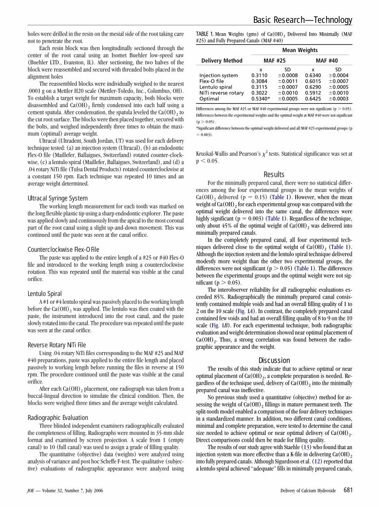
The interobserver reliability for all radiographic evaluations ex-eeded 85%. Radiographically the minimally prepared canal consis-ently contained multiple voids and had an overall filling quality of 1 toon the 10 scale (Fig. 1A). In contrast, the completely prepared canal
ontained few voids and had an overall filling quality of 8 to 9 on the 10cale (Fig. 1B). For each experimental technique, both radiographicvaluation and weight determination showed near optimal placement ofa(OH)2. Thus, a strong correlation was found between the radio-raphic appearance and the weight.
DiscussionThe results of this study indicate that to achieve optimal or near
ptimal placement of Ca(OH)2, a complete preparation is needed. Re-ardless of the technique used, delivery of Ca(OH)2 into the minimallyrepared canal was ineffective.
No previous study used a quantitative (objective) method for as-essing the weight of Ca(OH)2 fillings in mature permanent teeth. Theplit-tooth model enabled a comparison of the four delivery techniquesn a standardized manner. In addition, two different canal conditions,
inimal and complete preparation, were tested to determine the canalize needed to achieve optimal or near optimal delivery of Ca(OH)2.irect comparisons could then be made for filling quality.
The results of our study agree with Staehle (13) who found that annjection system was more effective than a K-file in delivering Ca(OH)2nto fully prepared canals. Although Sigurdsson et al. (12) reported that
ABLE 1. Mean Weights (gms) of Ca(OH)2 Delivered Into Minimally (MAF25) and Fully Prepared Canals (MAF #40)
Mean Weights
Delivery Method MAF #25 MAF #40
x SD x SDInjection system 0.3110 �0.0008 0.6340 �0.0004Flex-O file 0.3084 �0.0011 0.6015 �0.0007Lentulo spiral 0.3115 �0.0007 0.6290 �0.0005NiTi reverse rotary 0.3022 �0.0010 0.5912 �0.0010Optimal 0.5340* �0.0005 0.6425 �0.0003
ifferences among the MAF #25 or MAF #40 experimental groups were not significant (p � 0.05).
ifferences between the experimental weights and the optimal weight at MAF #40 were not significant
p � 0.05).
Significant difference between the optimal weight delivered and all MAF #25 experimental groups (p
0.003).
lentulo spiral achieved “adequate” fills in minimally prepared canals,
Delivery of Calcium Hydroxide 681

opsrpcAiCrpa
aplwmtnq
amascdt
oncmd
BttR
1
1
1
1
1
Fm
Basic Research—Technology
6
ur study does not support those findings. An explanation for this dis-arity could be in the differences in experimental technique. In theirtudy, each experimental procedure was repeated three times before aadiographic assessment was made. In our study, each experimentalrocedure was discontinued as soon as Ca(OH)2 was visualized at theanal orifice, which more closely approximates the clinical condition.nother explanation could be the dissimilar mixtures of Ca(OH)2 used
n the two experiments. Sigurdsson used an aqueous mixture ofa(OH)2, which has higher flow properties than the more viscous Ult-acal mixture. The aqueous mixture could flow into a minimally pre-ared canal more easily. Thus, it could give a better overall radiographicppearance than what we observed in our minimally prepared canal.
Our study found a strong correlation between the radiographicppearance and the weight of Ca(OH)2 delivered. In the minimallyrepared canal, only 45% of the optimal amount of Ca(OH)2 was de-ivered. This correlated well with the overall radiographic appearance,hich rated 1 to 2 on a scale of 10. Regardless of technique used in theinimally prepared canal, the radiographic appearance correlated with
he amount of Ca(OH)2 placed. In the completely prepared canal, theear optimal weight of Ca(OH)2 delivered and the overall radiographicuality also strongly correlated.
If the clinician uses Ca(OH)2 as an interappointment medication,technique should be selected that will deliver the maximum amount ofaterial for a given preparation. If time is limited, which is common in
n emergency appointment, instrumentation procedures must be cho-en that will permit an adequate amount of Ca(OH)2 to be placed in theanal. According to our study, a complete preparation is needed toeliver close to optimal amounts of Ca(OH)2 with the physical proper-ies of Ultracal using any of the four delivery techniques.
In conclusion, a complete preparation is needed to deliver a nearptimal amount of Ca(OH)2 into the root canal system. The four tech-iques tested can all achieve that desirable degree of filling. The clini-ian can be confident in radiographically evaluating the quality of place-ent because of the strong correlation between the weight of Ca(OH)2
igure 1. Radiographic examples of minimally (A) and completely preparedinimally prepared canal.
elivered and the radiographic appearance.1
82 Simcock and Hicks
AcknowledgmentsThe authors would like to thank Drs. Stephen Niemczyk, Peter
rothman, Chris Ward, and Ellen Teverovsky for their assistance inhis research, and Leonard Braitman, PhD, for statistical analysis ofhe data. This study was supported, in part, by the I. B. Benderesearch Fund.
References1. Cvek M, Hollander L, Nord CE. Treatment of non-vital permanent incisors with
calcium hydroxide. Odontol Revy 1976;27:93–108.2. Matsumiya S, Kitamura M. Histo-pathological and histo-bacteriological studies of the
relation between the conditions of sterilization of the interior of the root canal and thehealing process of peripheral tissues in experimentally infected root canal treatment.Bull Tokyo Dent Coll 1960;1:1–19.
3. Heithersay GS. Calcium hydroxide in the treatment of pulpless teeth with associatedpathology. J Br Endod Soc 1975;8:74 –93.
4. Sjogren U, Figdor D, Spangberg L, Sundqvist G. The antimicrobial effect of calciumhydroxide as a short-term intracanal dressing. Int Endo J 1991;24:119 –25.
5. Safavi KE, Dowden WE, Introcaso JH, Langeland K. A comparison of antimicrobialeffects of calcium hydroxide and iodine-potassium iodide. J Endod 1985;11:454 – 6.
6. Bystrom A, Claesson R, Sundqvist G. The antibacterial effect of camphorated par-amonochlorophenol, camphorated phenol and calcium hydroxide in the treatmentof infected root canals. Endo Dent Traumatol 1985;1:170 –5.
7. Bystrom A, Sundqvist G. Bacteriologic evaluation of the efficacy of mechanical rootcanal instrumentation in endodontic therapy. Scand J Dent Res 1981;89:321– 8.
8. Dumsha TC, Gutmann JL. Clinical techniques for the placement of calcium hydroxide.Compend Cont Educ Dent 1985;6:482–9.
9. Webber RT, Schwiebert KA, Cathey GM. A technique for placement of calcium hy-droxide in the root canal system. J Am Dent Assoc 1981;103:417–21.
0. Kleier DJ, Averbach RE, Kawulok TC. Efficient calcium hydroxide placement withinthe root canal. J Prosth Dent 1981;53:509 –10.
1. Krell KV, Madison S. The use of the Messing gun in placing calcium hydroxidepowder. J Endod 1985;11:233– 4.
2. Sigurdsson A, Stancil R, Madison S. Intracanal placement of calcium hydroxide: acomparison of techniques. J Endod 1992;18:367–70.
3. Staehle HJ, Thoma C, Muller HP. Comparative in vitro investigation of different meth-ods for temporary root canal filling with aqueous suspensions of calcium hydroxide.Endod Dent Traumatol 1997;13:106 –112.
4. Torres CP, Apicella MJ, Yancich PP, Parker MH. Intracanal placement of calciumhydroxide: a comparison of techniques revisited. J Endod 2004;30:225–7.
nals filled with Ca(OH)2. Note large areas of unfilled canal (arrows) in the
(B) ca5. Budd CS, Weller RN, Kulild JC. A comparison of thermoplasticized injectable gutta-percha obturation techniques. J Endod 1991;17:260 – 4.
JOE — Volume 32, Number 7, July 2006