calicotyle californiensis n. sp. and calicotyle urobati n. sp
TRANSCRIPT

University of Nebraska - LincolnDigitalCommons@University of Nebraska - LincolnFaculty Publications from the Harold W. ManterLaboratory of Parasitology Parasitology, Harold W. Manter Laboratory of
1999
Calicotyle californiensis n. sp. and Calicotyle urobati n.sp. (Monogenea: Calicotylinae) fromElasmobranchs in the Gulf of CaliforniaStephen A. BullardGulf Coast Research Laboratory, [email protected]
Robin M. OverstreetGulf Coast Research Laboratory, [email protected]
Follow this and additional works at: http://digitalcommons.unl.edu/parasitologyfacpubs
Part of the Parasitology Commons
This Article is brought to you for free and open access by the Parasitology, Harold W. Manter Laboratory of at DigitalCommons@University ofNebraska - Lincoln. It has been accepted for inclusion in Faculty Publications from the Harold W. Manter Laboratory of Parasitology by an authorizedadministrator of DigitalCommons@University of Nebraska - Lincoln.
Bullard, Stephen A. and Overstreet, Robin M., "Calicotyle californiensis n. sp. and Calicotyle urobati n. sp. (Monogenea: Calicotylinae)from Elasmobranchs in the Gulf of California" (1999). Faculty Publications from the Harold W. Manter Laboratory of Parasitology. 408.http://digitalcommons.unl.edu/parasitologyfacpubs/408

J. Parasitol., 86(5), 2000, p. 939-944 ? American Society of Parasitologists 2000
CALICOTYLE CALIFORNIENSIS N. SP. AND CALICOTYLE UROBATI N. SP. (MONOGENEA: CALICOTYLINAE) FROM ELASMOBRANCHS IN THE GULF OF CALIFORNIA
Stephen A. Bullard* and Robin M. Overstreet Gulf Coast Research Laboratory, The Department of Coastal Sciences, The University of Southern Mississippi, P.O. Box 7000, Ocean Springs, Mississippi 39566
ABSTRACT: Two new species of Calicotyle (Monocotylidae: Calicotylinae) are described from elasmobranchs in the western Gulf of California. Calicotyle californiensis n. sp. is described from a single specimen collected from a gray smoothhound shark (Mustelus californicus, Carcharhiniformes: Triakidae). It is distinguished from its congeners by the combination of having vaginal pores opening within the intercecal space, distal regions of the vaginae twisting, proximal regions of the vaginae fusing medially to form a kidney bean-shaped structure, and a relatively long male copulatory organ recurving 3 times and passing between the distal penis bulb and the seminal vesicle. Calicotyle urobati n. sp. is described from 16 specimens collected from at least the cloaca and rectum of the round rays Urobatis halleri and Urobatis maculatus (Rajiformes: Urolophidae). It is distinguished from its congeners by the combination of having vaginal pores opening outside the intercecal space and proximal regions of the vaginae terminating at the level of the ceca. Members of Calicotyle have not been reported previously from the eastern Pacific Ocean or from these hosts. In the past, species of Calicotyle have been distinguished based primarily on the shape and length of the male copulatory organ and hamuli. Divisions of the vaginae and the positions of the vaginal pores are also useful in distinguishing members of the genus.
In the most recent review of Calicotylinae Monticelli, 1903
(Monocotylidae Taschenberg, 1879), Chisholm et al. (1997) recognized 14 species of Calicotyle Diesing, 1850. Members of this genus infect the cloaca, rectum, rectal gland, spiral intes-
tine, and oviducts of chimaeras, rays, and sharks (Chisholm et
al., 1997). In the present paper, we describe 1 new species from the body cavity of a gray smoothhound shark (Mustelus cali-
fornicus) and another from the cloaca and rectum of the round
rays Urobatis halleri and Urobatis maculatus. All were col- lected in the western Gulf of California.
MATERIALS AND METHODS
The shark host (M. californicus) was captured with a gill net and identified according to Compagno (1984). The round rays (U. halleri and U. maculatus) were captured with spear gun, trident, or gill net and identified using the key of McEachran and Notarbartolo di Sciara (1996). Parasites were removed carefully with forceps, heat-killed with- out pressure using hot water, and fixed in 10% neutral buffered formalin. All specimens but 1 were stained overnight in a solution of Van Cleave's hematoxylin with several additional drops of Ehrlich's hema- toxylin and dehydrated to 70% ethanol. Several drops of aqueous sat- urated lithium carbonate were added first, followed by several drops of 6% butyl-amine solution to keep the specimens basic. Specimens were then fully dehydrated in an ethanol series, cleared in clove oil, and mounted in neutral Canada balsam. The remaining worm was trans- ferred to and cleaned with 70% ethanol, brushed to remove debris, postfixed in osmium tetroxide, rinsed in 0.1 M sodium cacodylate buff- er, dehydrated through an ethanol series, placed in a critical point drier, sputter-coated with gold, and viewed under a JEOL JSM-T330 scanning electron microscope to search for haptoral hooklets. Drawings were made with the aid of a drawing tube. Whole-mounted specimens were measured with an ocular micrometer. Measurements are reported in mi- crometers. Where applicable, measurements are given as ranges, fol- lowed by the number of specimens measured in parentheses. As close as possible, measurements follow the curves of structures. Specimens of related species were loaned from the United States National Parasite Collection (USNPC), USDA, Beltsville, Maryland, and the Harold W. Manter Laboratory (HWML) of The University of Nebraska State Mu- seum, Lincoln, Nebraska. Holotypes and a paratype were deposited in
Received 15 November 1999; revised 24 March 2000; accepted 24 March 2000.
* Gulf Coast Research Laboratory, The University of Southern Missis- sippi, P.O. Box 7000, Ocean Springs, Mississippi 39566.
the Instituto de Biologia, Universidad Nacional Autonoma de Mexico, Mexico City (IBUNAM), and other paratypes were deposited in the USNPC and HWML.
DESCRIPTIONS
Calicotyle californiensis n. sp. (Figs. 1-6)
Based on 1 preserved, stained, whole-mounted adult specimen. Body 5,483 long (including haptor and accounting for slight folding of an- terior and posterior ends of specimen), 2,470 wide at widest portion just anterior to midbody (Fig. 1). Haptor oval, 810 long, 1,031 wide; septa muscular, dividing ventral surface of haptor into 1 central and 7 peripheral loculi. Hamuli single pair (not drawn in profile) 228 and 218 long (Fig. 2), in septa along sides of posterior median loculus (Fig. 1). Marginal hooklets not evident. Mouth not visible. Pharynx bulbous, 346 in diameter. Esophagus indistinct. Esophageal gland cells numerous, weakly stained in holotype, posterolateral to pharynx, with ducts lead- ing into posterior portion of pharynx. Ceca 2, unbranched, with 1 on each side of body, extending posteriorly approximately parallel with lateral body margins, terminating blindly in posterior region of body; cecal bifurcation immediately posterior to pharynx. Eyespots or dis- persed pigment granules not visible.
Testicular mass follicular, 1,449 in maximum width, occupying in- tercecal space from anterior third of body to region just anterior to blind ends of ceca (Fig. 1). Vas deferens emerging from anterosinistral portion of testicular mass, extending parallel to and near sinistral cecum, thick- ening anteriorly, 55 in maximum width anteriorly (Figs. 1, 4, 6). Sem- inal vesicle 104 in maximum width, extending posteriorly, narrowing to 32 in width before connecting to proximal penis bulb; penis bulb with distinct proximal and distal portions; proximal penis bulb 99 in maximum width, with 2 subspherical chambers, each 42 in maximum width (Figs. 4, 5); distal penis bulb extending anteriorly, ventral to seminal vesicle; penis bulb tube hollow, sclerotized, 134 long, origi- nating from junction of seminal vesicle and proximal penis bulb, pass- ing within proximal and distal penis bulbs (Figs. 4, 5). Male copulatory organ sclerotized, a hollow tube, 640 long, continuous with penis bulb tube, coiled slightly more than 1 turn, extending anteriorly for short distance before recurving and coiling ventral to distal penis bulb, pass- ing anteriorly between seminal vesicle and distal penis bulb before re- curving posteriorly and ventrally at level of proximal penis bulb, ter- minating at level of ootype (Figs. 4, 5, 6).
Ovary convoluted, possessing numerous germ cells; blind end lobed in sinistral half of body, looping over right cecum dorsoventrally (Fig. 1). Vitellarium dense, in extracecal bands, not confluent anteriorly or posteriorly, consisting of vitelline ducts and vitelline cells; vitelline ducts dendritic, approximately 37 in diameter, located along each side
939

940 THE JOURNAL OF PARASITOLOGY, VOL. 86, NO. 5, OCTOBER 2000
1
FIGURES 1-6. Holotype of Calicotyle califoriensis n. sp. from Mustelus californicus. 1. Ventral view of body (correcting for folding of anterior and posterior ends of specimen). Bar = 2,000 pLm. 2. Hamulus (not in profile). Bar = 100 pLm. 3. Diagrammatic representation of female reproductive system to show relative position of vaginal pore (VP), common genital pore (GP), ootype (OT), distal region of vaginae (DV), ovary (OV), proximal region of vagina (PV), and seminal receptacle (SR). 4. Ventral view of seminal vesicle, proximal and distal portions of penis bulb, spherical chambers, penis bulb tube (dashed lines), and male copulatory organ. Bar =100 p.m. 5. Diagrammatic representation of male reproductive system to show relative position of seminal vesicle (SV), proximal penis bulb (PPB), spherical chambers (SC), distal penis bulb (DPB), penis bulb tube (PBT), and beginning of male copulatory organ (P). 6. Partial ventral view of male and female reproductive systems. Bar = 250 pLm.
of body between body margin and ceca from level of pharynx to pos- terior end of ceca (Fig. 1); transverse vitelline ducts in anterior fourth of body; left transverse vitelline duct ventral to vas deferens (Fig. 6); vitelline cells irregularly shaped, approximately 3-4 (n = 5) long, in- terspersed within vitelline ducts. Vaginae 2, with each consisting of distinct proximal and distal regions; proximal regions meeting to form a common kidney bean-like pouch ventral to transverse vitelline ducts
medially, 224 wide and 109 long; distal regions approximately 400 (n = 2) long, 10 (n = 2) wide, following transverse vitelline ducts until midway to cecum, then turning anteriorly; pore of each vagina within intercecal space, opening ventrally at level of or immediately anterior to common genital pore, surrounded by glandular cells; glandular cells approximately 1-cell thick, surrounding each distal region from junction of proximal and distal region to approximately 2/3 total length of distal
BULLARD AND OVERSTREET-TWO NEW ELASMOBRANCH MONOGENEANS 941
region, bound by a membrane; membrane thin, weakly stained in ho- lotype (membrane not illustrated in Fig. 6); common genital pore im- mediately posterior to proximal penis bulb (Figs. 1, 6). Seminal recep- tacle 117 wide, 141 long, dorsal to transverse vitelline ducts (Figs. 3, 6). Mehlis' gland dispersed, located between ovary and transverse vi- telline ducts at level of seminal receptacle, connecting to base of ootype by collecting ducts (not drawn) (Fig. 1). Ootype 136 wide, 174 long, with triangular lumen, leading to common genital pore anteriorly (Figs. 1, 3, 6); egg apparently tetrahedral, collapsed, 154 long (2 presumed germ cells and 1 egg in specimen).
Taxonomic summary
Type host: Mustelus californicus Gill, 1864, gray smoothhound shark (Carcharhiniformes: Triakidae).
Site of infection: Body cavity (may be erroneous, see Discussion). Type locality: Western Gulf of California (Bahia de los Angeles),
Mexico. Type specimen: Holotype, IBUNAM CNHE 3907. Intensity and prevalence of infection: One worm in 1 of 20 speci-
mens of M. californicus. Etymology: The specific name californiensis refers to the Gulf of
California, the type locality.
Remarks
The following specimens of related species were examined for com- parison: 1 voucher specimen of Calicotyle asterii (Szidat, 1970) from the cloaca of Mustelus norrisi collected by T. Hansknecht in the north- ern Gulf of Mexico (USNPC 87188); holotype of Calicotyle ramsayi Robinson, 1961 from the cloaca of Squalus acanthias (Squaliformes: Squalidae) (as Acanthias lebruni) collected in Cook Straight, New Zea- land (USNPC 39429); 3 voucher specimens of Calicotyle stossichi Braun, 1899 from the rectal gland of M. norrisi plus 1 from the rectal gland of Mustelus mustelus collected in the eastern Atlantic Ocean by T Hansknecht (USNPC 87193 and 87194). The species of Calicotyle reported from sharks in the genus Mustelus Linck, 1790 (C. asterii, Calicotyle palombi Euzet and Williams, 1960, and C. stossichi) are morphologically more similar to C. californiensis n. sp. and to each other than they are to any other species in the genus in that they all possess an elongate body form, a small haptor and small hamuli relative to body size, U-shaped vaginae, proximal regions of the vaginae that join to form a compact medially located structure, and vaginal pores, which open ventrally within the intercecal space. The voucher speci- mens of C. stossichi concurred with the description by Euzet and Wil- liams (1960) in that the vaginal pores occurred within the intercecal space. Calicotyle californiensis is easily distinguished from C. asterii, C. palombi, and C. stossichi by the structure formed by the junction of the proximal regions of the vaginae medially being kidney bean-shaped and by a relatively long male copulatory organ (640 pum in the sexually mature specimen) that is completely coiled and passes between the dis- tal penis bulb and the seminal vesicle. The species can be distinguished from Calicotyle affinis Scott, 1911 and C. ramsayi by its possession of a smooth ceca that lacks lateral or medial pouches, from Calicotyle macrocotyle Cordero, 1944, Calicotyle quequeni (Szidat, 1972), Cali- cotyle similis (Szidat, 1972), and Calicotyle splendens (Szidat, 1970) by its possession of a vitellarium that does not encroach into the inter- cecal space and is not confluent posteriorly, and from Calicotyle aus- traliensis Rohde, Heap, Hayward, and Graham, 1992, Calicotyle aus- tralis Johnston, 1934, Calicotyle kroyeri Diesing, 1850, Calicotyle mit- sukurii Goto, 1894, and Calicotyle urolophi Chisholm, Beverley- Burton, and Last, 1991 by its possession of a small haptor and hamuli relative to its body size and by the location of the vaginal pores occur- ring within the intercecal space.
Eyespots and mouth were not evident in our specimen of C. califor- niensis; however, the anterior region was folded, thus making such structures difficult to distinguish.
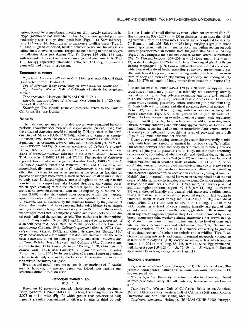
Calicotyle urobati n. sp. (Figs. 7-11)
Based on 16 preserved, stained, whole-mounted adult specimens. Body pyriform, 1,154-3,260 (n = 13) long (including haptor), 940- 2,075 (n = 14) wide (Fig. 7); width greater near posterior of body. Pigment granules concentrated or diffuse, in anterior third of body,
forming 2 pairs of small distinct eyespots when concentrated (Fig. 7). Haptor circular, 908-1,277 (n = 13) in diameter; septa muscular, divid- ing ventral surface of haptor into 1 central and 7 peripheral loculi (Fig. 7). Hamuli 1 pair, 208-308 (n = 25) long, varying slightly in shape among specimens, with each hamulus occurring within septum on both sides of posterior median loculus; hamulus guard 90-164 (n = 16) long (Figs. 7, 10). Marginal hooklets not evident. Mouth ventral, subterminal (Fig. 7). Pharynx bulbous, 109-209 (n = 12) long and 149-214 (n = 13) wide. Esophagus 25-75 (n = 5) long. Esophageal gland cells en- circling esophagus (Fig. 7). Ceca 2, unbranched and without diverticula, with 1 on each side of body, extending posteriorly approximately par- allel with lateral body margin until turning medially at level of posterior third of body and then abruptly turning posteriorly and ending blindly about 10-27% of length of body proper from junction of haptor (Fig. 7).
Testicular mass follicular, 445-1,129 (n = 9) wide, occupying inter- cecal space immediately posterior to midbody, not extending laterally beyond ceca (Fig. 7). Vas deferens extending anteriorly and medially to sinistral cecum (Figs. 7, 8). Seminal vesicle 27-55 (n = 11) in max- imum width, running posteriorly before connecting to penis bulb (Fig. 8). Penis bulb with proximal and distal portions; proximal portion 45- 65 (n = 12) wide, 32-50 (n = 8) long, with 2 subspherical to irregularly shaped chambers (Figs. 8, 9); distal portion 22-35 (n = 8) wide, 22- 32 (n = 8) long, connecting to male copulatory organ; male copulatory organ 110-210 (n = 16) long, sclerotized, tubelike, recurving once, initially running anteriorly and extending approximately 40% of its total length before recurving and extending posteriorly along ventral surface of distal penis bulb, ending roughly at level of proximal penis bulb (Figs. 8, 9). Penis bulb tube not evident.
Ovary convoluted, looping around right cecum dorsoventrally at mid- body, with blind end smooth in sinistral half of body (Fig. 7). Vitellar- ium located between ceca and body margin from immediately anterior to level of pharynx to posterior end of body, not confluent anteriorly or posteriorly, consisting of vitelline cells and vitelline ducts; vitelline cells spherical, approximately 2-4 (n = 12) in diameter, densely packed within vitelline ducts; vitelline ducts dendritic, 11-14 (n = 5) wide, combining ventral to ceca at level immediately anterior to ovary before forming transverse vitelline ducts; transverse vitelline ducts extending into intercecal space ventral to ceca and vas deferens, joining at midline. Mehlis' gland intercecal, located between transverse vitelline ducts and ovary. Mehlis' gland collecting ducts not evident. Common genital pore at level of proximal penis bulb (Fig. 7). Vaginae 2, each with a proximal and distal region; proximal region 159-318 (n = 11) long, 12-65 (n = 24) wide, directed laterally and parallel with transverse vitelline ducts, fusing at midline; ratio of length of proximal region of left vagina to intercecal width at level of vaginae 1:1.4-2.0 (n = 10); each distal region (Figs. 7, 8) a thin tube 45-149 (n = 21) long, 7-10 (n = 8) wide, beginning at level of ceca, running laterally and parallel with extended axis of transverse vitelline ducts; glandular cells surrounding distal regions of vaginae, approximately 1 cell thick, bounded by mem- brane; membrane thin, weakly staining (membrane not drawn in Fig. 8). Vaginal pores opening ventrally, just anterior to level of transverse vitelline ducts, between ceca and vitellarium (Figs 7, 8). Seminal re- ceptacle spherical, 27-55 (n = 11) in diameter, connecting to junction of proximal regions of vaginae posteriorly and at midline (Figs. 7, 8). Oviduct running anteriorly and ventral to seminal receptacle, connecting at midline with ootype (Fig. 8); ootype muscular, with nearly triangular lumen, 114-204 (n = 9) long, 85-148 (n = 14) wide. Egg tetrahedral, with longest edge 109-129 (n = 5), 75-104 (n = 5) wide, with filament approximately as long as egg proper (Fig. 11).
Taxonomic summary
Type host: Urobatis halleri (Cooper, 1863), Haller's round ray, (Ra- jiformes: Urolophidae). Other host: Urobatis maculatus Garman, 1913, spotted round ray.
Sites of infection: Primarily in rectum but also in cloaca and labeled as from pericardial cavity (the latter site may be erroneous, see Discus- sion).
Type locality: Western Gulf of California (Bahia de los Angeles), Mexico. Other localities: western Gulf of California (off Santa Rosalia, Puertocitos, and San Franciscaito), Mexico.
Specimens deposited: Holotype, IBUNAM CNHE 3908; Paratype,

942 THE JOURNAL OF PARASITOLOGY, VOL. 86, NO. 5, OCTOBER 2000 /
12
13
90
8
1O'
FIGURES 7-11. Calicotyle urobati n. sp. from Urobatis halleri. 7. Paratype, ventral view of body (correcting for slight folding of right posterior margin of haptor). Bar = 500 pLm. 8. Paratype, ventral view of portions of male and female reproductive tracts. Bar = 200 em. 9. Paratype, ventral view of proximal and distal portions of penis bulb, spherical chambers, and male copulatory organ. Bar =100 p.m. 10. Paratype, hamulus in profile. Bar = 100 p.m. 11. Paratype, egg. Bar = 100 pLm. FIGURES 12-14. Ventral view of distal and proximal regions of vaginae with respect

BULLARD AND OVERSTREET-TWO NEW ELASMOBRANCH MONOGENEANS 943
IBUNAM CNHE 3909, USNPC 89777, 89778, and 89779, and HWML 15365 and 15366.
Intensity and prevalence of infection: One to 7 worms in 20 of 67 specimens of U. halleri; 1 or 2 worms in 3 of 44 specimens of U. maculatus.
Etymology: The specific name urobati refers to the host genus Uro- batis.
Remarks
The following specimens were examined for comparison: 1 voucher specimen (USNPC 80510) of C. australis from the cloaca and rectum of Rhinobatos typus (Rajiformes: Rhinobatidae) (as Rhinobatos batil- lum, see Last and Stevens, 1994) collected by I. D. Whittington in Moreton Bay, Queensland, Australia; 2 voucher specimens (HWML 38527, slides labeled as Raia naevis) of C. kroyeri from the cloaca of Raja naevus (Rajiformes: Rajidae) collected by D. I. Gibson in the North Sea; 5 paratypes (HWML 31746 and 31747) of C. urolophi, 2 from the cloaca of Urolophus bucculentus (Rajiformes: Urolophidae) and 3 from the cloaca of Urolophus cruciatus collected by M. Beverley- Burton and P. Last from Bruny Island, Tasmania. Based on our obser- vations of these specimens and published descriptions of them, the 3 species are similar to C. urobati n. sp. by having a relatively large haptor, relatively large hamuli relative to body size, extracecal vaginal pores, and proximal and distal regions of vaginae that pass roughly parallel to the transverse vitelline ducts (Figs. 8, 12-14). Calicotyle urobati and C. urolophi differ by the extension of the proximal region of the vaginae relative to the cecum (Figs. 8, 12). Calicotyle urobati has vaginae with a proximal region that terminates ventral to the ceca (Fig. 8), but C. urolophi has vaginae with the proximal region lateral to the cecum (Fig. 12). Calicotyle kroyeri and C. australis are most easily distinguished from C. urobati because the proximal region of the vaginae in those species does not extend laterally as far as the ceca (Figs. 13, 14). The ratio of the length of the proximal region of the left vagina to the intercecal width at the level of the vaginae was 1:1.4-2.0 (n = 10) for C. urobati, 1:0.9-1.3 (n = 4) for C. urolophi, 1:2.2-2.5 (n = 2) for C. kroyeri, and 1:2.9 (n = 1) for C. australis. Even though not overlapping strictly, these ranges represent few specimens. Addi- tional ratios for these and other species might differentiate all species.
DISCUSSION
Based on the suite of morphological features for C. califor- niensis, the species of Calicotyle reported from sharks in Mus- telus form a natural group distinct from other members of the genus reported from nontriakid hosts. Szidat (1970) erected Paracalicotyle Szidat, 1970 based on body shape and haptor size. He established C. stossichi as the type species and includ- ed C. asterii and C. palombi. Our observations on specimens of C. asterii, C. californiensis, and C. stossichi (see Remarks section for C. californiensis) and the published description of C. palombi suggest to us that Paracalicotyle should be resur- rected. However, we herein continue to place the species in Calicotyle until more is known about the female reproductive anatomy of other species of Calicotyle and until more species of Calicotyle are collected from shark hosts.
Members of Calicotyle may provide insight into host phy- logeny. Calicotyle ramsayi from Squalus acanthias lacks many of the morphological characters shared among members of Cal- icotyle reported from species of Mustelus. The holotype and only reported specimen of C. ramsayi collected from S. acan- thias is in poor condition; however, several of its general fea- tures (e.g., pyriform body, extracecal vaginal pores, large haptor
relative to body size) are more similar to those of species of Calicotyle reported from rays than to those from species of Mustelus. The phylogenetic hypothesis of Shirai (1996) sepa- rated neoselachians into 2 superorders: Galea and Squalea. Ga- lea was comprised of sharks included in Heterodontiformes, Orectolobiformes, Lamniformes, and Carcharhiniformes (which includes Mustelus). Squalea was comprised of the remaining sharks, i.e., Chlamydoselachiformes, Hexanchiformes, Echinor- hiniformes, Dalatiiformes, Centrophoriformes, and Squalifor- mes (which includes members of Squalus), and all rays (Squa- tiniformes, Pristiophoriformes, and Rajiformes). The low de- gree of host specificity reported for some members of Calico- tylinae (Chisholm et al., 1997) discourages the formulation of coevolutionary hypotheses among members of this group and their hosts. However, the morphological similarities we ob- served among C. ramsayi and its congeners from rays are con- cordant with the system of Shirai (1996), in which S. acanthias is grouped with rays and squalean sharks rather than with gal- eans.
Calicotyle urobati and C. urolophi are morphologically the most similar among members of the genus, as are their respec- tive hosts. Calicotyle urobati occurs in the round rays U. halleri and U. maculatus from the Gulf of California, and C. urolophi is reported from the stingarees U. bucculentus, U. cruciatus, and Urolophus paucimaculatus from off the coast of southeast Tasmania, Australia (Chisholm et al., 1991). The hosts for C. urobati and C. urolophi are morphologically similar and were once grouped into the single genus Urolophus. Rays in both Urobatis and Urolophus are shallow-water coastal species that probably are not capable of trans-Pacific migration. Despite the geographic distance separating these parasite populations, C. urobati and C. urolophi have diverged only slightly, as have their respective hosts.
The existence and shape of the penis bulb tube may help distinguish species of Calicotyle. This structure previously had not been described and was not evident in other specimens that were examined. However, observations on additional specimens of congeneric species are needed so that the comparative shape of the penis bulb tube may be understood in greater detail.
Our observations suggest that the age of the worm may in- fluence the presence and shape of some features. Chisholm et al. (1997) reported that 14 marginal hooklets were present in all species of Calicotyle. We did not see a marginal hooklet using light microscopy on fixed adult specimens of C. califor- niensis or C. urobati and using scanning electron microscopy on a specimen of C. urobati; however, hooklets could have dissolved in the fixative or been present in juveniles worms. Living specimens or highly flattened juvenile and adult speci- mens mounted in Hoyer's or De Faures medium should be ex- amined for hooklets. Rohde et al. (1992) showed that the num- ber of coils in the male copulatory organ of C. australiensis increased with body length and that the organ became longer until specimens reached maturity at 1-2 mm total body length. As stated previously, the holotype of C. californiensis repre- sents a relatively large (nearly 5.5 mm in total body length,
to ceca and transverse vitelline ducts in 3 species of Calicotyle. 12. Calicotyle urolophi (paratype, HWML 31747). Bar = 300 p.m. 13. Calicotyle kroyeri (voucher specimen, HWML 38527). Bar = 300 pm. 14. Calicotyle australis (voucher specimen, USNPC 80510). Bar = 500 p.m.

944 THE JOURNAL OF PARASITOLOGY, VOL. 86, NO. 5, OCTOBER 2000
including haptor), sexually mature adult specimen. Because of this development, we are confident that the shape and length of the male copulatory organ and the kidney bean-shaped struc- ture formed by the junction of the proximal portions of the vaginae medially in the adult reliably distinguish the species from its apparently closely related congeners reported from oth- er species of Mustelus.
The body cavity and pericardial cavity are unusual sites for specimens of Calicotyle spp., if indeed they are accurate. The sites were based on labels occurring with the specimens. If these sites are correct, we cannot explain how the specimens gained access to those sites and why only 1 of 47 total speci- mens of C. urobati would occur in the pericardial cavity. Sev- eral species of Calicotyle have been reported from the rectal gland and oviduct, and none has been reported from the body cavity or pericardial cavity (Chisholm et al., 1997). In contrast, specimens of Dictyocotyle coeliaca Nybelin, 1941 (Calicotyli- nae) attach to the coelom and body cavity wall of several spe- cies of Raja (see Lawler, 1981). If our specimen of C. califor- niensis was attached similarly, the utilization of the same or of a similar microhabitat within M. californicus, e.g., the body cavity, could serve as a shared ecological characteristic that might support the hypothesized phylogenetic link between D. coeliaca and species of Calicotyle.
ACKNOWLEDGMENTS
We thank Janine Caira (University of Connecticut) and George Benz (Southeast Aquatic Research Institute, Chattanoo- ga, Tennessee) for allowing us the opportunity to take part in the collection and survey of metazoan parasites of elasmo- branchs from the Gulf of California; the artisanal fishermen of the Gulf of California and Loren Caira for catching elasmo- branchs for us to examine; Judy Benz, Paul Cislo, Kirsten Jen- sen, Valerie McKenzie, Peter Olson, Tim Rhunke, and Gaines Tyler for helping collect parasites; Gavin Naylor for aiding in the identification of the shark; John McEachran (Texas Coop- erative Wildlife Collection) for aiding in the identification of rays; G. Benz, Reg Blaylock (Gulf Coast Research Laboratory [GCRL]), Colin Dobson (University of Queensland), and two anonymous reviewers for helpful comments on the manuscript; Pam Monson (GCRL) for preparing a specimen for SEM; Ron- nie Palmer and Stephen Curran (both GCRL) for staining and mounting specimens; and Skip Sterner (HWML) and Ralph Lichtenfels and Pat Pilitt (both USNPC) for loaning specimens for study. This study was supported by National Science Foun-
dation grant DEB 9300796 to J. Caira, a Research Experiences for Undergraduates award to S.A.B. that was kindly facilitated by J. Caira, and a Gulf Coast Research Laboratory, Department of Coastal Sciences research assistantship to S.A.B. through R.M.O. The research in Mexico was conducted according to the conditions set forth in collecting permit number 120496- 213-03 issued to J. Caira by the Secretaria de Medio Ambiente Recursos Naturales y Pesca.
LITERATURE CITED CHISHOLM, L. A., M. BEVERLEY-BURTON, AND P. LAST. 1991. Calicotyle
urolophi n. sp. (Monogenea: Monocotylidae) from stingarees, Uro- lophus spp. (Elasmobranchii: Urolophidae) taken in coastal waters of southern Australia. Systematic Parasitology 20: 63-68.
, T. J. HANSKNECHT, I. D. WHITTINGTON, AND R. M. OVERSTREET.
1997. A revision of the Calicotylinae Monticelli, 1903 (Monoge- nea: Monocotylidae). Systematic Parasitology 38: 159-183.
COMPAGNO, L. J. V. 1984. FAO species catalogue, Vol. 4, Part 2: Sharks of the world. An annotated and illustrated catalogue of shark spe- cies known to date. FAO Fisheries Synopsis No. 125. United Na- tions Development Programme/Food and Agriculture Organization of the United Nations, Rome, Italy, 655 p.
EUZET, L., AND H. H. WILLIAMS. 1960. A re-description of the trematode Calicotyle stossichii Braun, 1899, with an account of Calicotyle palombi sp. nov. Parasitology 50: 21-30.
LAST, P. R., AND J. D. STEVENS. 1994. Sharks and rays of Australia, 1st ed. CSIRO Division of Fisheries, Canberra, Australia, 513 p.
LAWLER, A. R. 1981. Zoogeography and host-specificity of the super- family Capsaloidea Price, 1936 (Monogenea: Monopisthocotylea): An evaluation of the host-parasite locality records of the superfam- ily Capsaloidea Price, 1936, and their utility in determinations of host-specificity and zoogeography. Special Papers in Marine Sci- ence No. 6, Virginia Institute of Marine Science, Gloucester Point, Virginia, 650 p.
MCEACHRAN, J. D., AND G. NOTARBARTOLO DI SCIARA. 1996. Peces ba- toideos. In Guioia FAO para la identication de especies para las fenes de la pesca Pacifico central-oriental, W. Fisher, F. Groupp, W. Schnieder, C. Sommer, K. E. Carpenter, and V. H. Niem (eds.). Departemento di FAO and Instituto de Investigations, Senckenberg, Germany, p. 746-792.
ROHDE, K., M. HEAP, C. J. HAYWARD, AND K. J GRAHAM. 1992. Cali- cotyle australiensis n. sp. and Calicotyle sp. (Monogenea: Mono- pisthocotylea) from the rectum and rectal glands, and Rugogaster hydrolagi Schell, 1973 (Trematoda, Aspidogastrea) from the rectal glands of holocephalans off the coast of southeastern Australia. Systematic Parasitology 21: 69-79.
SHIRAI, S. 1996. Phylogenetic interrelationships of neoselachians (Chon- drichthyes: Euselachii). In Interrelationships of fishes, M. L. J. Stiassny, L. R. Parenti, and G. D. Johnson (eds.). Academic Press, San Diego, California, p. 9-34.
SZIDAT, L. 1970. Nuevas investigaciones sobre la subfamilia Calicoty- linae (Monogenea) y una nueva especie de Austrocalicotyle Szidat 1971. Anales del Instituto de Biologfa Universidad Nacional Au- t6noma de Mexico (Serie Zoologia) 41: 155-160.