campbell and reece chapter 17. process by which dna directs the synthesis of proteins or rna ...
TRANSCRIPT

From Gene to Protein
Campbell and ReeceChapter 17

process by which DNA directs the synthesis of proteins or RNA
synthesis of proteins 1. transcription2. translation
Gene Expression

Evidence from study of metabolic disorders:
1902: British physician 1st to suggest genes responsible for phenotype thru enzymes that catalyze specific chem rx in the cell
How Gene to Protein Figured Out
Garrod hypothesized that symptoms of an inherited disease are due to a gene that leads to inability to make a certain enzyme
1 of 1st to realize Mendel’s principle’s of heredity applied to more than pea plants
Inborn Errors of Metabolism

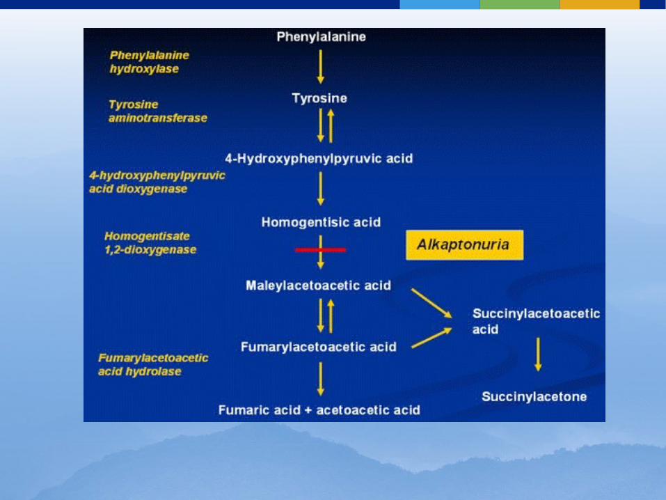
Signs & Symptoms: urine turns black when alkapton
(chemical in urine) reacts with air missing enzyme in pathway that
degrades phenylalanine (a.a.)
Alkaptonuria


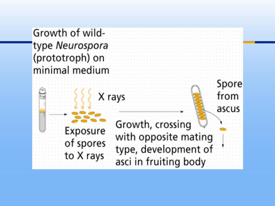
worked with a bread mold Neurospora crassa
bombarded it with radiation (already known to cause genetic changes)
then checked for survivors who had different nutritional needs from wild-type mold
Beadle & Tatum Experiment

individually put yeast in different mediums (agar with different nutrients)
identified mutants that could not survive on minimal nutrients placed them in complete growth medium (minimal med. + all 20 a.a. + few vitamins & minerals)
Beadle & Tatum Experiment


Beadle & Tatum’s results supported their hypothesis
1958: Nobel prize
1-Gene-1-Polypeptide

revised over time: not all proteins are enzymes some proteins have >1 polypeptide now: 1- gene-1-protein hypothesis not 100%: some eukaryotic genes can
each code for a set of closely related polypeptides via alternative splicing
1-Gene-1-Polypeptide

the synthesis of RNA using information in DNA
mRNA made using complimentary base pairing
Transcription: short version
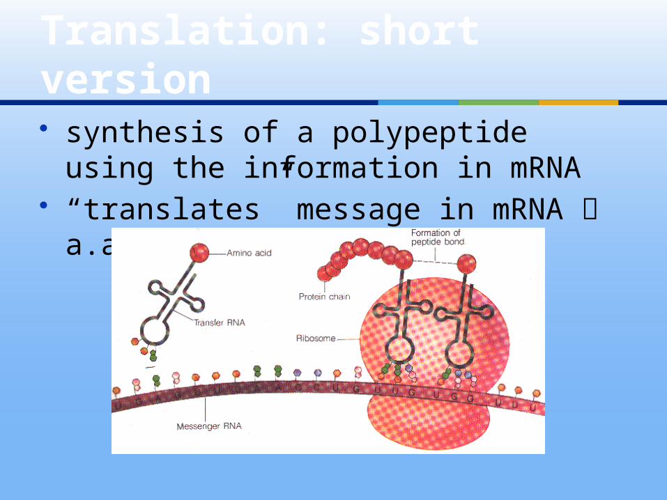
synthesis of a polypeptide using the information in mRNA
“translates” message in mRNA a.a.
Translation: short version

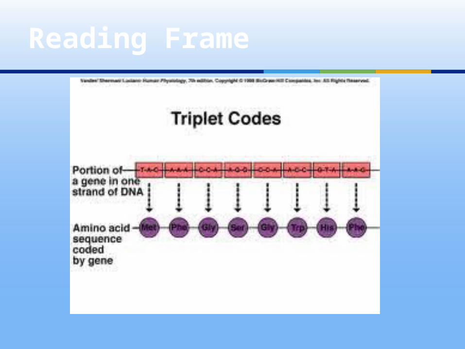
4 nucleotide bases to code for 20 a.a.
triplet code: 3 consecutive bases code for 1 of the a.a./ stop
The Genetic Code


during transcription: DNA helix unwound 1 strand only transcribed (could be
either side depending on the gene)
Template Strand

uracil added as compliment to adenine
ribose as its 5-carbon sugar single stranded
mRNA

nucleotide triplets of DNA or mRNA that specifies a particular amino acid or termination signal
basic unit of the genetic code written in 5’ 3’ direction (in DNA 3
bases read in 3’ 5’ direction)
Codons

Genetic Code

early 1960’s Nirenberg: synthesized mRNA using
only uracil (UUUUUUU…) added it to test tube with all 20 a.a.,
ribosomes translated into polypeptide made up of
phenyalanine now knew UUU = Phe did same for AAA= Lys, CCC = Pro, GGG
= Gly
Cracking the Code

all 64 a.a. deciphered by mid-1960’s 3 codons code for “stop” marking
end of translation AUG functions as “start” & Met
Met may or may not be clipped off later
Cracking the Code

>1 triplet codes for each of the a.a. but any 1 triplet codes for only 1 a.a
redundant triplets usually only differ in the 3rd base
Genetic Code is Redundant

translating the code in correct groupings
example: Did the red dog eat the bug?
Idt her edd oge att heb ug?
Reading Frame
Reading Frame

code is nearly universal: bacteria complex multicellular
organisms CAU = His insert genes into other species & get
same result (human insulin gene in bacteria)
exceptions: certain unicellular eukaryotes & in organelle genes of some species
Evolution of Genetic Code


unwinds 2 strands of DNA binds nucleotides together as build
mRNA only in 5’ 3’ direction (like DNA
polymerase)
RNA Polymerase

1. Initiation2. Elongation3. Termination
3 Stages of Transcription
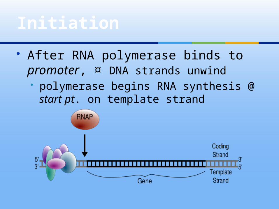
After RNA polymerase binds to promoter, ¤ DNA strands unwind polymerase begins RNA synthesis @
start pt. on template strand
Initiation

promoter: usually includes w/in it the transcription start point (a nucleotide where transcription begins) & extends several dozen or more nucleotide pairs upstream from start pt.
RNAP can assemble nucleotides only in 5’ 3’ direction (just like DNA polymerase)
unlike DNAP, RNAP does not require a primer
Initiation
nucleotide where RNA synthesis actually begins
RNAP binds in precise location & orientation on the promoter where determines where transcription starts & which of the 2 strands will be transcribed
Start Point

Bacteria: 1 single RNAP used to make all types
RNA Eukaryotic Cells:
@ least 3 types RNA polymerase II used for RNA synthesis I and III used to transcribe RNA not
used for protein synthesis
RNA Polymerase

Prokaryotes : RNAP recognizes & binds to the
promoter by itself Eukayotes:
collection of proteins , transcription factors, mediate the binding of RNAP & initiation of transcription
RNA Polymerase

must 1st attach to promoter b/4 RNAP II can bind to it RNAP II + transcription factors =
Transcription Initiation Complex TATA box: DNA sequence in
eukaryotic promoters crucial in forming the transcription initiation complex
Transcription Factors


RNAP moves downstrean, unwinding the DNA & elongating the RNA transcript 5’ 3’
~ 10 – 20 nucleotides exposed in wake of transcription the 2 DNA
strands spontaneously rewind length of DNA transcribed =
transcription unit
Elongation

Elongation

mechanism differs between prokaryotes & eukaryotes
Bacteria: transcription proceeds thru terminator sequence in the DNA the transcribed RNA functions as the terminator sequence causing RNAP to detach
prokaryotes have no further modification
Termination
RNAP II transcribes a portion of DNA called the polyadenylation signal (AAUAAA) in the pre-mRNA
~10 – 35 nucleotides downstream from that sequence proteins ass’c with transcription cut the pre-mRNA free from the polymerase
pre-mRNA then modified
Termination in Eukaryotes


in eukaryotes only both ends of primary transcript
altered certain interior sections cut out &
remaining parts spliced back together
RNA Processing

5’ end receives a 5’cap: modified G is added after ~ 20 – 40 nucleotides in mRNA
3’ end modified: enzyme adds 50 -250 A’s to the AAUAAA forming a poly-A tail
mRNA Ends
1. facilitate exit of mRNA from nucleus
2. protect mRNA from degradation of hydrolytic enzymes
3. help ribosomes attach to the 5’ end
Functions of Modified Ends of mRNA


cut-and-paste job removing segments of RNA that were transcribed
average size transcript: 27,000 nucleotides
average size protein: 1,200 nucleotides (400 a.a.)
RNA Splicing

noncoding, intervening sequence w/in primary transcript that is removed from the transcript during RNA processing; also refers to the region of DNA from which this sequence was transcribed
Introns

sequence w/in primary transcript that remains in the RNA after RNA processing; also refers to the region of DNA from which this sequence was transcribed
Exons

signal: short nucleotide sequence @ each end of an intron
particle called “snurp” recognizes splice sites small nuclear ribonucleoproteins
(snRNP’s) in nucleus made of RNA + protein small nuclear RNA ~150 nucleotides
RNA Splicing

combination of several different snRNP’s (almost size of ribosome)
interact with certain sites along intron releasing intron rapidly degraded
then joins ends of exons together
Spliceosome

RNA Splicing

RNA molecules that function like enzymes in some organisms
intron RNA can act like ribozyme & catalyze its own excision
Ribozymes

3 properties of RNA enables some RNA molecules to function as enzymes:
1. single-stranded: 1 sequence can interact w/another using base pairing
2. some of bases contain functional groups (like a.a) that could participate in catalysis
3. ability to form H-bonds adds specificity
Ribozymes

RNA
still having debate about importance of introns & RNA splicing in evolution
they both have adaptive benefits do not know functions of most
introns
Importance of Introns

single gene can encode >1 kind of polypeptide
know many genes that make 2 or more different polypeptides depending on what was removed as introns during gene splicing
called: alternative RNA splicing
Importance of Introns

Drosophila sex differences due to how RNA transcript is spliced
Human Genome Project: 1 of reasons humans get by with same # genes as a nematode
Alternative RNA Splicing

tRNA: transfers a.a. from cytoplasmic pool of a.a to ribosome where it’s a.a. is added to polypeptide chain
cell keeps supply of all 20 a.a. on hand degradation of other molecules synthesizes them using building blocks
in cytoplasm
Translation: Closer Look

brings specific a.a to ribosome 1 end has a.a./ other end has anticodon
which H-bonds with codon on ribosome tRNA translates the codes into the
corresponding a.a. tRNA is transcribed from DNA
templates & used repeatedly tRNA made of ~80 nucleotides long
with some regions folded back on self due to base pairing
tRNA

tRNA Structure
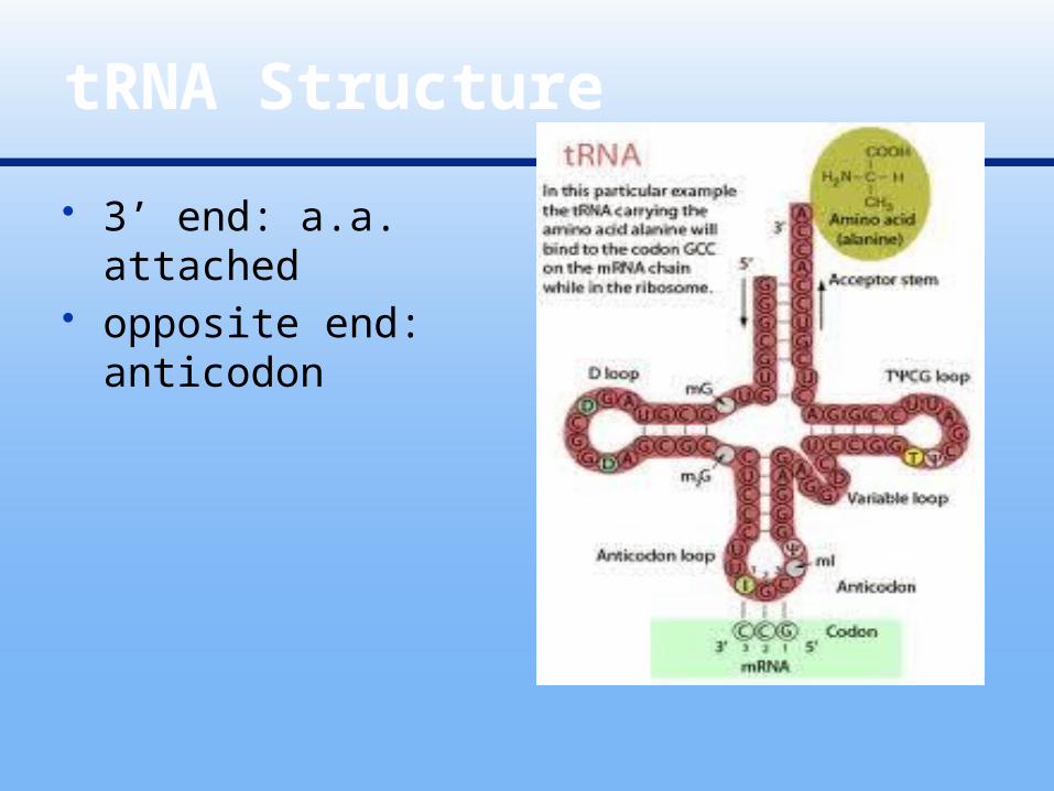
3’ end: a.a. attached
opposite end: anticodon
tRNA Structure
requires 2 instances of molecular recognition:
1. tRNA that binds to particular a.a. correct match made by group enzymes
called aminoacyl-tRNA synthetases: their active site fits only 1 of the 20 a.a.
2. pairing of tRNA anticodon with mRNA codon
Accurate Translation
~ 45 different ones (not 61 like genetic code would suggest) possible because pairing the 3rd base of
codon & 3rd base of anticodon: relaxed base pair rules
U can pair with A or G in 3’ end of codon (3rd position)
called a “wobble”
tRNA Wobble
subunits made in nucleolus rRNA transcribed & added to proteins
imported from cytoplasm ribosomal subunits cytoplasm, join
only when translating mRNA subunits ~1/3 protein & 2/3 rRNA
bacteria: 3 molecules rRNA eukaryotes: 4 molecules rRNA
Ribosomes

eukaryotic ribosomes slightly larger than prokaryotic ones
pharmaceutical products (antibiotics) designed to inactivate bacterial ribosomes that have no effect on ours Tetracyclines Streptomycin
Ribosome Structure

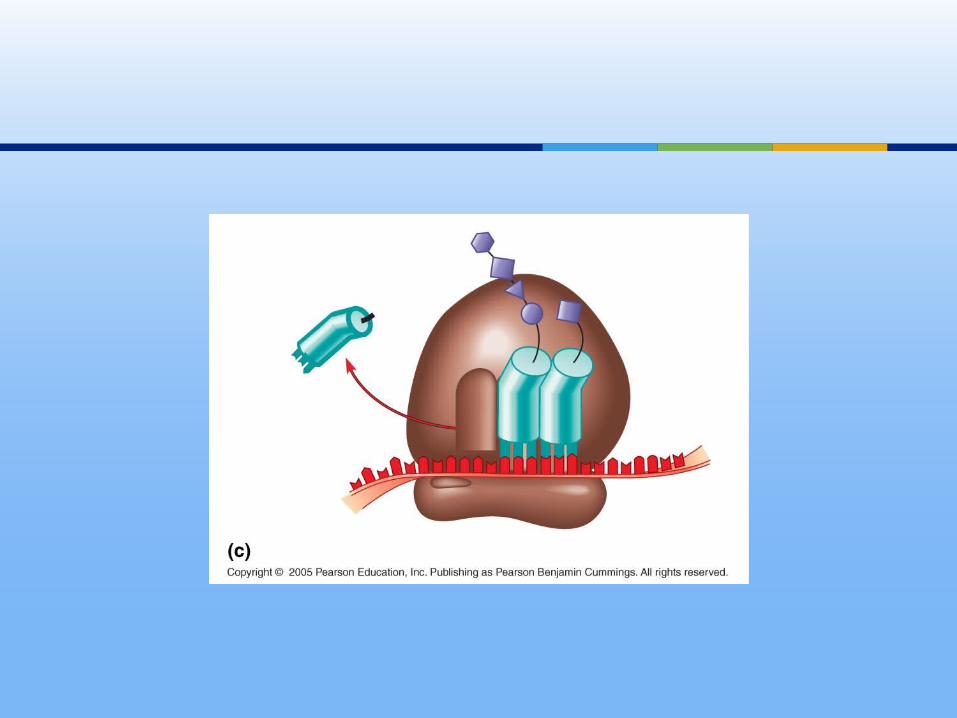
4 binding sites: (1st for mRNA, others for tRNA)
1. mRNA binding site2. P site: peptidyl-tRNA holds the
tRNA carrying the growing polypeptide chain
3. A site: aminoacyl-tRNA holds tRNA carrying next a.a to be added
4. E site: exit, where discharged tRNAs leave ribosome
Ribosome Structure


holds tRNA & mRNA in close proximity & catalyzes the formation of new peptide bond holding the 2 a.a together adding to carboxyl end of last a.a. in growing polypeptide chain
peptide chain passes thru exit tunnel in large subunit as it grows longer
Ribosome
3 Stages:1. Initiation2. Elongation3. Termination
Translation

small ribosomal subunit attaches to mRNA
downstream from this attachment is the start codon AUG
tRNA with UAC (Met) binds to it large ribosomal subunit attaches
(1GTP) initiation factors (proteins) required
to bring it all together
Initiation


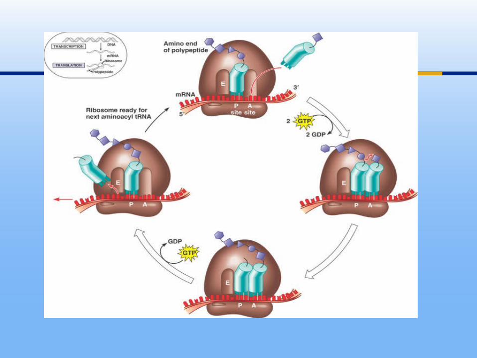
1. Codon recognition anticodon of incoming tRNA w/c’ base 1 GTP increases accuracy & efficiency
2. Peptide bond formation part of rRNA catalyzes reaction amino end of newest a.a + carboxyl end of
peptide chain transferring pep. chain to tRNA @ A site
3. Translocation ribosome moves so tRNA @ A site P site 1 GTP
Elongation

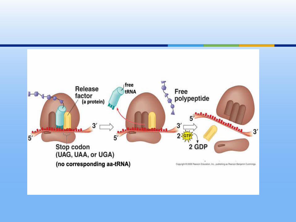
1. ribosome reaches stop codon the A site accepts a “release factor” (shaped like tRNA but does not have aminoacyl part)
2. promotes release of bond between P site, mRNA, & last tRNA
3. 2 ribosomal subunits & ass’c proteins come apart
Termination

http://bcs.whfreeman.com/thelifewire/content/chp12/1202003.html
http://highered.mcgraw-hill.com/olcweb/cgi/pluginpop.cgi?it=swf::535::535::/sites/dl/free/0072437316/120077/micro06.swf::Protein%20Synthesis
Animation Time!

http://www.wiley.com/college/boyer/0470003790/animations/translation/translation.htm
Try at home: interactive

1 ribosome can make polypeptide of average size: 1 min
typically many ribosomes are translating a single mRNA @ given time
1st ribosome gets far enough past start codon 2nd ribosome can get started
allow cell to make many copies of polypeptide very quickly
Polyribosomes

Polyribosomes

as polypeptide chain grows longer from ribosome it will spontaneously start to fold & coil as result of a.a side chain interactions
genes determine 1’ structure which then determines 2’, 3’ and 4’ structures
Primary Structure

proteins that help with the folding
Chaperonins

additional steps that may be required b/4 protein can do its job attachment of sugars, lipids, phosphate
groups to a.a enzymatic removal of 1 or more a.a.
from leading end (amino end)
Post-Translational Modifications

Modification of Insulin

free ribosomes make proteins used in cytoplasm
bound ribosomes (RER) attached to cytosolic side while polypeptide being released into endomembrane system
both have identical small & large subunits
Targeting Polypeptides Specific Locations in Cell

Ribosomes
growing polypeptide cues ribosome to attach to ER
polypeptides of proteins destined for endomembrane system have signal peptide: sequence of ~20 a.a. at or near leading end (N-terminus) is recognized by a protein-RNA complex called signal-recognition particle or SRP
Signal Peptide
escorts ribosome to receptor protein on ER membrane
receptor part of multiprotein translocation complex
ribosome continues to make polypeptide which enters ER thru protein pore
signal protein usually removed by enzyme
SRP


use other signal peptides for protein destined for chloroplast, mitochondria, or interior of nucleus
in these, proteins made in cytosol then to organelle
signal proteins target or “address” proteins for secretion or to cellular locations used by prokaryotes too
Proteins Organelles


ultimate source of new genes large scale mutations
chromosomal rearrangements: chap. 15 small scale mutations
1 or a few nucleotide bases changed
Mutations

http://www.bodrum-hotels.com/gene-mutations/gene-mutations-and-proteins-worksheet.html
Try @ Home

changes in single nucleotide pair if occurs in gamete or cell that gamete
will be passed on to offspring if mutation has adverse effect on
phenotype is called a genetic disorder or hereditary disease
if mutation causes organism to die before fully developed it is said to be lethal
if mutation results in no change in phenotype is said to be silent
Point Mutations


Sickle Cell Anemia

point mutation dominant possible cause of
sudden death of young athletes
Familial Cardiomyopathy

replacement of 1 nucleotide pair by another pair: a few will improve activity of protein it is coding for but most will be detrimental
some silent due to redundancy of genetic code
if changes 1 a.a. for another called missense mutation if substituted a.a. similar to real one no effect some substitutions will have major
consequences
Substitutions

Nucleotide-Pair Substitutions

1. Silent2. Missense:
most substitutions in this category
3. Nonsense: substitution changes from 1 a.a. stop codon
resulting polypeptide is shorter nearly all nonfunctional proteins
Nucleotide-Pair Substitution

(+) or (-) of nucleotide pairs in a gene
disastrous effects may alter reading frame triplet
codon shifts on mRNA called frameshift mutation
whenever insertion or deletion not in a multiple of 3
if not causes major missense
Insertions & Deletions


any chemical or physical agent that interacts with DNA & can cause a mutation
1920’s: Muller used x-rays to make mutant Drosophila & he discovered it does same in humans
mutagenic radiation includes: UV radiation cause thymine dimers in DNA
Mutagens

Thymine Dimers

nucleotide analogs similar to normal DNA nucleotides insert self into DNA
Chemical Mutagens
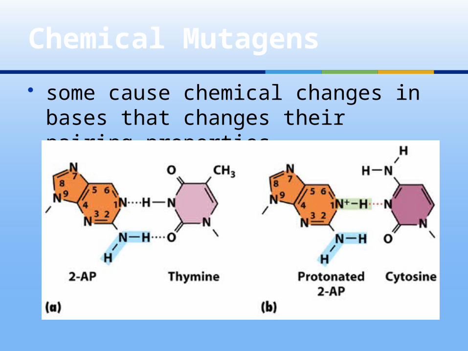
some cause chemical changes in bases that changes their pairing properties
Chemical Mutagens

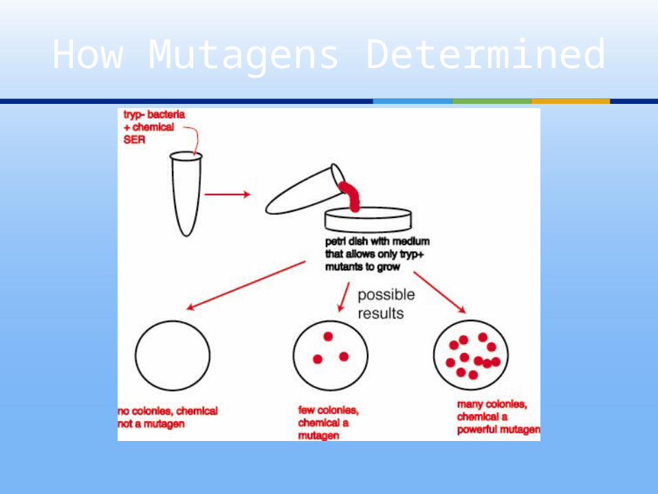
How Mutagens Determined


Gene Expression in 3 Domains

some in gene expression among eubacteria, archaea, and eukaryotes
if no nucleus: translation can begin b/4 transcription is over
Archaea show similarities to Eubacteria and eukaryotes in processes of gene expression
Differences

region of DNA whose final functional product is either a polypeptide or an ENA molecule
What is a Gene?
