cancer cell article - sarcoma uk · cancer cell article the hippo transducer yap1 transforms...
TRANSCRIPT

Please cite this article in press as: Tremblay et al., The Hippo Transducer YAP1 Transforms Activated Satellite Cells and Is a Potent Effector of Embry-onal Rhabdomyosarcoma Formation, Cancer Cell (2014), http://dx.doi.org/10.1016/j.ccr.2014.05.029
Cancer Cell
Article
The Hippo Transducer YAP1 TransformsActivated Satellite Cells and Is a Potent Effectorof Embryonal Rhabdomyosarcoma FormationAnnie M. Tremblay,1,2,3 Edoardo Missiaglia,4,5 Giorgio G. Galli,1,2,3 Simone Hettmer,2,3,9,10,11 Roby Urcia,7
Matteo Carrara,14 Robert N. Judson,7,15 Khin Thway,5,6 Gema Nadal,7 Joanna L. Selfe,5 Graeme Murray,8
Raffaele A. Calogero,14 Cosimo De Bari,8 Peter S. Zammit,12 Mauro Delorenzi,4,13 Amy J. Wagers,2,3,9 Janet Shipley,5
Henning Wackerhage,7 and Fernando D. Camargo1,2,3,*1Stem Cell Program, Boston Children’s Hospital, Boston, MA 02115, USA2Department of Stem Cell and Regenerative Biology, Harvard University, Cambridge, MA 02138, USA3Harvard Stem Cell Institute, Cambridge, MA 02138, USA4SIB Swiss Institute of Bioinformatics, 1015 Lausanne, Switzerland5Sarcoma Molecular Pathology Team, Divisions of Molecular Pathology and Cancer Therapeutics, The Institute of Cancer Research, Sutton,
Surrey SM2 5NG, UK6Department of Histopathology, Royal Marsden NHS Foundation Trust, London SW3 6JJ, UK7School of Medical Sciences, University of Aberdeen, Aberdeen, AB25 2ZD Scotland, UK8School of Medicine and Dentistry, University of Aberdeen, Aberdeen AB25 2ZD, Scotland, UK9Howard Hughes Medical Institute and Joslin Diabetes Center, Boston, MA 02115, USA10Department of Pediatric Oncology, Dana Farber Cancer Institute, Boston, MA 02115, USA11Division of Pediatric Hematology/Oncology, Children’s Hospital, Boston, MA 02115, USA12Randall Division of Cell and Molecular Biophysics, King’s College London, New Hunt’s House, Guy’s Campus, London SE1 1UL, UK13Ludwig Center for Cancer Research and Oncology Department, University of Lausanne, 1015 Lausanne, Switzerland14Molecular Biotechnology Center, Department of Biotechnology and Health Sciences, University of Torino, 10126 Torino, Italy15Biomedical Research Centre, Department of Medical Genetics, University of British Columbia, Vancouver BC V6T 1Z3, Canada
*Correspondence: [email protected]
http://dx.doi.org/10.1016/j.ccr.2014.05.029
SUMMARY
The role of the Hippo pathway effector YAP1 in soft tissue sarcomas is poorly defined. Here we report thatYAP1 activity is elevated in human embryonal rhabdomyosarcoma (ERMS). Inmice, sustained YAP1 hyperac-tivity in activated, but not quiescent, satellite cells induces ERMSwith high penetrance and short latency. Viaits transcriptional program with TEAD1, YAP1 directly regulates several major hallmarks of ERMS. YAP1-TEAD1 upregulate pro-proliferative and oncogenic genes and maintain the ERMS differentiation block byinterferingwithMYOD1andMEF2pro-differentiation activities. Normalization of YAP1 expression reduces tu-mor burden in human ERMSxenografts and allows YAP1-driven ERMS to differentiate in situ. Collectively, ourresults identify YAP1 as a potent ERMS oncogenic driver and a promising target for differentiation therapy.
INTRODUCTION
Rhabdomyosarcoma (RMS), the most common soft tissue sar-
coma in children and adolescents, is subdivided into embryonal
RMS (ERMS; z60%), alveolar RMS (ARMS; z20%), and other
Significance
The molecular and cellular origins of ERMS are incompletelybeen identified in association with familial syndromes. Currelead to life-altering sequelae for survivors. Over the past 30 yeaand less aggressive regimens, which highlights the need for imof the molecular changes underlying RMS formation and proplecular cause of ERMS, providing a rationale and mouse modeYAP1 inhibition in combination with other regimens for differe
(z20%) subtypes (Belyea et al., 2012). Most ARMS cases
(z80%) are associated with the presence of a PAX3/7-
FOXO1A fusion gene (ARMSp), which predicts clinical outcome
(Missiaglia et al., 2012) and is sufficient to cause ARMS at
low frequency in mice (Keller et al., 2004). However, z20%
understood, and only a few ERMS-causing pathways havent therapies for RMS are aggressive, highly cytotoxic, andrs, little progress has beenmade in identifying more efficientproved therapeutic approaches and a better understandingagation. Here we identify YAP1 hyperactivity as a major mo-ls for preclinical studies aimed at assessing the efficiency ofntiation therapy of ERMS.
Cancer Cell 26, 1–15, August 11, 2014 ª2014 Elsevier Inc. 1

Cancer Cell
High YAP1 Activity in Satellite Cells Causes ERMS
Please cite this article in press as: Tremblay et al., The Hippo Transducer YAP1 Transforms Activated Satellite Cells and Is a Potent Effector of Embry-onal Rhabdomyosarcoma Formation, Cancer Cell (2014), http://dx.doi.org/10.1016/j.ccr.2014.05.029
of histologically diagnosed ARMS cases are negative for the
expression of PAX3/7-FOXO1A fusion genes (ARMSn) and
are indistinguishable from ERMS at the molecular level (William-
son et al., 2010). In contrast to ARMS, the oncogenic drivers
in ERMS and ARMSn are still incompletely defined. A hallmark
of ERMS (and other RMS subtypes) is that the cells are locked
in a proliferating myoblast state despite the expression of
myogenic differentiation-regulating factors such as MYOD1
and Myogenin (Saab et al., 2011). While the precise mechanism
of differentiation inhibition is uncertain, it is believed that
the relative abundance of MYOD1 activating or inhibiting E-pro-
tein heterodimer partners in RMS is shifted toward repressive
heterodimers (Yang et al., 2009). Overcoming the terminal dif-
ferentiation block in RMS would open the door for differentia-
tion therapy, which aims to halt tumor progression by forcing
the cells to differentiate into nondividing muscle fibers (Al-Ta-
han et al., 2012; Saab et al., 2011). However, our understanding
of the mechanisms underlying this differentiation block in RMS
remains insufficient to achieve better treatments.
The core Hippo pathway is regulated by many upstream
signal transduction proteins in response to cues from the cellular
microenvironment (Yu and Guan, 2013). It generally functions
to limit organ growth and tumorigenesis by inducing the
cytosolic localization of the transcriptional cofactor YAP1,
especially but not solely through Ser127 phosphorylation (Zhao
et al., 2007). Nuclear YAP1 promotes proliferation and inhibits
apoptosis primarily by coactivating the TEAD family of transcrip-
tion factors (Tremblay and Camargo, 2012). Consistent with this,
the constitutively active YAP1 S127A mutant is mostly nuclear
(Zhao et al., 2007) and, with the exception of the intestine (Barry
et al., 2013; Zhou et al., 2011), promotes proliferation, expands
undifferentiated stem cells and progenitors, and leads to tumor
formation in epithelial tissues (Camargo et al., 2007; Dong
et al., 2007; Schlegelmilch et al., 2011). However, the role of
YAP1 in soft tissue sarcomas is still poorly defined. In C2C12
myoblasts and cultured activated satellite cells (Judson et al.,
2012; Watt et al., 2010), YAP1 S127A drives proliferation and
inhibits differentiation. Given that a pro-proliferation, antidiffer-
entiation myoblast phenotype is the hallmark of RMS, here
we tested whether YAP1 was involved in the development of
rhabdomyosarcoma.
RESULTS
Increased YAP1 Copy Number and Elevated Expressionin Human ERMSTo explore the role of YAP1 in human RMS, we first assessed
the abundance and cellular localization of YAP1 protein in
78 ARMS and 196 ERMS samples. Representative YAP1 im-
munostainings are shown for human ERMS and ARMS tumors
of each intensity category (Figure 1A). ERMS tumors expressed
a significantly higher level of YAP1 protein than ARMS
(Figure 1B) (p = 0.0046, Mann-Whitney U test). ARMS cases
displayed a higher proportion of predominantly cytoplasmic
staining (C), while ERMS cases displayed a greater proportion
of nuclear (N) and dual nuclear/cytoplasmic staining (N/C).
Indeed, there was a significant tendency for N/C expression
to have a strong intensity score (chi-square test for trend, p =
0.0002), and YAP1 was more nuclear (N and N/C) in ERMS
2 Cancer Cell 26, 1–15, August 11, 2014 ª2014 Elsevier Inc.
than ARMS (Figure 1C) (chi-square test, p < 0.0001, n = 227).
We also found an association between the proliferation marker
Ki67 and YAP1 staining within the ERMS subgroup, but not the
ARMS (Figure 1D) (chi-square test for trend, p = 0.079, n =
184). Array CGH analysis from 79 human RMS samples (Wil-
liamson et al., 2010) revealed a significant increase in YAP1 lo-
cus copy number in ERMS and in fusion-negative RMS, which
includes ERMS and fusion-negative ARMS (ARMSn), but not in
fusion-positive ARMS (ARMSp) (Figure 1E) (Fisher’s exact test,
p = 0.012). These data demonstrate a higher YAP1 expression
and activity in a high proportion of ERMS cases, but not ARMS,
suggesting a causative role for YAP1 in ERMS development
and/or propagation.
Expression of Activated YAP1 in Mouse Skeletal MuscleCauses ERMSTo test for a causal role of high YAP1 activity in RMS, we gener-
ated doxycycline-inducible (DOX-inducible) Myf5-hYAP1 S127A
mice (Figure 2A), in which DOX administration in adult mice
induces the overexpression of the TetO-hYAP1 S127A trans-
gene in the mature skeletal musculature, as well as in adult
quiescent and activated satellite cells and their myoblast prog-
eny (Beauchamp et al., 2000; Cornelison andWold, 1997; Kuang
et al., 2007; Tajbakhsh et al., 1996). After 4–8 weeks of DOX
administration, adult Myf5-hYAP1 S127A mice developed a
rapidly progressing and severe gait defect with marked loss of
limb flexibility (Figures S1A and S1B available online), which
did not allow obvious tumors to form before euthanasia was
required. Hematoxylin and eosin staining (H&E) revealed exten-
sive muscle damage, loss of mature myofibers, and the pres-
ence of newly formed fibers with centrally located nuclei,
indicative of ongoing muscle regeneration (Figures 2B and 2C,
arrowheads) (Judson et al., 2013). Additionally, a massive
expansion of mononucleated cells was detected in the interstitial
compartment of all muscles analyzed, including the tibialis ante-
rior (TA) (Figure 2B), abdominal (Figure 2C), and gastrocnemius
(GAS) muscles (Figure 2D).
Small well-demarcated tumor nodules located in the crural
and/or hamstring muscles were macroscopically apparent at
necropsy (Figures 2E and 2F). These tumors expressed high
levels of YAP1 (Figure 2G), Desmin (Figure 2H), Myogenin (Fig-
ure 2I), and Caveolin-1 (Figure 2J) (Rossi et al., 2011). Patholo-
gists identified the interstitial expansion and primary tumors as
non-alveolar RMS histologically similar to human embryonal
RMS (ERMS) of the spindle cell variant accompanied by focal
pleomorphic features. The brown fat pads, also derived from
the MYF5 lineage (Seale et al., 2008), appeared normal with no
apparent myogenic differentiation or neoplastic lesions (Figures
S1C and S1D). Dissociated cells (1 3 106) from the expanded
interstitial compartment of DOX-induced Myf5-hYAP1 S127A
mice produced tumors in nonobese diabetic/severe combined
immunodeficient (NOD/SCID) mice with a latency ranging from
36 to 54 days (Figure S1E). These also resembled human
ERMS (Figure S1F), expressed high levels of YAP1 (Figure S1G),
Desmin (Figure S1H), and Myogenin (Figure S1I) and were trans-
plantable (Figure S1J). Indeed, serial dilutions propagated the
same type of secondary tumors with strikingly short latency
and 100% penetrance after transplanting as few as 100 cells
(Figure S1K).

ARMSCases: 56
ERMSCases: 171
Nuclear (N)Dual (N/C)
Cytosolic (C)
14
14 28
60
83
28
0
25
50
75
100
%o
fsu
bty
pe
ERMSARMS
0 1 2 3YAP1 StainingIntensity
100
0
25
50
75
+-Ki67:
%K
i67/c
ate
gory
0 1 2 3YAP1 StainingIntensity
ARMS
ERMS
0 1 2 3YAP1 Staining
Intensity:
38 127
22 25
196 78
126
17 27
77
11
104
5 11
46
9
80
11
14
A
B C D
5.5 6.0 6.5 7.0 7.5
−1.
0−
0.5
0.0
0.5
1.0
YAP1 Log Intensity
CN
Log
rat
io −
YAP
1 lo
cus
Pearson coeff. 0.61p < 0.001
ERMS
3.0 3.5 4.0 4.5 5.0 5.5 6.0 6.5
−1.
0−
0.5
0.0
0.5
1.0
YAP1 Log Intensity
CN
Log
rat
io −
YA
P1
locu
s
Pearson coeff. 0.07p = 0.709
ARMSp
YAP1 Log Intensity
Pearson coeff. 0.57p < 0.001
5.0 6.0 7.0 8.0
−1.
0−
0.5
0.0
0.5
1.0
CN
Log
rat
io −
YAP
1 lo
cus
Fusion-Negative RMSE
Figure 1. YAP1 Is Expressed at Higher Levels and Trends with Increased Proliferation Index in Human ERMS
(A) Immunostaining for YAP1 in human RMS samples classified by intensity level and subtype. Scale bars, 50 mm.
(B) Distribution of cases in each subtype according to YAP1 staining intensity. The number of cases is indicated on the bars.
(C) Distribution of cases according to YAP1 localization in each subtype. The number of cases is indicated on the charts.
(D) Distribution of Ki67� and Ki67+ cases in ERMS according to YAP1 staining intensity. The number of cases is indicated on the bars.
(E) Plot of the log intensity expression level and local segmented copy number for YAP1 locus in ERMS, ARMSn (fusion-negative), and ARMSp (fusion-positive)
tumors. The gain and losses in each subtype were categorized based on a segmented ratio above 0.25 and below �0.25, respectively (Fisher’s exact test
p = 0.012).
Cancer Cell
High YAP1 Activity in Satellite Cells Causes ERMS
Please cite this article in press as: Tremblay et al., The Hippo Transducer YAP1 Transforms Activated Satellite Cells and Is a Potent Effector of Embry-onal Rhabdomyosarcoma Formation, Cancer Cell (2014), http://dx.doi.org/10.1016/j.ccr.2014.05.029
High YAP1Activity in theMYOD1 Lineage Induces ERMSTo confirm that RMS can arise as a result of persistent YAP1
hyperactivity in activated satellite cells or myoblasts, we addi-
tionally generated Myod1-hYAP1 S127A mice (Figure 2K) (Kani-
sicak et al., 2009). Within 4 weeks of DOX treatment, Myod1-
hYAP1 S127A mice developed a gait phenotype similar to
Myf5-hYAP1 S127A mice, with obvious primary tumors in the
limbs (Figures S1L and S1M) and in abdominal muscles at
Cancer Cell 26, 1–15, August 11, 2014 ª2014 Elsevier Inc. 3

Figure 2. High YAP1 Activity in the MYF5 and MYOD1 Lineages Induces Non-Alveolar Rhabdomyosarcoma
(A) Mating strategy and alleles of the Myf5-hYAP1 S127A mice.
(B–D) Cross-section of the TA (B), abdominal wall (C), and gastrocnemius (D) of Myf5-hYAP1 S127A mice induced for 8 weeks, stained by H&E. Yellow
arrowheads mark centrally located nuclei. Scale bars, 100 mm.
(E and F) Cross-section of hamstring with primary tumor outlined by dashed line (E) and at higher magnification (F) from Myf5-hYAP1 S127A mice induced for
8 weeks (n = 3). Scale bars, 100 mm.
(G–J) As in (F), immunostained for YAP1 (G), Desmin (H), Myogenin (I), and Caveolin-1 (J) (n = 3). Scale bars, 100 mm.
(K) Mating strategy and alleles of the Myod1-hYAP1 S127A mice (n = 5).
(L–N) Cross-sections of the abdominal wall of Myod1-hYAP1 S127A mice induced for 4 weeks, stained by H&E, and shown at 4X (L), 10X (M), and 20X (N)
magnification. Scale bars, 200 mm (L and M). Scale bar, 50 mm (N).
(O–Q) As in (N), immunostained for YAP1 (O), Desmin (P), and Myogenin (Q). Scale bars, 50 mm.
(R–U) Sections of primary tumors from Myod1-hYAP1 S127A mice induced for 4 weeks, immunostained for PCNA (R), PAX7 (S), MYOD1 (T), and Caveolin1 (U).
Scale bars, 50 mm (R), and 100 mm (S–U).
See also Figure S1.
Cancer Cell
High YAP1 Activity in Satellite Cells Causes ERMS
4 Cancer Cell 26, 1–15, August 11, 2014 ª2014 Elsevier Inc.
Please cite this article in press as: Tremblay et al., The Hippo Transducer YAP1 Transforms Activated Satellite Cells and Is a Potent Effector of Embry-onal Rhabdomyosarcoma Formation, Cancer Cell (2014), http://dx.doi.org/10.1016/j.ccr.2014.05.029

Cancer Cell
High YAP1 Activity in Satellite Cells Causes ERMS
Please cite this article in press as: Tremblay et al., The Hippo Transducer YAP1 Transforms Activated Satellite Cells and Is a Potent Effector of Embry-onal Rhabdomyosarcoma Formation, Cancer Cell (2014), http://dx.doi.org/10.1016/j.ccr.2014.05.029
necropsy. H&E revealed signs of muscle damage, myofiber
regeneration, and importantly, a widespread amplification of
mononucleated cells in the interstitial compartment of the TA
(Figures S1N and S1O) and abdominal muscles (Figures 2L–
2N), along with the presence of centrally located nuclei in the
remaining fibers (Figure S1O, arrowheads). As in Myf5-hYAP1
S127A mice, cells in the amplified interstitial compartment
expressed YAP1 (Figure 2O), Desmin (Figure 2P), and Myogenin
(Figure 2Q). In addition, PCNA (Figure 2R), PAX7 (Figure 2S),
MYOD1 (Figure 2T), and Caveolin-1 (Figure 2U) expression
suggested extensive proliferation and a low degree of myogenic
differentiation, compatible with an expansion of activated
satellite cells and/or myoblasts. Dissociated primary tumor cells
(0.5 3 106) propagated tumors in NOD/SCID (Figure S1P) with a
similar histology as the secondary tumors from the Myf5-hYAP1
S127A model (Figure S1Q) and similar expression of YAP1 (Fig-
ure S1R), Desmin (Figure S1S), and Myogenin (Figure S1T).
Collectively, these results reveal that MYF5+ and MYOD1+ acti-
vated satellite cells or myoblasts can be the cells of origin of
YAP1-ERMS, which is further supported by the enhanced soft
agar colony-forming potential of C2C12 myoblasts expressing
hYAP1 S127A (Figure S1U).
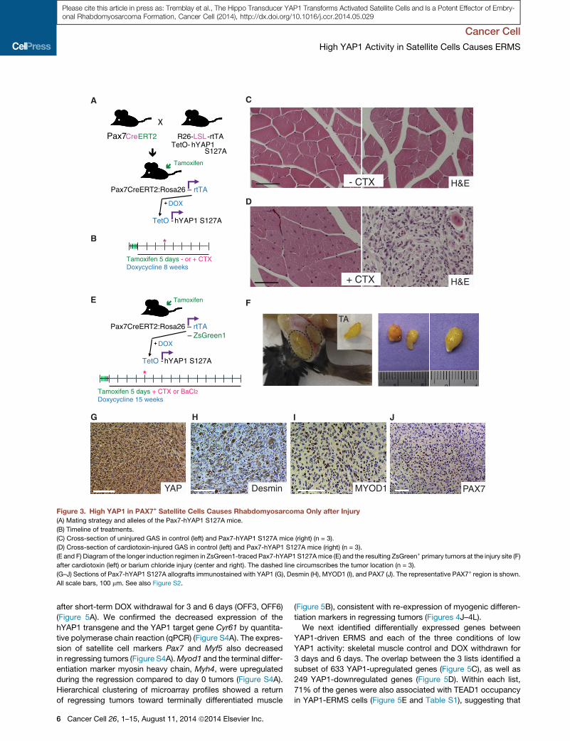
High YAP1 Activity Does Not Induce Satellite CellsActivation but Transforms Activated Satellite CellsNext we addressed the question of whether persistent YAP1
hyperactivity is sufficient to both activate and transform satellite
cells. To overexpress hYAP1 S127A in quiescent and activated
satellite cells, we generated DOX-inducible Pax7-hYAP1
S127A mice (Figure 3A) (Lepper and Fan, 2010). DOX was given
for 8 weeks, andmuscle injury was induced via cardiotoxin (CTX)
injection to the right hindlimb 3 weeks after the hYAP1 S127A
induction in PAX7+ cells (Figure 3B). Surprisingly, the muscle
morphology in the uninjured left hindlimb was unaffected after
8weeks of DOX, andH&E staining showed no signs of expansion
of the interstitial compartment (Figure 3C). Likewise, the percent-
age of satellite cells, assessed by the ratio of PAX7+ nuclei/total
nuclei, remained unchanged (Figures S2A and S2B). In contrast,
the injured muscles of Pax7-hYAP1 S127A overexpressing mice
displayedmassive amplification ofmononucleated cells 5 weeks
after injury (Figure 3D), which was histologically consistent with
RMS as determined by pathological examination. The core of
the injured muscle expressed high levels of YAP1 (Figure S2C),
PCNA (Figure S2D), Desmin (Figure S2E), and Myogenin (Fig-
ure S2F) with a low level of PAX7 expression (Figure S2G).
We additionally introduced a ZsGreen1 reporter allele in Pax7-
hYAP1 S127A mice in order to track the activated progeny of
quiescent PAX7+ cells after CTX injury (Figure 3E). A longer
DOX induction period after injury led to the development of
ZsGreen1+ primary tumors at the injury site by using either
CTX or a barium chloride injury method (Figure 3F). Sorted
ZsGreen1+ cells from those primary tumors produced secondary
ZsGreen1+ tumors expressing high levels of YAP1 (Figure 3G),
Desmin (Figure 3H), and MYOD1 (Figure 3I), while PAX7 expres-
sion was overall low (Figure 3J shows a representative patch of
PAX7+ cells). Human ERMS tumors notably express PAX7 at var-
iable levels (Tiffin et al., 2003), and while viewed as indicative of
a satellite cell origin, PAX7 expression is not a diagnostic criteria
for RMS classification. Collectively, our results on three trans-
genic mouse models (Myf5-, Myod1-, and Pax7-driven hYAP1
S127A expression) and two types of injury strongly suggest
that persistent hyperactivity of YAP1 in activated but not quies-
cent satellite cells can potently cause ERMS-like tumors with
remarkably short latency in mice.
Normalization of YAP1 Activity in YAP1-Driven ERMSReleases the YAP1-Imposed Differentiation BlockIn SituTo test whether YAP1 reduction causes tumor regression, we
withdrew DOX from host mice bearing secondary tumors
following transplantation of sorted ZsGreen1+ Pax7-hYAP1
S127A primary tumor cells from the injury site (Figure 4A, top
panel). DOX-withdrawn tumors regressed to z 50% of their
original size in 10 days (Figure 4A, bottom panel) and coex-
pressed the myogenic differentiation marker sarcomeric myosin
heavy chain (MyHC) and ZsGreen1 in myofibers at their core
(Figure 4B). We observed a similar regression in Myf5-hYAP1
S127A allograft tumors 12days after DOXwithdrawal (Figure 4C).
These tumors, after 6 days OFF DOX (OFF6) (Figure 4D), showed
a marked decrease in the expression of YAP1 (Figure 4E), PCNA
(Figure 4F), and Caveolin-1 (Figure 4G). Expression of Myogenin
(Figure 4H) appeared unchanged, while Desmin immunostaining
was suggestive of newly formed myofibers (Figure 4I). Consis-
tently, Caveolin-3 (Figure 4J) and MyHC (Figure 4K) expression
increased. Furthermore, coexpression of MyHC and ZsGreen1
(Figure 4L) at the core of the regressing tumors (Figures S3A
and 3SB) confirmed the ability of the YAP1-ERMS tumor cells
to differentiate into mature skeletal muscle in situ once YAP1
expression is normalized. In culture, ZsGreen1+ YAP1-ERMS
cells from Myf5-hYAP1 S127A secondary tumors could be
expanded in the presence of DOX (Figure S3C), but did not pro-
liferate as well in its absence (Figure S3D). These cells also
formed MyHC+ and ZsGreen1+ double positive myotubes
when plated at high density and subjected to differentiation con-
ditions for 4 days in the absence of DOX (Figure S3E). These re-
sults suggest that persistent hYAP1 S127A hyperactivity causes
ERMS-like tumors in mice both by promoting proliferation and
imposing a differentiation block, which can be overcome once
YAP1 activity is reduced.
Identification of YAP1 Target Genes in YAP1-ERMSTo characterize the mechanisms by which YAP1 drives ERMS,
we aimed to identify the YAP1 target genes that could explain
the sustained myoblast proliferation and differentiation-block
phenotype of ERMS. We combined global gene expression an-
alyses with chromatin immunoprecipitation-sequencing (ChIP-
Seq) for the YAP1 cofactor TEAD1. YAP1 and TEAD1 mostly
co-occupy the same promoters in MCF10A cells (>80% overlap)
(Zhao et al., 2008) and directly interact in proliferating mouse
myoblasts (Judson et al., 2012). YAP1 does not bind DNA
directly, so interaction with TEAD factors is responsible for
most, if not all, of YAP’s functions in vivo (Schlegelmilch et al.,
2011), and identification of YAP1 target genes is reliably associ-
ated with TEAD binding sites (Lian et al., 2010; Zhao et al., 2008).
We first used microarray analysis to identify potential YAP1
target genes in the YAP1-ERMS tumors. We reasoned that
YAP1 direct target genes would show a transcriptional response
to reduction of transgenic YAP1 levels in regressing YAP1-ERMS
Cancer Cell 26, 1–15, August 11, 2014 ª2014 Elsevier Inc. 5
Tamoxifen 5 days - or + CTXDoxycycline 8 weeks
*
XPax7CreERT2 R26-LSL-rtTA
TetO- hYAP1
Tamoxifen
Pax7CreERT2:Rosa26 – rtTA
TetO - hYAP1 S127A
+ DOX
A
S127A
B
G H I
H&E
H&E- CTX
+ CTX
DesminYAP MYOD1 PAX7
Tamoxifen 5 days + CTX or BaCl2Doxycycline 15 weeks
*
Tamoxifen
Pax7CreERT2:Rosa26 – rtTAZsGreen1
TetO - hYAP1 S127A
+ DOX–
E
TA
F
C
J
D
Figure 3. High YAP1 in PAX7+ Satellite Cells Causes Rhabdomyosarcoma Only after Injury
(A) Mating strategy and alleles of the Pax7-hYAP1 S127A mice.
(B) Timeline of treatments.
(C) Cross-section of uninjured GAS in control (left) and Pax7-hYAP1 S127A mice (right) (n = 3).
(D) Cross-section of cardiotoxin-injured GAS in control (left) and Pax7-hYAP1 S127A mice (right) (n = 3).
(E and F) Diagram of the longer induction regimen in ZsGreen1-traced Pax7-hYAP1 S127Amice (E) and the resulting ZsGreen+ primary tumors at the injury site (F)
after cardiotoxin (left) or barium chloride injury (center and right). The dashed line circumscribes the tumor location (n = 3).
(G–J) Sections of Pax7-hYAP1 S127A allografts immunostained with YAP1 (G), Desmin (H), MYOD1 (I), and PAX7 (J). The representative PAX7+ region is shown.
All scale bars, 100 mm. See also Figure S2.
Cancer Cell
High YAP1 Activity in Satellite Cells Causes ERMS
Please cite this article in press as: Tremblay et al., The Hippo Transducer YAP1 Transforms Activated Satellite Cells and Is a Potent Effector of Embry-onal Rhabdomyosarcoma Formation, Cancer Cell (2014), http://dx.doi.org/10.1016/j.ccr.2014.05.029
after short-term DOX withdrawal for 3 and 6 days (OFF3, OFF6)
(Figure 5A). We confirmed the decreased expression of the
hYAP1 transgene and the YAP1 target gene Cyr61 by quantita-
tive polymerase chain reaction (qPCR) (Figure S4A). The expres-
sion of satellite cell markers Pax7 and Myf5 also decreased
in regressing tumors (Figure S4A).Myod1 and the terminal differ-
entiation marker myosin heavy chain, Myh4, were upregulated
during the regression compared to day 0 tumors (Figure S4A).
Hierarchical clustering of microarray profiles showed a return
of regressing tumors toward terminally differentiated muscle
6 Cancer Cell 26, 1–15, August 11, 2014 ª2014 Elsevier Inc.
(Figure 5B), consistent with re-expression of myogenic differen-
tiation markers in regressing tumors (Figures 4J–4L).
We next identified differentially expressed genes between
YAP1-driven ERMS and each of the three conditions of low
YAP1 activity: skeletal muscle control and DOX withdrawn for
3 days and 6 days. The overlap between the 3 lists identified a
subset of 633 YAP1-upregulated genes (Figure 5C), as well as
249 YAP1-downregulated genes (Figure 5D). Within each list,
71% of the genes were also associated with TEAD1 occupancy
in YAP1-ERMS cells (Figure 5E and Table S1), suggesting that

OF
F13
MyHCCaveolin-3
Day
0O
FF
6D
ay 0
OF
F12
ZsGreen / MyHC / DAPI
Overlay
C
J KL
OF
F10
Day
0
A B Overlay DAPI ZsGreen MyHCO
FF
10D
ay 0
Desmin
Myogenin
Caveolin-1
PCNA
H&E
YAP1
E
F
H
I
Day0 OFF6 Day0 OFF6D G
Figure 4. Normalization of YAP1 Expression Allows Differentiation of YAP1-ERMS Tumors In Situ
(A) Representative picture of Pax7-hYAP1 S127A secondary tumors in situ at day 0 (top panel) and Pax7-hYAP1 S127A secondary tumors in situ following DOX
withdrawal for 10 days (bottom panel).
(B) Frozen section of tumors in (A) immunostained for MyHC (red) and DAPI (blue). ZsGreen and MyHC double positive fibers are circled.
(C) Representative picture of the Myf5-hYAP1 S127A secondary tumors in situ before and after DOX withdrawal for 12 days. The dashed line circumscribes the
tumor location.
(D) H&E staining of tumor sections at day 0 and after 6 days of DOX withdrawal (OFF6).
(E–K) As in (D), immunostained for YAP1 (E), Desmin (F), Myogenin (G), PCNA (H), Caveolin-1 (I), Caveolin-3 (J), and MyHC (K).
(L) Frozen section of ZsGreen-traced tumors after 13 days of DOX withdrawal (OFF 13), immunostained for MyHC (red) and DAPI (blue). ZsGreen and MyHC
double positive fibers are circled.
All scale bars, 50 mm. See also Figure S3.
Cancer Cell
High YAP1 Activity in Satellite Cells Causes ERMS
Please cite this article in press as: Tremblay et al., The Hippo Transducer YAP1 Transforms Activated Satellite Cells and Is a Potent Effector of Embry-onal Rhabdomyosarcoma Formation, Cancer Cell (2014), http://dx.doi.org/10.1016/j.ccr.2014.05.029
most of these genes are likely direct transcriptional targets of
YAP1-TEAD1. Gene ontology (GO) analysis revealed enrichment
for terms related to cellular proliferation, cell cycle, andmitosis in
the YAP1-ERMS-upregulated target gene list (Figure 5F). In
contrast, the YAP1-ERMS-repressed target gene list was en-
riched for GO terms related to muscle contraction (Figure 5G)
Cancer Cell 26, 1–15, August 11, 2014 ª2014 Elsevier Inc. 7

A
Day0
OFF6
OFF3YAP1-ERMS host + DOX
DOXwithdrawal
Log21-1
TUM
OF
F3
OF
F6
SK
M
71% 29% 71% 29%
TEAD1-boundNot bound
TEAD1-boundNot bound
YAP1-ERMSactivated
YAP1-ERMSrepressed
B
E
02468
101214
1618 YAP1-ERMS-activated
Genome***
******
***** ***
0
2
4
6
8
10
12***
******
YAP1-ERMSrepressedGenome
% w
ith te
rm
% w
ith te
rm
cell
cycl
e
cell c
ycle
pro
cess
mito
tic c
ell c
ycle
cell
divis
ion
mito
tic M
pha
sem
itosi
s
mus
cle
cont
ract
ion
regu
latio
n of
mus
cle c
ontra
ctio
n
stria
ted
mus
cle
con
tract
ion
0
1
2
3
4
5
Pou5f
1 (N
eg)
Cyr61
Ankrd
1
AjubaBirc
5
Ccnd1Cdc
6M
yog
Myo
m2
Tnnc
2
Tnni1M
yh2
Myb
phM
yl4
Hspb2
YAP1 targets/Proliferation
Myogenic Differentiation
0
10
20
30
40
50
60100140
Fold
Enrich
ment
YAP1 targets/Proliferation
Myogenic Differentiation
Fold
Enrich
ment
F G
HI
Pou5f
1 (N
eg)
Cyr61
Ankrd
1
AjubaBirc
5
Ccnd1Cdc
6M
yog
Myo
m2
Tnnc
2
Tnni1M
yh2
Myb
phM
yl4
Hspb2
UP in Tumors (Day0) vs:
SKM
OFF3103 41
23062
111
1947
633
OFF6
DOWN in Tumors (Day0) vs:
OFF3 OFF6
1180
171
249 157
287165 79
SKM
C
E2f1
E2f1
D
Figure 5. Combined Gene Expression Profiling and TEAD1 ChIP-Seq Analyses Identify YAP1-TEAD1 Direct Target Genes Signatures in
YAP1-Driven ERMS
(A) Diagram of the tumor regression experiments used in the microarray analysis.
(B) Heat map showing hierarchical clustering of global gene expression in normal skeletal muscle (SKM), day 0 (TUM), and DOX-withdrawn (OFF3 and OFF6)
tumors.
(C and D) Venn diagram showing the overlap between the genes upregulated (C) and downregulated (D) in the YAP1-ERMS tumors (TUM) versus normal skeletal
muscle (SKM) and the regressed tumors at day 3 (OFF3) and 6 (OFF6).
(E) Pie charts representing the percentage of common YAP1-regulated genes (TUM versus SKM-OFF3-OFF6) bound by TEAD1.
(F and G) Gene ontology analysis of the YAP1-ERMS-activated (F) and YAP1-ERMS-repressed (G) signatures (**p < 0.01; ***p < 0.001).
(H and I) ChIP-qPCR validations of YAP1 (H) and TEAD1 (I) occupancy on selected loci associated with upregulated (red) and downregulated (blue) genes in the
YAP1-ERMS microarray. Data were presented in fold enrichment over IgG ± SD.
See also Figure S4 and Table S1.
Cancer Cell
High YAP1 Activity in Satellite Cells Causes ERMS
Please cite this article in press as: Tremblay et al., The Hippo Transducer YAP1 Transforms Activated Satellite Cells and Is a Potent Effector of Embry-onal Rhabdomyosarcoma Formation, Cancer Cell (2014), http://dx.doi.org/10.1016/j.ccr.2014.05.029
and contained mostly target genes of myogenic regulatory
factors MYOD1, Myogenin, MEF2, and SRF (Figure S4B), sug-
gesting a decreased activity of these factors when YAP1 activity
is high. We next performed ChIP-qPCR validations for YAP1
(Figure 5H) and TEAD1 (Figure 5I) occupancy on a subset of
peaks selected by the combination of microarray and ChIP-
8 Cancer Cell 26, 1–15, August 11, 2014 ª2014 Elsevier Inc.
Seq analyses, confirming the correlation between TEAD1 bind-
ing peaks and YAP1 occupancy. Collectively, this suggests
that YAP1-TEAD1 not only directly drives gene expression
necessary for sustained proliferation but also contributes to
blocking myogenic differentiation by repressing genes that are
normally expressed in differentiated skeletal muscle.

Cancer Cell
High YAP1 Activity in Satellite Cells Causes ERMS
Please cite this article in press as: Tremblay et al., The Hippo Transducer YAP1 Transforms Activated Satellite Cells and Is a Potent Effector of Embry-onal Rhabdomyosarcoma Formation, Cancer Cell (2014), http://dx.doi.org/10.1016/j.ccr.2014.05.029
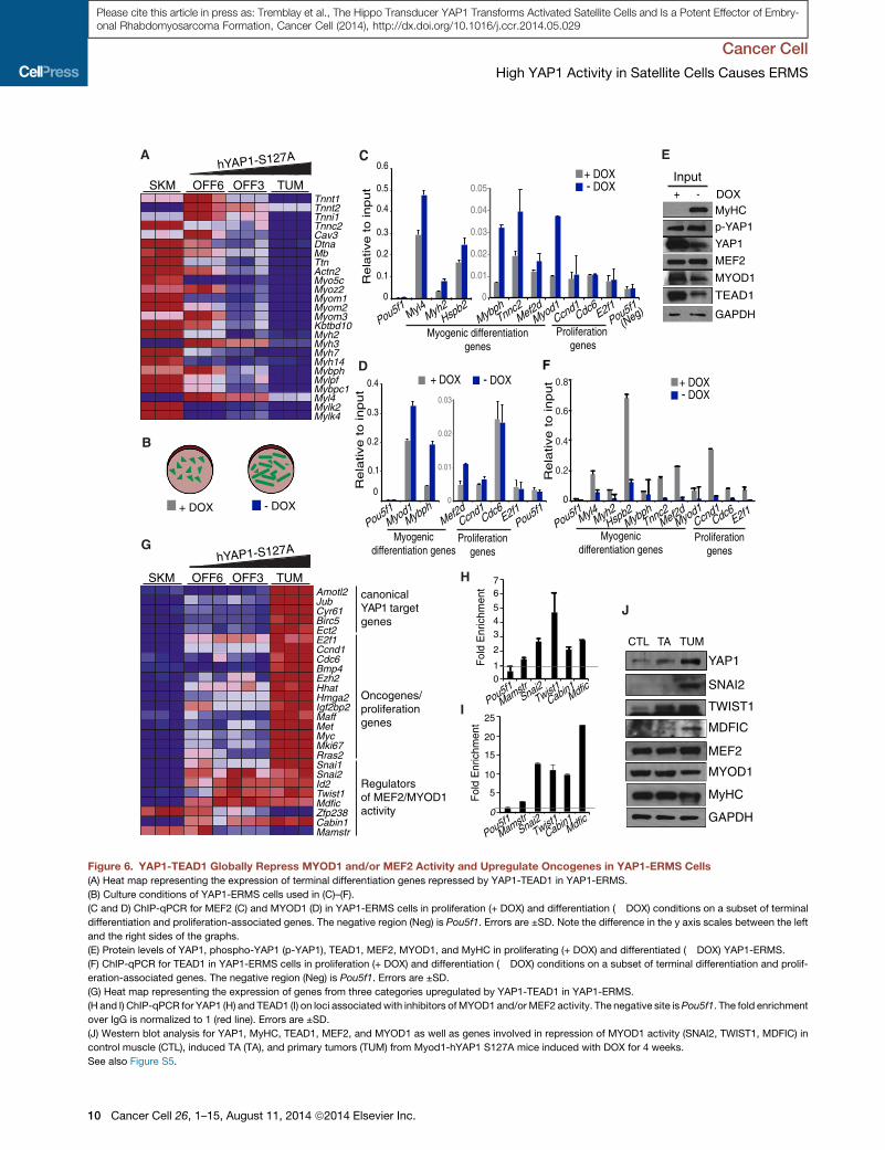
YAP1 Is Integrated in the Myogenic DifferentiationProgram and Represses Terminal Differentiation GenesStudies since the late 1980s reported occurrence of TEAD DNA
binding sites, named muscle CAT (MCAT) elements, in the
promoters of genes that are selectively expressed in differenti-
ated skeletal muscle (Yoshida, 2008). Here we confirm and
extend this observation genome-wide. Using motif enrichment
analyses, we identified the overrepresentation of Myogenin,
MEF2, or SRF consensus sequences within TEAD peaks or
around the promoter of TEAD1-associated genes (Figure S5A).
Nearly one-third (27%) of TEAD1 peaks in ’’undifferentiated’’
YAP1-ERMS cells are aligned with MYOD1, Myogenin, and/
or SRF binding peaks in differentiated C2C12 myotubes (Fig-
ure S5B), suggesting that YAP1-TEAD1 globally regulates
muscle lineage-specific gene expression in YAP1-ERMS. GO
analysis of the shared TEAD1-MYOD1-Myogenin-SRF loci re-
turned terms compatible with the YAP1-ERMS phenotype and
with the role of YAP1 in the repression of the myogenic differen-
tiation program (Figures S5C and S5D), while the TEAD1-only
sites associated generally with immune processes and signaling
pathways (Figure S5E).
Among the genes harboring adjacent binding sites for TEAD1-
MYOD1-Myogenin-SRF, a large number of repressed genes
were associated with terminal muscle contraction and differenti-
ation. The expression of these genes was reduced in YAP1-
driven ERMS but increased when YAP1 activity was lowered
by DOX withdrawal (Figure 6A). Interestingly, MYOD1 and Myo-
genin binding occupancy increases during myogenic differentia-
tion frommyoblasts tomyotubes, on loci sharedwith TEAD1 (Fig-
ure S5F). This correlates with the concomitant decrease in YAP1
activity duringmyogenic differentiation (Judson et al., 2012;Watt
et al., 2010).We thus hypothesized that YAP1-TEAD1occupancy
could impinge on MYOD1 and MEF2 binding on shared sites
associated with repressed muscle differentiation genes. To test
this we evaluated the occupancy of MEF2 and MYOD1 on a se-
lection of genes associated with myogenic differentiation or pro-
liferationwhile altering the levels of YAP1 in culturedYAP1-ERMS
cells via DOX withdrawal (Figure 6B). This analysis revealed that
reduction of hYAP1 S127A expression, leading to differentiation
of YAP1-ERMS cells, resulted in increased occupancy of MEF2
(Figure 6C) and/or MYOD1 (Figure 6D) specifically on myogenic
differentiation genes, but not on proliferation-related genes.
Such functional interplay between YAP1 and myogenic factors
at shared loci cannot be explained by changes in MEF2 or
MYOD1 expression levels as their expression did not increase
during YAP1-ERMS differentiation (Figure 6E). However, both
expression (Figure 6E) and occupancy of TEAD1 (Figure 6F)
decreased during YAP1-ERMS cell differentiation on all genes
tested, including proliferation-associated genes. Our data indi-
cate that the sustained presence of YAP1-TEAD1 plays a role in
repressing differentiation genes, partly by impinging on thediffer-
entiation promoting activities ofmyogenic transcription factors at
muscle differentiation-associated loci.
YAP1 Upregulates Oncogenes and Inhibitors ofMyogenic Regulatory Factors in Mouse YAP1-ERMS andHuman ERMS CellsAdditionally, we observed upregulation of several canonical
YAP1 target genes and RMS-related oncogenes (Figures 6G
and S5D) in YAP1-ERMS, identifying YAP1 as a meta-oncogene
and supporting the potency of YAP1 to trigger ERMS in mice.
Among YAP1 upregulated genes, we identified a subset that
encode known inhibitors of MYOD1 and MEF2 activity,
including Id2, Twist1, Mdfic, Snai1, Snai2, and Cabin1 (Fig-
ure 6G). Loci associated with the latter category of genes
were also bound by YAP1 (Figure 6H) and TEAD1 (Figure 6I)
in YAP1-ERMS cells and were upregulated at the protein level
in induced muscle and YAP1-ERMS primary tumors from
Myod1-hYAP1 S127A mice (Figure 6J). This suggests that
YAP1 can block differentiation not only by affecting the occu-
pancy of MYOD1 and MEF2 at myogenic differentiation-associ-
ated loci but also by upregulating the expression of inhibitors of
myogenic transcription factors.
Interestingly, most YAP1-TEAD1 bound target genes vali-
dated in mouse YAP1-ERMS (Figures 5H, 5I, 6H, and 6I) were
also bound by YAP1 (Figure S5G) and TEAD1 (Figure S5H)
in human ERMS RD cells, and some of these loci were also
shared by MEF2 (Figure S5I) and MYOD1 (Figure S5J). In
addition, gene expression analysis of the validated YAP1-
TEAD1 target genes in RD cells (Figure S5K) and human
ERMS (Figure S5L) shows regulation similar to mouse YAP1-
ERMS (Figures 6A and 6G), suggesting that the transcriptional
program of YAP1 and its role in blocking differentiation is similar
in human ERMS. Indeed, using the YAP1-ERMS signatures
identified in mouse (Figure 5E and Table S1), we could assess
YAP1 activity in a microarray of human fetal myoblasts under-
going differentiation. Consistent with the impeding effect of
YAP1 S127A on the differentiation of mouse myoblasts in vitro
(Judson et al., 2012; Watt et al., 2010), expression of YAP1
mRNA (Figure S5M) and the YAP1-ERMS-activated signature
(Figure S5N) decreased progressively when human fetal myo-
blasts differentiated. Concurrently, expression of the YAP1-
ERMS-repressed signature increased (Figure S5O). These
data validate use of the YAP1-ERMS signatures as YAP1 activ-
ity readout in human cells and suggest that a decrease in YAP1
activity is also associated with myogenic differentiation in hu-
man myoblasts, as in mouse.
These analyses in YAP1-ERMS and human RD cells demon-
strate that YAP1-TEAD1 binding is associated with three
categories of direct target genes. YAP1-TEAD1 binding is asso-
ciated first with expression of genes that promote proliferation
and/or transformation, second, with repression of genes that
are normally expressed in differentiated skeletal muscle, and
third, with increased expression of MYOD1 and MEF2 inhibitors.
Collectively, this suggests that persistent YAP1 hyperactivity not
only drives proliferation and transformation, but also represses
differentiated muscle lineage-specific genes, imposing a global
differentiation block, which is a hallmark of RMS.
High YAP1 Activity Associates with Higher Stage inFusion-Negative RMS and Trends with Poorer PrognosisWe next confirmed the importance of YAP1 activity in human
RMS using two large series of human RMS primary tumors
totaling 235 samples. First, YAP1 expression was higher in
human ERMS and ARMSn compared to ARMSp (Figure 7A).
YAP1 activity was next assessed using the YAP1-ERMS-
activated and YAP1-ERMS-repressed signatures. YAP1 activity
was high in all RMS subtypes compared to skeletal muscle
Cancer Cell 26, 1–15, August 11, 2014 ª2014 Elsevier Inc. 9
F
Myh3
Myl4
Tnnt2
Cav3Dtna
Tnni1
Myom3
Tnnt1
Actn2
Myh2
Myh14Mybph
Myoz2
Mb
MylpfMybpc1
Kbtbd10
Myo5c
Tnnc2
Myom2
Myh7
Ttn
Myom1
Mylk2Mylk4
SKM TUMOFF3OFF6
0
0.1
0.2
0.3
0.4
0.5
0.6
Myl4Myh2
Hspb2
0.01
0.02
0.03
0.04
0.05
MybphTnnc2
Mef2dMyod1
Ccnd1Cdc6E2f1
tu
pni
ote
vital
eR
Myogenic differentiationgenes
Proliferation genes
0.2
0.3
0.4
Myod1Mybph
Proliferationgenes
Myogenicdifferentiation genes
tu
pni
ote
vital
eR
0.01
0.02
0.03
Mef2dCcnd1
Cdc6E2f1
hYAP1-S127A
+ DOX - DOX
A C
D
SKM TUMOFF3OFF6
hYAP1-S127A
Mki67
Mdfic
Jub
Ect2E2f1
Cyr61
Maff
Amotl2
Bmp4
Myc
Snai1Snai2
Hhat
Ccnd1
Id2Twist1
Met
Ezh2
Birc5
Igf2bp2
Rras2
Cdc6
Hmga2
Zfp238Cabin1Mamstr
canonicalYAP1 targetgenes
Oncogenes/proliferationgenes
Regulatorsof MEF2/MYOD1activity
E
G
0.1
+ DOX- DOX
+ DOX - DOX
B
0
0.2
0.4
0.6
0.8
Myl4Myh2Hspb2
MybphTnnc2
Mef2dMyod1
Ccnd1Cdc6
E2f1
+ DOX- DOX
Proliferationgenes
Myogenicdifferentiation genes
0
0
0
Pou5f1Pou5f1
Pou5f1Pou5f1
(Neg)
tu
pni
ote
vital
eR
Pou5f1
MEF2
MYOD1
GAPDH
YAP1
p-YAP1
MyHC
Input
TEAD1
+ - DOX
Fol
d E
nric
hmen
t
H
I
Fol
d E
nric
hmen
t
Pou5f1
MamstrSnai2
Twist1Cabin1
Mdfic
Pou5f1Mamstr
Snai2Twist1
Cabin1Mdfic
25
20
15
10
5
0
76
543
2
10
TWIST1
SNAI2
MDFIC
MYOD1
MEF2
YAP1
TA TUMCTL
GAPDH
J
MyHC
Figure 6. YAP1-TEAD1 Globally Repress MYOD1 and/or MEF2 Activity and Upregulate Oncogenes in YAP1-ERMS Cells
(A) Heat map representing the expression of terminal differentiation genes repressed by YAP1-TEAD1 in YAP1-ERMS.
(B) Culture conditions of YAP1-ERMS cells used in (C)–(F).
(C and D) ChIP-qPCR for MEF2 (C) and MYOD1 (D) in YAP1-ERMS cells in proliferation (+ DOX) and differentiation (� DOX) conditions on a subset of terminal
differentiation and proliferation-associated genes. The negative region (Neg) is Pou5f1. Errors are ±SD. Note the difference in the y axis scales between the left
and the right sides of the graphs.
(E) Protein levels of YAP1, phospho-YAP1 (p-YAP1), TEAD1, MEF2, MYOD1, and MyHC in proliferating (+ DOX) and differentiated (� DOX) YAP1-ERMS.
(F) ChIP-qPCR for TEAD1 in YAP1-ERMS cells in proliferation (+ DOX) and differentiation (� DOX) conditions on a subset of terminal differentiation and prolif-
eration-associated genes. The negative region (Neg) is Pou5f1. Errors are ±SD.
(G) Heat map representing the expression of genes from three categories upregulated by YAP1-TEAD1 in YAP1-ERMS.
(H and I) ChIP-qPCR for YAP1 (H) and TEAD1 (I) on loci associated with inhibitors ofMYOD1 and/orMEF2 activity. The negative site isPou5f1. The fold enrichment
over IgG is normalized to 1 (red line). Errors are ±SD.
(J) Western blot analysis for YAP1, MyHC, TEAD1, MEF2, and MYOD1 as well as genes involved in repression of MYOD1 activity (SNAI2, TWIST1, MDFIC) in
control muscle (CTL), induced TA (TA), and primary tumors (TUM) from Myod1-hYAP1 S127A mice induced with DOX for 4 weeks.
See also Figure S5.
Cancer Cell
High YAP1 Activity in Satellite Cells Causes ERMS
10 Cancer Cell 26, 1–15, August 11, 2014 ª2014 Elsevier Inc.
Please cite this article in press as: Tremblay et al., The Hippo Transducer YAP1 Transforms Activated Satellite Cells and Is a Potent Effector of Embry-onal Rhabdomyosarcoma Formation, Cancer Cell (2014), http://dx.doi.org/10.1016/j.ccr.2014.05.029

D
−2
−1
01
23
Sta
ndar
dize
d sc
ore
Stage
I
Stage
II
Stage
III
Stage
IV
Spearman rho 0.25, p= 0.002
E
F
0 2 4 6 8 10
0
0.2
0.4
0.6
0.8
1
Follow−up (Years)
Sur
viva
l pr o
babi
lity Fusion-
Negative RMS
Fusion-Positive RMS
0 2 4 6 8 10
0
0.2
0.4
0.6
0.8
1
Follow−up (Years)
Sur
viva
l pr o
babi
lity
Low expressionFusion-NegativeRMS
Fusion-PositiveRMS
Low exp.
High expression
Fus-Neg: High vs Low HR=2.3 95%CI (0.9-6.2) p=0.09Fus-Pos: High vs Low HR=1.2 95%CI (0.5-2.7) p=0.7
Fus-Neg: High vs Low HR=0.3 95%CI (0.1-1.0) p=0.05Fus-Pos: High vs Low HR=0.7 95%CI (0.3-1.6) p=0.4
A
34
56
78
Log
inte
nsity
5.0
5.5
6.0
6.5
7.0
7.5
Log
inte
nsity
56
78
ARMSnERMSSkeletal Muscle
ARMS PAX3-FOXO1A+ARMS PAX7-FOXO1A +
Log
inte
nsity
B
C
High expression
Low expression
High expression
Low expression
High expression
ERMS vs ARMS-PAX3-FOXO, p<0.001ERMS vs ARMS-PAX7-FOXO, p=0.0046
ERMS vs ARMS-Pax3-FOXO, p=0.0028ERMS vs ARMS-Pax7-FOXO, p<0.001
Figure 7. The YAP1-ERMS Signatures Cluster Fusion-Negative RMS
Tumors According to Stage and Severity
(A–C) Expression score of YAP1 mRNA (A), YAP1-ERMS-activated (B), and
YAP1-ERMS-repressed signature (C) scores in in human RMS tumors in the
ITCC/CIT data set. Differences were tested by Wilcoxon signed-rank test.
(D) Box plot showing the association between the YAP1-ERMS-activated
signature and tumor stage in fusion-negative RMS cases (Spearman rho 0.25,
p = 0.002).
(E and F) Kaplan-Meier curves of survival in cases of fusion-negative and
fusion-positive RMS with high and low expression scores of the YAP1-ERMS-
activated (G) and YAP1-ERMS-repressed (H) signatures.
See also Figure S6 and Tables S2 and S3. All whisker bars represent 1.5 times
the interquartile range (1.5 IQR), and the central lines represent the median
value.
Cancer Cell
High YAP1 Activity in Satellite Cells Causes ERMS
Please cite this article in press as: Tremblay et al., The Hippo Transducer YAP1 Transforms Activated Satellite Cells and Is a Potent Effector of Embry-onal Rhabdomyosarcoma Formation, Cancer Cell (2014), http://dx.doi.org/10.1016/j.ccr.2014.05.029
control, but also significantly elevated in ERMS and ARMSn over
ARMSp (Figure 7B), while the expression of the YAP1-ERMS-
repressed signature was low in all subtypes (Figure 7C). The
YAP1-ERMS-activated signature was significantly enriched in
all RMS subtypes considered together versus skeletal muscle,
along with the gene expression signatures of embryonic stem
cells, Notch, and satellite cells activation (Figure S6A and Table
S2). Remarkably, the YAP1-ERMS-activated signature was en-
riched specifically in ERMS versus ARMSp (Figure S6B), and
more significantly than the satellite cells activation signature,
which was previously reported as a hallmark of ERMS (Hatley
et al., 2012; Rubin et al., 2011). These results show that YAP1
activity is elevated in human ERMS and suggest that our YAP1-
ERMS signatures could constitute a useful indicator of YAP1 ac-
tivity in human RMS tumors as well.
We then investigated whether YAP1 activity is associated
with prognosis and/or outcome in human RMS. Indeed, stage
3 and 4 tumors were significantly positively associated with
higher YAP1 activity based on a multivariable linear regression
model including clinicopathological features linked to RMS
outcome (Missiaglia et al., 2012) (Table S3 and Figure 7D),
such as tumor histology, age, and tumor location. We next
defined HIGH expression and LOW expression subgroups for
both YAP1-ERMS signatures (activated and repressed) within
each RMS subtypes and identified a trend (log rank test, p =
0.09) toward a poorer outcome for the subgroup with a higher
expression score of the YAP1-ERMS-activated signature in
fusion-negative, but not in fusion-positive, tumors (Figure 7E).
Consistently, the reverse analysis using the YAP1-ERMS-
repressed signature showed that the low expression subgroup
associated with poorer outcome (log rank test, p = 0.05) in
fusion-negative tumors, but not in fusion-positive tumors (Fig-
ure 7F). Taken together, these results suggest that the YAP1-
ERMS signatures can better segregate the fusion-negative
RMS subtypes in terms of stage and outcome, possibly identi-
fying tumors with higher proliferation rates and lower differenti-
ation levels.
YAP1 Knockdown Decreases Proliferation andTumorigenicity while Increasing DifferentiationCapacity of Human ERMSWe next hypothesized that lowering YAP1 in human ERMS
would release the terminal differentiation block. We stably intro-
duced a DOX-inducible hYAP1 knockdown construct in RD cells
(RD-TetO-shYAP1). DOX treatment of RD-TetO-shYAP1 cells for
5 days induced a marked reduction of YAP1 protein (Figure 8A)
and mRNA levels (Figure 8B). Expression of the proliferation
marker E2F1 (Figure 8B) and EdU incorporation also decreased
(Figure 8C), while the number of MyHC+ cells per myotubes
increased, demonstrating an enhanced myogenic differentiation
(Figure 8D). DOX-treated RD-TetO-shYAP1 cells cultured in soft
agar formed fewer and smaller colonies (Figures 8E and 8F), sug-
gesting that YAP1 knockdown additionally decreases tumorige-
nicity in vitro.
To assess the effects of YAP1 knockdown on ERMS tumor-
igenicity in vivo, we transplanted either control or hYAP1
knockdown RD cells into the gastrocnemius muscle of NOD/
SCID mice. Orthotopic xenografts from the hYAP1 knockdown
RD cells were 42% smaller than tumors originating from control
cells (Figure 8G). Efficient knockdown of YAP1 protein in the
DOX-treated xenografts was confirmed by immunostaining for
YAP1 (Figure 8H), which also correlated with decreased
expression of human Ki67 (Figure 8I) and increased expression
of MyHC (Figures 8J and 8K), indicating a greater degree of
differentiation of tumors in the YAP1 knockdown condition.
These results show that the tumorigenicity and low differentia-
tion potential of ERMS can be rescued in vitro and in vivo by
decreasing YAP1 expression, which functionally validates the
potential benefits of YAP1 inhibition for differentiation therapy
of ERMS.
Cancer Cell 26, 1–15, August 11, 2014 ª2014 Elsevier Inc. 11

A
B
C
D E
F
G H
I
K
J
Figure 8. YAP1 Knockdown in ERMS Cells Decreases Tumor Burden and Rescues the ERMS Differentiation Block
(A) Western blot analysis of YAP1 levels in RD-TetO-shYAP1 cells, untreated and DOX treated for 5 days.
(B) YAP1 and E2F1 mRNA levels in DOX-treated RD-TetO-shYAP1 cells versus untreated cells.
(C) EdU incorporation in RD-TetO-shYAP1 cells in the presence and absence of DOX and quantification of EdU+ cells (n = 3).
(D) Immunostaining for MyHC in RD-TetO-shYAP1 cells in the absence and presence of DOX in differentiation conditions.
(E) Soft agar colony formation assay of RD-TetO-shYAP1 in the presence and absence of DOX. Scale bars, 400 mm.
(F) Quantification of the number of colonies in soft agar colony formation assay as in (E), for increasing number of plated cells.
(G) Macroscopic picture of the xenograft tumors obtained from DOX-treated and untreated RD-TetO-shYAP1 cells and bar graph showing tumor weight at
excision (n = 8 for DOX treated; n = 10 for untreated).
(H–J) Cross-sections of tumors in (G), immunostained for YAP1 (H), Ki67 (I), and MyHC (J). Scale bars, 50 mm.
(K) Quantification of MyHC staining as in (J).
All error bars are represented as ±SEM.
Cancer Cell
High YAP1 Activity in Satellite Cells Causes ERMS
12 Cancer Cell 26, 1–15, August 11, 2014 ª2014 Elsevier Inc.
Please cite this article in press as: Tremblay et al., The Hippo Transducer YAP1 Transforms Activated Satellite Cells and Is a Potent Effector of Embry-onal Rhabdomyosarcoma Formation, Cancer Cell (2014), http://dx.doi.org/10.1016/j.ccr.2014.05.029
Cancer Cell
High YAP1 Activity in Satellite Cells Causes ERMS
Please cite this article in press as: Tremblay et al., The Hippo Transducer YAP1 Transforms Activated Satellite Cells and Is a Potent Effector of Embry-onal Rhabdomyosarcoma Formation, Cancer Cell (2014), http://dx.doi.org/10.1016/j.ccr.2014.05.029
DISCUSSION
Here we show that YAP1 expression and activity are highest in
human ERMS versus ARMS and find that many human ERMS,
but not ARMS, harbor a copy number gain of the YAP1 locus.
In addition, high YAP1 activity is associated with poorer prog-
nosis in human ERMS cases, but not in ARMS. Consistent with
this, we show that high YAP1 activity causes ERMS-like tumors
in mice with surprisingly short latency and high penetrance
compared to other mouse models of RMS, identifying YAP1 as
an exceptionally potent ERMS oncogene.
UsingMyf5-,Myod1-, and Pax7-driven Cre alleles in combina-
tion with muscle injury, we identify activated, but not quiescent,
satellite cells as the cell of origin of YAP1-driven ERMS. Overex-
pressing a constitutively active hYAP1 S127Amutant in other tis-
sues such as liver (Camargo et al., 2007; Dong et al., 2007) and
skin (Schlegelmilch et al., 2011) increases the proliferation of
progenitors and organ size without the need for a concomitant
activating signal or injury. This might represent inherent differ-
ences in the basal cycling activity of these tissues. Indeed, adult
skeletal muscle has a low cellular turnover (Spalding et al., 2005)
as satellite cells are normallymitotically quiescent and only rarely
become activated to fulfill the sporadic demands for hypertrophy
or repair. Intriguingly, this partly mirrors human pathogenesis as
ERMS generally occurs in children and adolescents, where post-
natal muscle growth is dependent on satellite cell activity (Par-
ham and Ellison, 2006). Additionally, dystrophic mice, in which
a large fraction of satellite cells is activated (Pallafacchina
et al., 2010), spontaneously form RMS (Chamberlain et al.,
2007; Hosur et al., 2012), and further studies are required to
establish the role of aberrant YAP1 activity in muscular dystro-
phies and their associated RMS.
Although insufficient to activate quiescent satellite cells under
homeostatic conditions in adult mice, YAP1 is sufficient to drive
RMS from activated satellite cells by maintaining the myoblast
state in a global manner. Indeed, YAP1 not only promotes persis-
tent proliferation of activated satellite cells but also arguably acts
as ameta-oncogene by promoting the expression of a number of
oncogenes associated with RMS (Myc, Met, Rras2, Maff, Birc5/
Survivin). In parallel, YAP1 also imposes a differentiation block in
a direct and indirect manner via its transcriptional program.
First, YAP1-TEAD1 bind to the promoters of genes normally
expressed only in terminally differentiated muscle and appear
to repress the activity of myogenic factors by impinging on the
recruitment of MYOD1 and MEF2 on myogenic differentiation-
associated loci. While it was known since the late 1980s that a
few differentiated skeletal muscle genes contain TEAD-targeted
MCAT elements (Yoshida, 2008), our study demonstrates that
YAP1-TEAD1 operate genome-wide to regulate lineage-specific
gene expression in skeletal muscle. The repressive effect of
YAP1-TEAD1 binding specifically on myogenic differentiation-
associated loci also suggests that YAP1 is not only acting as a
co-activator for the TEAD family of transcription factors. The
transcriptional outcome of YAP1-TEAD1 binding most likely de-
pends on the general chromatin context. Indeed, a repressive
role of YAP1-TEAD1was only reported recently in ES cells (Beyer
et al., 2013), and taken together with our results, this supports
the concept that YAP1-TEAD1 can be integrated into larger
repressive complexes in a context- and lineage-specificmanner.
The notion that YAP1 can act with the same partner in multiple
transcriptional mechanisms is intriguing, although in agreement
with the different effects of YAP1 overexpression across various
tissues (Barry et al., 2013; Camargo et al., 2007; Schlegelmilch
et al., 2011).
Second, YAP1 also blocks the myogenic differentiation pro-
gram in an indirect manner by upregulating the expression of
known repressors of MYOD1 pro-differentiation activity (Twist1,
Mdfic, Snai1, and Snai2). This is consistent with the idea that
myogenic factors are expressed but inactive at completing the
differentiation program in RMS and that the balance between
MYOD1 activating or repressing heterodimers is shifted toward
repression in RMS (Yang et al., 2009). Twist1 is a known repres-
sive heterodimer partner of MYOD1 (Yang et al., 2009) and was
previously reported to play a role in RMS development (Maestro
et al., 1999). CABIN1 and MASTR are coregulators of MEF2 fac-
tors (Creemers et al., 2006; Jang et al., 2007; Meadows et al.,
2008; Mokalled et al., 2012), and SNAI1 and SNAI2 were recently
reported to direct MYOD1 away from its pro-differentiation tar-
gets genes in proliferating myoblasts (Soleimani et al., 2012).
Thus, under high YAP1 activity, upregulation of TWIST1 and
SNAI1 and SNAI2 expression by YAP1-TEAD1 likely adds
another repressive layer for MYOD1 transcriptional activity on
differentiation genes. Our study clearly demonstrates that the
transcriptional program of YAP1-TEAD1 in ERMS can globally
sustain the myoblast phenotype by acting simultaneously on
several interlinked cellular functions including proliferation,
transformation and terminal differentiation.
In addition, our study identifies YAP1 inhibition as a promising
strategy for differentiation therapy of ERMS. Despite the recent
report that the Hippo pathway, via its upstream regulator
RASSF4, was involved in fusion-positive alveolar RMS (Crose
et al., 2014), a direct link between ARMS development and
YAP1 was not conclusively demonstrated. Our results might
suggest a higher importance of YAP1 in ERMS development
and propagation. Indeed, higher stage ERMS, which usually
require more aggressive therapies, display elevated YAP1 activ-
ity and are associated with poorer prognosis, while the fusion
positive tumors displaying high or low YAP1 activity appear
equally aggressive. Reducing YAP1 activity in YAP1-ERMS
tumors as well as in human ERMS RD cells was sufficient to
decrease their proliferation and transformed state and resulted
in myogenic differentiation. Importantly, we confirmed that dif-
ferentiation therapy through YAP1 inhibition is effective not
only in YAP1-driven mouse ERMS but also in more complex
human ERMS tumors via orthotopic xenograft assays. Our re-
sults collectively support that YAP1 inhibition should be explored
in preclinical studies as a differentiation therapy approach to
restore differentiation of fusion-negative RMS in cooperation
with other therapeutic modalities.
EXPERIMENTAL PROCEDURES
Mouse Strains, Animal Procedures, and Human Samples
The Pax7-cre/ERT2 (stock 012476), Myf5-Cre (stock 007893), Myod1-iCre
(stock 014140), ZsGreen1 reporter (stock 007906), and NOD/SCID (stock
001303) mice were purchased from the Jackson Laboratory. The R26-Stop-
rtTA and Col1a-TetO-hYAP1 S127A mice were previously described (Schle-
gelmilch et al., 2011). The Pax7-creERT2 mice were treated with tamoxifen
(Sigma) solubilized in corn oil (Sigma) and administered as previously
Cancer Cell 26, 1–15, August 11, 2014 ª2014 Elsevier Inc. 13
Cancer Cell
High YAP1 Activity in Satellite Cells Causes ERMS
Please cite this article in press as: Tremblay et al., The Hippo Transducer YAP1 Transforms Activated Satellite Cells and Is a Potent Effector of Embry-onal Rhabdomyosarcoma Formation, Cancer Cell (2014), http://dx.doi.org/10.1016/j.ccr.2014.05.029
described (Lepper and Fan, 2010). Doxycycline hyclate (Sigma) was adminis-
tered in drinking water. Cardiotoxin from Naja naja mossambica (4.5 mM)
(Sigma) was injected intra-muscularly 24 hr prior to transplantation (25 ml).
All procedures were conducted in accordance with the Guidelines for the
Care and Use of Laboratory Animals and were approved by the Boston Chil-
dren’s Hospital and Joslin Diabetes Center Institutional Animal Care and
Use Committee. The human RMS sample collection from UK centers through
the Children’s Cancer and Leukemia Group was performed under the Local
Research Ethics Committee protocol Nos. 1836 and 2015 and Multi-Regional
Research Ethics Committee/06/4/71, with consent, as previously described
(Tonelli et al., 2012; Wachtel et al., 2006).
Primary Cells Preparation, Stable Cell Line Generation, and
Xenograft Assay
For transplantation in NOD/SCID hosts, muscle interstitial cells from DOX-
induced donor mice (8 weeks induction) were prepared from a two-digest
protocol as previously described (Sherwood et al., 2004). The tumors were
dissociated using the same protocol except that a single combined digestion
step was performed for 60–75 min at 37 degree. Cells were either transplanted
into NOD/SCID hosts directly or sorted for expression of ZsGreen1 by flow
cytometry as indicated. Sorted tumor cells were transplanted or cultured (Dul-
becco’s modified Eagle’s medium supplemented with 20% fetal bovine
serum, pen and strep, glutamine) in the presence of DOX (2 mg/mL). RD cells
were infected with an inducible YAP1-TRIPz-TetO-shRNA lentivirus (Open
Biosystems) expressing red fluorescent protein (RFP) and a short hairpin tar-
geting YAP1 upon DOX induction. Cells were selected with puromycin for
5 days, then DOX was added for an additional 4 days before RFP positive cells
were sorted by flow cytometry. Sorted RD-TetO-shYAP1 cells were cultured
for 4 days in the presence or absence of DOX, and then 4.4 million cells
were transplanted into the GAS of injured NOD/SCID recipients.
Human RMS Tissue Microarray and Gene Expression Analysis
The human RMS tissue microarray construction and human samples collec-
tion were previously described (Tonelli et al., 2012; Wachtel et al., 2006). The
gene expression profile of 235 RMS patients from two publicly available
data sets were analyzed: one containing 101 samples [Innovative Therapies
for Children with Cancer/Carte d’Identite des Tumeurs (ITCC/CIT)] (Williamson
et al., 2010) and a second with 134 samples [Children’s Oncology Group/Inter-
group Rhabdomyosarcoma Study Group (COG/IRSG)] (Davicioni et al., 2009).
Raw data for the COG/IRSG collection was obtained from the NCI Cancer
Array Database and from Prof. M.J. Anderson. Raw data for the ITCC/CIT
collection are in ArrayExpress (E-TABM-1202). The two data sets were normal-
ized separately using a robust multiarray average. Gene expression profiles of
normal skeletal muscles and other relevant controls were obtained from public
resources; details are reported in the Supplemental Experimental Procedures.
ACCESSION NUMBERS
Microarray and ChIP-Seq data are deposited in the Gene Expression Omnibus
database (GSE47198 and GSE55186, respectively).
SUPPLEMENTAL INFORMATION
Supplemental Information includes Supplemental Experimental Procedures,
six figures, and three tables and can be found with this article online at
http://dx.doi.org/10.1016/j.ccr.2014.05.029.
ACKNOWLEDGMENTS
We thank Kriti Shrestha for technical assistance. This research was funded by
a Stand Up to Cancer-AACR initiative grant (F.D.C.), NIH grants AR064036
(F.D.C.) and DK099559 (F.D.C.), a Canadian Institutes of Health Research
(CIHR) fellowship (A.M.T.), a Medical Research Council project grant (99477,
H.W., P.S.Z., C.D.B.), an American-Italian Cancer Foundation postdoctoral
research fellowship (G.G.G.), an Oliver Bird PhD studentship (R.J.), a Friends
of Anchor pilot grant and a Sarcoma UK grant (H.W., G.M., C.D.B.), a Cancer
Research UK project grant (C5066/A10399) (J.S.), a Chris Lucas Trust grant
14 Cancer Cell 26, 1–15, August 11, 2014 ª2014 Elsevier Inc.
(J.S.), a Stand Up to Cancer-AACR Innovative Research Grant (SU2C-
AACR-IRG1111) and NIH New Innovator Award (DP2 OD004345-01)
(A.J.W.), and grants from P.A.L.S. Bermuda/St. Baldrick’s and Alex’s
Lemonade Stand Foundation (S.H.). Mouse microarray studies were per-
formed by the Molecular Genetics Core Facility at Children’s Hospital Boston
supported by NIH-P50-NS40828 and NIH-P30-HD18655. The Children’s Can-
cer and Leukemia Group, and UK and NHS funding to the NIHR Biomedical
Research Centre, assisted with human tissue collection. Confocal imaging
was performed at the Children’s Hospital Boston Imaging Core facility.
Received: May 14, 2013
Revised: April 8, 2014
Accepted: May 29, 2014
Published: July 31, 2014
REFERENCES
Al-Tahan, A., Sarkis, O., Harajly, M., Baghdadi, O.K., Zibara, K., Boulos, F.,
Dighe, D., Kregel, S., Bazarbachi, A., El-Sabban, M., et al. (2012). Retinoic
acid fails to induce cell cycle arrest with myogenic differentiation in rhabdo-
myosarcoma. Pediatr. Blood Cancer 58, 877–884.
Barry, E.R., Morikawa, T., Butler, B.L., Shrestha, K., de la Rosa, R., Yan, K.S.,
Fuchs, C.S., Magness, S.T., Smits, R., Ogino, S., et al. (2013). Restriction of
intestinal stem cell expansion and the regenerative response by YAP. Nature
493, 106–110.
Beauchamp, J.R., Heslop, L., Yu, D.S., Tajbakhsh, S., Kelly, R.G., Wernig, A.,
Buckingham, M.E., Partridge, T.A., and Zammit, P.S. (2000). Expression of
CD34 andMyf5 defines themajority of quiescent adult skeletal muscle satellite
cells. J. Cell Biol. 151, 1221–1234.
Belyea, B., Kephart, J.G., Blum, J., Kirsch, D.G., and Linardic, C.M. (2012).
Embryonic signaling pathways and rhabdomyosarcoma: contributions to can-
cer development and opportunities for therapeutic targeting. Sarcoma 2012,
406239.
Beyer, T.A., Weiss, A., Khomchuk, Y., Huang, K., Ogunjimi, A.A., Varelas, X.,
and Wrana, J.L. (2013). Switch enhancers interpret TGF-b and Hippo
signaling to control cell fate in human embryonic stem cells. Cell Reports
5, 1611–1624.
Camargo, F.D., Gokhale, S., Johnnidis, J.B., Fu, D., Bell, G.W., Jaenisch, R.,
and Brummelkamp, T.R. (2007). YAP1 increases organ size and expands
undifferentiated progenitor cells. Current biology: CB 17, 2054–2060.
Chamberlain, J.S., Metzger, J., Reyes, M., Townsend, D., and Faulkner, J.A.
(2007). Dystrophin-deficient mdx mice display a reduced life span and are
susceptible to spontaneous rhabdomyosarcoma. FASEB journal: official pub-
lication of the Federation of American Societies for Experimental Biology 21,
2195–2204.
Cornelison, D.D., andWold, B.J. (1997). Single-cell analysis of regulatory gene
expression in quiescent and activated mouse skeletal muscle satellite cells.
Dev. Biol. 191, 270–283.
Creemers, E.E., Sutherland, L.B., McAnally, J., Richardson, J.A., and Olson,
E.N. (2006). Myocardin is a direct transcriptional target of Mef2, Tead and
Foxo proteins during cardiovascular development. Development 133, 4245–
4256.
Crose, L.E., Galindo, K.A., Kephart, J.G., Chen, C., Fitamant, J., Bardeesy, N.,
Bentley, R.C., Galindo, R.L., Chi, J.T., and Linardic, C.M. (2014). Alveolar rhab-
domyosarcoma-associated PAX3-FOXO1 promotes tumorigenesis via Hippo
pathway suppression. J. Clin. Invest. 124, 285–296.
Davicioni, E., Anderson, M.J., Finckenstein, F.G., Lynch, J.C., Qualman, S.J.,
Shimada, H., Schofield, D.E., Buckley, J.D., Meyer, W.H., Sorensen, P.H., and
Triche, T.J. (2009). Molecular classification of rhabdomyosarcoma—geno-
typic and phenotypic determinants of diagnosis: a report from the Children’s
Oncology Group. Am. J. Pathol. 174, 550–564.
Dong, J., Feldmann, G., Huang, J., Wu, S., Zhang, N., Comerford, S.A.,
Gayyed, M.F., Anders, R.A., Maitra, A., and Pan, D. (2007). Elucidation of a
universal size-control mechanism in Drosophila and mammals. Cell 130,
1120–1133.
Cancer Cell
High YAP1 Activity in Satellite Cells Causes ERMS
Please cite this article in press as: Tremblay et al., The Hippo Transducer YAP1 Transforms Activated Satellite Cells and Is a Potent Effector of Embry-onal Rhabdomyosarcoma Formation, Cancer Cell (2014), http://dx.doi.org/10.1016/j.ccr.2014.05.029
Hatley, M.E., Tang, W., Garcia, M.R., Finkelstein, D., Millay, D.P., Liu, N., Graff,
J., Galindo, R.L., and Olson, E.N. (2012). A mouse model of rhabdomyosar-
coma originating from the adipocyte lineage. Cancer Cell 22, 536–546.
Hosur, V., Kavirayani, A., Riefler, J., Carney, L.M., Lyons, B., Gott, B., Cox,
G.A., and Shultz, L.D. (2012). Dystrophin and dysferlin double mutant mice:
a novel model for rhabdomyosarcoma. Cancer Genet 205, 232–241.
Jang, H., Choi, D.E., Kim, H., Cho, E.J., and Youn, H.D. (2007). Cabin1 re-
presses MEF2 transcriptional activity by association with a methyltransferase,
SUV39H1. J. Biol. Chem. 282, 11172–11179.
Judson, R.N., Tremblay, A.M., Knopp, P., White, R.B., Urcia, R., De Bari, C.,
Zammit, P.S., Camargo, F.D., andWackerhage, H. (2012). The Hippo pathway
member Yap plays a key role in influencing fate decisions in muscle satellite
cells. J. Cell Sci. 125, 6009–6019.
Judson, R.N., Gray, S.R., Walker, C., Carroll, A.M., Itzstein, C., Lionikas, A.,
Zammit, P.S., De Bari, C., andWackerhage, H. (2013). Constitutive expression
of Yes-associated protein (Yap) in adult skeletal muscle fibres induces muscle
atrophy and myopathy. PLoS ONE 8, e59622.
Kanisicak, O., Mendez, J.J., Yamamoto, S., Yamamoto, M., and Goldhamer,
D.J. (2009). Progenitors of skeletal muscle satellite cells express the muscle
determination gene, MyoD. Dev. Biol. 332, 131–141.
Keller, C., Arenkiel, B.R., Coffin, C.M., El-Bardeesy, N., DePinho, R.A., and
Capecchi, M.R. (2004). Alveolar rhabdomyosarcomas in conditional
Pax3:Fkhr mice: cooperativity of Ink4a/ARF and Trp53 loss of function.
Genes Dev. 18, 2614–2626.
Kuang, S., Kuroda, K., Le Grand, F., and Rudnicki, M.A. (2007). Asymmetric
self-renewal and commitment of satellite stem cells in muscle. Cell 129,
999–1010.
Lepper, C., and Fan, C.M. (2010). Inducible lineage tracing of Pax7-descendant
cells reveals embryonic origin of adult satellite cells. Genesis 48, 424–436.
Lian, I., Kim, J., Okazawa, H., Zhao, J., Zhao, B., Yu, J., Chinnaiyan, A., Israel,
M.A., Goldstein, L.S., Abujarour, R., et al. (2010). The role of YAP transcription
coactivator in regulating stem cell self-renewal and differentiation. Genes Dev.
24, 1106–1118.
Maestro, R., Dei Tos, A.P., Hamamori, Y., Krasnokutsky, S., Sartorelli, V.,
Kedes, L., Doglioni, C., Beach, D.H., and Hannon, G.J. (1999). Twist is a poten-
tial oncogene that inhibits apoptosis. Genes Dev. 13, 2207–2217.
Meadows, S.M., Warkman, A.S., Salanga, M.C., Small, E.M., and Krieg, P.A.
(2008). The myocardin-related transcription factor, MASTR, cooperates with
MyoD to activate skeletal muscle gene expression. Proc. Natl. Acad. Sci.
USA 105, 1545–1550.
Missiaglia, E., Williamson, D., Chisholm, J., Wirapati, P., Pierron, G., Petel, F.,
Concordet, J.P., Thway, K., Oberlin, O., Pritchard-Jones, K., et al. (2012).
PAX3/FOXO1 fusion gene status is the key prognostic molecular marker in
rhabdomyosarcoma and significantly improves current risk stratification.
J. Clin. Oncol. 30, 1670–1677.
Mokalled, M.H., Johnson, A.N., Creemers, E.E., and Olson, E.N. (2012).
MASTR directs MyoD-dependent satellite cell differentiation during skeletal
muscle regeneration. Genes Dev. 26, 190–202.
Pallafacchina, G., Francois, S., Regnault, B., Czarny, B., Dive, V., Cumano, A.,
Montarras, D., andBuckingham,M. (2010). An adult tissue-specific stem cell in
its niche: a gene profiling analysis of in vivo quiescent and activated muscle
satellite cells. Stem Cell Res. (Amst.) 4, 77–91.
Parham, D.M., and Ellison, D.A. (2006). Rhabdomyosarcomas in adults and
children: an update. Arch. Pathol. Lab. Med. 130, 1454–1465.
Rossi, S., Poliani, P.L., Cominelli, M., Bozzato, A., Vescovi, R., Monti, E., and
Fanzani, A. (2011). Caveolin 1 is a marker of poor differentiation in
Rhabdomyosarcoma. Eur. J. Cancer 47, 761–772.
Rubin, B.P., Nishijo, K., Chen, H.I., Yi, X., Schuetze, D.P., Pal, R., Prajapati,
S.I., Abraham, J., Arenkiel, B.R., Chen, Q.R., et al. (2011). Evidence for an
unanticipated relationship between undifferentiated pleomorphic sarcoma
and embryonal rhabdomyosarcoma. Cancer Cell 19, 177–191.
Saab, R., Spunt, S.L., and Skapek, S.X. (2011). Myogenesis and rhabdomyo-
sarcoma the Jekyll and Hyde of skeletal muscle. Curr. Top. Dev. Biol. 94,
197–234.
Schlegelmilch, K., Mohseni, M., Kirak, O., Pruszak, J., Rodriguez, J.R., Zhou,
D., Kreger, B.T., Vasioukhin, V., Avruch, J., Brummelkamp, T.R., and
Camargo, F.D. (2011). Yap1 acts downstream of a-catenin to control
epidermal proliferation. Cell 144, 782–795.
Seale, P., Bjork, B., Yang, W., Kajimura, S., Chin, S., Kuang, S., Scime, A.,
Devarakonda, S., Conroe, H.M., Erdjument-Bromage, H., et al. (2008).
PRDM16 controls a brown fat/skeletal muscle switch. Nature 454, 961–967.
Sherwood, R.I., Christensen, J.L., Conboy, I.M., Conboy, M.J., Rando, T.A.,
Weissman, I.L., and Wagers, A.J. (2004). Isolation of adult mouse myogenic
progenitors: functional heterogeneity of cells within and engrafting skeletal
muscle. Cell 119, 543–554.
Soleimani, V.D., Yin, H., Jahani-Asl, A., Ming, H., Kockx, C.E., van Ijcken, W.F.,
Grosveld, F., and Rudnicki, M.A. (2012). Snail regulates MyoD binding-site oc-
cupancy to direct enhancer switching and differentiation-specific transcription
in myogenesis. Mol. Cell 47, 457–468.
Spalding, K.L., Bhardwaj, R.D., Buchholz, B.A., Druid, H., and Frisen, J. (2005).
Retrospective birth dating of cells in humans. Cell 122, 133–143.
Tajbakhsh, S., Bober, E., Babinet, C., Pournin, S., Arnold, H., andBuckingham,
M. (1996). Gene targeting the myf-5 locus with nlacZ reveals expression of this
myogenic factor in mature skeletal muscle fibres as well as early embryonic
muscle. Dev. Dyn. 206, 291–300.
Tiffin, N., Williams, R.D., Shipley, J., and Pritchard-Jones, K. (2003). PAX7
expression in embryonal rhabdomyosarcoma suggests an origin in muscle
satellite cells. Br. J. Cancer 89, 327–332.
Tonelli, R., McIntyre, A., Camerin, C., Walters, Z.S., Di Leo, K., Selfe, J.,
Purgato, S., Missiaglia, E., Tortori, A., Renshaw, et al.. (2012). Antitumor activ-
ity of sustained N-myc reduction in rhabdomyosarcomas and transcriptional
block by antigene therapy. Clin. Cancer Res. 18, 796–807.
Tremblay, A.M., and Camargo, F.D. (2012). Hippo signaling in mammalian
stem cells. Semin. Cell Dev. Biol. 23, 818–826.
Wachtel, M., Runge, T., Leuschner, I., Stegmaier, S., Koscielniak, E., Treuner,
J., Odermatt, B., Behnke, S., Niggli, F.K., and Schafer, B.W. (2006). Subtype
and prognostic classification of rhabdomyosarcoma by immunohistochem-
istry. J. Clin. Oncol. 24, 816–822.
Watt, K.I., Judson, R., Medlow, P., Reid, K., Kurth, T.B., Burniston, J.G.,
Ratkevicius, A., De Bari, C., and Wackerhage, H. (2010). Yap is a novel regu-
lator of C2C12 myogenesis. Biochem. Biophys. Res. Commun. 393, 619–624.
Williamson, D., Missiaglia, E., de Reynies, A., Pierron, G., Thuille, B.,
Palenzuela, G., Thway, K., Orbach, D., Lae, M., Freneaux, P., et al. (2010).
Fusion gene-negative alveolar rhabdomyosarcoma is clinically and molecu-
larly indistinguishable from embryonal rhabdomyosarcoma. J. Clin. Oncol.
28, 2151–2158.
Yang, Z., MacQuarrie, K.L., Analau, E., Tyler, A.E., Dilworth, F.J., Cao, Y.,
Diede, S.J., and Tapscott, S.J. (2009). MyoD and E-protein heterodimers
switch rhabdomyosarcoma cells from an arrested myoblast phase to a differ-
entiated state. Genes Dev. 23, 694–707.
Yoshida, T. (2008). MCAT elements and the TEF-1 family of transcription
factors in muscle development and disease. Arterioscler. Thromb. Vasc.
Biol. 28, 8–17.
Yu, F.X., and Guan, K.L. (2013). The Hippo pathway: regulators and regula-
tions. Genes Dev. 27, 355–371.
Zhao, B., Wei, X., Li, W., Udan, R.S., Yang, Q., Kim, J., Xie, J., Ikenoue, T., Yu,
J., Li, L., et al. (2007). Inactivation of YAP oncoprotein by the Hippo pathway is
involved in cell contact inhibition and tissue growth control. Genes Dev. 21,
2747–2761.
Zhao, B., Ye, X., Yu, J., Li, L., Li, W., Li, S., Yu, J., Lin, J.D., Wang, C.Y.,
Chinnaiyan, A.M., et al. (2008). TEADmediates YAP-dependent gene induction
and growth control. Genes Dev. 22, 1962–1971.
Zhou, D., Zhang, Y., Wu, H., Barry, E., Yin, Y., Lawrence, E., Dawson, D., Willis,
J.E., Markowitz, S.D., Camargo, F.D., and Avruch, J. (2011). Mst1 and Mst2
protein kinases restrain intestinal stem cell proliferation and colonic tumori-
genesis by inhibition of Yes-associated protein (Yap) overabundance. Proc.
Natl. Acad. Sci. USA 108, E1312–E1320.
Cancer Cell 26, 1–15, August 11, 2014 ª2014 Elsevier Inc. 15