cancer risks associated with external radiation from diagnostic
TRANSCRIPT

Cancer Risks Associated With External Radiation FromDiagnostic Imaging Procedures
Martha S Linet MD MPH1 Thomas L Slovis MD2 Donald L Miller MD FSIR3 Ruth Kleinerman MPH4Choonsik Lee PhD5 Preetha Rajaraman PhD6 Amy Berrington de Gonzalez DPhil7
The 600 increase in medical radiation exposure to the US population since 1980 has provided immense benefit but increased poten-
tial future cancer risks to patients Most of the increase is from diagnostic radiologic procedures The objectives of this review are to
summarize epidemiologic data on cancer risks associated with diagnostic procedures describe how exposures from recent diagnostic
procedures relate to radiation levels linked with cancer occurrence and propose a framework of strategies to reduce radiation from
diagnostic imaging in patients We briefly review radiation dose definitions mechanisms of radiation carcinogenesis key epidemiologic
studies of medical and other radiation sources and cancer risks and dose trends from diagnostic procedures We describe cancer risks
from experimental studies future projected risks from current imaging procedures and the potential for higher risks in genetically sus-
ceptible populations To reduce future projected cancers from diagnostic procedures we advocate the widespread use of evidence-
based appropriateness criteria for decisions about imaging procedures oversight of equipment to deliver reliably the minimum radiation
required to attain clinical objectives development of electronic lifetime records of imaging procedures for patients and their physicians
and commitment by medical training programs professional societies and radiation protection organizations to educate all stakeholders
in reducing radiation from diagnostic procedures CA Cancer J Clin 20126275-100 Published 2012 American Cancer Societydagger
Introduction
Since the discoveries of x-rays radium and radioactivity from uranium salts during the late 19th century remarkable exper-
imental clinical and technological developments in radiologic imaging have continued to transform medicine as summa-
rized in Table 112 A few years after x-rays were first used for radiologic imaging physicians and other medical radiation
workers developed skin carcinomas leukemia dermatitis cataracts and other adverse health effects7-10 Despite early recom-
mendations to decrease stray radiation to the patient and restrict the x-ray beam811 25 years passed before these recommenda-
tions were implemented1 and radiation protection committees were established12 With the development and evolution of
measures of radiation dose film badge monitoring and personal (eg lead aprons) and general (eg lead shields) radiation pro-
tection equipment2 occupational doses declined dramatically31314 and the excesses of leukemia skin cancer and female breast
cancer in medical radiation workers employed before 1950 were no longer apparent in subsequent medical radiation workers3
From 1956 to the present epidemiologic studies have also linked diagnostic x-rays with cancer increases in patients
including modest excesses of pediatric leukemia in the offspring of mothers undergoing diagnostic x-rays during preg-
nancy15-19 and increased breast cancer risks in women with tuberculosis who were monitored using fluoroscopy20-23 and in
women with scoliosis who were evaluated with repeated x-rays24 During the past 30 years newer imaging modalities (such
as computed tomography [CT] myocardial perfusion scans positron emission tomography [PET] and other radiologic
procedures) dramatically increased These procedures have provided immense clinical benefit but also higher ionizing radia-
tion exposures to patients Medical radiation now comprises almost 50 of the per capita radiation dose compared with
15 in the early 1980s (Fig 1)25 Although the individual risk of developing radiation-related cancer from any single medi-
cal imaging procedure is extremely small the substantial increase in the per capita effective dose between 1980 and 2006 as
well as reports of a substantial fraction of patients undergoing repeated higher dose examinations motivate this review2526
1Chief and Senior Investigator Radiation Epidemiology Branch Division of Cancer Epidemiology and Genetics National Cancer Institute Bethesda MD2Chief Pediatric Radiology Department of Radiology Childrenrsquos Hospital of Michigan Detroit MI 3Acting Chief Diagnostic Devices Branch Division of
Mammography Quality and Radiation Program Center for Devices and Radiological Health Food and Drug Administration Silver Spring MD 4Epidemiologist
Radiation Epidemiology Branch Division of Cancer Epidemiology and Genetics National Cancer Institute Bethesda MD 5Investigator Radiation Epidemiology
Branch Division of Cancer Epidemiology and Genetics National Cancer Institute Bethesda MD 6Investigator Radiation Epidemiology Branch Division of
Cancer Epidemiology and Genetics National Cancer Institute Bethesda MD 7Senior Investigator Radiation Epidemiology Branch Division of Cancer
Epidemiology and Genetics National Cancer Institute Bethesda MD
Corresponding author Martha S Linet MD MPH Radiation Epidemiology Branch Division of Cancer Epidemiology and Genetics National Cancer Institute6120 Executive Blvd EPS 7048 Bethesda MD 20892-7238 linetmmailnihgov
We are grateful to Annelie Landgren MPH and Stephanie Glagola BA for technical support
DISCLOSURES This review was supported by the Intramural Research Program of the National Institutes of Health and the National Cancer Institute
Published 2012 American Cancer Society Inc daggerThis article is a US Government work and as such is in the public domain in the United States of Americadoi103322caac21132 Available online at httpcacancerjournalcom
VOLUME 62 _ NUMBER 2 _ MARCHAPRIL 2012 75
CA CANCER J CLIN 20126275ndash100
The objectives of this review are to summarize the key
epidemiologic and experimental data on cancer risks associ-
ated with diagnostic radiologic procedures to relate radia-
tion exposures from recent and current imaging procedures
to radiation levels statistically associated with cancer risks
and to propose a framework of strategies for reducing
future cancer risks projected from current levels of diagnos-
tic imaging procedures in patients
TABLE 1 Key Discoveries and Technological Developments in Diagnostic Radiography
YEAR DISCOVERIES AND TECHNOLOGICAL DEVELOPMENTS
1895 Roentgen x-rays
1896 Edison calcium tungstate
1904 Self-regulated gas tubes
1908 Snook generator provides selectable kV and mA
1913 Coolidge first successful roentgen-ray tube
1914-1916 Patterson fluoroscopic screen
1917 Potter-Bucky diaphragm reduce scatter by secondary radiation
1917 Kodak double emulsion acetate film
1924 Film changer for serial x-rays
1928 Siemens 3-phase generators
1929 Hans Bouwers (at Philips research laboratories) rotating anode x-ray tube shielding provided by tube housing integrated in tube assembly
1934 Ziedes des Plantes optical subtraction of radiographic film to aid in visualization of small blood vessels
1941 First automatic film processor
1942 X-ray phototimers
1947 Xeroradiography
1948 Coltman image intensifier tube for fluoroscopy
1960 DuPont polyester film base replaces acetate
1964 Kodak 90-s Xomat processor
1964-1968 Cormack and Hounsfield CT scanner
1969 Dedicated mammographic unit with molybdenum target tube and compression cone
1971 Xeroradiography system for mammography
1972 Screen film system for mammography
1973 Buchanan rare earth screen phosphors
1979 Fuji Photo Film Co digital subtraction angiography
1982 Ultrafast CT scanner
1984 Computed radiography systems
1985 American College of RadiologyndashNational Electrical Manufacturers Association Digital Imaging and Communication Standard to developstandards for medical picture archiving and communications (PACS)
1989 Heiken et al slip-ring helical CT volume imaging
1993 Solid state digital x-ray detectors
1999 4-slice CT system
2000 Digital mammography system
2001 16-slice CT system with submillimeter collimation
2004 64-slice CT system
kV indicates kilovolts mA milliamperes CT computed tomography PACS picture archiving and communication system
Modified from Linet MS Kim KP Miller DL Kleinerman RA Simon SL de Gonzalez AB Historical review of occupational exposures and cancer risks in medicalradiation workers Radiat Res 2010174793-8083 Sources Seibert JA One hundred years of medical diagnostic imaging technology Health Phys199569695-7201 Hall E Giaccia AJ Milestones in the radiation sciences In Radiobiology for the Radiologist 6th ed Philadelphia Lippincott Williams ampWilkins 20061-42 Haus AG Historical technical developments in mammography Technol Cancer Res Treat 20021119-1264 Wang Y Best DE Hoffman JGet al ACR-NEMA digital imaging and communications standards minimum requirements Radiology 1988166529-5325 and Flohr TG Ohnesorge BM Imagingof the heart with computed tomography Basic Res Cardiol 2008103161-1736
Cancer Risks and Diagnostic Imaging
76 CA A Cancer Journal for Clinicians
Background
Radiation Dose Measures
The radiation dose is the amount of energy absorbed in the
body from radiation interactions Early nonquantitative
measures of dose based on skin erythema were replaced by
measures of exposure (eg the ability of x-rays to ionize air
measured in roentgens [or R]) and measures of absorbed
dose (eg energy absorption measured initially in radiation
absorbed dose [or rad] and more recently in gray [Gy] or
milligray [mGy] [1 Gyfrac14 100 rad 1 radfrac14 10 mGy or
001 Gy])2 Shown in Table 2 are definitions of the key
dose quantities and units Different types of radiation may
produce different biological effects and the magnitude of
the effect can vary according to the rate at which radiation
is received (dose rate) The dose rate is a primary factor in
determining the biological effects of a given absorbed dose
For example as the dose rate is reduced and the exposure
time extended the biologic effect of a given dose is gener-
ally reduced Relative biological effectiveness which
denotes the ability of a given type of radiation to produce a
specific biological outcome compared with x-rays or gamma
rays is taken into account by the sievert (Sv) a metric for
biological equivalent dose that can be used to measure
mixed types of radiation exposure The effective dose is the
sum of the equivalent doses to each tissue and organ
exposed multiplied by the appropriate tissue-weighting fac-
tor or in other words the whole-body dose of x-rays that
would have to be delivered to produce the same carcino-
genic risk as the partial dose that was delivered This quan-
tity provides an easy assessment of overall risk and makes
the comparison of risks much simpler Although effective
dose is emphasized in many surveys because this metric is
related to the risk of carcinogenic effects effective dose
cannot be measured and cannot be used for individual risk
assessment Only absorbed dose to a given tissue or organ
can be used for estimating cancer risks3031
Biological Mechanisms of RadiationCarcinogenesis
Ionizing radiation is an established carcinogen based on ani-
mal studies and studies of early radiologists radium dial work-
ers (who used radium-containing paint for glow-in-the-dark
FIGURE 1 US Annual Per Capita Effective Radiation Dose in Millisieverts (mSv) From Various Sources for 1980 and 2006 The source for the estimated annual per capitanatural background exposure of 24 mSv in both time periods is the 1988 United Nations Scientific Committee on the Effects of Atomic Radiation (UNSCEAR) report27 Thesource for the estimated annual per capita total radiation exposure of 30 mSv for 1980 is National Council on Radiation Protection and Measurements (NCRP) Report No 93published in 198728 The source for the estimated annual per capita total radiation exposure of 56 mSv for 2006 is NCRP Report No 160 published in 200925 CT indicatescomputed tomography FampIF fluoroscopy and interventional fluoroscopy Reprinted with permission from Mettler FA Jr Bhargavan M Faulkner K et al Radiologic and nuclearmedicine studies in the United States and worldwide frequency radiation dose and comparison with other radiation sourcesndash1950-2007 Radiology 2009253520-53129
CA CANCER J CLIN 20126275-100
VOLUME 62 _ NUMBER 2 _ MARCHAPRIL 2012 77
watch dials) uranium miners the Japanese atomic bomb
survivors patients treated with radiotherapy and those
undergoing repeated fluoroscopic or radiographic diagnos-
tic examinations132332-34 Two types of cellular damage
deterministic and stochastic effects are produced by radia-
tion in the absence of adequate repair Deterministic effects
occur above a threshold dose and are characterized by a
dose-related increasing risk and associated severity of out-
come A long-recognized adverse deterministic effect is
radiation-induced dermatitis35 which was initially described
in 19027 After radiotherapy or fluoroscopically guided inter-
ventional procedures generalized erythema may occur within
hours and then fade within hours to days followed by a sec-
ond phase of sustained erythema manifesting 10 to 14 days
after the exposure The early erythema is considered to be
an acute inflammatory reaction with an increase in vascu-
lar permeability while the more sustained erythema with-
out other epidermal changes is thought to be mediated by
cytokines36 Radiation cataractogenesis particularly the
occurrence of posterior subcapsular opacities has been
considered to be another classic example of a deterministic
late effect Formerly the threshold was reported to be
2 Gy for acute radiation exposure 4 Gy for fractionated
doses and even higher levels for long-term exposure31
but recent human and mechanistic studies suggest a lower
(eg around 05 Gy) or no threshold37
Stochastic effects including cancer and hereditary
effects are caused by a mutation or other permanent change
in which the cell remains viable The probability of a stochas-
tic effect increases with dose (probably with no threshold an
assumption based on molecular knowledge of carcinogenesis
a very small x-ray dose can cause a base change in DNA) but
the severity of the outcome is not related to the dose2 For
many years radiation dose-related cancer risks at low doses
were generally estimated from results of the follow-up studies
of the atomic bomb survivors and of patients treated
with moderate- to high-dose radiation Major national and
international radiation expert committees concluded in com-
prehensive reviews published during 2005 to 2008 that the
available biological and biophysical data support a linear
no-threshold risk model for cancer (eg dose response at low
levels occurs in a generally linear pattern without evidence of
a threshold313839) and that this combined with an uncertain
dose and dose rate effectiveness factor for extrapolation from
high doses continues to be considered a conservative basis for
radiation protection at low doses and dose rates Some recent
TABLE 2 Quantities and Units Used in Radiation Protectiona
QUANTITY DEFINITION
UNIT
NEW OLD
ABSORBED DOSE ENERGY PER UNIT MASS GRAYb RADb
FOR INDIVIDUALS
Equivalent dose(radiation weighted dose)
Average absorbed dose multiplied by the radiation weighting factor Svc Rem
Effective dose Sum of equivalent doses to organs and tissues exposed each multiplied by theappropriate tissue weighting factor
Sv Rem
Committed equivalent dose Equivalent dose integrated over 50 y takes into account absorbed dose from irradiationfrom internally deposited radionuclides
Sv Rem
Committed effective dose Effective dose integrated over 50 y takes into account committed equivalent doses toindividual organs or tissues from irradiation from internally deposited radionuclides multipliedby appropriate tissue weighting factors and then summed
Sv Rem
FOR POPULATIONS
Collective equivalent dose Product of the average equivalent dose to a population and the no of persons exposed Person-Sv Man-rem
Collective effective dose Product of the average effective dose to a population and the no of persons exposed Person-Sv Man-rem
Collective committed effective dose Effective dose over the entire population out to a period of 50 y takes into account effectivedoses from ingested or inhaled radionuclides that deposit dose over a prolonged period of time
Person-Sv Man-rem
Rad indicates radiation absorbed dose Sv sievert Rem roentgen equivalent man Person-Sv previously designated as Man-rem is the sum of all individualexposures or collective dose in a population (collective dose is the product of the average dose to a population and the number of persons exposed (if 100persons receive an average equivalent dose of 01 Sv [10 Rem] the collective effective dose is 10 Person-Sv [1000 Man-rem]))
aCaveat effective doses allow for the comparison of doses from partial body exposures (eg different anatomic sites) but are not appropriate estimates ofabsorbed radiation doses to organs or tissues Collective doses are useful for estimating average annual population doses but caution must be exercisedwhen using collective dose estimates for calculating the probability of cancer in a population
bOne gray (Gy)frac14 100 rad 1 radfrac14 10 milligray or 001 Gy
cSv is a metric for biological equivalent dose and mixed types of radiation exposures
Source Hall E Giaccia AJ Milestones in the radiation sciences In Radiobiology for the Radiologist 6th ed Philadelphia Lippincott Williams amp Wilkins 20062
Cancer Risks and Diagnostic Imaging
78 CA A Cancer Journal for Clinicians
reports based mostly on findings from radiobiology suggest
that there is substantially greater complexity regarding low
dose and low-dose rate effects from nontargeted effects of
low-dose radiation (eg effects in nonirradiated cells near and
at distant sites from irradiated cells)4041
Epidemiologic literature on low-dose and low-dose rate
effects is hampered by limited statistical power at cumulative
lifetime radiation levels of less than 100 millisieverts (mSv)
even for very large studies Nevertheless despite wide confi-
dence limits the results of individual large and pooled studies
of radiation workers reveal modest exposure-related increases
in the risk of solid tumors at low-dose levels4243 More
research is needed on radiobiologic effects along with con-
tinuing follow-up of existing and newer studies of radiation
workers to clarify the shape of the dose-response relationship
at low dose and low-dose rate radiation levels41
Epidemiologic studies have shown minimum latency
periods of 2 to 5 years between radiation exposure and the
onset of leukemias with many of the excess leukemias
occurring within the first 2 decades of exposure There is
variation in the temporal pattern of radiation-related leuke-
mia risks between exposures in childhood and adulthood
(with the decline in risk occurring sooner and in more pro-
nounced manner for the former than the latter) and for dif-
ferent major subtypes of leukemia (with the excess risk of
chronic myeloid leukemia decreasing substantially about
10 years after exposure the excess risk declining much
more slowly for acute myeloid leukemia and the excess risk
of acute lymphocytic leukemia decreasing with attained age
based on data from follow-up of the atomic bomb survi-
vors)134445 Minimum latency periods are longer for solid
tumors ranging from 10 years to many years after the ini-
tial radiation exposure Risks of most solid tumors continue
to increase throughout the radiation-exposed personrsquos
lifetime46 Radiation-related cancers generally occur at the
same ages as non-radiation-related cancers
Cancer Risks Associated With External RadiationFrom Sources Other Than Diagnostic RadiologicProcedures Highlights From KeyEpidemiological Studies
Much is known about cancer risks associated with a single
high-dose rate external radiation exposure from studies of
the Japanese atomic bomb survivors444647 fractionated
high-dose external radiation exposures in patients treated
with radiotherapy for benign or malignant disorders132223
and to a lesser extent chronic low-dose low dose rate expo-
sures4243 The Life Span Study of more than 105000
atomic bomb survivors (including 30000 children) remains
one of the richest sources of information because of the
wide dose range (less than 0005 Gy to 2-4 Gy [mean
02 Gy]) wide range in age at exposure and long-term
follow-up This study has demonstrated evidence of a linear
dose response for all solid tumors combined including a
statistically significant dose response for survivors with esti-
mated doses under 015 Gy (Table 3)44-47 For the 17448
incident first primary cancers diagnosed between 1958 and
1998 (including 850 cancers or 11 diagnosed in individu-
als with estimated doses greater than 0005 Gy attributable
to the atomic bomb radiation exposure) significant
radiation-associated excess risks were observed for most
but not all specific types of solid tumors46 Excess relative
risks (ERRs) per Gy (excess compared with baseline
population risks) and excess absolute rates (EARs) varied
according to organ or tissue and by age at exposure ERRs
per Sv for acute lymphoid acute myeloid and chronic
myeloid leukemias were 91 33 and 62 respectively
while excess absolute rates per 10000 person-year Sv were
06 11 and 09 respectively44 Minimum latency periods
of 2 to 5 years were apparent for the leukemias (excluding
chronic lymphocytic leukemia) but were longer for
solid tumors Excess risk persisted throughout life for
most malignancies
Among approximately 2500 atomic bomb survivors who
were in utero at the time of the bombings there was no evi-
dence of a radiation dose-related increase in cancer mortal-
ity among persons aged younger than 15 years at the time
of follow-up49 In a follow-up of cancer incidence in this
population during 1958 through 199947 that compared
solid cancer incidence risks among in utero cohort members
(based on 94 incident cancers) with risks following post-
natal exposures among survivors aged younger than 6 years
at the time of the bombings (based on 649 incident can-
cers) the investigators found that the ERRs per Sv at the
same attained age of 50 years were higher for the children
exposed postnatally (17 per Sv 95 confidence interval
[95 CI] 11 Sv-25 Sv) than for those exposed in utero
(042 per Sv 95 CI 00 Sv to 20 Sv) The EARs per
10000 person-years per Sv increased markedly with
attained age among those exposed in early childhood
(EAR 56 95 CI 36-79) but showed a substantially
lower increase with attained age among those exposed in
utero (EAR 68 95 CI 0002-48) This landmark study
demonstrated that in utero radiation exposure from the
bombings was associated with an increased adult-onset solid
tumor risk47 but could not provide detailed radiation-related
childhood cancer incidence risk estimates in the absence of
complete incidence between 1945 and 1957 (the period after
the bombings but before the establishment of population-
based cancer registries in Hiroshima and Nagasaki)
The dose response patterns for cancer risks associated
with high-dose fractionated radiotherapy are generally sim-
ilar to those of the atomic bomb survivors but the ERRs
per Gy are lower for patients treated with high-dose frac-
tionated radiotherapy compared with those for atomic
bomb survivors likely due to cell killing (Table 3) At high
CA CANCER J CLIN 20126275-100
VOLUME 62 _ NUMBER 2 _ MARCHAPRIL 2012 79
doses radiation kills cancer cells by irrevocably damaging
DNA so the cells are nonviable whereas at lower doses cells
may undergo DNA damage but a large proportion of irra-
diated cells remain viable In radiotherapy extensive efforts
are usually made to limit lower dose lsquolsquoradiation scatterrsquorsquo to
surrounding tissue so that only a small proportion of cells
irradiated receive low doses
Nuclear workers have experienced radiation dose-related
incidence and mortality risk increases for leukemias
(excluding chronic lymphocytic leukemia) In the United
Kingdom incidence was slightly more elevated (ERR per
Gy 1712 90 CI 006-429) than the dose-associated
risks of the atomic bomb survivors (ERR per Gy 14 90
CI 01-34) These workers also had statistically significant
increases for all cancers combined other than leukemia4243
Dose-associated increases were also apparent for lung
cancer in the 15-country study4243 although the associa-
tions with lung cancer may have been confounded by
smoking (Table 3)
Patterns and Trends in DiagnosticRadiologic Procedures
Prior to 1980 exposures to the US general population from
environmental sources of ionizing radiation (eg radon nat-
ural background gamma radiation and cosmic rays) were
estimated at about 28 mSv per capita versus 053 mSv
from medical sources (the latter comprising about 15 of
the estimated 36 mSv total)25 The estimated per capita
dose from medical radiation in the United States increased
approximately 600 from about 053 mSv in the early
TABLE 3 Summary of Findings From Key Epidemiological Studies Assessing Cancer Risks From Sources of External Radiation(X-Rays or Gamma Rays) Other Than Studies of Diagnostic Radiologic Procedures
KEY STUDIES WEIGHTED ORGAN DOSES HIGHLIGHTS
Japanese atomicbomb survivorsPreston 200746
Preston 199444
40 of population lt 5 mGy 3of population gt 1 Gy
ndashTotal solid cancer risk shows linear dose response
ndashDose-response for solid cancers is significantly increased at low doses(eg 015 Gy similar doses to multiple CT scans)
ndashSignificant radiationndashassociated excesses seen for most solid tumors
ndashRisks higher for exposure at early ages (except lung which rose with age)
ndashData support a radiationndashassociated solid tumor increase throughout life
ndashApproximately 11 of solid tumors due to the atomic radiation
ndashSignificantly elevated and high ERRs per Gy for AML ALL and CML
ndashDose-response excess persisted for several decades for ALL and CML butpeaked at 10 y after the bombings for AML
ndashHigh proportion of leukemia attributable to the atomic bombndashrelated radiation
Radiotherapy for benignconditionsRon 200322
Organ doses to cancer sitesranged from 1-15 Gy
ndashBenign conditions treated include ankylosing spondylitis benign gynecologicdisorders and peptic ulcer and in children and adolescents skin hemangiomastinea capitis tonsils acne and enlarged thymus
ndashPartial body irradiation fractionated doses
ndashERRs per Gy generally consistent with findings from atomic bombsurvivors significant variation in risks for specific anatomic sites genderage at exposure and attained age
ndashSome evidence although not consistent that fractionation reduced risk
Radiotherapy for cancerBoice 200623
Organ doses to second cancer sitesranged from 2 to 200 Gy
ndashFirst cancers treated include uterine cervix and endometrial Hodgkin lymphomanonndashHodgkin lymphoma and breast testicular and pediatric cancers
ndashPartial body irradiation fractionated doses
ndashSmall absolute no of second cancers
ndashERRs per Gy notably less than risks for atomic bomb survivors ofsimilar age at exposure likely due to cell killing risks by anatomic siteand age at exposure similar to atomic bomb survivors
Nuclear workersCardis 200548
Cardis 200743
Muirhead 200942
Weighted organ doses ranged from0 to 500 mSv mean lifetimedose ranged from 15-25 mSv
ndashSignificantly increased ERR per Sv for all cancers combined otherthan leukemias4243
ndashSignificantly increased ERR per Sv for leukemias excluding chroniclymphocytic leukemia42
ndashSignificantly increased ERR per Sv for lung cancer mortality43
mGy indicates milligray Gy gray CT computed tomography RR relative risk ERR excess relative risk AML acute myeloid leukemia ALL acute lymphocyticleukemia CML chronic myeloid leukemia mSv millisieverts Sv sievert
Cancer Risks and Diagnostic Imaging
80 CA A Cancer Journal for Clinicians
1980s to about 30 mSv in 2006 (the latter including about
15 mSv per capita from CT scans 08 mSv from nuclear
medicine procedures 04 mSv from interventional proce-
dures and 03 mSv from standard radiographic procedures)
(Fig 1) Within the 25-year period the proportion of per
capita individual radiation exposure from medical sources
increased from 15 to close to 50 (Fig 1)25
Although US surveys for specific categories of radiologic
procedures have been conducted periodically since the early
1950s comprehensive assessment across different radio-
logic procedures has been relatively infrequent Comparison
of the estimated annual numbers and per capita doses
for categories of procedures performed during 1980 to
1982 with the annual numbers performed in 2006 showed
more than 2-fold increases in the total numbers of all
radiographic examinations excluding dental procedures a
20-fold increase in CT scans a 5-fold increase in dental
radiographic examinations and a 15-fold increase in nuclear
medicine procedures accompanied by a notable change in
the specific types of nuclear medicine procedures2529
Compared with an estimated 33 million CT scans per-
formed between 1980 and 1982 there were an estimated
80 million CT scans performed in 201050 The nearly
6-fold increase in the annual estimated per capita effective
dose from all sources of medical radiation between 1980
through 1982 and 2006 was due mostly to the nearly
100-fold increase in per capita dose from CT scans and the
5-fold and 25-fold increases from nuclear medicine and inter-
ventional procedures respectively2529 Although usage has also
increased in other countries average annual per capita exposure
in the United States is 50 higher than in other high-income
countries (3 mSv vs 2 mSv per year respectively)29 Recently
however there has been evidence of a decline in the per-
centage of annual increase in CT imaging among Medicare
fee-for-service beneficiaries from a compound annual
growth rate of 95 during 1998 to 2005 to 43 during
2005 to 200851 Among the Medicare beneficiaries the
decline in the compound annual growth rate for all non-
invasive procedures was greater for tests ordered by radiol-
ogists (from a 34 annual growth rate during 1998-2005
to 08 annually during 2005-2008) than for tests ordered
by all other physicians (from a 66 annual growth rate
during 1998-2005 to 18 annually during 2005-2008)
Survey data from the United Kingdom and the United
States demonstrate substantial variation in estimated effective
doses for different radiologic procedures (Table 4)1352-55
For a given type of radiologic procedure estimated effec-
tive doses differ by the anatomic site examined (Table 4)
by age at examination (particularly for children and ado-
lescents) (Table 5) and by the facility where the examina-
tion was performed (Fig 2) Variation among hospitals in
estimated effective doses associated with a specific radio-
logic procedure has been recognized for decades6061 despite
early recommendations to restrict the x-ray beam to ana-
tomic sites under study reduce the numbers of x-ray pro-
jections incorporate standardized protocols and improve
physician training61 Notable variation in estimated effec-
tive doses persists as was reported in 1999 for fetal doses
from radiologic examinations62 and more recently for CT
scans in adults (Fig 2)63
TABLE 4 Typical Effective Doses From Some Medical ImagingExaminations
TYPE OF EXAMINATIONEFFECTIVEDOSE (mSv)
NO OF CHESTX-RAYS RESULTINGIN SAMEEFFECTIVE DOSEa
Radiography
Skull AP or PA 0015 1
Chest PA 0013 1
L-spine AP 044 30
Abdomen AP 046 35
Pelvis AP 048 35
Mammography (4 views)b
Screening 02 15
Dental radiographyc
Intraoral 0013 1
Panoramic 0012 1
Diagnostic fluoroscopy procedures
Barium swallowd 1 70
Barium enemad 5 350
Angiography cardiacc 7 500
CTe
Head 2 150
Chest 10 750
Abdomen 10 750
Pelvis 7 500
Abdomenpelvis 15 1100
C-spine 5 400
T-spine 8 550
L-spine 7 500
mSv indicates millisieverts AP anteroposterior PA posteroanterior CT com-puted tomography
aNumber in the third column indicates the equivalent number of chest x-raysfor that procedure
bEffective dose was calculated using the mean glandular dose found in theMammography Quality Standards Act (MQSA) inspection in 2006 in theUnited States54
cAverage effective dose health care level I countries United Nations Scien-tific Committee on the Effects of Atomic Radiation (UNSCEAR) report 200013
dEffective dose was calculated using entrance surface dose nationwide sur-vey (2001-2006 United Kingdom) and effective dose conversion factor5253
eAverage effective doses for axial and helical scans from a nationwide surveybetween 2000 and 2001 in the United States55
CA CANCER J CLIN 20126275-100
VOLUME 62 _ NUMBER 2 _ MARCHAPRIL 2012 81
Epidemiologic Studies of CancerRisks Associated With DiagnosticRadiologic Procedures
The key studies examining the association between various
diagnostic radiological procedures and subsequent cancer
risk are reviewed below according to age at radiation exposure
Methodologic issues related to the quality and importance of
the studies include the source of information about the radio-
logic procedures (self-reported vs those collected from medical
records) the study design (case-control vs cohort studies) the
method for estimating doses (dose reconstruction for individ-
ual patients vs other approach) the timing of exposure in rela-
tion to the cancer and adequacy of the sample size
In Utero X-Rays and Pediatric Cancer Risks
Case-Control Studies
During the late 1940s through the 1960s obstetricians fre-
quently evaluated pregnancy-related medical problems with
whole-fetal imaging using abdominal radiographs and
gauged the likelihood of successful vaginal delivery with
TABLE 5 Radiation Dose to Children by Age at Diagnostic Examination
TYPE OF EXAMINATION DOSE QUANTITYa
RADIATION DOSE TO CHILDREN (BY AGE AT EXPOSURE)
0 YEARS 1 YEAR 5 YEARS 10 YEARS 15 YEARS ADULTS
Radiographyb
Skull AP ED (mSv) - 0037 0058 - - 0084
Skull LAT ED (mSv) - 0025 0031 - - 0041
Chest PA ED (mSv) 0023 0024 0037 0025 0026 0051
Abdomen AP ED (mSv) 0077 0197 0355 0509 0897 2295
Pelvis AP ED (mSv) 0085 0121 0230 0309 0556 1783
Dental radiographyc
Intraoral ED (mSv) 0008d 0011
Panoramic ED (mSv) 0015d 0015
Diagnostic fluoroscopy procedures
MCUc ED (mSv) 0807 0763 0688 0640 0677 2789
Barium swallowc ED (mSv) 0645 0589 0303 0760 0581 1632
Barium mealc ED (mSv) 2209 2226 1427 2137 2386 5158
Cardiac-ASD occlusione ED (mSv) 388d
Cardiac-PDA occlusione ED (mSv) 321d
Cardiac-VSD occlusione ED (mSv) 121d
CTf
Brain ED (mSv) 23 22 19 20 22 19
Facial bonesinuses ED (mSv) 14 05 05 05 06 09
Chest ED (mSv) 19 22 25 30 33 59
Entire abdomen ED (mSv) 36 48 54 58 67 104
Spine ED (mSv) 44 114 8 76 69 101
AP anteroposterior ED effective dose mSv millisieverts LAT lateral PA posteroanterior MCU micturating cystourethrography ASD atrial septal defectPDA patent ductus arteriosus VSD ventricular septal defect CT computed tomography
aDosimetric quantities are all shown as the ED
bSource Hart D Hillier MC Dose to Patients From Medical X-Ray Examinations in the UK-2000 Review Chilton UK National Radiological Protection Board 200752and Hart D Hillier MC Dose to Patients From Medical X-Ray Examinations in the UK-2002 Review Chilton UK National Radiological Protection Board 200256
cSource Hart D Hillier MC Dose to Patients From Medical X-Ray Examinations in the UK-2000 Review Chilton UK National Radiological Protection Board 200752
dAge is not specified
eSource Onnasch DG Schroder FK Fischer G Kramer HH Diagnostic reference levels and effective dose in paediatric cardiac catheterization Br J Radiol200780177-18557 The mean age of patients is 25 years
fSource Galanski M Nagel HD Stamm G Paediatric CT Exposure Practice in the Federal Republic of GermanyndashResults of a Nation-Wide Survey in 20052006 Hannover Germany Hannover Medical School 200658 Radiation doses to adults are based on a German nationwide survey on multislice CT59 Theradiation dose in each age group category is the dose administered to pediatric patients who are newborn (the 0-y category) those ages gt0-1 (the 1-y cate-gory) those ages 2 to 5 y (the 5-y category) those ages 6 to 10 y (the 10-y category) and those ages 11 to 15 y (the 15-y category)
Cancer Risks and Diagnostic Imaging
82 CA A Cancer Journal for Clinicians
radiographic imaging of the maternal pelvis and fetal struc-
tures within the pelvis (pelvimetry) More than 50 years
ago Stewart et al in the large Oxford Survey of Childhood
Cancers (OSCC) case-control study15 described a 2-fold
statistically significantly higher risk of total pediatric cancer
mortality in the offspring of women who underwent diag-
nostic x-ray procedures compared with risk in the offspring
of women who did not undergo radiographic procedures
during pregnancy Radiation doses to maternal and fetal
gonads from pelvimetry based on nationwide UK surveys
in the 1950s ranged from 14 mGy to 22 mGy per
exposure depending upon the projection and number of
exposures61 There was also notable variation within and
among countries19 and over time6465 in the proportion of
pregnant women undergoing pelvimetry or abdominal
x-rays Although the interview-based 2-fold increase in risk
reported by Stewart et al15 was initially received with skep-
ticism more notice was taken when the significant risk
excess (RR 139 95 CI 131-147) persisted after the
accrual of more than 15000 pediatric cancer cases in the
OSCC between 1953 and 19816667 maternal self-reports
correlated well with radiologic reports67 and a similar
14-fold significantly increased risk of total pediatric cancer
based on medical records was reported in the offspring of
mothers undergoing prenatal radiographic examinations in
the northeast United States17 Subsequently other studies
from the United Kingdom the United States Finland and
Sweden1968 replicated the findings
A 2008 meta-analysis of 32 case-control studies of pediatric
leukemia (excluding the hypothesis-generating OSCC
study)18 revealed a similar (RR 132 95 CI 119-146)
albeit slightly lower risk based on the 4052 pediatric leukemia
cases in the OSCC (RR 149 95 CI 133-167)66 The risk
of pediatric leukemia from fetal diagnostic x-ray exposure in
case-control studies of twins69-71 was comparable to the risks
observed in singletons In the OSCC the estimated RR for
all solid tumors (147 95 CI 134-162) was similar to the
risk of leukemia (RR 149 95 CI 133-167) A few early
studies reported modest 20 to 30 increased risks of pediat-
ric central nervous system tumors in the offspring of mothers
undergoing diagnostic radiologic procedures with abdominal
radiation176672 but more recent studies generally found no
increase in risk7374 A limited number of case-control studies
with small numbers of cases have assessed the risks of other
pediatric tumors associated with in utero diagnostic x-rays19
OSCC data showed a dramatically declining risk of total
pediatric cancer associated with fetal radiation exposure
over time from a 54-fold excess among offspring born
between 1946 and 1947 to a 13-fold increase among chil-
dren born between 1962 and 196364 Compared with the
15-fold to 22-fold increased risk of pediatric acute lym-
phoblastic leukemia in the offspring of mothers undergoing
abdominal or pelvic diagnostic x-ray procedures reported in
earlier studies667576 risks were substantially lower or not
increased in more recent studies6577-79 possibly due to
decreases in estimated radiation dose levels
FIGURE 2 Variation in Estimated Effective Radiation Dose in Millisieverts (mSv) Associated With 11 Common Types of Diagnostic Computed Tomography StudiesPerformed on 1119 Adult Patients in 4 San Francisco Bay Area Hospitals Shown are the median values interquartile ranges and minimum and maximum valuesReprinted with permission from Smith-Bindman R Lipson J Marcus R et al Radiation dose associated with common computed tomography examinations and theassociated lifetime attributable risk of cancer Arch Intern Med 20091692078-208663 VC 2009 American Medical Association All rights reserved
CA CANCER J CLIN 20126275-100
VOLUME 62 _ NUMBER 2 _ MARCHAPRIL 2012 83
Cohort Studies
Cohort studies of pediatric cancer risks associated with in
utero diagnostic x-rays have included a few hundred to
39166 exposed children but the findings were based on
13 or fewer total pediatric cancer cases and 9 or fewer pedi-
atric leukemia cases in each cohort Summary RR were ini-
tially reported by Doll and Wakeford68 (RR 12 95 CI
07-20) and subsequently by the International Commission
on Radiological Protection (ICRP) 2003 report80 for a
larger number of studies (RR 108 95 CI 078-150)
The estimated RRs for the combined cohort studies were
not significantly increased although the confidence inter-
vals were compatible with both the 40 increase from the
case-control studies and with a decreased risk due to lim-
ited power and substantial uncertainty6880 A recent record
linkage study from Ontario that reported a nonsignificantly
reduced risk of total pediatric cancer (based on 4 childhood
cancer cases) in the offspring of 5590 mothers exposed to
major radiologic procedures in pregnancy compared with
cancer occurrence in the offspring of 183 million non-
exposed mothers also had wide 95 CIs81
Because the association between in utero diagnostic
x-ray exposure and pediatric cancer risk could be con-
founded by maternal or fetal medical conditions prompting
diagnostic x-ray examinations epidemiologic studies of
twins were recommended to clarify whether confounding
could explain the association since a high proportion of
twins underwent pelvimetry in early years to determine fetal
positioning rather than for medical conditions82 Cancer
risks have been investigated in twin cohorts ranging in size
from 13000 to more than 125000 with total pediatric
cancer cases ranging from 14 to 166 and pediatric leukemia
cases ranging from 3 to 5583-89 RRs ranged from 070 to
096 for total cancer and from 07 to 114 for leukemia
Cancer risks in twins have not changed over time as
pelvimetry has been replaced with ultrasonography85 but
lower pediatric leukemia risks in twins compared with sin-
gletons may reflect biologic or clinical characteristics of
twins such as low birth weight intrauterine growth restric-
tion 5-fold higher mortality in the first year of life or
genetic factors which may outweigh potentially carcino-
genic risks associated with in utero radiation exposure8790
Confounding and Uncertainties
To address concerns that the observed associations between
fetal diagnostic x-ray exposure and elevated pediatric cancer
risk in offspring might be confounded by medical indications
for the x-rays additional analyses were undertaken that dem-
onstrated that the associations were still apparent when the
reasons for the diagnostic radiologic examinations were con-
sidered67 In the medical record-based northeast US study the
associations were specific for childhood cancer and not other
causes of death in children and there was no evidence of
confounding by many other factors17 The studies of diagnos-
tic x-rays in utero and the risk of pediatric leukemia and other
cancers are characterized by several uncertainties the most
important being a lack of dose measurement data1868
Summary of Findings From Studies of In UteroX-Rays and Cancer Risks in Offspring
In utero diagnostic x-rays in earlier decades have been con-
sistently linked with a small excess of pediatric leukemia in
offspring There continues to be debate about whether a
radiation dose estimated to be approximately 10 mGy could
give rise to cancer91 Doll and Wakeford had previously
estimated that the lifetime excess risk of cancer for those
exposed in utero was 668 which is 2-fold to 3-fold higher
than the ICRP lifetime excess risk estimate for exposure in
childhood80 but data from the recent follow-up of the
atomic bomb survivors comparing ERRs and EARs of
those children exposed in utero and those exposed in early
childhood do not support a projection of a higher lifetime
risk for the former compared with the latter47 Additional
follow-up is needed to quantify lifetime risks in the atomic
bomb survivors exposed early in life Although ultrasound
replaced abdominal x-rays and pelvimetry several decades
ago there recently have been reports of increasing levels of
radiologic imaging in pregnant women in the United
States Investigators leading a large survey at one institution
reported that CT increased by 25 per year and nuclear
medicine by 12 per year during 1997 through 200692
Understanding the cancer risks from in utero exposures
therefore remains important
Childhood and Adolescent X-Rays and Pediatricand Lifetime Cancer Risks
Early Postnatal X-Rays and Pediatric Cancer Risks
The OSCC found no association between early life diag-
nostic exposure and risks of total pediatric cancer as
reported in interviews of mothers16 Postnatal diagnostic
x-rays of children born between 1980 and 1983 in the
United Kingdom were associated with a nonsignificant
2-fold increase (95 CI 032-1251) of childhood cancer
risk based on interview data but this association was largely
attenuated (RR 111 95 CI 032-363) when risks were
recalculated for maternal reports of radiologic examinations
that were confirmed in medical records93 More recently a
nonsignificant modest increase in the risk of all pediatric
cancer (RR 119 95 CI 082-174) was found in 2690
UK childhood cancer patients born between 1976 and 1996
based on evaluation of medical records79 There was a slight
excess of cancer in 4891 Canadian children with congenital
heart disease who underwent cardiac catheterization during
1946 through 1968 and additional follow-up of a subset
revealed a nonsignificant 60 excess of leukemia (90 CI
043-414 based on 3 cases among 5 total pediatric
Cancer Risks and Diagnostic Imaging
84 CA A Cancer Journal for Clinicians
cancer cases)94 Among 675 Israeli children who underwent
cardiac catheterization for congenital anomalies during
1950 through 1970 there was a significant cancer excess
(observed vs expected 23 95 CI 12-41) due to
increased risks of lymphomas and melanomas based on
very small numbers of these malignancies95
While 2 interview-based studies of early postnatal diag-
nostic x-rays found a significantly elevated risk of leuke-
mia9697 and a third observed a significant excess of acute
lymphoblastic leukemia (but not acute myeloid leukemia)98
with exposure to diagnostic radiation other investigations
including studies based on medical record assessment have
not found significant increases1779 Few studies have
investigated whether early postnatal exposure to diagnostic
x-rays was linked with an increased risk of specific subtypes
of pediatric acute lymphocytic leukemia but Shu et al65
found that the risk was significantly elevated for pre-B-cell
acute lymphoblastic leukemia and Bartley et al98 reported
that the risk was significantly increased for B-cell acute
lymphocytic leukemia Postnatal radiation exposure from
diagnostic radiographs has generally not been linked to an
increased risk of childhood brain tumors1999 There have
been relatively few studies of pediatric cancers following
postnatal radiation other than leukemia and brain tumors
and most have had small numbers of exposed cases includ-
ing 2 studies that found an increased risk of lymphoma79100
Childhood or Adolescent Diagnostic Radiologic andOther Radiation Exposures and Lifetime Cancer Risks
Epidemiologic studies of atomic bomb survivors exposed as
young children47 and children treated with radiotherapy for
benign conditions22 or cancer101 found that children exposed
at young ages to ionizing radiation were at an increased risk
of developing radiation-related cancer later in life Other evi-
dence also indicates that exposure to diagnostic radiation in
childhood or adolescence may have implications for lifetime
cancer risk Repeated diagnostic radiology examinations in
adolescents and young women monitored for scoliosis102 and
for tuberculosis20 have been associated with increased breast
cancer risks later in life The ERR per Gy for breast cancer
incidence was 286 (Pfrac14 058) in those monitored for scolio-
sis (mean dose to the breast was 120 mGy) and risks
remained elevated for at least 5 decades following exposure
Risks of lung cancer and leukemia however were not ele-
vated in either of these 2 groups of patients103104
Summary of Findings From Studies of PostnatalX-Rays and Cancer Risks
Overall studies of pediatric cancer risks in children under-
going radiographic examinations have produced ambivalent
results1819105 perhaps due in part to methodologic limita-
tions or differences (eg insufficient age matching recall
bias incorporation of varying latency periods differing
types of radiologic examinations evaluated and reductions
in radiation doses over time for standard radiologic proce-
dures) In addition if diagnostic radiation exposures are truly
associated with very small risk increases many epidemiologic
studies may be too small to detect these increases Few epide-
miologic studies of diagnostic radiation exposures in young
children have followed the population for sufficiently long
periods to assess risks in adulthood2047102 There are major
initiatives currently underway around the world however to
assess the cancer risks from CT scans received in childhood
These studies address many of the limitations described above106
Adult X-Rays and Cancer Risks
Repeated Fluoroscopic Imaging Proceduresand Cancer Risks
There have been several large retrospective cohort studies
of patients with tuberculosis who were monitored fre-
quently using fluoroscopy2021 There was a wide range in
the number of examinations The mean dose to the most
highly exposed organs (the breast and the lung) was close
to 1 Gy Significant dose-response relationships were found
for breast cancer (RR 129 95 CI 11-15) but there
was no evidence of an increased risk of lung cancer There
have been no other epidemiologic studies assessing cancer
risks in patients undergoing repeated fluoroscopic imaging
procedures Epidemiologic studies of adults undergoing non-
fluoroscopic imaging procedures have provided more limited
information due to the limited size of such studies the lower
sensitivity of adults to the carcinogenic effects of ionizing
radiation compared with children the lack of individual
patient dosimetry and the potential for recall bias Findings
from larger studies characterized by stronger methodology
and efforts to minimize biases are summarized below
Adult Diagnostic X-Rays and Leukemia Risks
In a large case-control study conducted in a health mainte-
nance organization in which over 25000 x-ray procedures
were abstracted from medical records and each x-ray proce-
dure was assigned a score based on estimated bone marrow
dose there were small nonsignificant elevations in risk of
leukemias other than chronic lymphocytic leukemia using
different lag periods (3-month lag RR 117 [95 CI
08-18] 2-year lag RR 142 [95 CI 09-22] and 5-year
lag RR 104 [95 CI 06-18]) but no evidence of
dose-response relationships109 Preston-Martin and Pogoda
found that risks rose with increasing estimated doses to bone
marrow to a 24-fold excess risk associated with an estimated
dose of 20 mGy in the 3 to 20 years prior to diagnosis in a
medical record-based case-control study of adult-onset acute
myeloid leukemia in Los Angeles that utilized a unique data-
base of estimated doses and dose ranges based on review of
the dosimetry literature and consultation with radiology
experts107 Radiographic procedures of the gastrointestinal
tract and multiple spinal x-rays were linked with an increased
risk of chronic myeloid leukemia in a case-control study in
CA CANCER J CLIN 20126275-100
VOLUME 62 _ NUMBER 2 _ MARCHAPRIL 2012 85
Los Angeles108 Three of 4 earlier studies of chronic myeloid
leukemia and diagnostic radiographic procedures (2 of which
examined medical records) found evidence of small risks and
one found a dose-response relationship with an increasing
number of x-ray films in the 20 years prior to diagnosis108
Adult Diagnostic X-Rays and Cancers OtherThan Leukemia
From the large case-control study by Boice et al small non-
significant increases were apparent for multiple myeloma for
all lag periods and dose-response trends approached statistical
significance due to high RRs of patients in the highest expo-
sure score category There was no significant dose-response
relationship for non-Hodgkin lymphoma109 In Sweden
the cumulative number of x-ray examinations (derived from
medical record review) was not linked with thyroid cancer
risk110 Meningiomas111112 and parotid tumors in adults in
Los Angeles113 were associated with full-mouth and substan-
tial numbers of dental x-rays prior to age 20 years or before
1945 Comparison of interview data with dental records
showed similar levels of agreement for cases and controls sug-
gesting that the findings were not due to recall bias114
Summary of Findings From Studies of AdultX-Rays and Cancer Risks
Overall the most compelling results are the significant dose
response associations with breast cancer but not lung can-
cer in the cohort studies of patients undergoing repeated
fluoroscopic imaging examinations for tuberculosis Incon-
sistent findings limited numbers of epidemiologic studies
and relatively small numbers of substantially exposed leuke-
mia cases other than chronic lymphocytic leukemia make it
difficult to draw clear conclusions about diagnostic radiog-
raphy and the risk of leukemia other than chronic lympho-
cytic leukemia Limited data suggest a possible risk of chronic
myeloid leukemia There are too few studies examining risks
of non-Hodgkin lymphoma multiple myeloma thyroid can-
cer parotid tumors or meningiomas to draw conclusions
Recently a statistical association was reported between chro-
mosome translocation frequencies in cultures of peripheral
blood lymphocytes and increasing radiation dose score based
on numbers and types of diagnostic x-ray examinations in a
cohort of US radiologic technologists115116 Mechanistic
approaches in conjunction with epidemiologic and genetic
studies in selected populations may provide insights about the
role of low-dose radiation procedures and genetic susceptibil-
ity in breast thyroid and other radiogenic cancer risks
Animal Studies
Results of Key Studies
Excess risks of liver pituitary and ovarian cancers have
been reported in the offspring of pregnant mice who were
irradiated with a single whole-body dose of 03 to 27 Gy
in utero on days 16 to 18 postcoitus117-119 In contrast the
offspring of mice irradiated with 10 Gy on each day of
gestation experienced no significant increase in their
incidence of tumors as adults120 The offspring of
1343 pregnant Beagle dogs irradiated with a single dose
of 016 or 081 Gy on days 8 28 or 55 after breeding and
2 70 and 365 days postpartum (120 dogs in each dose
and treatment day group) had a significant increase in
their incidence of benign and malignant neoplasms
including fatal malignancies at young ages and during
their lifetime121 Statistically significant increases in the
risk of lymphoma were seen in the beagles irradiated at
55 days postcoitus and significant increases of hemangio-
sarcomas occurred at 8 and 55 days postcoitus respec-
tively but a significantly increasing trend with increasing
dose was seen only for hemangiosarcoma among dogs
irradiated on day 8 postcoitus121
Studies examining the effects of radiation exposure of
05 to 3 Gy in mice during gestation have demonstrated
various effects consistent with radiation-related genomic
instability in fetal murine hematopoietic cells that are trans-
ferred though cell migration to postnatal bone marrow and
seen subsequently as chromosomal abnormalities in adult
bone marrow but to date studies have not shown the
induction of leukemia from prenatal irradiation122 Efforts
to track explicit chromosomal aberrations from fetus to
adult revealed that cells with these aberrations are elimi-
nated during the early postnatal stage123 Nakano et al124
showed that mean translocation frequencies in peripheral
blood T cells spleen cells and bone marrow cells evaluated
in mice at 20 weeks of age were very low when the mice
had been exposed to 1 or 2 Gy of x-rays during the fetal
or early postnatal stages but translocation frequencies
increased with increasing age at irradiation and then pla-
teaued for mice irradiated at 6 weeks of age or older These
findings in mice were consistent with the absence of a radi-
ation dose-related increase in the frequency of chromosome
translocations in atomic bomb survivors exposed in utero
(and studied at age 40 years) although the mothers of these
offspring were found to have a radiation dose-associated
increase in chromosomal translocations125
Summary of Animal Studies and Future Directionsfor Experimental Studies
Studies of laboratory animals have demonstrated the shape
of radiation-associated dose-response curves for cancer over a
broad range of doses carcinogenic effects of acute single-dose
versus fractionated or protracted doses the radiation-related
dose response for cancer according to age at exposure sex
organ irradiated genetic background physiological condi-
tion and environment of the animals and cellular and
molecular mechanisms of carcinogenesis39 Unfortunately few
studies have exposed animals to radiation levels in the range
Cancer Risks and Diagnostic Imaging
86 CA A Cancer Journal for Clinicians
of diagnostic radiologic procedures (less than 010 Gy)
In more recent years investigators have developed experi-
mental models to study the effects of radiation cellular
interactions and mechanisms at the cancer progenitor cell
level for studies of carcinogenic initiation From these stud-
ies accumulating data suggest that processes other than the
induction of specific locus mutations may be important
Such processes may include increased transcription of spe-
cific genes altered DNA methylation delayed genomic
instability (eg radiation-induced chromosomal alterations
changes in ploidy or mini- and microsatellite instabilities
or other changes occurring at delayed times after irradiation
and manifest in the progeny of exposed cells) and
bystander effects (eg nontargeted cellular effects usually
associated with direct exposure to ionizing radiation but
occurring in nonirradiated cells)39
Risk Projection Studies
Rationale and Approach to Risk Projection
As described above because the risks to individuals from
diagnostic radiation exposures are generally small it is
often difficult to study them directly However because
of the large number of people exposed annually even
small risks could translate into a considerable number of
future cancers Risk projection models which utilize the
wealth of existing information on the long-term cancer
risks after radiation exposure can provide a more timely
assessment of the magnitude of the potential risks
A number of expert committees have developed method-
ologies to estimate the future cancer risks from low-dose
radiation exposures The National Academy of Science
BEIR VII committee was the most recent to develop
models for the US population38 and the United Nations
Scientific Committee on the Effects of Atomic Radia-
tion13 has also published models for a number of differ-
ent populations These reports were used in most of the
examples described below
Based on the frequency of x-ray use in the United States
in the early 1990s Berrington de Gonzalez and Darby126
estimated that about 1 of cancers in the United States
might be related to diagnostic x-rays and CT scans At that
time only very basic US survey data were available Using
newly available detailed estimates of the frequency of diag-
nostic medical radiation exposures in the United States25
and state-of-the-art risk projection models for cancer risks
associated with low-dose radiation exposure to the US pop-
ulation38 they recently published updated risk projections
for current levels of diagnostic radiation exposures in the
United States127128 The projected levels of risk and confi-
dence limits assume a linear dose-response relationship for
solid tumors although there is uncertainty about the mag-
nitude of the risk at low doses41
Diagnostic Radiologic Procedures
These recent estimates suggest that the 70 million CT
scans performed in the United States in 2007 could result
in approximately 29000 future cancers (95 uncertainty
limits 15000-45000)128 One-third of the projected can-
cers were from scans performed at ages 35 to 54 years com-
pared with 15 from scans performed before age 18 years
abdomenpelvis scans in adults contributed almost one-half
of the total risk If CT scan use remains at the current level
these results suggest that eventually about 2 (95 uncer-
tainty limits 1-3) of the 14 million cancers diagnosed
annually in the United States129 could be related to CT
scans128 The most common projected cancers in decreasing
order were lung cancer colon cancer and leukemias
Screening Procedures
Risk projection models have been used in a number of stud-
ies to estimate the potential radiation risks from repeated
screening The results of those studies (eg screening frequen-
cies and age ranges) are shown in Table 6130-134 The risks
range from about 40 radiation-related cancers per 100000
screened for annual coronary artery calcification from ages
45 to 70 years131 to 1900 cancers per 100000 for annual
whole-body CT screening from ages 45 to 70 years133
The decision to expose large numbers of asymptomatic
individuals to radiation from screening tests such as CT
colonography needs careful assessment since most of the
persons screened will not develop the disease of interest In
general the benefits where established should outweigh
all risks including the radiation risks from the radiologic
screening test For example the mortality reduction from
regular mammographic screening in women aged 50 years
or older is much greater than the estimated risk of radia-
tion-related breast cancer134 This may not be the case
however for some screening tests or for screening at ages
younger than the recommended ages because the radiation
risks are higher but the absolute benefits from screening are
typically lower135 Whole-body CT screening is not cur-
rently recommended as a screening tool as no clear benefit
has been established
Genetic Susceptibility and Radiation-RelatedCancer Risks
Patients With Chromosome Instability
Evidence for an association between radiation and cancer in
genetically susceptible populations with radiation sensitivity
comes primarily from studies of individuals with chromo-
some instability disorders such as ataxia telangiectasia
(AT) and Nijmegen breakage syndrome (NBS)136-138
These rare autosomal recessive diseases predispose to
malignancies (leukemia and lymphoma for AT and B-cell
lymphoma prior to age 15 years for NBS) and in vitro
CA CANCER J CLIN 20126275-100
VOLUME 62 _ NUMBER 2 _ MARCHAPRIL 2012 87
studies indicate that individuals with these disorders are
unusually sensitive to ionizing radiation139140 Clinical sen-
sitivity to radiation has been observed following radiother-
apy in these individuals141 but it is not known whether
they are unusually sensitive to the lower radiation doses
typically received from diagnostic exposures Defects in
DNA repair genes may predispose individuals to radiogenic
cancer or lower the threshold for the development of deter-
ministic effects34142 Patients with serious and unanticipated
radiation injuries may be among the 1 of the population
that is heterozygous for the AT mutated (ATM) gene an
autosomal recessive gene responsible for AT or may harbor
some other ATM abnormality34142 Other clinical disorders
with a genetic component affecting DNA breakage or repair
also increase radiation sensitivity including Fanconi anemia
Bloom syndrome and xeroderma pigmentosum34142143
Patients with familial polyposis Gardner syndrome heredi-
tary malignant melanoma and dysplastic nevus syndrome
may also be characterized by increased radiation sensitivity142
Patients With Hereditary Syndromes
Increased cancer risks associated with radiotherapy have
been noted for individuals with hereditary cancer syn-
dromes including retinoblastoma (Rb) neurofibromatosis
type 1 (NF1) Li-Fraumeni syndrome (LFS) and nevoid
basal cell carcinoma syndrome (NBCCS)144 Genetic pre-
disposition has a substantial impact on cancer risk in these
populations which is further increased by radiotherapy A
study of patients with hereditary Rb found a notably and
statistically significant radiation dose response for bone and
soft tissue sarcomas145 Patients with NF1 who were irradi-
ated for optic pathway gliomas are at increased risks of
developing other cancers including gliomas soft tissue sar-
comas leukemia and malignant peripheral nerve sheath
tumors146 Elevated risks of developing second and third
cancers were observed in a cohort of 200 LFS family mem-
bers especially children possibly related to radiotherapy147
Children with NBCCS are very sensitive to radiation and
develop multiple basal cell cancers in irradiated areas148 Due
to improved survival patients with these syndromes are at
risk of second and third cancers and they generally undergo
periodic imaging to detect new tumors Although the associ-
ation between diagnostic radiation and cancer risk has not
been evaluated in these populations magnetic resonance
imaging (MRI) scans have been recommended in place of
imaging studies that produce ionizing radiation exposures to
follow up symptoms evaluate abnormal physical findings or
monitor the effects of cancer treatment particularly in Rb
survivors149 and children with NBCCS especially those who
have been diagnosed with medulloblastoma150 In contrast
[F-18]-fluorodeoxyglucose (18FDG )-PET scans have been
recommended for the detection of tumors in patients with
LFS151 and NF1152
Low Penetrance Genetic Alleles RadiationExposure and Cancer Risk
Despite much interest in the possibility that common
genetic variants confer an increased risk of radiation-
induced cancer142 there has been little empirical evidence
to date particularly within the context of diagnostic radia-
tion One study of childhood leukemia reported a potential
modification of the relationship between diagnostic x-rays
and risk of leukemia by variants in the DNA mismatch
repair genes human mutS homolog 3 (hMSH3) (exon23
variant) and human MutL homolog 1 (hMLH1) (exon8
variant) but results from the study were sex-specific and
were not consistent between the first and second phases of
the study96153 A population-based study of breast can-
cer154 and a series of nested case-control studies in US
radiologic technologists have suggested that common
variants in genes involved in DNA damage repair155156
TABLE 6 Estimated Risks of Radiation-Related Cancers From Repeated Screening
STUDY SCREENING TEST FREQUENCY AGE YEARSRADIATION-RELATED CANCERS(PER 100000 SCREENED)
Brenner 2004130 Lung CT (smokers) Annual 50-70 230 (males)
850 (females)
Kim 2009131 Coronary artery calcification CT Annual 45-70 (males) 40 (males)
55-70 (females) 60 (females)
Berrington de Gonzalez 2011132 CT colonography Every 5 y 50-70 150
Brenner amp Elliston 2004133 Whole-body CT Annual 45-70 1900
Yaffe amp Mainprize 2011134 Mammography Annual at age lt 55 y 45-74 90 (females)
Biannual at age 55 y
CT indicates computed tomography
Cancer Risks and Diagnostic Imaging
88 CA A Cancer Journal for Clinicians
apoptosis and proliferation157 may alter the risk of
radiation-related breast cancer from diagnostic radiation
procedures but these results need to be replicated
Similarly there is some indication that single nucleotide
polymorphisms in the O 6-methylguanine DNA methyl-
transferase (MGMT) and poly (ADP-ribose) polymerase 1
(PARP1) DNA repair genes could modify the relationship
between diagnostic radiation exposure and risk of
glioma158 but this has not been reported in other studies
Summary of Findings on Genetic Susceptibilityand Cancer Risk
A few rare genetic variants associated with human cancer
susceptibility syndromes appear to increase radiation sus-
ceptibility in individuals with chromosome instability dis-
orders and certain hereditary cancer syndromes Although
these syndromes affect only a small proportion of the gen-
eral population it is important to identify such individuals
and reduce their medical radiation exposure to the extent
possible Genetic pathways including DNA damage repair
radiation fibrogenesis oxidative stress and endothelial cell
damage have been implicated in cell tissue and gene stud-
ies of radiosensitivity159 indicating that at least some part
of the genetic contribution defining radiation susceptibility
is likely to be polygenic with elevated risk resulting from
the inheritance of several low-penetrance risk alleles (the
lsquolsquocommon-variant-common-diseasersquorsquo model) While com-
mon genetic variation underlying this susceptibility is
likely identifying this variation is not straightforward It is
essential that future studies addressing this question be
large in size and have sufficient power to adequately address
variation in demographic factors and also include high-
quality radiation exposure information
How Do Radiation Exposures From Imaging ProceduresCompare With Radiation Levels AssociatedWith Cancer Risks
Radiation dose levels associated with significantly increased
cancer risks are shown in Table 7182042-444666102160-162
These data are derived from epidemiologic studies assessing
low-dose radiation and cancer risks Based on epidemiolog-
ical data an international multidisciplinary group of radia-
tion science experts concluded that the lowest dose of x- or
gamma radiation for which there is good evidence of
increased cancer risks in humans is approximately 10 to
50 mSv for an acute exposure and approximately 50 to
100 mSv for a protracted exposure but they recognized
the uncertainties of these estimates and the difficulties of
increasing precision in estimating radiation dose response91
Data from the most recent follow-up of solid cancer inci-
dence in the atomic bomb survivors revealed a statistically
significant dose response in the range of 0 to 150 mGy and
the pattern of the trend at low doses was consistent with the
trend for the full dose range46 Although a linear extrapola-
tion of cancer risks from intermediate to low radiation doses
appears to be the most reasonable hypothesis it is acknowl-
edged that there is uncertainty about the true relationship41
From Table 4 the range of estimated effective doses from a
single CT scan is 2 to 15 mSv Mettler et al have reported
that 30 of patients who undergo CT scans have at least 3
scans 7 have at least 5 scans and 4 have at least 9
scans26 Patients who undergo multiple CT scans as
described in studies assessing the use of CT among patients
with a wide range of medical disorders163-166 may be
exposed to radiation doses associated with increased cancer
risks A single CT examination may comprise multiple CT
scan sequences Data from 2008 Medicare claims revealed
that some hospitals were performing 2-scan sequences for a
chest CT examination more than 80 of the time even
though the national average is 54 Overall 2009 Medi-
care data showed little change from the 2008 data167
Strategies For Reducing Radiation ExposureFrom Diagnostic Imaging Procedures
Key Concepts
Justification
The referring medical practitioner is responsible for ensur-
ing that a diagnostic procedure involving ionizing radiation
is necessary for a patientrsquos care and that the radiation dose
from the procedure is expected to do more good than harm
a concept designated as justification by the ICRP31
Optimization
The radiological medical practitioner (who is not always a
radiologist) is responsible for ensuring that the radiologic
procedure provides images adequate for diagnosis and treat-
ment while keeping the radiation dose as low as reasonably
achievable (ALARA) a concept designated as optimization
by the ICRP31 Optimization requires identifying imaging
parameters and using procedures and protocols to produce
the clinically required information while keeping radiation
doses as low as possible
In addition the imaging equipment must be properly set
up and maintained To achieve optimization radiological
medical practitioners and radiologic technologists with
substantial input from manufacturers must work closely
with medical physicists to ensure rigorous oversight of
radiation-producing imaging units This includes accuracy
of settings safeguards calibration and maintenance as
highlighted in reports of excess radiation during CT brain
perfusion scans168169 In the United States there are 2
more avenues for optimization of the CT unit One is the
yearly state requirements for the evaluation of dose by a
physicist and by inspections For CT accreditation of tech-
nologists is rapidly becoming mandatory while
CA CANCER J CLIN 20126275-100
VOLUME 62 _ NUMBER 2 _ MARCHAPRIL 2012 89
accreditation of the CT unit is now voluntary but will be
mandated for payment by Medicare in 2014
Implementation of Justification and Optimization
Referring medical practitioners need guidance to determine
whether an imaging study is needed and if an imaging study
is required which type of imaging study will yield the neces-
sary clinical information at the lowest achievable radiation
dose Unfortunately it has been well documented that many
physicians are often not conversant with the pros and cons of
various imaging modalities with the types of imaging modal-
ities producing ionizing radiation exposure or with the levels
of radiation associated with specific imaging modalities170-
172 Therefore one of the most important roles of the radio-
logical medical practitioner is to provide advice to the refer-
ring medical practitioner about the appropriate test for the
patient The advice from the radiologic medical practitioner
can be provided in several ways An efficient method would
be for the radiologic medical practitioner to screen requests
for lsquolsquohigh-dosersquorsquo examinations such as CT and if the appro-
priate indication is not given or if the patient has had the
same or similar radiologic procedures recently to contact the
referring medical practitioner and discuss the case
Reducing radiation exposure from diagnostic procedures
is a shared responsibility of the referring medical practi-
tioner and the radiological medical practitioner173 To
assist referring medical practitioners in decision-making
about imaging in the management of patients the
American College of Radiology (ACR)174175 and the
American College of Cardiology (ACC) in collaboration
with other professional societies176177 in the United States
and the Royal College of Radiologists178 in the United
Kingdom have developed evidence- andor consensus-based
guidelines These guidelines produced by a panel of experts
generally take the form of identifying which modalities are
most appropriate Below we summarize key elements of the
strategy to guide referring medical practitioners in selecting
the optimal imaging tests needed for clinical diagnosis and
TABLE 7 Radiation Dose Levels Associated With Increased Cancer Risks in Epidemiologic Studies Assessing Low-Dose Radiation andCancer Risk
STUDY POPULATION SIZE MEAN DOSE mGy CANCER OUTCOME ERRGy (90 CI)
Preston 200746 Atomic bomb survivors 308 weighted colon All solid cancers adult incidence 047 (040-054) for totalpopulation
Allfrac14 105427 Statistically significantdose response whenanalyses limited to cohortmembers with doses inrange of 0-150 mGy
Cardis 200743 15-country nuclear workersfrac14 407391 20 weighted colon All solid cancer mortality 087 (003-19)
Muirhead 200942 NRRW radiation workersfrac14 174541 249 weighted colon All solid cancers mortality 0275 (002-056)
Krestinina 2005160 Population radionuclide wastefrac14 29873 30 stomach All solid cancers mortality 092 (02-17)
Krestinina 2007161 Population radionuclide wastefrac14 17433 40 stomach All solid cancers incidence 10 (03-19)
Preston 199444 Atomic bomb survivors 305 bone marrow Non-CLL leukemia mortality(Nfrac14 261)
14 (01-34)
Cardis 200743 15-country nuclear workers 15 mSv whole body Non-CLL leukemia mortality(Nfrac14 196)
193 (lt 0-714)
Muirhead 200942 NRRW radiation workersfrac14 174541 249 mSv whole body Non-CLL leukemia incidence(Nfrac14 177)
1782 (017-436)
Krestinina 2005160 Population radionuclide wastefrac14 29756 300 bone marrow Non-CLL leukemia incidence(Nfrac14 70)
49 (16-143)
Bithell amp Stewart 197566 OSCC case-control study of diagnostic inutero radiation and risk of pediatric cancers
Approximately 10 bonemarrow
All leukemias mortality(Nfrac14 4052)
RRfrac14 149 (133-167)
Wakeford 200818 Meta-analysis of epidemiologic studies ofdiagnostic in utero radiation and pediatricleukemias 32 studies excluding OSCC
Unknown All leukemias mortality andincidence
RRfrac14 132 (119-146)
Preston 200746 Atomic bomb survivorsfrac14 105427 308 breast Breast cancer incidence (Nfrac14 527) 087 (055-13)
Ostroumova 2008162 Population radionuclide wastefrac14 9908 40 stomach dose Breast cancer incidence (Nfrac14 131) 135 (25-278)
Ronckers 2008102 Patients undergoing x-rays tomonitor scoliosis
121 breast Breast cancer incidence (Nfrac14 78) 286 (007 to 862)
mGy indicates milligray ERR excess relative risk Gy gray 95 CI 95 confidence interval NRRW National Registry for Radiation Workers non-CLL leuke-mias other than chronic lymphocytic leukemia mSv millisieverts OSCC Oxford Survey of Childhood Cancers RR relative risk TB tuberculosis (patientsunderwent repeated fluoroscopic examinations to monitor lung collapse treatment)
Cancer Risks and Diagnostic Imaging
90 CA A Cancer Journal for Clinicians
treatment while limiting associated radiation exposures to
levels as low as reasonably achievable A few examples of the
relevant literature base are provided but the scope of this
review precludes comprehensive assessment
Evidence Justifying Selection of ImagingProcedures Data Are Limited
Justification Evidence Basis
In general only limited data provide strong evidence to
conclusively indicate who needs an imaging examination
involving ionizing radiation instead of an alternative that
does not expose the patient to ionizing radiation Clearly it
is inappropriate to utilize an imaging test in lieu of obtain-
ing a detailed medical history and a carefully performed
physical examination (absent major trauma or a patient in
extremis) The concept of the benefitrisk ratio should
underlie justification decisions If there is no difference in
the expected benefit the least invasive imaging tests (or
those that do not require ionizing radiation) should be pre-
ferred over more invasive imaging tests (or those that do
expose patients to ionizing radiation) An effort should also
be made to avoid repeating the same examination for a
given constellation or bout of symptoms and to consider
the clinical urgency of the need for an imaging test
(eg ordering a test that can be performed immediately [often
a CT]) versus another test free of radiation-related risk to
be undertaken when an appointment is available (eg ultra-
sound) or scheduled within a few days (eg MRI which
does not involve ionizing radiation)
Because children and adolescents are at higher risk of
developing radiation-associated cancers than older per-
sons46 there has been substantial debate about the optimal
type of imaging tests for children and adolescents for cer-
tain indications (eg CT scan vs ultrasound for suspected
appendicitis)179 The recognition that children are at higher
risk of developing cancer following exposure to radiation
than adults has led to increasing reliance on clinical history
and physical examination for children suspected of appen-
dicitis and only if necessary the use of laboratory tests and
imaging to confirm the diagnosis180-182
Examples of Important Aspects of Justification
Two examples illustrate important aspects of justification
1) if higher dose imaging examinations are needed at all
(eg certain pediatric head trauma patients) or 2) if 2 or
more higher dose imaging tests are needed at the same time
(eg posttreatment response in pediatric cancer patients) A
third example guidelines for breast cancer screening using
mammography illustrates some complexities associated
with justification given knowledge gaps
Head trauma is one of the most common reasons that a
CT scan is ordered While there is little argument that
patients with a more severe head injury (eg Glasgow coma
score less than 13) will experience a greater benefit from a
CT scan than any future radiation-related cancer risk there
is a substantial debate concerning routine CT for a child
with a less severe injury (eg Glasgow coma score greater
than 14) In a prospective cohort study of 42412 children
presenting with Glasgow coma scale scores of 14 to 15
trained investigators recorded patient history injury mech-
anism and symptoms and signs before imaging results were
known and followed children to ascertain outcomes
(including death neurosurgery intubation for more than
24 hours or hospital admission of 2 nights or more)183 CT
scans were obtained at the discretion of the emergency
department clinician (nfrac14 14969 patients) and interpreted
onsite (780 patients had traumatic brain injuries on CT
scan) The investigators derived and validated age-specific
prediction rules for clinically important traumatic brain
injury The prediction rules identified children at very low
risk for whom the investigators concluded that CT scans
were not required183
Patients with pediatric cancer are frequently treated with
radiotherapy depending upon the diagnosis and treatment
protocol implemented Regardless of the specific treat-
ments patients with pediatric cancer also undergo extensive
imaging for diagnosis and clinical staging treatment
response assessment and follow-up monitoring after treat-
ment has ended This assessment entails significant cumu-
lative radiation doses184 Developing an evidence-based
approach to the diagnosis and ongoing monitoring of pedi-
atric oncology patients is critical to limit cumulative radia-
tion dose but there is extensive debate184 Although it is
clear that CT or PETCT scans are valuable for diagnostic
purposes and during the early stages of treatment it may
not be necessary to obtain diagnostic contrast-enhanced
CT at the same time as PET imaging184 As noted earlier
it is particularly important to consider alternative imaging
procedures for cancer patients who are at high risk of devel-
oping radiation-related second malignancies The high
incidence of radiation-related second tumors in patients
with hereditary Rb has led pediatric ophthalmologists and
pediatric radiologists to propose guidelines that call for the
use of MRI rather than CT in such patients149
Strong evidence from randomized trials has shown that
screening mammography from ages 40 to 69 years reduces
mortality from breast cancer185 There are differing inter-
pretations of the evidence and some differences among the
guidelines with regard to screening intervals and ages at
which to start and stop screening Nevertheless there is
good agreement about screening for women ages 50 to 74
years186-188 Reasons for the differences are mostly due to
the absence of data from multiple large randomized trials to
address the following knowledge gaps lack of accurate and
reproducible measures of the sensitivity of mammography
screening for the identification of breast cancer particularly in
CA CANCER J CLIN 20126275-100
VOLUME 62 _ NUMBER 2 _ MARCHAPRIL 2012 91
those with dense breast tissue and insufficient evidence about
the benefits versus harms of screening mammography in older
women (aged 75 years and older) annual versus biennial
screening and overdiagnosis (eg limited knowledge about
which ductal carcinomas in situ will go on to become invasive
and the rapidity of spread of invasive breast cancers) Given
these gaps the screening guidelines that have been proposed
are based on expert consensus informed by critical assessment
of the literature186 or on statistical modeling187189 The esti-
mated radiation dose associated with a single view in mam-
mography is presently about 2 mGy190 As indicated above
the risk of radiation-induced breast cancer from routine
mammographic screening of women ages 50 to 74 years is
small compared with the expected mortality reduction from
screening in the general population134135 but the benefit
may not outweigh the risk of screening female BRCA muta-
tion carriers younger than age 35 years191
Optimization of Radiation Dose
Need for Protocols Tailored to Patient Characteristics
Once the decision has been made that a CT scan is appro-
priate the radiological medical practitioner must tailor the
CT parameters (milliamperes kilovoltage peak automatic
exposure control and others) and protocol (cover only the
anatomic region necessary) to the patientrsquos size and age
There should be as few phases as possible (usually one) as
each run (without contrast with contrast delayed)
multiplies the dose These considerations should be
applied to all patients but young children pregnant
women and obese patients require further protocol
modifications to optimize dose192 Technological improve-
ments including automatic tube current modulation
(which modifies the dose depending on the thickness of
the anatomic site to be examined) and noise reduction
filters193-195 will reduce further the doses from CT while
continuing to improve images
It is important to include the dose report on all CT and
other radiation-producing diagnostic procedures As the
dose cannot be determined by the appearance of the
images this is the only way to verify that the correct proto-
col was used For CT the current metric is the volume-
weighted CT dose (CTDIvol) In the future better metrics
such as size-specific dose estimates CTDIvol as advocated
by the American Association of Physicists in Medicine196
will hopefully become the norm
Example of Successful Dose Reduction
A prospective controlled nonrandomized study enrolled
4995 sequential patients undergoing cardiac CT angiography
(CCTA) at 15 hospital imaging centers during a 2-month
control period followed by an 8-month intervention period
using a best-practice CCTA scan model (including minimized
scan range heart rate reduction electrocardiographic-gated
tube current modulation and reduced tube voltage) and
then a 2-month follow-up period Compared with the ini-
tial control period patientsrsquo estimated effective dose was
reduced from 21 mSv to 10 mSv with the most notable
reduction in dose occurring at low-volume sites197
Diagnostic Reference Levels
In 1990 the metric of normative values for patient radia-
tion dose from a given procedure was introduced in the
United Kingdom and was subsequently recommended by
the ICRP198 These normative measures designated
lsquolsquodiagnostic reference levelsrsquorsquo typically correspond to the
75th percentile of the distribution of measured dose values
for particular imaging procedures199 Diagnostic reference
levels serve as benchmarks for comparing dose levels for
imaging tests at a given facility with the broad range of
dose levels from many other institutions Such benchmarks
should be regularly evaluated and if exceeded addressed by
medical physicists and radiological medical practitioners as
part of a facilityrsquos quality assurance program in radiation
protection200 These benchmarks should be periodically
reevaluated and reduced as current practices will certainly
lower the 75th percentile dose
Appropriateness Criteria and Evidence-BasedRadiology
History
The observation of striking regional (including small area)
variation in the use of medical procedures201 and debate
about overuse underuse and the lsquolsquorightrsquorsquo level of use202 led
to the concept of lsquolsquoappropriateness of medical proceduresrsquorsquo
This concept was defined to mean that the expected health
benefits from procedures should exceed by a sufficiently
wide margin the expected negative consequences of per-
forming the procedures203 The RAND Corporation and
the University of California at Los Angeles operationalized
the concept of appropriateness of a specific medical proce-
dure for specific indications by basing it on a quantitative
score provided by expert panels (drawn from multiple med-
ical specialties and including physicians who did and those
who did not perform the procedure) that were guided by
formal literature review Each specific procedureindication
for use category was established for a homogeneous group
of patients meeting the criteria for appropriateness there
could be many specific indications for a given procedure A
rigorous reproducible statistical technique was used to
obtain a consensus score on an ordinal scale The approach
has demonstrated good reliability validity and predictive
power and has confirmed the efficiency of the method for
estimating the appropriateness of a variety of specific proce-
dures for medical care204 Randomized trials comparing
general guidelines with specific appropriateness criteria in
Cancer Risks and Diagnostic Imaging
92 CA A Cancer Journal for Clinicians
decisions about diagnostic testing have found that
appropriateness criteria were effective in achieving more
appropriate test ordering205
Description of ACR Appropriateness Criteria
In 1993 the ACR developed the scientific-based ACR
Appropriateness Criteria to guide decisions about ordering
imaging procedures These guidelines are comprehensive
currently address more than 175 topics with over 850
variants are produced through consensus of panels of rec-
ognized experts are updated regularly and incorporate
medical practice guidelines used by the Agency for Health-
care Research and Quality as designed by the Institute of
Medicine The approach relies not only on evidence-based
assessment of the scientific evidence but also on expert con-
sensus when data from scientific outcome and technology
assessment studies are insufficient206
Limitations
The ACR Appropriateness Criteria have been criticized for
not utilizing the rigorous methodology of the evidence-
based medicine approach for radiology207 Although there is
support for the development of a systematic evidence-based
approach to evaluate each specific radiologic procedure
indication it is acknowledged that there is a lack of
even limited measures such as sensitivity and specificity for
certain procedures let alone more rigorous types of evaluation
such as randomized trials These major limitations in con-
junction with the rapid adoption and use of new imaging
technologies limit more comprehensive use of evidence-based
approaches208209 Similar limitations apply to the Appropriate
Use Criteria for Cardiac Computed Tomography developed
by the ACC and other collaborating organizations Studies
have identified large proportions of clinical indications for
which matching clinical fields or variants cannot be identified
in the ACR or ACC Appropriateness Criteria210211 Another
major problem is the low utilization of the ACR and perhaps
the ACC appropriateness criteria likely due to a lack of
awareness of these resources212
Examples Illustrating Important Aspects ofAppropriateness Criteria
To evaluate a child with a first nonfebrile seizure (which
occurs in 1-2 of children and is generally idiopathic)
unless a child is at high risk (eg the presence of a predis-
posing condition) an emergent CT is not indicated and
well-appearing children who meet low-risk criteria can be
discharged if follow-up is assured213 For low-risk children
an evidence-based assessment demonstrates that MRI is a
sensitive neuroimaging modality that can detect neurodeve-
lopmental lesions (eg heterotopic gray matter cortical dys-
plasia and polymicrogyria among others) some of which
may be difficult to detect on CT214215 Since many of the
causes of seizures are not seen as well or at all on CT
the use of CT exposes children to risk without adequate
benefit That is CT in these children is not justified Simi-
larly for a child with new onset of headaches the Quality
Standards Subcommittee of the American Academy of
Neurology and the Practice Committee of the Child Neu-
rology Society have concluded that routine neuroimaging is
not indicated for children with recurrent headaches and a
normal neurologic examination216 Neuroimaging evalua-
tion is justified in children with an abnormal neurologic ex-
amination or other physical findings or symptoms that may
suggest disease of the central nervous system MRI in this
situation will provide more information without radiation
exposure compared with CT
Studies Reporting Inappropriate Orderingof CT Examinations
Evaluation of data from the National Hospital Ambulatory
Medical Care Survey (1998-2007) provides indirect evi-
dence of inappropriate ordering of CT or MRI examina-
tions in emergency department visits These data revealed
that there was no change during the period in the preva-
lence of patients admitted to the hospital or intensive care
unit from emergency departments whereas the prevalence
of CT or MRI use in the emergency departments increased
from 6 to 15217 Review of data to assess the use of
screening cervical CT examinations performed after trauma
revealed that close to 24 of the CT scans of patients that
were negative for an acute injury had no written documen-
tation of any of the 5 criteria established by the National
Emergency X-Radiography Utilization Study to identify
patients with a low probability of cervical spine injury who
do not require cervical spine imaging218 Retrospective
review of the medical records from 459 outpatient CT and
MRI examinations from primary care physicians in the
state of Washington using appropriateness criteria from a
radiology benefit management company similar to the
ACR Appropriateness Criteria revealed that 74 of the
imaging examinations were considered appropriate while
26 were not considered appropriate (examples of the lat-
ter included brain CT for chronic headache lumbar spine
MRI for acute back pain knee or shoulder MRI in patients
with osteoarthritis and CT for hematuria during a urinary
tract infection)219 The investigators followed up the results
of the examinations and found that 58 of the appropriate
studies but only 24 of the inappropriate studies had posi-
tive results and affected subsequent management
Alternatives and Enhancements of theAppropriateness Criteria
For some patients with chronic remitting and relapsing
disorders such as Crohn disease who may require
multiple imaging examinations evaluation of appropriate-
ness criteria may be less important than consideration of
CA CANCER J CLIN 20126275-100
VOLUME 62 _ NUMBER 2 _ MARCHAPRIL 2012 93
alternate imaging procedures that provide the data for clini-
cal decision-making while reducing radiation-related
risks220 Despite the ACR Appropriateness Criteria the
continuing increase in imaging has led to consideration of
preauthorization programs based on Appropriateness Crite-
ria Utilization patterns of CT and MRI before and after
implementation of an Israeli managed care preauthorization
program based on the ACR Appropriateness Criteria and
the UK Royal College of Radiology guidelines demon-
strated that annual performance rates of CT and MRI
decreased from 259 and 7 examinations respectively per
1000 in the year 2000 to 173 and 56 examinations respec-
tively per 1000 in 2003 representing reductions of 33 for
CT and 9 for MRI221 Decision support software that uses
the ACR Appropriateness Criteria has been built into a
computerized radiology examination ordering system mak-
ing it available at the time the imaging study is
requested222223 This method has been shown to be effective
in decreasing the rate of imaging utilization223 It is also
essential for reports of all CT and other radiologic examina-
tions to be incorporated into medical records immediately to
reduce the frequency of repetition of the same or similar
diagnostic radiologic procedures
Other Strategies to Reduce Radiation DosesFrom Diagnostic Examinations
Radiation Safety Alliances and Campaignsby Professional Organizations
The Society for Pediatric Radiology sponsored the first
ALARA conference on CT dose reduction in 2001 bring-
ing together physicists radiation biologists manufacturers
and members of the US Food and Drug Administration
(FDA) the National Cancer Institute and the National
Council on Radiation Protection and Measurements with
referring and radiologic practitioners The Society has con-
tinued to sponsor biennial conferences focusing on various
topics to limit unnecessary procedures and decrease radia-
tion doses from CT224-227
A crucial offshoot of these efforts was the formation of
the Alliance for Radiation Safety in Pediatric Imaging in
2007 By 2008 this advocacy group was formalized with the
founding organizations including the Society for Pediatric
Radiology the American Society of Radiologic Technolo-
gists the ACR and the American Association of Physicists
in Medicine This coalition of professional health care
organizations joined with manufacturers of imaging equip-
ment to work together for both appropriate imaging and for
reducing the radiation dose from imaging procedures The
organization has continued to grow and now includes more
than 65 organizations committed to reducing radiation
dose228229 The Image Gently campaign is an initiative of
this organization (available at wwwimagegentlyorg)
The Society for Pediatric Radiology has a program
to expose second- and third-year medical students to in-
formation about imaging and radiation-producing tests
The Society is also working with the nationwide Child-
renrsquos Oncology Group to devise dose-reducing protocols
for the diagnosis treatment and surveillance of patients
with pediatric cancers
The ACR the Radiological Society of North America
the American Association of Physicists in Medicine and
the American Society of Radiologic Technologists have
collaborated with the Image Gently campaign of the Alli-
ance for Radiation Safety in Pediatric Imaging to create the
Image Wisely campaign whose objectives are to apply the
same principles of appropriate and lower radiation doses to
diagnostic procedures undertaken in adults
Summit of 60 Organizations to Discuss Causesand Effects of Overutilization of Imaging
A 2009 summit cosponsored by the American Board of
Radiology Foundation the National Institute of Bio-
medical Imaging and Bioengineering and the American
Board of Radiology identified several contributors to
overutilization including the payment system and
reimbursement of procedures on a procedure basis little
control over the number of imaging devices available in
populations of patients high reimbursement for imaging
procedures encouraging nonradiologists to add imaging
to services provided to patients little legislative or
regulatory action to control inappropriate financially
motivated self-referral practices that have led to higher
utilization230 defensive medicine practices (43 of 824
physicians completing a survey on defensive medicine
reported using imaging technology in clinically unneces-
sary circumstances231 and 28 of CT scans were ordered
primarily for defensive purposes in one state232) lack of
education of referring medical practitioners from medical
school through residency training practice and continu-
ing medical education at meetings failure to educate
referring medical practitioners when inappropriate tests
are ordered failure of radiologists to review imaging
requests for appropriateness failure to educate patients
who demand imaging tests about benefits and risks and
inadvertent or deliberate duplication of imaging studies
(20 of all patients surveyed in 2007 had duplicate imag-
ing examinations)233234 Areas for improvement identi-
fied by summit participants included better education and
training of referring medical practitioners a national col-
laborative effort to develop comprehensive evidence-based
appropriateness criteria for imaging greater use of prac-
tice guidelines in requesting and conducting imaging
studies decision support at the point of care education of
patients and the public235 accreditation of imaging facili-
ties management of self-referral and defensive medicine
Cancer Risks and Diagnostic Imaging
94 CA A Cancer Journal for Clinicians
by the physician community acting in concert or by
legislative action to place restrictions on self-referral and
payment reform234
FDA Center for Devices and Radiological HealthInitiative to Reduce Unnecessary Radiation ExposureFrom Medical Imaging
In February 2010 the FDA launched an Initiative to
Reduce Unnecessary Radiation Exposure The overarching
goals are to promote the safe use of medical imaging devi-
ces support informed clinical decision-making and
increase patient awareness To promote the safe use of
medical imaging devices the FDA will establish require-
ments for manufacturers of CT and fluoroscopic devices to
incorporate additional safeguards into equipment design
labeling and user training partner with the Centers for
Medicare and Medicaid Services to incorporate key qual-
ity assurance practices into accreditation and participation
criteria for imaging facilities and hospitals and
recommend that the health care professional community
in collaboration with the FDA continue efforts to develop
diagnostic reference levels for CT fluoroscopy and nuclear
medicine procedures locally and also through a national
radiation dose registry To support informed clinical
decision-making the FDA will establish requirements for
manufacturers of CT and fluoroscopic devices to record
radiation dose information for use in patient medical records
or a radiation dose registry and will recommend that the
health care community continue to develop and adopt crite-
ria for the appropriate use of CT fluoroscopy and nuclear
medicine procedures that use these techniques To increase
patient awareness the FDA will provide patients with tools
to track their personal medical imaging history
Summary of Strategies for Reducing Radiation Exposurefrom Diagnostic Imaging Procedures
Professionals and professional organizations that play a key
role in the appropriate utilization of medical imaging are
the referring medical practitioners who are responsible for
ensuring that a diagnostic procedure involving ionizing radi-
ation is necessary for a patientrsquos care and should be expected
to do more good than harm (designated as justification) and
the radiological medical practitioners who together with
qualified medical physicists and manufacturers of x-ray
equipment provide images adequate for diagnosis and
treatment while keeping the radiation dose at levels as low
as reasonably achievable (designated as optimization) Only
limited data provide strong evidence about which categories
of patients should be evaluated with an imaging examination
involving ionizing radiation instead of an alternative
Approaches for optimizing doses from imaging procedures
have undergone limited assessment Diagnostic reference lev-
els (corresponding to the 75th percentile of the distribution
of doses from all such examinations) provide normative
values and serve as benchmarks for comparing dose levels
and for investigating imaging practices if these levels are
exceeded The history methodology and limitations of the
ACR Appropriateness Criteria program to guide decisions
about ordering imaging procedures are described Growing
evidence provides documentation that a substantial propor-
tion of imaging examinations are inappropriately ordered
and performed Imaging examinations that do not require
ionizing radiation should be preferred when appropriate
for patients with chronic disorders who require repeated
imaging for diagnostic and treatment purposes Strategies
that can reduce unnecessary imaging examinations include
preauthorization and the use of decision support software
Finally efforts to reduce radiation doses from diagnostic
procedures include those by radiation safety alliances of
radiologists physicists radiobiologists clinicians and
manufacturers a summit of 60 organizations to discuss the
causes and effects of overutilization of imaging and to
identify areas for improvement and the FDA Center for
Devices and Radiological Health Initiative to promote the
safe use of medical imaging devices support informed
clinical decision-making and increase patient awareness of
radiation exposures from medical imaging
Recommendations for Clinicians
1 Become knowledgeable about the radiation doses for
the imaging studies
2 Consider ultrasound and MRI when these are appropri-
ate alternatives since these procedures do not subject
the patient to ionizing radiation172236
3 Do not order a higher radiation dose study if a lower
dose study (or an imaging study that does not use ion-
izing radiation) can provide the clinical information
needed
4 All requests for imaging studies should be justified
(eg when all benefits and risks are considered the
study should be expected to do more good than harm)
5 Available aids for justification such as the ACRrsquos Appro-
priateness Criteria and the ACCrsquos Appropriate Use
Criteria for Cardiac Computed Tomography should be
utilized to provide guidance for choosing the most appro-
priate imaging examination
6 Unnecessary imaging studies (duplicate studies and
those that are not medically necessary) should not be
performed
7 In general neither screening nor elective x-ray exami-
nations should be performed on pregnant women
8 Refer patients who require imaging studies to a facility
that strives to optimize radiation dose so that imaging is
performed with the least amount of radiation necessary to
provide adequate image quality n
CA CANCER J CLIN 20126275-100
VOLUME 62 _ NUMBER 2 _ MARCHAPRIL 2012 95
References1 Seibert JA One hundred years of medical
diagnostic imaging technology HealthPhys 199569695-720
2 Hall E Giaccia AJ Milestones in the radia-tion sciences In Radiobiology for theRadiologist 6th ed Philadelphia Lippin-cott Williams amp Wilkins 20061-4
3 Linet MS Kim KP Miller DL KleinermanRA Simon SL de Gonzalez AB Historicalreview of occupational exposures and can-cer risks in medical radiation workersRadiat Res 2010174793-808
4 Haus AG Historical technical develop-ments in mammography Technol CancerRes Treat 20021119-126
5 Wang Y Best DE Hoffman JG et al ACR-NEMA digital imaging and communica-tions standards minimum requirementsRadiology 1988166529-532
6 Flohr TG Ohnesorge BM Imaging of theheart with computed tomography BasicRes Cardiol 2008103161-173
7 Frieben A Demonstration eines Cancroidsdes rechten Handruckens das sich nachlangdauernder Einwirkung von Rontgen-strahlen entwickelt hatte Fortsch Roent-genstr 19026106-111
8 Rollins W Notes on X-Light CambridgeMA University Press 1904
9 Scott SG Notes on a case of x-ray dermati-tis with fatal termination Arch RoentgenRay 191115443-444
10 Von Jagic N Schwartz G Siebenrock LBlutbefunde bei Rontgenologen Berl KlinWchnschr 1911481220
11 Kathren RLWilliam H Rollins (1852-1929) x-ray protection pioneer J HistMed Allied Sci 196419287-294
12 Walker JS The controversy over radiationsafety A historical overview JAMA 1989262664-668
13 United Nations Scientific Committee on theEffects of Atomic Radiation (UNSCEAR)Sources and Effects of Ionizing RadiationVol I New York United Nations ScientificCommittee on the Effects of Atomic Radia-tion 2000
14 Simon SL Weinstock RM Doody MM et alEstimating historical radiation doses to acohort of US radiologic technologistsRadiat Res 2006166(1 pt 2)174-192
15 Stewart AMWebb J Giles D Hewitt D Ma-lignant disease in childhood and diagnosticirradiation in utero Lancet 1956ii447
16 Stewart A Webb J Hewitt D A survey ofchildhood malignancies Br Med J 195811495-1508
17 MacMahon B Prenatal x-ray exposure andchildhood cancer J Natl Cancer Inst 1962281173-1191
18 Wakeford R Childhood leukaemia follow-ing medical diagnostic exposure to ioniz-ing radiation in utero or after birth RadiatProt Dosimetry 2008132166-174
19 Linet MS Kim KP Rajaraman P Child-renrsquos exposure to diagnostic medical radi-ation and cancer risk epidemiologic anddosimetric considerations Pediatr Radiol200939(suppl 1)S4-S26
20 Boice JD Jr Preston D Davis FG MonsonRR Frequent chest X-ray fluoroscopy andbreast cancer incidence among tuberculo-sis patients in Massachusetts Radiat Res1991125214-222
21 Howe GR McLaughlin J Breast cancermortality between 1950 and 1987 after ex-posure to fractionated moderate-dose-rateionizing radiation in the Canadian fluoros-copy cohort study and a comparison withbreast cancer mortality in the atomicbomb survivors study Radiat Res 1996145694-707
22 Ron E Cancer risks from medical radia-tion Health Phys 20038547-59
23 Boice JD Jr Ionizing radiation In Schotten-feld D Fraumeni JF Jr eds Cancer Epide-miology and Prevention 3rd ed New YorkOxford University Press 2006259-293
24 Doody MM Lonstein JE Stovall M HackerDG Luckyanov N Land CE Breast cancermortality after diagnostic radiography find-ings from the US Scoliosis Cohort StudySpine (Phila Pa 1976) 2000252052-2063
25 National Council on Radiation Protectionand Measurements Ionizing Radiation Ex-posure of the Population of the UnitedStates NCRP Report No 160 BethesdaMD National Council on Radiation Pro-tection and Measurements 2009
26 Mettler FA Jr Wiest PW Locken JA Kel-sey CA CT scanning patterns of use anddose J Radiol Prot 200020353-359
27 United Nations Scientific Committeeon the Effects of Atomic Radiation(UNSCEAR) Sources and Effects of Ioniz-ing Radiation Vol I New York UnitedNations Scientific Committee on theEffects of Atomic Radiation 1988
28 National Council on Radiation Protectionand Measurements Ionizing radiation ex-posure of the population of the UnitedStates NCRP Report No 93 BethesdaMD National Council on Radiation protec-tion and Measurements 1987
29 Mettler FA Jr Bhargavan M Faulkner Ket al Radiologic and nuclear medicine stud-ies in the United States and worldwidefrequency radiation dose and comparisonwith other radiation sourcesndash1950-2007Radiology 2009253520-531
30 International Commission on RadiationProtection Recommendations of the Inter-national Commission on Radiological Pro-tection ICRP Pub No 60 Ann ICRP 1991601-193
31 International Commission on RadiationProtection The 2007 Recommendations ofthe International Commission on Radio-logical Protection ICRP Pub No 103 AnnICRP 2007371-332
32 IARC Working group on the evaluation ofcarcinogenic risks to humans ionizingradiation Part I X- and gamma- radiationand neutrons Lyon France 26 May-2June 1999 IARC Monogr Eval CarcinogRisks Hum 200075(pt 1)1-448
33 IARC Working group on the evaluation ofcarcinogenic risks to humans ionizingradiation Part 2 some internally depos-ited radionuclides Views and expert opin-ions of an IARC Working Group on theEvaluation of Carcinogenic Risks toHumans Lyon France 14-21 June 2000IARC Monogr Eval Carcinog Risks Hum2001781-559
34 United Nations Scientific Committee on theEffects of Atomic Radiation Sources andEffects of Ionizing Radiation Annex AEpidemiological Studies of Radiation Carci-nogenesis New York United NationsScientific Committee on the Effects ofAtomic Radiation 199411-183
35 Hymes SR Strom EA Fife C Radia-tion dermatitis clinical presentation
pathophysiology and treatment 2006J Am Acad Dermatol 20065428-46
36 Balter S Hopewell JW Miller DL WagnerLK Zelefsky MJ Fluoroscopically guidedinterventional procedures a review ofradiation effects on patientsrsquo skin andhair Radiology 2010254326-341
37 Ainsbury EA Bouffler SD Dorr W et alRadiation cataractogenesis a review ofrecent studies Radiat Res 20091721-9
38 BEIR VII Committee to Assess HealthRisks From Exposure to Low Levels ofIonizing Radiation Health Risks FromExposure to Low Levels of Ionizing Radia-tion BEIR VII Washington DC NationalResearch Council 2006
39 United Nations Scientific Committee on theEffects of Atomic Radiation Sources andEffects of Ionizing Radiation Vol I NewYork United Nations Scientific Committeeon the Effects of Atomic Radiation 2008
40 Tubiana M The report of the FrenchAcademy of Science lsquoProblems associatedwith the effects of low doses of ionisingradiationrsquo J Radiol Prot 199818243-248
41 Dauer LT Brooks AL Hoel DG MorganWF Stram D Tran P Review and evalua-tion of updated research on the healtheffects associated with low-dose ionisingradiation Radiat Prot Dosimetry 2010140103-136
42 Muirhead CR OrsquoHagan JA Haylock RGet al Mortality and cancer incidence fol-lowing occupational radiation exposurethird analysis of the National Registry forRadiation Workers Br J Cancer 2009100206-212
43 Cardis E Vrijheid M Blettner M et al The15-Country Collaborative Study of CancerRisk among Radiation Workers in the Nu-clear Industry estimates of radiation-relatedcancer risks Radiat Res 2007167396-416
44 Preston DL Kusumi S Tomonaga M et alCancer incidence in atomic bomb survi-vors Part III Leukemia lymphoma andmultiple myeloma 1950-1987 Radiat Res1994137(suppl 2)S68-S97
45 Richardson D Sugiyama H Nishi N et alIonizing radiation and leukemia mortalityamong Japanese Atomic Bomb Survivors1950-2000 Radiat Res 2009172368-382
46 Preston DL Ron E Tokuoka S et al Solidcancer incidence in atomic bomb survivors1958-1998 Radiat Res 20071681-64
47 Preston DL Cullings H Suyama A et alSolid cancer incidence in atomic bomb sur-vivors exposed in utero or as young chil-dren J Natl Cancer Inst 2008100428-436
48 Cardis E Vrijheid M Blettner M et alRisk of cancer after low doses of ionisingradiation retrospective cohort study in 15countries BMJ 200533177
49 Kato H Mortality in children exposed tothe A-bombs while in utero 1945-1969Am J Epidemiol 197193435-442
50 Brenner DJ Hricak H Radiation exposurefrom medical imaging time to regulateJAMA 2010304208-209
51 Levin DC Rao VM Parker L Frangos AJSunshine JH Bending the curve therecent marked slowdown in growth ofnoninvasive diagnostic imaging AJR Am JRoentgenol 2011196W25-W29
52 Hart D Hillier MC Dose to Patients FromMedical X-Ray Examinations in the UK-2000 Review Chilton UK National Radi-ological Protection Board 2007
Cancer Risks and Diagnostic Imaging
96 CA A Cancer Journal for Clinicians
53 Hart D Jones DG Normalised OrganDoses for Medical X-Ray ExaminationsCalculated Using Monte Carlo TechniquesChilton UK National Radiological Protec-tion Board 1994
54 Spelic DC Dose and Image Quality inMammography Trends During the FirstDecade of MQSA Rockville MD US Foodand Drug Administration 2006
55 Conference of Radiation Control ProgramDirectors Nationwide Evaluation of X-RayTrends (NEXT) Tabulation and GraphicalSummary of 2000 Survey of ComputedTomography Frankfort KY Conferenceof Radiation Control Program DirectorsInc 2007154
56 Hart D Hillier MC Dose to Patients FromMedical X-Ray Examinations in the UK-2002 Review Chilton UK National Radi-ological Protection Board 2002
57 Onnasch DG Schroder FK Fischer GKramer HH Diagnostic reference levelsand effective dose in paediatric cardiaccatheterization Br J Radiol 200780177-185
58 Galanski M Nagel HD Stamm G Paediat-ric CT Exposure Practice in the FederalRepublic of GermanyndashResults of a Nation-Wide Survey in 20052006 HannoverGermany Hannover Medical School2006 Available at httpikrwebuni-muensterde Accessed December 172011
59 Brix G Nagel HD Stamm G et al Radia-tion exposure in multi-slice versus single-slice spiral CT results of a nationwide sur-vey Eur Radiol 2003131979-1991
60 Matthews JC Miller H Radiation hazardsfrom diagnostic radiology A repeat surveyover a small area Br J Radiol 196942814-817
61 Mole RH Childhood cancer after prenatalexposure to diagnostic X-ray examinationsin Britain Br J Cancer 199062152-168
62 Osei EK Faulkner K Fetal doses from radi-ological examinations Br J Radiol 199972773-780
63 Smith-Bindman R Lipson J Marcus Ret al Radiation dose associated with com-mon computed tomography examinationsand the associated lifetime attributablerisk of cancer Arch Intern Med 20091692078-2086
64 Totter JR MacPherson HG Do childhoodcancers result from prenatal x-raysHealth Phys 198140511-524
65 Shu XO Potter JD Linet MS et al Diagnos-tic X-rays and ultrasound exposure and riskof childhood acute lymphoblastic leukemiaby immunophenotype Cancer EpidemiolBiomarkers Prev 200211177-185
66 Bithell JF Stewart AM Pre-natal irradia-tion and childhood malignancy a reviewof British data from the Oxford Survey BrJ Cancer 197531271-287
67 Knox EG Stewart AM Kneale GW GilmanEA Prenatal irradiation and childhood can-cer J Soc Radiol Prot 19877177-189
68 Doll R Wakeford R Risk of childhoodcancer from fetal irradiation Br J Radiol199770130-139
69 Mole RH Antenatal irradiation and child-hood cancer causation or coincidence BrJ Cancer 197430199-208
70 Harvey EB Boice JD Jr Honeyman MFlannery JT Prenatal x-ray exposure andchildhood cancer in twins N Engl J Med1985312541-545
71 Rodvall Y Pershagen G Hrubec Z Ahl-bom A Pedersen NL Boice JD PrenatalX-ray exposure and childhood cancer inSwedish twins Int J Cancer 199046362-365
72 Preston-Martin S Yu MC Benton B Hen-derson BE N-Nitroso compounds andchildhood brain tumors a case-controlstudy Cancer Res 1982425240-5245
73 Bunin GR Buckley JD Boesel CP RorkeLB Meadows AT Risk factors for astro-cytic glioma and primitive neuroectoder-mal tumor of the brain in young childrena report from the Childrenrsquos CancerGroup Cancer Epidemiol Biomarkers Prev19943197-204
74 Schuz J Kaletsch U Kaatsch P Meinert RMichaelis J Risk factors for pediatrictumors of the central nervous systemresults from a German population-basedcase-control study Med Pediatr Oncol200136274-282
75 van Steensel-Moll HA Valkenburg HA vanZanen GE Childhood leukemia and paren-tal occupation A register-based case-controlstudy Am J Epidemiol 1985121216-224
76 Shu XO Gao YT Brinton LA et al A pop-ulation-based case-control study of child-hood leukemia in Shanghai Cancer 198862635-644
77 Magnani C Pastore G Luzzatto L Terra-cini B Parental occupation and other envi-ronmental factors in the etiology ofleukemias and non-Hodgkinrsquos lymphomasin childhood a case-control study Tumori199076413-419
78 Naumburg E Bellocco R Cnattingius SHall P Boice JD Jr Ekbom A Intrauterineexposure to diagnostic X rays and risk ofchildhood leukemia subtypes Radiat Res2001156718-723
79 Rajaraman P Simpson J Neta G et alEarly life exposure to diagnostic radiationand ultrasound scans and risk of child-hood cancer case-control study BMJ2011342d472
80 International Commission on RadiationProtection Biological effects after prenatalirradiation (embryo and fetus) ICRP PubNo 90 Ann ICRP 2003167-170
81 Ray JG Schull MJ Urquia ML You JJGuttmann A Vermeulen MJ Major radio-diagnostic imaging in pregnancy and therisk of childhood malignancy a popula-tion-based cohort study in Ontario PLoSMed 20107e1000337
82 Committee on the Biological Effects of Ion-izing Radiation Low Levels of IonizingRadiation The Effects on Populations ofExposure to Low Levels of Ionizing Radia-tion 1980 Washington DC NationalResearch Council 1980
83 Hewitt D Lashof JC Stewart AM Childhoodcancer in twins Cancer 196619157-161
84 Norris FD Jackson EW Childhood cancerdeaths in California-born twins A furtherreport on types of cancer found Cancer197025212-218
85 Inskip PD Harvey EB Boice JD Jr et alIncidence of childhood cancer in twinsCancer Causes Control 19912315-324
86 Rodvall Y Hrubec Z Pershagen G AhlbomA Bjurman A Boice JD Jr Childhood can-cer among Swedish twins Cancer CausesControl 19923527-532
87 Murphy MF Whiteman D Hey K et alChildhood cancer incidence in a cohortof twin babies Br J Cancer 2001841460-1462
88 Neale RE Mineau G Whiteman DCBrownbill PA Murphy MF Childhoodand adult cancer in twins evidence fromthe Utah genealogy Cancer Epidemiol Bio-markers Prev 2005141236-1240
89 Murphy MF Bunch KJ Chen B Hem-minki K Reduced occurrence of childhoodcancer in twins compared to singletonsprotection but by what mechanismPediatr Blood Cancer 20085162-65
90 Puumala SE Carozza SE Chow EJ et alChildhood cancer among twins and higherorder multiples Cancer Epidemiol Bio-markers Prev 200918162-168
91 Brenner DJ Doll R Goodhead DT et alCancer risks attributable to low doses ofionizing radiation assessing what wereally know Proc Natl Acad Sci U S A200310013761-13766
92 Lazarus E Debenedectis C North D SpencerPK Mayo-Smith WW Utilization of imagingin pregnant patients 10-year review of 5270examinations in 3285 patientsndash1997-2006Radiology 2009251517-524
93 Hartley AL Birch JM McKinney PA et alThe Inter-Regional Epidemiological Study ofChildhood Cancer (IRESCC) past medicalhistory in children with cancer J EpidemiolCommunity Health 198842235-242
94 Spengler RF Cook DH Clarke EA OlleyPM Newman AM Cancer mortality fol-lowing cardiac catheterization a prelimi-nary follow-up study on 4891 irradiatedchildren Pediatrics 198371235-239
95 Modan B Keinan L Blumstein T SadetzkiS Cancer following cardiac catheteriza-tion in childhood Int J Epidemiol 200029424-428
96 Infante-Rivard C Diagnostic x rays DNArepair genes and childhood acute lympho-blastic leukemia Health Phys 20038560-64
97 Polhemus DW Koch R Leukemia and med-ical radiation Pediatrics 195923453-461
98 Bartley K Metayer C Selvin S Ducore JBuffler P Diagnostic X-rays and risk ofchildhood leukaemia Int J Epidemiol 2010391628-1637
99 Little J Epidemiology of Childhood Can-cer IARC Scientific Pub No 149 LyonFrance International Agency for Researchon Cancer 1999
100 Shu XO Jin F Linet MS et al DiagnosticX-ray and ultrasound exposure and risk ofchildhood cancer Br J Cancer 199470531-536
101 Friedman DL Whitton J Leisenring Wet al Subsequent neoplasms in 5-year sur-vivors of childhood cancer the ChildhoodCancer Survivor Study J Natl Cancer Inst20101021083-1095
102 Ronckers CM Doody MM Lonstein JEStovall M Land CE Multiple diagnostic X-rays for spine deformities and risk of breastcancer Cancer Epidemiol Biomarkers Prev200817605-613
103 Davis FG Boice JD Jr Hrubec Z MonsonRR Cancer mortality in a radiation-exposedcohort of Massachusetts tuberculosispatients Cancer Res 1989496130-6136
104 Ronckers CM Land CE Miller JS StovallM Lonstein JE Doody MM Cancer mor-tality among women frequently exposedto radiographic examinations for spinaldisorders Radiat Res 201017483-90
105 Schulze-Rath R Hammer GP Blettner MAre pre- or postnatal diagnostic X-rays arisk factor for childhood cancer A systematic
CA CANCER J CLIN 20126275-100
VOLUME 62 _ NUMBER 2 _ MARCHAPRIL 2012 97
review Radiat Environ Biophys 200847301-312
106 National Cancer Institute Radiation Epi-demiology Branch Pediatric CT ScansCancer Incidence Among Children Under-going Computed Tomography (CT) ScansAvailable at httpdcegcancergovrebresearchionizingmedical4 AccessedApril 19 2011
107 Preston-Martin S Pogoda JM Estimationof radiographic doses in a case-controlstudy of acute myelogenous leukemiaHealth Phys 200384245-259
108 Preston-Martin S Thomas DC Yu MCHenderson BE Diagnostic radiography asa risk factor for chronic myeloid andmonocytic leukaemia (CML) Br J Cancer198959639-644
109 Boice JD Jr Morin MM Glass AG et alDiagnostic x-ray procedures and risk ofleukemia lymphoma and multiple my-eloma JAMA 19912651290-1294
110 Inskip PD Ekbom A Galanti MR Grime-lius L Boice JD Jr Medical diagnostic xrays and thyroid cancer J Natl CancerInst 1995871613-1621
111 Preston-Martin S Paganini-Hill A Hen-derson BE Pike MC Wood C Case-controlstudy of intracranial meningiomas inwomen in Los Angeles County CaliforniaJ Natl Cancer Inst 19806567-73
112 Preston-Martin S Yu MC Henderson BERoberts C Risk factors for meningiomasin men in Los Angeles County J Natl Can-cer Inst 198370863-866
113 Preston-Martin S Henderson BE Bern-stein L Medical and dental x rays as riskfactors for recently diagnosed tumors ofthe head Natl Cancer Inst Monogr 198569175-179
114 Preston-Martin S Bernstein L MaldonadoAA Henderson BE White SC A dental x-ray validation study Comparison of infor-mation from patient interviews and dentalcharts Am J Epidemiol 1985121430-439
115 Sigurdson AJ Bhatti P Preston DL et alRoutine diagnostic X-ray examinationsand increased frequency of chromosometranslocations among US radiologic tech-nologists Cancer Res 2008688825-8831
116 Bhatti P Doody MM Rajaraman P et alNovel breast cancer risk alleles and inter-action with ionizing radiation among USradiologic technologists Radiat Res 2010173214-224
117 Di Majo V Coppola M Rebessi S CovelliV Age-related susceptibility of mouseliver to induction of tumors by neutronsRadiat Res 1990124227-234
118 Sasaki S Influence of the age of mice atexposure to radiation on life-shorteningand carcinogenesis J Radiat Res (Tokyo)199132(suppl 2)73-85
119 Nitta Y Kamiya K Yokoro K Carcino-genic effect of in utero 252Cf and 60Coirradiation in C57BL6N x C3HHe F1(B6C3F1) mice J Radiat Res (Tokyo) 199233319-333
120 Rugh R Duhamel L Skaredoff L Relationof embryonic and fetal x-irradiation to lifetime average weights and tumor incidencein mice Proc Soc Exp Biol Med 1966121714-718
121 Benjamin SA Lee AC Angleton GMSaunders WJ Keefe TJ Mallinckrodt CHMortality in beagles irradiated duringprenatal and postnatal development IIContribution of benign and malignant ne-oplasia Radiat Res 1998150330-348
122 Uma Devi P Radiosensitivity of the devel-oping haemopoietic system in mammalsand its adult consequences animal stud-ies Br J Radiol 200376366-372
123 Uma Devi P Hossain M Satyamitra M LowDose Fetal Irradiation Chromosomal Insta-bility and Carcinogenesis in the MouseRadiation and Homeostatis InternationalCongress Series No 1236 2002123-126
124 Nakano M Kodama Y Ohtaki K et alChromosome aberrations do not persist inthe lymphocytes or bone marrow cells ofmice irradiated in utero or soon afterbirth Radiat Res 2007167693-702
125 Ohtaki K Kodama Y Nakano M et alHuman fetuses do not register chromo-some damage inflicted by radiation expo-sure in lymphoid precursor cells exceptfor a small but significant effect at lowdoses Radiat Res 2004161373-379
126 Berrington de Gonzalez A Darby S Riskof cancer from diagnostic X-rays esti-mates for the UK and 14 other countriesLancet 2004363345-351
127 Berrington de Gonzalez A Kim KP Yee JCT colonography perforation rates andpotential radiation risks GastrointestEndosc Clin N Am 201020279-291
128 Berrington de Gonzalez A Mahesh MKim KP et al Projected cancer risks fromcomputed tomographic scans performedin the United States in 2007 Arch InternMed 20091692071-2077
129 American Cancer Society Cancer Facts ampFigures 2010 Atlanta GA American Can-cer Society 2010
130 Brenner DJ Radiation risks potentiallyassociated with low-dose CT screening ofadult smokers for lung cancer Radiology2004231440-445
131 Kim KP Einstein AJ Berrington de Gonza-lez A Coronary artery calcification screen-ing estimated radiation dose and cancerrisk Arch Intern Med 20091691188-1194
132 Berrington de Gonzalez A Kim KP Knud-sen AB et al Radiation-related cancerrisks from CT colonography screening arisk-benefit analysis AJR Am J Roent-genol 2011196816-823
133 Brenner DJ Elliston CD Estimated radia-tion risks potentially associated with full-body CT screening Radiology 2004232735-738
134 Yaffe MJ Mainprize JG Risk of radiation-induced breast cancer from mammographicscreening Radiology 201125898-105
135 Berrington de Gonzalez A Reeves GMammographic screening before age 50years in the UK comparison of the radia-tion risks with the mortality benefits Br JCancer 200593590-596
136 Concannon P Gatti R Nijmegen breakagesyndrome In Pagon RA Bird TD DolanCR Stephens K eds Gene Reviews [inter-net] 20100320 ed Seattle WA Univer-sity of Washington at Seattle 1993
137 Gatti R Candidates for the moleculardefect in ataxia telangiectasia Adv Neurol199361127-132
138 International Commission on RadiologicalProtection Genetic Susceptibility to Can-cer ICRP Pub No 79 Ann ICRP 1998281-157
139 Taylor AM Harnden DG Arlett CF et alAtaxia telangiectasia a human mutationwith abnormal radiation sensitivity Na-ture 1975258427-429
140 Bakhshi S Cerosaletti KM Concannon Pet al Medulloblastoma with adverse reac-tion to radiation therapy in nijmegenbreakage syndrome J Pediatr HematolOncol 200325248-251
141 Pollard JM Gatti RA Clinical radiationsensitivity with DNA repair disorders anoverview Int J Radiat Oncol Biol Phys2009741323-1331
142 Allan JM Genetic susceptibility to radio-genic cancer in humans Health Phys200895677-686
143 Alter BP Radiosensitivity in Fanconirsquosanemia patients Radiother Oncol 200262345-347
144 Kleinerman RA Radiation-sensitive geneti-cally susceptible pediatric sub-populationsPediatr Radiol 200939(suppl 1)S27-S31
145 Wong FL Boice JD Jr Abramson DHet al Cancer incidence after retinoblas-toma Radiation dose and sarcoma riskJAMA 19972781262-1267
146 Evans DG Baser ME McGaughran J SharifS Howard E Moran A Malignant periph-eral nerve sheath tumours in neurofibroma-tosis 1 J Med Genet 200239311-314
147 Hisada M Garber JE Fung CY FraumeniJF Jr Li FP Multiple primary cancers infamilies with Li-Fraumeni syndromeJ Natl Cancer Inst 199890606-611
148 Strong LC Genetic and environmentalinteractions Cancer 1977401861-1866
149 de Graaf P Goricke S Rodjan F et alGuidelines for imaging retinoblastomaimaging principles and MRI standardiza-tion [published online ahead of print Au-gust 18 2011] Pediatr Radiol
150 Stavrou T Bromley CM Nicholson HS et alPrognostic factors and secondary malignan-cies in childhood medulloblastoma J PediatrHematol Oncol 200123431-436
151 Masciari S Van den Abbeele AD DillerLR et al F18-fluorodeoxyglucose-positronemission tomographycomputed tomogra-phy screening in Li-Fraumeni syndromeJAMA 20082991315-1319
152 Ferner RE Lucas JD OrsquoDoherty MJet al Evaluation of (18)fluorodeoxyglucosepositron emission tomography ((18)FDGPET) in the detection of malignant periph-eral nerve sheath tumours arising fromwithin plexiform neurofibromas in neuro-fibromatosis 1 J Neurol Neurosurg Psychi-atry 200068353-357
153 Infante-Rivard C Mathonnet G Sinnett DRisk of childhood leukemia associatedwith diagnostic irradiation and polymor-phisms in DNA repair genes EnvironHealth Perspect 2000108495-498
154 Millikan RC Player JS Decotret AR TseCK Keku T Polymorphisms in DNArepair genes medical exposure to ioniz-ing radiation and breast cancer risk Can-cer Epidemiol Biomarkers Prev 2005142326-2334
155 Bhatti P Struewing JP Alexander BHet al Polymorphisms in DNA repair genesionizing radiation exposure and risk ofbreast cancer in US radiologic technolo-gists Int J Cancer 2008122177-182
156 Rajaraman P Bhatti P Doody MM et alNucleotide excision repair polymorphismsmay modify ionizing radiation-relatedbreast cancer risk in US radiologic technolo-gists Int J Cancer 20081232713-2716
157 Sigurdson AJ Bhatti P Doody MMet al Polymorphisms in apoptosis-and proliferation-related genes ionizing
Cancer Risks and Diagnostic Imaging
98 CA A Cancer Journal for Clinicians
radiation exposure and risk of breastcancer among US radiologic technolo-gists Cancer Epidemiol Biomarkers Prev2007162000-2007
158 Liu Y Scheurer ME El-Zein R et al Asso-ciation and interactions between DNArepair gene polymorphisms and adult gli-oma Cancer Epidemiol Biomarkers Prev200918204-214
159 Barnett GC West CM Dunning AM et alNormal tissue reactions to radiotherapytowards tailoring treatment dose by geno-type Nat Rev Cancer 20099134-142
160 Krestinina LY Preston DL OstroumovaEV et al Protracted radiation exposureand cancer mortality in the Techa RiverCohort Radiat Res 2005164602-611
161 Krestinina LY Davis F Ostroumova Eet al Solid cancer incidence and low-dose-rate radiation exposures in the TechaRiver cohort 1956-2002 Int J Epidemiol2007361038-1046
162 Ostroumova E Preston DL Ron E et alBreast cancer incidence following low-dose rate environmental exposure TechaRiver Cohort 1956-2004 Br J Cancer2008991940-1945
163 Peloquin JM Pardi DS Sandborn WJet al Diagnostic ionizing radiation expo-sure in a population-based cohort ofpatients with inflammatory bowel diseaseAm J Gastroenterol 20081032015-2022
164 Palmer L Herfarth H Porter CQ FordhamLA Sandler RS Kappelman MD Diagnos-tic ionizing radiation exposure in a popu-lation-based sample of children withinflammatory bowel diseases Am J Gas-troenterol 20091042816-2823
165 Loftus ML Minkowitz S Tsiouris AJ MinRJ Sanelli PC Utilization guidelines forreducing radiation exposure in the evalua-tion of aneurysmal subarachnoid hemor-rhage a practice quality improvementproject AJR Am J Roentgenol 2010195176-180
166 Eisenberg MJ Afilalo J Lawler PR Abra-hamowicz M Richard H Pilote L Cancerrisk related to low-dose ionizing radiationfrom cardiac imaging in patients afteracute myocardial infarction CMAJ 2011183430-436
167 Bogdanich W Craven McGinty J Medi-care claims show overuse for CT scanningNew York Times June 18 2011A1 Avail-able at httpwwwnytimescom20110618health18radiationhtml AccessedDecember 20 2011
168 Bogdanich W After stroke scans patientsface serious health risks New York TimesAugust 1 2010A1 Available at httpwwwnytimescom20100801health01radiationhtml Accessed December20 2011
169 US Food and Drug Administration SafetyInvestigation of CT Brain Perfusion ScansUpdate 1192010 Available at wwwfdagovmedicaldevicessafetyalertsandnoticesucm185898htm Accessed December 202011
170 Lee CI Haims AH Monico EP Brink JAForman HP Diagnostic CT scans assess-ment of patient physician and radiologistawareness of radiation dose and possiblerisks Radiology 2004231393-398
171 Thomas KE Parnell-Parmley JE Haidar Set al Assessment of radiation dose aware-ness among pediatricians Pediatr Radiol200636823-832
172 Krille L Hammer GP Merzenich H Zeeb HSystematic review on physicianrsquos knowl-edge about radiation doses and radiationrisks of computed tomography Eur J Radiol20107636-41
173 Holmberg O Czarwinski R Mettler F Theimportance and unique aspects of radia-tion protection in medicine Eur J Radiol2010766-10
174 Sistrom CL The ACR appropriateness cri-teria translation to practice and researchJ Am Coll Radiol 2005261-67
175 Schueler BA Incorporating radiationdose assessments into the ACR appropri-ateness criteria J Am Coll Radiol 20085775-776
176 Hendel RC Patel MR Kramer CM et alAmerican College of Cardiology Founda-tion Quality Strategic Directions Commit-tee Appropriateness Criteria WorkingGroup American College of Radiology So-ciety of Cardiovascular Computed Tomog-raphy Society for Cardiovascular MagneticResonance American Society of NuclearCardiology North American Society forCardiac Imaging Society for CardiovascularAngiography and Interventions Societyof Interventional Radiology ACCFACRSCCTSCMRASNCNASCISCAISIR 2006appropriateness criteria for cardiac com-puted tomography and cardiac magneticresonance imaging a report of the Ameri-can College of Cardiology FoundationQuality Strategic Directions CommitteeAppropriateness Criteria Working GroupAmerican College of Radiology Society ofCardiovascular Computed TomographySociety for Cardiovascular Magnetic Reso-nance American Society of Nuclear Cardi-ology North American Society for CardiacImaging Society for Cardiovascular Angi-ography and Interventions and Society ofInterventional Radiology J Am Coll Car-diol 2006481475-1497
177 Taylor AJ Cerqueira M Hodgson JMet al ACCFSCCTACRAHAASEASNCNASCISCAISCMR 2010 appropriate usecriteria for cardiac computed tomographyA report of the American College of Cardi-ology Foundation Appropriate Use CriteriaTask Force the Society of CardiovascularComputed Tomography the AmericanCollege of Radiology the American HeartAssociation the American Society ofEchocardiography the American Societyof Nuclear Cardiology the North Ameri-can Society for Cardiovascular Imagingthe Society for Cardiovascular Angiogra-phy and Interventions and the Society forCardiovascular Magnetic Resonance J AmColl Cardiol 2010561864-1894
178 The Royal College of Radiologists RCRReferral Guidelines Available at httpwwwrcracukcontentaspxPageIDfrac14995Accessed December 20 2011
179 Howell JM Eddy OL Lukens TW ThiessenME Weingart SD Decker WW AmericanCollege of Emergency Physicians Clinicalpolicy critical issues in the evaluation andmanagement of emergency departmentpatients with suspected appendicitis AnnEmerg Med 20105571-116
180 Raja AS Wright C Sodickson AD et alNegative appendectomy rate in the era ofCT an 18-year perspective Radiology2010256460-465
181 Klein MD Clinical approach to a childwith abdominal pain who might have ap-pendicitis Pediatr Radiol 20073711-14
182 Lander A The role of imaging in childrenwith suspected appendicitis the UK per-spective Pediatr Radiol 2007375-9
183 Kuppermann N Holmes JF Dayan PS etal Pediatric Emergency Care AppliedResearch Network (PECARN) Identifica-tion of children at very low risk of clini-cally-important brain injuries after headtrauma a prospective cohort study Lancet20093741160-1170
184 Voss SD Reaman GH Kaste SC Slovis TLThe ALARA concept in pediatric oncologyPediatr Radiol 2009391142-1146
185 Nelson HD Tyne K Naik A Chan BKHumphrey L US Preventive ServicesTask Force Screening for breast canceran update for the US Preventive ServicesTask Force Ann Intern Med 2009151727-737W237-W242
186 Bevers TB Anderson BO Bonaccio E et alChan BK Humphrey L US PreventiveServices Task Force NCCN clinical prac-tice guidelines in oncology breast cancerscreening and diagnosis J Natl ComprCanc Netw 200971060-1096
187 US Preventive Services Task ForceScreening for breast cancer US Preven-tive Services Task Force recommendationstatement Ann Intern Med 2009151716-726W-236
188 Smith RA Cokkinides V Brooks D Sas-low D Brawley OW Cancer screening inthe United States 2010 a review of cur-rent American Cancer Society guidelinesand issues in cancer screening CA CancerJ Clin 20106099-119
189 Mandelblatt JS Cronin KA Bailey S et alBreast Cancer Working Group of the CancerIntervention and Surveillance Modeling Net-work Effects of mammography screeningunder different screening schedules modelestimates of potential benefits and harmsAnn Intern Med 2009151738-747
190 Thierry-Chef I Simon SL Weinstock RMKwon D Linet MS Reconstruction ofabsorbed doses to fibroglandular tissue ofthe breast of women undergoing mam-mography (1960 to the present) [pub-lished online ahead of print October 122011] Radiat Res
191 Berrington de Gonzalez A Berg CD Visva-nathan K Robson M Estimated risk ofradiation-induced breast cancer frommammographic screening for youngBRCA mutation carriers J Natl CancerInst 2009101205-209
192 McCollough CH Primak AN Braun NKofler J Yu L Christner J Strategies forreducing radiation dose in CT Radiol ClinNorth Am 20094727-40
193 Hara AK Paden RG Silva AC Kujak JLLawder HJ Pavlicek W Iterative recon-struction technique for reducing bodyradiation dose at CT feasibility study AJRAm J Roentgenol 2009193764-771
194 Fink C Krissak R Henzler T et al Radia-tion dose at coronary CT angiography sec-ond-generation dual-source CT versussingle-source 64-MDCT and first-genera-tion dual-source CT AJR Am J Roentgenol2011196W550-W557
195 Neefjes LA Dharampal AS Rossi A et alImage quality and radiation exposure usingdifferent low-dose scan protocols in dual-source CT coronary angiography random-ized study Radiology 2011261779-786
196 Strauss KJ Goske MJ Estimated pediatricradiation dose during CT Pediatr Radiol201141(suppl 2)472-482
CA CANCER J CLIN 20126275-100
VOLUME 62 _ NUMBER 2 _ MARCHAPRIL 2012 99
197 Raff GL Chinnaiyan KM Share DA et alAdvanced Cardiovascular Imaging Con-sortium Co-Investigators Radiation dosefrom cardiac computed tomography beforeand after implementation of radiation dose-reduction techniques JAMA 20093012340-2348
198 International Commission on RadiationProtection Radiological Protection andSafety in Medicine ICRP Pub No 73 AnnICRP 1996261-47
199 Wall BF Shrimpton PC The historical de-velopment of reference doses in diagnosticradiology Radiat Prot Dosimetry 19988015-20
200 American College of Radiology ACR Prac-tice Guideline for Diagnostic ReferenceLevels in Medical X-Ray Imaging Availableat httpwwwacrorgSecondaryMainMenuCategoriesquality_safetyguidelinesmed_physreference_levelsaspx AccessedDecember 20 2011
201 Wennberg J Gittelsohn A Small area var-iations in health care delivery Science19731821102-1108
202 Fink A Kosecoff J Chassin M Brook RHConsensus methods characteristics andguidelines for use Am J Public Health198474979-983
203 Kahn KL Kosecoff J Chassin MR et alMeasuring the clinical appropriateness ofthe use of a procedure Can we do it MedCare 198826415-422
204 Ayanian JZ Landrum MB Normand SLGuadagnoli E McNeil BJ Rating theappropriateness of coronary angiography-do practicing physicians agree with anexpert panel and with each other N Engl JMed 19983381896-1904
205 Junghans C Feder G Timmis AD et alEffect of patient-specific ratings vs conven-tional guidelines on investigation decisionsin angina Appropriateness of Referral andInvestigation in Angina (ARIA) Trial ArchIntern Med 2007167195-202
206 Cascade PN The American College of Ra-diology ACR Appropriateness Criteriaproject Radiology 2000214(suppl)3-46
207 Blackmore CC Medina LS Evidence-basedradiology and the ACR Appropriateness Cri-teria J Am Coll Radiol 20063505-509
208 Bettmann MA The ACR AppropriatenessCriteria view from the committee chairJ Am Coll Radiol 20063510-512
209 Sistrom CL American College of Radiol-ogy In support of the ACR Appropriate-ness Criteria J Am Coll Radiol 20085630-635 discussion 636-637
210 Levy G Blachar A Goldstein L et al Non-radiologist utilization of American Collegeof Radiology Appropriateness Criteria in apreauthorization center for MRI requestsapplicability and effects AJR Am J Roent-genol 2006187855-858
211 Miller JA Raichlin E Williamson EEet al Evaluation of coronary CTA Appro-priateness Criteria in an academic medicalcenter J Am Coll Radiol 20107125-131
212 Bautista AB Burgos A Nickel BJ Yoon JJTilara AA Amorosa JK American Collegeof Radiology Appropriateness Do cliniciansuse the American College of RadiologyAppropriateness criteria in the manage-ment of their patients AJR Am J Roent-genol 20091921581-1585
213 Sharma S Riviello JJ Harper MB BaskinMN The role of emergent neuroimagingin children with new-onset afebrile seiz-ures Pediatrics 20031111-5
214 Heinz ER Heinz TR Radtke R et al Effi-cacy of MR vs CT in epilepsy AJR Am JRoentgenol 1989152347-352
215 Bernal B Altman NR Evidence-basedmedicine neuroimaging of seizures Neu-roimaging Clin N Am 200313211-224
216 Lewis DW Ashwal S Dahl G et al Qual-ity Standards Subcommittee of the Ameri-can Academy of Neurology PracticeCommittee of the Child Neurology SocietyPractice parameter evaluation of childrenand adolescents with recurrent headachesreport of the Quality Standards Subcom-mittee of the American Academy of Neu-rology and the Practice Committee of theChild Neurology Society Neurology 200259490-498
217 Korley FK Pham JC Kirsch TD Use ofadvanced radiology during visits to USemergency departments for injury-relatedconditions 1998-2007 JAMA 20103041465-1471
218 Griffith B Bolton C Goyal N Brown MLJain R Screening cervical spine CT in alevel I trauma center overutilization AJRAm J Roentgenol 2011197463-467
219 Lehnert BE Bree RL Analysis of appropri-ateness of outpatient CT and MRI referredfrom primary care clinics at an academicmedical center how critical is the need forimproved decision support J Am CollRadiol 20107192-197
220 Guimaraes LS Fidler JL Fletcher JG et alAssessment of appropriateness of indicationsfor CT enterography in younger patientsInflamm Bowel Dis 201016226-232
221 Blachar A Tal S Mandel A et al Preau-thorization of CT and MRI examinationsassessment of a managed care preauthori-zation program based on the ACR Appro-priateness Criteria and the Royal Collegeof Radiology guidelines J Am Coll Radiol20063851-859
222 Sistrom CL Dang PA Weilburg JB DreyerKJ Rosenthal DI Thrall JH Effect of com-puterized order entry with integrated deci-sion support on the growth of outpatientprocedure volumes seven-year time seriesanalysis Radiology 2009251147-155
223 Vartanians VM Sistrom CL Weilburg JBRosenthal DI Thrall JH Increasing theappropriateness of outpatient imagingeffects of a barrier to ordering low-yieldexaminations Radiology 2010255842-849
224 Slovis TL Berdon WE The ALARA (aslow as reasonably achievable) concept inpediatric CT intelligent dose reductionPediatr Radiol 200132217-317
225 Strauss KJ Kaste SC The ALARA (as low asreasonably achievable) concept in pediatricinterventional and fluoroscopic imagingstriving to keep radiation doses as low aspossible during fluoroscopy of pediatricpatients-a white paper executive summaryPediatr Radiol 200636(suppl 2)110-112
226 Frush DP Frush KS The ALARA conceptin pediatric imaging building bridgesbetween radiology and emergency medi-cine consensus conference on imagingsafety and quality for children in the emer-gency setting Feb 23-24 2008 OrlandoFL-Executive Summary Pediatr Radiol200838(suppl 4)S629-S632
227 Kaste SC The ALARA (as low as reason-ably achievable) concept in pediatricimaging oncology Pediatr Radiol 200939(suppl 1)S1-S81
228 Goske M Applegate K Bulas D et alImage Gently progress and challenges inCT education and advocacy Pediatr Radiol201141461-466
229 The Alliance for Radiation Safety in Pedi-atric Imaging Image Gently Available athttpwwwpedradorgassociations5364ig Accessed December 20 2011
230 Levin DC Rao VM The effect of self-refer-ral on utilization of advanced diagnosticimaging AJR Am J Roentgenol 2011196848-852
231 Studdert DM Mello MM Sage WM et alDefensive medicine among high-risk spe-cialist physicians in a volatile malpracticeenvironment JAMA 20052932609-2617
232 Massachusetts Medical Society Investiga-tion of Defensive Medicine in Massachu-setts November 2008 Available athttpwwwmassmedorgAMTemplatecfmSectionfrac14Search8ampCONTENTIDfrac1427797ampTEMPLATEfrac14CMContentDisplaycfmAccessed December 20 2011
233 The Commonwealth Fund Commission ona High Performance Health System WhyNot the Best Results from a National Score-card on US Health System PerformanceNew York The Commonwealth Fund2006 Available at httpwwwcommonwealthfundorgusr_docCommission_whynotthebest_951pdf Accessed December20 2011
234 Hendee WR Becker GJ Borgstede JPet al Addressing overutilization in medi-cal imaging Radiology 2010257240-245
235 Dauer LT Thornton RH Hay JL Balter RWilliamson MJ St Germain J Fears feel-ings and facts interactively communicat-ing benefits and risks of medical radiationwith patients AJR Am J Roentgenol 2011196756-761
236 Chun-Sing W Bingsheng H Ho-Kwan SWai-Lam W Ka-Ling Y Tiffany CY Aquestionnaire study assessing local physi-cians radiologists and internsrsquo knowledgeand practice pertaining to radiation expo-sure related to radiological imaging [pub-lished online ahead of print March 232011] Eur J Radiol
Cancer Risks and Diagnostic Imaging
100 CA A Cancer Journal for Clinicians
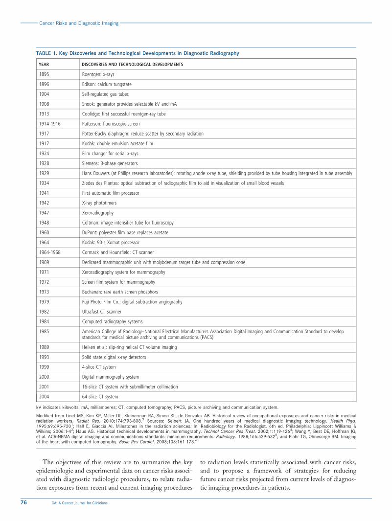
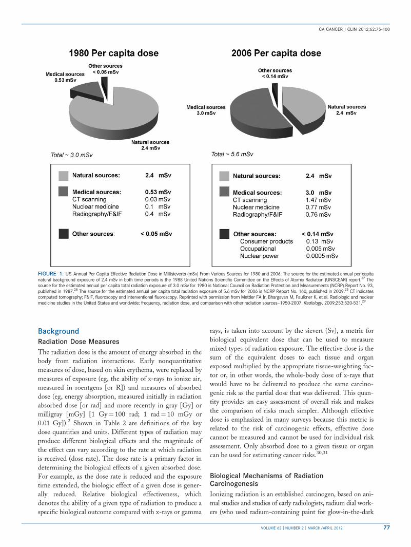