canine bacterial pyoderma - pdfs.semanticscholar.org · canine bacterial pyoderma dr heidi...
TRANSCRIPT
Canine Bacterial Pyoderma
Dr Heidi Schroeder BVSc MMedVet(Med)Willow Park Veterinary Hospital, Willow Glen, Pretoria
DEFINITIONBacterial pyoderma is a pyogenic bacterial infection of the dog’s skin. It occurs due to the multiplication of bacteria on or in the epidermis and its appendages and with invasion of the dermis. Bacterial pyoderma is second to flea allergic dermatitis the most common dermatosis of dogs.
ETIOLOGYThe most important pathogen is a coagulase-positive staphylococcus specific to the dog namely Staphylococcus intermedius. In rare cases Staphylococcus aureus or S. hyicus may be the pathogens. Others, mainly gram-negative bacteria, can be involved, but are usually secondary to S. intermedius infection.
DEVELOPMENT OF PYODERMAS. intermedius belongs to the normal resident flora on a dog’s skin and mucosa. It becomes pathogenic when the cutaneous ecosystem equilibrium is disturbed by changes in the surface micro-environment. This disturbance usually occurs due to primary causes such as allergic dermatoses. This is the reason why pyodermas are essentially secondary to underlying dermatoses.
Development of pyoderma occurs in 2 stages. Firstly the pathogen colonises areas of the body surface. It may then become a surface pyoderma. The 2 most important of these surface pyodermas are skin fold pyodermas (intertrigo) and pyotraumatic dermatitis. The second phase occurs when the most superficial layer of the skin (the stratum corneum) is invaded to cause impetigo and/or invasion of the hair follicles causing folliculitis. The latter 2 are classified as superficial pyoderma. When the infected hair follicle ruptures, the infection spreads into the dermis (furunculosis) or spreads more deeply along tissue planes (cellulitis). The latter 2 are termed deep pyodermas. Resolution depends on the non-specific defenses (neutrophils and macrophages) and on the specific defenses (cell mediated immunity and antibodies). Resolution is accompanied by an inflammatory response, which is responsible for the clinical signs of pyoderma.
CLASSIFICATION OF PYODERMACanine pyoderma can be classified based on various criteria, but the classification based on the depth of bacterial involvement is most useful clinically. This is because clinical information referable to diagnosis, likelihood of underlying disease, prognosis and response to therapy correlate with depth of infection.
SURFACE PYODERMA This term is applied to very superficial erosions of the skin. Although pathogenic bacteria can be cultured from these lesions, bacterial involvement is secondary. Pyotraumatic dermatitis, intertrigo and mucocutaneous pyoderma are classified as surface pyodermas. Dogs with surface pyodermas are rarely a major diagnostic or therapeutic problem.
Pyotraumatic dermatitis Lesions are exudative, erythematous, painful and swollen with acute onset and rapid progression and are usually self-inflicted. This dermatitis is common in thick coated dogs, especially in the summer months. Flea allergy dermatitis and otitis externa are the most common underlying causes.
IntertrigoIntertrigo is seen in dogs with anatomic defects, such as lip folds in Spaniels, facial folds in Bulldogs and vulva folds in obese bitches. The skin folds create a warm, moist, dark environment which is ideal for bacterial proliferation. These infections are characterized by exudative, odiferous, erythematous lesions within the skin folds.
Mucocutaneous pyoderma This is an infection of unknown etiology that predominantly involves the lips and perioral skin. It may be present alone or together with lip fold intertrigo.
SUPERFICIAL PYODERMASuperficial pyodermas are the most common canine bacterial skin diseases and among the most common canine dermatoses. Impetigo, superficial folliculitis and superficial spreading pyoderma are classified as superficial pyodermas.
Impetigo Impetigo is used to describe non-follicular bacterial micro abscessation involving the superficial layers of the epidermis. The infection is just under the stratum corneum and is characterized by a non-follicular pustule usually seen in hairless areas, particularly in young puppies.
Superficial folliculitisThis is the single most common bacterial skin disease in the dog. The infection is within the hair follicle. It starts as a papule, has a short pustular phase and then develops a crust. As the pustular phase is of short duration, the diagnosis is not easy and may be confused with a number of papular dermatoses. A wide range of clinical appearances can be seen with superficial folliculitis. Intact pustules are fragile and rupture easily eventually resulting in crusted papules. The papules may not be crusted in appearance because the crusts may have been removed by self trauma or have fallen off. Therefore, papules with or without adherent crusts, are often the most commonly seen clinical features of superficial folliculitis. Alopecia is a common feature and characterised by distinct circular
pathes of hair loss surrounding previously affected hair follicles. Chronicity leads to a generalised moth-eaten appearance. This is more obvious in short coated breeds. Careful examination of the periphery of these alopecic areas may reveal individual peripheral collarettes and coalescing collarettes.
Superficial spreading pyodermaA visually distinctive clinical subset of superficial pyoderma termed superficial spreading pyoderma, is characterised by centrifugally progressive erythema and expanding peripheral epidermal collarettes. These lesions can be seen alone or together with superficial folliculitis.
DEEP PYODERMAIn deep pyoderma the infection from the distal parts of the hair follicle extends beneath and beyond the confines of the hair follicle. Follicular rupture may lead to a granulomatous tissue response directed against free keratin from the root sheath and hair shaft fragments, which fosters the formation of scar tissue with sequestered pyogranulomas. Consequently, the dual problems of infection and foreign body granulomas coexist.
It is much less common than superficial pyoderma and is further subdivided into deep folliculitis, furunculosis and cellulitis. Lesions typically involve large groups of hair follicles in a region, poorly defined erythematous plaques form and individual pustules and nodules are less obvious. Lesions often ulcerate which leads to draining fistulas. Pain or pruritus is commonly present, as well as regional or generalised lymphadenopathy.
Certain types of deep bacterial folliculitis and furunculosis are clinically distinctive enough to warrant individual classification. These syndromes include muzzle folliculitis and furunculosis (canine acne), pyotraumatic folliculitis, pedal folliculitis and furunculosis, callus pyoderma (pressure point pyoderma), German shepherd dog pyoderma and nasal folliculitis and furunculosis.
DIAGNOSISCytologic examination, skin scrapings, bacterial culture and sensitivity and skin biopsies are the most consistently useful diagnostic procedures in the evaluation of a case of suspected pyoderma. The presence of bacterial pyoderma is always secondary to an underlying cause. It is thus important to also determine the predisposing cause if at all possible in order to prevent reinfection.
Skin cytologyCytology is reliable, rapid, cost-effective and is considered an excellent method to support the clinical diagnosis of pyoderma. Samples can be collected by direct impression smears (in cases of surface pyodermas) or from intact pustules which are opened gently with a 25- or 26-gauge needle. The pustular content is transferred to a glass slide and a smear is made. The smears are stained with diff quick stain. Smears are examined for the presence of cells and bacteria.
The following may be observed in samples taken from cases with pyodermas:Neutrophils - may be degenerate (“impaired”) or non-degenerate. The degenerate neutrophils indicate an active immune status. They do not take up stain well, appear bulky and have hypersegmented and pyknotic nuclei. The non-degenerate neutrophils appear healthy and well stained. Macrophages and /or lymphocytes - seen more often in chronic, deep and/or severe conditions.Eosinophils – indicate a possible allergic component to the pyoderma. Bacteria – the finding of cocci is highly suggestive of pyoderma caused by Staphylococcus intermedius. They may be intracellular where bacteria are inside a degenerate neutrophil. This is called phagocytosis and it indicates an immune response by the host and suggests an active disease status. The engulfed bacteria can be seen as pathogens and a diagnosis of a pyoderma can be made. Where bacteria are extracellular, they indicate colonisation. This is typically seen in surface pyodermas. The presence of abnormally large numbers of cocci within pustules indicates the likelihood of underlying endogenous or iatrogenic hyperadrenocorticism. This can also be seen in cases receiving immunosuppressive therapy for cancer or autoimmune disease.
Skin scrapingsDeep scrapings should be made in every case of pyoderma for underlying demodecosis. Especially in cases of deep pyoderma, demodecosis can be the underlying cause. It should always be remembered that it may be difficult to demonstrate the mites with skin scrapings in cases of severe, deep pododermatitis and in certain breeds, such as the Sharpei. In these cases a skin biopsy for histopathology is indicated.
Bacterial culture and sensitivityThis is not a diagnostic procedure as such. It reinforces the presumption and is rather a guide to treatment. Situations where bacterial culture and sensitivity are indicated include therapeutic failure, cases refractory to treatment, multiple relapses and severe deep pyoderma. In general, bacterial cultures should not be performed on open lesions. Cultures of biopsies from intact pustules, furuncles or nodules are more likely to yield useful information.
Skin biopsy for histopathologyThis is required when support of clinical diagnosis is needed, particularly if therapy has failed in spite of a strong suspicion. Early primary lesions more frequently yield diagnostic information. Skin biopsy may provide rapid establishment of a definitive diagnosis and help to rule out other potentially serious diseases and identify unexpected underlying diseases.
Evaluation of immunocompetenceSerum immunoglobulin quantitation, complete blood count, serum electrophoresis, and neutrophil function tests have all been suggested to evaluate immunocompetence in the dog. The most readily available and practical
is the complete blood count and serum electrophoresis. Theoretically, an absolute neutrophilia with a lymphocyte count of at least 1000 cells/µl to 1500 cells /µl should be seen in immunologically normal dogs adequately responding to ongoing or recurrent pyoderma. In electrophoresis a broad-based elevation in the serum electrophoretic pattern in the ß and γ range should be seen in normal dogs responding to bacterial infection.
MANAGEMENTAppropriate management of most cases of superficial and deep pyoderma requires the use of systemic antibiotics usually in combination with topical shampoo therapy. Immunomodulatory therapy, although rarely successful, may be used in an attempt to prevent or diminish the frequency of recurrent infections.
Systemic antibioticsThe basic principles of successful systemic antibiotic therapy include choice of proper antibiotic, establishment of an effective dosage, and long enough duration of treatment to ensure cure rather than simply transient remission.
The choice of antibiotic may be empiric or based on results of a culture and sensitivity. Over 90% of cases of canine pyoderma are caused by Staphylococcus intermedius. Therefore the antibiotic should be effective against this bacterium and should not be inactivated by ß-lactamase, as 50 % of S intermedius strains produce this enzyme. Ideally the chosen antibiotic should also have a narrow spectrum of activity, minimal side effects and be cost effective.
Useful antibiotics in dermatology include the following:Macrolides and lincosamides: erythromycin is a bacteriostatic macrolide which is relatively cheap and has few side effects. Resistance is increasing and cross resistance exists with other macrolides and lincomycin. Lincomycin and clindamycin are good choices, but cost is a major disadvantage.
Diaminopyrimidines potentiated sulphonamides: a good first choice, but resistance is increasing. Very cost effective. Major disadvantages are serious side effects especially with long term use. These include keratoconjunctivitis sicca, nonseptic polyarthritis (especially in Dobermans) and numerous dermatologic side effects ranging from minor papular rashes to life threatening erythema multiforme and toxic epidermal necrolysis.
Penicillins resistant to ß-lactamases: amoxicillin is not effective for treatment of pyodermas. Amoxicillin with clavulanic acid is very effective, has few side effects, but is not cheap.
Cephalosporins (cephalexin and cephadroxil) are broad spectrum, first generation cephalosporins with excellent clinical efficacy. Advantages include twice daily administration, rarity of resistance, and moderate cost as a generic.
Fluoroquinolones are new, broad-spectrum bactericidal antibiotics. Advantages include once daily dosing, outstanding tissue penetration, activity against Staphylococcus intermedius and multiple gram-negative secondary invading organisms, and rarity of resistance. This should not be used in growing dogs because articular damage may occur. It should only be used in severe, chronic, deep pyodermas where no other antibiotics are effective any longer. The flouroquinolones are likely to be the final pharmacological group of antimicrobials to be developed. Therefore their use should be reserved for severe cases and not as first line treatment of superficial pyodermas. The establishment of the appropriate antibiotic dosage is very important. All dogs should be weighed. Dosages should be as close to established recommendations as possible, but not less than recommended. Underdosing is likely to lead to diminished efficacy, whereas overdosing may increase the likelihood of adverse reactions with some antibiotics. In some cases of pyoderma with severe scarring and sequestered infection, antibiotics such as cephalexin and enrofloxacin may be used at higher dosages for better effects.
Antibiotic therapy must be maintained until complete elimination of bacterial infection rather than simply transient remission is achieved. It is recommended to continue therapy for at least 1 week beyond clinical cure for superficial pyoderma and a minimum of 2 weeks beyond clinical cure in all cases of deep pyoderma because nonvisible sequestered foci of infection often remain after most lesions have cleared.
Topical therapy ShampoosShampoos are used in pyoderma cases to reduce the cutaneous bacterial population, to remove tissue debris, to allow direct contact between the active ingredient and the organism and to promote drainage.
Some cases of superficial pyoderma can be treated with shampoos alone, however in most cases systemic antibiotics are administered as well to ensure a more prompt clinical response, with the shampoo playing a supportive role.
A common indication for the long term use of shampoos is in the dog suffering from recurrent superficial folliculitis (idiopathic of secondary to endocrine or allergic skin disease). Here antibacterial shampoos may have a prophylactic effect if used regularly e.g. weekly or every two weeks.
Cases of deep pyoderma need to be clipped before using shampoos. This prevents the formation of sealing crusts and allows the active ingredients to come in contact with the lesions. Shampoos should be used very frequently in these cases. Initially twice weekly, thereafter once a week shampoos are recommended.
Antibacterial shampoos effective for pyodermas, which are currently available in South Africa, contain either chlorhexidene, benzoyl peroxide, or benzoyl peroxide and sulphur.
Chlorhexidene is an antiseptic that is very effective against most bacteria (gram + and -), as well as against Malassezia pachydermatis. It is bactericidal by action on the cytoplasmic membrane which causes leakage of intracellular components. It has a prolonged residual action on the skin, is non-toxic and non-irritant. The 3% formulation has been shown to be the most effective.
Benzoyl peroxide is metabolized in the skin to benzoic acid. Much of its microbiocidal activity derives from the lowered skin pH which disrupts microbial cell membranes. It is also an oxidizing agent, which releases nascent oxygen into the skin and produces a series of chemical reactions resulting in permeability changes and rupture of bacterial membranes. It has an excellent prophylactic effect. Its follicular flushing and keratolytic degreasing and comedolytic activity are additional benefits. Benzoyl peroxide and sulphur offers an excellent combination with additional degreasing effects. It is generally used in concentrations of 2 to 3%, which are well tolerated, but irritation can occur at higher concentrations (erythema, pruritus and pain).
Antibacterial creams and ointmentsCreams and ointments containing antibiotics may be used to treat small areas of infection. Cost, messiness and time of application limit their usefulness when larger areas are involved. Pyodermas where topical creams and ointments have some use include intertrigo, mucocutaneous pyoderma, muzzle folliculitis and furunculosis (canine acne), callus pyoderma, pyotraumatic folliculitis and pedal folliculitis and furunculosis.
Mupirocin is a newer, potent topical antibiotic available in a polyethylene glycol base. It is formed as a fermentation product of Pseudomonas flourescens. Mupirocin inhibits bacterial protein synthesis and shows no cross reactivity with any other group of antibiotics. Superior penetrating ability allows mupirocin to be used topically for deep as well as superficial pyoderma. It is not formulated for use on mucosal surfaces and is contraindicated for treatment of large areas where absorption of large amounts of the polyethylene glycol base is possible because of the potential of nephrotoxicity.
Immunomodulatory therapyImmunomodulatory treatment remains controversial in the management of canine pyoderma. Most discussions of efficacy have been subjective or anecdotal because clinicians commonly use immunomodulatory therapy in combination with systemic antibiotics and topical therapy.
Bacterin preparations containing killed bacteria are the most commonly used immunomodulatory therapy for canine pyodermas. According to leading
dermatologists 30 to 50% of dogs with recurrent pyoderma ultimately benefit from bacterin treatment. Autogenous vaccines also have been advocated for the management of pyoderma. These preparations are made from the specific staphylococcal organism isolated from a dog with pyoderma for the same dog. Some reports suggest efficacy in 30 to 50% of cases, but controlled or blinded studies have not been performed.
Nonbacterial immunostimulants are also very controversial as controlled blinded studies have not performed to determine efficacy. Most reports are anecdotal as these immunostimulants are always used in combination with other therapies.
Levamisole, a large animal antihelminthic, has been used for this purpose. This agent may alter lymphocyte and phagocyte immune function by modifying leukocyte intracellular cyclic nucleotides. The recommended dosage is 2, 2 mg/kg per os every other day. Efficacy is believed to be less than 20%.
Cimetidine is a H2 histamine receptor blocker. Lymphocytes have surface H2
receptors and therefore cimetidine theoretically could reduce immunosuppression by down-regulating suppressor T lymphocytes, thereby modulating cytokine production. Recommended dosages are 3 to 4 mg/kg per os twice daily for at least 10 weeks. This is safe, but expensive and controlled studies have not been performed.
Factors complicating management of pyodermaTherapeutic failure and recurrence of pyoderma are most often associated with inappropriate initial therapy. Errors commonly made include faulty antibiotic selection, dosage used and duration of therapy. In cases of deep pyoderma sequestered foci of bacterial infection are a common cause of recurrence of a deep pyoderma. If the initial therapy seems appropriate, other factors should be considered. The lack of identification of underlying disease or contributing factors is another frequent cause of treatment failure. These include underlying allergies (atopy, food allergy flea allergy dermatitis), ectoparasites (demodex, sarcoptes), diseases altering cornification (seborrheic disorders), and hair follicles (follicular dysplasias), chronic cutaneous inflammation (sebaceous adenitis), other infectious diseases (Malassezia dermatitis, dermatophytosis) and diseases that lead to a weaker immune system such as endogenous or iatrogenic hyperadrenocorticism and hypothyroidism or congenital or acquired immunodeficiency (systemic disease, neoplasia, immunosuppressive therapy).
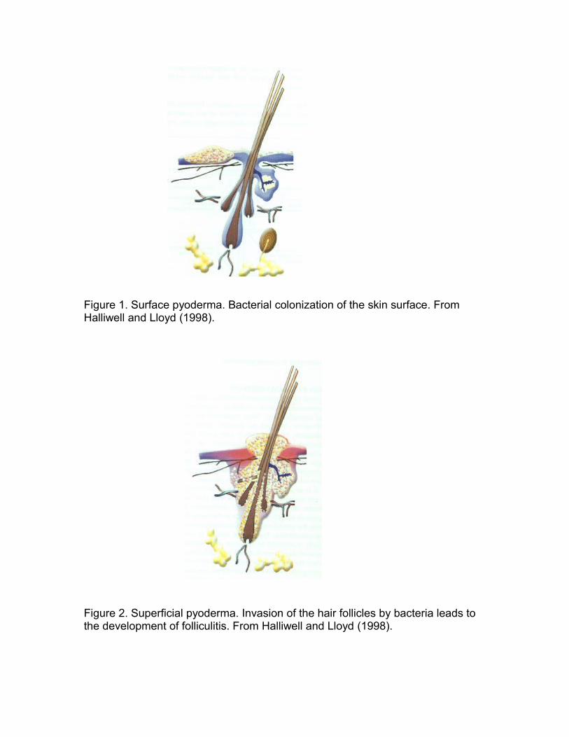
Figure 1. Surface pyoderma. Bacterial colonization of the skin surface. From Halliwell and Lloyd (1998).
Figure 2. Superficial pyoderma. Invasion of the hair follicles by bacteria leads to the development of folliculitis. From Halliwell and Lloyd (1998).
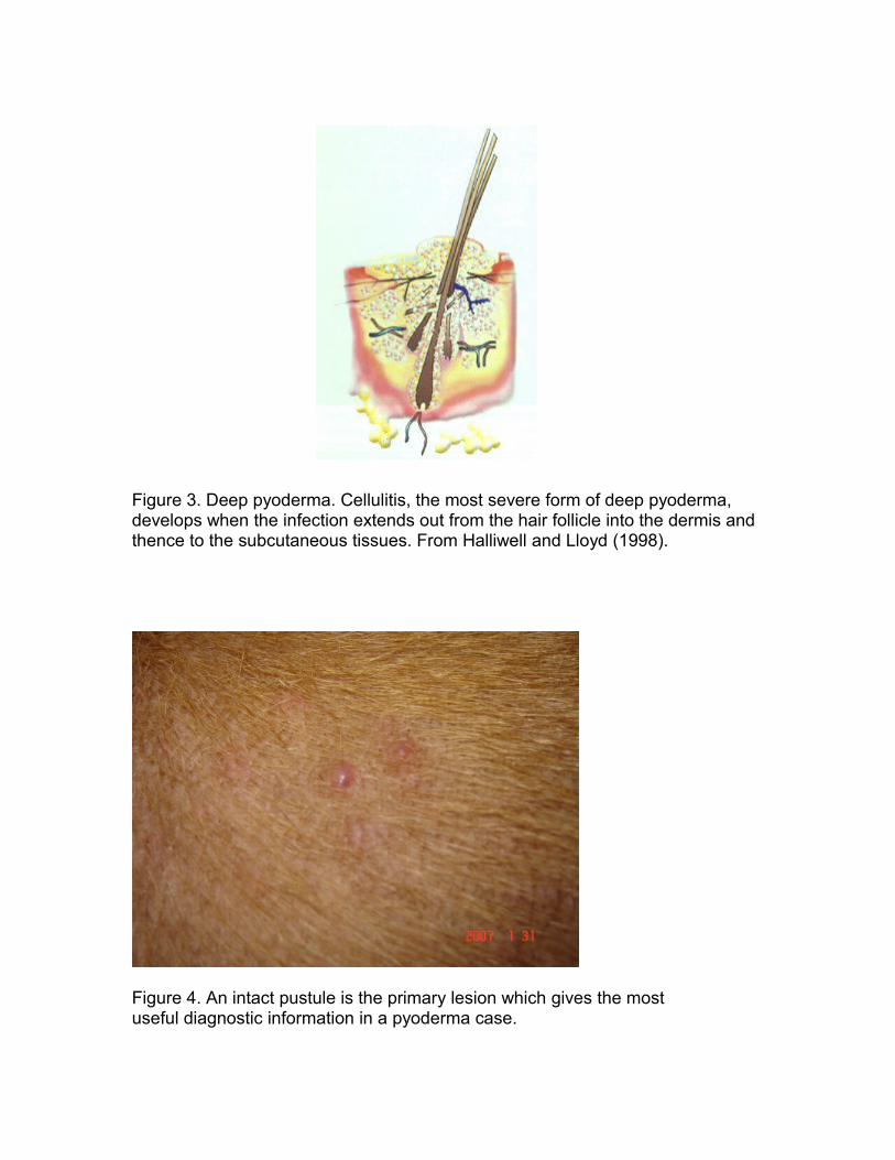
Figure 3. Deep pyoderma. Cellulitis, the most severe form of deep pyoderma, develops when the infection extends out from the hair follicle into the dermis and thence to the subcutaneous tissues. From Halliwell and Lloyd (1998).
Figure 4. An intact pustule is the primary lesion which gives the most useful diagnostic information in a pyoderma case.
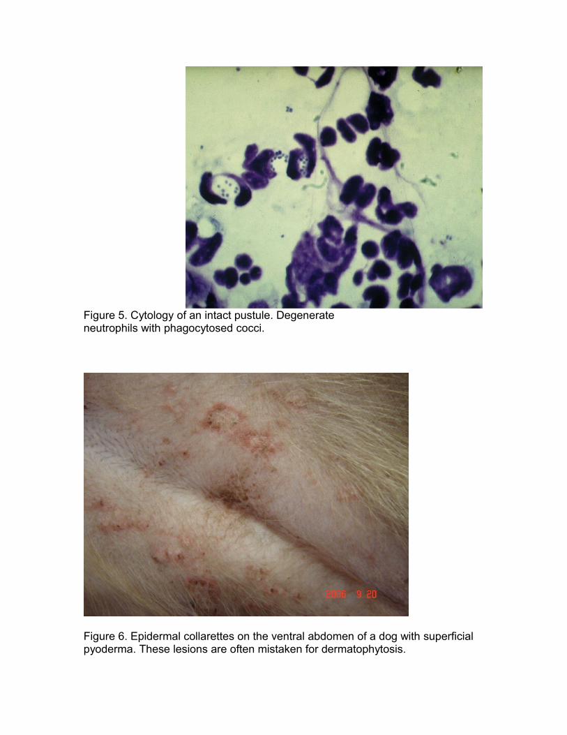
Figure 5. Cytology of an intact pustule. Degenerate neutrophils with phagocytosed cocci.
Figure 6. Epidermal collarettes on the ventral abdomen of a dog with superficial pyoderma. These lesions are often mistaken for dermatophytosis.
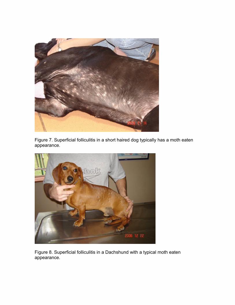
Figure 7. Superficial folliculitis in a short haired dog typically has a moth eaten appearance.
Figure 8. Superficial folliculitis in a Dachshund with a typical moth eaten appearance.
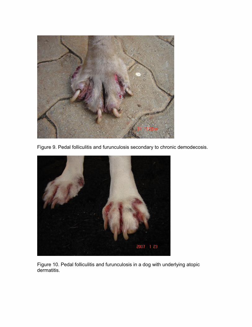
Figure 9. Pedal folliculitis and furunculosis secondary to chronic demodecosis.
Figure 10. Pedal folliculitis and furunculosis in a dog with underlying atopic dermatitis.
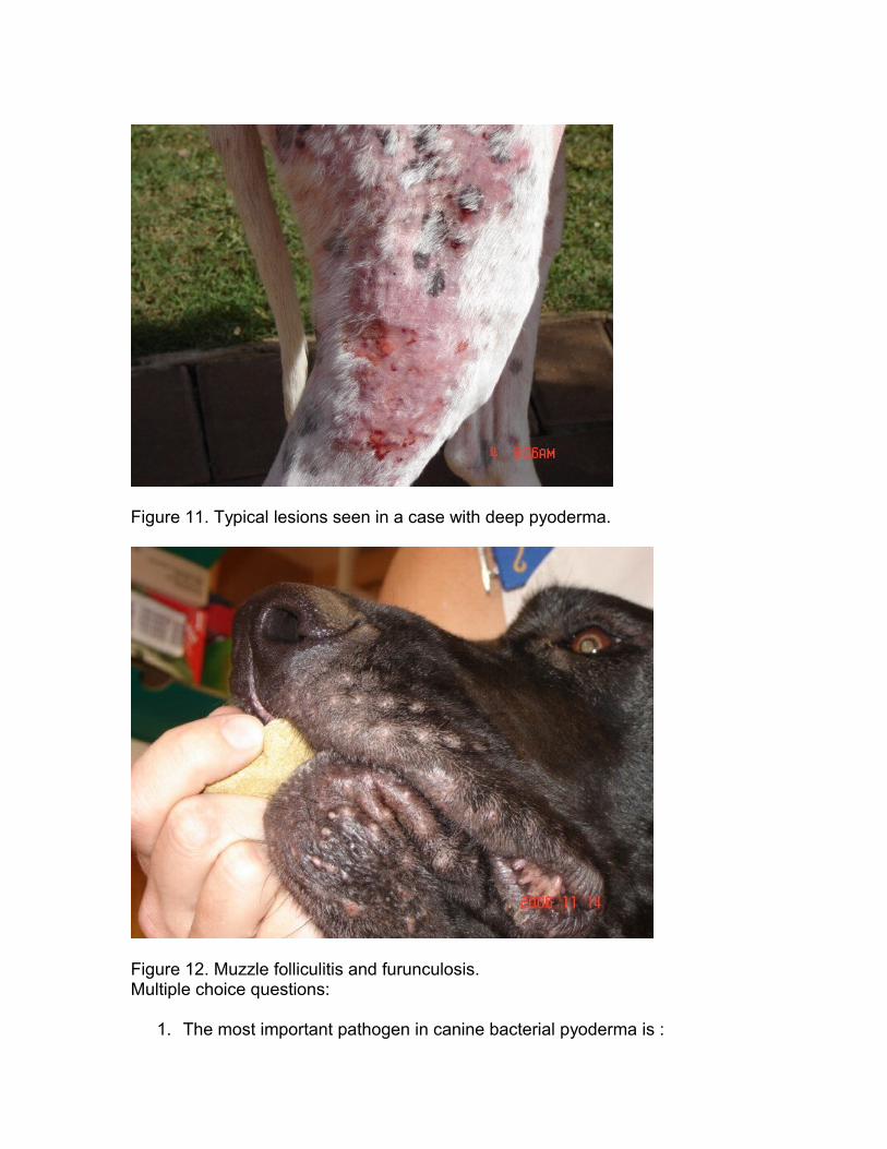
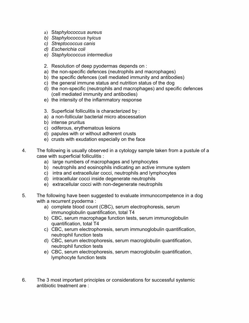
Figure 11. Typical lesions seen in a case with deep pyoderma.
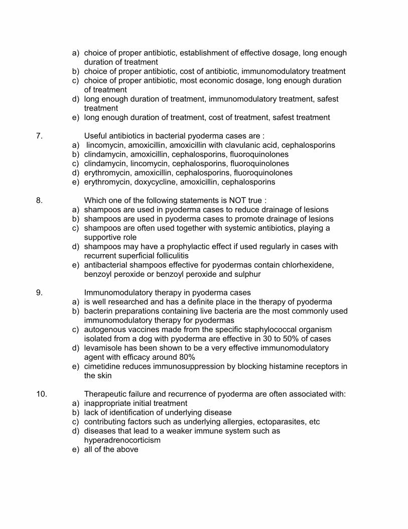
Figure 12. Muzzle folliculitis and furunculosis. Multiple choice questions:
1. The most important pathogen in canine bacterial pyoderma is :
a) Staphylococcus aureus b) Staphylococcus hyicus c) Streptococcus canis d) Escherichia coli e) Staphylococcus intermedius
2. Resolution of deep pyodermas depends on :a) the non-specific defences (neutrophils and macrophages)b) the specific defences (cell mediated immunity and antibodies)c) the general immune status and nutrition status of the dogd) the non-specific (neutrophils and macrophages) and specific defences
(cell mediated immunity and antibodies)e) the intensity of the inflammatory response
3. Superficial folliculitis is characterized by :a) a non-follicular bacterial micro abscessationb) intense pruritusc) odiferous, erythematous lesionsd) papules with or without adherent crustse) crusts with exudation especially on the face
4. The following is usually observed in a cytology sample taken from a pustule of a case with superficial folliculitis :
a) large numbers of macrophages and lymphocytesb) neutrophils and eosinophils indicating an active immune systemc) intra and extracellular cocci, neutrophils and lymphocytesd) intracellular cocci inside degenerate neutrophilse) extracellular cocci with non-degenerate neutrophils
5. The following have been suggested to evaluate immunocompetence in a dog with a recurrent pyoderma :
a) complete blood count (CBC), serum electrophoresis, serum immunoglobulin quantification, total T4
b) CBC, serum macrophage function tests, serum immunoglobulin quantification, total T4
c) CBC, serum electrophoresis, serum immunoglobulin quantification, neutrophil function tests
d) CBC, serum electrophoresis, serum macroglobulin quantification, neutrophil function tests
e) CBC, serum electrophoresis, serum macroglobulin quantification, lymphocyte function tests
6. The 3 most important principles or considerations for successful systemic antibiotic treatment are :
a) choice of proper antibiotic, establishment of effective dosage, long enough duration of treatment
b) choice of proper antibiotic, cost of antibiotic, immunomodulatory treatmentc) choice of proper antibiotic, most economic dosage, long enough duration
of treatmentd) long enough duration of treatment, immunomodulatory treatment, safest
treatmente) long enough duration of treatment, cost of treatment, safest treatment
7. Useful antibiotics in bacterial pyoderma cases are :a) lincomycin, amoxicillin, amoxicillin with clavulanic acid, cephalosporinsb) clindamycin, amoxicillin, cephalosporins, fluoroquinolonesc) clindamycin, lincomycin, cephalosporins, fluoroquinolonesd) erythromycin, amoxicillin, cephalosporins, fluoroquinolonese) erythromycin, doxycycline, amoxicillin, cephalosporins
8. Which one of the following statements is NOT true :a) shampoos are used in pyoderma cases to reduce drainage of lesionsb) shampoos are used in pyoderma cases to promote drainage of lesionsc) shampoos are often used together with systemic antibiotics, playing a
supportive roled) shampoos may have a prophylactic effect if used regularly in cases with
recurrent superficial folliculitise) antibacterial shampoos effective for pyodermas contain chlorhexidene,
benzoyl peroxide or benzoyl peroxide and sulphur
9. Immunomodulatory therapy in pyoderma casesa) is well researched and has a definite place in the therapy of pyodermab) bacterin preparations containing live bacteria are the most commonly used
immunomodulatory therapy for pyodermasc) autogenous vaccines made from the specific staphylococcal organism
isolated from a dog with pyoderma are effective in 30 to 50% of casesd) levamisole has been shown to be a very effective immunomodulatory
agent with efficacy around 80%e) cimetidine reduces immunosuppression by blocking histamine receptors in
the skin
10. Therapeutic failure and recurrence of pyoderma are often associated with:a) inappropriate initial treatmentb) lack of identification of underlying diseasec) contributing factors such as underlying allergies, ectoparasites, etcd) diseases that lead to a weaker immune system such as
hyperadrenocorticisme) all of the above
Answers to multiple choice questions:
Question 1: eQuestion 2: dQuestion 3: dQuestion 4: dQuestion 5: cQuestion 6: aQuestion 7: cQuestion 8: aQuestion 9: cQuestion 10: e