cannulation of the right internal jugular vein for central ... · a flush solution for the cvp...
TRANSCRIPT
Cannulation of the right internal jugular vein for central venouspressure-A reviewWILLIAM G. RONK, CRNAROBERT DAYTON, CRNACooperstown, New York
The authors review in a step-by-stepillustrated manner a techniqueinvolved for placing a catheter tomonitor central venous pressure inthe internal jugular vein; theyelaborate on both the advantagesand disadvantages of the technique.
Central venous pressure (CVP) monitor.ing is a useful tool in the anestheticmanagement of the seriously ill patientfor both emergency and elective proce-dures. A working knowledge of anatomy,various techniques, and the proper indi-cation for CVP must be foremost in theanesthetist's mind. There are a numberof techniques used in the placement ofthe CVP catheter. The purpose of thisarticle is to review these areas, withemphasis on placement in the internaljugular vein (IJV), elaborating on tech-nique, advantages and disadvantages.
The indications for the insertionand monitoring of the central venouspressure (CVP) are as follows: (1) pa-tients undergoing surgery in which amajor volume shift may occur (suchas in the Whipple procedure) ; (2) op-erations where the potential for hypo-volemia exists (such as in a bowel re-section); (3) patients in shock (suchas in trauma) ; (4) operations per-formed with the patient in the sittingposition with risk of air embolization;and (5) some selected patients with con-gestive heart failure. A Swan-Ganz cath-eter rather than a CVP catheter prob-
ably would be placed for assessment ofleft heart filling pressures in patientswith severe left ventricular dysfunction.
Alternatives to the internal jugularvein for placement of the CVPcatheter
Many techniques have been de-scribed for placing a catheter in thesuperior vena cava or right atrium. Fre-quently used routes include the femoral,basilic, cephalic, external jugular, sub-clavian and internal jugular veins.
These first four techniques have hada low incidence of successful centralvenous cannulation. The distance be-tween the anticubital veins and the su-perior vena cava is the chief disadvan-tage in the use of the basilic and cephalicroutes. In addition, these veins are rel-atively small and tolerate indwellingcatheters for only short periods of time.The veins leading to the inferior venacava have been virtually abandoned assites for indwelling catheters because ofthe dangers associated with phlebitis andmechanical obstruction during abdom-inal surgery.
The external jugular vein is usuallyvisible or easily palpable in the neckwhen the patient is supine. Central ve-nous cannulation by this route is diffi-cult, however, since the external jugularvein contains numerous valves andmakes an acute angle where it joins thesubclavian vein. The subclavian veinhas the advantage of its large size andrelatively high flow, rendering it suitable
Journal of the American Association of Nurse Anesthetists144
for indwelling catheters. However, nu-merous complications including pneu-mothorax, hemothorax, hematoma, me-diastinal infiltration, subcutaneousemphysema and air embolism, brachialplexus injury, septicemia, and cellulitishave been reported.
In 1966, Hermosura and co-work.ers" described a method of placing apolyethylene catheter in the internaljugular vein. In 1969, English and col-leagues 9 described two methods of in-ternal jugular vein percutaneous can-nulation, with no serious complicationsin 200 insertions. 8 Anatomic locationhas been shown to be advantageous be-cause of its straight course to the rightatrium, easy accessibility (even in obesepatients), and its definite external an-atomic landmarks.
Use of the right internal jugularvein for CVP
The right internal jugular vein ispreferred over the left internal jugularvein because: (1) the risk of lacerationof thoracic duct is greater on the leftthan the right; (2) the carotid arteryis consistently deep and medial to theIJV on the right; and (3) the right leadsstraight to the superior vena cava. Kap-lan and Miller' have described the tech-nique we currently employ. We shalldescribe Kaplan's technique in a step bystep illustrated manner.
Anatomical reviewThe internal jugular vein is located
under the medial border of the clavicularhead of the sternocleidomastoid muscle.The vein ending is behind the medial
April/1978
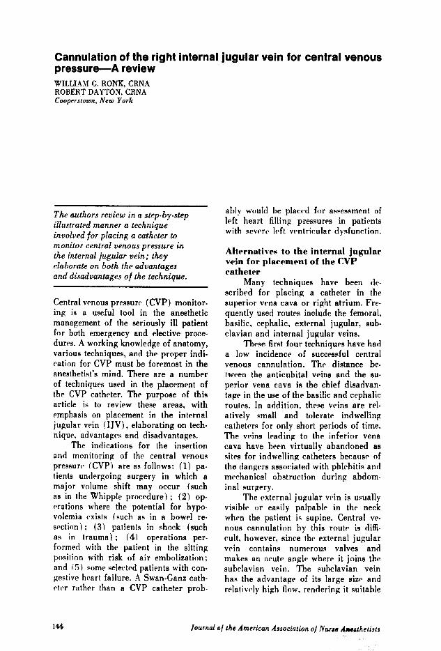
Figure 1.Line drawing showing the relationship of the right internal jugular vein to thecarotid artery and sternocleidomastoid muscle. With the patient's head turnedtoward the left, the vein passes under the apex of a triangle forward by thesternal and clavicular heads of the SCM and lateral to the common carotid artery.
Internal JugularVein
145

end of the clavicle. The thoracic duct onthe left and the right lymphatic ductenter the internal jugular and subclavianveins at their confluence posteriorly.
The right side is preferred becausethe internal jugular, innominate, andsuperior vena cava are almost in astraight line. The carotid artery is con-sistently deep and medial to the internaljugular vein and is deep under the ster-nal head medial to the sternocleidomas-toid muscle and the clavicle. The triangleformed by the two heads of this muscleand the clavicle encompass the internaljugular vein. (Figure 1.)
Technique of cannulationStep 1. Place the patient in a 150
Trendelenberg position with the neck ex-tended and turned sharply to the left.This distends the vein during cannula-tion. The anatomy is demonstrated moreeasily in the awake patient. The proce-dure is no more uncomfortable thanstarting a peripheral IV. If the patientis to be anesthetized first, mark out theanatomy and triangle when he is awake.(Figure 2.)
Step 2. Using a surgical skin mark-er, mark out the lateral border of thesternal head and the medial border ofthe clavicular head of the sternocleido-
Figure 2.
mastoid muscle. Palpate the location ofthe carotid artery. The puncture is madeat the point where the two marked mus-cle borders meet (about two fingerbreadths above the clavicle). This pointshould be well lateral to the carotidpulse.
Step .3. After the skin has been pre-pared carefully with Betadine® and thepatient has been draped, a skin wheal israised at the predetermined point. Theinitial puncture should be made with a22-gauge needle on a 5-cc syringe con-taining 1% Xylocaine.® (Figure 3.) Theneedle is advanced in a caudal directionat a 30° angle to the skin, angled towardthe right nipple. Constant suction ismaintained on the syringe as the needleis advanced toward the vein.
If the vein is difficult to find, a Val.salva maneuver by the patient may mark-edly distend the internal jugular andease identification (Figure 2). Aspira-tion of dark venous blood signals entryinto the internal jugular vein. After lo-cating the vein, infiltrate with Xylo-caineR as the needle is withdrawn.
Step 4. The patient's skin located un-der your left hand should not be per-mitted to move, or the relationship ofthe underlying anatomy will change. A14-gauge, 8-inch Intracath is then in-
Steps 1 & 2: Anesthetist's eyeview of the sternal (1) and clavicular (2) heads of the SCMwith "X" marking the location for IJV puncture.
Journal of the American Association of Nurse Anesthetists

serted at the same 30° angle toward theright nipple and the same depth into thevein. (Figure 4.) Two needle "pops"may be noted; these emanate from (1)the carotid sheath, and (2) the internaljugular vein, identified by a free returnof dark venous blood through the cath-eter.
The full length of the cathetershould thread easily into the superiorvena cava. If threading is difficult, twistthe catheter clockwise as it is inserted.This action will prevent the catheterfrom hitting the vein wall. To avoidlaceration of the catheter, it shouldnever he withdrawn through the needle.
Figure 3.Step 3: Right internal jugular vein is identified with a #22-gauge needle.
Figure 4.Step 4: IJV punctured with a #14-gauge thin wall needle and catheter advanced into thevein. Note the dark venous backflow into catheter.
April/1978

If venipuncture is unsuccessful, both theneedle and catheter should be withdrawntogether.
Step 5. Once the catheter is placed,the needle is removed and the IV tubingis connected. (Figure 5.) Having thepatient hold his breath while you attachthe catheter to the IV will avoid airembolism. The plastic guard is attachedat its proper place to avoid bending ordisconnection of the catheter at bevelpoint.
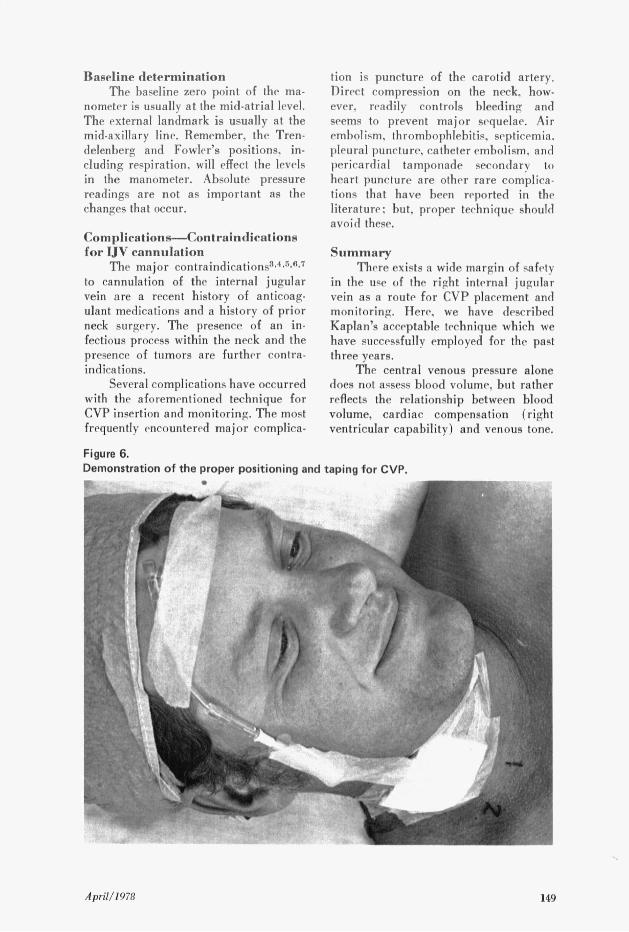
Once established, a good check forproper insertion of the catheter is toplace the IV bottle below the head ofthe patient to insure free flow of bloodthrough the catheter. If free flow is notpresent, take the IV down and slowlywithdraw the catheter until flow is ob-tained, assuring that the catheter is notcurled and/or lodged against the veinwall. Another valuable check is to ex-amine the postoperative chest x-ray forconfirmation of catheter location. Theskin is treated with tincture of benzoin.a sterile dressing is applied, and thecatheter is taped along the anterior bor-der of the ear with the IV tubing tapedacross the mid-forehead. (Figure 6.)
A flush solution for the CVP shouldcontain 1-unit of heparin per ml of fluidand may be either normal saline. Ringer'slactate solution or Normosol R'. Cardio-green or methylene blue can he addedto the manometer to make it easier toread. Some anesthetists prefer to placea black silk suture through the skin andaround the catheter in the similar fash-ion used by surgeons to secure drains.
Catheter tip placementVaughan and Weygandt 2 described
electrocardiogram changes that occurredduring the placement of CVP catheters.They stressed the importance of EKGmonitoring to detect arrhythmias as thecatheter tip enters the right auricle orventricle. Following insertion of thecatheter, the P-wave is observed as thecatheter is withdrawn into its properplace.
Placement should he just above thesuperior cavo-atrial junction in the su-perior vena cava. Changes in the QRScomplex. ST segment. and T-wave some-times occur but arc less dramatic andmore difficult to interpret than P-wavechanges.
Figure 5.Step 5: Catheter is connected to the IV flush solution.
Journal of the American Association of Nurse Anesthetists
Baseline determinationThe baseline zero point of the ma-
nometer is usually at the mid-atrial level.The external landmark is usually at themid-axillary line. Remember, the Tren-delenberg and Fowler's positions, in-cluding respiration, will effect the levelsin the manometer. Absolute pressurereadings are not as important as thechanges that occur.
Complications-Contraindicationsfor IJV cannulation
The major contraindications3 .4" ",7
to cannulation of the internal jugularvein are a recent history of anticoag-ulant medications and a history of priorneck surgery. The presence of an in-fectious process within the neck and thepresence of tumors are further contra-indications.
Several complications have occurredwith the aforementioned technique forCVP insertion and monitoring. The mostfrequently encountered major complica-
Figure 6.
tion is puncture of the carotid artery.Direct compression on the neck, how-ever, readily controls bleeding andseems to prevent major sequelae. Airembolism, thrombophlebitis, septicemia.pleural puncture, catheter embolism, andpericardial tamponade secondary toheart puncture are other rare complica-tions that have been reported in theliterature; but, proper technique shouldavoid these.
SummaryThere exists a wide margin of safety
in the use of the right internal jugularvein as a route for CVP placement andmonitoring. Here, we have describedKaplan's acceptable technique which wehave successfully employed for the pastthree years.
The central venous pressure alonedoes not assess blood volume, but ratherreflects the relationship between bloodvolume, cardiac compensation (rightventricular capability) and venous tone.
Demonstration of the proper positioning and taping for CVP.
April/1978

The CVP is a function of four measur-able and independent forces: (1) thevolume of blood in the veins, (2) thedistensibility and contractility of theright heart chambers, (3) venometeractivity in the central veins, and (4)intrathoracic pressure. The CVP reflectsprincipally the volume of blood return-ing to the heart and the ability of boththe right and left ventricles to propel it.The normal central venous pressure is6-16 cm of water or 5-12 mm of mer-cury.
Hypovolemia is characterized by alow CVP, low blood pressure, low car-diac output, and high peripheral resis-tance-all of which respond favorablyto adequate and rapid volume replace-ment. In contrast, cardiac failure ischaracterized by low blood pressure witha high CVP and low cardiac output.Additional fluid load will not improvecardiac performance in this instance.However, cardiac stimulants, such ascalcium, dopamine, and digitalis, willimprove blood pressure and result in areturn toward normal CVP.
As an index of right ventricularfilling pressure, the CVP may be helpfulto diagnose cardiac and volume-relatedchanges responsible for alterations incardiac output. It may also prevent vol-ume overloading and subsequent pul-monary edema.
The usefulness of the central venous
pressure has been clearly demonstratedas an adjunct to routine anesthetic man-agement of our patients. We advocateusing the CVP on those patients whosecardiac or fluid balance status requirecareful monitoring during anesthesia.
REFERENCES(1) Kaplan, J. and Miller. E. 1976. InternalJugular Vein Catheterization. AnesthesiologyReview. May, 21-23.(2) Vaughan, R. and Weygandt, G. 1973. Re-liable Percutaneous Central Venous PressureMeasurement. Anes. & Analg. 52: 709-716.(3) Civetta, J., et al. 1972. Internal JugularVein Puncture with a Margin of Safety. Anes-thesiology. 36: 622-623.(4) Thomas, C., et al. 1969. Pericardial Tam-ponade from Central Venous Catheters. Anes.& Analg. 48: 761-762.(5) Die Coyanes, A., et al. 1969. Complica-tions of Catheterization for Central VenousPressure; A Reprint of Three Cases. Anes.& Analg. 48: 563-565.(6) Kuramoto, T. and Sakahe, T. 1975. Com-parison of Success in Jugular Versus BasilisVein Technics for Central Venous PressureCatheter Positioning. Anes. & Analg. 54: 696-697.(7) Jernigan, W., et al. 1970. Use of theInternal Jugular Vein for Placement of Cen-tral Venous Catheter. Surgery, Gynecology &Obstetrics. March, 520-524.(8) Hermosura, B., and Vanga, L. 1966. Mea-surement of Pressure during Intravenous Ther-apy. JAMA. 195: 321-323.(9) English, I., et al. 1969. Percutaneous Can-nulation of the Internal Jugular Vein. Thorax.24: 296-297.
AUTHORS
William Ronk, CRNA, attended DutchessCommunity College, Poughkeepsie, New York.He is a graduate of Hudson River State Hos-pital School of Nursing in Poughkeepsie, NewYork and the United Hospital of NewarkSchool of Nurse Anesthesia, in Newark, NewJersey. Since September, 1975, he has beena staff anesthetist at Mary Imogene BassettHospital, Cooperstown, New York, affiliatedwith Columbia University of New York.
Robert Dayton, CRNA, received his Asso-ciate Degree of Nursing from Mohawk ValleyCommunity College, Utica, New York. He isa graduate of Albany Medical Center Schoolof Anesthesia, Albany, New York. Since July,1972, he has been a staff anesthetist at MaryImogene Bassett Hospital, Cooperstown, NewYork, affiliated with Columbia University ofNew York.
Journal of the American Association of Nurse Anesthetists